
Search
- Page Path
- HOME > Search
Research Articles
- The recovery effect of dentin biomodifiers on microtensile bond strength and sealer-penetration depth of coronal and radicular dentin: an in vitro experimental study
- Mona Rizk Aboelwafa, Yasmin Tawfik Mohamed Sobh
- Restor Dent Endod 2026;51(2):e15. Published online April 7, 2026
- DOI: https://doi.org/10.5395/rde.2026.51.e15
-
 Abstract
 PDF
PDF PubReader
 ePub
ePub - Objectives
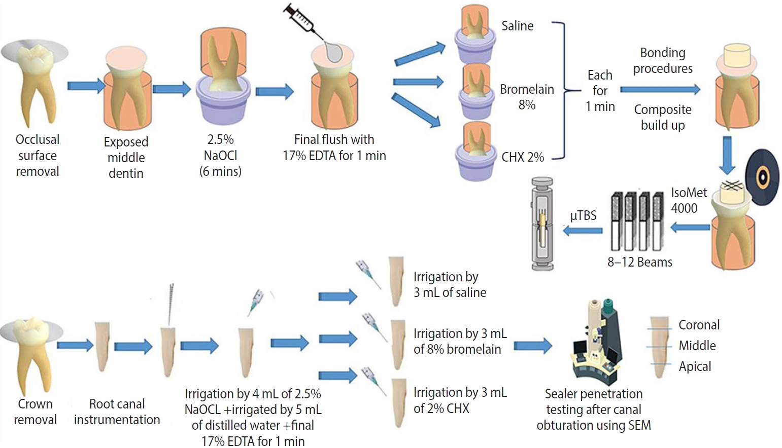
This study aimed to assess the outcomes of bromelain enzyme and chlorhexidine (CHX) following endodontic irrigation by evaluating coronal dentin microtensile bond strength (μTBS) and radicular dentin sealer penetration depth.
Methods
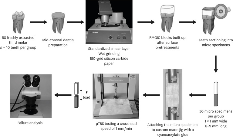
Fifty-one human molars with flat mid-dentin surfaces were soaked in sodium hypochlorite, then randomly assigned to three groups relying on the biomodification approach (n = 17): group 1, saline; group 2, 8% bromelain; and group 3, 2% CHX. After bonding and resin composite build-ups, the μTBS, failure mode, and bond interface were evaluated. Forty-two root canals of human molars were mechanically prepared and randomly distributed among three groups (n = 14), similar to the coronal-dentin biomodification protocol. The sealer-penetration depth was measured utilizing the scanning electron microscope. One- and two-way analyses of variance and the pairwise t- and chi-square tests were utilized.
Results
The bromelain group showed the highest statistically significant resin-dentin μTBS values, followed by the CHX and control groups. For sealer-penetration assessment, the bromelain group showed the highest penetration at the middle and apical root levels, whereas CHX demonstrated the highest penetration at the coronal level.
Conclusions
Bromelain biomodification positively influenced the resin-dentin bond strength and the sealer-penetration depth in apical and middle levels.
- 603 View
- 95 Download

- Bonding of a resin-modified glass ionomer cement to dentin using universal adhesives
- Muhittin Ugurlu
- Restor Dent Endod 2020;45(3):e36. Published online June 15, 2020
- DOI: https://doi.org/10.5395/rde.2020.45.e36
-
Abstract
PDFPubReaderePub
Objectives This study aims to assess the effect of universal adhesives pretreatment on the bond strength of resin-modified glass ionomer cement to dentin.
Materials and Methods Fifty caries-free human third molars were employed. The teeth were randomly assigned into five groups (
n = 10) based on dentin surface pretreatments: Single Bond Universal (3M Oral Care), Gluma Bond Universal (Heraeus Kulzer), Prime&Bond Elect (Dentsply), Cavity Conditioner (GC) and control (no surface treatment). After Fuji II LC (GC) was bonded to the dentin surfaces, the specimens were stored for 7 days at 37°C. The specimens were segmented into microspecimens, and the microspecimens were subjugated to microtensile bond strength testing (1.0 mm/min). The modes of failure analyzed using a stereomicroscope and scanning electron microscopy. Data were statistically analyzed with one-way analysis of variance and Duncan tests (p = 0.05).Results The surface pretreatments with the universal adhesives and conditioner increased the bond strength of Fuji II LC to dentin (
p < 0.05). Single Bond Universal and Gluma Bond Universal provided higher bond strength to Fuji II LC than Cavity Conditioner (p < 0.05). The bond strengths obtained from Prime&Bond Elect and Cavity Conditioner were not statistically different (p > 0.05).Conclusions The universal adhesives and polyacrylic acid conditioner could increase the bond strength of resin-modified glass ionomer cement (RMGIC) to dentin. The use of universal adhesives before the application of RMGIC may be more beneficial in improving bond strength.
-
Citations
Citations to this article as recorded by
- Bioactive restorative materials in dentistry: a comprehensive review of mechanisms, clinical applications, and future directions
Dina Abozaid, Amr Azab, Mohammad A. Bahnsawy, Mohamed Eldebawy, Abdullah Ayad, Romesa soomro, Enas Elwakeel, Maged Ahmed Mohamed
Odontology.2026; 114(2): 349. CrossRef - Impact of nanochitosan incorporation on the performance of resin-modified glass ionomer luting cement: a comprehensive in vitro study
Mostafa A. Abdelshafi, Nesma Elgohary, Ahmed Shams
BMC Oral Health.2026;[Epub] CrossRef - Coronal cavity pretreatment agents and restoration protocols effect on microleakage of endodontically treated teeth
Lena Bal, Cangül Keskin, Aybüke Karaca Sakallı, Osman Fatih Aydın
Journal of Medicine and Palliative Care.2026; 7(1): 40. CrossRef - Comparative evaluation of effect of natural collagen cross-linker and silver diamine fluoride pre-treatment before resin-modified glass ionomer cement restoration on dentin of primary teeth: An in vitro study
Mrunal Pawar, N. D. Shashikiran, Sachin Chandrashekar Gugwad, Namrata Gaonkar, Savita Hadakar, Pali Nikose, Sonali Kisan Waghmode, Ankita Maurya
Journal of Indian Society of Pedodontics and Preventive Dentistry.2026; 44(1): 132. CrossRef - Clinical evaluation of giomer-based injectable resin composite versus resin-modified glass ionomer in class V carious lesions over 18 months: A randomized clinical trial
Reham Hendam, Rania Mosallam, Dina Kamal
Journal of Conservative Dentistry and Endodontics.2025; 28(1): 50. CrossRef - Push-Out Bond Strength of Different Luting Cements Following Post Space Irrigation with 2% Chitosan: An In Vitro Study
Shimaa Rifaat, Ahmed Rahoma, Hind Muneer Alharbi, Sawsan Jamal Kazim, Shrouq Ali Aljuaid, Basmah Omar Alakloby, Faraz A. Farooqi, Noha Taymour
Prosthesis.2025; 7(1): 18. CrossRef - A Comparative Evaluation of Marginal Leakage and Shear Bond Strength of Cention N, Resin-Modified Glass Ionomer Cement (RMGIC), and Conventional Glass Ionomer Cement (GIC): An In Vitro Study
Khushboo Singh, Debapriya Pradhan, Saurabh Tiwari, Raksha Thakur, Priyamvada Sharma, Devika Agrawal, Mahima Singh, Devshree Jawalikar, Delphina Michael Kapoor, Jyoti Priiya Kodimela
Cureus.2025;[Epub] CrossRef - Assessment of Nanosilver Fluoride Application on the Microtensile Bond Strength of Glass Ionomer Cement and Resin-modified Glass Ionomer Cement on Primary Carious Dentin: An In Vitro Study
Anushka Das, Neeraja Ramamurthy, Ila Srinivasan, Yuthi Milit
International Journal of Clinical Pediatric Dentistry.2024; 17(5): 565. CrossRef - Effect of Surface Treatments on Shear-bond Strength of Glass Ionomer Cements to Silver Diamine Fluoride-treated Simulated Carious Dentin
WT Koh, OT Yeoh, NA Yahya, AU Yap
Operative Dentistry.2024; 49(6): 714. CrossRef - Desensitizing agents’ post-bleaching effect on orthodontic bracket bond strength
Gufa Bagus Pamungkas, Dyah Karunia, Sri Suparwitri
Dental Journal.2024; 57(1): 45. CrossRef - Successful Rehabilitation of Traumatized Immature Teeth by Different Vital Pulp Therapies in Pediatric Patients
Mohammad Kamran Khan
Journal of the Scientific Society.2023; 50(1): 111. CrossRef - Do bioactive materials show greater retention rates in restoring permanent teeth than non-bioactive materials? A systematic review and network meta-analysis of randomized controlled trials
Juliana Benace Fernandes, Sheila Mondragón Contreras, Manuela da Silva Spinola, Graziela Ribeiro Batista, Eduardo Bresciani, Taciana Marco Ferraz Caneppele
Clinical Oral Investigations.2023;[Epub] CrossRef - Effects of tooth preparation on the microleakage of fissure sealant
Gesti Kartiko Sari, Sri Kuswandari, Putri Kusuma Wardani Mahendra
Dental Journal (Majalah Kedokteran Gigi).2022; 55(2): 67. CrossRef - Rheological Properties, Surface Microhardness, and Dentin Shear Bond Strength of Resin-Modified Glass Ionomer Cements Containing Methacrylate-Functionalized Polyacids and Spherical Pre-Reacted Glass Fillers
Whithipa Thepveera, Wisitsin Potiprapanpong, Arnit Toneluck, Somruethai Channasanon, Chutikarn Khamsuk, Naruporn Monmaturapoj, Siriporn Tanodekaew, Piyaphong Panpisut
Journal of Functional Biomaterials.2021; 12(3): 42. CrossRef
- Bioactive restorative materials in dentistry: a comprehensive review of mechanisms, clinical applications, and future directions
- 5,420 View
- 48 Download
- 14 Crossref
- Influence of 10-MDP concentration on the adhesion and physical properties of self-adhesive resin cements
- Kazuhiko Shibuya, Naoko Ohara, Serina Ono, Kumiko Matsuzaki, Masahiro Yoshiyama
- Restor Dent Endod 2019;44(4):e45. Published online November 12, 2019
- DOI: https://doi.org/10.5395/rde.2019.44.e45
-
Abstract
PDFPubReaderePub
Objectives Self-adhesive resin cements contain functional monomers that enable them to adhere to the tooth structure without a separate adhesive or etchant. One of the most stable functional monomers used for chemical bonding to calcium in hydroxyapatite is 10-methacryloyloxydecyl dihydrogen phosphate (10-MDP). The aim of this study was to evaluate the influence of the10-MDP concentration on the bond strength and physical properties of self-adhesive resin cements.
Materials and Methods We used experimental resin cements containing 3 different concentrations of 10-MDP: 3.3 wt% (RC1), 6.6 wt% (RC2), or 9.9 wt% (RC3). The micro-tensile bond strength of each resin cement to dentin and a hybrid resin block (Estenia C&B, Kuraray Noritake Dental) was measured, and the fractured surface morphology was analyzed. Further, the flexural strength of the resin cements was measured using the three-point bending test. The water sorption and solubility of the cements following 30 days of immersion in water were measured.
Results The bond strength of RC2 was significantly higher than that of RC1. There was no significant difference between the bond strength of RC2 and that of RC3. The water sorption of RC3 was higher than that of any other cement. There were no significant differences in the three-point bending strength or water solubility among all three types of cements.
Conclusions Within the limitations of this study, it is suggested that 6.6 wt% 10-MDP showed superior properties than 3.3 wt% or 9.9 wt% 10-MDP in self-adhesive resin cement.
-
Citations
Citations to this article as recorded by- Material properties and finite element analysis of adhesive cements used for zirconia crowns on dental implants
Megha Satpathy, Hai Pham, Shreya Shah
Computer Methods in Biomechanics and Biomedical Engineering.2026; 29(3): 582. CrossRef - Bonding effectiveness of 10-MDP containing resin composite cements: a systematic review with meta-analysis
Sofia Bignotto de Carvalho, Lívia Maiumi Uehara, João Marcos Carvalho-Silva, Andréa Cândido dos Reis
International Journal of Adhesion and Adhesives.2026; 146: 104260. CrossRef - Comparative Evaluation of Color Stability in Bioactive and Conventional Resin Cements Under Thermal Stress Conditions
Alaa Turkistani, Hanin E. Yeslam
Biomimetics.2025; 10(7): 432. CrossRef - Influence of temperature and curing modes on polymerization of self-adhesive resin cements
Hae-In Kim, Jin-Woo Kim, Se-Hee Park, Kyung-Mo Cho
Korean Journal of Dental Materials.2025; 52(3): 143. CrossRef - Clinical Performance and Retention of Partial Implant Restorations Cemented with Fuji Plus® and DentoTemp™: A Retrospective Clinical Study with Mechanical Validation
Sergiu-Manuel Antonie, Laura-Cristina Rusu, Ioan-Achim Borsanu, Remus Christian Bratu, Emanuel-Adrian Bratu
Medicina.2025; 61(12): 2183. CrossRef - A thorough assessment of 10-MDP primers in modern dental adhesive systems
Ahmed A Abduljawad, Harraa SM Salih, Omar F Tawfiq
Journal of Baghdad College of Dentistry.2024; 36(3): 79. CrossRef - Clinical reliability of self-adhesive luting resins compared to other adhesive procedures: A systematic review and meta-analysis
Mohammed Ahmed Alghauli, Ahmed Yaseen Alqutaibi, Sebastian Wille, Matthias Kern
Journal of Dentistry.2023; 129: 104394. CrossRef - Influence of autoclave sterilization on bond strength between zirconia frameworks and Ti-base abutments using different resin cements
Reinhold Lang, Karl-Anton Hiller, Lena Kienböck, Katrin Friedl, Karl-Heinz Friedl
The Journal of Prosthetic Dentistry.2022; 127(4): 617.e1. CrossRef - Bond Strength Comparison Among 10-MDP-Containing and Non-10-MDP-Containing Adhesives in Different Degrees of Dental Fluorosis
Luis Javier Solís-Martínez, Karla Yareli Valles Flores, Nohé Vargas Chávez, Oscar Eduardo Almeda-Ojeda, Víctor Hiram Barajas-Pérez, Edgar García-Torres
Odovtos - International Journal of Dental Sciences.2022; 25(1): 212. CrossRef - Varying 10-methacryloyloxydecyl dihydrogen phosphate (10-MDP) level improves polymerisation kinetics and flexural strength in self-adhesive, remineralising composites
António H.S. Delgado, Nazanin Owji, Paul Ashley, Anne M. Young
Dental Materials.2021; 37(9): 1366. CrossRef - Investigating a Commercial Functional Adhesive with 12-MDPB and Reactive Filler to Strengthen the Adhesive Interface in Eroded Dentin
Madalena Belmar da Costa, António HS Delgado, Tomás Amorim Afonso, Luís Proença, Ana Sofia Ramos, Ana Mano Azul
Polymers.2021; 13(20): 3562. CrossRef
- Material properties and finite element analysis of adhesive cements used for zirconia crowns on dental implants
- 3,626 View
- 28 Download
- 11 Crossref
- Microtensile bond strength of CAD/CAM-fabricated polymer-ceramics to different adhesive resin cements
- Leyla Sadighpour, Farideh Geramipanah, Zahra Ghasri, Mehrnoosh Neshatian
- Restor Dent Endod 2018;43(4):e40. Published online September 3, 2018
- DOI: https://doi.org/10.5395/rde.2018.43.e40
-
Abstract
PDFPubReaderePub
Objectives This study evaluated the microtensile bond strength (µTBS) of polymer-ceramic and indirect composite resin with 3 classes of resin cements.
Materials and Methods Two computer-aided design/computer-aided manufacturing (CAD/CAM)-fabricated polymer-ceramics (Enamic [ENA; Vita] and Lava Ultimate [LAV; 3M ESPE]) and a laboratory indirect composite resin (Gradia [GRA; GC Corp.]) were equally divided into 6 groups (
n = 18) with 3 classes of resin cements: Variolink N (VAR; Vivadent), RelyX U200 (RXU; 3M ESPE), and Panavia F2 (PAN; Kuraray). The μTBS values were compared between groups by 2-way analysis of variance and thepost hoc Tamhane test (α = 0.05).Results Restorative materials and resin cements significantly influenced µTBS (
p < 0.05). In the GRA group, the highest μTBS was found with RXU (27.40 ± 5.39 N) and the lowest with VAR (13.54 ± 6.04 N) (p < 0.05). Similar trends were observed in the ENA group. In the LAV group, the highest μTBS was observed with VAR (27.45 ± 5.84 N) and the lowest with PAN (10.67 ± 4.37 N) (p < 0.05). PAN had comparable results to those of ENA and GRA, whereas the μTBS values were significantly lower with LAV (p = 0.001). The highest bond strength of RXU was found with GRA (27.40 ± 5.39 N,p = 0.001). PAN showed the lowest µTBS with LAV (10.67 ± 4.37 N;p < 0.001).Conclusions When applied according to the manufacturers' recommendations, the µTBS of polymer-ceramic CAD/CAM materials and indirect composites is influenced by the luting cements.
-
Citations
Citations to this article as recorded by- Enhancing severely compromised premolar strength: role of cusp reduction design in CAD/CAM composite restorations
Mohamed F. Haridy, Ahmed Refaat Mohamed, Shehabeldin Saber, Edgar Schafer, Samar Elsayed Swelam, Youssef M. Haridy, Hend S. Ahmed
Odontology.2026; 114(2): 852. CrossRef - Effect of hydrofluoric acid and self-etch ceramic primers on the flexural strength and fatigue resistance of glass ceramics: A systematic review and meta-analysis of in vitro studies
Paulo Matias Moreira, Gabriela Luiza Moreira Carvalho, Rodrigo de Castro Albuquerque, Carolina Bosso André
Japanese Dental Science Review.2024; 60: 198. CrossRef - Light transmittance through resin-matrix composite onlays adhered to resin-matrix cements or flowable composites
Rita Fidalgo-Pereira, Susana O. Catarino, Óscar Carvalho, Nélio Veiga, Orlanda Torres, Annabel Braem, Júlio C.M. Souza
Journal of the Mechanical Behavior of Biomedical Materials.2024; 151: 106353. CrossRef - Effect of thermocycling on the mechanical properties of permanent composite-based CAD-CAM restorative materials produced by additive and subtractive manufacturing techniques
Tuğba Temizci, Hatice Nalan Bozoğulları
BMC Oral Health.2024;[Epub] CrossRef - Effect of different surface treatments on resin-matrix CAD/CAM ceramics bonding to dentin: in vitro study
Hanan Fathy, Hamdi H. Hamama, Noha El-Wassefy, Salah H. Mahmoud
BMC Oral Health.2022;[Epub] CrossRef - Digital image analysis of fluorescence of ceramic veneers with different ceramic materials and resin cements
Jiao ZHANG, Qing YU
Dental Materials Journal.2022; 41(6): 868. CrossRef - Fatigue Behavior of Monolithic Zirconia-Reinforced Lithium Silicate Ceramic Restorations: Effects of Conditionings of the Intaglio Surface and the Resin Cements
F Dalla-Nora, LF Guilardi, CP Zucuni, LF Valandro, MP Rippe
Operative Dentistry.2021; 46(3): 316. CrossRef
- Enhancing severely compromised premolar strength: role of cusp reduction design in CAD/CAM composite restorations
- 2,654 View
- 8 Download
- 7 Crossref
- Effects of endodontic tri-antibiotic paste on bond strengths of dentin adhesives to coronal dentin
- Parvin Mirzakoucheki, Ricardo Walter, Navid Khalighinejad, Maryam Zare Jahromi, Sanaz Mirsattari, Navid Akbarzadeh
- Restor Dent Endod 2015;40(2):136-142. Published online February 12, 2015
- DOI: https://doi.org/10.5395/rde.2015.40.2.136
-
Abstract
PDFPubReaderePub
Objectives The aim of this study was to evaluate the effects of tri-antibiotic paste (TAP) on microtensile bond strengths (MTBS) of dental adhesives to dentin.
Materials and Methods Sixty extracted molars had their occlusal surfaces flattened to expose dentin. They were divided into two groups, i.e., control group with no dentin treatment and experimental group with dentin treatment with TAP. After 10 days, specimens were bonded using self-etch (Filtek P90 adhesive) or etch-and-rinse (Adper Single Bond Plus) adhesives and restored with composite resin. Teeth were sectioned into beams, and the specimens were subjected to MTBS test. Data were analyzed using two-way ANOVA and post hoc Tukey tests.
Results There was a statistically significant interaction between dentin treatment and adhesive on MTBS to coronal dentin (
p = 0.003). Despite a trend towards worse MTBS being noticed in the experimental groups, TAP application showed no significant effect on MTBS (p = 0.064).Conclusions The etch-and-rinse adhesive Adper Single Bond Plus presented higher mean bond strengths than the self-etch adhesive Filtek P90, irrespective of the group. The superior bond performance for Adper Single Bond when compared to Filtek P90 adhesive was confirmed by a fewer number of adhesive failures. The influence of TAP in bond strength is insignificant.
-
Citations
Citations to this article as recorded by- Efecto antimicrobiano como medicación intraconducto de la pasta triantibiótica.
Paúl Sebastián Ulloa Amores, Diana Álvarez Álvarez, María Elizabeth Moscoso Abad, Magda Zulay Bastidas Calva
Revista de la Asociación Dental Mexicana.2024; 81(4): 211. CrossRef - Effect of Intracanal Medicaments on Push-out Bond Strength of Calcium Silicate-based Materials
Hyuntae Jeong, Sunmi Yang, Seonmi Kim, Namki Choi, Jaehwan Kim
THE JOURNAL OF THE KOREAN ACADEMY OF PEDTATRIC DENTISTRY.2018; 45(4): 455. CrossRef
- Efecto antimicrobiano como medicación intraconducto de la pasta triantibiótica.
- 1,780 View
- 6 Download
- 2 Crossref
- Effect of chlorhexidine application on the bond strength of resin core to axial dentin in endodontic cavity
- Yun-Hee Kim, Dong-Hoon Shin
- Restor Dent Endod 2012;37(4):207-214. Published online November 21, 2012
- DOI: https://doi.org/10.5395/rde.2012.37.4.207
-
Abstract
PDFPubReaderePub
Objectives This study evaluated the influence of chlorhexidine (CHX) on the microtensile bonds strength (µTBS) of resin core with two adhesive systems to dentin in endodontic cavities.
Materials and Methods Flat dentinal surfaces in 40 molar endodontic cavities were treated with self-etch adhesive system, Contax (DMG) and total-etch adhesive system, Adper Single Bond 2 (3M ESPE) after the following surface treatments: (1) Priming only (Contax), (2) CHX for 15 sec + rinsing + priming (Contax), (3) Etching with priming (Adper Single Bond 2), (4) Etching + CHX for 15 sec + rinsing + priming (Adper Single Bond 2). Resin composite build-ups were made with LuxaCore (DMG) using a bulk method and polymerized for 40 sec. For each condition, half of specimens were submitted to µTBS after 24 hr storage and half of them were submitted to thermocycling of 10,000 cycles between 5℃ and 55℃ before testing. The data were analyzed using ANOVA and independent
t -test at a significance level of 95%.Results CHX pre-treatment did not affect the bond strength of specimens tested at the immediate testing period, regardless of dentin surface treatments. However, after 10,000 thermocycling, all groups showed reduced bond strength. The amount of reduction was greater in groups without CHX treatments than groups with CHX treatment. These characteristics were the same in both self-etch adhesive system and total-etch adhesive system.
Conclusions 2% CHX application for 15 sec proved to alleviate the decrease of bond strength of dentin bonding systems. No significant difference was shown in µTBS between total-etching system and self-etching system.
-
Citations
Citations to this article as recorded by- Physical characterization and bond performance of a non-methacrylate dental adhesive in long-term biochemical and thermal aging models
Zach Gouveia, Rastin Rahiminejad, Lingyun Zhu, Jesse Barker, Yoav Finer, J. Paul Santerre
Dental Materials.2026; 42(5): 877. CrossRef - Micro Tensile bond strength and microleakage assessment of total-etch and self-etch adhesive bonded to carious affected dentin disinfected with Chlorhexidine, Curcumin, and Malachite green
Zeeshan Qamar, Nishath Sayed Abdul, R Naveen Reddy, Mahesh Shenoy, Saleh Alghufaili, Yousef Alqublan, Ali Barakat
Photodiagnosis and Photodynamic Therapy.2023; 43: 103636. CrossRef - The Classification and Selection of Adhesive Agents; an Overview for the General Dentist
Naji Ziad Arandi
Clinical, Cosmetic and Investigational Dentistry.2023; Volume 15: 165. CrossRef - Influence of chlorhexidine 2% and sodium hypochlorite 5.25% on micro-tensile bond strength of universal adhesive system (G-Premio Bond)
Nafiseh Fazelian, Abbas Rahimi Dashtaki, MohammadAmin Eftekharian, Batool Amiri
Brazilian Journal of Oral Sciences.2022;[Epub] CrossRef - Comparative evaluation of the effects of different methods of post space preparation in primary anterior teeth on the fracture resistance of tooth restorations
Bahman Seraj, Sara Ghadimi, Ebrahim Najafpoor, Fatemeh Abdolalian, razieh khanmohammadi
Journal of Dental Research, Dental Clinics, Dental Prospects.2019; 13(2): 141. CrossRef - Chemical, microbial, and host‐related factors: effects on the integrity of dentin and the dentin–biomaterial interface
Marcela T. Carrilho, Fabiana Piveta, Leo Tjäderhane
Endodontic Topics.2015; 33(1): 50. CrossRef - MMP Inhibitors on Dentin Stability
A.F. Montagner, R. Sarkis-Onofre, T. Pereira-Cenci, M.S. Cenci
Journal of Dental Research.2014; 93(8): 733. CrossRef - Thermal cycling for restorative materials: Does a standardized protocol exist in laboratory testing? A literature review
Anna Lucia Morresi, Maurizio D'Amario, Mario Capogreco, Roberto Gatto, Giuseppe Marzo, Camillo D'Arcangelo, Annalisa Monaco
Journal of the Mechanical Behavior of Biomedical Materials.2014; 29: 295. CrossRef
- Physical characterization and bond performance of a non-methacrylate dental adhesive in long-term biochemical and thermal aging models
- 1,846 View
- 5 Download
- 8 Crossref
- Effect of different chlorhexidine application times on microtensile bond strength to dentin in Class I cavities
- Hyun-Jung Kang, Ho-Jin Moon, Dong-Hoon Shin
- Restor Dent Endod 2012;37(1):9-15. Published online March 2, 2012
- DOI: https://doi.org/10.5395/rde.2012.37.1.9
-
Abstract
PDFPubReaderePub
Objectives This study evaluated the effect of 2% chlorhexidine digluconate (CHX) with different application times on microtensile bonds strength (MTBS) to dentin in class I cavities and intended to search for ideal application time for a simplified bonding protocol.
Materials and Methods Flat dentinal surfaces with class I cavities (4 mm × 4 mm × 2 mm) in 40 molar teeth were bonded with etch-and-rinse adhesive system, Adper Single Bond 2 (3M ESPE) after: (1) etching only as a control group; (2) etching + CHX 5 sec + rinsing; (3) etching + CHX 15 sec + rinsing; (4) etching + CHX 30 sec + rinsing; and (5) etching + CHX 60 sec + rinsing. Resin composite was built-up with Z-250 (3M ESPE) using a bulk method and polymerized for 40 sec. For each condition, half of the specimens were immediately submitted to MTBS test and the rest of them were assigned to thermocycling of 10,000 cycles between 5℃ and 55℃ before testing. The data were analyzed using two-way ANOVA, at a significance level of 95%.
Results There was no significant difference in bond strength between CHX pre-treated group and control group at the immediate testing period. After thermocycling, all groups showed reduced bond strength irrespective of the CHX use. However, groups treated with CHX maintained significantly higher MTBS than control group (
p < 0.05). In addition, CHX application time did not have any significant influence on the bond strength among groups treated with CHX.Conclusion Application of 2% CHX for a short time period (5 sec) after etching with 37% phosphoric acid may be sufficient to preserve dentin bond strength.
-
Citations
Citations to this article as recorded by- Evaluation of Fracture Resistance of Tooth Fragment Reattached with Cavity Disinfectants in Primary and Permanent Teeth: An In Vitro Study
Komal P Bhosale, Vishnu R Chamarthi, Dhanraj Kalaivanan, Santham Krishnamoorthy, Sumaiyya Saleem, Santhosh Priya AKR, Sai SK Kothimbakkam
The Journal of Contemporary Dental Practice.2026; 27(3): 269. CrossRef - Effect of nonthermal atmospheric plasma application at different time intervals on the dentinal shear bond strength pretreated with 2% chlorhexidine as cavity disinfectant: An in vitro study
Roopadevi Garlapati, Nagesh Bolla, Gali Praveen Kumar, Mayana Aameena Banu, Bandlapally Sreenivasa Guptha Anila, Shaik Afreen Kamal
Journal of Conservative Dentistry and Endodontics.2024; 27(7): 769. CrossRef - Comparative evaluation ofEmblica officinalisas an etchant and an MMP inhibitor with orthophosphoric acid and chlorhexidine on the microshear bond strength of composite resin: anex vivostudy
Divya Sangeetha Rajkumar, Annapoorna Ballagere Mariswamy
Restorative Dentistry & Endodontics.2021;[Epub] CrossRef - Effect of Cavity Disinfectants on Adhesion to Primary Teeth—A Systematic Review
Ana Coelho, Inês Amaro, Ana Apolónio, Anabela Paula, José Saraiva, Manuel Marques Ferreira, Carlos Miguel Marto, Eunice Carrilho
International Journal of Molecular Sciences.2021; 22(9): 4398. CrossRef - Effect of Different Matrix Metalloproteinase Inhibitors on Shear Bond Strength of Composite Attached to Primary Teeth Dentin
Najmeh Mohammadi, Zahra Parsaie, Dana Jafarpour, Fatemeh Bizolm
European Journal of General Dentistry.2020; 9(03): 147. CrossRef
- Evaluation of Fracture Resistance of Tooth Fragment Reattached with Cavity Disinfectants in Primary and Permanent Teeth: An In Vitro Study
- 2,506 View
- 6 Download
- 5 Crossref
Basic Researchs
- Bonding efficacy of cured or uncured dentin adhesives in indirect resin
- Ji-Hyun Jang, Bin-Na Lee, Hoon-Sang Chang, Yun-Chan Hwang, Won-Mann Oh, In-Nam Hwang
- J Korean Acad Conserv Dent 2011;36(6):490-497. Published online November 30, 2011
- DOI: https://doi.org/10.5395/JKACD.2011.36.6.490
-
Abstract
PDFPubReaderePub
Objectives This study examined the effect of the uncured dentin adhesives on the bond interface between the resin inlay and dentin.
Materials and Methods Dentin surface was exposed in 24 extracted human molars and the teeth were assigned to indirect and direct resin restoration group. For indirect resin groups, exposed dentin surfaces were temporized with provisional resin. The provisional restoration was removed after 1 wk and the teeth were divided further into 4 groups which used dentin adhesives (OptiBond FL, Kerr; One-Step, Bisco) with or without light-curing, respectively (Group OB-C, OB-NC, OS-C and OS-NC). Pre-fabricated resin blocks were cemented on the entire surfaces with resin cement. For the direct resin restoration groups, the dentin surfaces were treated with dentin adhesives (Group OB-D and OS-D), followed by restoring composite resin. After 24 hr, the teeth were assigned to microtensile bond strength (µTBS) and confocal laser scanning microscopy (CLSM), respectively.
Results The indirect resin restoration groups showed a lower µTBS than the direct resin restoration groups. The µTBS values of the light cured dentin adhesive groups were higher than those of the uncured dentin adhesive groups (
p < 0.05). CLSM analysis of the light cured dentin adhesive groups revealed definite and homogenous hybrid layers. However, the uncured dentin adhesive groups showed uncertain or even no hybrid layer.Conclusions Light-curing of the dentin adhesive prior to the application of the cementing material in luting a resin inlay to dentin resulted in definite, homogenous hybrid layer formation, which may improve the bond strength.
- 1,993 View
- 10 Download
- Microtensile bond strength of resin inlay bonded to dentin treated with various temporary filling materials
- Tae-Woo Kim, Bin-Na Lee, Young-Jung Choi, So-Young Yang, Hoon-Sang Chang, Yun-Chan Hwang, In-Nam Hwang, Won-Mann Oh
- J Korean Acad Conserv Dent 2011;36(5):419-424. Published online September 30, 2011
- DOI: https://doi.org/10.5395/JKACD.2011.36.5.419
-
Abstract
PDFPubReaderePub
Objectives This study was aimed to determine the effects of temporary sealing materials on microtensile bond strength between resin-coated dentin and resin inlay and to compare the bonding effectiveness of delayed dentin sealing and that of immediate dentin sealing.
Materials and Methods The teeth were divided into 4 groups: group 1, specimens were prepared using delayed dentin sealing after temporary sealing with zinc oxide eugenol (ZOE); group 2, specimens were prepared using immediate dentin sealing and ZOE sealing; group 3, specimens were prepared using immediate dentin sealing and Dycal (Dentsply) sealing; group 4, specimens were prepared using immediately sealed, and then temporarily sealed with a resin-based temporary sealing material.
After removing the temporary sealing material, we applied resin adhesive and light-cured. Then the resin inlays were applied and bonded to the cavity with a resin-based cement. The microtensile bond strength of the sectioned specimens were measured with a micro-tensile tester (Bisco Inc.). Significance between the specimen groups were tested by means of one-way ANOVA and multiple Duncan's test.
Results Group 1 showed the lowest bond strength, and group 4 showed the highest bond strength (
p < 0.01). When temporary sealing was performed with ZOE, immediate dentin sealing showed a higher bonding strength than delayed dentin sealing (p < 0.01).Conclusions Based on these results, immediate dentin sealing is more recommended than delayed dentin sealing in bonding a resin inlay to dentin. Also, resin-based temporary sealing materials have shown the best result.
- 1,348 View
- 2 Download
- Effect of adhesive hydrophobicity on microtensile bond strength of low-shrinkage silorane resin to dentin
- So-Yeun Cho, Hyun-Young Kang, Kyoung-A Kim, Mi-Kyung Yu, Kwang-Won Lee
- J Korean Acad Conserv Dent 2011;36(4):280-289. Published online July 31, 2011
- DOI: https://doi.org/10.5395/JKACD.2011.36.4.280
-
Abstract
PDFPubReaderePub
Objectives The purpose of this study was to evaluate µTBS (microtensile bond strength) of current dentin bonding adhesives which have different hydrophobicity with low-shrinkage silorane resin.
Materials and Methods Thirty-six human third molars were used. Middle dentin was exposed. The teeth were randomly assigned to nine experimental groups: Silorane self-etch adhesives (SS), SS + phosphoric acid etching (SS + pa), Adper easy bond (AE), AE + Silorane system bonding (AE + SSb), Clearfil SE bond (CSE), CSE + SSb, All-Bond 2 (AB2), AB2 + SSb, All-Bond 3 (AB3). After adhesive's were applied, the clinical crowns were restored with Filtek LS (3M ESPE). The 0.8 mm × 0.8 mm sticks were submitted to a tensile load using a Micro Tensile Tester (Bisco Inc.). Water sorption was measured to estimate hydrophobicity adhesives.
Results µTBS of silorane resin to 5 adhesives: SS, 23.2 MPa; CSE, 19.4 MPa; AB3, 30.3 MPa; AB2 and AE, no bond. Additional layering of SSb: CSE + SSb, 26.2 MPa; AB2 + SSb, 33.9 MPa; AE + SSb, no bond. High value of µTBS was related to cohesive failure. SS showed the lowest water sorption. AE showed the highest solubility.
Conclusions The hydrophobicity of adhesive increased, and silorane resin bond-strength was also increased. Additional hydrophobic adhesive layer did not increase the bond-strength to silorane resin except AB2 + SSb. All-Bond 3 showed similar µTBS & water sorption with SS. By these facts, we could reach a conclusion that All-Bond 3 is a competitive adhesive which can replace the Silorane adhesive system.
-
Citations
Citations to this article as recorded by- Microtensile bond strength of silorane-based composite specific adhesive system using different bonding strategies
Laura Alves Bastos, Ana Beatriz Silva Sousa, Brahim Drubi-Filho, Fernanda de Carvalho Panzeri Pires-de-Souza, Lucas da Fonseca Roberti Garcia
Restorative Dentistry & Endodontics.2015; 40(1): 23. CrossRef
- Microtensile bond strength of silorane-based composite specific adhesive system using different bonding strategies
- 1,957 View
- 1 Download
- 1 Crossref
- Influence of application methods of one-step self-etching adhesives on microtensile bond strength
- Chul-Kyu Choi, Sung-Ae Son, Jin-Hee Ha, Bock Hur, Hyeon-Cheol Kim, Yong-Hun Kwon, Jeong-Kil Park
- J Korean Acad Conserv Dent 2011;36(3):203-210. Published online May 31, 2011
- DOI: https://doi.org/10.5395/JKACD.2011.36.3.203
-
Abstract
PDFPubReaderePub
Objectives The purpose of this study was to evaluate the effect of various application methods of one-step self-etch adhesives to microtensile resin-dentin bond strength.
Materials and Methods Thirty-six extracted human molars were used. The teeth were assigned randomly to twelve groups (
n = 15), according to the three different adhesive systems (Clearfil Tri-S Bond, Adper Prompt L-Pop, G-Bond) and application methods. The adhesive systems were applied on the dentin as follows: 1) The single coating, 2) The double coating, 3) Manual agitation, 4) Ultrasonic agitation. Following the adhesive application, light-cure composite resin was constructed. The restored teeth were stored in distilled water at room temperature for 24 hours, and prepared 15 specimens per groups. Then microtensile bond strength was measured and the failure mode was examined.Results Manual agitation and ultrasonic agitation of adhesive significantly increased the microtensile bond strength than single coating and double coating did. Double coating of adhesive significantly increased the microtensile bond strength than single coating did and there was no significant difference between the manual agitation and ultrasonic agitation group. There was significant difference in microtensile bonding strength among all adhesives and Clearfil Tri-S Bond showed the highest bond strength.
Conclusions In one-step self-etching adhesives, there was significant difference according to application methods and type of adhesives. No matter of the material, the manual or ultrasonic agitation of the adhesive showed significantly higher microtensile bond strength.
-
Citations
Citations to this article as recorded by- Effect of Baicalein on Bond Strength of Indirect Ceramic Restoration
Nuray Zulkadir Ergin, Aslı Seçilmiş
Süleyman Demirel Üniversitesi Sağlık Bilimleri Dergisi.2025; 16(3): 356. CrossRef - The Classification and Selection of Adhesive Agents; an Overview for the General Dentist
Naji Ziad Arandi
Clinical, Cosmetic and Investigational Dentistry.2023; Volume 15: 165. CrossRef
- Effect of Baicalein on Bond Strength of Indirect Ceramic Restoration
- 2,600 View
- 14 Download
- 2 Crossref
- Effect of 2% chlorhexidine application on microtensile bond strength of resin composite to dentin using one-step self-etch adhesives
- Soon-Ham Jang, Bock Hur, Hyeon-Cheol Kim, Yong-Hun Kwon, Jeong-Kil Park
- J Korean Acad Conserv Dent 2010;35(6):486-491. Published online November 30, 2010
- DOI: https://doi.org/10.5395/JKACD.2010.35.6.486
-
Abstract
PDFPubReaderePub
Objectives This study examined the effect of 2% chlorhexidine on the µTBS of a direct composite restoration using one-step self-etch adhesives on human dentin.
Materials and Methods Twenty-four extracted permanent molars were used. The teeth were assigned randomly to six groups (
n = 10), according to the adhesive system and application of chlorhexidine. With or without the application of chlorhexidine, each adhesive system was applied to the dentin surface. After the bonding procedure, light-cure composite resin buildups were produced. The restored teeth were stored in distilled water at room temperature for 24 hours, and then cut and glued to the jig of the microtensile testing machine. A tensile load was applied until the specimen failed. The failure mode was examined using an operating microscope. The data was analyzed statistically using one-way ANOVA, Student'st -test (p < 0.05) and Scheffé's test.Results Regardless of the application of chlorhexidine, the Clearfil S3 Bond showed the highest µTBS, followed by G-Bond and Xeno V. Adhesive failure was the main failure mode of the dentin bonding agents tested with some samples showing cohesive failure.
Conclusions The application of 2% chlorhexidine did not affect the µTBS of the resin composite to the dentin using a one-step self-etch adhesive.
-
Citations
Citations to this article as recorded by- Effects of Collagen Cross-Linking Agents on Dentin-Composite Interface Strength and Morphology
Faisal Ali bin Abbooud AlQhtani, Zuhair Motlak Alkahtani, Muhammad Abdullah Kamran, Ahmed Ali M. Albariqi, Khalid M. Abdelaziz
Calcified Tissue International.2026;[Epub] CrossRef
- Effects of Collagen Cross-Linking Agents on Dentin-Composite Interface Strength and Morphology
- 2,003 View
- 6 Download
- 1 Crossref
- The study of fractural behavior of repaired composite
- Sang-Soon Park, Wook Nam, Ah-Hyang Eom, Duck-Su Kim, Gi-Woon Choi, Kyoung-Kyu Choi
- J Korean Acad Conserv Dent 2010;35(6):461-472. Published online November 30, 2010
- DOI: https://doi.org/10.5395/JKACD.2010.35.6.461
-
Abstract
PDFPubReaderePub
Objectives This study evaluated microtensile bond strength (µTBS) and short-rod fracture toughness to explain fractural behavior of repaired composite restorations according to different surface treatments.
Materials and Methods Thirty composite blocks for µTBS test and sixty short-rod specimens for fracture toughness test were fabricated and were allocated to 3 groups according to the combination of surface treatment (none-treated, sand blasting, bur roughening). Each group was repaired immediately and 2 weeks later. Twenty-four hours later from repair, µTBS and fracture toughness test were conducted. Mean values analyzed with two-way ANOVA / Tukey's B test (α = 0.05) and correlation analysis was done between µTBS and fracture toughness. FE-SEM was employed on fractured surface to examine the crack propagation.
Results The fresh composite resin showed higher µTBS than the aged composite resin (
p < 0.001). Mechanically treated groups showed higher bond strength than non-mechanically treated groups except none-treated fresh group in µTBS (p < 0.05). The fracture toughness value of mechanically treated surface was higher than that of non-mechanically treated surface (p < 0.05). There was no correlation between fracture toughness and microtensile bond strength values. Specimens having high KIC showed toughening mechanism including crack deviation, microcracks and crack bridging in FE-SEM.Conclusions Surface treatment by mechanical interlock is more important for effective composite repair, and the fracture toughness test could be used as an appropriate tool to examine the fractural behavior of the repaired composite with microtensile bond strength.
- 1,329 View
- 1 Download
- The effect of the removal of chondroitin sulfate on bond strength of dentin adhesives and collagen architecture
- Jong-Ryul Kim, Sang-Jin Park, Gi-Woon Choi, Kyoung-Kyu Choi
- J Korean Acad Conserv Dent 2010;35(3):211-221. Published online May 31, 2010
- DOI: https://doi.org/10.5395/JKACD.2010.35.3.211
-
Abstract
PDFPubReaderePub
Proteoglycan is highly hydrophilic and negatively charged which enable them attract the water. The objective of study was to investigate the effects of Proteoglycan on microtensile bond strength of dentin adhesives and on architecture of dentin collagen matrix of acid etched dentin by removing the chondroitin sulphate attached on Proteoglycan. A flat dentin surface in mid-coronal portion of tooth was prepared. After acid etching, half of the specimens were immersed in 0.1 U/mL chondroitinase ABC (C-ABC) for 48 h at 37℃, while the other half were stored in distilled water. Specimens were bonded with the dentin adhesive using three different bonding techniques (wet, dry and re-wet) followed by microtensile bond strength test. SEM examination was done with debonded specimen, resin-dentin interface and acid-etched dentin surface with/without C-ABC treatment.
For the subgroups using wet-bonding or dry-bonding technique, microtensile bond strength showed no significant difference after C-ABC treatment (p > 0.05). Nevertheless, the subgroup using rewetting technique after air dry in the Single Bond 2 group demonstrated a significant decrease of microtensile bond strength after C-ABC treatment. Collagen architecture is loosely packed and some fibrils are aggregated together and relatively collapsed compared with normal acid-etched wet dentin after C-ABC treatment. Further studies are necessary for the contribution to the collagen architecture of noncollagenous protein under the various clinical situations and several dentin conditioners and are also needed about long-term effect on bond strength of dentin adhesive.
- 1,402 View
- 2 Download
- Effect of surface treatments of fiber posts on bond strength to composite resin cores
- Hye-Jo Keum, Hyun-Mi Yoo
- J Korean Acad Conserv Dent 2010;35(3):173-179. Published online May 31, 2010
- DOI: https://doi.org/10.5395/JKACD.2010.35.3.173
-
Abstract
PDFPubReaderePub
The purpose of the present study was to compare the influence of post-surface treatment with silane, hydrogen peroxide, hydrofluoric acid or sandblasting and to investigate the effect of silane in combination of the other treatments on the microtensile bond strength between fiber posts and composite resins for core build-up. Thirty-two glass-fiber posts (FRC Postec Plus, Ivoclar Vivadent, Schaan, Liechtenstein) were divided into eight groups according to the different surface pretreatments performed: silane application (S); immersion in 28% hydrogen peroxide (HP); immersion in hydrogen peroxide followed by application of silane (HP-S); immersion in 4% hydrofluoric acid gel (HF); immersion in hydrofluoric acid gel followed by application of silane (HF-S); sandblasting with aluminum oxide particles (SB); sandblasting followed by application of silane (SB-S). In control group, no surface treatment was performed. The composite resin (Tetric Flow, Ivoclar Vivadent, Schaan, Liechtenstein) was applied onto the posts to produce the composite cylinder specimen. It was sectioned into sticks to measure the microtensile bond strength. The data was analyzed with one-way ANOVA and LSD test for post hoc comparison (p < 0.05). Post pretreatment with sandblasting enhanced the interfacial strength between the fiber posts and core materials. Moreover, sand-blasting followed by application of silane appears to be the most effective method that can improve the clinical performance of glass fiber posts.
-
Citations
Citations to this article as recorded by- Preparation and properties of glass fiber-reinforced endodontic (root canal therapy) posts
Jae-Yong Son, Kyoung-Ja Kim, Kyoung-Hun Kim, Joo-Seok Park, Kwang-Bo Shim
Journal of the Korean Crystal Growth and Crystal Technology.2015; 25(3): 105. CrossRef - Fracture resistance of upper central incisors restored with different posts and cores
Maryam Rezaei Dastjerdi, Kamran Amirian Chaijan, Saeid Tavanafar
Restorative Dentistry & Endodontics.2015; 40(3): 229. CrossRef - Effect of surface treatment of FRC-Post on bonding strength to resin cements
Chan-Hyun Park, Se-Hee Park, Jin-Woo Kim, Kyung-Mo Cho
Journal of Korean Academy of Conservative Dentistry.2011; 36(2): 125. CrossRef
- Preparation and properties of glass fiber-reinforced endodontic (root canal therapy) posts
- 1,866 View
- 6 Download
- 3 Crossref
- The effect of Er,Cr:YSGG irradiation on microtensile bond strength of composite resin restoration
- Jeong-Hye Son, Hyeon-Cheol Kim, Bock Hur, Jeong-Kil Park
- J Korean Acad Conserv Dent 2010;35(2):134-142. Published online March 31, 2010
- DOI: https://doi.org/10.5395/JKACD.2010.35.2.134
-
Abstract
PDFPubReaderePub
The purpose of this study was to evaluate the effect of Er,Cr:YSGG laser irradiation with hypersensitivity mode on microtensile bond strength of composite resin. Twenty extracted permanent molars were randomly assigned to six groups, according to the irradiation of Er,Cr:YSGG laser, adhesive system (Optibond FL or Clearfil SE bond) and application time of etchant (15 sec or 20 sec). Then composite resin was build up on each conditioned surface. The restored teeth were stored in distilled water at room temperature for 24 h and twelve specimens for each group were prepared. All specimens were subjected to microtensile bond strength and the fracture modes were evaluated. Also, the prepared dentin surface and laser irradiated dentin surface were examined under SEM.
The results were as follows:
The microtensile bond strength of laser irradiated group was lower than that of no laser irradiated group.
Regardless of laser irradiation, the microtensile bond strength of Optibond FL was higher than that of Clearfil SE bond. And the microtensile bond strength of 20 sec etching group was higher than that of 15 sec etching group when using Optibond FL.
The SEM image of laser irradiated dentin surface showed prominent peritubular dentin, opened dentinal tubules and no smear layer.
-
Citations
Citations to this article as recorded by- Enamel pretreatment with Er:YAG laser: effects on the microleakage of fissure sealant in fluorosed teeth
Mahtab Memarpour, Nasrin Kianimanesh, Bahareh Shayeghi
Restorative Dentistry & Endodontics.2014; 39(3): 180. CrossRef
- Enamel pretreatment with Er:YAG laser: effects on the microleakage of fissure sealant in fluorosed teeth
- 2,018 View
- 7 Download
- 1 Crossref
- Effect of the exponential curing of composite resin on the microtensile dentin bond strength of adhesives
- So-Rae Seong, Duck-kyu Seo, In-Bog Lee, Ho-Hyun Son, Byeong-Hoon Cho
- J Korean Acad Conserv Dent 2010;35(2):125-133. Published online March 31, 2010
- DOI: https://doi.org/10.5395/JKACD.2010.35.2.125
-
Abstract
PDFPubReaderePub
Objectives Rapid polymerization of overlying composite resin causes high polymerization shrinkage stress at the adhesive layer. In order to alleviate the shrinkage stress, increasing the light intensity over the first 5 seconds was suggested as an exponential curing mode by an LED light curing unit (Elipar FreeLight2, 3M ESPE). In this study, the effectiveness of the exponential curing mode on reducing stress was evaluated with measuring microtensile bond strength of three adhesives after the overlying composite resin was polymerized with either continuous or exponential curing mode.
Methods Scotchbond Multipurpose Plus (MP, 3M ESPE), Single Bond 2 (SB, 3M ESPE), and Adper Prompt (AP, 3M ESPE) were applied onto the flat occlusal dentin of extracted human molar. The overlying hybrid composite (Denfil, Vericom, Korea) was cured under one of two exposing modes of the curing unit. At 48h from bonding, microtensile bond strength was measured at a crosshead speed of 1.0 mm/min. The fractured surfaces were observed under FE-SEM.
Results There was no statistically significant difference in the microtensile bond strengths of each adhesive between curing methods (Two-way ANOVA, p > 0.05). The microtensile bond strengths of MP and SB were significantly higher than that of AP (p < 0.05). Mixed failures were observed in most of the fractured surfaces, and differences in the failure mode were not observed among groups.
Conclusion The exponential curing method had no beneficial effect on the microtensile dentin bond strengths of three adhesives compared to continuous curing method.
-
Citations
Citations to this article as recorded by- The effect of the strength and wetting characteristics of Bis-GMA/TEGDMA-based adhesives on the bond strength to dentin
Eun-Sook Park, Chang-Keun Kim, Ji-Hyun Bae, Byeong-Hoon Cho
Journal of Korean Academy of Conservative Dentistry.2011; 36(2): 139. CrossRef
- The effect of the strength and wetting characteristics of Bis-GMA/TEGDMA-based adhesives on the bond strength to dentin
- 1,595 View
- 1 Download
- 1 Crossref
- Microtensile bond strength of self-etching and self-adhesive resin cements to dentin and indirect composite resin
- Jae-Gu Park, Young-Gon Cho, Il-Sin Kim
- J Korean Acad Conserv Dent 2010;35(2):106-115. Published online March 31, 2010
- DOI: https://doi.org/10.5395/JKACD.2010.35.2.106
-
Abstract
PDFPubReaderePub
The purpose of this study was to evaluate the microtensile bond strength (µTBS), failure modes and bonding interfaces of self-etching and three self-adhesive resin cements to dentin and indirect composite resin.
Cylindrical composite blocks (Tescera, Bisco Inc.) were luted with resin cements (PA: Panavia F 2.0, Kuraray Medical Inc., RE: RelyX Unicem Clicker, 3M ESPE., MA: Maxem, Kerr Co., BI: BisCem, Bisco Inc.) on the prepared occlusal dentin surfaces of 20 extracted molars. After storage in distilled water for 24 h, 1.0 mm × 1.0 mm composite-dentin beams were prepared. µTBS was tested at a cross-head speed of 0.5 mm/min. Data were analyzed with one-way ANOVA and Tukey's HSD test. Dentin sides of all fractured specimens and interfaces of resin cements-dentin or resin cements-composite were examined at FE-SEM (Field Emission-Scanning Electron Microscope).
In conclusion, PA and RE showed higher bond strength and closer adaptation than MA and BI when indirect composite blocks were luted to dentin using a self-etching and three self-adhesive resin cements.
- 1,441 View
- 5 Download
Original Articles
- The effect of concentration and application time of hydrogen peroxide on the microtensile bond strength of resin restorations to the dentin at different depths
- Jeong-Lyong Son, Gye-Young Lee, Yu-Mi Kang, Young-Taek Oh, Kwang-Won Lee, Tae-Gun Kim
- J Korean Acad Conserv Dent 2009;34(5):406-414. Published online September 30, 2009
- DOI: https://doi.org/10.5395/JKACD.2009.34.5.406
-
Abstract
PDFPubReaderePub
The purpose of this study was to examine the effect of hydrogen peroxide at different application time and concentrations on the microtensile bond strength of resin restorations to the deep and the pulp chamber dentin.
A conventional endodontic access cavity was prepared in each tooth, and then the teeth were randomly divided into 1 control group and 4 experimental groups as follows: Group 1, non treated; Group 2, with 20% Hydrogen peroxide(H2O2); Group 3, with 10% H2O2; Group 4, with 5% H2O2; Group 5, with 2.5% H2O2; the teeth of all groups except group 1 were treated for 20, 10, and 5min. The treated teeth were filled using a Superbond C&B (Sun medical Co., Shiga, Japan). Thereafter, the specimens were stored in distilled water at 37℃ for 24-hours and then sectioned into the deep and the chamber dentin. The microtensile bond strength values of each group were analyzed by 3-way ANOVA and Tukey post hoc test(p < 0.05).
In this study, the microtensile bond strength of the deep dentin (D1) was significantly greater than that of the pulp chamber dentin (D2) in the all groups tested. The average of microtensile bond strength was decreased as the concentration and the application time of H2O2 were increased. Analysis showed significant correlation effect not only between the depth of the dentin and the concentration of H2O2 but also between the concentration of H2O2 and the application time(p < 0.05), while no significant difference existed among these three variables(p > 0.05). The higher H2O2 concentration, the more opened dentinal tubules under a scanning electron microscope(SEM) examination.
-
Citations
Citations to this article as recorded by- Changes in µ-TBS to pulp chamber dentin after the application of NaOCl & reversal effect by using sodium ascorbate
Su-Mi Kwon, Tae-Gun Kim, Mi-Kyung Yu, Kwang-Won Lee
Journal of Korean Academy of Conservative Dentistry.2009; 34(6): 515. CrossRef
- Changes in µ-TBS to pulp chamber dentin after the application of NaOCl & reversal effect by using sodium ascorbate
- 1,574 View
- 3 Download
- 1 Crossref
- Effect of an intermediate bonding resin and flowable resin on the compatibility of two-step total etching adhesives with a self-curing composite resin
- Sook-Kyung Choi, Ji-Wan Yum, Hyeon-Cheol Kim, Bock Hur, Jeong-Kil Park
- J Korean Acad Conserv Dent 2009;34(5):397-405. Published online September 30, 2009
- DOI: https://doi.org/10.5395/JKACD.2009.34.5.397
-
Abstract
PDFPubReaderePub
This study compared the effect of an activator, intermediate bonding resin and low-viscosity flowable resin on the microtensile bond strength of a self-curing composite resin used with two-step total etching adhesives. Twenty extracted permanent molars were used. The teeth were assigned randomly to nine groups (n=10) according to the adhesive system and application of additional methods (activator, intermediate adhesive, flowable resin). The bonding agents and additional applications of each group were applied to the dentin surfaces. Self-curing composite resin buildups were made for each tooth to form a core, 5mm in height. The restored teeth were then stored in distilled water at room temperature for 24h before sectioning. The microtensile bond strength of all specimens was examined. The data was analyzed statistically by one-way ANOVA and a Scheffe's test. The application of an intermediate bonding resin (Optibond FL adhesive) and low-viscosity flowable resin (Tetric N-flow) produced higher bond strength than that with the activator in all groups. Regardless of the method selected, Optibond solo plus produced the lowest µTBS to dentin. The failure modes of the tested dentin bonding agents were mostly adhesive failure but there were some cases showed cohesive failure in the resin.
- 1,780 View
- 10 Download
- Effect of a desensitizer on dentinal bond strength in cementation of composite resin inlay
- Sae-Hee Han, Young-Gon Cho
- J Korean Acad Conserv Dent 2009;34(3):223-231. Published online May 31, 2009
- DOI: https://doi.org/10.5395/JKACD.2009.34.3.223
-
Abstract
PDFPubReaderePub
The purpose of this study was to evaluate the effect of a desensitizer on dentinal bond strength in cementation of composite resin inlay. Fifty four molar teeth were exposed the occlusal dentin. Class I inlay cavities were prepared and randomly divided into six groups. Control group ; no agent, Group 1 ; Isodan, Group 2 ; One-step, Group 3 ; All-Bond SE, Group 4 ; Isodan + One-step, Group 5 ; Isodan + All-Bond SE.
Desensitizing agent and dentin bonding agents were applied immediately after the completion of the preparations. Impressions were then made. The composite resin inlays (Tescera, Bisco) were fabricated according to the manufacturers' guidelines. Cementation procedures followed a standard protocol by using resin cement (Bis-Cem, Bisco). Specimens were stored in distilled water at 37℃ for 24 hours.
All specimens were sectioned to obtained sticks with 1.0 × 1.0 mm2 cross sectional area. The microtensile bond strength (µTBS) was tested at crosshead speed of 1 mm/min. The data was analyzed using oneway ANOVA and Tukey's test. Scanning electron microscopy analysis was made to examine the details of the bonding interface.
1. Group 1 showed significantly lower µTBS than other groups (p<0.05).
2. There was no significant difference between the µTBS of Group 3 and Group 5.
3. The µTBS of Group 4 showed significantly lower than that of Group 2 (p<0.05).
In conclusion, a desensitizer (Isodan) might have an adverse effect on the bond strength of composite resin inlay to dentin.
-
Citations
Citations to this article as recorded by- Microtensile bond strength of self-etching and self-adhesive resin cements to dentin and indirect composite resin
Jae-Gu Park, Young-Gon Cho, Il-Sin Kim
Journal of Korean Academy of Conservative Dentistry.2010; 35(2): 106. CrossRef
- Microtensile bond strength of self-etching and self-adhesive resin cements to dentin and indirect composite resin
- 1,654 View
- 4 Download
- 1 Crossref
- THE EFFECT OF PRIMING ETCHED DENTIN WITH SOLVENT ON THE MICROTENSILE BOND STRENGTH OF HYDROPHOBIC DENTIN ADHESIVE
- Eun-Sook Park, Ji-Hyun Bae, Jong-Soon Kim, Jae-Hoon Kim, In-Bog Lee, Chang-Keun Kim, Ho-Hyun Son, Byeong-Hoon Cho
- J Korean Acad Conserv Dent 2009;34(1):42-50. Published online January 14, 2009
- DOI: https://doi.org/10.5395/JKACD.2009.34.1.042
-
Abstract
PDFPubReaderePub
Abstract Deterioration of long-term dentin adhesion durability is thought to occur by hydrolytic degradation within hydrophilic domains of the adhesive and hybrid layers. This study investigated the hypothesis that priming the collagen network with an organic solvent displace water without collapse and thereby obtain good bond strength with an adhesive made of hydrophobic monomers and organic solvents. Three experimental adhesives were prepared by dissolving two hydrophobic monomers, bisphenol-A-glycidylmethacrylate (Bis-GMA) and triethylenegly-col dimethacrylate (TEGDMA), into acetone, ethanol or methanol. After an etching and rinsing procedure, the adhesives were applied onto either wet dentin surfaces (wet bonding) or dentin surfaces primed with the same solvent (solvent-primed bonding). Microtensile bond strength (MTBS) was measured at 48 hrs, 1 month and after 10,000 times of thermocycles. The bonded interfaces were evaluated using a scanning electron microscope (SEM). Regardless of bonding protocols, well-developed hybrid layers were observed at the bonded interface in most specimens. The highest mean MTBS was observed in the adhesive containing ethanol at 48 hrs. With solvent-primed bonding, increased MTBS tendencies were seen with thermocycling in the adhesives containing ethanol or methanol. However, in the case of wet bonding, no increase in MTBS was observed with aging.
- 1,306 View
- 4 Download
- EFFECT OF DENTIN SURFACE WETNESS ON TENSILE BOND STRENGTH OF SELF ADHESIVE RESIN CEMENTS
- Sung-Young Yoon, Se-Hee Park, Jin-Woo Kim, Kyung-Mo Cho
- J Korean Acad Conserv Dent 2009;34(2):113-119. Published online January 14, 2009
- DOI: https://doi.org/10.5395/JKACD.2009.34.2.113
-
Abstract
PDFPubReaderePub
Abstract The purpose of this study was to compare the tensile bond strength of several self-adhesive resin cements bonded to dentin surfaces with different wet conditions.
Three self-adhesive resin cements; Rely-X Unicem (3M ESPE, St. Paul, MN, USA), Embrace Wetbond (Pulpdent, Oakland, MA, USA), Maxcem (Kerr, Orange, CA, USA) were used. Extracted sixty human molars were used. Each self-adhesive resin cement was adhered to the dentin specimens (two rectangular sticks from each molar) in different wet conditions.
Tensile bond strength were measured using universal testing machine (EZ Test, Shimadzu corporation, Kyoto, Japan) at a crosshead speed of 1.0mm/min. After the testing, bonding failures of specimens were observed by Operative microscope (OPMI pro, Carl Zeiss, Oberkochen, Germany). T-test was used to evaluate the effect of dentin surface wetness. One-way ANOVA test was used to evaluate the tensile bond strength of self-adhesive resin cements in the same condition. Scheffe's test was used for statistical analyzing at the 95% level of confidence.
The result showed that wetness of dentin surface didn't affect tensile bond strength of self-adhesive resin cements and Maxcem showed the lowest tensile bond strength.
-
Citations
Citations to this article as recorded by- Impact of Overdried Preparation and Thermocycling on the Fracture of CAD–CAM Hybrid Ceramic Occlusal Veneer Restorations
Daranee Tantbirojn, Antheunis Versluis, Paul D Edgerley, David R Cagna
International Journal of Prosthodontics and Restorative Dentistry.2019; 9(2): 38. CrossRef - Effects of dentin moisture on the push-out bond strength of a fiber post luted with different self-adhesive resin cements
Sevinç Aktemur Türker, Emel Uzunoğlu, Zeliha Yılmaz
Restorative Dentistry & Endodontics.2013; 38(4): 234. CrossRef - 'Wet or Dry tooth surface?' - for self-adhesive resin cement
Jeong-Won Park
Restorative Dentistry & Endodontics.2012; 37(4): 249. CrossRef
- Impact of Overdried Preparation and Thermocycling on the Fracture of CAD–CAM Hybrid Ceramic Occlusal Veneer Restorations
- 1,787 View
- 2 Download
- 3 Crossref
- The Effect of Temporary Filling Materials on The Adhesion between Dentin Adhesive-coated Surface and Resin Inlay
- Tae-Gun Kim, Kwang-Won Lee, Mi-Kyung Yu
- J Korean Acad Conserv Dent 2008;33(6):553-559. Published online November 30, 2008
- DOI: https://doi.org/10.5395/JKACD.2008.33.6.553
-
Abstract
PDFPubReaderePub
The purpose of this research was to compare the microtensile bond strength of resin coated surface and resin inlay according temporary filling materials prior to applying self-adhesive resin cement. Caviton(GC, Japan), Provifil(Promedica, Neumunster, Germany), Provifil(Promedica, Neumunster, Germany) & petrolatum, and Eugenol-based cement, Tembond(Kerr, Orange CA, USA) were used as temporary filling materials. After fabrication of Tescera(Bisco, Schamburg IL, USA), it was bonded with a self-adhesive resin cement, Rely X unicem(3M, St. Paul. Minn, USA). After this procedure, the microtensile bond strength was measured and it was analyzed through one-way ANOVA and Duncan test(p<0.05).
Caviton(GC, Tokyo, Japan) showed statistical difference except for the control(group I) and the saliva(group II)(p<0.05). Provifil(group IV), Provifil & petroneum(group V), Tembond(group VI) had lower microtensile bond strength.
-
Citations
Citations to this article as recorded by- Shear bond strength of a self-adhesive resin cement to resin-coated dentin
Jee-Youn Hong, Cheol-Woo Park, Jeong-Uk Heo, Min-Ki Bang, Jae-Jun Ryu
The Journal of Korean Academy of Prosthodontics.2013; 51(1): 27. CrossRef - Microtensile bond strength of resin inlay bonded to dentin treated with various temporary filling materials
Tae-Woo Kim, Bin-Na Lee, Young-Jung Choi, So-Young Yang, Hoon-Sang Chang, Yun-Chan Hwang, In-Nam Hwang, Won-Mann Oh
Journal of Korean Academy of Conservative Dentistry.2011; 36(5): 419. CrossRef
- Shear bond strength of a self-adhesive resin cement to resin-coated dentin
- 1,591 View
- 9 Download
- 2 Crossref
- Influence of Sodium Ascorbate on Microtensile Bond Strengths to Pulp Chamber Dentin treated with NaOCl
- Soo-Yeon Jeon, Kwang-Won Lee, Mi-Kyung Yu
- J Korean Acad Conserv Dent 2008;33(6):545-552. Published online November 30, 2008
- DOI: https://doi.org/10.5395/JKACD.2008.33.6.545
-
Abstract
PDFPubReaderePub
The purpose of this study was to evaluate the influence of sodium ascorbate on microtensile bond strengths of total-etching adhesive system to pulp chamber dentin treated with NaOCl.
Pulp chambers of extracted human non-caries permanent molars were treated as follows: group 1, with 0.9% NaCl; group 2, with 5.25% NaOCl; group 3, with 5.25% NaOCl and 10% sodium ascorbate for 1min; group 4, with 5.25% NaOCl and 10% sodium ascorbate for 1 min and 10ml of water; group 5, with 5.25% NaOCl and 10% sodium ascorbate for 5 min; group 6, with 5.25% NaOCl and 10% sodium ascorbate for 5 min and 10ml of water; group 7, with 5.25% NaOCl and 10% sodium ascorbate for 10 min; group 8, with 5.25% NaOCl and 10% sodium ascorbate for 10 min and 10ml of water. Treated specimens were dried, bonded with a total-etching adhesive system (Single bond), restored with a composite resin(Z250) and kept for 24h at 100% humidity to measure the microtensile bond strength.
NaOCl-treated group (group 2) demonstrated significantly lower strength than the other groups. No significant difference in microtensile bond strengths was found between NaCl-treated group (group 1) and sodium ascorbate-treated groups (group 3-8). The results of this study indicated that dentin treated with NaOCl reduced the microtensile bond strength of Single bond. Application of 10% sodium ascorbate restored the bond strength of Single bond on NaOCl-treated dentin. Application time of sodium ascorbate did not have a significant effect.
-
Citations
Citations to this article as recorded by- Influence of Sodium Hypochorite & EDTA on the Microtensile Bond Strength of Ethanol Wet Bonding
Deok-Joong Kim, Yong-Beom Song, Sang-Hee Park, Hyoung-Sun Kim, Hye-Yoon Lee, Mi-Kyung Yu, Kwang-Won Lee
Journal of Dental Rehabilitation and Applied Science.2013; 29(1): 37. CrossRef - Changes in µ-TBS to pulp chamber dentin after the application of NaOCl & reversal effect by using sodium ascorbate
Su-Mi Kwon, Tae-Gun Kim, Mi-Kyung Yu, Kwang-Won Lee
Journal of Korean Academy of Conservative Dentistry.2009; 34(6): 515. CrossRef
- Influence of Sodium Hypochorite & EDTA on the Microtensile Bond Strength of Ethanol Wet Bonding
- 1,631 View
- 1 Download
- 2 Crossref
- The effect of various bonding systems on the microtensile bond strength of immediate and delayed dentin sealing
- Jin-hee Ha, Hyeon-Cheol Kim, Bock Hur, Jeong-Kil Park
- J Korean Acad Conserv Dent 2008;33(6):526-536. Published online November 30, 2008
- DOI: https://doi.org/10.5395/JKACD.2008.33.6.526
-
Abstract
PDFPubReaderePub
The purpose of this study was to compare the effect of various dentin bonding systems on microtensile bond strength of immediate dentin sealing (IDS) and delayed dentin sealing (DDS). Eighteen extracted permanent molars were used in this study. The teeth for DDS group were restored with a provisional restorations, and immersed in saline solution for 1 week, and divided into 3 subgroups according to various dentin bonding adhesives; SB subgroup (3 step total-etch adhesive), SE subgroup (2 step self-etch adhesive), XE subgroup (1 step self-etch adhesive). In IDS group, the teeth were divided into 3 subgroups, and applied with bonding adhesives as in DDS group. The teeth were restored with provisional restorations, and immersed in saline solution for 1 week. Indirect composite disc was cemented with resin cement, and all specimens were subjected to microtensile bond strength. The data were statistically analyzed with one-way ANOVA and Student t-test.
The results were as follows:
The IDS group showed significantly higher µTBS than DDS group in 3 step total-etch and 2 step self-etch adhesive (p < 0.05).
In IDS and DDS group, 3 step total-etch adhesive showed the highest µTBS value, followed by 2 step self-etch, and 1 step self-etch adhesive. In IDS group, the µTBS value for 1 step self-etch adhesive was significantly different from those of the other subgroups (p < 0.05), and in DDS group, there were statistical differences in all subgroup (p < 0.05).
Failure modes of tested dentin bonding adhesives were mostly mixed failure and only 1 step self-etch adhesive showed adhesive failure.
-
Citations
Citations to this article as recorded by- The effect of Er,Cr:YSGG irradiation on microtensile bond strength of composite resin restoration
Jeong-Hye Son, Hyeon-Cheol Kim, Bock Hur, Jeong-Kil Park
Journal of Korean Academy of Conservative Dentistry.2010; 35(2): 134. CrossRef
- The effect of Er,Cr:YSGG irradiation on microtensile bond strength of composite resin restoration
- 2,230 View
- 9 Download
- 1 Crossref
- The influence of cavity configuration on the microtensile bond strength between composite resin and dentin
- Yemi Kim, Jeong-won Park, Chan young Lee, Yoon jung Song, Deok Kyu Seo, Byoung-Duck Roh
- J Korean Acad Conserv Dent 2008;33(5):472-480. Published online September 30, 2008
- DOI: https://doi.org/10.5395/JKACD.2008.33.5.472
-
Abstract
PDFPubReaderePub
This study was conducted to evaluate the influence of the C-factor on the bond strength of a 6th generation self-etching system by measuring the microtensile bond strength of four types of restorations classified by different C-factors with an identical depth of dentin.
Eighty human molars were divided into four experimental groups, each of which had a C-factor of 0.25, 2, 3 or 4. Each group was then further divided into four subgroups based on the adhesive and composite resin used. The adhesives used for this study were AQ Bond Plus (Sun Medical, Japan) and Xeno III (DENTSPLY, Germany). And composite resins used were Fantasista (Sun Medical, Japan) and Ceram-X mono (DENTSPLY, Germany).
The results were then analyzed using one-way ANOVA, a Tukey's test, and a Pearson's correlation test and were as follows.
There was no significant difference among C-factor groups with the exception of groups of Xeno III and Ceram-X mono (p < 0.05).
There was no significant difference between any of the adhesives and composite resins in groups with C-factor 0.25, 2 and 4.
There was no correlation between the change in C-factor and microtensile bond strength in the Fantasista groups.
It was concluded that the C-factor of cavities does not have a significant effect on the microtensile bond strength of the restorations when cavities of the same depth of dentin are restored using composite resin in conjunction with the 6th generation self-etching system.
-
Citations
Citations to this article as recorded by- Effect of different chlorhexidine application times on microtensile bond strength to dentin in Class I cavities
Hyun-Jung Kang, Ho-Jin Moon, Dong-Hoon Shin
Restorative Dentistry & Endodontics.2012; 37(1): 9. CrossRef - Effect of Er:YAG lasing on the dentin bonding strength of two-step adhesives
Byeong-Choon Song, Young-Gon Cho, Myung-Seon Lee
Journal of Korean Academy of Conservative Dentistry.2011; 36(5): 409. CrossRef - Effect of Chlorhexidine Application Methods on Microtensile Bond Strength to Dentin in Class I Cavities
Y-E. Chang, D-H. Shin
Operative Dentistry.2010; 35(6): 618. CrossRef - Difference in bond strength according to filling techniques and cavity walls in box-type occlusal composite resin restoration
Eun-Joo Ko, Dong-Hoon Shin
Journal of Korean Academy of Conservative Dentistry.2009; 34(4): 350. CrossRef
- Effect of different chlorhexidine application times on microtensile bond strength to dentin in Class I cavities
- 1,458 View
- 1 Download
- 4 Crossref
- The effect of different curing modes on composite resin/dentin bond strength in class icavities
- Shin-young Baek, Young-Gon Cho, Byeong-Choon Song
- J Korean Acad Conserv Dent 2008;33(5):428-434. Published online September 30, 2008
- DOI: https://doi.org/10.5395/JKACD.2008.33.5.428
-
Abstract
PDFPubReaderePub
The purpose of this study was to compare the microtensile bond strength in Class I cavities associated with different light curing modes of same light energy density.
Occlusal enamel was removed to expose a flat dentin surface and twenty box-shaped Class I cavities were prepared in dentin. Single Bond (3M Dental product) was applied and Z 250 was inserted using bulk technique. The composite was light-cured using one of four techniques; pulse delay (PD group), soft-start (SS group), pulse cure (PC group) and standard continuous cure (CC group). The light-curing unit capable of adjusting time and intensity (VIP, Bisco Dental product) was selected and the light energy density for all curing modes was fixed at 16 J/cm2. After storage for 24 hours, specimens were sectioned into beams with a rectangular cross-sectional area of approximately 1 mm2. Microtensile bond strength (µTBS) test was performed using a universal testing machine (EZ Test, Shimadzu Co.). The results were analyzed using oneway ANOVA and Tukey's test at significance level 0.05. The µTBS of PD group and SS group was higher than that of PC group and CC group.
Within the limitations of this in vitro study, modification of curing modes such as pulse delay and soft start polymerization can improve resin/dentin bond strength in Class I cavities by controlling polymerization velocity of composite resin.
- 1,244 View
- 4 Download
- Effect of chlorhexidine on microtensile bond strength of dentin bonding systems
- Eun-Hwa Oh, Kyoung-Kyu Choi, Jong-Ryul Kim, Sang-Jin Park
- J Korean Acad Conserv Dent 2008;33(2):148-161. Published online March 31, 2008
- DOI: https://doi.org/10.5395/JKACD.2008.33.2.148
-
Abstract
PDFPubReaderePub
The purpose of this study was to evaluate the effect of chlorhexidine (CHX) on microtensile bond strength (µTBS) of dentin bonding systems.
Dentin collagenolytic and gelatinolytic activities can be suppressed by protease inhibitors, indicating that MMPs (Matrix metalloproteinases) inhibition could be beneficial in the preservation of hybrid layers. Chlorhexidine (CHX) is known as an inhibitor of MMPs activity
in vitro. The experiment was proceeded as follows:
At first, flat occlusal surfaces were prepared on mid-coronal dentin of extracted third molars. GI (Glass Ionomer) group was treated with dentin conditioner, and then, applied with 2% CHX. Both SM (Scotchbond Multipurpose) and SB (Single Bond) group were applied with CHX after acid-etched with 37% phosphoric acid. TS (Clearfil Tri-S) group was applied with CHX, and then, with adhesives. Hybrid composite Z-250 and resin-modified glass ionomer Fuji-II LC was built up on experimental dentin surfaces. Half of them were subjected to 10,000 thermocycle, while the others were tested immediately. With the resulting data, statistically two-way ANOVA was performed to assess the µTBS before and after thermocycling and the effect of CHX. All statistical tests were carried out at the 95% level of confidence. The failure mode of the testing samples was observed under a scanning electron microscopy (SEM).
Within limited results, the results of this study were as follows;
In all experimental groups applied with 2% chlorhexidine, the microtensile bond strength increased, and thermocycling decreased the microtensile bond strength (P > 0.05).
Compared to the thermocycling groups without chlorhexidine, those with both thermocycling and chlorhexidine showed higher microtensile bond strength, and there was significant difference especially in GI and TS groups.
SEM analysis of failure mode distribution revealed the adhesive failure at hybrid layer in most of the specimen, and the shift of the failure site from bottom to top of the hybrid layer with chlorhexidine groups.
2% chlorhexidine application after acid-etching proved to preserve the durability of the hybrid layer and microtensile bond strength of dentin bonding systems.
-
Citations
Citations to this article as recorded by- The effect of the removal of chondroitin sulfate on bond strength of dentin adhesives and collagen architecture
Jong-Ryul Kim, Sang-Jin Park, Gi-Woon Choi, Kyoung-Kyu Choi
Journal of Korean Academy of Conservative Dentistry.2010; 35(3): 211. CrossRef
- The effect of the removal of chondroitin sulfate on bond strength of dentin adhesives and collagen architecture
- 2,110 View
- 11 Download
- 1 Crossref
- A study on fractural behavior of dentin-resin interface
- Gil-Joo Ryu, Gi-Woon Choi, Sang-Jin Park, Kyung-Kyu Choi
- J Korean Acad Conserv Dent 2007;32(3):208-221. Published online May 31, 2007
- DOI: https://doi.org/10.5395/JKACD.2007.32.3.208
-
Abstract
PDFPubReaderePub
The fracture toughness test is believed as a clinically relevant method for assessing the fracture resistance of the dentinal restoratives. The objectives of this study were to measure the fracture toughness (K1C) and microtensile bond strength of dentin-resin composite interface and compare their relationship for their use in evaluation of the integrity of the dentin-resin bond.
A minimum of six short-rod specimens for fracture toughness test and fifteen specimens for microtensile bond strength test was fabricated for each group of materials used. After all specimens storing for 24 hours in distilled water at 37℃, they were tensile-loaded with an EZ tester universal testing machin. Statistical analysis was performed using ANOVA and Tukey's test at the 95% confidence level, Pearson's coefficient was used to verify the correlation between the mean of fracture toughness and microtensile bond strength. FE-SEM was employed on fractured surface to describe the crack propagation.
Fracture toughness value of Clearfil SE Bond (SE) was the highest, followed by Adper Single Bond 2 (SB), OptiBond Solo (OB), ONE-STEP PLUS (OS), ScotchBond Multi-purpose (SM) and there was significant difference between SE and other 4 groups (p < 0.05). There were, however, no significant difference among SB, OB, OS, SM (p > 0.05). Microtensile bond strength of SE was the highest, followed by SB, OB, SM, OS and OS only showed significant lower value (p < 0.05). There was no correlation between fracture toughness and microtensile bond strength values. FE-SEM examination revealed that dentin bonding agent showed different film thickness and different failure pattern according to the film thickness.
From the limited results of this study, it was noted that there was statistically no correlation between K1C and µTBS. We can conclude that for obtaining the reliability of bond strength test of dentin bonding agent, we must pay more attention to the test procedure and its profound scrutiny.
-
Citations
Citations to this article as recorded by- The study of fractural behavior of repaired composite
Sang-Soon Park, Wook Nam, Ah-Hyang Eom, Duck-Su Kim, Gi-Woon Choi, Kyoung-Kyu Choi
Journal of Korean Academy of Conservative Dentistry.2010; 35(6): 461. CrossRef
- The study of fractural behavior of repaired composite
- 1,749 View
- 5 Download
- 1 Crossref
- The effect of adhesive thickness on microtensile bond strength to the cavity wall
- Hwa-Eon Lee, Hyeon-Cheol Kim, Bock Hur, Jeong-Kil Park
- J Korean Acad Conserv Dent 2007;32(1):9-18. Published online January 31, 2007
- DOI: https://doi.org/10.5395/JKACD.2007.32.1.009
-
Abstract
PDFPubReaderePub
The purposes of this study were to examine the variability of adhesive thickness on the different site of the cavity wall when used total-etch system without filler and simplified self-etch system with filler and to evaluate the relationship between variable adhesive thickness and microtensile bond strength to the cavity wall.
A class I cavity in six human molars was prepared to expose all dentinal walls. Three teeth were bonded with a filled adhesive, Clearfil™ SE bond and the other three teeth were bonded with unfilled adhesives, Scotchbond™ Multi Purpose. Morphology and thickness of adhesive layer were examined using fluorescence microscope. Bonding agent thickness was measured at three points along the axial cavity wall, edge of cavity margin
(rim) (hlf) (ang) rim, hlf ang For both bonding agents, adhesive thickness of
ang rim hlf Adhesive thickness of internal angle of the cavity was significantly thicker than that of the cavity margin and the halfway cavity wall for both bonding agents. Microtensile bond strength of the thick adhesive layer at the internal angle of the cavity was higher than that of the thin adhesive layer at the cavity margin and the halfway cavity in the two bonding systems.
-
Citations
Citations to this article as recorded by- Bond strength of a 3-step total-etch bonding system to dentine – An improved approach
H. Hassan Elnadif, W. Palin, M.A. Hadis, B.W. Darvell
Dental Materials.2025; 41(5): 483. CrossRef - Evaluation of bonding effectiveness of a self-etch and an etch-and-rinse adhesive resin to un-treated and Er:Yag laser treated dentin using mini-interfacial fracture toughness test
Marjan Behroozibakhsh, Sotoudeh Davaie, Abbas Monzavi, Tayebeh Abazari, Sima Shahabi, Maryam Pirmoradian
Journal of Adhesion Science and Technology.2019; 33(11): 1201. CrossRef - The effect of Er,Cr:YSGG irradiation on microtensile bond strength of composite resin restoration
Jeong-Hye Son, Hyeon-Cheol Kim, Bock Hur, Jeong-Kil Park
Journal of Korean Academy of Conservative Dentistry.2010; 35(2): 134. CrossRef
- Bond strength of a 3-step total-etch bonding system to dentine – An improved approach
- 2,378 View
- 7 Download
- 3 Crossref
- Effects of one or two applications of all-in-one adhesive on microtensile bond strength to unground enamel
- Chang-Yong Son, Hyeon-Cheol Kim, Bock Hur, Jeong-Kil Park
- J Korean Acad Conserv Dent 2006;31(6):445-451. Published online November 30, 2006
- DOI: https://doi.org/10.5395/JKACD.2006.31.6.445
-
Abstract
PDFPubReaderePub
The purposes of this study were to compare the effects of one or two applications of all-in-one adhesives on microtensile bond strengths (µTBS) to unground enamel and to investigate the morphological changes in enamel surfaces treated with these adhesives using a scanning electron microscopy (SEM).
Twenty-five noncarious, unrestored human mandibular molars were used. The unground enamel surfaces were cleansed with pumice. The following adhesives were applied to lingual, mid-coronal surfaces according to manufacture's directions; Clearfil SE bond in SE group, Adper Prompt L-Pop™1 coat in LP1 group, 2 coats in LP2 group, Xeno® III 1 coat in XN1 group, and 2 coats in XN2 group. After application of the adhesives, a hybrid light-activated resin composite was built up on the unground enamel. Each tooth was sectioned to make a cross-sectional area of approximately 1.0 mm2 for each stick. The microtensile bond strength was determined. Each specimen was observed under SEM to examine the morphological changes. Data were analyzed with one-way ANOVA.
The results of this study were as follows;
1. The microtensile bond strength values were; SE (19.77±2.44 MPa), LP1 (13.88±3.67 MPa), LP2 (14.50±2.52 MPa), XN1 (14.42±2.51 MPa) and XN2 (15.28±2.79 MPa). SE was significantly higher than the other groups in bond strength (p < 0.05). All groups except SE were not significantly different in bond strength (p < 0.05).
2. All groups were characterized as shallow and irregular etching patterns.
-
Citations
Citations to this article as recorded by- The effect of various bonding systems on the microtensile bond strength of immediate and delayed dentin sealing
Jin-hee Ha, Hyeon-Cheol Kim, Bock Hur, Jeong-Kil Park
Journal of Korean Academy of Conservative Dentistry.2008; 33(6): 526. CrossRef
- The effect of various bonding systems on the microtensile bond strength of immediate and delayed dentin sealing
- 1,850 View
- 0 Download
- 1 Crossref
- Aging effect on the microtensile bond strength of self-etching adhesives
- JS Park, JS Kim Kim, HH Son, HC Kwon, BH Cho
- J Korean Acad Conserv Dent 2006;31(6):415-426. Published online November 30, 2006
- DOI: https://doi.org/10.5395/JKACD.2006.31.6.415
-
Abstract
PDFPubReaderePub
In this study, the changes in the degree of conversion (DC) and the microtensile bond strength (MTBS) of self-etching adhesives to dentin was investigated according to the time after curing. The MTBS of Single Bond (SB, 3M ESPE, USA), Clearfil SE Bond (SE, Kuraray, Japan), Xeno-III (XIII, Dentsply, Germany), and Adper Prompt (AP, 3M ESPE, USA) were measured at 48h, at 1 week and after thermocycling for 5,000 cycles between 5℃ and 55℃. The DC of the adhesives were measured immediately, at 48h and at 7 days after curing using a Fourier Transform Infra-red Spectrometer. The fractured surfaces were also evaluated with scanning electron microscope. The MTBS and DC were significantly increased with time and there was an interaction between the variables of time and material (MTBS, 2-way ANOVA, p = 0.018; DC, Repeated Measures ANOVA, p < 0.001). The low DC was suggested as a cause of the low MTBS of self-etching adhesives, XIII and AP, but the increase in the MTBS of SE and AP after 48h could not be related with the changes in the DC. The microscopic maturation of the adhesive layer might be considered as the cause of increasing bond strength.
-
Citations
Citations to this article as recorded by- Effect of Plasma Deposition Using Low-Power/Non-thermal Atmospheric Pressure Plasma on Promoting Adhesion of Composite Resin to Enamel
Geum-Jun Han, Jae-Hoon Kim, Sung-No Chung, Bae-Hyeock Chun, Chang-Keun Kim, Byeong-Hoon Cho
Plasma Chemistry and Plasma Processing.2014; 34(4): 933. CrossRef - The effect of priming etched dentin with solvent on the microtensile bond strength of hydrophobic dentin adhesive
Eun-Sook Park, Ji-Hyun Bae, Jong-Soon Kim, Jae-Hoon Kim, In-Bog Lee, Chang-Keun Kim, Ho-Hyun Son, Byeong-Hoon Cho
Journal of Korean Academy of Conservative Dentistry.2009; 34(1): 42. CrossRef - Effect of curing methods of resin cements on bond strength and adhesive interface of post
Mun-Hong Kim, Hae-Jung Kim, Young-Gon Cho
Journal of Korean Academy of Conservative Dentistry.2009; 34(2): 103. CrossRef - Difference in bond strength according to filling techniques and cavity walls in box-type occlusal composite resin restoration
Eun-Joo Ko, Dong-Hoon Shin
Journal of Korean Academy of Conservative Dentistry.2009; 34(4): 350. CrossRef - The effect of various bonding systems on the microtensile bond strength of immediate and delayed dentin sealing
Jin-hee Ha, Hyeon-Cheol Kim, Bock Hur, Jeong-Kil Park
Journal of Korean Academy of Conservative Dentistry.2008; 33(6): 526. CrossRef
- Effect of Plasma Deposition Using Low-Power/Non-thermal Atmospheric Pressure Plasma on Promoting Adhesion of Composite Resin to Enamel
- 1,748 View
- 1 Download
- 5 Crossref
- Effect of additional coating of bonding resin on the microtensile bond strength of self-etching adhesives to dentin
- Moon-Kyung Jung, Byeong-Hoon Cho, Ho-Hyun Son, Chung-Moon Um, Young-Chul Han, Sae-Joon Choung
- J Korean Acad Conserv Dent 2006;31(2):103-112. Published online January 14, 2006
- DOI: https://doi.org/10.5395/JKACD.2006.31.2.103
-
Abstract
PDFPubReaderePub
Abstract This study investigated the hypothesis that the dentin bond strength of self-etching adhesive (SEA) might be improved by applying additional layer of bonding resin that might alleviate the pH difference between the SEA and the restorative composite resin. Two SEAs were used in this study; Experimental SEA (Exp, pH: 1.96) and Adper Prompt (AP, 3M ESPE, USA, pH: 1.0). In the control groups, they were applied with two sequential coats. In the experimental groups, after applying the first coat of assigned SEAs, the D/E bonding resin of All-Bond 2 (Bisco Inc., USA, pH: 6.9) was applied as the intermediate adhesive. Z-250 (3M ESPE, USA) composite resin was built-up in order to prepare hourglass-shaped specimens. The microtensile bond strength (MTBS) was measured and the effect of the intermediate layer on the bond strength was analyzed for each SEA using t-test. The fracture mode of each specimen was inspected using stereomicroscope and Field Emission Scanning Electron Microscope (FE-SEM). When D/E bonding resin was applied as the second coat, MTBS was significantly higher than that of the control groups. The incidence of the failure between the adhesive and the composite or between the adhesive and dentin decreased and that of the failure within the adhesive layer increased. According to the results, applying the bonding resin of neutral pH can increase the bond strength of SEAs by alleviating the difference in acidity between the SEA and restorative composite resin.
-
Citations
Citations to this article as recorded by- Effect of an intermediate bonding resin and flowable resin on the compatibility of two-step total etching adhesives with a self-curing composite resin
Sook-Kyung Choi, Ji-Wan Yum, Hyeon-Cheol Kim, Bock Hur, Jeong-Kil Park
Journal of Korean Academy of Conservative Dentistry.2009; 34(5): 397. CrossRef - Aging effect on the microtensile bond strength of self-etching adhesives
JS Park, JS Kim, MS Kim, HH Son, HC Kwon, BH Cho
Journal of Korean Academy of Conservative Dentistry.2006; 31(6): 415. CrossRef
- Effect of an intermediate bonding resin and flowable resin on the compatibility of two-step total etching adhesives with a self-curing composite resin
- 1,847 View
- 2 Download
- 2 Crossref
- Compatibility of self-etching dentin adhesives with resin luting cements
- Do-Wan Kim, Sang-Jin Park, Kyoung-Kyu Choi
- J Korean Acad Conserv Dent 2005;30(6):493-504. Published online November 30, 2005
- DOI: https://doi.org/10.5395/JKACD.2005.30.6.493
-
Abstract
PDFPubReaderePub
This study was performed to investigate the compatibility between 4 dentin adhesives and 4 resin luting cements.
Dentin adhesives used in this study were All-Bond 2 (Bisco Inc., Schaumbrug, IL, USA), Clearfil SE-Bond (Kuraray Medical Inc, Osaka, Japan), Prompt L-Pop (3M Dental Products, St. Paul, MN, USA), One-Up Bond F (Tokuyama corp., Tokyo, Japan). Resin luting cements used in this study were Choice (Bisco Inc., Schaumbrug, IL, USA), Panavia F (Kuraray Medical Inc, Osaka, Japan), RelyX ARC (3M Dental Products, St. Paul, MN, USA), Bistite II DC (Tokuyama corp., Tokyo, Japan). Combination of each dentin adhesive and corresponding resin cement was made to 16 experimental groups.
Flat dentin surfaces was created on mid-coronal dentin of extracted mandibular third molars, then dentin surface was polished with 320-grit silicon carbide abrasive papers.
Indirect resin composite block (Tescera, Bisco) was fabricated. Its surface for bonding to tooth was polished with silicon carbide abrasive papers. Each dentin adhesive was treated on tooth surface and resin composite overlay were luted with each resin cement. Each bonded specimen was poured in epoxy resin and sectioned occluso-gingivally into 1.0 mm thick slab, then further sectioned into 1.0 × 1.0 mm2 composite-dentin beams. Microtensile bond strength was tested at a crosshead speed of 1.0 mm/min. The data were analysed by one-way ANOVA and Duncan's multiple comparison tests.
The results of this study were as follows;
2-step self-etching dentin adhesive which has additional bonding resin is more compatible than 1-step self-etching dentin adhesive.
-
Citations
Citations to this article as recorded by- Microtensile bond strength of resin inlay bonded to dentin treated with various temporary filling materials
Tae-Woo Kim, Bin-Na Lee, Young-Jung Choi, So-Young Yang, Hoon-Sang Chang, Yun-Chan Hwang, In-Nam Hwang, Won-Mann Oh
Journal of Korean Academy of Conservative Dentistry.2011; 36(5): 419. CrossRef - Effect of the additional etching procedure on push-out bond strength of one-step resin cement
Soon-Il Kang, Jeong-Kil Park, Bock Hur, Hyeon-Cheol Kim
Journal of Korean Academy of Conservative Dentistry.2008; 33(5): 443. CrossRef - The study of shear bond strength of a self-adhesive resin luting cement to dentin
Hee-Sun In, Jong-Il Park, Jong-In Choi, Hye-Won Cho, Jin-Keun Dong
The Journal of Korean Academy of Prosthodontics.2008; 46(5): 535. CrossRef - Comparison of bond strength of a fiber post cemented with various resin cements
Hyun-A Lee, Young-Gon Cho
Journal of Korean Academy of Conservative Dentistry.2008; 33(6): 499. CrossRef - The Effect of Temporary Filling Materials on The Adhesion between Dentin Adhesive-coated Surface and Resin Inlay
Tae-Gun Kim, Kwang-Won Lee, Mi-Kyung Yu
Journal of Korean Academy of Conservative Dentistry.2008; 33(6): 553. CrossRef - The bonding durability of resin cements
Min-Woo Cho, Sang-Hyuk Park, Jong-Ryul Kim, Kyoung-Kyu Choi
Journal of Korean Academy of Conservative Dentistry.2007; 32(4): 343. CrossRef
- Microtensile bond strength of resin inlay bonded to dentin treated with various temporary filling materials
- 2,054 View
- 6 Download
- 6 Crossref
- Effect of increasing application time of single bottle adhesives to microtensile bond strength of dried dentin
- Hak-Geun Kim, Dong-Jun Kim, Yun-Chan Hwang, Wonmann Oh, In-Nam Hwang
- J Korean Acad Conserv Dent 2005;30(6):435-441. Published online November 30, 2005
- DOI: https://doi.org/10.5395/JKACD.2005.30.6.435
-
Abstract
PDFPubReaderePub
The purpose of this study was to evaluate the effect of increasing application time of single bottle adhesives (SBA) to microtensile bond strength (MTBS) of dried dentin. To expose the superficial dentin surfaces, human molars were sectioned perpendicular to the long axis of tooth. 32% phosphoric acid gels were applied for 15s and rinsed. The teeth were randomly assigned to 3 groups ; S group (Single Bond), O group (One-Step), P group (Prime & Bond NT). Each group was divided to 3 subgroups (W: dentin wipe with wet gauge and light cured immediately, D: dentin dried for 30s and light cured immediately, 30: dentin dried for 30s and light cured after applying SBA for 30s). Composite resin was built up on the dentin surface and sectioned to obtain 20 specimens with 1 mm2 cross sectional area and the MTBS was measured.
For Single Bond, the mean MTBS of S-W and S-30 group were higher than that of S-D group statistically (P < 0.05). For One-Step, the mean MTBS of O-D group was statistically lower than that of O-W group (P < 0.05). For Prime & Bond NT, the mean MTBS of P-30 group was statistically lower than that of P-D group (P < 0.05).
- 1,262 View
- 0 Download
- Comparative bond strength of single step adhesives to different dentinal depths
- Young-Gon Cho, Cheol-Hee Jin, Jung-Bum Min
- J Korean Acad Conserv Dent 2005;30(4):319-326. Published online July 30, 2005
- DOI: https://doi.org/10.5395/JKACD.2005.30.4.319
-
Abstract
PDFPubReaderePub
This study compared the microtensile bond strength (µTBS) of single step adhesives to different dentin depths.
Superficial or deep dentin was exposed in 30 molar teeth by sectioning immediately under the DEJ or 1.5 mm area from central pit, respectively. After polishing with 600-grit SiC paper, the dentin surfaces were assigned to three groups: AQ group-AQ Bond, L-Pop group-Adper Prompt L-Pop, Xeno group-Xeno III.
The bonded specimens were sectioned into sticks and subjected to µTBS testing with a crosshead speed of 1 mm/minute. The results of this study were as follows;
The µTBS to superficial dentin was higher than that to deep dentin in all group.
The µTBS of Xeno group was significantly higher than that of L-Pop group and AQ group in both superficial and deep dentin (p < 0.05).
- 1,113 View
- 1 Download
- Microtensile bond strength of single step adhesives to dentin
- Young-Gon Cho, Young-Jae Kee
- J Korean Acad Conserv Dent 2005;30(4):312-318. Published online July 30, 2005
- DOI: https://doi.org/10.5395/JKACD.2005.30.4.312
-
Abstract
PDFPubReaderePub
This study compared the microtensile bond strength (µTBS) of three single step adhesives to dentin.
Occlusal superficial dentin was exposed in fifteen human molars. They were assigned to three groups by used adhesives: Xeno group (Xeno III), Prompt group (Adper Prompt L-Pop), AQ group (AQ Bond).
Each adhesive was applied to dentin surface, and composite of same manufacturer was constructed. The bonded specimens were sectioned into sticks with an interface area approximately 1 mm2, and subjected to µTBS testing with a crosshead speed of 1 mm/minute. The results of this study were as follows;
The µTBS to dentin was 48.78 ± 9.83 MPa for Xeno III, 30.22 ± 4.52 MPa for Adper Prompt L-Pop, and 26.31 ± 7.07 MPa for AQ Bond.
The mean µTBS of Xeno group was significantly higher than that of Prompt group and AQ group (p < 0.05).
There was no significant difference between the µTBS of Prompt group and AQ group.
- 1,336 View
- 5 Download
- Microtensile bond strength of all-in-one adhesive to caries-affected dentin
- Ji-Deok Moon, Jeong-Kil Park, Bock Hur, Hyeon-Cheol Kim
- J Korean Acad Conserv Dent 2005;30(1):49-57. Published online January 31, 2005
- DOI: https://doi.org/10.5395/JKACD.2005.30.1.049
-
Abstract
PDFPubReaderePub
The purpose of this study was to evaluate the effect of multiple application of all-in-one dentin adhesive system on microtensile bond strength to caries-affected dentin.
Twenty one extracted human molars with occlusal caries extending into mid-dentin were prepared by grinding the occlusal surface flat. The carious lesions were excavated with the aid of caries detector dye. The following adhesives were applied to caries-affected dentin according to manufacturer's directions; Scotchbond™ Multi-Purpose in SM group, Adper Prompt L-Pop™ 1 coat in LP1 group, 2 coats in LP2 group, 3 coats in LP3 group, Xeno® III 1 coat in XN1 group, 2 coats in XN2 group, and 3 coats in XN3 group. After application of the adhesives, a cylinder of resin-based composite was built up on the occlusal surface. Each tooth was sectioned vertically to obtain the 1 × 1 mm2 sticks. The microtensile bond strength was determined. Each specimen was observed under SEM to examine the failure mode. Data were analyzed with one-way ANOVA.
The results of this study were as follows;
1. The microtensile bond strength values were; SM (14.38 ± 2.01 MPa), LP1 (9.15 ± 1.81 MPa), LP2 (14.08 ± 1.75 MPa), LP3 (14.06 ± 1.45 MPa), XN1 (13.65 ± 1.95 MPa), XN2 (13.98 ± 1.60 MPa), XN3 (13.88 ± 1.66 MPa). LP1 was significantly lower than the other groups in bond strength (p < 0.05). All groups except LP1 were not significantly different in bond strength (p > 0.05).
2. In LP1, there were a higher number of specimens showing adhesive failure. Most specimens of all groups except LP1 showed mixed failure.
- 1,371 View
- 8 Download
- THE CHANGE OF ADAPTABILITY CHANGE IN ADHESIVE SYSTEMS TO DENTIN SUBSTRTE ACCORDING TO STORAGE TIME
- Young-Gon Cho, Il-Hwan Ban, Mi-Kyung Yu
- J Korean Acad Conserv Dent 2005;30(3):204-214. Published online January 14, 2005
- DOI: https://doi.org/10.5395/JKACD.2005.30.3.204
-
Abstract
PDFPubReaderePub
ABSTRACT This study compared the microtensile bond strength (μTBS) and microscopic change of two 2-step and two 1-step self-etching adhesives to dentin according to storage times in distilled water.
Occlusal dentin was exposed in 48 human molars. They were divided to four groups by different adhesives: SE Bond group (Clearfil SE Bond), AdheSE group (AdheSE), Adper group (Adper Prompt L-Pop), and Xeno group (Xeno III). Each group was stored in 37℃ distilled water for 1, 15, and 30 days.
Resin-bonded specimens were sectioned into beams and subjected to μTBS testing with a crosshead speed of 1 mm/minute. For SEM observation, one specimen was selected and sectioned in each group after each stroage time. Resin-dentin interface was observed under FE-SEM.
In all storage times, mean μTBS of SE group was significantly higher than those of other groups (p < 0.05). There was no significant difference between mean μTBS of SE group and AdheSE group among all storage times, but significant difference between 1- and 30-day storage in mean μTBS of Adper group and Xeno group (p < 0.05). For 1-and 15-day storage, all groups showed the close adaptation between resin-dentin interfaces. For 30-day storage, resin-dentin interfaces showed wide gap in Adper group and separate pattern in Xeno III group.
-
Citations
Citations to this article as recorded by- Effect of a desensitizer on dentinal bond strength in cementation of composite resin inlay
Sae-Hee Han, Young-Gon Cho
Journal of Korean Academy of Conservative Dentistry.2009; 34(3): 223. CrossRef - The effect of thermocycling on the durability of dentin adhesive systems
Young-Hoon Moon, Jong-Ryul Kim, Kyung-Kyu Choi, Sang-Jin Park
Journal of Korean Academy of Conservative Dentistry.2007; 32(3): 222. CrossRef
- Effect of a desensitizer on dentinal bond strength in cementation of composite resin inlay
- 1,533 View
- 0 Download
- 2 Crossref
- The effect of adhesive property on microtensile bond strength to human dentin
- Hyoun-Jin Kim, Bock Hur, Hyun-Cheol Kim
- J Korean Acad Conserv Dent 2004;29(3):281-287. Published online May 31, 2004
- DOI: https://doi.org/10.5395/JKACD.2004.29.3.281
-
Abstract
PDFPubReaderePub
The purposes of this study were to evaluate the effect of adhesive property on microtensile bond strength and to determine the failure mode.
Flat occlusal dentin surfaces were prepared using low-speed diamond saw. The dentin was etched with 37% phosphoric acid. The following adhesives were applied to the etched dentin to manufacturer's directions; Scotchbond Multi-Purpose in group SM, Prime&Bond NT in group NT, Scotchbond Multi-Purpose followed by Tetric-flow in group TR. After adhesive application, a cylinder of resin-based composite was built up on the occlusal surface. Each tooth was sectioned vertically to obtain the 1 × 1mm2 "sticks". Microtensile bond strength were determined. Each specimen was observed under stereomicroscope and scanning electron microscope (SEM) to examine the failure mode. Data were analyzed using one way ANOVA.
The results of this study were as follows;
1. The microtensile bond strength value were; group SM (18.98 ± 3.01MPa), group NT (16.01 ± 4.82MPa) and group TR (17.56 ± 3.22MPa). No significant statistical differences were observed among the groups (P>0.05).
2. Most of specimens showed mixed failure. In group TR, there was a higher number of specimens showing areas of cohesive failure in resin.
- 1,262 View
- 5 Download
- The etching effects and microtensile bond strength of total etching and self-etching adhesive system on unground enamel
- Sun-Kyong Oh, Bock Hur, Hyeon-Cheol Kim
- J Korean Acad Conserv Dent 2004;29(3):273-280. Published online May 31, 2004
- DOI: https://doi.org/10.5395/JKACD.2004.29.3.273
-
Abstract
PDFPubReaderePub
The purpose of this study was to evaluate the etching effects and bond strength of total etching and self-etching adhesive system on unground enamel using scanning electron microscopy and microtensile bond strength test.
The buccal coronal unground enamel from human extracted molars were prepared using low-speed diamond saw. Scotchbond Multi-Purpose (group SM), Clearfil SE Bond (group SE), or Adper Prompt L-Pop (group LP) were applied to the prepared teeth, and the blocks of resin composite (Filtek Z250) were built up incrementally. Resin tag formation was evaluated by scanning electron microscopy, after removal of enamel surface by acid dissolution and dehydration. For microtensile bond strength test, resin-bonded teeth were sectioned to give a bonded surface area of 1mm2. Microtensile bond strength test was perfomed.
The results of this study were as follows.
1. A definite etching pattern was observed in Scotchbond Multi-Purpose group.
2. Self-etching groups were characterized as shallow and irregular etching patterns.
3. The results (mean) of microtensile bond strength were SM; 26.55 MPa, SE; 18.15 MPa, LP; 15.57 MPa. SM had significantly higher microtensile bond strength than SE and PL (p < 0.05), but there was no significant differance between SE and PL.
-
Citations
Citations to this article as recorded by- Physical properties of different self-adhesive resin cements and their shear bond strength on lithium disilicate ceramic and dentin
Hye-Jin Shin, Chang-Kyu Song, Se-Hee Partk, Jin-Woo Kim, Kyung-Mo Cho
Journal of Korean Academy of Conservative Dentistry.2009; 34(3): 184. CrossRef - Effects of one or two applications of all-in-one adhesive on microtensile bond strength to unground enamel
Chang-Yong Son, Hyeon-Cheol Kim, Bock Hur, Jeong-Kil Park
Journal of Korean Academy of Conservative Dentistry.2006; 31(6): 445. CrossRef
- Physical properties of different self-adhesive resin cements and their shear bond strength on lithium disilicate ceramic and dentin
- 1,474 View
- 2 Download
- 2 Crossref
- The effect of delayed composite resin filling on microtensile bond strength
- Hyun-Sik Park, Young-Gon Cho, Byung-Cheul Park, Jong-Uk Kim, Hee-Young Choi, Jong-Jin Kim, Cheul-Hee Jin, Sang-Hoon Yoo, Young-Jae Ki
- J Korean Acad Conserv Dent 2004;29(3):233-238. Published online May 31, 2004
- DOI: https://doi.org/10.5395/JKACD.2004.29.3.233
-
Abstract
PDFPubReaderePub
The purpose of this study was to evaluate the effect of immediate or delayed composite resin filling on dentinal microtensile bond strength (µTBS) after applied the adhesive.
The coronal dentin of human third molars was exposed. Single-Bond or One-Step was applied on the dentin surfaces, and composite resin were constructed immediately (group 1) or 5 min., 10 min., 15 min., 20 min. and 30 min. (groups 2-6) after an adhesive was applied. The specimens were sectioned and made bar-shaped. Each surface area of them was about 1mm2. The µTBS test was performed by EZ test. The results were analysed by One-way ANOVA and Tukey's test at 95% significance level.
The results suggested that the µTBS of Single-Bond to dentin was decreased when the composite resin was constructed 20 min. and 30 min. after Single-Bond was applied. But the µTBS of One-Step was not affected by delayed composite resin filling.
- 1,189 View
- 1 Download
- Effect of rewetting agent on dentinal microtensile bond strength
- Hee-Young Kang, Young-Gon Cho, Jong-Uk Kim, Byung-Cheul Park, Sang-Hoon Yoo, Cheul-Hee Jin, Hee-Young Choi, Young-Jae Ki
- J Korean Acad Conserv Dent 2004;29(2):153-161. Published online March 31, 2004
- DOI: https://doi.org/10.5395/JKACD.2004.29.2.153
-
Abstract
PDFPubReaderePub
This study investigated that the effect of rewetting agent on dentinal microtensile bond strength (µTBS). Human molars were sectioned to expose the superficial dentin surfaces.
Samples were divided into two groups according to type of adhesives-Single Bond (S) and One-Step (O)], and again subdivided into five groups by different dentin surface treatment-dry for 15s (D), blot dry (BD) or dry for 15s, and rewet with different rewetting agents [distilled water (DW), Gluma Desensitizer (GD) and Aqua-Prep (AP)] for 30s. After application of adhesive, composite resin was built up on the bonding surface. Each tooth was sectioned to obtain stick with 1 mm2 cross sectional area and the µTBS was determined by EZ test.
In the S group, the mean µTBS of GD, AP and BD group was significantly higher than that of DW and D group (p < 0.05). In the O group, the mean µTBS of AP, GD, BD and DW group was significantly higher than that of D group (p < 0.05).
The data suggested that Gluma Desensitizer and Aqua-Prep could be successfully used as rewetting agents, and Distilled water could be acceptable in aceton based adhesive system only.
- 1,148 View
- 1 Download
- INFLUENCE OF APPICATION TIME OF SELF-ETCHING PRIMERS ON DENTINAL MICROTENSILE BOND STRENGTH
- Young-Gon Cho, Young-Gon Lee, Jong-Uk Kim, Byung-Cheul Park, Jong-Jin Kim, Hee-Young Choi, Cheul-Hee Jin, Sang-Hoon Yoo
- J Korean Acad Conserv Dent 2004;29(5):430-438. Published online January 14, 2004
- DOI: https://doi.org/10.5395/JKACD.2004.29.5.430
-
Abstract
PDFPubReaderePub
ABSTRACT This study evaluated the influence of application time of self-etching primers on microtensile bond strength (μTBS) to dentin using three self-etching primer adhesive systems.
Dentin surfaces were exposed from forty-eight human molars. They were conditioned with three self-etching primers (Clearfil SE Bond [SE], Unifil Bond [UF], Tyrian SPE + One Step Plus [TY]) and different primining times (10s, 20s, 30s and 40s). Composite resins were bonded to dentin surfaces and specimens were made. μTBS was tested and statistically compared using by one-way ANOVA and Tukey’s Test.
The results of this study presented that priming time for 10s in SE and UF groups and for 30s and 40s in TY group was highly decreased μTBS to dentin.
-
Citations
Citations to this article as recorded by- The utility of dental health capacity of the first permanent molars as an oral health indicator among children and adolescents
Sun-Ho Lee, Heung-Soo Lee, Hyo-Won Oh, Hyun-Jeong Ju, So-Young Park
Journal of Korean Academy of Oral Health.2015; 39(3): 174. CrossRef - Logistic regression analysis of factors affecting the survival of first molars in Korean adults
Sung-Suk Chung, Jong-Bae Kim, Young-Sam Kim, Young-Ran Jung, Jin-Sil Hong, Ju-Yeon Kim, Kee-Wan Chang
Journal of Korean Academy of Oral Health.2015; 39(2): 88. CrossRef - Dental health capacity of the first permanent molars among children and adolescents in Korea for the year 2010
Sun-Ho Lee, Hyun-Jeong Ju, So-Young Park, Hyo-Won Oh, Heung-Soo Lee
Journal of Korean Academy of Oral Health.2013; 37(2): 103. CrossRef
- The utility of dental health capacity of the first permanent molars as an oral health indicator among children and adolescents
- 1,522 View
- 0 Download
- 3 Crossref
- THE EFFECT OF MULTIPLE APPLICATION ON MICROTENSILE BOND STRENGTH OF ALL-IN-ONE DENTIN ADHESIVE SYSTEMS
- Sung-Ae Son, Bock Hur
- J Korean Acad Conserv Dent 2004;29(5):423-429. Published online January 14, 2004
- DOI: https://doi.org/10.5395/JKACD.2004.29.5.423
-
Abstract
PDFPubReaderePub
ABSTRACT The purpose of this study was to evaluate the effect of multiple application of all-in-one dentin adhesive system on microtensile bond strength using confocal laser scanning microscope and microtensile bond strength test. Flat occlusal dentin surfaces were prepared using low-speed diamond saw. In group I, Scotchbond Multipurpose (SM) was applied by manufacturer’s recommendation. In group II, after Adper Prompt L-Pop was applied for 15s and light cured for 10s, the second coat was re-applied and light-cured. In group III, after light-curing the second layer, the third coat was re-applied and light-cured. Specimens bonded with a resin-composite were sectioned into resin-dentin stick for measuring the adhesive layer thickness by confocal laser scanning microscope and evaluating micro-tensile bond strength. The adhesive layers of three-step dentin adhesive system, 3 coats of Adper Prompt L-Pop had significantly thicker than SM, 2 coats of Adper Prompt L-Pop (p < 0.05). However, there was no significant differences in bond strengths between SM and 3 coats of Adper Prompt L-Pop (p > 0.05). And SM, 3 coats of Adper Prompt L-Pop had significantly higher than 2 coats of Adper Prompt L-Pop in bond strengths (p < 0.05).
-
Citations
Citations to this article as recorded by- Influence of application methods of one-step self-etching adhesives on microtensile bond strength
Chul-Kyu Choi, Sung-Ae Son, Jin-Hee Ha, Bock Hur, Hyeon-Cheol Kim, Yong-Hun Kwon, Jeong-Kil Park
Journal of Korean Academy of Conservative Dentistry.2011; 36(3): 203. CrossRef
- Influence of application methods of one-step self-etching adhesives on microtensile bond strength
- 1,486 View
- 1 Download
- 1 Crossref
- The effect of hybrid layer thickness on microtensile bond strength of three-step and self-etching dentin adhesive systems
- Hye-Jung Lee, Jeong-Kil Park, Bock Hur
- J Korean Acad Conserv Dent 2003;28(6):491-497. Published online November 30, 2003
- DOI: https://doi.org/10.5395/JKACD.2003.28.6.491
-
Abstract
PDFPubReaderePub
The purpose of this study was to evaluate the correlation between hybrid layer thickness and bond strength using confocal laser scanning microscope and microtensile bond strength test of two adhesive systems.
The dentin surface of human molars, sectioned to remove the enamel from the occlusal surface. Either Scotchbond Multi-Purpose(3M Dental Product, St. Paul, MN, U.S.A) or Clearfil SE Bond(Kuraray, Osaka, Japan) was bonded to the surface, and covered with resin-composite. The resin-bonded teeth were serially sliced perpendicular to the adhesive interface to measure the hybrid layer thickness by confocal laser scanning microscope. The specimen were trimmed to give a bonded cross-sectional surface area of 1mm2, then the micro-tensile bone test was performed at a crosshead speed of 1.0 mm/min. All fractured surfaces were also observed by stereomicroscope.
There was no significant differences in bond strengths the materials(p>0.05). However, the hybrid layers of three-step dentin adhesive system, SM, had significantly thicker than self-etching adhesive system, CS(p<0.05). Pearson's correlation coefficient showed no correlation between hybrid layer thickness and bond strengths(p>0.05). Bond strengths of dentin adhesive systems were not dependent on the thickness of hybrid layer.
-
Citations
Citations to this article as recorded by- Techniques for the restorative management of localized and generalized tooth wear
Alex Milosevic
Dental Update.2023; 50(10): 842. CrossRef
- Techniques for the restorative management of localized and generalized tooth wear
- 1,827 View
- 5 Download
- 1 Crossref
 First
First Prev
Prev

