Articles
- Page Path
- HOME > Restor Dent Endod > Volume 33(2); 2008 > Article
- Original Article Effect of chlorhexidine on microtensile bond strength of dentin bonding systems
- Eun-Hwa Oh, Kyoung-Kyu Choi, Jong-Ryul Kim, Sang-Jin Park
-
2008;33(2):-161.
DOI: https://doi.org/10.5395/JKACD.2008.33.2.148
Published online: March 31, 2008
Department of Conservative Dentistry, Division of Dentistry, Graduate of Kyung Hee University, Korea.
- Corresponding Author: Sang-Jin Park. Professor of Division of Dentistry, Graduate School of KyungHee University, 1, Hoegi-Dong, Dongdaemun-Gu, Seoul 130-702, Korea. Tel: 82-2-958-9335, psangjin@khu.ac.kr
Copyright © 2008 Korean Academy of Conservative Dentistry
- 1,491 Views
- 9 Download
- 1 Crossref
Abstract
-
The purpose of this study was to evaluate the effect of chlorhexidine (CHX) on microtensile bond strength (µTBS) of dentin bonding systems.Dentin collagenolytic and gelatinolytic activities can be suppressed by protease inhibitors, indicating that MMPs (Matrix metalloproteinases) inhibition could be beneficial in the preservation of hybrid layers. Chlorhexidine (CHX) is known as an inhibitor of MMPs activity in vitro.The experiment was proceeded as follows:At first, flat occlusal surfaces were prepared on mid-coronal dentin of extracted third molars. GI (Glass Ionomer) group was treated with dentin conditioner, and then, applied with 2% CHX. Both SM (Scotchbond Multipurpose) and SB (Single Bond) group were applied with CHX after acid-etched with 37% phosphoric acid. TS (Clearfil Tri-S) group was applied with CHX, and then, with adhesives. Hybrid composite Z-250 and resin-modified glass ionomer Fuji-II LC was built up on experimental dentin surfaces. Half of them were subjected to 10,000 thermocycle, while the others were tested immediately. With the resulting data, statistically two-way ANOVA was performed to assess the µTBS before and after thermocycling and the effect of CHX. All statistical tests were carried out at the 95% level of confidence. The failure mode of the testing samples was observed under a scanning electron microscopy (SEM).Within limited results, the results of this study were as follows;
In all experimental groups applied with 2% chlorhexidine, the microtensile bond strength increased, and thermocycling decreased the microtensile bond strength (P > 0.05).
Compared to the thermocycling groups without chlorhexidine, those with both thermocycling and chlorhexidine showed higher microtensile bond strength, and there was significant difference especially in GI and TS groups.
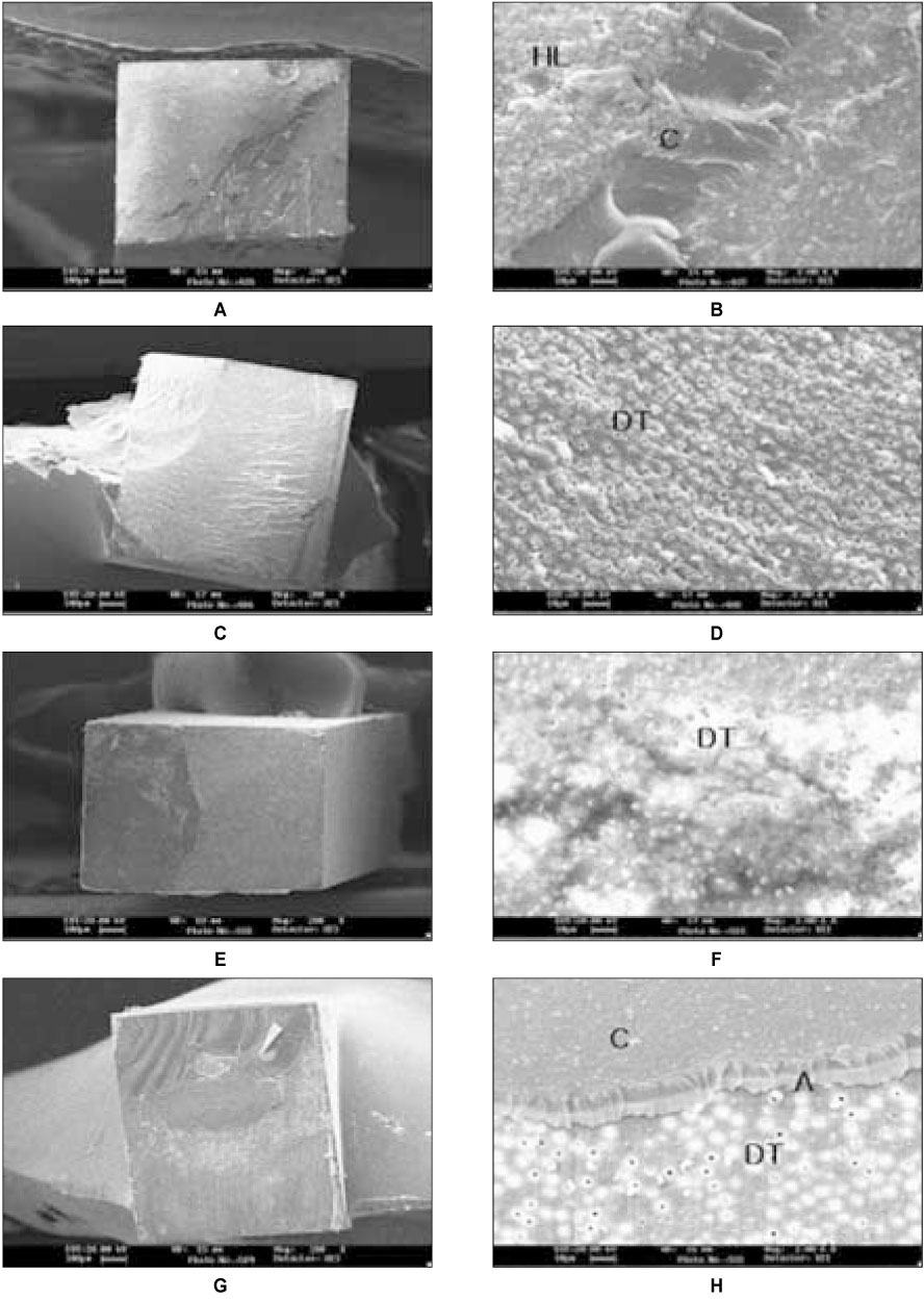
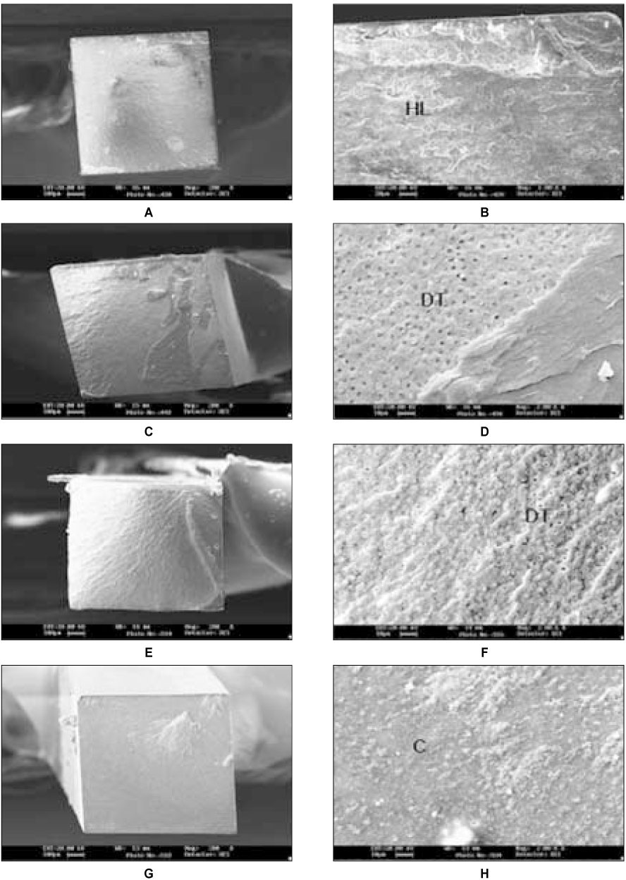
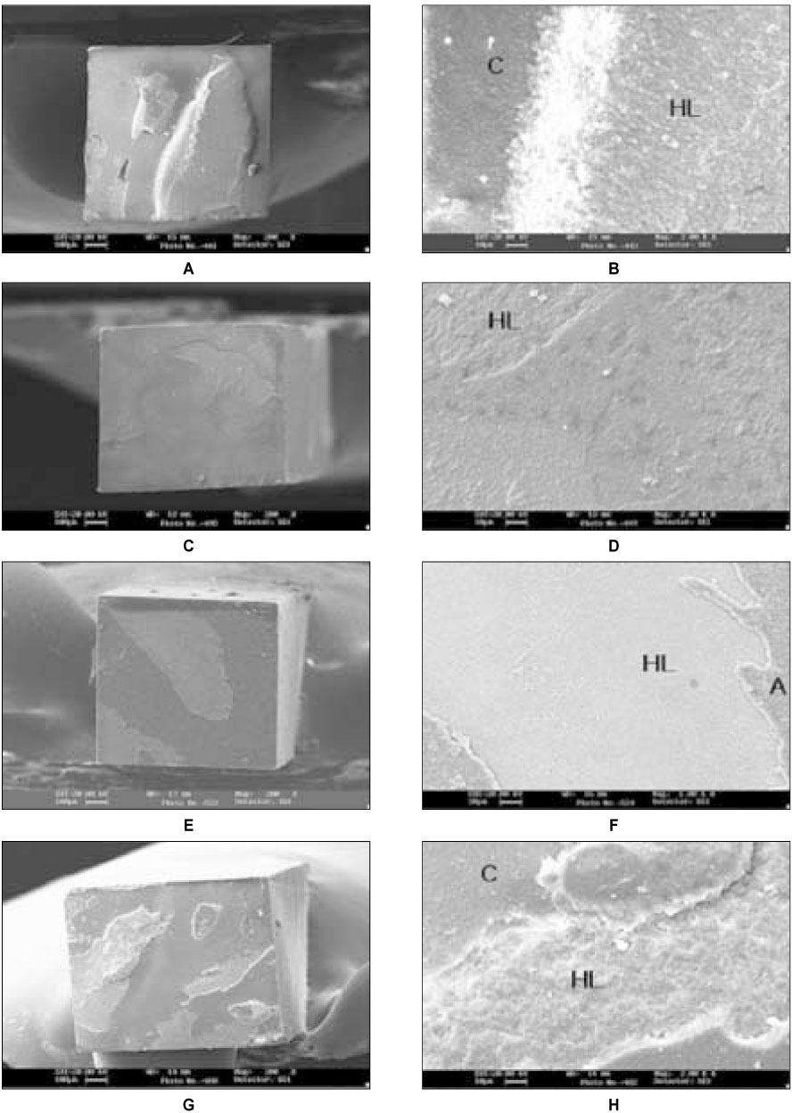
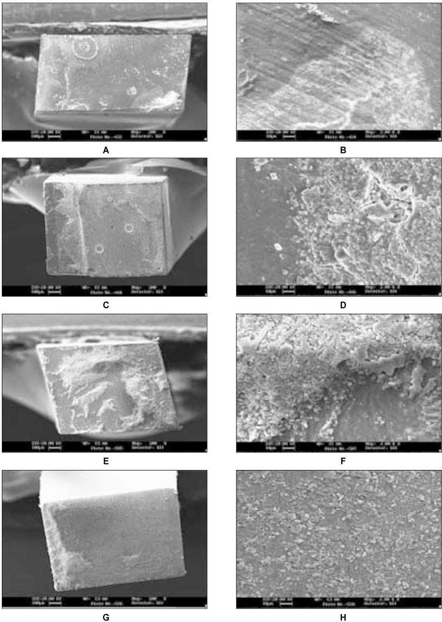
SEM analysis of failure mode distribution revealed the adhesive failure at hybrid layer in most of the specimen, and the shift of the failure site from bottom to top of the hybrid layer with chlorhexidine groups.
2% chlorhexidine application after acid-etching proved to preserve the durability of the hybrid layer and microtensile bond strength of dentin bonding systems.
- 1. Van Meerbeek B, Perdigao J, Lambrechts P. The clinical performance of adhesives. J Dent. 1998;26: 1-20.ArticlePubMed
- 2. Hashimoto M, Ohno H, Sano H. In vitro degradation of resin-dentin bonds analyzed by microtensile bond test, scanning and transmission electron microscopy. Biomaterials. 2003;24: 3795-3803.ArticlePubMed
- 3. Okuda M, Pereira PNR, Nakajima M. Long-term durability of resin dentin interface: nanoleakage vs microtensile bond strength. Oper Dent. 2002;27: 289-296.PubMed
- 4. De Munck J, Van Meerbeek B, Yoshida Y. Four-year Water Degradation of Total-etch Adhesives Bonded to Dentin. J Dent Res. 2003;2: 136-140.
- 5. Pashley DH, Tay FR, Yiu C. Callagen Degradation by Host-derived Enzymes during Aging. J Dent Res. 2004;83: 216-221.ArticlePubMedPDF
- 6. Gendron R, Grenier D, Sorsa T. Inhibition of the activities of matrix metalloproteinases 2, 8, and 9 by chlorhexidine. Clin Diagn Lab Immunol. 1999;437-439.ArticlePubMedPMCPDF
- 7. Hashimoto M, Ohno H, Kaga M. In vivo degradation of resin-dentin bonds in humans over 1 to 3 years. J Dent Res. 2000;79: 1385-1391.ArticlePubMedPDF
- 8. Chaussain-Miller C, Fioretti F, Goldberg M. The role of matrix metalloproteinases(MMPs) in Human Caries. J Dent Res. 2006;85: 22-32.ArticlePubMedPDF
- 9. Sorsa T, Tjaderhane L, Salo T. Matrix metalloproteinases(MMPs) in oral disease. Oral Dis. 2004;10: 311-318.PubMed
- 10. Hebling J, Pashley DH, Tjaderhane L. Chlorhexidine arrests subclinical degradation of dentin hybrid layers in vivo. J Dent Res. 2005;84: 741-746.ArticlePubMedPDF
- 11. Carrilho MR, Carvalho RM. Chlorhexidine preserves dentin bond in vitro. J Dent Res. 2007;86: 90-94.ArticlePubMedPDF
- 12. Carrilho MR, Tay FR, Pashley DH. Mechnical stability of resin-dentin bond components. Dent Mater. 2005;21: 232-241.PubMed
- 13. Carrilho MR, Carvalho RM, Tay FR. Durability of resin-dentin bonds related to water and oil storage. Am J Dent. 2005;18: 315-319.PubMed
- 14. De Castro F, De Andrade M. Effect of 2% chlorhexidine on microtensile bond strength of composite to dentin. J Adhes Dent. 2003;5: 129-138.PubMed
- 15. Martin-De Las Heras S, Valenzuela A, Overall CM. The matrix metalloproteinases gelatinases A in human dentin. Arch Oral Biol. 2000;45: 757-765.PubMed
- 16. De Munck J, Van Landuyt K, Peumans M. A critical review of the durability of adhesion to tooth tissue: methods and results. J Dent Res. 2005;84: 118-132.ArticlePubMedPDF
- 17. Nagase H, Visse R, Mruphy G. Structure and function of matrix metalloproteinases and TIMPs. Cardiovasc Res. 2006;69: 562-573.ArticlePubMed
- 18. Nishitani Y, Yoshiyama M, Wadgaonkar B. Activation of gelatinolytic/collagenolytic activity in dentin by self-etching adhesives. Eur J Oral Sci. 2006;114: 160-166.ArticlePubMed
- 19. Dumas J, Hurion N, Weill R. Collagenases in mineralized tissues of human teeth. FEBS Lett. 1985;187: 51-55.PubMed
- 20. Sulkala M, Wahlgren J, Larmas M. The effects of MMP inhibitors on human salivary MMP activity and caries progression in rat. J Dent Res. 2001;80: 1545-1549.ArticlePubMedPDF
- 21. Traderhane L, Larjava H, Sorsa T. The activation and function of host matrix metalloproteinases in dentin matrix breakdown in caries lesions. J Dent Res. 1998;77: 1622-1629.ArticlePubMedPDF
- 22. Van Strijp AJ, Van Steenbergen TJ, De Graaff J. Bacterial colonization and degradation of demineralized dentin matrix in situ. Caries Res. 1994;28: 21-27.ArticlePubMed
- 23. Van Strijp AJ, Van Steenbergen TJ, ten Cate JM. Bacterial colonization of mineralized and completely demineralized dentin in situ. Caries Res. 1997;31: 349-355.ArticlePubMed
- 24. Birkedal-Hansen H, Moore WG, Bodden MK. Matrix metalloproteinases: a review. Crit Rev Oral Biol Med. 1993;4: 197-250.ArticlePubMedPDF
- 25. Bode W, Fernandez-Catalan C, Tschesche H. Structural properties of matrix metalloproteinases. Cell Mol Life Sci. 1999;55: 639-652.ArticlePubMedPMCPDF
- 26. Van Strijp AJ, Jansen DC, De Groot J. Host-derived proteinases and degradation of dentine collagen in situ. Caries Res. 2003;37: 58-65.ArticlePubMedPDF
- 27. Brackett WW, Tay FR, Brackett MG. The effect of chlorhexidine on dentin hybrid layers in vivo. Oper Dent. 2007;32: 107-111.ArticlePubMedPDF
- 28. Carrilho MR, Geraldeli S, Tay F. In vivo preservation of the hybrid layer by chlorhexidine. J Dent Res. 2007;86: 529-533.ArticlePubMedPDF
- 29. Inoue S, Van Meerbeek B, Abe Y. Effect of remaining dentin thickness and the use of conditioner on microtensile bond strength of glass ionomer adhesive. Dent Mater. 2001;17: 445-455.PubMed
- 30. Lin A, Mclntyre NS, Davidson RD. Studies on the adhesion of glass ionomer cements to dentin. J Dent Res. 1992;71: 1836-1841.ArticlePubMedPDF
- 31. Van Meerbeek B, Vargas S, Inoue S. Adhesives and cements to promote preservation dentistry. Oper Dent. 2001;26: Suppl 6. S119-S144.
- 32. Yoshida Y, Van Meerbeek B, Nakayama Y. Evidence of chemical bonding at biomaterial-hard tissue interfaces. J Dent Res. 2000;79: 709-714.ArticlePubMedPDF
- 33. Gale MS, Darvell BW. Thermal cycling procedures for laboratory testing of dental restorations. J Dent. 1999;27: 89-99.ArticlePubMed
- 34. Van Meerbeek B, Conn LJ Jr, Duke ES. Correlative transmission electron microscopy examination of nondemineralized and demineralized resin-dentin interfaces formed by two dentin adhesive systems. J Dent Res. 1996;75: 879-888.ArticlePubMedPDF
- 35. Eliades G, Vougiouklakis G, Palaghias G. Heterogenous distribution of single-bottle adhesive monomers in the resin-dentin interdiffusion zone. Dent Mater. 2001;17: 277-283.PubMed
REFERENCES
Tables & Figures
REFERENCES
Citations

- The effect of the removal of chondroitin sulfate on bond strength of dentin adhesives and collagen architecture
Jong-Ryul Kim, Sang-Jin Park, Gi-Woon Choi, Kyoung-Kyu Choi
Journal of Korean Academy of Conservative Dentistry.2010; 35(3): 211. CrossRef
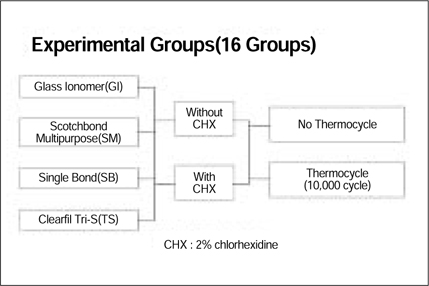
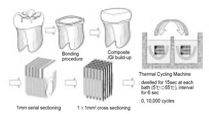
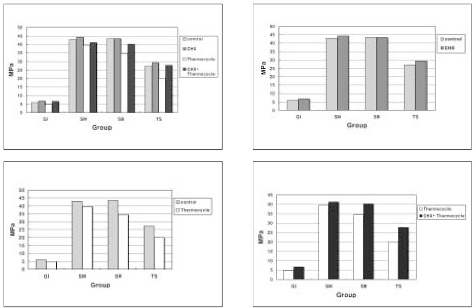


Figure 1
Figure 2
Figure 3
Figure 4
Figure 5
Figure 6
Figure 7
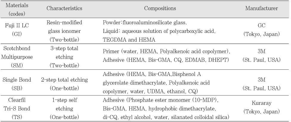
Materials used in this study
Microtensile Bond Strengths (MPa, mean ± SD) of 16 Experimental Groups
Different superscript letters were significantly different (p < 0.05).
Different superscript letters were significantly different (p < 0.05).

 KACD
KACD



 ePub Link
 Cite
Cite

