Articles
- Page Path
- HOME > Restor Dent Endod > Volume 43(4); 2018 > Article
- Research Article Microtensile bond strength of CAD/CAM-fabricated polymer-ceramics to different adhesive resin cements
-
Leyla Sadighpour1
 , Farideh Geramipanah2
 , Zahra Ghasri3, Mehrnoosh Neshatian4
 -
Restor Dent Endod 2018;43(4):e40.
DOI: https://doi.org/10.5395/rde.2018.43.e40
Published online: September 3, 2018
1Dental Research Center, Dentistry Research Institute, Department of Prosthodontics, School of Dentistry, Tehran University of Medical Sciences, Tehran, Iran.
2Dental Implant Research Center, Department of Prosthodontics, School of Dentistry, Tehran University of Medical Sciences, Tehran, Iran.
3Department of Restorative Dentistry, Faculty of Dentistry, Shahed University, Tehran, Iran.
4Matrix Dynamic Group, Faculty of Dentistry, University of Toronto, Toronto, ON, Canada.
- Correspondence to Farideh Geramipanah, DDS, MS. Professor, Dental Implant Research Center, Department of Prosthodontics, School of Dentistry, Tehran University of Medical Sciences, Kargar Shomali, Hakim HWY, 143995591, Tehran, Iran. Geramipa@tums.ac.ir, lsadigh27@yahoo.com
Copyright © 2018. The Korean Academy of Conservative Dentistry
This is an Open Access article distributed under the terms of the Creative Commons Attribution Non-Commercial License (https://creativecommons.org/licenses/by-nc/4.0/) which permits unrestricted non-commercial use, distribution, and reproduction in any medium, provided the original work is properly cited.
- 1,947 Views
- 6 Download
- 7 Crossref
Abstract
-
Objectives This study evaluated the microtensile bond strength (µTBS) of polymer-ceramic and indirect composite resin with 3 classes of resin cements.
-
Materials and Methods Two computer-aided design/computer-aided manufacturing (CAD/CAM)-fabricated polymer-ceramics (Enamic [ENA; Vita] and Lava Ultimate [LAV; 3M ESPE]) and a laboratory indirect composite resin (Gradia [GRA; GC Corp.]) were equally divided into 6 groups (n = 18) with 3 classes of resin cements: Variolink N (VAR; Vivadent), RelyX U200 (RXU; 3M ESPE), and Panavia F2 (PAN; Kuraray). The μTBS values were compared between groups by 2-way analysis of variance and the post hoc Tamhane test (α = 0.05).
-
Results Restorative materials and resin cements significantly influenced µTBS (p < 0.05). In the GRA group, the highest μTBS was found with RXU (27.40 ± 5.39 N) and the lowest with VAR (13.54 ± 6.04 N) (p < 0.05). Similar trends were observed in the ENA group. In the LAV group, the highest μTBS was observed with VAR (27.45 ± 5.84 N) and the lowest with PAN (10.67 ± 4.37 N) (p < 0.05). PAN had comparable results to those of ENA and GRA, whereas the μTBS values were significantly lower with LAV (p = 0.001). The highest bond strength of RXU was found with GRA (27.40 ± 5.39 N, p = 0.001). PAN showed the lowest µTBS with LAV (10.67 ± 4.37 N; p < 0.001).
-
Conclusions When applied according to the manufacturers' recommendations, the µTBS of polymer-ceramic CAD/CAM materials and indirect composites is influenced by the luting cements.
INTRODUCTION
Materials used in this study
MATERIALS AND METHODS
RESULTS
Bond strength values in MPa and the results of multiple comparisons, according to the 2 variables of restorative material and cement type
| Restorative material | Cement | ||
|---|---|---|---|
| VAR | RXU | PAN | |
| ENA | 10.70 ± 3.40Aa | 19.58 ± 4.61Ba | 19.40 ± 7.87Bb |
| LAV | 27.45 ± 5.84Cb | 20.23 ± 3.32Ba | 10.67 ± 4.37Aa |
| GRA | 13.45 ± 6.04Aa | 27.40 ± 5.39Cb | 19.20 ± 5.83Bb |
The frequency of failure types
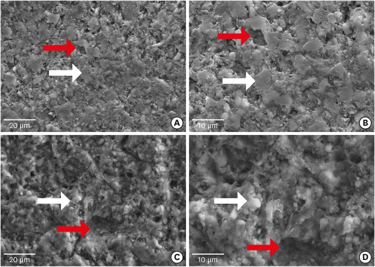
Scanning electron microscope images of computer-aided design/computer-aided manufacturing polymer ceramics before cutting. (A) Enamic (×2,500) showed a homogeneous distribution of the ceramic phase (white arrow) and the polymer phase (red arrow); (B) With higher magnification (×5,000), the ceramic particles with sharp edges (white arrow) and amorphous polymer phases (red arrow) were displayed; (C) LAVA Ultimate (×2,500) showed distributed roughness on the surface; (D) With higher magnification (×5,000), organic fibers (white arrow) embedded in the polymer matrix were observed.

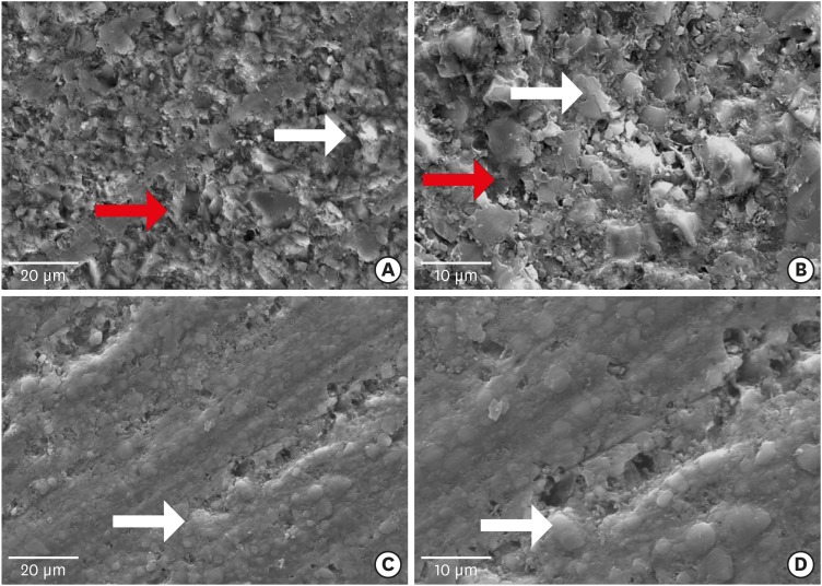
Scanning electron microscope images of computer-aided design/computer-aided manufacturing polymer ceramics after cutting. (A) Enamic (×2,500); (B) With higher magnification (×5,000), more roughness than in the as-block specimen was seen (white arrow indicates ceramic particles and red arrow indicates polymer phase); (C) LAVA Ultimate (×2,500); (D) With higher magnification (×5,000), the cut surface of Lava Ultimate showed less roughness. More areas of porosity were distributed across the specimen than in the as-block specimen (whiter arrow indicates ceramic fiber).

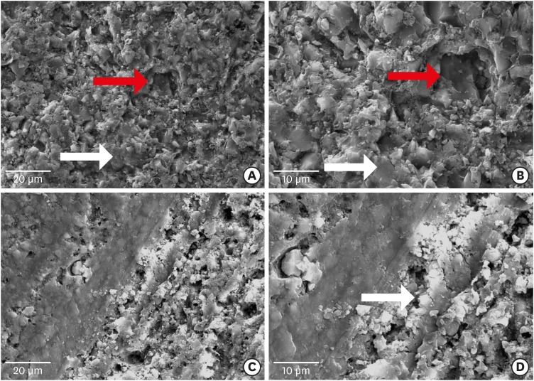
Scanning electron microscope images of computer-aided design/computer-aided manufacturing polymer-ceramic after treatment. (A) Etched surface of Enamic (×2,500); (B) With higher magnification (×5,000), larger holes (white arrow) within a relatively unaffected polymer phase (red arrow) are seen; (C) Lava Ultimate after sand blasting (×2,500); (D) With higher magnification (×5,000), Lava Ultimate showed more roughness and porosity than untreated specimens. The cracks in the matrix could be seen (white arrow).

DISCUSSION
CONCLUSIONS
ACKNOWLEDGEMENT
-
Funding: This research was supported by a grant (No. #32381) from Tehran University of Medical Sciences.
-
Conflict of Interest: No potential conflict of interest relevant to this article was reported.
-
Author Contributions:
Conceptualization: Sadighpour L, Geramipanah F.
Data curation: Sadighpour L, Geramipanah F, Ghasri Z, Neshatian M.
Funding acquisition: Sadighpour L, Geramipanah F.
Investigation: Sadighpour L, Ghasri Z, Neshatian M.
Methodology: Sadighpour L, Geramipanah F, Ghasri Z, Neshatian M.
Project administration: Sadighpour L.
Resources: Sadighpour L.
Supervision: Sadighpour L, Geramipanah F.
Validation: Sadighpour L, Geramipanah F.
Visualization: Ghasri Z, Neshatian M.
Writing - original draft: Sadighpour L, Geramipanah F.
Writing - review & editing: Sadighpour L, Geramipanah F, Ghasri Z, Neshatian M.
- 1. Della Bona A, Corazza PH, Zhang Y. Characterization of a polymer-infiltrated ceramic-network material. Dent Mater 2014;30:564-569.ArticlePubMedPMC
- 2. Fron Chabouis H, Smail Faugeron V, Attal JP. Clinical efficacy of composite versus ceramic inlays and onlays: a systematic review. Dent Mater 2013;29:1209-1218.ArticlePubMed
- 3. Sripetchdanond J, Leevailoj C. Wear of human enamel opposing monolithic zirconia, glass ceramic, and composite resin: an in vitro study. J Prosthet Dent 2014;112:1141-1150.ArticlePubMed
- 4. Elsaka SE. Repair bond strength of resin composite to a novel CAD/CAM hybrid ceramic using different repair systems. Dent Mater J 2015;34:161-167.ArticlePubMed
- 5. Angeletaki F, Gkogkos A, Papazoglou E, Kloukos D. Direct versus indirect inlay/onlay composite restorations in posterior teeth. A systematic review and meta-analysis. J Dent 2016;53:12-21.ArticlePubMed
- 6. Gracis S, Thompson VP, Ferencz JL, Silva NR, Bonfante EA. A new classification system for all-ceramic and ceramic-like restorative materials. Int J Prosthodont 2015;28:227-235.ArticlePubMed
- 7. Cekic-Nagas I, Ergun G, Egilmez F, Vallittu PK, Lassila LV. Micro-shear bond strength of different resin cements to ceramic/glass-polymer CAD-CAM block materials. J Prosthodont Res 2016;60:265-273.ArticlePubMed
- 8. Swain MV, Coldea A, Bilkhair A, Guess PC. Interpenetrating network ceramic-resin composite dental restorative materials. Dent Mater 2016;32:34-42.ArticlePubMed
- 9. Awada A, Nathanson D. Mechanical properties of resin-ceramic CAD/CAM restorative materials. J Prosthet Dent 2015;114:587-593.ArticlePubMed
- 10. Kumbuloglu O, Özcan M. Clinical survival of indirect, anterior 3-unit surface-retained fibre-reinforced composite fixed dental prosthesis: up to 7.5-years follow-up. J Dent 2015;43:656-663.ArticlePubMed
- 11. Jongsma LA, Kleverlaan CJ, Feilzer AJ. Clinical success and survival of indirect resin composite crowns: results of a 3-year prospective study. Dent Mater 2012;28:952-960.ArticlePubMed
- 12. Frankenberger R, Hartmann VE, Krech M, Krämer N, Reich S, Braun A, Roggendorf M. Adhesive luting of new CAD/CAM materials. Int J Comput Dent 2015;18:9-20.PubMed
- 13. Elsaka SE. Bond strength of novel CAD/CAM restorative materials to self-adhesive resin cement: the effect of surface treatments. J Adhes Dent 2014;16:531-540.PubMed
- 14. Yoshihara K, Nagaoka N, Maruo Y, Nishigawa G, Irie M, Yoshida Y, Van Meerbeek B. Sandblasting may damage the surface of composite CAD-CAM blocks. Dent Mater 2017;33:e124-e135.ArticlePubMed
- 15. Peumans M, Valjakova EB, De Munck J, Mishevska CB, Van Meerbeek B. Bonding effectiveness of luting composites to different CAD/CAM materials. J Adhes Dent 2016;18:289-302.PubMed
- 16. Flury S, Schmidt SZ, Peutzfeldt A, Lussi A. Dentin bond strength of two resin-ceramic computer-aided design/computer-aided manufacturing (CAD/CAM) materials and five cements after six months storage. CAD/CAM-materials after storage. Dent Mater J 2016;35:728-735.PubMed
- 17. da Silva EM, Miragaya L, Sabrosa CE, Maia LC. Stability of the bond between two resin cements and an yttria-stabilized zirconia ceramic after six months of aging in water. J Prosthet Dent 2014;112:568-575.ArticlePubMed
- 18. Ferracane JL, Stansbury JW, Burke FJ. Self-adhesive resin cements - chemistry, properties and clinical considerations. J Oral Rehabil 2011;38:295-314.ArticlePubMed
- 19. Frassetto A, Navarra CO, Marchesi G, Turco G, Di Lenarda R, Breschi L, Ferracane JL, Cadenaro M. Kinetics of polymerization and contraction stress development in self-adhesive resin cements. Dent Mater 2012;28:1032-1039.ArticlePubMed
- 20. Campos F, Almeida CS, Rippe MP, de Melo RM, Valandro LF, Bottino MA. Resin bonding to a hybrid ceramic: effects of surface treatments and aging. Oper Dent 2016;41:171-178.ArticlePubMedPDF
- 21. Keul C, Martin A, Wimmer T, Roos M, Gernet W, Stawarczyk B. Stawarcxyk. Tensile bond strength of PMMA- and composite-based CAD/CAM materials to luting cements after different conditioning methods. Int J Adhes Adhes 2013;46:122-127.
- 22. El Zohairy AA, De Gee AJ, Mohsen MM, Feilzer AJ. Microtensile bond strength testing of luting cements to prefabricated CAD/CAM ceramic and composite blocks. Dent Mater 2003;19:575-583.ArticlePubMed
- 23. Stawarczyk B, Basler T, Ender A, Roos M, Ozcan M, Hämmerle C. Effect of surface conditioning with airborne-particle abrasion on the tensile strength of polymeric CAD/CAM crowns luted with self-adhesive and conventional resin cements. J Prosthet Dent 2012;107:94-101.ArticlePubMed
- 24. Pereira SG, Fulgêncio R, Nunes TG, Toledano M, Osorio R, Carvalho RM. Effect of curing protocol on the polymerization of dual-cured resin cements. Dent Mater 2010;26:710-718.ArticlePubMed
- 25. Kumbuloglu O, Lassila LV, User A, Vallittu PK. A study of the physical and chemical properties of four resin composite luting cements. Int J Prosthodont 2004;17:357-363.PubMed
- 26. Gilbert S, Keul C, Roos M, Edelhoff D, Stawarczyk B. Bonding between CAD/CAM resin and resin composite cements dependent on bonding agents: three different in vitro test methods. Clin Oral Investig 2016;20:227-236.ArticlePubMedPDF
- 27. El Zohairy AA, de Gee AJ, de Jager N, van Ruijven LJ, Feilzer AJ. The influence of specimen attachment and dimension on microtensile strength. J Dent Res 2004;83:420-424.ArticlePubMedPDF
REFERENCES
Tables & Figures
REFERENCES
Citations

- Enhancing severely compromised premolar strength: role of cusp reduction design in CAD/CAM composite restorations
Mohamed F. Haridy, Ahmed Refaat Mohamed, Shehabeldin Saber, Edgar Schafer, Samar Elsayed Swelam, Youssef M. Haridy, Hend S. Ahmed
Odontology.2025;[Epub] CrossRef - Effect of hydrofluoric acid and self-etch ceramic primers on the flexural strength and fatigue resistance of glass ceramics: A systematic review and meta-analysis of in vitro studies
Paulo Matias Moreira, Gabriela Luiza Moreira Carvalho, Rodrigo de Castro Albuquerque, Carolina Bosso André
Japanese Dental Science Review.2024; 60: 198. CrossRef - Light transmittance through resin-matrix composite onlays adhered to resin-matrix cements or flowable composites
Rita Fidalgo-Pereira, Susana O. Catarino, Óscar Carvalho, Nélio Veiga, Orlanda Torres, Annabel Braem, Júlio C.M. Souza
Journal of the Mechanical Behavior of Biomedical Materials.2024; 151: 106353. CrossRef - Effect of thermocycling on the mechanical properties of permanent composite-based CAD-CAM restorative materials produced by additive and subtractive manufacturing techniques
Tuğba Temizci, Hatice Nalan Bozoğulları
BMC Oral Health.2024;[Epub] CrossRef - Effect of different surface treatments on resin-matrix CAD/CAM ceramics bonding to dentin: in vitro study
Hanan Fathy, Hamdi H. Hamama, Noha El-Wassefy, Salah H. Mahmoud
BMC Oral Health.2022;[Epub] CrossRef - Digital image analysis of fluorescence of ceramic veneers with different ceramic materials and resin cements
Jiao ZHANG, Qing YU
Dental Materials Journal.2022; 41(6): 868. CrossRef - Fatigue Behavior of Monolithic Zirconia-Reinforced Lithium Silicate Ceramic Restorations: Effects of Conditionings of the Intaglio Surface and the Resin Cements
F Dalla-Nora, LF Guilardi, CP Zucuni, LF Valandro, MP Rippe
Operative Dentistry.2021; 46(3): 316. CrossRef


Figure 1
Figure 2
Figure 3
Materials used in this study
| Product name | Manufacturer | Composition | Batch number |
|---|---|---|---|
| GC Gradia | GC Corp. | Polymer (UDMA and EDMA), filler (75 wt%; ceramic, SiO2, and prepolymerized particles) | 140618A |
| Enamic | Vita Zahnfabrik | Polymer (14 wt%; TEGDMA and UDMA), ceramic (86 wt%; SiO2, ZrO2, Al2O3, Na2O, K2O, and CaO) | 48720 |
| Lava Ultimate | 3M ESPE | Polymer (20 wt%; Bis-GMA, UDMA, Bis-EMA, and TEGDMA), filler (80 wt%; SiO2 and ZrO2) | N515648 |
| Variolink N | Vivadent/Ivoclar | Polymer (Bis-GMA. TEGDMA, and UDMA), filler (73.4 wt%; barium glass, ytterbium trifluoride, Ba-Al-fluorosilicate glass, and spheroid mixed oxide), initiators, stabilizers, pigments | U16084 & T38947 |
| Panavia | Kuraray Noritake Dental Inc. | Base: polymer (10-MDP, 5-NMSA, and dimethacrylates), initiator, filler (Silica) | 990024 |
| Catalyst: polymer (dimethacrylates), filler (73 wt%; barium glass and sodium fluoride), BPO | 970114 | ||
| Relyx U200 | 3M ESPE | Paste A: polymer (HEMA), filler (fluoroaluminasilicate [FAS] glass), proprietary reduction agent, opacifying agent | 603039 |
| Paste B: polymer (methacrylate polycarboxylic acid, Bis-GMA, and HEMA), zirconia silica filler, potassium persulfate |
UDMA, urethane dimethacrylate; EDMA, ethylene glycol dimethacrylate; TEGDMA, triethylene glycol dimethacrylate; Bis-GMA, bisphenol A diglycidyl methacrylate; Bis-EMA, ethoxylated bisphenol A glycol dimethacrylate; 10-MDP, 10-methacryloyloxydecyl dihydrogenphosphate; 5-NMSA, 5-N-methacryloyl-5-aminosalicylic acid; HEMA, 2-hydroxyethyl methacrylate.
Bond strength values in MPa and the results of multiple comparisons, according to the 2 variables of restorative material and cement type
| Restorative material | Cement | ||
|---|---|---|---|
| VAR | RXU | PAN | |
| ENA | 10.70 ± 3.40Aa | 19.58 ± 4.61Ba | 19.40 ± 7.87Bb |
| LAV | 27.45 ± 5.84Cb | 20.23 ± 3.32Ba | 10.67 ± 4.37Aa |
| GRA | 13.45 ± 6.04Aa | 27.40 ± 5.39Cb | 19.20 ± 5.83Bb |
Data are shown as means ± standard deviations (n = 18). The values in each column and row with different superscript letters are significantly different at a 95% level of confidence. Differences within each row are shown in uppercase superscript letters and differences within each column are shown in lowercase superscript letters.
GRA, GC Gradia, GC Corp., Tokyo, Japan; ENA, Enamic, Vita Zahnfabrik, Bad Sackingen, Germany; LAV, Lava Ultimate, 3M ESPE, St. Paul, MN, USA; VAR, Variolink N, Vivadent/Ivoclar, Schaan, Liechtenstein; RXU, RelyX U200, 3M ESPE; PAN, Panavia F2, Kuraray Noritake Dental Inc., Okayama, Japan.
The frequency of failure types
| Restorative material | Cement | Failure type | ||
|---|---|---|---|---|
| Type 1 | Type 2 | Type 3 | ||
| GRA | VAR | - | - | 18 (100) |
| RXU | - | - | 18 (100) | |
| PAN | - | - | 16 (100) | |
| ENA | VAR | 1 (5.6) | 17 (94.4) | - |
| RXU | 1 (5.6) | 17 (94.4) | - | |
| PAN | 2 (11.1) | 16 (90.0) | - | |
| LAV | VAR | 2 (11.1) | 16 (90.0) | - |
| RXU | 2 (11.1) | 16 (90.0) | - | |
| PAN | 3 (16.7) | 15 (83.3) | - | |
Values are presented as number (%).
GRA, GC Gradia, GC Corp., Tokyo, Japan; ENA, Enamic, Vita Zahnfabrik, Bad Sackingen, Germany; LAV, Lava Ultimate, 3M ESPE, St. Paul, MN, USA; VAR, Variolink N, Vivadent/Ivoclar, Schaan, Liechtenstein; RXU, RelyX U200, 3M ESPE; PAN, Panavia F2, Kuraray Noritake Dental Inc., Okayama, Japan; Type 1, adhesive failure, in which the surface of the CAD/CAM material was visible; Type 2, mixed failure in CAD/CAM material and cement surfaces, in which resin cement was partially visible in certain areas; Type 3, cohesive failure within the resin layer, in which almost all of the fracture surface was covered with cement; CAD/CAM, computer-aided design/computer-aided manufacturing.
UDMA, urethane dimethacrylate; EDMA, ethylene glycol dimethacrylate; TEGDMA, triethylene glycol dimethacrylate; Bis-GMA, bisphenol A diglycidyl methacrylate; Bis-EMA, ethoxylated bisphenol A glycol dimethacrylate; 10-MDP, 10-methacryloyloxydecyl dihydrogenphosphate; 5-NMSA, 5-N-methacryloyl-5-aminosalicylic acid; HEMA, 2-hydroxyethyl methacrylate.
Data are shown as means ± standard deviations (
GRA, GC Gradia, GC Corp., Tokyo, Japan; ENA, Enamic, Vita Zahnfabrik, Bad Sackingen, Germany; LAV, Lava Ultimate, 3M ESPE, St. Paul, MN, USA; VAR, Variolink N, Vivadent/Ivoclar, Schaan, Liechtenstein; RXU, RelyX U200, 3M ESPE; PAN, Panavia F2, Kuraray Noritake Dental Inc., Okayama, Japan.
Values are presented as number (%).
GRA, GC Gradia, GC Corp., Tokyo, Japan; ENA, Enamic, Vita Zahnfabrik, Bad Sackingen, Germany; LAV, Lava Ultimate, 3M ESPE, St. Paul, MN, USA; VAR, Variolink N, Vivadent/Ivoclar, Schaan, Liechtenstein; RXU, RelyX U200, 3M ESPE; PAN, Panavia F2, Kuraray Noritake Dental Inc., Okayama, Japan; Type 1, adhesive failure, in which the surface of the CAD/CAM material was visible; Type 2, mixed failure in CAD/CAM material and cement surfaces, in which resin cement was partially visible in certain areas; Type 3, cohesive failure within the resin layer, in which almost all of the fracture surface was covered with cement; CAD/CAM, computer-aided design/computer-aided manufacturing.

 KACD
KACD
 ePub Link
 Cite
Cite

