Articles
- Page Path
- HOME > Restor Dent Endod > Volume 31(2); 2006 > Article
- Original Article Effect of additional coating of bonding resin on the microtensile bond strength of self-etching adhesives to dentin
- Moon-Kyung Jung1, Byeong-Hoon Cho1,2,3,*, Ho-Hyun Son1,2, Chung-Moon Um1,2, Young-Chul Han1, Sae-Joon Choung1
-
J Korean Acad Conserv Dent 2006;31(2):-112.
DOI: https://doi.org/10.5395/JKACD.2006.31.2.103
Published online: January 14, 2006
1Department of Conservative Dentistry, School of Dentistry, Seoul National University, Seoul, Korea
2Dental Research Institute, Seoul National University, Seoul, Korea
3Intellectual Biointerface Engineering Center, Seoul National University, Seoul, Korea
- *Corresponding Author: Byeong-Hoon Cho, Department of Conservative Dentistry, School of Dentistry, and Dental Research Institute, Seoul National University. 28-2 Yeongun-dong, Chongro-gu, Seoul, Korea, 110-749, Tel: +82-2-2072-3514 Fax: +82-2-764-3514, E-mail: chobh@snu.ac.kr
Copyright © 2006 The Korean Academy of Conservative Dentistry
This is an Open Access article distributed under the terms of the Creative Commons Attribution Non-Commercial License (http://creativecommons.org/licenses/by-nc/3.0) which permits unrestricted non-commercial use, distribution, and reproduction in any medium, provided the original work is properly cited.
- 1,230 Views
- 2 Download
- 2 Crossref
Abstract
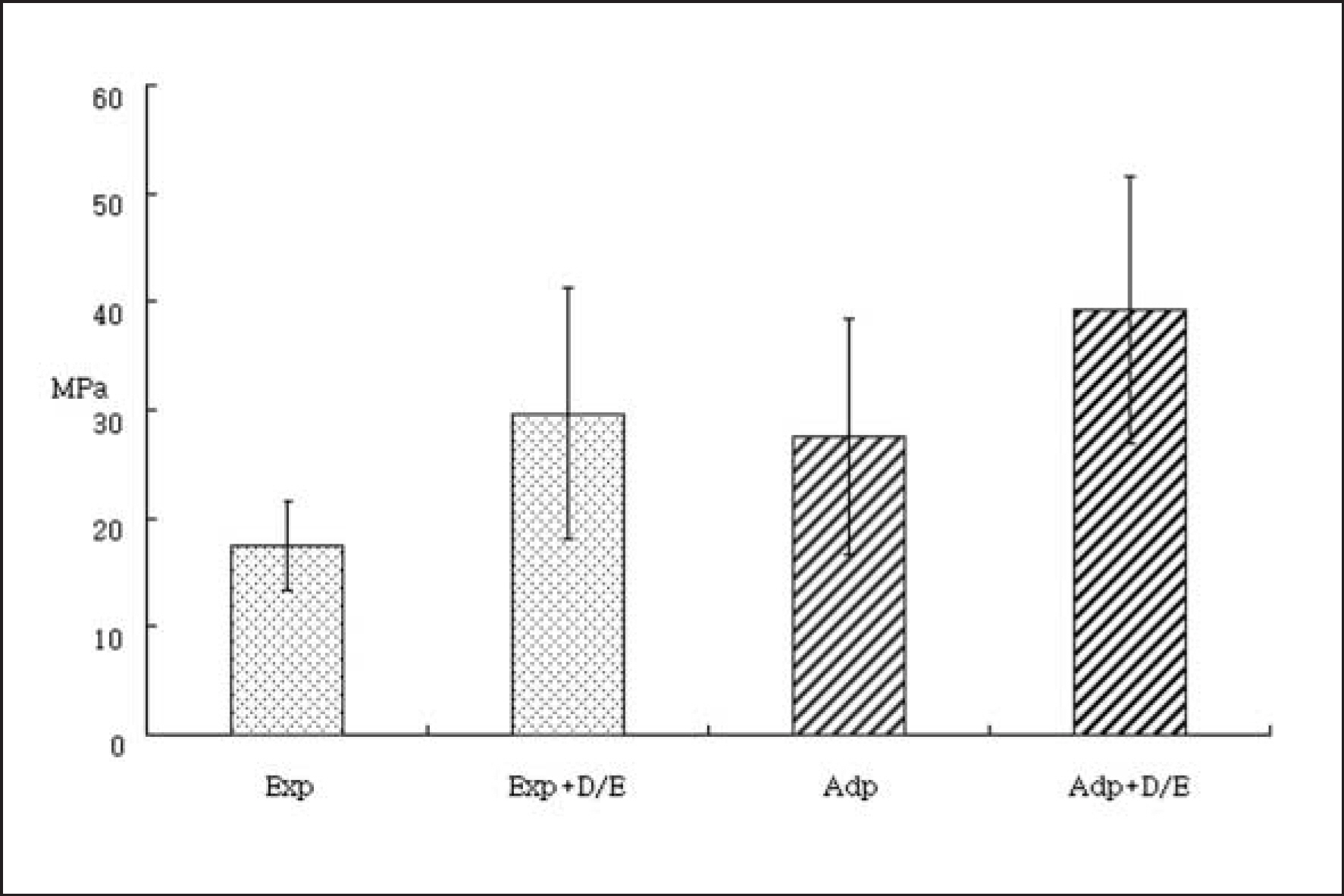
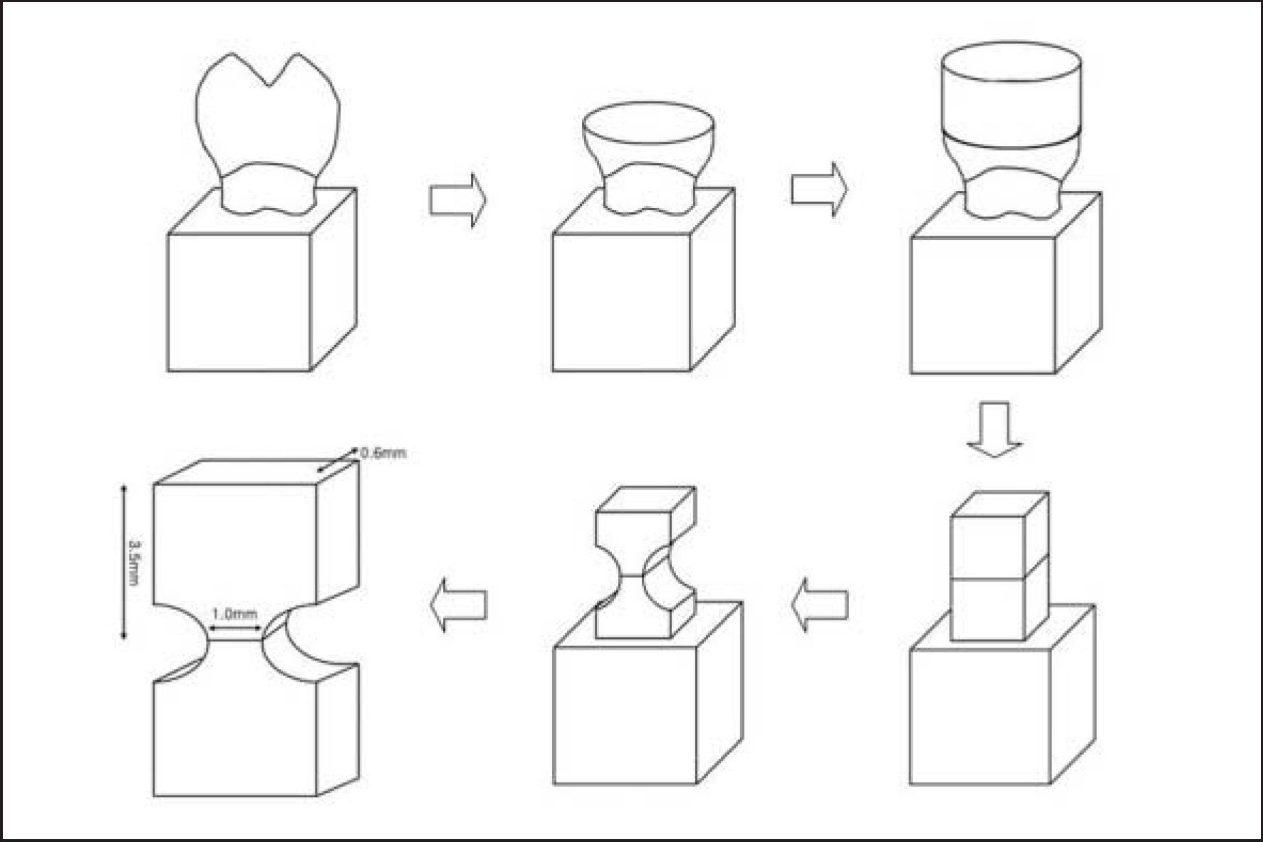
- This study investigated the hypothesis that the dentin bond strength of self-etching adhesive (SEA) might be improved by applying additional layer of bonding resin that might alleviate the pH difference between the SEA and the restorative composite resin. Two SEAs were used in this study; Experimental SEA (Exp, pH: 1.96) and Adper Prompt (AP, 3M ESPE, USA, pH: 1.0). In the control groups, they were applied with two sequential coats. In the experimental groups, after applying the first coat of assigned SEAs, the D/E bonding resin of All-Bond 2 (Bisco Inc., USA, pH: 6.9) was applied as the intermediate adhesive. Z-250 (3M ESPE, USA) composite resin was built-up in order to prepare hourglass-shaped specimens. The microtensile bond strength (MTBS) was measured and the effect of the intermediate layer on the bond strength was analyzed for each SEA using t-test. The fracture mode of each specimen was inspected using stereomicroscope and Field Emission Scanning Electron Microscope (FE-SEM). When D/E bonding resin was applied as the second coat, MTBS was significantly higher than that of the control groups. The incidence of the failure between the adhesive and the composite or between the adhesive and dentin decreased and that of the failure within the adhesive layer increased. According to the results, applying the bonding resin of neutral pH can increase the bond strength of SEAs by alleviating the difference in acidity between the SEA and restorative composite resin.

Abbreviations. Exp: Experimental self-etching adhesive; D/E: D/E bonding resin of All-Bond 2; Adp: Adper Prompt.


- 1. Fritz UB, Finger WJ. Bonding efficiency of single-bottle enamel/dentin adhesive. Am J Dent 12:277-282. 1999.PubMed
- 2. Bouillaguet S, Gysi P, Wataha JC, Ciucchi B, Cattani M, Godin C, Meyer JM. Bond strength of composite to dentin using conventional, one-step, and self-etching adhesive system. J Dent 29:55-61. 2001.PubMed
- 3. Tomoko A, Shigeru U, Hidehiko S. Comparison of bonding efficacy of an all-in-one adhesive with a self-etching primer system. Eur J Oral Sci 112:286-292. 2004.PubMed
- 4. Pashley EL, Agee KA, Pashley DH, Tay FR. Effects of one versus two application of an unfilled, all-in-one adhesive on dentin bonding. J Dent 30:83-90. 2002.PubMed
- 5. Frankenberger R, Perdigao J, Rosa BT, Lopes M. ‘No-bottle’vs‘multi-bottle’dentin adhesives-a microten-sile bond strength and morphological study. Dent Mater 17:373-380. 2001.ArticlePubMed
- 6. Choi KK, Condon JR, Ferracane JL. The effects of adhesive thickness on polymerization contraction stress of composite. J Dent Res 79:812-817. 2000.ArticlePubMedPDF
- 7. Cho BH, Dickens SH. Effects of the acetone content of single solution dentin bonding agents on the adhesive layer thickness and the microtensile bond strength. Dent Mater 20:107-115. 2004.ArticlePubMed
- 8. Zheng L, Pereira PN, Nakajima M, Sano H, Tagami J. Relationship between adhesive thickness and microten-sile bond strength. Oper Dent 26:97-104. 2001.PubMed
- 9. Chersoni S, Suppa P, Grandini W, Goracci C, Monticelli F, Yiu C, Huang C, Prati C, Breschi L, Ferrari M, Pashley DH, Tay FR. In vivo and in vitro permeability of one-step self-etch adhesives. J Dent Res 83:459-64. 2004.ArticlePubMedPDF
- 10. Tay FR, King NM, Suh BI, Pashley DH. Effect of delayed activation of light-cured resin composites on bonding of all-in-one adhesives. J Adhesive Dent 3:207-225. 2001.
- 11. Tay FR, Pashely DH. Aggressiveness of contemporary self-etching system. I: Depth of penetration beyond dentin smear layers. Dent Mater 17:296-308. 2001.PubMed
- 12. Pashley DH, Sano H, Ciucchi B, Yoshiyama M, Carvalho RM. Adhesion testing of dentin bonding agents: A review. Dent Mater 11:117-125. 1995.ArticlePubMed
- 13. Pashley DH, Carvalho RM, Sano H, Nakajima M, Yoshiyama M, Shono Y, Fernandes CA, Tay FR. The microtensile bond test: A review. J Adhesive Dent 1:299-309. 1999.
- 14. Tay FR, Pashley DH. Water treeing–a potential mechanism for degradation of dentin adhesives. Am J Dent 16:6-12. 2003.PubMed
- 15. Tay FR, Pashley DH, Suh BI, Carvalho RM, Itthagarun A. Single-step adhesives are permeable membranes. J Dent 30:371-82. 2002.ArticlePubMed
- 16. Tay FR, Pashley DH, Peters MC. Adhesive permeability affects composite coupling to dentin treated with a self-etch adhesive. Oper Dent 28:610-621. 2003.PubMed
- 17. Simo-Alfonso E, Gelfi C, Sebastiano T, Citterio A, Righetti PG. Novel acrylamido monomers with higher hydrophilicity and improved hydrolytic stability: II. Properties fo N-acryloylaminopropanol. Electrophoresis 17:732-737. 1996.ArticlePubMed
- 18. Tay FR, Pashley DH. Have dentin adhesives become too hydrophilic? J Can Dent Assoc 69:726-731. 2003.PubMed
- 19. Hagge MS, Lindemuth JS. Shear bond strength of an autopolymerizing core buildup composite bonded to dentin with 9 dentin adhesive systems. J Prosthet Dent 86:620-623. 2001.ArticlePubMed
- 20. Keefe KL, Powers JM. Adhesion of resin composite core materials to dentin. Int J Prosthod 14:451-456. 2001.
- 21. Swift EJ Jr, Perdigao J, Combe EC, Simpson CH 3rd, Nunes MF. Effects of restorative and adhesive curing methods on dentin bond strengths. Am J Dent 14:137-40. 2001.PubMed
- 22. Sanares AME, King NM, Itthagarun A, Tay FR, Pashley DH. Adverse surface interactions between one-bottle light-cured adhesives and chemical-cured composites. Dent Mater 17:542-556. 2001.ArticlePubMed
- 23. Dickens SH, Cho BH. Interpretation of bond failure through conversion and residual solvent measurements and Weibull analyses of flexural and microtensile bond strengths of bonding agents. Dent Mater 21:354-364. 2005.ArticlePubMed
REFERENCES
Tables & Figures
REFERENCES
Citations

- Effect of an intermediate bonding resin and flowable resin on the compatibility of two-step total etching adhesives with a self-curing composite resin
Sook-Kyung Choi, Ji-Wan Yum, Hyeon-Cheol Kim, Bock Hur, Jeong-Kil Park
Journal of Korean Academy of Conservative Dentistry.2009; 34(5): 397. CrossRef - Aging effect on the microtensile bond strength of self-etching adhesives
JS Park, JS Kim, MS Kim, HH Son, HC Kwon, BH Cho
Journal of Korean Academy of Conservative Dentistry.2006; 31(6): 415. CrossRef


Figure 1.
Figure 2.
Figure 3.
Figure 4.
Figure 5.
Figure 6.
| Composition of the materials | Manufacturer | |
|---|---|---|
| Adper Prompt | Liquid 1: | 3M ESPE, |
| Methacrylated phosphoric esters | St. Paul. MN, USA | |
| Bis-GMA | ||
| Initiators based on camphoroquinone | ||
| Stabilizers | ||
| Liquid 2: | ||
| Water | ||
| 2-Hydroxyethyl methacrylate (HEMA) | ||
| Polyalkenoic acid | ||
| Stabilizers | ||
| Experimental self-etching adhesive | Ethylene glycol methacrylate Phosphate (EGMP) | |
| MONO-2-(Methacryloyloxy) Ethyl phthalate (MEP) | ||
| Urethane dimethacrylate | ||
| 2-Hydroxyethylmethacrylate Ethanol | ||
| D/E resin of All-Bond2 | Bisphenol A diglycidylmethacrylate | Bisco, Itasca, IL, USA |
| Urethane dimetyhacrylate | ||
| Hydroxyethyl methacrylate | ||
| Z-250 | Matrix: | 3M ESPE, |
| UDMA, Bis-EMA and Bis-GMA | St. Paul. MN, USA | |
| Filler: | ||
| Zirconium glass and Coloidal silica |
| Fracture modes | Exp 2coat | Exp + D/E | Adp 2coat | Adp + D/E |
|---|---|---|---|---|
| Composite resin cohesive | 4 | 0 | 3 | 6 |
| Composite resin - Adhesive layer | 0 | 3 | 16 | 0 |
| Within adhesive layer | 0 | 7 | 1 | 7 |
| Adhesive layer - Dentin | 16 | 6 | 2 | 5 |
| Dentin cohesive | 0 | 1 | 0 | 0 |
| Mixed |
1 |
5 |
0 |
3 |
| Total | 21 | 22 | 22 | 21 |
Abreviations. Exp: Experimental adhesive; D/E: D/E bonding resin of All-Bond2; Adp: Adper Prompt.

 KACD
KACD

 ePub Link
 Cite
Cite

