Warning: mkdir(): Permission denied in /home/virtual/lib/view_data.php on line 81

Warning: fopen(upload/ip_log/ip_log_2024-12.txt): failed to open stream: No such file or directory in /home/virtual/lib/view_data.php on line 83

Warning: fwrite() expects parameter 1 to be resource, boolean given in /home/virtual/lib/view_data.php on line 84
Effect of 2% chlorhexidine application on microtensile bond strength of resin composite to dentin using one-step self-etch adhesives
Skip Navigation
Skip to contents

Restor Dent Endod : Restorative Dentistry & Endodontics

OPEN ACCESS

Articles

Page Path
HOME > Restor Dent Endod > Volume 35(6); 2010 > Article
Basic Research Effect of 2% chlorhexidine application on microtensile bond strength of resin composite to dentin using one-step self-etch adhesives
Soon-Ham Jang, DDS, PhD1, Bock Hur, DDS, PhD1, Hyeon-Cheol Kim, DDS, PhD1, Yong-Hun Kwon, PhD2, Jeong-Kil Park, DDS, PhD1
Journal of Korean Academy of Conservative Dentistry 2010;35(6):486-491.
DOI: https://doi.org/10.5395/JKACD.2010.35.6.486
Published online: November 30, 2010

1Department of Conservative Dentistry, Pusan National University School of Dentistry, Yangsan, Korea.

2Department of Dental Materials, Pusan National University School of Dentistry, Yangsan, Korea.

Correspondence to Jeong-Kil Park, DDS, PhD. Associate Professor, Department of Conservative Dentistry, Pusan National University School of Dentistry, Beomeo-li, Mulgem-up, Yangsan, Korea 626-770. TEL, +82-55-360-5213; FAX, +82-55-360-5214; jeongkil@pusan.ac.kr
• Received: September 21, 2010   • Revised: October 19, 2010   • Accepted: October 20, 2010

Copyright © 2010 Korean Academy of Conservative Dentistry

  • 24 Views
  • 0 Download
prev next
  • Objectives
    This study examined the effect of 2% chlorhexidine on the µTBS of a direct composite restoration using one-step self-etch adhesives on human dentin.
  • Materials and Methods
    Twenty-four extracted permanent molars were used. The teeth were assigned randomly to six groups (n = 10), according to the adhesive system and application of chlorhexidine. With or without the application of chlorhexidine, each adhesive system was applied to the dentin surface. After the bonding procedure, light-cure composite resin buildups were produced. The restored teeth were stored in distilled water at room temperature for 24 hours, and then cut and glued to the jig of the microtensile testing machine. A tensile load was applied until the specimen failed. The failure mode was examined using an operating microscope. The data was analyzed statistically using one-way ANOVA, Student's t-test (p < 0.05) and Scheffé's test.
  • Results
    Regardless of the application of chlorhexidine, the Clearfil S3 Bond showed the highest µTBS, followed by G-Bond and Xeno V. Adhesive failure was the main failure mode of the dentin bonding agents tested with some samples showing cohesive failure.
  • Conclusions
    The application of 2% chlorhexidine did not affect the µTBS of the resin composite to the dentin using a one-step self-etch adhesive.
Pre-treatment of the tooth surface with antibacterial agents is useful for eliminating the harmful effects caused by either the residual bacteria or bacterial microleakage.1 Chlorhexidine is one of the most widely used broad spectrum antibacterial or antiseptic agents in dentistry.2 The efficacy of chlorhexidine in caries prevention had been established in clinical trials.3
The latest development in dental adhesion is based on simplification and a reduced application time. The first self-etch adhesives were composed of two solutions, an acidic primer and bonding resin. Recently, many clinicians have shifted to one-step self-etch systems (all-in-one adhesives), in which the manufacturers have attempted to incorporate all the primary components of an adhesive system (etchant, primer, and bonding resin) into a single solution.
One potential problem with the use of chlorhexidine with dentin bonding agents is the possibility of adverse effects on the bond strength of the resin composites. Previous studies4-10 were carried out using etch-and-rinse and self-etch adhesives with conflicting results being reported. However, few studies have examined the effect of chlorhexidine application on the microtensile bond strength of resin composites to dentin using one-step self-etch adhesives. As the use of one-step self-etch adhesives has become popular at clinical practice recently, a study about the effect of chlorhexidine on resin composite restored with one-step self-etch adhesive is necessary.
Therefore, this study examined the effect of chlorhexidine on the microtensile bond strength (µTBS) of direct composite restorations in vitro using a one-step self-etch adhesive on human dentin.
1. Tooth preparation
Twenty-four extracted noncarious human permanent molars were washed and stored in distilled water. A plastic mold was filled with an autopolymerizing resin (Tokuso curefast; Tokuyama, Tokyo, Japan) and the root surface was embedded in acrylic resin, leaving the clinical crown exposed. After removing the plastic mold, the teeth were sectioned horizontally at mid-coronal level using a diamond-saw (Accutom-50; Struers, R.dovre, Denmark) under continuous water cooling. The flat dentin surface was exposed using 600 grit silicon carbide paper under running water.
2. Dentin bonding and resin composite buildups
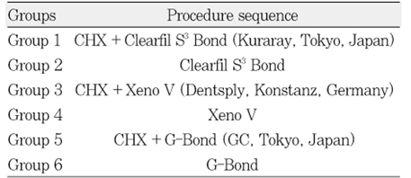
The teeth were then assigned randomly to six groups according to the adhesive system and application of chlorhexidine. Tables 1 and 2 listed the materials and groups used in this study, respectively. In the chlorhexidine applied groups, 1 mL of a 2% chlorhexidine solution was applied to the exposed dentin surface for 10 seconds and dried for 10 seconds without rinsing. Each adhesive system was applied according to the manufacturer's instruction. After the bonding procedure, the light-cure composite resin (Premise; Kerr, Orange, CA, USA) was bonded to the treated dentin surface in 1 mm increments. Each 1 mm increment was polymerized for 20 seconds using a light-emitting diode (LED) visible light-polymerizing unit (Bluephase; Ivoclar Vivadent, Schaan, Liechtenstein), and the height of the total resin build up was approximately 5 mm. The restored teeth were then stored in distilled water at room temperature for 24 hours.
3. µTBS testing
The restored teeth were sectioned longitudinally to make approximately 1×1 mm thick and 10 mm long specimens using a diamond-saw under copious amounts of water. Each group contained 10 specimens. Each specimen was glued to the jig of the microtensile testing machine (BISCO Inc; Bisco, Schaumburg, IL, USA) using cyanoacrylate cement (Zapit; Dental Ventures of America, Corona, CA, USA). A tensile load was applied at a 1.0 mm/min cross-head speed until bonding failure occurred and the maximum load at failure was recorded.
4. Failure mode investigation
The fractured test specimens were examined using an operating microscope (OPMI pico; Carl Zeiss, Obercohen, Germany) under×25 magnification, and the failure mode was classified as follows: adhesive, if the composite resin cone fractured at the adhesive-tooth adhesivetooth interface; cohesive, if the composite resin cone fractured inside the composite resin or dentin; or mixed, a combination of adhesive and cohesive failure.
5. Statistical analysis
Statistical analysis of the µTBS between chlorhexidine application groups and non-application groups were performed using a Student's t-test. For a comparison of the dentin adhesives, one-way ANOVA was performed and a Scheffé's test was used for a post-hoc multiple comparison. The level of significance was set to p < 0.05.
Table 3 lists the µTBS of the chlorhexidine application and non-application groups of each dentin bonding agent. In each group, the chlorhexidine non-application group showed a higher µTBS than the application group. However there was no significant difference between application and non-application groups (p > 0.05). In the chlorhexidine application groups, Clearfil S3 Bond (group 1) showed the highest µTBS, followed by a G-Bond (group 5) and Xeno V (group 3). The µTBS values of the three adhesives were significant different (p < 0.05). Similarly, in the chlorhexidine non-application groups, Clearfil S3 Bond (group 2) showed the highest µTBS, followed by G-Bond (group 6) and Xeno V (group 4). The µTBS of the Clearfil S3 Bond was significant higher than that of the G-Bond (p < 0.05). However, there was no significant difference between the G-Bond and Xeno V (p > 0.05).
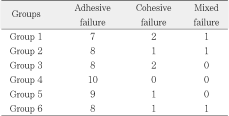
Table 4 shows the failure mode. Adhesive failure was the main failure mode observed in all groups. All specimens in group 4 showed adhesive failure.
In many cases, an antiseptic can be useful for eliminating the effects caused by bacteria and provide a better prognosis for the minimal restorative treatment of dental caries. Chlorhexidine is a broad spectrum antiseptic with pronounced antimicrobial effects,11 and has been shown to be effective in reducing cariogenic bacteria.12
In addition to its antibacterial effect, chlorhexidine also functions as a potent matrix metalloproteinase (MMP) inhibitor.13 Despite the remarkable advances that have increased the dentin-resin bond strength, the premature loss of bond strength is a major problems with adhesive restorations. The loss of bond strength was attributed mainly to the degradation of the hybrid layer at the dentin-adhesive interface. Mild acids can activate MMPs.14,15 Both the etch-and-rinse adhesives and self-etching adhesives have mild acidity, and can release and activate endogenous MMPs during dentin bonding.16-18 These activated MMPs can slowly hydrolyze any unprotected collagen fibrils of the dentin hybrid layers, which are believed to be responsible for the thinning and disappearance of collagen fibrils from the incompletely infiltrated hybrid layers in aged, bonded dentin. Auto-degradation of the collagen matrix can be suppressed by synthetic protease inhibitors, such as chlorhexidine, indicating that MMP inhibition can be beneficial for preserving hybrid layers.19-21
According to these results, the application of chlorhexidine did not affect the µTBS value significantly. However, the difference in µTBS between the bonding agents was significant as follows: Clearfil S3 Bond showed the highest µTBS, followed by G-Bond and Xeno V. Xeno V had the lowest pH (pH < 2), followed by G-Bond (pH = 2.3), and Clearfil S3 Bond (pH = 2.4). Low-pH self-etch adhesives have rather low bond strengths, particularly to dentin.22 This may be why Clearfil S3 Bond showed the highest µTBS, followed by G-Bond and Xeno V.
Previous studies4,5,7,10 reported the association of chlorhexidine application and bond strength to be a contentious issue. There are studies that insist that the application of chlorhexidine affects the bond strength. Gurgan et al reported that the use of 2% chlorhexidine before or after acid etching may decrease the bond strength by altering the ability of the hydrophilic resin to seal dentin.7 Meiers and Shook indicated that bonding with Tenure (two-step etch-and-rinse adhesive) was unaffected by the application of chlorhexidine, whereas bonding with Syntac (two-step self-etch adhesive) was affected.10
However, Perdigão et al stated that chlorhexidine has strong affinity to the tooth surfaces, which is increased by acid-etching.5 Chlorhexidine also increases the surface free energy of the enamel and can have a similar effect on dentin. These properties might improve the adhesion of primers. However, the loss of calcium from hydroxyapatite and these effects are negated by the presence of chlorhexidine residue. According to their results, chlorhexidine had no significant effect on the shear bond strengths of the composite to dentin using the All-Bond 2 adhesive system. Say et al found that chlorhexidine applied before the acid etching of dentin caused changes in the smear layer, but an acid treatment of the surface nullified any effect of chlorhexidine on the smear layer.4 In addition, in the present study, there were no differences between the two groups, regardless of chlorhexidine application.
In this study, adhesive failure was the main failure mode in all groups. Adhesive failure may be due to the lower bond strength or a weak stability of the adhesive layer.
In conclusion, within the limits of this study, the application of 2% chlorhexidine did not affect the µTBS of the resin composite to the dentin using a one-step self-etch adhesive. Because chlorhexidine has antimicrobial properties and does not significantly affect the bond strength to dentin, the use of a chlorhexidine solution with a one-step self-etch adhesive might have the added potential of preventing collagen fibril degradation in the dentin hybrid layers. Further studies will be needed to determine the effects of chlorhexidine on the bond durability.

This work was supported for two years by Pusan National University Research Grant.

  • 1. Ersin NK, Uzel A, Aykut A, Candan U, Eronat C. Inhibition of cultivable bacteria by chlorhexidine treatment of dentin lesions treated with the ART technique. Caries Res. 2006;40(2):172-177.PubMed
  • 2. Mandel ID. Antimicrobial mouthrinses: overview and update. J Am Dent Assoc. 1994;125: Suppl 2. 2S-10S.ArticlePubMed
  • 3. Baca P, Junco P, Bravo M, Baca AP, Muñoz MJ. Caries incidence in permanent first molars after discontinuation of a school-based chlorhexidine-thymol varnish program. Community Dent Oral Epidemiol. 2003;31(3):179-183.ArticlePubMedPDF
  • 4. Say EC, Koray F, Tarim B, Soyman M, Gulmez T. In vitro effect of cavity disinfectants on the bond strength of dentin bonding systems. Quintessence Int. 2004;35(1):56-60.PubMed
  • 5. Perdigao J, Denehy GE, Swift EJ Jr. Effects of chlorhexidine on dentin surfaces and shear bond strengths. Am J Dent. 1994;7(2):81-84.PubMed
  • 6. El-Housseiny AA, Jamjoum H. The effect of caries detector dyes and a cavity cleansing agent on composite resin bonding to enamel and dentin. J Clin Pediatr Dent. 2000;25(1):57-63.ArticlePubMedPDF
  • 7. Gurgan S, Bolay S, Kiremitci A. Effect of disinfectant application methods on the bond strength of composite to dentin. J Oral Rehabil. 1999;26(10):836-840.ArticlePubMed
  • 8. de Castro FL, de Andrade MF, Duarte Junior SL, Vaz LG, Ahid FJ. Effect of 2% chlorhexidine on microtensile bond strength of composite to dentin. J Adhes Dent. 2003;5(2):129-138.PubMed
  • 9. Carrilho MR, Carvalho RM, de Goes MF, di Hipolito V, Geraldeli S, Tay FR, Pashley DH, Tjaderhane L. Chlorhexidine preserves dentin bond in vitro. J Dent Res. 2007;86(1):90-94.PubMed
  • 10. Meiers JC, Shook LW. Effect of disinfectants on the bond strength of composite to dentin. Am J Dent. 1996;9: 11-14.PubMed
  • 11. Ercan E, Ozekinci T, Atakul F, Gul Kadri. Antibacterial activity of 2% chlorhexidine gluconate and 5.25% sodium hypochlorite in infected root canal: In vivo study. J Endod. 2004;30(2):84-87.ArticlePubMed
  • 12. Attin R, Tuna A, Attin T, Brunner E, Noack MJ. Efficacy of differently concentrated chlorhexidine varnishes in decreasing mutans streptococci and lactobacilli counts. Arch Oral Biol. 2003;48(7):503-509.ArticlePubMed
  • 13. Gendron R, Grenier D, Sorsa T, Mayrand D. Inhibition of the activities of matrix metalloproteinases 2, 8 and 9 by chlorhexidine. Clin Diagn Lab Immunol. 1999;6(3):437-439.PubMedPMC
  • 14. Davis GE. Identification of an abundant latent 94-kDa gelatin-degrading metalloprotease in human saliva which is activated by acid exposure: implications for a role in digestion of collagenous proteins. Arch Biochem Biophys. 1991;286(2):551-554.ArticlePubMed
  • 15. Gunja-Smith Z, Woessner JF Jr. Activation of cartilage stromelysin-1 at acid pH and its relation to enzyme pH optimum and osteoarthritis. Agents Actions. 1993;40(3-4):228-231.ArticlePubMedPDF
  • 16. Mazzoni A, Pashley DH, Nishitani Y, Breschi L, Mannello F, Tjäderhane L, Toledano M, Pashley EL, Tay FR. Reactivation of inactivated endogenous proteolytic activities in phosphoric acid-etched dentine by etch-and-rinse adhesives. Biomaterials. 2006;27(25):4470-4476.ArticlePubMed
  • 17. Nishitani Y, Yoshiyama M, Wadgaonkar B, Breschi L, Mannello F, Mazzoni A, Carvalho RM, Tjäerhane L, Tay FR, Pashley DH. Activation of gelatinolytic/collagenolytic activity in dentin by self-etching adhesives. Eur J Oral Sci. 2006;114(2):160-166.PubMed
  • 18. Tay FR, Pashley DH, Loushine RJ, Weller RN, Monticelli F, Osorio R. Self-etching adhesives increase collagenolytic activity in radicular dentin. J Endod. 2006;32(9):862-868.ArticlePubMed
  • 19. Hashimoto M, Ohno H, Sano H, Kaga M, Oguchi H. In vitro degradation of resin-dentin bonds analyzed by microtensile bond test, scanning and transmission electron microscopy. Biomaterials. 2003;24(21):3795-3803.ArticlePubMed
  • 20. Armstrong SR, Vargas MA, Chung I, Pashley DH, Campbell JA, Laffoon JE, Qian F. Resin-dentin interfacial ultrastructure and microtensile dentin bond strength after five-year water storage. Oper Dent. 2004;29(6):705-712.PubMed
  • 21. Pashley DH, Tay FR, Yiu C, Hashimoto M, Breschi L, Carvalho RM, Ito S. Collagen degradation by host-derived enzymes during aging. J Dent Res. 2004;83(3):216-221.PubMed
  • 22. De Munck J, Vargas M, Iracki J, Van Landuyt K, Poitevin A, Lambrechts P, Van Meerbeek B. One-day bonding effectiveness of new self-etch adhesives to bur-cut enamel and dentin. Oper Dent. 2005;30(1):39-49.PubMed
Table 1
Materials used in this study

*Abbreviations: 10-MDP, 10-methacryloyloxydecyl dihydrogen phosphate; HEMA, 2-hydroxyethyl methacrylate; DMA, dimethacrylate; bis-GMA, bisphenol-A-glycidyl ether dimethacrylate; 4-MET, 4-methacryloxyethyl trimellitic acid; UDMA, urethane dimethacrylate; TEGDMA, triethylene glycol dimethacrylate; PPF, prepolymerized filler.

jkacd-35-486-i001.jpg
Table 2
Groups used in this study according to the type of adhesive and application of chlorhexidine
jkacd-35-486-i002.jpg
Table 3
Mean microtensile bond strength (MPa) and standard deviation (n = 10)

µTBS with same superscript in the same vertical row were not significantly different (p > 0.05).

jkacd-35-486-i003.jpg
Table 4
Failure mode (the number of the specimen of failure)
jkacd-35-486-i004.jpg

Tables & Figures

REFERENCES

    Citations

    Citations to this article as recorded by  

      • ePub LinkePub Link
      • Cite
        CITE
        export Copy Download
        Close
        Download Citation
        Download a citation file in RIS format that can be imported by all major citation management software, including EndNote, ProCite, RefWorks, and Reference Manager.

        Format:
        • RIS — For EndNote, ProCite, RefWorks, and most other reference management software
        • BibTeX — For JabRef, BibDesk, and other BibTeX-specific software
        Include:
        • Citation for the content below
        Effect of 2% chlorhexidine application on microtensile bond strength of resin composite to dentin using one-step self-etch adhesives
        J Korean Acad Conserv Dent. 2010;35(6):486-491.   Published online November 30, 2010
        Close
      • XML DownloadXML Download
      Related articles
      Effect of 2% chlorhexidine application on microtensile bond strength of resin composite to dentin using one-step self-etch adhesives
      Effect of 2% chlorhexidine application on microtensile bond strength of resin composite to dentin using one-step self-etch adhesives

      Materials used in this study

      *Abbreviations: 10-MDP, 10-methacryloyloxydecyl dihydrogen phosphate; HEMA, 2-hydroxyethyl methacrylate; DMA, dimethacrylate; bis-GMA, bisphenol-A-glycidyl ether dimethacrylate; 4-MET, 4-methacryloxyethyl trimellitic acid; UDMA, urethane dimethacrylate; TEGDMA, triethylene glycol dimethacrylate; PPF, prepolymerized filler.

      Groups used in this study according to the type of adhesive and application of chlorhexidine

      Mean microtensile bond strength (MPa) and standard deviation (n = 10)

      µTBS with same superscript in the same vertical row were not significantly different (p > 0.05).

      Failure mode (the number of the specimen of failure)

      Table 1 Materials used in this study

      *Abbreviations: 10-MDP, 10-methacryloyloxydecyl dihydrogen phosphate; HEMA, 2-hydroxyethyl methacrylate; DMA, dimethacrylate; bis-GMA, bisphenol-A-glycidyl ether dimethacrylate; 4-MET, 4-methacryloxyethyl trimellitic acid; UDMA, urethane dimethacrylate; TEGDMA, triethylene glycol dimethacrylate; PPF, prepolymerized filler.

      Table 2 Groups used in this study according to the type of adhesive and application of chlorhexidine

      Table 3 Mean microtensile bond strength (MPa) and standard deviation (n = 10)

      µTBS with same superscript in the same vertical row were not significantly different (p > 0.05).

      Table 4 Failure mode (the number of the specimen of failure)


      Restor Dent Endod : Restorative Dentistry & Endodontics
      Close layer
      TOP