-
Analysis of temperature change during polymerization according to resin thickness: an in vitro experimental study
-
Kkot-Byeol Bae, Eun-Young Noh, Young-Tae Cho, Bin-Na Lee, Hoon-Sang Chang, Yun-Chan Hwang, Won-Mann Oh, In-Nam Hwang
-
Restor Dent Endod 2025;50(4):e34. Published online November 12, 2025
-
DOI: https://doi.org/10.5395/rde.2025.50.e34
-
-
 Abstract Abstract
 PDF PDF PubReader PubReader ePub ePub
- Objectives
This study aimed to analyze the temperature changes during the light curing of conventional flowable composite resin and bulk-fill composite resin of various thicknesses using an infrared thermographic camera.
Methods
Flowable composite resin (G-aenial Flo, GC Co.) and bulk-fill composite resin (SDR, Dentsply Caulk) were used. Specimens with thicknesses from 0.5 mm to 5.0 mm were prepared. The infrared thermographic camera measured the temperature changes at the maximum temperature rise point during light curing. The data were analyzed for maximum temperature, time to peak temperature, and temperature rise patterns.
Results
For G-aenial Flo, the maximum temperature tended to decrease with increasing thickness, whereas for SDR, the maximum temperature decreased up to 2.0 mm and then remained relatively consistent from 2.0 mm to 5.0 mm. At thicknesses of 1.5 mm or less, both resins showed a rapid temperature increase within the first 5 seconds, followed by a reduced rate of increase up to 80 seconds. At thicknesses of 2.0 mm or greater, the temperature peaked and then gradually decreased. Across all thicknesses, SDR was observed to reach peak temperature more rapidly than G-aenial Flo.
Conclusions
Observable differences in polymerization dynamics were identified between the two resin types, particularly at greater thicknesses. Although no statistical analysis was performed, these descriptive findings suggest that infrared thermographic cameras may be useful for indirectly assessing polymerization dynamics during resin polymerization.
-
Citations
Citations to this article as recorded by  - Mapping the horizontal thermal distribution profile of composite resins during polymerization using real-time infrared thermography
Kkot-Byeol Bae, Young-Tae Cho, Bin-Na Lee, Hoon-Sang Chang, Yun-Chan Hwang, In-Nam Hwang
Journal of Dental Rehabilitation and Applied Science.2026; 42(2): 79. CrossRef
-
1,996
View
-
100
Download
-
1
Crossref
-
Impact of post adhesion on stress distribution: an in silico study
-
Kkot-Byeol Bae, Jae-Yoon Choi, Young-Tae Cho, Bin-Na Lee, Hoon-Sang Chang, Yun-Chan Hwang, Won-Mann Oh, In-Nam Hwang
-
Restor Dent Endod 2025;50(2):e19. Published online May 21, 2025
-
DOI: https://doi.org/10.5395/rde.2025.50.e19
-
-
Abstract
PDFPubReaderePub
- Objectives
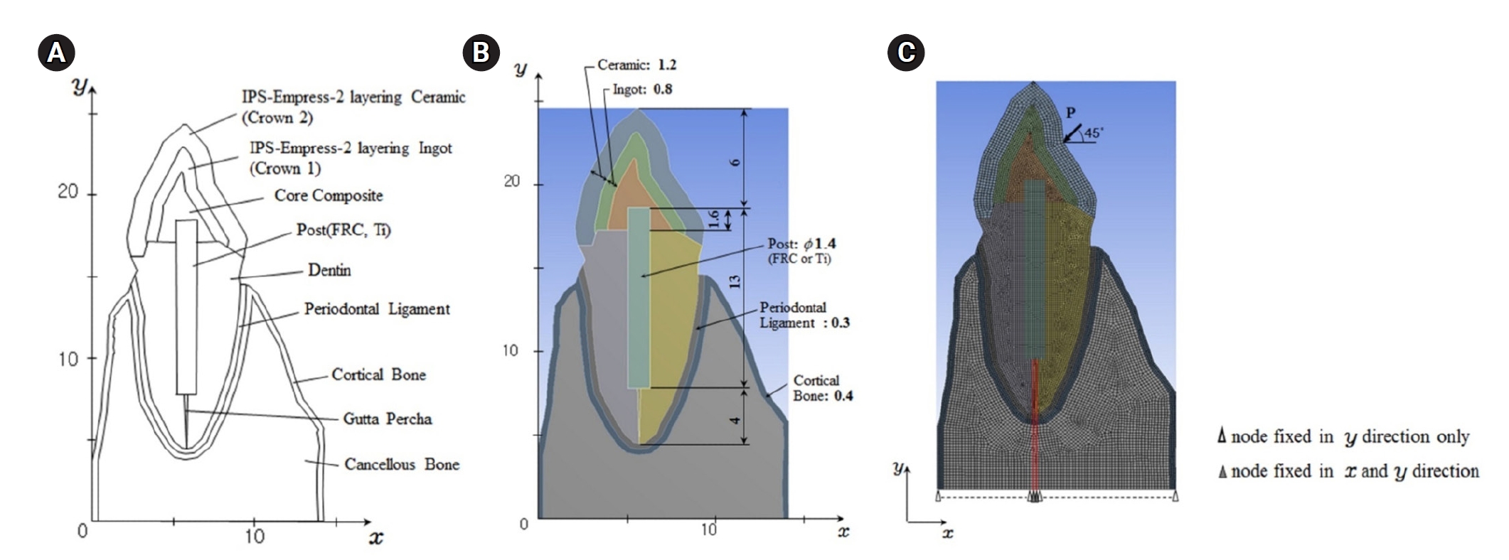
This study aimed to evaluate the stress distribution in teeth restored with different post materials and bonding conditions using finite element analysis (FEA).
Methods
A two-dimensional FEA model of a maxillary central incisor restored with IPS-Empress-2 crown (Ivoclar Vivadent), composite resin core, and posts were created. The model simulated bonded and non-bonded conditions for both fiber-reinforced composite (FRC) and titanium (Ti) posts. Stress distribution was analyzed using ANSYS 14.0 software under a 100-N load applied at a 45° angle to the long axis of the tooth.
Results
The results revealed that stress concentration was significantly higher in non-bonded posts compared to bonded ones. FRC posts exhibited stress values closer to those of dentin, whereas Ti posts demonstrated higher stress concentration, particularly in non-bonded states, increasing the potential risk of damage to surrounding tissues.
Conclusions
FRC posts, with elastic properties similar to dentin and proper adhesion, minimize stress concentration and potential damage to surrounding tissues. Conversely, materials with higher elastic modulus like Ti, can cause unfavorable stress concentrations if not properly bonded, emphasizing the importance of post adhesion in tooth restoration.
-
Citations
Citations to this article as recorded by - Advances in Ecofriendly and High-Strength Dental Composites: Structural and Functional Perspectives
Sayem A. Mulla, Amit Patil, Himmat Jaiswal, Bhavani Sangala Nagendra, Ashima Jakhar, Waseem Z. Khan
European Journal of General Dentistry.2026;[Epub] CrossRef
-
3,102
View
-
109
Download
-
1
Crossref
-
Effects of CTHRC1 on odontogenic differentiation and angiogenesis in human dental pulp stem cells
-
Jong-soon Kim, Bin-Na Lee, Hoon-Sang Chang, In-Nam Hwang, Won-Mann Oh, Yun-Chan Hwang
-
Restor Dent Endod 2023;48(2):e18. Published online April 28, 2023
-
DOI: https://doi.org/10.5395/rde.2023.48.e18
-
-
Abstract
PDFPubReaderePub
- Objectives
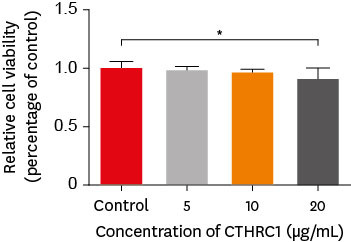
This study aimed to determine whether collagen triple helix repeat containing-1 (CTHRC1), which is involved in vascular remodeling and bone formation, can stimulate odontogenic differentiation and angiogenesis when administered to human dental pulp stem cells (hDPSCs). Materials and MethodsThe viability of hDPSCs upon exposure to CTHRC1 was assessed with the WST-1 assay. CTHRC1 doses of 5, 10, and 20 µg/mL were administered to hDPSCs. Reverse-transcription polymerase reaction was used to detect dentin sialophosphoprotein, dentin matrix protein 1, vascular endothelial growth factor, and fibroblast growth factor 2. The formation of mineralization nodules was evaluated using Alizarin red. A scratch wound assay was conducted to evaluate the effect of CTHRC1 on cell migration. Data were analyzed using 1-way analysis of variance followed by the Tukey post hoc test. The threshold for statistical significance was set at p < 0.05. ResultsCTHRC1 doses of 5, 10, and 20 µg/mL had no significant effect on the viability of hDPSCs. Mineralized nodules were formed and odontogenic markers were upregulated, indicating that CTHRC1 promoted odontogenic differentiation. Scratch wound assays demonstrated that CTHRC1 significantly enhanced the migration of hDPSCs. ConclusionsCTHRC1 promoted odontogenic differentiation and mineralization in hDPSCs.
-
Hard tissue formation after direct pulp capping with osteostatin and MTA in vivo
-
Ji-Hye Yoon, Sung-Hyeon Choi, Jeong-Tae Koh, Bin-Na Lee, Hoon-Sang Chang, In-Nam Hwang, Won-Mann Oh, Yun-Chan Hwang
-
Restor Dent Endod 2021;46(2):e17. Published online February 25, 2021
-
DOI: https://doi.org/10.5395/rde.2021.46.e17
-
-
Abstract
PDFPubReaderePub
- Objectives
In recent in vitro study, it was reported that osteostatin (OST) has an odontogenic effect and synergistic effect with mineral trioxide aggregate (MTA) in human dental pulp cells. Therefore, the aim of this study was to evaluate whether OST has a synergistic effect with MTA on hard tissue formation in vivo. Materials and MethodsThirty-two maxillary molars of Spraque-Dawley rats were used in this study. An occlusal cavity was prepared and the exposed pulps were randomly divided into 3 groups: group 1 (control; ProRoot MTA), group 2 (OST 100 μM + ProRoot MTA), group 3 (OST 10 mM + ProRoot MTA). Exposed pulps were capped with each material and cavities were restored with resin modified glass ionomer. The animals were sacrificed after 4 weeks. All harvested teeth were scanned with micro-computed tomography (CT). The samples were prepared and hard tissue formation was evaluated histologically. For immunohistochemical analysis, the specimens were sectioned and incubated with primary antibodies against dentin sialoprotein (DSP). ResultsIn the micro-CT analysis, it is revealed that OST with ProRoot MTA groups showed more mineralized bridge than the control (p < 0.05). In the H&E staining, it is showed that more quantity of the mineralized dentin bridge was formed in the OST with ProRoot MTA group compared to the control (p < 0.05). In all groups, DSP was expressed in newly formed reparative dentin area. ConclusionsOST can be a supplementary pulp capping material when used with MTA to make synergistic effect in hard tissue formation.
-
Citations
Citations to this article as recorded by - Trends and Future Perspectives in Additive Modifications of Calcium Silicate‐Based Cements: A Scoping Review (2015–2024)
Kenta Tsuchiya, Salvatore Sauro, Jukka P. Matinlinna, Monica Yamauti, Hidehiko Sano, Kyung‐San Min, Atsushi Tomokiyo
Australian Endodontic Journal.2026;[Epub] CrossRef - Pulpal responses to mineral trioxide aggregate with and without zinc oxide addition in mature canine teeth after full pulpotomy
Behnam Bolhari, Neda Kardouni Khouzestani, Hadi Assadian, Saeed Farzad-Mohajeri, Mohammad Mehdi Dehghan, Soheil Niavarzi, Behnam Dorost, Venkateshbabu Nagendrababu, Henry F. Duncan, Artak Heboyan, Antonio Signore, Stefano Benedicenti
Scientific Reports.2025;[Epub] CrossRef - Research Advancements in Peptides for Promoting Reparative Dentin Regeneration in Direct Pulp Capping: A Narrative Review
Jiawen Wang, Shuwei Qiao, Tianjia Huang, Junjie Lian, Song Zhu
International Journal of Peptide Research and Therapeutics.2025;[Epub] CrossRef - Biocompatibility and pro-mineralization effects of premixed calcium silicate-based materials on human dental pulp stem cells: An in vitro and in vivo study
Nyein Chan KO, Sonoko NODA, Yamato OKADA, Kento TAZAWA, Nobuyuki KAWASHIMA, Takashi OKIJI
Dental Materials Journal.2024; 43(5): 729. CrossRef - Osteostatin, a peptide for the future treatment of musculoskeletal diseases
Daniel Lozano, Arancha R. Gortazar, Sergio Portal-Núñez
Biochemical Pharmacology.2024; 223: 116177. CrossRef - Comparison of bioactive material failure rates in vital pulp treatment of permanent matured teeth – a systematic review and network meta-analysis
Péter Komora, Orsolya Vámos, Noémi Gede, Péter Hegyi, Kata Kelemen, Adél Galvács, Gábor Varga, Beáta Kerémi, János Vág
Scientific Reports.2024;[Epub] CrossRef - Hard tissue formation in pulpotomized primary teeth in dogs with nanomaterials MCM-48 and MCM-48/hydroxyapatite: an in vivo animal study
Sahar Talebi, Nosrat Nourbakhsh, Ardeshir Talebi, Amir Abbas Nourbakhsh, Abbas Haghighat, Maziar Manshayi, Hamid Reza Bakhsheshi, Razieh Karimi, Rahman Nazeri, Kenneth J.D. Mackenzie
BMC Oral Health.2024;[Epub] CrossRef - Reparative Mineralized Tissue Characterization by Different Bioactive Direct Pulp-capping Agents
Mrunal Shinde, Varsha Pandit, Sarita Singh, Aniket Jadhav, Sarah Marium, Smita Patil
Journal of the International Clinical Dental Research Organization.2024; 16(1): 8. CrossRef - Effects of mineral trioxide aggregate and methyl sulfonyl methane on pulp exposure via RUNX2 and RANKL pathways
Altar Ateş, Ayca Kurt, Tolga Mercantepe
Odontology.2024; 112(3): 895. CrossRef - Effects of barium titanate on the dielectric constant, radiopacity, and biological properties of tricalcium silicate-based bioceramics
Yoorina CHOI, Yun-Chan HWANG, Mi-Kyung YU, Kwang-Won LEE, Kyung-San MIN
Dental Materials Journal.2023; 42(1): 55. CrossRef - Bioactive potential of Bio‐C Pulpo is evidenced by presence of birefringent calcite and osteocalcin immunoexpression in the rat subcutaneous tissue
Marcela Borsatto Queiroz, Rafaela Nanami Handa Inada, Camila Soares Lopes, Juliane Maria Guerreiro‐Tanomaru, Estela Sasso‐Cerri, Mário Tanomaru‐Filho, Paulo Sérgio Cerri
Journal of Biomedical Materials Research Part B: Applied Biomaterials.2022; 110(10): 2369. CrossRef - The Influence of New Bioactive Materials on Pulp–Dentin Complex Regeneration in the Assessment of Cone Bone Computed Tomography (CBCT) and Computed Micro-Tomography (Micro-CT) from a Present and Future Perspective—A Systematic Review
Mirona Paula Palczewska-Komsa, Bartosz Gapiński, Alicja Nowicka
Journal of Clinical Medicine.2022; 11(11): 3091. CrossRef - A Breakthrough in the Era of Calcium Silicate-Based Cements: A Critical Review
Payal S Chaudhari, Manoj G Chandak, Akshay A Jaiswal, Nikhil P Mankar, Priyanka Paul
Cureus.2022;[Epub] CrossRef - Effectiveness of Direct Pulp Capping Bioactive Materials in Dentin Regeneration: A Systematic Review
Ermin Nie, Jiali Yu, Rui Jiang, Xiangzhen Liu, Xiang Li, Rafiqul Islam, Mohammad Khursheed Alam
Materials.2021; 14(22): 6811. CrossRef
-
3,656
View
-
41
Download
-
13
Web of Science
-
14
Crossref
-
Oral manifestation and root canal therapy of the patient with mucopolysaccharidosis
-
Ji-Hye Yoon, Hyo-Il Lee, Ji-Hyun Jang, Sung-Hyeon Choi, Hoon-Sang Chang, Yun-Chan Hwang, In-Nam Hwang, Bin-Na Lee, Won-Mann Oh
-
Restor Dent Endod 2019;44(2):e14. Published online April 4, 2019
-
DOI: https://doi.org/10.5395/rde.2019.44.e14
-
-
Abstract
PDFPubReaderePub
Mucopolysaccharidosis (MPS) is an inherited metabolic disorder caused by a deficiency in enzymes that participate in the degradation of glycosaminoglycans (GAGs) such as heparin sulfate and dermatan sulfate. Left untreated, patients show progressive mental and physical deterioration due to deposition of GAGs in organs. Death often occurs due to cardiac or respiratory failure before patients reach their early twenties. MPS has several oral and dental manifestations. An enlarged head, short neck, and open mouth associated with a large tongue are major characteristics of MPS patients. Dental complications can be severe, including unerupted dentition, dentigerous cyst-like follicles, malocclusions, condylar defects, and gingival hyperplasia. A 21-year-old female patient with MPS was described in this article, with special emphasis on oral manifestations and dental treatment. -
Citations
Citations to this article as recorded by - Pediatric Interventions in a Sanfilippo Syndrome Patient Under General Anesthesia: A Case Report
Ahmad Al Malak, Hassan Issawi, Mohammad Hassoun, Mohammad Al Halabi, Darko Macan
Case Reports in Dentistry.2025;[Epub] CrossRef - Behavioural disorders and sleep problems in Sanfilippo syndrome: overlaps with some other conditions and importance indications
Karolina Wiśniewska, Jakub Wolski, Paulina Anikiej-Wiczenbach, Magdalena Żabińska, Grzegorz Węgrzyn, Karolina Pierzynowska
European Child & Adolescent Psychiatry.2025; 34(6): 1795. CrossRef - Hurler syndrome: Oral and radiographic findings of a rare clinical case
W. Kabbassi, H. Hessissen, J. Hammouti
Medical Reports.2025; 14: 100325. CrossRef - Sanfilippo syndrome: consensus guidelines for clinical care
Nicole Muschol, Roberto Giugliani, Simon A. Jones, Joseph Muenzer, Nicholas J. C. Smith, Chester B. Whitley, Megan Donnell, Elise Drake, Kristina Elvidge, Lisa Melton, Cara O’Neill
Orphanet Journal of Rare Diseases.2022;[Epub] CrossRef - Manifestaciones bucales de pacientes con mucopolisacaridosis. Serie de casos
Andrea Verónica Ríos, Mariana Llorensi
Revista de la Asociación Odontológica Argentina.2021;[Epub] CrossRef
-
2,201
View
-
7
Download
-
5
Crossref
-
Dental management of patients with X-linked hypophosphatemia
-
Bin-Na Lee, Hye-Yoon Jung, Hoon-Sang Chang, Yun-Chan Hwang, Won-Mann Oh
-
Restor Dent Endod 2017;42(2):146-151. Published online January 6, 2017
-
DOI: https://doi.org/10.5395/rde.2017.42.2.146
-
-
Abstract
PDFPubReaderePub
X-linked hypophosphatemia (XLH) is a hereditary metabolic disease caused by the loss of phosphate through the renal tubules into the urine, and an associated decrease in serum calcium and potassium phosphate. Its dental features include spontaneous dental abscesses that occur in the absence of trauma or dental caries. The aim of this case report was to describe the dental problems of XLH patients and to evaluate limitations in their treatment. A 14 year old male and a 38 year old female with XLH were referred to the Department of Conservative Dentistry for endodontic treatment. The dental findings were periapical abscesses without obvious trauma or caries. Conservative endodontic treatment was performed in teeth with pulp necrosis and abscess. In case 1, the treated teeth showed improvements in bone healing, without clinical symptoms. However, in case 2, the implants and the treated tooth showed hypermobility, and the final restoration was therefore postponed. Early diagnosis, periodic examinations, and communication with the patient's pediatrician are important in the dental management of patients with XLH. -
Citations
Citations to this article as recorded by - Targeted Alkaline Phosphatase Therapy Enhances Alveolar Bone Healing in X‐Linked Hypophosphatemia in Mice
Aonjittra Phanrungsuwan, Bella Donnelly, José Luis Millán, Brian L. Foster
Journal of Periodontal Research.2026; 61(3): 309. CrossRef - Real-world effectiveness of burosumab across age groups: X-linked hypophosphatemia (XLH) Disease Monitoring Program
Leanne M Ward, Thomas O Carpenter, Hamilton Cassinelli, Pablo Florenzano, Erik A Imel, Aliya A Khan, Ben Johnson, Erru Yang, Marc Vincent, Heather M Heerssen, Zhiyi Li, Jill H Simmons
The Journal of Clinical Endocrinology & Metabolism.2026;[Epub] CrossRef - Oral manifestations of X-linked hypophosphatemic rickets: diagnostic and therapeutic strategies from the perspective of systemic homeostatic imbalance
Yunfan Cai, Yue Feng, Baize Zhang, Jingrui Kang, Ling Li, Xiang Li, Zhiting Li, Shengquan Cheng, Xin Sun, Yujiang Chen, Tao Ye, Li’an Wu, Kaiyan Wang, Lina Niu
Oral Science and Homeostatic Medicine.2026;[Epub] CrossRef - Oral Features in Children with X-Linked Hypophosphatemic Rickets: An 8-Year Follow-Up Case Report
Soumaya Kachti, Manel Chalbi, Soumaya Boussaid, Faten Awled Brahim, Mohamed Ali Chemli
OBM Genetics.2025; 09(02): 1. CrossRef - Dental implant considerations in patients with systemic diseases: An updated comprehensive review
Seyed Ali Mosaddad, Sahar Talebi, Seied Omid Keyhan, Hamid Reza Fallahi, Mohammad Darvishi, Seyedeh Sara Aghili, Narges Tavahodi, Reza Abdollahi Namanloo, Artak Heboyan, Amirhossein Fathi
Journal of Oral Rehabilitation.2024; 51(7): 1250. CrossRef - Inherited fibroblast growth factor 23 excess
Kripa Elizabeth Cherian, Thomas Vizhalil Paul
Best Practice & Research Clinical Endocrinology & Metabolism.2024; 38(2): 101844. CrossRef - Dental abnormalities in rare genetic bone diseases: Literature review
Eiji Iwata, Shyam Kishor Sah, I‐Ping Chen, Ernst Reichenberger
Clinical Anatomy.2024; 37(3): 304. CrossRef - Implant Survival in Patients with Chronic Kidney Disease: A Case Report and Systematic Review of the Literature
Iris Alla, Felice Lorusso, Sergio Alexandre Gehrke, Francesco Inchingolo, Maristella Di Carmine, Antonio Scarano
International Journal of Environmental Research and Public Health.2023; 20(3): 2401. CrossRef - X-linked hypophosphatemic rickets: Orthodontic considerations and management. A case report
Clara Gibson, Suhaym Mubeen, Robert Evans
Journal of Orthodontics.2022; 49(2): 205. CrossRef - X-chromosomale Hypophosphatämie (XLH)/Phosphatdiabetes – Eine lebenslange Erkrankung
Adalbert Raimann, Roland Kocijan, Gabriel T. Mindler
Journal für Klinische Endokrinologie und Stoffwechsel.2022; 15(2): 63. CrossRef - Dental Manifestations and Oral Management of X-Linked Hypophosphatemia
Rena Okawa, Kazuhiko Nakano
Endocrines.2022; 3(4): 654. CrossRef - Prospective Analysis of Muscle Adiposity in Children With X-linked Hypophosphatemic Rickets vs Control Children
Virginie Nguyen-Khac, Aurore Bonnet-Lebrun, Agnès Linglart, Marine de Tienda, Jugurtha Berkenou, Inès Mannes, Catherine Adamsbaum, Philippe Wicart, Wafa Skalli
Journal of the Endocrine Society.2022;[Epub] CrossRef - Dental and periodontal features and management in XLH children and adults
Martin Biosse Duplan, Elvire Le Norcy, Frédéric Courson, Catherine Chaussain
International Journal of Bone Fragility.2021; 1(2): 74. CrossRef - Hiding in plain sight: Gene panel and genetic markers reveal 26-year undiagnosed tumor-induced osteomalacia of the rib concurrently misdiagnosed as X-linked hypophosphatemia
Juan M. Colazo, Joseph A. DeCorte, Erin A. Gillaspie, Andrew L. Folpe, Kathryn M. Dahir
Bone Reports.2021; 14: 100744. CrossRef - X-linked hypophosphatemia and burosumab: Practical clinical points from the French experience
Justine Bacchetta, Anya Rothenbuhler, Iva Gueorguieva, Peter Kamenicky, Jean-Pierre Salles, Karine Briot, Agnès Linglart
Joint Bone Spine.2021; 88(5): 105208. CrossRef - Presentation and non‐surgical endodontic treatment of two patients with X‐linked hypophosphatemia: a case report
H. Bradley, A. Dutta, R. Philpott
International Endodontic Journal.2021; 54(8): 1403. CrossRef - Periodontal status evaluation in adolescents with hereditary rickets-like diseases
E.V. Vislobokova, L.P. Kiselnikova, D.A. Lezhnev, S.S. Murtazaev, N.A. Sholokhova
Stomatologiya.2021; 100(6): 63. CrossRef - Diagnosis, treatment-monitoring and follow-up of children and adolescents with X-linked hypophosphatemia (XLH)
Anya Rothenbuhler, Dirk Schnabel, Wolfgang Högler, Agnès Linglart
Metabolism.2020; 103: 153892. CrossRef - Long-term outcomes for Asian patients with X-linked hypophosphataemia: rationale and design of the SUNFLOWER longitudinal, observational cohort study
Takuo Kubota, Seiji Fukumoto, Hae Il Cheong, Toshimi Michigami, Noriyuki Namba, Nobuaki Ito, Shin Tokunaga, Yoshimi Gibbs, Keiichi Ozono
BMJ Open.2020; 10(6): e036367. CrossRef - X-Linked Hypophosphatemic Rickets Manifesting as Sclerotic Bone Disease and Enthesopathy
Hiya Boro, Shailendra Singh Naik, Charandeep Singh, Saurav Khatiwada, Rajesh Khadgawat
Cureus.2020;[Epub] CrossRef - X-linked hypophosphatemia diagnosed after identification of dental symptoms
Kaoruko Wato, Rena Okawa, Saaya Matayoshi, Yuko Ogaya, Ryota Nomura, Kazuhiko Nakano
Pediatric Dental Journal.2020; 30(2): 115. CrossRef - X-Linked Hypophosphatemia: A New Era in Management
Kathryn Dahir, Mary Scott Roberts, Stan Krolczyk, Jill H Simmons
Journal of the Endocrine Society.2020;[Epub] CrossRef - Prosthetic rehabilitation of a patient with X-linked hypophosphatemia using dental implants: a case report and review of the literature
Martin James, Reza Vahid Roudsari
International Journal of Implant Dentistry.2019;[Epub] CrossRef - Oral symptoms and oral health-related quality of life of individuals with x-linked hypophosphatemia
Marcel Hanisch, Lauren Bohner, Martin M. I. Sabandal, Johannes Kleinheinz, Susanne Jung
Head & Face Medicine.2019;[Epub] CrossRef - Outcome of adult patients with X‐linked hypophosphatemia caused by PHEX gene mutations
Douglas Chesher, Michael Oddy, Ulpee Darbar, Parag Sayal, Adrian Casey, Aidan Ryan, Annalisa Sechi, Charlotte Simister, Aoife Waters, Yehani Wedatilake, Robin H. Lachmann, Elaine Murphy
Journal of Inherited Metabolic Disease.2018; 41(5): 865. CrossRef
-
3,565
View
-
19
Download
-
25
Crossref
-
Evaluation of reparative dentin formation of ProRoot MTA, Biodentine and BioAggregate using micro-CT and immunohistochemistry
-
Jia Kim, Young-Sang Song, Kyung-San Min, Sun-Hun Kim, Jeong-Tae Koh, Bin-Na Lee, Hoon-Sang Chang, In-Nam Hwang, Won-Mann Oh, Yun-Chan Hwang
-
Restor Dent Endod 2016;41(1):29-36. Published online January 4, 2016
-
DOI: https://doi.org/10.5395/rde.2016.41.1.29
-
-
Abstract
PDFPubReaderePub
- Objectives
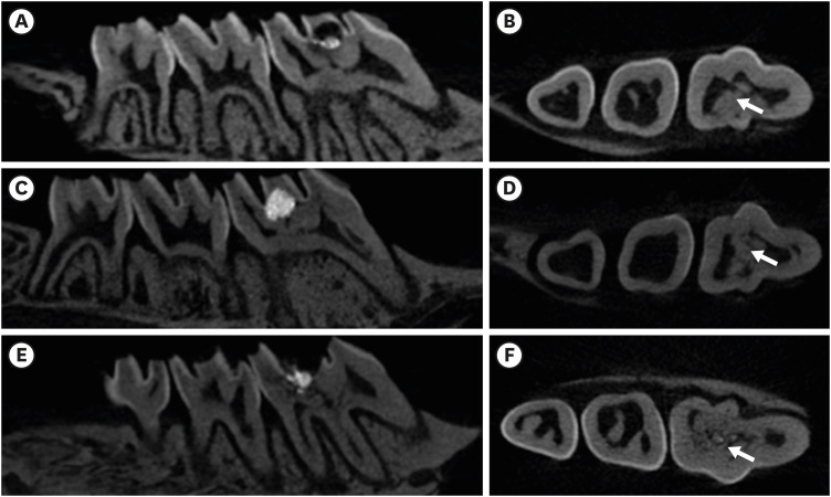
The purpose of this study was to assess the ability of two new calcium silicate-based pulp-capping materials (Biodentine and BioAggregate) to induce healing in a rat pulp injury model and to compare them with mineral trioxide aggregate (MTA). Materials and MethodsEighteen rats were anesthetized, cavities were prepared and the pulp was capped with either of ProRoot MTA, Biodentine, or BioAggregate. The specimens were scanned using a high-resolution micro-computed tomography (micro-CT) system and were prepared and evaluated histologically and immunohistochemically using dentin sialoprotein (DSP). ResultsOn micro-CT analysis, the ProRoot MTA and Biodentine groups showed significantly thicker hard tissue formation (p < 0.05). On H&E staining, ProRoot MTA showed complete dentin bridge formation with normal pulpal histology. In the Biodentine and BioAggregate groups, a thick, homogeneous hard tissue barrier was observed. The ProRoot MTA specimens showed strong immunopositive reaction for DSP. ConclusionsOur results suggest that calcium silicate-based pulp-capping materials induce favorable effects on reparative processes during vital pulp therapy and that both Biodentine and BioAggregate could be considered as alternatives to ProRoot MTA.
-
Citations
Citations to this article as recorded by - Evaluation of microhardness, monomer conversion, and antibacterial properties of an experimental pulp-capping material containing collagen–hydroxyapatite nanocomposite and/or chlorhexidine
Hacer Balkaya, Sezer Demirbuğa, Fatih Duman, Ahmet Ceylan, Ömer Aydın
Odontology.2026; 114(1): 204. CrossRef - Histopathologic responses of the dental pulp to an experimental calcium aluminate–calcium silicate based capping material in comparison to mineral trioxide aggregate
Reham S. Saleh, Eyad A. Elbattawy, Shymaa Hamza, Yousra Aly
Scientific Reports.2026;[Epub] CrossRef - Biological effects of a premixed calcium silicate pulp‐capping material containing dimethyl sulphoxide as a vehicle: In vitro and in vivo study
Min‐Ji Choi, Eun‐Sook Kang, Min‐Kyeong Kim, Kenta Tsuchiya, Jukka P. Matinlinna, Kwang‐Won Lee, Kyung‐San Min
European Journal of Oral Sciences.2026;[Epub] CrossRef - Clinical applications and classification of calcium silicate-based cements based on their history and evolution: a narrative review
Kenta Tsuchiya, Salvatore Sauro, Hidehiko Sano, Jukka P. Matinlinna, Monica Yamauti, Shuhei Hoshika, Yu Toida, Rafiqul Islam, Atsushi Tomokiyo
Clinical Oral Investigations.2025;[Epub] CrossRef - Impact of diode laser irradiation along with Biodentine on dental pulp stem cell proliferation and pluripotent gene expression
Ladan Alborzy, Sedighe Sadat Hashemikamangar, Mahshid Hodjat, Nasim Chiniforush, Behnaz Behniafar
Scientific Reports.2025;[Epub] CrossRef - The effect of different treatment methods on apical closure and treatment success in immature permanent first molars with reversible pulpitis
Muhammed ALAGOZ, Sera SIMSEK DERELIOĞLU
BMC Oral Health.2025;[Epub] CrossRef - Novelties in pulp capping materials
Vani Grover, Namith Rai, Nidambur Ballal
Acta stomatologica Naissi.2025; 41(91): 3086. CrossRef - Histological evaluation of pulp response to alendronate and Biodentine as pulp capping agents: an animal study
Thangavel Boopathi, Sekar Manimaran, Joseline Charles Kerena, Mathew Sebeena, Kumaravadivel Karthick, Natesan Thangaraj Deepa
Restorative Dentistry & Endodontics.2024;[Epub] CrossRef - Comparative Clinical and Radiographic Success Rate of Bioceramic Premix vs Biosilicate-based Medicament as Indirect Pulp Treatment Materials in Primary Molars: A Double-blind Randomized Trial with a Follow-up of 12 Months
Payal Kothari, Aditi Mathur, Sunnypriyatham Tirupathi, Rashmi Chauhan, Meenakshi Nankar, Ashrita Suvarna
International Journal of Clinical Pediatric Dentistry.2024; 17(7): 748. CrossRef - Effects of mineral trioxide aggregate and methyl sulfonyl methane on pulp exposure via RUNX2 and RANKL pathways
Altar Ateş, Ayca Kurt, Tolga Mercantepe
Odontology.2024; 112(3): 895. CrossRef - Evaluation of biocompatibility and bioactive potential of Well-Root PT by comparison with ProRoot MTA and Biodentine
Yong Kwon Chae, Ju Ri Ye, Ok Hyung Nam
Journal of Dental Sciences.2024; 19(4): 2218. CrossRef - Dentine Remineralisation Induced by “Bioactive” Materials through Mineral Deposition: An In Vitro Study
Marta Kunert, Ireneusz Piwonski, Louis Hardan, Rim Bourgi, Salvatore Sauro, Francesco Inchingolo, Monika Lukomska-Szymanska
Nanomaterials.2024; 14(3): 274. CrossRef - Different pulp capping agents and their effect on pulp inflammatory response: A narrative review
Mustafa Tariq Mutar, Anas F Mahdee
The Saudi Dental Journal.2024; 36(10): 1295. CrossRef - Clinical application of calcium silicate-based bioceramics in endodontics
Xinyuan Wang, Yizhi Xiao, Wencheng Song, Lanxiang Ye, Chen Yang, Yuzhen Xing, Zhenglin Yuan
Journal of Translational Medicine.2023;[Epub] CrossRef - Evaluation of the pulp response following direct pulp capping with exogenous nitric oxide and Mineral Trioxide Aggregate (MTA) a histologic study
Amirah Alnour, Ghassan Almohammad, Anas Abdo, Kinda Layous
Heliyon.2023; 9(7): e17458. CrossRef - Histological evaluation of dental pulp response to Biodentine, enamel matrix derivative (Emdogain), and mineral trioxide aggregate as direct pulp-capping agents – A randomized clinical trial
Takhellambam Premlata Devi, Amandeep Kaur, Shamurailatpam Priyadarshini, B. S. Deepak, Sumita Banerjee, Ngairangbam Sanjeeta
Journal of Medical Society.2023; 37(3): 107. CrossRef - Effect of Intracoronal Sealing Biomaterials on the Histological Outcome of Endodontic Revitalisation in Immature Sheep Teeth—A Pilot Study
Elanagai Rathinam, Sivaprakash Rajasekharan, Heidi Declercq, Christian Vanhove, Peter De Coster, Luc Martens
Journal of Functional Biomaterials.2023; 14(4): 214. CrossRef - Restorative management of the posterior tooth that has undergone a pulpotomy
Nicholas N Longridge, James S Hyde, Fadi Jarad, Sondos Albadri
Dental Update.2023; 50(11): 932. CrossRef - Direct pulp capping procedures – Evidence and practice
Rafiqul Islam, Md Refat Readul Islam, Toru Tanaka, Mohammad Khursheed Alam, Hany Mohamed Aly Ahmed, Hidehiko Sano
Japanese Dental Science Review.2023; 59: 48. CrossRef - A novel analysis of the formation and resorption changes in dental hard tissue using longitudinal in vivo micro computed tomography
Yeon-Jee YOO, Joonil HWANG, So-Hyun PARK, Jaehong HWANG, Seungryong CHO, Sun-Young KIM
Dental Materials Journal.2023; 42(5): 708. CrossRef - Evaluation of pH and Calcium Ion Diffusion from Intracanal MTA and Bioaggregate to Simulated External Resorption Cavities Through Dentinal Tubules
Umut AKSOY, Kaan POLATOĞLU, Feridun ŞAKLAR
European Annals of Dental Sciences.2022; 49(3): 108. CrossRef - Pulpa Kuafajı ve Kuafaj Materyallerine Güncel Bir Bakış: Derleme
Dilek AKIN, Çiğdem ATALAYIN ÖZKAYA
Selcuk Dental Journal.2022; 9(2): 617. CrossRef - The Influence of New Bioactive Materials on Pulp–Dentin Complex Regeneration in the Assessment of Cone Bone Computed Tomography (CBCT) and Computed Micro-Tomography (Micro-CT) from a Present and Future Perspective—A Systematic Review
Mirona Paula Palczewska-Komsa, Bartosz Gapiński, Alicja Nowicka
Journal of Clinical Medicine.2022; 11(11): 3091. CrossRef - Evaluation of shear bond strength of e-mineral trioxide aggregate and biodentine with glass ionomer cement
Hemalatha Hiremath, Aishwarya Singh Solanki, Shivangi Trivedi, Devansh Verma
Endodontology.2022; 34(2): 127. CrossRef - Multiple growth factors accommodated degradable submicron calcium sulfate hemihydrate/porous hydroxyapatite for dentin-pulp regeneration
Chih-Wen Chi, Bharathi Priya Lohanathan, Ching-Ching Wong, Che-Lun Chen, Hsun-Chang Lin, Yu-Chih Chiang
Biomaterials Advances.2022; 140: 213045. CrossRef - THE EFFECT OF BLOOD CONTAMINATION ON SHEAR BOND STRENGTH OF CALCIUM SILICATE-BASED PULP CAPPING MATERIALS
Hasan Fatih YAVUZ, Güneş BULUT EYÜBOĞLU
Cumhuriyet Dental Journal.2022; 24(4): 371. CrossRef - Comparison of Four Dental Pulp-Capping Agents by Cone-Beam Computed Tomography and Histological Techniques—A Split-Mouth Design Ex Vivo Study
Jayanandan Muruganandhan, Govindarajan Sujatha, Saravanan Poorni, Manali Ramakrishnan Srinivasan, Nezar Boreak, Ahmed Al-Kahtani, Mohammed Mashyakhy, Hitesh Chohan, Shilpa Bhandi, A. Thirumal Raj, Alessio Zanza, Luca Testarelli, Shankargouda Patil
Applied Sciences.2021; 11(7): 3045. CrossRef - Effect of Naturally Occurring Biogenic Materials on Human Dental Pulp Stem Cells (hDPSC): an In Vitro Study.
Prasanna T. Dahake, Vinod V. Panchal, Yogesh J. Kale, Mahesh V. Dadpe, Shrikant B. Kendre, Vijay M. Kumbar
Regenerative Engineering and Translational Medicine.2021; 7(4): 506. CrossRef - Influence of Ultrasonic Activation on the Physicochemical Properties of Calcium Silicate-Based Cements
Fredson Márcio Acris De Carvalho, Yara Teresinha Corrêa Silva-Sousa, Carlos Eduardo Saraiva Miranda, Paulo Henrique Miller Calderon, Ana Flávia Simões Barbosa, Luciana Martins Domingues De Macedo, Fuad Jacob Abi Rached-Junior, Boonlert Kukiattrakoon
International Journal of Dentistry.2021; 2021: 1. CrossRef - Tailored 70S30C Bioactive glass induces severe inflammation as pulpotomy agent in primary teeth: an interim analysis of a randomised controlled trial
Yasmine Elhamouly, Rania M. El Backly, Dalia M. Talaat, Samia S. Omar, Maha El Tantawi, Karin M. L. Dowidar
Clinical Oral Investigations.2021; 25(6): 3775. CrossRef - Response of dental pulp capped with calcium-silicate based material, calcium hydroxide and adhesive resin in rabbit teeth
Cynthia Kassis, Pierre Khoury, Karim Corbani, Charbel Mansour, Louis Hardan, Ghassan Yared, Carole Chakar
Brazilian Journal of Oral Sciences.2021;[Epub] CrossRef - Effectiveness of Direct Pulp Capping Bioactive Materials in Dentin Regeneration: A Systematic Review
Ermin Nie, Jiali Yu, Rui Jiang, Xiangzhen Liu, Xiang Li, Rafiqul Islam, Mohammad Khursheed Alam
Materials.2021; 14(22): 6811. CrossRef - Immunohistochemical expression of non-collagenous extracellular matrix molecules involved in tertiary dentinogenesis following direct pulp capping: a systematic review
C. Călin, M. Sajin, V.T. Moldovan, C. Coman, S.I. Stratul, A.C. Didilescu
Annals of Anatomy - Anatomischer Anzeiger.2021; 235: 151674. CrossRef - Recent Advances in Indirect Pulp Treatment Materials for Primary Teeth: A Literature Review
Afnan M Saber, Omar A El Meligy, Sumer M Alaki
International Journal of Clinical Pediatric Dentistry.2021; 14(6): 795. CrossRef - Chitosan-Based Accelerated Portland Cement Promotes Dentinogenic/Osteogenic Differentiation and Mineralization Activity of SHED
Hasan Subhi, Adam Husein, Dasmawati Mohamad, Nik Rozainah Nik Abdul Ghani, Asma-Abdullah Nurul
Polymers.2021; 13(19): 3358. CrossRef - Histological evaluation of the regenerative potential of a novel treated dentin matrix hydrogel in direct pulp capping
Ahmed A. Holiel, Elsayed M. Mahmoud, Wegdan M. Abdel-Fattah, Khadiga Y. Kawana
Clinical Oral Investigations.2021; 25(4): 2101. CrossRef - Minimal Intervention in Dentistry: A Literature Review on Biodentine as a Bioactive Pulp Capping Material
Naji Ziad Arandi, Mohammad Thabet, Mona Abbassy
BioMed Research International.2021;[Epub] CrossRef - Potential of tailored amorphous multiporous calcium silicate glass for pulp capping regenerative endodontics—A preliminary assessment
Jie Liu, Chao-An Chen, Xiaofei Zhu, Brian R. Morrow, Ukrit Thamma, Tia J. Kowal, Hassan M. Moawad, Matthias M. Falk, Himanshu Jain, George T.-J. Huang
Journal of Dentistry.2021; 109: 103655. CrossRef - Tomographic evaluation of direct pulp capping using a novel injectable treated dentin matrix hydrogel: a 2-year randomized controlled clinical trial
Ahmed A. Holiel, Elsayed M. Mahmoud, Wegdan M. Abdel-Fattah
Clinical Oral Investigations.2021; 25(7): 4621. CrossRef - Hard tissue formation after direct pulp capping with osteostatin and MTA in vivo
Ji-Hye Yoon, Sung-Hyeon Choi, Jeong-Tae Koh, Bin-Na Lee, Hoon-Sang Chang, In-Nam Hwang, Won-Mann Oh, Yun-Chan Hwang
Restorative Dentistry & Endodontics.2021;[Epub] CrossRef - Hydraulic cements for various intra-coronal applications: Part 1
Stephen J Bonsor, Josette Camilleri
Dental Update.2021; 48(8): 653. CrossRef - In vivo Biocompatibility and Bioactivity of Calcium Silicate-Based Bioceramics in Endodontics
Wencheng Song, Wei Sun, Lili Chen, Zhenglin Yuan
Frontiers in Bioengineering and Biotechnology.2020;[Epub] CrossRef - Evaluation of dentinogenesis inducer biomaterials: an in vivo study
Anabela B. Paula, Mafalda Laranjo, Carlos-Miguel Marto, Siri Paulo, Ana M. Abrantes, Bruno Fernandes, João Casalta-Lopes, Manuel Marques-Ferreira, Maria Filomena Botelho, Eunice Carrilho
Journal of Applied Oral Science.2020;[Epub] CrossRef - Micro-computed tomographic evaluation of a new system for root canal filling using calcium silicate-based root canal sealers
Mario Tanomaru-Filho, Fernanda Ferrari Esteves Torres, Jader Camilo Pinto, Airton Oliveira Santos-Junior, Karina Ines Medina Carita Tavares, Juliane Maria Guerreiro-Tanomaru
Restorative Dentistry & Endodontics.2020;[Epub] CrossRef - Bio-Inductive Materials in Direct and Indirect Pulp Capping—A Review Article
Marta Kunert, Monika Lukomska-Szymanska
Materials.2020; 13(5): 1204. CrossRef - Micro-computed tomographic evaluation of the flow and filling ability of endodontic materials using different test models
Fernanda Ferrari Esteves Torres, Juliane Maria Guerreiro-Tanomaru, Gisselle Moraima Chavez-Andrade, Jader Camilo Pinto, Fábio Luiz Camargo Villela Berbert, Mario Tanomaru-Filho
Restorative Dentistry & Endodontics.2020;[Epub] CrossRef - Release of Transforming Growth Factor Beta 1 from Human Tooth Dentin after Application of Either ProRoot MTA or Biodentine as a Coronal Barrier
Kunlada Wattanapakkavong, Tanida Srisuwan
Journal of Endodontics.2019; 45(6): 701. CrossRef - Effect of Leptin on Odontoblastic Differentiation and Angiogenesis: An In Vivo Study
Sung-Hyeon Choi, Ji-Hyun Jang, Jeong-Tae Koh, Hoon-Sang Chang, Yun-Chan Hwang, In-Nam Hwang, Bin-Na Lee, Won-Mann Oh
Journal of Endodontics.2019; 45(11): 1332. CrossRef - Análise da composição química dos cimentos MTA Angelus® branco, cinza e HP Repair® através de Microscopia Eletrônica de Varredura (MEV) acoplada a Espectrômetro de Energia Dispersiva (EDS)
Gabriela Duarte Rocha SARZEDA, Marcelo Santos BAHIA, Paulo Victor Teixeira DORIGUÊTTO, Karina Lopes DEVITO, Anamaria Pessoa Pereira LEITE
Revista de Odontologia da UNESP.2019;[Epub] CrossRef - Direct Pulp Capping: Which is the Most Effective Biomaterial? A Retrospective Clinical Study
Anabela Paula, Eunice Carrilho, Mafalda Laranjo, Ana M. Abrantes, João Casalta-Lopes, Maria Filomena Botelho, Carlos Miguel Marto, Manuel M. Ferreira
Materials.2019; 12(20): 3382. CrossRef - Characterization of Odontoblast-like Cell Phenotype and Reparative Dentin Formation In Vivo: A Comprehensive Literature Review
Dimitrios Tziafas
Journal of Endodontics.2019; 45(3): 241. CrossRef - Mineral trioxide aggregate and other bioactive endodontic cements: an updated overview – part I: vital pulp therapy
M. Parirokh, M. Torabinejad, P. M. H. Dummer
International Endodontic Journal.2018; 51(2): 177. CrossRef - Effects of calcium silicate cements on dental pulp cells: A systematic review
Ramy Emara, Karim Elhennawy, Falk Schwendicke
Journal of Dentistry.2018; 77: 18. CrossRef - Biodentine™ material characteristics and clinical applications: a 3 year literature review and update
S. Rajasekharan, L. C. Martens, R. G. E. C. Cauwels, R. P. Anthonappa
European Archives of Paediatric Dentistry.2018; 19(1): 1. CrossRef - The Relationship of Surface Characteristics and Antimicrobial Performance of Pulp Capping Materials
Cher Farrugia, Christie Y.K. Lung, Pierre Schembri Wismayer, Maria Teresa Arias-Moliz, Josette Camilleri
Journal of Endodontics.2018; 44(7): 1115. CrossRef - Effect of iRoot Fast Set root repair material on the proliferation, migration and differentiation of human dental pulp stem cells in vitro
Yan Sun, Tao Luo, Ya Shen, Markus Haapasalo, Ling Zou, Jun Liu, Gianpaolo Papaccio
PLOS ONE.2017; 12(10): e0186848. CrossRef - Bioactive-glass in Endodontic Therapy and Associated Microsurgery
Andrea Corrado Profeta, Gian Marco Prucher
The Open Dentistry Journal.2017; 11(1): 164. CrossRef - Influence of Biodentine® - A Dentine Substitute - On Collagen Type I Synthesis in Pulp Fibroblasts In Vitro
Frangis Nikfarjam, Kim Beyer, Anke König, Matthias Hofmann, Manuel Butting, Eva Valesky, Stefan Kippenberger, Roland Kaufmann, Detlef Heidemann, August Bernd, Nadja Nicole Zöller, Dimitrios Karamichos
PLOS ONE.2016; 11(12): e0167633. CrossRef - Effect of an Experimental Direct Pulp-capping Material on the Properties and Osteogenic Differentiation of Human Dental Pulp Stem Cells
Fan Yu, Yan Dong, Yan-wei Yang, Ping-ting Lin, Hao-han Yu, Xiang Sun, Xue-fei Sun, Huan Zhou, Li Huang, Ji-hua Chen
Scientific Reports.2016;[Epub] CrossRef
-
4,163
View
-
42
Download
-
59
Crossref
-
Treatment of non-vital immature teeth with amoxicillin-containing triple antibiotic paste resulting in apexification
-
Hyon-Beom Park, Bin-Na Lee, Yun-Chan Hwang, In-Nam Hwang, Won-Mann Oh, Hoon-Sang Chang
-
Restor Dent Endod 2015;40(4):322-327. Published online August 28, 2015
-
DOI: https://doi.org/10.5395/rde.2015.40.4.322
-
-
Abstract
PDFPubReaderePub
A recent treatment option for non-vital immature teeth in young patients is revascularization with triple antibiotic paste (TAP). However, tooth discoloration was reported with the use of conventional minocycline-containing TAP. In this case report, amoxicillin-containing TAP was used for revascularization of non-vital immature teeth to prevent tooth discoloration. At the 1 yr follow up, the teeth were asymptomatic on clinical examination and showed slight discoloration of the crown due to mineral trioxide aggregate (MTA) filling rather than amoxicillin-containing TAP. Radiographic examination revealed complete resolution of the periapical radiolucency, and closed apex with obvious periodontal ligament space. However, the root growth was limited, and the treatment outcome was more like apexification rather than revascularization. These results may be due to unstable blood clot formation which could not resist the condensation force of MTA filling, whether or not a collagen matrix was in place. These cases showed that although revascularization was not successful, apexification could be expected, resulting in the resolution of the periapical radiolucency and the closure of the apex. Therefore, it is worthwhile attempting revascularization of non-vital immature teeth in young patients. -
Citations
Citations to this article as recorded by - Comparative evaluation of effect of modified triple antibiotic paste and calcium hydroxide as intracanal medicament on microhardness of root dentin: An in vitro study
Aparna Palekar, Piyush Mantri, Minal Awinashe, Basawaraj Biradar, Mukund Singh
Endodontology.2024; 36(2): 143. CrossRef - Healing of Large Endodontic Lesions Using Long‐Term Application of a New Combination of Triple Antibiotics: A Series of Cases
Saeed Asgary, Ardavan Parhizkar, Maria Beatriz Duarte Gavião
Case Reports in Dentistry.2023;[Epub] CrossRef - Comparative Evaluation of Two Antibiotic Pastes for Root Canal Disinfection
Mridula Goswami, CP Baveja, Urvashi Bhushan, Sadhna Sharma
International Journal of Clinical Pediatric Dentistry.2022; 15(S1): S12. CrossRef - Regenerative Endodontics as the Future Treatment of Immature Permanent Teeth
Justyna Zbańska, Katarzyna Herman, Piotr Kuropka, Maciej Dobrzyński
Applied Sciences.2021; 11(13): 6211. CrossRef - Antimicrobial efficacy of triple antibiotic paste in teeth with primary endodontic infection: A systematic review
Rhythm Bains, Aseem P. Tikku, Promila Verma, Pragya Pandey
Asian Journal of Oral Health and Allied Sciences.2021; 11: 2. CrossRef - Effectiveness of MTA apical plug in dens evaginatus with open apices
Khoa Van Pham, Thu Anh Tran
BMC Oral Health.2021;[Epub] CrossRef - Lesion Sterilization and Tissue Repair: A Literature Review
Ankit Rawat, Jyoti Nagpal, Shreeya Mehta, Divya Vyas, Abhishek Kumar, Fathima Amal
Journal of Research and Advancement in Dentistry.2021; 12(3): 6. CrossRef - Spectrophotometric assessment of Tooth discoloration induced by various Antibiotic pastes
Ravi Gupta, Radhika Kewalramani, Dishant Patel
Research Journal of Pharmacy and Technology.2021; : 1979. CrossRef - Comparative evaluation of calcium release of the apical plugs formed by mineral trioxide aggregate, Biodentine, and EndoSequence root repair material with and without 2% triple antibiotic powder: An in vitro study
PoojaNitin Mapara, ND Shashikiran, Sachin Gugawad, Namrata Gaonkar, Savita Hadakar, Swapnil Taur, Dhanshri Khade
Journal of Indian Society of Pedodontics and Preventive Dentistry.2020; 38(2): 132. CrossRef - Effect of triple antibiotic loaded apatitic nanocarriers on Enterococcus faecalis biofilm – An In vitro study
S. Nagarathinam, V. Sujatha, K. Madhumathi, S. Mahalaxmi, P.Pranav Vanajassun, T.S.Sampath Kumar
Journal of Drug Delivery Science and Technology.2019; 51: 499. CrossRef - Coronal tooth discoloration induced by regenerative endodontic treatment using different scaffolds and intracanal coronal barriers: a 6-month ex vivo study
Noushin Shokouhinejad, Hassan Razmi, Maryam Farbod, Marzieh Alikhasi, Josette Camilleri
Restorative Dentistry & Endodontics.2019;[Epub] CrossRef - Triple antibiotic paste: momentous roles and applications in endodontics: a review
Ardavan Parhizkar, Hanieh Nojehdehian, Saeed Asgary
Restorative Dentistry & Endodontics.2018;[Epub] CrossRef - Mineral trioxide aggregate and other bioactive endodontic cements: an updated overview – part II: other clinical applications and complications
M. Torabinejad, M. Parirokh, P. M. H. Dummer
International Endodontic Journal.2018; 51(3): 284. CrossRef - Alternative to Avoid Tooth Discoloration after Regenerative Endodontic Procedure: A Systematic Review
Luciane Geanini Pena dos Santos, Luiz Alexandre Chisini, Camila Guerner Springmann, Beatriz Dulcineia Mendes de Souza, Fernanda Geraldo Pappen, Flávio Fernando Demarco, Mara Cristina Santos Felippe, Wilson Tadeu Felippe
Brazilian Dental Journal.2018; 29(5): 409. CrossRef - Regenerative Endodontic Treatment or Mineral Trioxide Aggregate Apical Plug in Teeth with Necrotic Pulps and Open Apices: A Systematic Review and Meta-analysis
Mahmoud Torabinejad, Ali Nosrat, Prashant Verma, Oyoyo Udochukwu
Journal of Endodontics.2017; 43(11): 1806. CrossRef - Revascularization in Immature Permanent Teeth with Necrotic Pulp and Apical Pathology: Case Series
López Carmen, Mendoza Asunción, Solano Beatriz, Yáñez-Vico Rosa, Jiiang H. Jeng
Case Reports in Dentistry.2017;[Epub] CrossRef
-
2,749
View
-
12
Download
-
16
Crossref
-
A review of the regenerative endodontic treatment procedure
-
Bin-Na Lee, Jong-Wook Moon, Hoon-Sang Chang, In-Nam Hwang, Won-Mann Oh, Yun-Chan Hwang
-
Restor Dent Endod 2015;40(3):179-187. Published online March 16, 2015
-
DOI: https://doi.org/10.5395/rde.2015.40.3.179
-
-
Abstract
PDFPubReaderePub
Traditionally, apexification has been used to treat immature permanent teeth that have lost pulp vitality. This technique promotes the formation of an apical barrier to close the open apex so that the filling materials can be confined to the root canal. Because tissue regeneration cannot be achieved with apexification, a new technique called regenerative endodontic treatment was presented recently to treat immature permanent teeth. Regenerative endodontic treatment is a treatment procedure designed to replace damaged pulp tissue with viable tissue which restores the normal function of the pulp-dentin structure. After regenerative endodontic treatment, continued root development and hard tissue deposition on the dentinal wall can occur under ideal circumstances. However, it is difficult to predict the result of regenerative endodontic treatment. Therefore, the purpose of this study was to summarize multiple factors effects on the result of regenerative endodontic treatment in order to achieve more predictable results. In this study, we investigated the features of regenerative endodontic treatment in comparison with those of other pulp treatment procedures and analyzed the factors that have an effect on regenerative endodontic treatment. -
Citations
Citations to this article as recorded by - Evaluation of the Clinical Outcomes of Regenerative Endodontic Procedures Using Autologous Platelet Concentrate: A Systematic Review and Meta-Analysis
Elnaz Mousavi, Navid Nasrabadi, Samira Jamali, Arian Haddadi
Pesquisa Brasileira em Odontopediatria e Clínica Integrada.2025;[Epub] CrossRef - Antimicrobial 3D printed gelatin scaffolds for root canal disinfection in regenerative endodontics procedures
Mateo Dallos Ortega, Jenny Aveyard, Raghda Magdy Abdelgawad, Reem El-Gendy, Alexander Ciupa, David Whetnall, Julia Behnsen, Robert J. Poole, Raechelle A. D'Sa
Biomaterials Science.2025; 13(14): 3795. CrossRef - Regenerative Endodontic Therapies: Harnessing Stem Cells, Scaffolds, and Growth Factors
Rosana Farjaminejad, Samira Farjaminejad, Franklin Garcia-Godoy
Polymers.2025; 17(11): 1475. CrossRef - Effects of combining hyaluronic acid hydrogel with injectable platelet rich fibrin on apical papilla stem cells proliferation and differentiation
Azal H. Al-Masoody, Nasrin Asadi, Hadiseh Mohammadpour, Mahshid Hodjat, Tahereh Sadat Jafarzadeh Kashi
BMC Oral Health.2025;[Epub] CrossRef - Experts consensus on management of tooth luxation and avulsion
Ruijie Huang, Chenchen Zhou, Ling Zhan, Yuan Liu, Xian Liu, Qin Du, Jun Wang, Wei Zhao, Guangtai Song, Li-an Wu, Beizhan Jiang, Yanhong Li, Hongmei Zhang, Jing Zou
International Journal of Oral Science.2024;[Epub] CrossRef - A review of tissue engineering in regenerative endodontic treatment
Eric Priyo Prasetyo, Dian Agustin Wahjuningrum, Galih Sampoerno, Wilson Sukandar, Shafy Shariz Bin Sharizal, Nurfahira Paidal, Menza Fadiyan Amriel, Nathania Elita Gunawan, Ketut Suardita, Evelyn Tjendronegoro
Conservative Dentistry Journal.2024; 14(1): 1. CrossRef - Innovative Paradigms and Established Strategies in Tooth Revitalization: A Review
Ahmad Shah Khan, Zahid Mehmood Khan, Palwasha Ishaque, Muhammad Zubair, Syeda Fatima Tu Zahra, Sana Ashfaq
Dental Update.2024; 51(8): 570. CrossRef - Explore the most recent developments and upcoming outlooks in the field of dental nanomaterials
Ali Alsuraifi, Zainab M. Sulaiman, Noor Alhuda R. Mohammed, Jassim Mohammed, Sarah Kareem Ali, Yousef Husam Abdualihamaid, Fatimah Husam, Abdullah Ayad
Beni-Suef University Journal of Basic and Applied Sciences.2024;[Epub] CrossRef - Recent Advances in Regenerative Endodontics: A Review of Current Techniques and Future Directions
Firas A Alothman, Lamia S Hakami, Ali Alnasser, Faris M AlGhamdi, Abdullah A Alamri, Basel M Almutairii
Cureus.2024;[Epub] CrossRef - Regenerative Potential of Dental Pulp Stem Cells in Response to a Bioceramic Dental Sealer and Photobiomodulation: An In Vitro Study
Hamed A Alshawkani, Mohamed Mansy, Mahmoud Al Ankily, Mohamed Shamel
The Journal of Contemporary Dental Practice.2024; 25(4): 313. CrossRef - Marginal adaptation of customized gutta percha cone with calcium silicate based sealer versus MTA and biodentine apical plugs in simulated immature permanent teeth (an in vitro study)
Mary M. Mina, Sybel M. Moussa, Mahmoud R. Aboelseoud
BMC Oral Health.2024;[Epub] CrossRef - GelMA‐based hydrogel biomaterial scaffold: A versatile platform for regenerative endodontics
Lei Huang, Xuan Chen, XiaoXia Yang, Yinchun Zhang, Xiaoling Qiu
Journal of Biomedical Materials Research Part B: Applied Biomaterials.2024;[Epub] CrossRef - Regenerative Endodontic Treatment in Dentinogenesis Imperfecta‐Induced Apical Periodontitis
Ying Liao, Ting Pan, Xianghui Xing, Sivakumar Nuvvula
Case Reports in Dentistry.2024;[Epub] CrossRef - In vitro and in vivo evaluation of iRoot BP Plus as a coronal sealing material for regenerative endodontic procedures
Ning Yang, Wenxiao Yang, Rou Shen, Shengcai Zhang, Tianchi Ma, Yao Liu
Clinical Oral Investigations.2024;[Epub] CrossRef - Comparative evaluation of pH and Ca+ ion release from MTA on interaction with platelet-rich fibrin and blood clot: an in vitro study
Sonia Khatri, Sylvia Mathew, Shruthi Nagaraja, Swaroop Hegde, Soumyadeep Ghosh, Kavimalar Ravichandran
F1000Research.2023; 12: 364. CrossRef - Photobiomodulation Therapy and Pulp-Regenerative Endodontics: A Narrative Review
Jiawen Yong, Sabine Gröger, Zuping Wu, Sabine Ruf, Yuer Ye, Xiaoyan Chen
Bioengineering.2023; 10(3): 371. CrossRef - Efficacy of disinfection procedures performed prior to regenerative endodontic therapy: An integrative review
Ketillyn da Silva Magalhães, Ana Clara Kuerten Gil, Taynara Santos Goulart, Daniela Peressoni Vieira Schuldt, Beatriz Serrato Coelho, Daniela de Rossi Figueiredo, Lucas da Fonseca Roberti Garcia, Josiane de Almeida
Australian Endodontic Journal.2023; 49(2): 418. CrossRef - Newer Prospects of Regenerative Endodontics: A Comprehensive and Updated Review of Literature
Mohammad Kamran Khan, Mahendra Kumar Jindal
Journal of the Scientific Society.2023; 50(3): 299. CrossRef - Comparative evaluation of pH and Ca+ ion release from MTA on interaction with platelet-rich fibrin and blood clot: an in vitro study
Sonia Khatri, Sylvia Mathew, Shruthi Nagaraja, Swaroop Hegde, Soumyadeep Ghosh, Kavimalar Ravichandran
F1000Research.2023; 12: 364. CrossRef - Effects of CEM cement and emdogain on proliferation and differentiation of human stem cells from the apical papilla: a comparative in vitro study
Elham Khoshbin, Leila Ghasemi, Rezvan Najafi, Hamed Karkehabadi
Biotechnology Letters.2023; 45(1): 69. CrossRef - Comparative Evaluation of the Regenerative Potential of Blood Clot and Platelet-rich Fibrin in Young Permanent Teeth Based on the Revised American Academy of Endodontics Clinical Considerations for Regenerative Procedure: 2016
Aarathi J Prakash, Saraswathi V Naik, Prabhakar Attiguppe
International Journal of Clinical Pediatric Dentistry.2023; 16(S2): S149. CrossRef - Biomechanical characterization of a fibrinogen–blood hydrogel for human dental pulp regeneration
Sofia Silvia Piglionico, Bela Varga, Orsolya Pall, Olivier Romieu, Csilla Gergely, Frédéric Cuisinier, Bernard Levallois, Ivan Vladislavov Panayotov
Biomaterials Science.2023; 11(20): 6919. CrossRef - Intracellular bacterial eradication using a novel peptide in vitro
Wing Nok Isaac Ng, Shanthini Kalimuthu, Carmen Oi Kwan Law, Angeline Hui Cheng Lee, Terrence Chi Kong Lau, Yiu Yan Leung, Gary Shun Pan Cheung, Prasanna Neelakantan
International Endodontic Journal.2023; 56(11): 1360. CrossRef - Regenerative Endodontic Treatment of Previously Treated Mature Permanent Tooth: A Case Report with 3-year Follow Up
Myung-Jin Lee
The Korean Journal of Oral and Maxillofacial Pathology.2023; 47(6): 133. CrossRef - Clinical Outcome and Comparison of Regenerative and Apexification Intervention in Young Immature Necrotic Teeth—A Systematic Review and Meta-Analysis
Pratima Panda, Lora Mishra, Shashirekha Govind, Saurav Panda, Barbara Lapinska
Journal of Clinical Medicine.2022; 11(13): 3909. CrossRef - Evaluation of Attitude and Knowledge of Endodontic, Pedodontic and SBARD Residents in Saudi Arabia toward Regenerative Endodontics—A National Survey
Ali A. Assiry, Mohmed Isaqali Karobari, Niher Tabassum Snigdha, Roshan Noor Mohamed, Syed Nahid Basheer, Mohammed Zameer
Medicina.2022; 58(4): 545. CrossRef - Effects of Intracanal Antimicrobials on Viability and Differentiation of Stem Cells From the Apical Papilla: An In Vitro Study
Gavin Raddall, Isabel Mello, Brendan M. Leung
Journal of Endodontics.2022; 48(7): 880. CrossRef - Awareness and Acceptance of Vital Pulp Therapy and Regenerative Endodontic Procedures among Dental Professionals in India: A Web-based Survey
Saloni Rathi, Priya Chauhan, Suparna Ganguly Saha, Rolly Agarwal, Simar Kaur Manocha, Mrinali Chaddha
Journal of Research and Advancement in Dentistry.2022; 14(1): 10. CrossRef - Exosomes as Biochemistry Tools for Stem Cell Differentiation: A Novel Cell-Based Treatment for Diseases
Saeed Azandeh, Darioush Bijan Nejad, Samaneh Karimi, Fereshtesadat Fakhredini
Jentashapir Journal of Cellular and Molecular Biology.2022;[Epub] CrossRef - Effect of biodentine coated with emdogain on proliferation and differentiation of human stem cells from the apical papilla
Hamed Karkehabadi, Erfan Ahmadyani, Rezvan Najafi, Elham Khoshbin
Molecular Biology Reports.2022; 49(5): 3685. CrossRef - Evaluation of the Effectiveness of Laser‐Assisted Bleaching of the Teeth Discolored due to Regenerative Endodontic Treatment
Noushin Shokouhinejad, Mehrfam Khoshkhounejad, Fatemeh Hamidzadeh, Murilo Baena Lopes
International Journal of Dentistry.2022;[Epub] CrossRef - Triple Antibiotic Paste: A Suitable Medicament for Intracanal Disinfection
Krutika Malu, Monika Khubchandani
Cureus.2022;[Epub] CrossRef - A Comparative Evaluation of Microhardness and Chemical Structure of Radicular Dentin with Two Combinations of TAP and MTAP: An In Vitro Study
Muthumula Daneswari, Nagireddy Venugopal Reddy, Annie P Chris, Nikhila V Reddy, Saigeeta Kondamadugu, P Niharika
International Journal of Clinical Pediatric Dentistry.2022; 15(S2): S151. CrossRef - Comparing Antibiotic Pastes with Electrospun Nanofibers as Modern Drug Delivery Systems for Regenerative Endodontics
Nura Brimo, Dilek Çökeliler Serdaroğlu, Busra Uysal
Current Drug Delivery.2022; 19(9): 904. CrossRef - The Advances of Blood Clots Used as Biomaterials in Regenerative Medicine
Eliza VanZweden, Rachael Tolsma, Victor Hung, Peter Awad, Robert Sawyer, Yong Li
Regenerative Medicine.2022; 17(12): 957. CrossRef - Microstructure and color stability of calcium silicate-based dental materials exposed to blood or platelet-rich fibrin
Noushin Shokouhinejad, Ibrahim Abu Tahun, Shima Saber Tahan, Fatemeh Mohandes, Mohammad H. Nekoofar, Paul M. H. Dummer
Clinical Oral Investigations.2022; 27(3): 1193. CrossRef - Results of “proroot mta” application in treatment of chronic periodontitis in teeth with incomplete root formation
N.M. Korneeva, E.A. Novikova, D.S. Popova, K.S. Rabadanova, L.Ya Rzaeva
Stomatology for All / International Dental review.2022; (2(99)): 10. CrossRef - Antimicrobial Effect of Calcium Hydroxide Combined with Electrolyzed Superoxidized Solution at Neutral pH on Enterococcus faecalis Growth
Héctor Armando Jimenez-Gonzalez, María Argelia Akemi Nakagoshi-Cepeda, Sergio Eduardo Nakagoshi-Cepeda, Víctor Hugo Urrutia-Baca, Myriam Angélica De La Garza-Ramos, Juan Manuel Solis-Soto, Ricardo Gomez-Flores, Patricia Tamez-Guerra, Yeliz Guven
BioMed Research International.2021;[Epub] CrossRef - Unpredictable Outcomes of a Regenerative Endodontic Treatment
Zahra Mohammadi, Hadi Assadian, Behnam Bolhari, Mohammadreza Sharifian, Mehrfam Khoshkhounejad, Nazanin Chitsaz, Andrea Scribante
Case Reports in Dentistry.2021;[Epub] CrossRef - Revascularization of nonvital immature incisor with asymptomatic apical periodontitis
Ema Mulyawati, Pribadi Santosa, Tunjung Nugraheni
Scientific Dental Journal.2020; 4(3): 134. CrossRef - Comparative analysis of calcium hydroxide apexification and regenerative endodontic procedure for root dentine growth stimulation in immature incisors with pulp necrosis
M.S. Rakhmanova, M.V. Korolenkova
Stomatologiya.2020; 99(6): 55. CrossRef - Antimicrobial Efficacy of a Novel Antibiotic‐Eluting Injectable Platelet‐Rich Fibrin Scaffold against a Dual‐Species Biofilm in an Infected Immature Root Canal Model
Azade Rafiee, Mahtab Memarpour, Yasaman Najibi, Bahman Khalvati, Sedigheh Kianpour, Mohammad Hossein Morowvat, Sung-Hwan Choi
BioMed Research International.2020;[Epub] CrossRef - Exosomes Derived from Stem Cells from the Apical Papilla Promote Dentine-Pulp Complex Regeneration by Inducing Specific Dentinogenesis
Xueying Zhuang, Lingli Ji, Huan Jiang, Yao Liu, Xuemei Liu, Jing Bi, Weidong Zhao, Zhenjiang Ding, Xu Chen
Stem Cells International.2020; 2020: 1. CrossRef - Injectable Biomaterials for Dental Tissue Regeneration
Håvard Jostein Haugen, Poulami Basu, Mousumi Sukul, João F Mano, Janne Elin Reseland
International Journal of Molecular Sciences.2020; 21(10): 3442. CrossRef - Viability and Stimulation of Human Stem Cells from the Apical Papilla (hSCAPs) Induced by Silicate-Based Materials for Their Potential Use in Regenerative Endodontics: A Systematic Review
José Luis Sanz, Leopoldo Forner, Alicia Almudéver, Julia Guerrero-Gironés, Carmen Llena
Materials.2020; 13(4): 974. CrossRef - An Innovative Drug Delivery System Loaded with a Modified Combination of Triple Antibiotics for Use in Endodontic Applications
Ardavan Parhizkar, Hanieh Nojehdehian, Fahimeh Tabatabaei, Saeed Asgary
International Journal of Dentistry.2020; 2020: 1. CrossRef - Defining Endodontic Residents' Clinical Experiences: A National Survey
Jonathan D. Blacher, Kamran E. Safavi, Robert H. Aseltine, Blythe M. Kaufman
Journal of Dental Education.2019; 83(5): 504. CrossRef - Coronal tooth discoloration induced by regenerative endodontic treatment using different scaffolds and intracanal coronal barriers: a 6-month ex vivo study
Noushin Shokouhinejad, Hassan Razmi, Maryam Farbod, Marzieh Alikhasi, Josette Camilleri
Restorative Dentistry & Endodontics.2019;[Epub] CrossRef - Comparative Study between Revitalization of Necrotic Immature Permanent Anterior Teeth with and without Platelet Rich Fibrin: A Randomized Controlled Trial
Rasha Adel Ragab, Amr Ezzat Abd El Lattif, Norhan Abd El Wahab El Dokky
Journal of Clinical Pediatric Dentistry.2019; 43(2): 78. CrossRef - Biomaterials and Scaffold Design Strategies for Regenerative Endodontic Therapy
Gavin Raddall, Isabel Mello, Brendan M. Leung
Frontiers in Bioengineering and Biotechnology.2019;[Epub] CrossRef - Iloprost Induces Dental Pulp Angiogenesis in a Growth Factor–free 3-Dimensional Organ Culture System
Sonntana Seang, Prasit Pavasant, Chalida N. Limjeerajarus
Journal of Endodontics.2018; 44(5): 759. CrossRef - Ratio and Rate of Induced Root Growth in Necrotic Immature Teeth
Eun Jung Sang, Ji-Soo Song, Teo Jeon Shin, Young-Jae Kim, Jung-Wook Kim, Ki-Taeg Jang, Sang-Hoon Lee, Hong-Keun Hyun
THE JOURNAL OF THE KOREAN ACADEMY OF PEDTATRIC DENTISTRY.2018; 45(2): 225. CrossRef - Triple antibiotic paste: momentous roles and applications in endodontics: a review
Ardavan Parhizkar, Hanieh Nojehdehian, Saeed Asgary
Restorative Dentistry & Endodontics.2018;[Epub] CrossRef - Traumatic avulsion and delayed replantation of maxillary incisors in an eleven-year-old child
Gokcen Deniz Bayrak
Edorium Journal of Dentistry.2018; 5(1): 1. CrossRef - Influence of Apical Diameter on the Outcome of Regenerative Endodontic Treatment in Teeth with Pulp Necrosis: A Review
Yanjun Fang, Xinhuan Wang, Jingjing Zhu, Chaonan Su, Ying Yang, Liuyan Meng
Journal of Endodontics.2018; 44(3): 414. CrossRef - Assessment of Regaining Pulp Sensibility in Mature Necrotic Teeth Using a Modified Revascularization Technique with Platelet-rich Fibrin: A Clinical Study
Mohamed Nageh, Geraldine M. Ahmed, Alaa A. El-Baz
Journal of Endodontics.2018; 44(10): 1526. CrossRef - Local drug delivery in endodontics: A literature review
Shin Hye Chung, Young-Seok Park
Journal of Drug Delivery Science and Technology.2017; 39: 334. CrossRef - Regenerative Endodontics
Kristina Feigin, Bonnie Shope
Journal of Veterinary Dentistry.2017; 34(3): 161. CrossRef - Intentional Replantation of an Avulsed Immature Permanent Incisor: A Case Report
Claudio Maniglia-Ferreira, Fabio de Almeida Gomes, Marcelo de Morais Vitoriano
Journal of Endodontics.2017; 43(8): 1383. CrossRef - Effect of acidic solutions on the microhardness of dentin and set OrthoMTA and their cytotoxicity on murine macrophage
Soram Oh, Hiran Perinpanayagam, Yoon Lee, Jae-Won Kum, Yeon-Jee Yoo, Sang-Min Lim, Seok Woo Chang, Won-Jun Shon, Woocheol Lee, Seung-Ho Baek, Kee-Yeon Kum
Restorative Dentistry & Endodontics.2016; 41(1): 12. CrossRef - Questioning the spot light on Hi-tech endodontics
Jojo Kottoor, Denzil Albuquerque
Restorative Dentistry & Endodontics.2016; 41(1): 80. CrossRef - Effects of a Bioactive Scaffold Containing a Sustained Transforming Growth Factor-β1–releasing Nanoparticle System on the Migration and Differentiation of Stem Cells from the Apical Papilla
Craig Bellamy, Suja Shrestha, Calvin Torneck, Anil Kishen
Journal of Endodontics.2016; 42(9): 1385. CrossRef - Effects of Novel 3-dimensional Antibiotic-containing Electrospun Scaffolds on Dentin Discoloration
Margaret Louise A. Porter, Eliseu A. Münchow, Maria T.P. Albuquerque, Kenneth J. Spolnik, Anderson T. Hara, Marco C. Bottino
Journal of Endodontics.2016; 42(1): 106. CrossRef
-
7,989
View
-
129
Download
-
63
Crossref
-
Changes in SIRT gene expression during odontoblastic differentiation of human dental pulp cells
-
Young-Eun Jang, Su-Hee Go, Bin-Na Lee, Hoon-Sang Chang, In-Nam Hwang, Won-Mann Oh, Yun-Chan Hwang
-
Restor Dent Endod 2015;40(3):223-228. Published online July 15, 2015
-
DOI: https://doi.org/10.5395/rde.2015.40.3.223
-
-
Abstract
PDFPubReaderePub
- Objectives
The aim of this study was to investigate the expression of 7 different sirtuin genes (SIRT1-SIRT7) in human dental pulp cells (HDPCs), and to determine the role of SIRTs in the odontoblastic differentiation potential of HDPCs. Materials and MethodsHDPCs were isolated from freshly extracted third molar teeth of healthy patients and cultulred in odontoblastic differentiation inducing media. Osteocalcin (OCN) and dentin sialophosphoprotein (DSPP) expression was analyzed to evaluate the odontoblastic differentiation of HDPCs by reverse transcription-polymerase chain reaction (RT-PCR), while alizarin red staining was used for the mineralization assay. To investigate the expression of SIRTs during odontoblastic differentiation of HDPCs, real time PCR was also performed with RT-PCR. ResultsDuring the culture of HDPCs in the differentiation inducing media, OCN, and DSPP mRNA expressions were increased. Mineralized nodule formation was also increased in the 14 days culture. All seven SIRT genes were expressed during the odontogenic induction period. SIRT4 expression was increased in a time-dependent manner. ConclusionsOur study identified the expression of seven different SIRT genes in HDPCs, and revealed that SIRT4 could exert an influence on the odontoblast differentiation process. Further studies are needed to determine the effects of other SIRTs on the odontogenic potential of HDPCs.
-
Citations
Citations to this article as recorded by - Biodegradable Zn‐5Dy Alloy with Enhanced Osteo/Angio‐Genic Activity and Osteointegration Effect via Regulation of SIRT4‐Dependent Mitochondrial Function
Yue Han, Xian Tong, Runqi Zhou, Yilin Wang, Yuge Chen, Liang Chen, Xinhua Hong, Linmei Wu, Zhiqiang Lin, Yichi Zhang, Xuejia Zhang, Chaoming Hu, Bin Li, Yifan Ping, Zelin Cao, Zhou Ye, Zhongchen Song, Yuncang Li, Cuie Wen, Yongsheng Zhou, Jixing Lin, Shen
Advanced Science.2024;[Epub] CrossRef - The Role of Histone Acetylation Modification in Dental Tissue-Derived Mesenchymal Stem Cells and Odontogenesis
Haoling Chen, Zijing Huang, Chuxiao Chen
Cellular Reprogramming.2023; 25(1): 11. CrossRef - Metabolic Remodeling Impacts the Epigenetic Landscape of Dental Mesenchymal Stem Cells
Haiyun Luo, Yachuan Zhou, Wenjing Liu, Jun Wang
Stem Cells International.2022; 2022: 1. CrossRef - SIRT4 regulates rat dental papilla cell differentiation by promoting mitochondrial functions
Haoling Chen, Jun Kang, Fuping Zhang, Tong Yan, Wenguo Fan, Hongwen He, Fang Huang
The International Journal of Biochemistry & Cell Biology.2021; 134: 105962. CrossRef - Sirtuins as Interesting Players in the Course of HIV Infection and Comorbidities
Karolina Jurkowska, Beata Szymańska, Brygida Knysz, Amadeusz Kuźniarski, Agnieszka Piwowar
Cells.2021; 10(10): 2739. CrossRef - Robust expression of SIRT6 inhibits pulpitis via activation of the TRPV1 channel
Jia Hu, Weiran Chen, Zailing Qiu, Hongbing Lv
Cell Biochemistry and Function.2020; 38(5): 676. CrossRef - Downregulation of microRNA‐143‐5p is required for the promotion of odontoblasts differentiation of human dental pulp stem cells through the activation of the mitogen‐activated protein kinases 14‐dependent p38 mitogen‐activated protein kinases signaling pa
Bao‐Liang Wang, Zhi Wang, Xi Nan, Qing‐Cai Zhang, Wei Liu
Journal of Cellular Physiology.2019; 234(4): 4840. CrossRef - A potential role for the silent information regulator 2 homologue 1 (SIRT1) in periapical periodontitis
H. Kudo, O. Takeichi, K. Hatori, K. Makino, K. Himi, B. Ogiso
International Endodontic Journal.2018; 51(7): 747. CrossRef - Overexpressed Sirt1 in MSCs Promotes Dentin Formation in Bmi1-Deficient Mice
H. Wang, C. Lv, Y. Gu, Q. Li, L. Xie, H. Zhang, D. Miao, W. Sun
Journal of Dental Research.2018; 97(12): 1365. CrossRef - Expression of silent information regulator 2 homolog 1 (SIRT1) in periapical granulomas
Hiroshi Kudo, Osamu Takeichi, Kosuke Makino, Keisuke Hatori, Bunnai Ogiso
Journal of Oral Science.2018; 60(3): 411. CrossRef - TET1 knockdown inhibits the odontogenic differentiation potential of human dental pulp cells
Li-Jia Rao, Bai-Cheng Yi, Qi-Meng Li, Qiong Xu
International Journal of Oral Science.2016; 8(2): 110. CrossRef
-
2,238
View
-
7
Download
-
11
Crossref
-
Cytotoxicity and physical properties of tricalcium silicate-based endodontic materials
-
Young-Eun Jang, Bin-Na Lee, Jeong-Tae Koh, Yeong-Joon Park, Nam-Eok Joo, Hoon-Sang Chang, In-Nam Hwang, Won-Mann Oh, Yun-Chan Hwang
-
Restor Dent Endod 2014;39(2):89-94. Published online March 21, 2014
-
DOI: https://doi.org/10.5395/rde.2014.39.2.89
-
-
Abstract
PDFPubReaderePub
- Objectives
The aim of this study was to evaluate the cytotoxicity, setting time and compressive strength of MTA and two novel tricalcium silicate-based endodontic materials, Bioaggregate (BA) and Biodentine (BD). Materials and MethodsCytotoxicity was evaluated by using a 2,3-bis(2-methoxy-4-nitro-5-sulfophenyl)-5-((phenylamino)carbonyl)-2H-tetrazolium hydroxide (XTT) assay. Measurements of 9 heavy metals (arsenic, cadmium, chromium, copper, iron, lead, manganese, nickel, and zinc) were performed by inductively coupled plasma-mass spectrometry (ICP-MS) of leachates obtained by soaking the materials in distilled water. Setting time and compressive strength tests were performed following ISO requirements. ResultsBA had comparable cell viability to MTA, whereas the cell viability of BD was significantly lower than that of MTA. The ICP-MS analysis revealed that BD released significantly higher amount of 5 heavy metals (arsenic, copper, iron, manganese, and zinc) than MTA and BA. The setting time of BD was significantly shorter than that of MTA and BA, and the compressive strength of BA was significantly lower than that of MTA and BD. ConclusionsBA and BD were biocompatible, and they did not show any cytotoxic effects on human periodontal ligament fibroblasts. BA showed comparable cytotoxicity to MTA but inferior physical properties. BD had somewhat higher cytotoxicity but superior physical properties than MTA.
-
Citations
Citations to this article as recorded by - Zinc bioactive glass modified with an organic resin for pulp capping in comparison to other bioceramic cements: biological fluid -mediated performance
Shamsodin Heydari, Behnam Bolhari, Parham Pedram, Foozie Zahedi, Faranak Noori
Materials & Design.2026; 266: 116178. CrossRef - Effect of Vital Pulp Therapy Biomaterials on Tooth Discolouration: A Review of the Literature
Maedeh Gilvari Sarshari, Kiana Shakeri, Ardavan Parhizkar, Naresh Kasoju
International Journal of Biomaterials.2025;[Epub] CrossRef - Evaluation of the physical properties of bromelain-modified biodentine for direct pulp capping
Paridhi Agrawal, Manoj Chandak, Aditya Patel, Jay Bhopatkar
BMC Oral Health.2024;[Epub] CrossRef - Evaluation of bioactivity, biocompatibility, and antibacterial properties of tricalcium silicate bone cement modified with wollastonite/ fluorapatite glass and glass-ceramic
H.K. Abd El-Hamid, A.M. Fayad, R.L. Elwan
Ceramics International.2024; 50(14): 25322. CrossRef - Evaluation of the chemical, physical, and biological properties of a newly developed bioceramic cement derived from cockle shells: an in vitro study
Monthip Wannakajeepiboon, Chankhrit Sathorn, Chatvadee Kornsuthisopon, Busayarat Santiwong, Thanakorn Wasanapiarnpong, Pairoj Linsuwanont
BMC Oral Health.2023;[Epub] CrossRef - Strength of a cement-based dental material: Early age testing and first micromechanical modeling at mature age
Petr Dohnalík, Christian Hellmich, Gilles Richard, Bernhard L. A. Pichler
Frontiers in Bioengineering and Biotechnology.2023;[Epub] CrossRef - Calcium silicate and calcium aluminate cements for dentistry reviewed
Carolyn Primus, James L. Gutmann, Franklin R. Tay, Anna B. Fuks
Journal of the American Ceramic Society.2022; 105(3): 1841. CrossRef - Biomimetic Approaches in Clinical Endodontics
Naresh Kumar, Nazrah Maher, Faiza Amin, Hani Ghabbani, Muhammad Sohail Zafar, Francisco Javier Rodríguez-Lozano, Ricardo E. Oñate-Sánchez
Biomimetics.2022; 7(4): 229. CrossRef - Effect of different manipulations on the physical, chemical and microstructural characteristics of Biodentine
Mariana Domingos Pires, Joana Cordeiro, Isabel Vasconcelos, Mariana Alves, Sérgio André Quaresma, António Ginjeira, Josette Camilleri
Dental Materials.2021; 37(7): e399. CrossRef - Minimal Intervention in Dentistry: A Literature Review on Biodentine as a Bioactive Pulp Capping Material
Naji Ziad Arandi, Mohammad Thabet, Mona Abbassy
BioMed Research International.2021;[Epub] CrossRef - Chitosan-Based Accelerated Portland Cement Promotes Dentinogenic/Osteogenic Differentiation and Mineralization Activity of SHED
Hasan Subhi, Adam Husein, Dasmawati Mohamad, Nik Rozainah Nik Abdul Ghani, Asma-Abdullah Nurul
Polymers.2021; 13(19): 3358. CrossRef - Material Pulp Cells and Tissue Interactions
Nastaran Meschi, Biraj Patel, Nikita B. Ruparel
Journal of Endodontics.2020; 46(9): S150. CrossRef - Biological Effects of Tricalcium Silicate Nanoparticle-Containing Cement on Stem Cells from Human Exfoliated Deciduous Teeth
Yoonsun Jung, Ji-Young Yoon, Kapil Dev Patel, Lan Ma, Hae-Hyoung Lee, Jongbin Kim, Jung-Hwan Lee, Jisun Shin
Nanomaterials.2020; 10(7): 1373. CrossRef - Physicochemical, mechanical and cytotoxicity evaluation of chitosan-based accelerated portland cement
Hasan Subhi, Adam Husein, Dasmawati Mohamad, Asma-Abdullah Nurul
Journal of Materials Research and Technology.2020; 9(5): 11574. CrossRef - Tricalcium silicate cements: osteogenic and angiogenic responses of human bone marrow stem cells
Mohamed R. W. Ali, Manal Mustafa, Asgeir Bårdsen, Athanasia Bletsa
European Journal of Oral Sciences.2019; 127(3): 261. CrossRef - Bioactive tri/dicalcium silicate cements for treatment of pulpal and periapical tissues
Carolyn M. Primus, Franklin R. Tay, Li-na Niu
Acta Biomaterialia.2019; 96: 35. CrossRef - Effect of phytic acid on the setting times and tensile strengths of calcium silicate‐based cements
Ozgur Uyanik, Emre Nagas, Selen Kucukkaya Eren, Zafer C. Cehreli, Pekka K. Vallittu, Lippo V.J. Lassila
Australian Endodontic Journal.2019; 45(2): 241. CrossRef - Effects of four novel root-end filling materials on the viability of periodontal ligament fibroblasts
Makbule Bilge Akbulut, Pembegul Uyar Arpaci, Ayce Unverdi Eldeniz
Restorative Dentistry & Endodontics.2018;[Epub] CrossRef - Biodentine™ material characteristics and clinical applications: a 3 year literature review and update
S. Rajasekharan, L. C. Martens, R. G. E. C. Cauwels, R. P. Anthonappa
European Archives of Paediatric Dentistry.2018; 19(1): 1. CrossRef - Root perforations: a review of diagnosis, prognosis and materials
Carlos Estrela, Daniel de Almeida Decurcio, Giampiero Rossi-Fedele, Julio Almeida Silva, Orlando Aguirre Guedes, Álvaro Henrique Borges
Brazilian Oral Research.2018;[Epub] CrossRef - Effects of chelating agent and acids on Biodentine
V Ballal, JN Marques, CN Campos, CO Lima, RA Simão, M Prado
Australian Dental Journal.2018; 63(2): 170. CrossRef - Biological interactions of a calcium silicate based cement (Biodentine™) with Stem Cells from Human Exfoliated Deciduous teeth
Eirini Athanasiadou, Maria Paschalidou, Anna Theocharidou, Nikolaos Kontoudakis, Konstantinos Arapostathis, Athina Bakopoulou
Dental Materials.2018; 34(12): 1797. CrossRef - Retention of BioAggregate and MTA as coronal plugs after intracanal medication for regenerative endodontic procedures: an ex vivo study
Suzan Abdul Wanees Amin, Shaimaa Ismail Gawdat
Restorative Dentistry & Endodontics.2018;[Epub] CrossRef - Management of Dens Invaginatus Type II Associated with Immature Apex and Large Periradicular Lesion Using Platelet-rich Fibrin and Biodentine
Shruti Goel, Ruchika Roongta Nawal, Sangeeta Talwar
Journal of Endodontics.2017; 43(10): 1750. CrossRef - Brain aluminium accumulation and oxidative stress in the presence of calcium silicate dental cements
K Demirkaya, B Can Demirdöğen, Z Öncel Torun, O Erdem, E Çırak, YM Tunca
Human & Experimental Toxicology.2017; 36(10): 1071. CrossRef - Calcium silicate‐based cements: composition, properties, and clinical applications
Alaa E. Dawood, Peter Parashos, Rebecca H.K. Wong, Eric C. Reynolds, David J. Manton
Journal of Investigative and Clinical Dentistry.2017;[Epub] CrossRef - Biological response of commercially available different tricalcium silicate‐based cements and pozzolan cement
Serhat Köseoğlu, Tuğba Pekbağr?yan?k, Ebru Kucukyilmaz, Mehmet Sağlam, Sukru Enhos, Ayşe Akgün
Microscopy Research and Technique.2017; 80(9): 994. CrossRef - Modified tricalcium silicate cement formulations with added zirconium oxide
Xin Li, Kumiko Yoshihara, Jan De Munck, Stevan Cokic, Pong Pongprueksa, Eveline Putzeys, Mariano Pedano, Zhi Chen, Kirsten Van Landuyt, Bart Van Meerbeek
Clinical Oral Investigations.2017; 21(3): 895. CrossRef - Cytotoxic effects of mineral trioxide aggregate, calcium enrichedmixture cement, Biodentine and octacalcium pohosphate onhuman gingival fibroblasts
Eshagh A. Saberi, Narges Farhadmollashahi, Faroogh Ghotbi, Hamed Karkeabadi, Roholla Havaei
Journal of Dental Research, Dental Clinics, Dental Prospects.2016; 10(2): 75. CrossRef - The effect of working time on the displacement of Biodentine™ beneath prefabricated stainless steel crown: a laboratory study
Alaa E. Dawood, David J. Manton, Peter Parashos, Rebecca H. K. Wong
Journal of Investigative and Clinical Dentistry.2016; 7(4): 391. CrossRef - Evaluation of reparative dentin formation of ProRoot MTA, Biodentine and BioAggregate using micro-CT and immunohistochemistry
Jia Kim, Young-Sang Song, Kyung-San Min, Sun-Hun Kim, Jeong-Tae Koh, Bin-Na Lee, Hoon-Sang Chang, In-Nam Hwang, Won-Mann Oh, Yun-Chan Hwang
Restorative Dentistry & Endodontics.2016; 41(1): 29. CrossRef
-
2,496
View
-
7
Download
-
31
Crossref
-
Autogenous tooth transplantation for replacing a lost tooth: case reports
-
Ji-Youn Kang, Hoon-Sang Chang, Yun-Chan Hwang, In-Nam Hwang, Won-Mann Oh, Bin-Na Lee
-
Restor Dent Endod 2013;38(1):48-51. Published online February 26, 2013
-
DOI: https://doi.org/10.5395/rde.2013.38.1.48
-
-
Abstract
PDFPubReaderePub
The autogenous tooth transplantation is an alternative treatment replacing a missing tooth when a suitable donor tooth is available. It is also a successful treatment option to save significant amount of time and cost comparing implants or conventional prosthetics. These cases, which required single tooth extraction due to deep caries and severe periodontal disease, could have good results by transplanting non-functional but sound donor tooth to the extraction site. -
Citations
Citations to this article as recorded by - Autogenous tooth transplantation of canines: a prospective clinical study on the influence of extraoral storage time-guided adjunctive antibiotic therapy and patient-related risk factors affecting success, survival, and prognosis after two years of follow
Sebastian Meinzer, Dirk Nolte, Karin Christine Huth
BMC Oral Health.2026;[Epub] CrossRef - Autogenous Tooth Transplantation of Canines—A Prospective Clinical Study on the Influence of Adjunctive Antibiosis and Patient-Related Risk Factors During Initial Healing
Sebastian Meinzer, Dirk Nolte, Karin Christine Huth
Journal of Clinical Medicine.2025; 14(3): 821. CrossRef - 13-year follow-up of autotransplantation using an immature third molar: a case report
Hojin Moon
Journal of Dental Rehabilitation and Applied Science.2025; 41(1): 72. CrossRef - Template-Guided Autogenous Tooth Transplantation Using a CAD/CAM Dental Replica in a Complex Anatomical Scenario: A Case Report
Michael Alfertshofer, Florian Gebhart, Dirk Nolte
Dentistry Journal.2025; 13(7): 281. CrossRef - Autogenous Transplantation of Teeth Across Clinical Indications: A Systematic Review and Meta-Analysis
Martin Baxmann, Karin Christine Huth, Krisztina Kárpáti, Zoltán Baráth
Journal of Clinical Medicine.2025; 14(14): 5126. CrossRef - Dental autotransplantation: case report and follow-up
Kassandra García Covarrubias, Erika Etcheverry Doger, Jennifer Antón Sarabia, Mario Alberto Lagunes López
Revista Odontología Pediátrica.2024;[Epub] CrossRef - Analysis and evaluation of the effectiveness of autotransplantation of teeth
Filipp V. Dulov, Roman B. Gurkin, Ekaterina S. Derbentsova, Ulia V. Budanova
Russian Journal of Dentistry.2023; 27(3): 193. CrossRef - Pre- and peri-operative factors influence autogenous tooth transplantation healing in insufficient bone sites
Thanapon Suwanapong, Aurasa Waikakul, Kiatanant Boonsiriseth, Nisarat Ruangsawasdi
BMC Oral Health.2021;[Epub] CrossRef - Third molar autotransplantation: An alternative to dental implant - 9 years follow up of a case
Sanjay Kumar, Mansi Jain, Suma Sogi, Prinka Shahi, Saru Dhir, Swati Rana
Annals of Maxillofacial Surgery.2020; 10(2): 529. CrossRef - Prognostic Factors for Clinical Outcomes in Autotransplantation of Teeth with Complete Root Formation: Survival Analysis for up to 12 Years
Youngjune Jang, Yoon Jeong Choi, Seung-Jong Lee, Byoung-Duck Roh, Sang Hyuk Park, Euiseong Kim
Journal of Endodontics.2016; 42(2): 198. CrossRef - Post-Odontoma autotransplantation of an impacted tooth: A case report
Waikhom Robindro Singh, Kirankumar Aheibam, Anthopia Nameirakpam
Journal of Oral Biology and Craniofacial Research.2015; 5(2): 120. CrossRef - Autotransplantation of a Mandibular Third Molar: A Case Report with 5 Years of Follow-up
Mauro Henrique Chagas e Silva, Mariane Floriano Lopes Santos Lacerda, Maria das Gracas Afonso Miranda Chaves, Celso Neiva Campos
Brazilian Dental Journal.2013; 24(3): 289. CrossRef
-
2,612
View
-
10
Download
-
12
Crossref
-
Power density of various light curing units through resin inlays with modified layer thickness
-
Sung-Ok Hong, Yonghui Oh, Jeong-Bum Min, Jin-Woo Kim, Bin-Na Lee, Yun-Chan Hwang, In-Nam Hwang, Won-Mann Oh, Hoon-Sang Chang
-
Restor Dent Endod 2012;37(3):130-135. Published online August 29, 2012
-
DOI: https://doi.org/10.5395/rde.2012.37.3.130
-
-
Abstract
PDFPubReaderePub
- Objectives
The purpose of this study was to enhance curing light penetration through resin inlays by modifying the thicknesses of the dentin, enamel, and translucent layers. Materials and MethodsTo investigate the layer dominantly affecting the power density of light curing units, resin wafers of each layer with 0.5 mm thickness were prepared and power density through resin wafers was measured with a dental radiometer (Cure Rite, Kerr). The dentin layer, which had the dominant effect on power density reduction, was decreased in thickness from 0.5 to 0.1 mm while thickness of the enamel layer was kept unchanged at 0.5 mm and thickness of the translucent layer was increased from 0.5 to 0.9 mm and vice versa, in order to maintain the total thickness of 1.5 mm of the resin inlay. Power density of various light curing units through resin inlays was measured. ResultsPower density measured through 0.5 mm resin wafers decreased more significantly with the dentin layer than with the enamel and translucent layers (p < 0.05). Power density through 1.5 mm resin inlays increased when the dentin layer thickness was reduced and the enamel or translucent layer thickness was increased. The highest power density was recorded with dentin layer thickness of 0.1 mm and increased translucent layer thickness in all light curing units. ConclusionsTo enhance the power density through resin inlays, reducing the dentin layer thickness and increasing the translucent layer thickness would be recommendable when fabricating resin inlays.
-
Citations
Citations to this article as recorded by - Effects of layering technique on the shade of resin overlays and the microhardness of dual cure resin cement
Hoon-Sang Chang, Sung-Ok Hong
Brazilian Oral Research.2014;[Epub] CrossRef - Early Hardness and Shear Bond Strength of Dual-cure Resin Cement Light Cured Through Resin Overlays With Different Dentin-layer Thicknesses
H-S Chang, J-W Kim
Operative Dentistry.2014; 39(4): 398. CrossRef - Light curing of dual cure resin cement
Hoon-Sang Chang
Restorative Dentistry & Endodontics.2013; 38(4): 266. CrossRef
-
1,854
View
-
1
Download
-
3
Crossref
-
Colorimetric comparison of single layered dental composite with double layered dental composite
-
Young-Sang Song, Ja-Hyun Kim, Bin-Na Lee, Ji-Hyun Jang, Hoon-Sang Chang, Yun-Chan Hwang, Won-Mann Oh, In-Nam Hwang
-
Restor Dent Endod 2012;37(2):84-89. Published online May 18, 2012
-
DOI: https://doi.org/10.5395/rde.2012.37.2.84
-
-
Abstract
PDFPubReaderePub
-
Objectives
This study analyzed the difference in color caused by different thickness in enamel layer of composite resins when applied with single and layering placement technique, and evaluated if the results agreed with the shade guide from the manufacturers to verify reliability of the color matching process of the manufacturers.
Materials and Methods
For single composite resin samples, 6 mm diameter and 4 mm thickness cylindrical samples were fabricated using Ceram-X mono (DENTSPLY DeTrey) and CIE L*a*b* values were measured with spectrophotometer. Same process was done for layering composite resin samples, making 3 dentinal shade samples, 4 mm thickness, for each shade using Ceram-X duo (DENTSPLY DeTrey) and enamel shade resins were layered in 2 mm thickness and CIE L*a*b* values were measured. These samples were ground to 0.2 mm thickness each time, and CIE L*a*b* values were measured to 1 mm thickness of enamel shade resin.
Results
Color difference (ΔE*) between single and layering composite resin was 1.37 minimum and 10.53 maximum when layering thicknesses were between 1 mm and 2 mm and 6 out of 10 same shade groups suggested by manufacturer showed remarkable color difference at any thickness (ΔE* > 3.3).
Conclusion
When using Ceram-X mono and duo for composite resin restoration, following the manufacturer's instructions for choosing the shade is not appropriate, and more accurate information for Ceram-X duo is needed on the variation and expression of the shades depending on the thickness of the enamel.
-
Citations
Citations to this article as recorded by - Improvement of mechanical strength and water repellency of Hanji (traditional Korean paper) through acetylation in supercritical CO2
Seungmok Shin, Hwi-Sung Lee, Hee Suk Woo, Mulugeta G. Aregay, Tae Jun Yoon, Youn-Woo Lee
The Journal of Supercritical Fluids.2022; 190: 105735. CrossRef - Color Change in Tooth Induced by Various Calcium Silicate-Based Pulp-Capping Materials
Jiyoon Jeon, Namki Choi, Seonmi Kim
THE JOURNAL OF THE KOREAN ACADEMY OF PEDTATRIC DENTISTRY.2021; 48(3): 280. CrossRef
-
1,503
View
-
5
Download
-
2
Crossref
-
Bonding efficacy of cured or uncured dentin adhesives in indirect resin
-
Ji-Hyun Jang, Bin-Na Lee, Hoon-Sang Chang, Yun-Chan Hwang, Won-Mann Oh, In-Nam Hwang
-
J Korean Acad Conserv Dent 2011;36(6):490-497. Published online November 30, 2011
-
DOI: https://doi.org/10.5395/JKACD.2011.36.6.490
-
-
Abstract
PDFPubReaderePub
-
Objectives
This study examined the effect of the uncured dentin adhesives on the bond interface between the resin inlay and dentin.
Materials and Methods
Dentin surface was exposed in 24 extracted human molars and the teeth were assigned to indirect and direct resin restoration group. For indirect resin groups, exposed dentin surfaces were temporized with provisional resin. The provisional restoration was removed after 1 wk and the teeth were divided further into 4 groups which used dentin adhesives (OptiBond FL, Kerr; One-Step, Bisco) with or without light-curing, respectively (Group OB-C, OB-NC, OS-C and OS-NC). Pre-fabricated resin blocks were cemented on the entire surfaces with resin cement. For the direct resin restoration groups, the dentin surfaces were treated with dentin adhesives (Group OB-D and OS-D), followed by restoring composite resin. After 24 hr, the teeth were assigned to microtensile bond strength (µTBS) and confocal laser scanning microscopy (CLSM), respectively.
Results
The indirect resin restoration groups showed a lower µTBS than the direct resin restoration groups. The µTBS values of the light cured dentin adhesive groups were higher than those of the uncured dentin adhesive groups (p < 0.05). CLSM analysis of the light cured dentin adhesive groups revealed definite and homogenous hybrid layers. However, the uncured dentin adhesive groups showed uncertain or even no hybrid layer.
Conclusions
Light-curing of the dentin adhesive prior to the application of the cementing material in luting a resin inlay to dentin resulted in definite, homogenous hybrid layer formation, which may improve the bond strength.
-
Microtensile bond strength of resin inlay bonded to dentin treated with various temporary filling materials
-
Tae-Woo Kim, Bin-Na Lee, Young-Jung Choi, So-Young Yang, Hoon-Sang Chang, Yun-Chan Hwang, In-Nam Hwang, Won-Mann Oh
-
J Korean Acad Conserv Dent 2011;36(5):419-424. Published online September 30, 2011
-
DOI: https://doi.org/10.5395/JKACD.2011.36.5.419
-
-
Abstract
PDFPubReaderePub
-
Objectives
This study was aimed to determine the effects of temporary sealing materials on microtensile bond strength between resin-coated dentin and resin inlay and to compare the bonding effectiveness of delayed dentin sealing and that of immediate dentin sealing.
Materials and Methods
The teeth were divided into 4 groups: group 1, specimens were prepared using delayed dentin sealing after temporary sealing with zinc oxide eugenol (ZOE); group 2, specimens were prepared using immediate dentin sealing and ZOE sealing; group 3, specimens were prepared using immediate dentin sealing and Dycal (Dentsply) sealing; group 4, specimens were prepared using immediately sealed, and then temporarily sealed with a resin-based temporary sealing material.
After removing the temporary sealing material, we applied resin adhesive and light-cured. Then the resin inlays were applied and bonded to the cavity with a resin-based cement. The microtensile bond strength of the sectioned specimens were measured with a micro-tensile tester (Bisco Inc.). Significance between the specimen groups were tested by means of one-way ANOVA and multiple Duncan's test.
Results
Group 1 showed the lowest bond strength, and group 4 showed the highest bond strength (p < 0.01). When temporary sealing was performed with ZOE, immediate dentin sealing showed a higher bonding strength than delayed dentin sealing (p < 0.01).
Conclusions
Based on these results, immediate dentin sealing is more recommended than delayed dentin sealing in bonding a resin inlay to dentin. Also, resin-based temporary sealing materials have shown the best result.
-
The effects of short-term application of calcium hydroxide on dentin fracture strength
-
Eun-Jung Shin, Yeong-Joon Park, Bin-Na Lee, Ji-Hyun Jang, Hoon-Sang Chang, In-Nam Hwang, Won-Mann Oh, Yun-Chan Hwang
-
J Korean Acad Conserv Dent 2011;36(5):425-430. Published online September 30, 2011
-
DOI: https://doi.org/10.5395/JKACD.2011.36.5.425
-
-
Abstract
PDFPubReaderePub
-
Objectives
This in vitro study investigated whether short-term application of calcium hydroxide in the root canal system for 1 and 4 wk affects the fracture strength of human permanent teeth.
Materials and Methods
Thirty two mature human single rooted mandibular premolars in similar size and dentin thickness without decay or restorations were hand and rotary instrumented and 16 teeth vertically packed with calcium hydroxide paste and sealed coronally with caviton to imitate the endodontic procedure and the other 16 teeth was left empty as a control group. The apicies of all the samples were sealed with resin, submerged in normal saline and put in a storage box at 37℃ to mimic the oral environment. After 1 and 4 wk, 8 samples out of 16 samples from each group were removed from the storage box and fracture strength test was performed. The maximum load required to fracture the samples was recorded and data were analysed statistically by the two way ANOVA test at 5% significance level.
Results
The mean fracture strengths of two groups after 1 wk and 4 wk were similar. The intracanal placement of calcium hydroxide weakened the fracture strength of teeth by 8.2% after 4 wk: an average of 39.23 MPa for no treatment group and 36.01 MPa for CH group. However there was no statistically significant difference between experimental groups and between time intervals.
Conclusions
These results suggest that short term calcium hydroxide application is available during endodontic treatment.
-
Citations
Citations to this article as recorded by - Effect of niobium pentoxide incorporated calcium hydroxide as an intracanal medicament on fracture resistance of root canal dentin
Nadimpalli Teja Varma, Venkatappan Sujatha, Kittappa Karthikeyan, Sekar Mahalaxmi
Journal of Conservative Dentistry and Endodontics.2025; 28(12): 1211. CrossRef - Effect of Calcium Hydroxide as an Intracanal Medication on Dentine Fracture Resistance: A Systematic Review and Network Meta-Analysis
Chayanit Sunlakawit, Chitpol Chaimanakarn, Natchalee Srimaneekarn, Sittichoke Osiri
Journal of Endodontics.2024; 50(12): 1714. CrossRef
-
4,019
View
-
30
Download
-
2
Crossref
-
The evaluation of color and color difference according to the layering placement of Incisal shade composites on the body composites of the indirect resin restoration
-
Su-Jung Park, Han-Young Lee, Myong-Yun Nah, Hoon-Sang Chang, Yun-Chan Hwang, Won-Mann Oh, In-Nam Hwang
-
J Korean Acad Conserv Dent 2011;36(1):37-49. Published online January 14, 2011
-
DOI: https://doi.org/10.5395/JKACD.2011.36.1.37
-
-
Abstract
PDFPubReaderePub
- Abstract
Objectives:
The aim of this study was to evaluate the surface color of indirect resin restoration according to the layering placement of different shade of incisal composite.
Materials and Methods:
In this study, CIE L*a*b* value of 16 Body composite of Tescera ATL (Bisco, Schaumburg IL, USA) was measured by spectrophotometer (NF999, Nippon Denshuku, Japan), and compared to CIE L*a*b* value of Vitapan shade guide. Nine shade Incisal composite of Tescera ATL were buildup to 1 mm thickness on Body composites inlay block, and CIE L*a*b* value was measured. Incisal composite was ground to 0.5 mm thickness and CIE L*a*b* value was re-measured. Color difference between Body composite and Incisal composites layered on Body composite was calculated as a function of thickness.
Results:
Color difference between corresponding shade of Tescera Body composite and Vitapan shade guide was from 6.88 to 12.80.
L* and b*value was decreased as layering thickness of Incisal composite on Body composite was increased. But, a* value did not show specific change tendency.
Conclusions:
Surface color difference between Body composites and Incisal composites layered on Body composite was increased as the layering thickness of Incisal composite increased (p < 0.05).
-
Citations
Citations to this article as recorded by - Evaluation of the color stability of materials used in the resin infiltration technique for artificial white spot lesions
Kkot-Byeol Bae, Tae-geon Kim, Su-Jung Park, Yun-Chan Hwang, In-Nam Hwang
Journal of Dental Rehabilitation and Applied Science.2026; 42(2): 90. CrossRef - Color stability of esthetic restorative materials after application of fluoride varnishes
Chul-Hoon Jang, Dong-Gil Jang, Bin-Na Lee, Hoon-Sang Chang, Yun-Chan Hwang, Won-Mann Oh, In-Nam Hwang
Korean Journal of Dental Materials.2021; 48(3): 147. CrossRef - Color Change in Tooth Induced by Various Calcium Silicate-Based Pulp-Capping Materials
Jiyoon Jeon, Namki Choi, Seonmi Kim
THE JOURNAL OF THE KOREAN ACADEMY OF PEDTATRIC DENTISTRY.2021; 48(3): 280. CrossRef - Discrimination between FRC-post and core according to the color difference
Jou-Hwe Kim, Jin-Woo Kim, Kyung-Mo Cho, Se-Hee Park
Journal of Dental Rehabilitation and Applied Science.2015; 31(2): 75. CrossRef - Optical characteristics of resin composite before and after polymerization
Ah-Hyang Eom, Duck-Su Kim, Soo-Hee Lee, Chang-Won Byun, Noh-Hoon Park, Kyoung-Kyu Choi
Journal of Korean Academy of Conservative Dentistry.2011; 36(3): 219. CrossRef
-
1,586
View
-
3
Download
-
5
Crossref
-
Physical and chemical properties of experimental mixture of mineral trioxide aggregate and glass ionomer cement
-
Yu-Na Jeong, So-Young Yang, Bum-Jun Park, Yeong-Joon Park, Yun-Chan Hwang, In-Nam Hwang, Won-Mann Oh
-
J Korean Acad Conserv Dent 2010;35(5):344-352. Published online September 30, 2010
-
DOI: https://doi.org/10.5395/JKACD.2010.35.5.344
-
-
Abstract
PDFPubReaderePub
-
Objectives
The purpose of this study was to determine the setting time, compressive strength, solubility, and pH of mineral trioxide aggregate (MTA) mixed with glass ionomer cement (GIC) and to compare these properties with those of MTA, GIC, IRM, and SuperEBA.
Materials and Methods
Setting time, compressive strength, and solubility were determined according to the ISO 9917 or 6876 method. The pH of the test materials was determined using a pH meter with specified electrode for solid specimen.
Results
The setting time of MTA mixed with GIC was significantly shorter than that of MTA. Compressive strength of MTA mixed with GIC was significantly lower than that of other materials at all time points for 7 days. Solubility of 1 : 1 and 2 : 1 specimen from MTA mixed with GIC was significantly higher than that of other materials. Solubility of 1 : 2 specimen was similar to that of MTA. The pH of MTA mixed with GIC was 2-4 immediately after mixing and increased to 5-7 after 1 day.
Conclusions
The setting time of MTA mixed with GIC was improved compared with MTA. However, other properties such as compressive strength and pH proved to be inferior to those of MTA. To be clinically feasible, further investigation is necessary to find the proper mixing ratio in order to improve the drawbacks of MTA without impairing the pre-existing advantages and to assess the biocompatibility.
-
Citations
Citations to this article as recorded by - Ex-vivo study investigating modified pulp-capping glass-ionomers
Jirachaya SOMUDORN, Hathaitip SRITANAUDOMCHAI, Puangwan LAPTHANASUPKUL, Parichart NARUPHONTJIRAKUL, Mariano N Simon PEDANO DE PIERO, Bart VAN MEERBEEK, Pong PONGPRUEKSA
Dental Materials Journal.2026;[Epub] CrossRef - Effect of particle size reduction on the physicochemical and mechanical properties of conventional glass ionomer cement
Nozimjon Tuygunov, Farangis Abdurahimova, Sevara Rizaeva, Zohaib Khurshid, Arief Cahyanto, Myrna Nurlatifah Zakaria, Bakhtinur Khudanov
Frontiers in Dental Medicine.2025;[Epub] CrossRef - Comparison of Setting Time, Compressive Strength, Solubility, and pH of Four Kinds of MTA
Jing-Ling Che, Jae-Hwan Kim, Seon-Mi Kim, Nam-ki Choi, Hyun-Joo Moon, Moon-Jin Hwang, Ho-Jun Song, Yeong-Joon Park
Korean Journal of Dental Materials.2016; 43(1): 61. CrossRef - Do conventional glass ionomer cements release more fluoride than resin-modified glass ionomer cements?
Maria Fernanda Costa Cabral, Roberto Luiz de Menezes Martinho, Manoel Valcácio Guedes-Neto, Maria Augusta Bessa Rebelo, Danielson Guedes Pontes, Flávia Cohen-Carneiro
Restorative Dentistry & Endodontics.2015; 40(3): 209. CrossRef - Synthesis and Properties of a New Dental Material Based on Nano‐Structured Highly Active Calcium Silicates and Calcium Carbonates
Vukoman Jokanović, Božana Čolović, Miodrag Mitrić, Dejan Marković, Bojana Ćetenović
International Journal of Applied Ceramic Technology.2014; 11(1): 57. CrossRef - Evaluation of the effect of blood contamination on the compressive strength of MTA modified with hydration accelerators
Kaveh Oloomi, Eshaghali Saberi, Hadi Mokhtari, Hamid Reza Mokhtari Zonouzi, Ali Nosrat, Mohammad Hossein Nekoofar, Paul Michael Howell Dummer
Restorative Dentistry & Endodontics.2013; 38(3): 128. CrossRef - Endodontic management of a maxillary lateral incisor with dens invaginatus and external root irregularity using cone-beam computed tomography
Young-Jun Lim, Sook-Hyun Nam, Sung-Ho Jung, Dong-Ryul Shin, Su-Jung Shin, Kyung-San Min
Restorative Dentistry & Endodontics.2012; 37(1): 50. CrossRef
-
2,114
View
-
4
Download
-
7
Crossref
-
Biocompatibility of experimental mixture of mineral trioxide aggregate and glass ionomer cement
-
Min-Jae Oh, Yu-Na Jeong, In-Ho Bae, So-Young Yang, Bum-Jun Park, Jeong-Tae Koh, Yun-Chan Hwang, In-Nam Hwang, Won-Mann Oh
-
J Korean Acad Conserv Dent 2010;35(5):359-367. Published online September 30, 2010
-
DOI: https://doi.org/10.5395/JKACD.2010.35.5.359
-
-
Abstract
PDFPubReaderePub
-
Objectives
The purpose of the present in vitro study was to evaluate the biocompatibility of mineral trioxide aggregate (MTA) mixed with glass ionomer cement (GIC), and to compare it with that of MTA, GIC, IRM and SuperEBA.
Materials and Methods
Experimental groups were divided into 3 groups such as 1 : 1, 2 : 1, and 1 : 2 groups depending on the mixing ratios of MTA powder and GIC powder. Instead of distilled water, GIC liquid was mixed with the powder. This study was carried out using MG-63 cells derived from human osteosarcoma. They were incubated for 1 day on the surfaces of disc samples and examined by scanning electron microscopy. To evaluate the cytotoxicity of test materials quantitatively, XTT assay was used. The cells were exposed to the extracts and incubated. Cell viability was recorded by measuring the optical density of each test well in reference to controls.
Results
The SEM revealed that elongated, dense, and almost confluent cells were observed in the cultures of MTA mixed with GIC, MTA and GIC. On the contrary, cells on the surface of IRM or SuperEBA were round in shape. In XTT assay, cell viability of MTA mixed with GIC group was similar to that of MTA or GIC at all time points. IRM and SuperEBA showed significantly lower cell viability than other groups at all time points (p < 0.05).
Conclusions
In this research MTA mixed with GIC showed similar cellular responses as MTA and GIC. It suggests that MTA mixed with GIC has good biocompatibility like MTA and GIC.
-
Citations
Citations to this article as recorded by - In vitrocytotoxicity of four calcium silicate-based endodontic cements on human monocytes, a colorimetric MTT assay
Sedigheh Khedmat, Somayyeh Dehghan, Jamshid Hadjati, Farimah Masoumi, Mohammad Hossein Nekoofar, Paul Michael Howell Dummer
Restorative Dentistry & Endodontics.2014; 39(3): 149. CrossRef - Endodontic management of a maxillary lateral incisor with dens invaginatus and external root irregularity using cone-beam computed tomography
Young-Jun Lim, Sook-Hyun Nam, Sung-Ho Jung, Dong-Ryul Shin, Su-Jung Shin, Kyung-San Min
Restorative Dentistry & Endodontics.2012; 37(1): 50. CrossRef
-
2,032
View
-
3
Download
-
2
Crossref
-
Comparison of Antibacterial effect of Listerine® with Various root canal irrigants
-
Young Hun Kim, Min-Kyung Kang, Eun-Kyoung Choi, So-Young Yang, Inseok Yang, In-Chol Kang, Yun-Chan Hwang, In-Nam Hwang, Won-Mann Oh
-
J Korean Acad Conserv Dent 2009;34(6):500-507. Published online November 30, 2009
-
DOI: https://doi.org/10.5395/JKACD.2009.34.6.500
-
-
Abstract
PDFPubReaderePub
The purpose of this study is to compare the antibacterial effect of Listerine® on two microorganisms (P. gingivalis and E. faecalis) with various root canal irrigants (NaOCl, CHX, EDTA) and to identify possibility of using Listerine® as a root canal irrigant. Porphyromonas gingivalis ATCC 3327 and Enterococcus faecalis ATCC 29212 were used in this experiment. For the test irrigants, 0.5%, 1%, 2.5%, 5.25% NaOCl, 0.1%, 0.2%, 1%, 2% CHX, 0.5M EDTA (18.6% EDTA) and Listerine® were prepared. Distiled water was used as control. Two methods-1) Comparison of turbidity in broth and 2) Agar diffusion test-were used to determine the extent of antibacterial effect of Listerine® and to compare it with that of NaOCl, CHX, and EDTA. All solutions tested were effective against two bacterial strains compared with control (p<0.001). Any concentration of NaOCl, CHX, and EDTA showed similarly high effectiveness against all bacterial strains. In all experiment, Listerine® showed significantly low antibacterial effect compared with the other root canal irrigants (p<0.05).
In conclusion, the results reflect remarkably low antibacterial effect of Listerine® as compared with root canal irrigants in general so it is not suitable for the root canal irrigant.
-
Influence of the color of composite resins applied to lingual surface on the labial tooth color
-
Seung-Hui Mun, Su-Jung Park, Hyun-Gu Cho, Yun-Chan Hwang, Won-Mann Oh, In-Nam Hwang
-
J Korean Acad Conserv Dent 2009;34(4):309-323. Published online July 31, 2009
-
DOI: https://doi.org/10.5395/JKACD.2009.34.4.309
-
-
Abstract
PDFPubReaderePub
In this study we evaluated the influence of both the thickness of residual enamel and the color of the composite resins applied to lingual surface on the labial surface color.
Background plates were made by randomly (A1, A2, A6D, B1, B2, B3, C1, C2, C6D) selected colors of Filtek Supreme (3M ESPE, St. Paul, U.S.A.) composite resin.
Crown portion of 9 maxillary central incisors were cut off and embedded with acrylic resin except labial surface. Samples of average thickness of 2.2 mm were obtained after cutting it in a thickness of 2.5 mm from the labial surface and sandpaper polish.
The shade of composite resin background was measured using Spectrophotometer (Spectrolino® GretagMacbeth, Regensdorf, Switzerland). And CIE L*a*b* value of 2.2 mm thickness tooth samples were measured on the 9 composite resin backgrounds. And then, the cutting side of tooth samples was ground to the extent of 1.9 mm, 1.6 mm, 1.3 mm, 1.0 mm and placed on composite resin backgrounds and measured L*a*b* values with the same method.
In all samples, L* value and b* value seemed to have a tendency of decreasing as thickness of tooth sample becomes thinner regardless of background colors (p < 0.05). But, a* value didn't show the significant differences depending on the thickness. -
Citations
Citations to this article as recorded by - Influence of Use of Opaque Single-Shade Composite Resin and Residual Dentin Thickness on the Esthetic Performance of Single-Shade Composite Resin
Suyeon Lee, Juhyun Lee, Minho Hong, Haeni Kim
THE JOURNAL OF THE KOREAN ACADEMY OF PEDTATRIC DENTISTRY.2025; 52(3): 253. CrossRef - Evaluation of the color stability of light cured composite resins according to the resin matrices
Da-Hye Yu, Hyun-Jin Jung, Sung-Hyeon Choi, In-Nam Hwang
Korean Journal of Dental Materials.2019; 46(2): 109. CrossRef
-
1,431
View
-
1
Download
-
2
Crossref
-
Color difference of the dental composites measured by different color measuring instruments
-
Su-Jung Park, Eun-Young Noh, Hyun-Gu Cho, Yun-Chan Hwang, Won-Mann Oh, In-Nam Hwang
-
J Korean Acad Conserv Dent 2009;34(3):199-207. Published online May 31, 2009
-
DOI: https://doi.org/10.5395/JKACD.2009.34.3.199
-
-
Abstract
PDFPubReaderePub
The objective of this study was to evaluate the effect of color measuring instrument by measuring the color of dental composite resins.
Nine shade light cured composite resin disks were prepared (diameter : 15 mm, thickness : 4 mm). CIE L*a*b* color scale of each disk was measured with 3 different types of spectrophotometer [MiniScan XE plus (Model 4000S, Hunter Lab, USA), CM-3500d (Minolta, Japan) and Specbos 2100 Miniature VIS Reflection spectrometer (Serial No: 319416, JETI Technishe VIS Instrumentic GmbH, Germany)]. Miniscan XE Plus and CM-3500d using identical measuring geometry with different size of viewing aperture. But Specbos 2100 using different measuring geometry.
Within the limitation of this study, there were color difference (ΔE*) from 2.4 to 7.8 between Miniscan XE Plus and CM-3500d, but L*, a*, b* values showed the high correlation. However, there were great color difference (ΔE*) in the extent of about 20 between instruments with the different measuring geometry.
Therefore, color scale measured by color measuring instrument should be used as a relative value rather than an absolute value in the field of dentistry. -
Citations
Citations to this article as recorded by - Color Change in Tooth Induced by Various Calcium Silicate-Based Pulp-Capping Materials
Jiyoon Jeon, Namki Choi, Seonmi Kim
THE JOURNAL OF THE KOREAN ACADEMY OF PEDTATRIC DENTISTRY.2021; 48(3): 280. CrossRef - Effects of the color components of light-cured composite resin before and after polymerization on degree of conversion and flexural strength
Ji-A Yoo, Byeong-Hoon Cho
Journal of Korean Academy of Conservative Dentistry.2011; 36(4): 324. CrossRef
-
1,409
View
-
5
Download
-
2
Crossref
-
Comparison of biocompatibility of four root perforation repair materials
-
Min-Kyung Kang, In-Ho Bae, Jeong-Tae Koh, Yun-Chan Hwang, In-Nam Hwang, Won-Mann Oh
-
J Korean Acad Conserv Dent 2009;34(3):192-198. Published online May 31, 2009
-
DOI: https://doi.org/10.5395/JKACD.2009.34.3.192
-
-
Abstract
PDFPubReaderePub
This study was carried out in order to determine in vitro biocompatibility of white mineral trioxide aggregate (MTA), and to compare it with that of the commonly used materials, i. e. calcium hydroxide liner (Dycal), glass ionomer cement (GIC), and Portland cement which has a similar composition of MTA. To assess the biocompatibility of each material, cytotoxicity was examined using MG-63 cells. The degree of cytotoxicity was evaluated by scanning electron microscopy (SEM) and a colorimetric method, based on reduction of the tetrazolium salt 2,3 bis {2methoxy 4nitro 5[(sulfenylamino) carbonyl] 2H tetrazolium hydroxide} (XTT) assay.
The results of SEM revealed the cells in contact with GIC, MTA, and Portland cement at 1 and 3 days were apparently healthy. In contrast, cells in the presence of Dycal appeared rounded and detached. In XTT assay, the cellular activities of the cells incubated with all the test materials except Dycal were similar, which corresponded with the SEM observation. The present study supports the view that MTA is a very biocompatible root perforation repair material. It also suggests that cellular response of Portland cement and GIC are very similar to that of MTA. -
Citations
Citations to this article as recorded by - The effect of several root-end filling materials on MG63 osteoblast-like cells
Jeong-Ho Lee, Won-Jun Shon, WooCheol Lee, Seung-Ho Baek
Journal of Korean Academy of Conservative Dentistry.2010; 35(3): 222. CrossRef - Biocompatibility of experimental mixture of mineral trioxide aggregate and glass ionomer cement
Min-Jae Oh, Yu-Na Jeong, In-Ho Bae, So-Young Yang, Bum-Jun Park, Jeong-Tae Koh, Yun-Chan Hwang, In-Nam Hwang, Won-Mann Oh
Journal of Korean Academy of Conservative Dentistry.2010; 35(5): 359. CrossRef - Physical and chemical properties of experimental mixture of mineral trioxide aggregate and glass ionomer cement
Yu-Na Jeong, So-Young Yang, Bum-Jun Park, Yeong-Joon Park, Yun-Chan Hwang, In-Nam Hwang, Won-Mann Oh
Journal of Korean Academy of Conservative Dentistry.2010; 35(5): 344. CrossRef
-
1,995
View
-
5
Download
-
3
Crossref
-
A comparative study on radiopacity of root canal sealers
-
Tae-Min Kim, Seo-Kyoung Kim, In-Nam Hwang, Yun-Chan Hwang, Byung-Cheol Kang, Suk-Ja Yoon, Jae-Seo Lee, Won-Mann Oh
-
J Korean Acad Conserv Dent 2009;34(1):61-68. Published online January 31, 2009
-
DOI: https://doi.org/10.5395/JKACD.2009.34.1.061
-
-
Abstract
PDFPubReaderePub
This study was performed to assess the radiopacity of a variety of root canal sealers according to the specification concerning root canal sealers.
Ten materials including Tubli-Seal™, Kerr Pulp Canal Sealer™, AH 26®, AH plus®, AH plus jet™, Ad sea l™, Sealapex™, NOGENOL™, ZOB seal™, Epiphany™ and dentin were evaluated in this study. In the first part, densitometric reading of an each step of aluminum step wedge on occlusal film was performed at different voltage and exposure time. In the second part, ten specimens were radiographed simultaneously with an aluminum step wedges on the occlusal films under decided condition. The mean radiographic den sity values of the materials were transformed into radiopacity expressed equivalent thickness of aluminum (mm Al).
The following results were obtained.
1. Among the various conditions, the appropriate voltage and exposure time that meet the requirement density was 60 kVp at 0.2 s
2. All of the materials had greater radiopacity than 3 mm Al requirement of ANSI/ADA specification No. 57 (2000) and ISO No. 6876 (2001) standards.
3. The radiopacity of materials increased as thickness of materials increased.
4. The mm Al value of each specimen at 1mm in thickness has a significant difference in the statistics.
It suggests that root canal sealers have a sufficient radiopacity that meet the requirement. -
Citations
Citations to this article as recorded by - Evaluation of radiopacity and discriminability of various fiber reinforced composite posts
Eun-Hye Lee, Hang-Moon Choi, Se-Hee Park, Jin-Woo Kim, Kyung-Mo Cho
Journal of Korean Academy of Conservative Dentistry.2010; 35(3): 188. CrossRef
-
1,825
View
-
4
Download
-
1
Crossref
-
Influence of adhesive application on shear bond strength of the resin cement to indirect resin composite
-
Mi-Hae Song, Su-Jung Park, Hyun-Gu Cho, Yun-Chan Hwang, Won-Mann Oh, In-Nam Hwang
-
J Korean Acad Conserv Dent 2008;33(5):419-427. Published online September 30, 2008
-
DOI: https://doi.org/10.5395/JKACD.2008.33.5.419
-
-
Abstract
PDFPubReaderePub
This study analyzed the influence of dental adhesive/primer on the bond strength between indirect resin composite and the resin cement.
Seventy disc specimens of indirect resin composite (Tescera Dentin, Bisco) were fabricated. And bonding area of all specimens were sandblasted and silane treated for one minute. The resin cements were used with or without application of adhesive/primer to bonding area of indirect resin restoration: Variolink-II (Ivoclar-Vivadent): Exite DSC, Panavia-F (Kuraray): ED-Primer, RelyX Unicem (3M ESPE): Single-Bond, Duolink (Bisco): One-step, Mulitlink (Ivoclar-Vivadent): Multilinh Primer.
Shear bond strength was measured by Instron universal testing machine.
Adhesive application improved shear bond strength (p < 0.05). But Variolink II and Panavia-F showed no statistically significant difference according to the adhesive application.
With the above results, when resin inlay is luted by resin cement it seems that application of dental adhesive/primer is necessary in order to improve the bond strength. -
Citations
Citations to this article as recorded by - Shear bond strength of a self-adhesive resin cement to resin-coated dentin
Jee-Youn Hong, Cheol-Woo Park, Jeong-Uk Heo, Min-Ki Bang, Jae-Jun Ryu
The Journal of Korean Academy of Prosthodontics.2013; 51(1): 27. CrossRef - Microtensile bond strength of self-etching and self-adhesive resin cements to dentin and indirect composite resin
Jae-Gu Park, Young-Gon Cho, Il-Sin Kim
Journal of Korean Academy of Conservative Dentistry.2010; 35(2): 106. CrossRef - Effect of a desensitizer on dentinal bond strength in cementation of composite resin inlay
Sae-Hee Han, Young-Gon Cho
Journal of Korean Academy of Conservative Dentistry.2009; 34(3): 223. CrossRef - Effect of curing methods of resin cements on bond strength and adhesive interface of post
Mun-Hong Kim, Hae-Jung Kim, Young-Gon Cho
Journal of Korean Academy of Conservative Dentistry.2009; 34(2): 103. CrossRef - Effect of dentin surface wetness on tensile bond strength of self adhesive resin cements
Sung-Young Yoon, Se-Hee Park, Jin-Woo Kim, Kyung-Mo Cho
Journal of Korean Academy of Conservative Dentistry.2009; 34(2): 113. CrossRef
-
1,585
View
-
3
Download
-
5
Crossref
-
Evaluating the reliability and repeatability of the digital color analysis system for dentistry
-
Joong-Jae Jeong, Su-Jung Park, Hyun-Gu Cho, Yun-Chan Hwang, Won-Mann Oh, In-Nam Hwang
-
J Korean Acad Conserv Dent 2008;33(4):352-368. Published online July 31, 2008
-
DOI: https://doi.org/10.5395/JKACD.2008.33.4.352
-
-
Abstract
PDFPubReaderePub
This study was done to evaluate the reliability of the digital color analysis system (ShadeScan, CYNOVAD, Montreal, Canada) for dentistry.
Sixteen tooth models were made by injecting the A2 shade chemical cured resin for temporary crown into the impression acquired from 16 adults. Surfaces of the model teeth were polished with resin polishing cloth. The window of the ShadeScan handpiece was placed on the labial surface of tooth and tooth images were captured, and each tooth shade was analyzed with the ShadeScan software. Captured images were selected in groups, and compared one another.
Two models were selected to evaluate repeatability of ShadeScan, and shade analysis was performed 10 times for each tooth.
And, to ascertain the color difference of same shade code analyzed by ShadeScan, CIE L*a*b*values of shade guide of Gradia Direct (GC, Tokyo, Japan) were measured on the white and black background using the Spectrolino (GretagMacbeth, USA), and Shade map of each shade guide was captured using the ShadeScan.
There were no teeth that were analyzed as A2 shade and unique shade. And shade mapping analyses of the same tooth revealed similar shade and distribution except incisal third.
Color difference (ΔE*) among the Shade map which analyzed as same shade by ShadeScan were above 3.
Within the limits of this study, digital color analysis instrument for dentistry has relatively high repeatability, but has controversial in accuracy. -
Citations
Citations to this article as recorded by - Development of polarization dental imaging modality and evaluation of its clinical feasibility
Eunji Kim, Taeyoon Son, Yoon Lee, Byungjo Jung
Journal of Dentistry.2012; 40: e18. CrossRef
-
1,772
View
-
2
Download
-
1
Crossref
-
Color stability of the resin cements with accelerated aging
-
Ha-Jeung Song, Su-Jung Park, Yun-Chan Hwang, Won-Mann Oh, In-Nam Hwang
-
J Korean Acad Conserv Dent 2008;33(4):389-396. Published online July 31, 2008
-
DOI: https://doi.org/10.5395/JKACD.2008.33.4.389
-
-
Abstract
PDFPubReaderePub
The purpose of this study was to evaluate the color stability of resin cements with accelerated test. Four dual curing resin cements: Panavia-F (KURARAY), Duolink (BISCO), Variolink-II (Ivoclar Vivadent), and RelyX Unicem (3M ESPE) and 1 self curing resin cement: Resiment CE (j. l. Blosser) were used in this study. In control group, Gradia Anterior (GC) composite resin and Tescera Dentin (Bisco) indirect composite were used. Ten disk shape specimens were made from each resin cement. The specimens were subjected to an accelerated aging process in a refrigerated bath circulator at 60℃ for 15 and 30 days. Spectrophotometric analyses were made before and after 15 days and 30 days of accelerated aging time.
The color characteristics (L*, a*, b*) and the color difference (ΔE*) of the specimens before and after immersion were measured and computed.
Regardless of type of the resin cements, L* value was decreased and a* value was increased, but there were no significant difference. But b* value was increased significantly (p < 0.05). Tescera inlay showed least color change (p < 0.05), but Gradia showed notable color change after 15 days.
After 30 days on accelerated aging, ΔE* value was increased (Panavia-F < Variolink-II < Resiment CE < Duolink < Unicem) (p < 0.05), but there were no significant difference among Panavia-F, Variolink-II, and Resiment CE groups. After 30 days of accelerated aging, ΔE* value of all resin cements were greater than 3.0 and could be perceived by the human eye. -
Citations
Citations to this article as recorded by - Influence of light-curing on the translucence change and color stability of amine-free dual-cured resin cements
Sung-Hyeon Choi, Bo-Ram Lee, Bin-Na Lee, Hoon-Sang Chang, Yun-Chan Hwang, Won-Mann Oh, In-Nam Hwang
Korean Journal of Dental Materials.2019; 46(3): 165. CrossRef - Effect of Accelerated Aging on the Color Stability of Dual-Cured Self-Adhesive Resin Cements
Ah-Rang Kim, Yong-Chan Jeon, Chang-Mo Jeong, Mi-Jung Yun, Jung-Bo Huh
Journal of Korean Dental Science.2015; 8(2): 49. CrossRef
-
1,765
View
-
7
Download
-
2
Crossref
-
A comparative study on radiopacity of canal filling and retrograde root-end filling materials
-
Yong-Sang Kim, Seo-Kyong Kim, Yun-Chan Hwang, In-Nam Hwang, Won-Mann Oh
-
J Korean Acad Conserv Dent 2008;33(2):107-114. Published online March 31, 2008
-
DOI: https://doi.org/10.5395/JKACD.2008.33.2.107
-
-
Abstract
PDFPubReaderePub
This study was performed to assess the radiopacity of a variety of canal filling and retrograde root-end filling materials according to the specification concerning root canal obturation materials.
Ten materials including Gutta-percha pellets, amalgam, Fuji II LC, Dyract® AP, Super EBA®, IRM®, AH 26®, Sealapex™, Tubli-Seal™ and dentin were evaluated in this study. In the first part, densitometric reading of an each step of aluminum step wedge on occlusal film were performed at 60 kVp (0.2, 0.3, 0.4 s), 70 kVp (0.2, 0.3, 0.33 s) to decide appropriate voltage and exposure time. In the second part, ten specimens which are 5 mm in diameter and 0.5, 1.0, 1.5, 2.0, 2.5, 3.0 mm in thickness, were fabricated from each material studied. The specimens were radiographed simultaneously with an aluminum step wedge under decided condition (60 kVp, 0.2 s). The mean radiographic density values of the materials were transformed into radiopacity expressed equivalent thickness of aluminum (mm Al).
The following results were obtained.
Among the various conditions including 0.2 s, 0.3 s, 0.4 s at 60 kVp and 0.2 s, 0.3 s, 0.33 s at 70 kVp, the appropriate voltage and exposure time that meet the requirement of density from 0.5 to 2.0 was 0.2 s at 60 kVp.
All of the materials in this study had greater radiopacity than the minimun level recommended by ISO No. 4049 standards.
Most of the materials had greater radiopacity than 3 mm Al requirement of ANSI/ADA specification No. 57 (2000) and ISO No. 6876 (2001) standards except for Fuji II LC and Dyract.
It suggests that all experimental canal filling and retrograde root-end filling materials have a sufficient radiopacity that meet the requirement concerning root canal obturation materials except for Fuji II LC and Dyract. -
Citations
Citations to this article as recorded by - Evaluation of prognosis related to compliance with supportive periodontal treatment in patients with chronic periodontitis: a clinical retrospective study
Jong-Bin Lee, Hye-Jung Shin, Dae-Yeob Kim, Eun-Kyoung Pang
Journal of Periodontal & Implant Science.2019; 49(2): 76. CrossRef
-
1,673
View
-
0
Download
-
1
Crossref
-
Effect of irrigation methods on the adhesion of Resilon/Epiphany sealer and gutta-percha/AH 26 sealer to intracanal dentin
-
Seo-Kyong Kim, Yun-Chan Hwang, In-Nam Hwang, Won-Mann Oh
-
J Korean Acad Conserv Dent 2008;33(2):98-106. Published online March 31, 2008
-
DOI: https://doi.org/10.5395/JKACD.2008.33.2.098
-
-
Abstract
PDFPubReaderePub
The purpose of this study was to evaluate whether intracanal irrigation method could affect the adhesion between intracanal dentin and root canal filling materials (Gutta-percha/AH 26 sealer and Resilon/Epiphany sealer).
Thirty extracted human incisor teeth were prepared. Canals were irrigated with three different irrigation methods as a final rinse and obturated with two different canal filling materials (G groups : Gutta-percha/AH 26 sealer, R groups : Resilon/Epiphany sealer) respectively.
Group G1, R1 - irrigated with 5.25% NaOCl
Group G2, R2 - irrigated with 5.25% NaOCl, sterile saline
Group G3, R3 - irrigated with 5.25% NaOCl, 17% EDTA, sterile saline
Thirty obturated roots were horizontally sliced and push-out bond strength test was performed in the universal testing machine. After test, the failure patterns of the specimens were observed using Image-analyzing microscope.
The results were as follows.
Gutta-percha/AH 26 sealer groups had significantly higher push-out bond strength compared with the Resilon/Epiphany sealer groups (p < 0.05).
Push-out bond strength was higher when using 17% EDTA followed by sterile saline than using NaOCl as a final irrigation solution in the Resilon/Epiphany sealer groups (p < 0.05).
In the failure pattern analysis, there was no cohesive failure in Group G1, G2, and R1. Gutta-percha/AH 26 sealer groups appeared to exhibit predominantly adhesive and mixed failure patterns, whereas Resilon/Epiphany sealer groups exhibited mixed failures with the cohesive failure occurred within the Resilon substrate.
-
A study of insertion depth of gutta percha cones after shaping by Ni-Ti rotary files in simulated canals
-
Hyun-Gu Cho, Yun-Chan Hwang, In-Nam Hwang, Won-Mann Oh
-
J Korean Acad Conserv Dent 2007;32(6):550-558. Published online November 30, 2007
-
DOI: https://doi.org/10.5395/JKACD.2007.32.6.550
-
-
Abstract
PDFPubReaderePub
The purpose of this study was to evaluate the insertion depth of several brands of master gutta percha cones after shaping by various Ni-Ti rotary files in simulated canals.
Fifty resin simulated J-shape canals were instrumented with ProFile, ProTaper and HEROShaper. Simulated canals were prepared with ProFile .04 taper #25 (n = 10), .06 taper #25 (n = 10), ProTaper F2 (n = 10), HEROShaper .04 taper #25 (n = 10) and .06 taper #25 (n = 10). Size #25 gutta percha cones with a .04 & .06 taper from three different brands were used: DiaDent; META; Sure-endo. The gutta percha cones were selected and inserted into the prepared simulated canals. The distance from the apex of the prepared canal to the gutta percha cone tip was measured by image analysis program.
Within limited data of this study, the results were as follows
1. When the simulated root canals were prepared with HEROShaper, gutta-percha cones were closely adapted to the root canal.
2. All brands of gutta percha cones fail to go to the prepared length in canal which was instrumented with ProFile, the cones extend beyond the prepared length in canal which was prepared with ProTaper.
3. In canal which was instrumented with HEROShaper .04 taper #25, Sure-endo .04 taper master gutta percha cone was well fitted (p < 0.05).
4. In canal which was instrumented with HEROShaper .06 taper #25, META .06 taper master gutta percha cone was well fitted (p < 0.05).
As a result, we concluded that the insertion depth of all brands of master gutta percha cone do not match the rotary instrument, even though it was prepared by crown-down technique, as recommended by the manufacturer. Therefore, the master cone should be carefully selected to match the depth of the prepared canal for adequate obturation.
-
Influence of the labial surface irregularity on the measurement of the tooth color by spectrometer
-
Yong-Jin Choi, Su-Jung Park, Hyun-Gu Cho, Yun-Chan Hwang, Won-Mann Oh, Byung-Ju Park, In-Nam Hwang
-
J Korean Acad Conserv Dent 2007;32(5):411-418. Published online September 30, 2007
-
DOI: https://doi.org/10.5395/JKACD.2007.32.5.411
-
-
Abstract
PDFPubReaderePub
The most scientific and reliable method for deciding the tooth color is the instrumental measurement. However, such color measuring instrument shows the difference of the measuring value according to the diversified measuring condition.
This study was conducted to evaluate what effect of the labial surface irregularity of the tooth to the result of the color measured by spectrometer.
11 models of the teeth were made by injecting the A2 shade Luxatemp Automix Plus (DMG, Germany) into the impression acquired from 11 adults. Standard disk samples (15 mm diameter, 7 mm thickness) were made with same material. CIE L*a*b* value was measured at the incisal, central, and gingival area of the central incisor, lateral incisor, canine and first premolar using Specbos 2100 (JETI, Germany) spectrometer. Color difference was calculated between labial surface and standard samples.
Among all models of the teeth, L* and b* value showed the reducing tendency as they go toward the gingival area, but a* value showed the increasing tendency.
Color difference between model teeth and standard samples showed the most difference at the incisal area, but the gingival area showed the least difference. And the canine showed the least color difference from the comparison of standard sample, and the central incisor showed the highest difference (p < 0.01).
Although the visually detectable difference of the measuring value showed notably depending on the type and measured area (p < 0.05), L* and a* value showed notable differences depending more on the measured areas than on the type of the teeth. -
Citations
Citations to this article as recorded by - A Study on Digital Color Reproduction for Recording Color Appearance of Cultural Heritage
Hyeong Rok Song, Young Hoon Jo
Journal of Conservation Science.2022; 38(2): 154. CrossRef
-
1,642
View
-
2
Download
-
1
Crossref
-
A study on transportation of apical foramen after overinstrumentation by ProFile®, ProTaper™ and K3TM in simulated canals with different curvatures
-
Hyun Yang, In-Seok Yang, Yun-Chann Hwang, In-Nam Hwang, Suk-Ja Yoon, Won-Jae Kim, Won-Mann Oh
-
J Korean Acad Conserv Dent 2007;32(2):87-94. Published online March 31, 2007
-
DOI: https://doi.org/10.5395/JKACD.2007.32.2.087
-
-
Abstract
PDFPubReaderePub
This study was done to evaluate transportation of the apical foramen after 0.5 mm overinstrumentation by ProFile, ProTaper and K3 in simulated resin root canal.
Sixty simulated resin root canal with a curvature of J and S-shape were divided into two groups. Each group consisted of three subgroups with 10 blocks according to the instruments used: ProFile®, ProTaper™, and K3TM. Simulated resin root canal was prepared by ProFile, ProTaper and K3 with 300 rpm by the crown-down preparation technique. Pre- and post-instrumentation apical foramen images were overlapped and recorded with Image-analyzing microscope 100X (Camcope, Sometech Inc, Korea). The amounts of difference in width and dimension on overlapped images were measured after reference points were determined by Image Analysis program (Image-Pro® Express, Media Cybernetic, USA). Data were analyzed using Kruskal-Wallis and Mann-Whitney U-test.
The results suggest that ProFile showed significantly less canal transportation and maintained original apical foramen shape better than K3 and ProTaper.
-
Pulp response of mineral trioxide aggregate, calcium sulfate or calcium hydroxide
-
Young-Ran Yun, In-Seok Yang, Yun-Chan Hwang, In-Nam Hwang, Hong-Ran Choi, Suk-Ja Yoon, Sun-Hun Kim, Won-Mann Oh
-
J Korean Acad Conserv Dent 2007;32(2):95-101. Published online March 31, 2007
-
DOI: https://doi.org/10.5395/JKACD.2007.32.2.095
-
-
Abstract
PDFPubReaderePub
This study was performed to verify the possibility of MTA and calcium sulfate as a pulp capping agent through comparing the dental pulp response in dogs after capping with MTA, calcium sulfate, and calcium hydroxide.
24 teeth of 2 dogs, 8 month old, were used in this study.
Under general anesthesia, cervical cavities were prepared and pulp was exposed with sterilized #2 round bur in a high speed handpiece.
MTA, calcium hydroxide, and calcium sulfate were applied on the exposed pulp. Then the coronal openings were sealed with IRM and light-cured composite.
Two months after treatment, the animals were sacrificed. The extracted teeth were fixed in 10% neutral-buffered formalin solution and were decalcified in formic acid-sodium citrate. They were prepared for histological examination in the usual manner. The sections were stained with haematoxylin and eosin.
In MTA group, a hard tissue bridges formation and newly formed odontoblasts layer was observed. There was no sign of pulp inflammatory reaction in pulp tissue.
In calcium hydroxide group, there was no odontoblast layer below the dentin bridge. In pulpal tissue, chronic inflammatory reaction with variable intensity and extension occurred in all samples.
In calcium sulfate group, newly formed odontoblast layer was observed below the bridge. Mild chronic inflammation with a few neutrophil infiltrations was observed on pulp tissue.
These results suggest that MTA is more biocompatible on pulp tissue than calcium hydroxide or calcium sulfate. -
Citations
Citations to this article as recorded by - Effects of the exposure site on histological pulpal responses after direct capping with 2 calcium-silicate based cements in a rat model
Panruethai Trongkij, Supachai Sutimuntanakul, Puangwan Lapthanasupkul, Chitpol Chaimanakarn, Rebecca Wong, Danuchit Banomyong
Restorative Dentistry & Endodontics.2018;[Epub] CrossRef - Conservative approach of a symptomatic carious immature permanent tooth using a tricalcium silicate cement (Biodentine): a case report
Cyril Villat, Brigitte Grosgogeat, Dominique Seux, Pierre Farge
Restorative Dentistry & Endodontics.2013; 38(4): 258. CrossRef - Comparison of gene expression profiles of human dental pulp cells treated with mineral trioxide aggregate and calcium hydroxide
Yong-Beom Kim, Won-Jun Shon, Woocheol Lee, Kee-Yeon Kum, Seung-Ho Baek, Kwang-Shik Bae
Journal of Korean Academy of Conservative Dentistry.2011; 36(5): 397. CrossRef - Pulp response of beagle dog to direct pulp capping materials: Histological study
Ji-Hyun Bae, Young-Gyun Kim, Pil-Young Yoon, Byeong-Hoon Cho, Yong-Hoon Choi
Journal of Korean Academy of Conservative Dentistry.2010; 35(1): 5. CrossRef - Gene expression profiling in human dental pulp cells treated with mineral trioxide aggregate
Yong-Beom Kim, Won-Jun Shon, WooCheol Lee, Kee-Yeon Kum, Seung-Ho Baek, Kwang-Shik Bae
Journal of Korean Academy of Conservative Dentistry.2010; 35(3): 152. CrossRef - Biocompatibility of experimental mixture of mineral trioxide aggregate and glass ionomer cement
Min-Jae Oh, Yu-Na Jeong, In-Ho Bae, So-Young Yang, Bum-Jun Park, Jeong-Tae Koh, Yun-Chan Hwang, In-Nam Hwang, Won-Mann Oh
Journal of Korean Academy of Conservative Dentistry.2010; 35(5): 359. CrossRef - Biocompatibility of bioaggregate cement on human pulp and periodontal ligament (PDL) derived cells
Choo-Ryung Chung, Euiseong Kim, Su-Jung Shin
Journal of Korean Academy of Conservative Dentistry.2010; 35(6): 473. CrossRef - Physical and chemical properties of experimental mixture of mineral trioxide aggregate and glass ionomer cement
Yu-Na Jeong, So-Young Yang, Bum-Jun Park, Yeong-Joon Park, Yun-Chan Hwang, In-Nam Hwang, Won-Mann Oh
Journal of Korean Academy of Conservative Dentistry.2010; 35(5): 344. CrossRef - The effect of several root-end filling materials on MG63 osteoblast-like cells
Jeong-Ho Lee, Won-Jun Shon, WooCheol Lee, Seung-Ho Baek
Journal of Korean Academy of Conservative Dentistry.2010; 35(3): 222. CrossRef - Effects of condensation techniques and canal sizes on the microleakage of orthograde MTA apical plug in simulated canals
Deuk-Lim Nam, Jeong-Kil Park, Bock Hur, Hyeon-Cheol Kim
Journal of Korean Academy of Conservative Dentistry.2009; 34(3): 208. CrossRef - Comparison of biocompatibility of four root perforation repair materials
Min-Kyung Kang, In-Ho Bae, Jeong-Tae Koh, Yun-Chan Hwang, In-Nam Hwang, Won-Mann Oh
Journal of Korean Academy of Conservative Dentistry.2009; 34(3): 192. CrossRef - A bioactivity study of Portland cement mixed with β-glycerophosphosphate on human pulp cell
Young-Hwan Oh, Young-Joo Jang, Yong-Bum Cho
Journal of Korean Academy of Conservative Dentistry.2009; 34(5): 415. CrossRef
-
2,051
View
-
8
Download
-
12
Crossref
-
Proposal of new dental color-space for aesthetic dental materials
-
Yun-Jeong Oh, Su-Jung Park, Dong-Jun Kim, Hyun-Gu Cho, Yun-Chan Hwang, Won-Mann Oh, In-Nam Hwang
-
J Korean Acad Conserv Dent 2007;32(1):19-27. Published online January 31, 2007
-
DOI: https://doi.org/10.5395/JKACD.2007.32.1.019
-
-
Abstract
PDFPubReaderePub
The purpose of this study is to develope new dental color-space system. Twelve kinds of dental composites and one kind of dental porcelain were used in this study. Disk samples (15 mm in diameter, 4 mm in thickness) of used materials were made and sample's CIE L*a*b* value was measured by Spectrocolorimeter (MiniScan XE plus, Model 4000S, diffuse/8° viewing mode, 14.3 mm Port diameters, Hunter Lab. USA). The range of measured color distribution was analyzed. All the data were applied in the form of T### which is expression unit in CNU Cons Dental Color Chart.
The value of L* lies between 80.40 and 52.70. The value of a* are between 10.60 and 3.60 and b* are between 28.40 and 2.21. The average value of L* is 67.40, and median value is 67.30. The value of a* are 2.89 and 2.91 respectively. And for the b*, 14.30 and 13.90 were obtained. The data were converted to T### that is the unit count system in CNU-Cons Dental Color Chart. The value of L* is converted in the first digit of the numbering system. Each unit is 2.0 measured values. The second digit is the value of a* and is converted new number by 1.0 measured value. For the third digit b* is replaced and it is 2.0 measured unit apart. T555 was set to the value of L* ranging from 66.0 to 68.0, value of a* ranging from 3 to 4 and b* value ranging from 14 to 16. -
Citations
Citations to this article as recorded by - Difference in color and translucency according to dental zirconia A3 colorant
Joo-Hee Lee, Jin-Young Park
Journal of Korean Acedemy of Dental Technology.2022; 44(4): 118. CrossRef
-
1,537
View
-
2
Download
-
1
Crossref
-
Influence of the Surface roughness on translucency and surface color of the dental composite resins
-
Kyu-Jeong Cho, Su-Jung Park, Hyun-Gu Cho, Dong-Jun Kim, Yun-Chan Hwang, Won-Mann Oh, In-Nam Hwang
-
J Korean Acad Conserv Dent 2006;31(4):312-322. Published online July 31, 2006
-
DOI: https://doi.org/10.5395/JKACD.2006.31.4.312
-
-
Abstract
PDFPubReaderePub
The objectives of this study were to evaluate the effect of surface roughness on the surface color and translucency of the composite resins.
Two composite resins (Esthet-X, Dentsply, Milford, USA and Charisma, Kulzer, Domagen, Germany) were used to investigate the surface color. Charisma was used to investigate the translucency. 40 disc samples (diameter: 8 mm, thickness: 5 mm) were made by each product to measure the surface color. Polymerized each sample's one side was treated by Sof-Lex finishing and polishing system (Group C, M, F, SF). 40 disc samples (diameter: 6 mm, thickness: 1 mm) were prepared to measure the opacity. 1 mm samples were ground one side with #600, #1000, #1500 and #2000 sandpapers. CIE L*a*b* values of each 5 mm thickness samples, and XYZ values of 1 mm thickness samples on the white and black background were measured with spectrophotometer (Spectrolino, GretagMacbeth, Regensdorf, Switzerland).
Mean surface roughness (Ra) of all samples before and after surface treatment was measured using the Surface Roughness Tester SJ-301 (Mytutoyo, Tokyo, Japan).
Regardless of type and shade of the composite resin, L* values measured in group C were higher than others (p < 0.05), and L* value decreased as the Ra value decreased except B3 shade of Esthet-X. But there were no significant difference in a* values among groups. In control group and SF, highest b* values were measured (p < 0.05), except B1 shade of Esthet-X.
Contrast ratio decreased as the Ra value decreased (p < 0.05).
With the above results, difference of surface roughness has influence on surface color and translucency of dental composite resins. -
Citations
Citations to this article as recorded by - Effects of Smokeless Tobacco on Color Stability and Surface Roughness of 3D-Printed, CAD/CAM-Milled, and Conventional Denture Base Materials: An In Vitro Study
Maryam H. Mugri, Saurabh Jain, Mohammed E. Sayed, Amjad Hussain Asiri Halawi, Safa Ahmed Ibrahim Hamzi, Raniya Abdulaziz Saad Aljohani, Zainab Mousa Ali Madkhali, Asaad Khalid, Hossam F. Jokhadar, Mai Almarzouki, Ghaida A. Alhumaidan, Ahid Amer Alshahrani
Biomedicines.2023; 11(2): 491. CrossRef - Color quality evaluation of composite resins used for splinting teeth
Ji-Hye Jung, Kyeong Jun Cheon, Yonghui Oh, Hoon-Sang Chang
Journal of Korean Society for Quality Management.2017; 45(4): 995. CrossRef - Optical characteristics of resin composite before and after polymerization
Ah-Hyang Eom, Duck-Su Kim, Soo-Hee Lee, Chang-Won Byun, Noh-Hoon Park, Kyoung-Kyu Choi
Journal of Korean Academy of Conservative Dentistry.2011; 36(3): 219. CrossRef - Surface roughness and color stability of various composite resins
Sung-Yi Lee, Hyeon-Cheol Kim, Bock Hur, Jeong-Kil Park
Journal of Korean Academy of Conservative Dentistry.2007; 32(6): 542. CrossRef
-
1,952
View
-
1
Download
-
4
Crossref
-
Effect of rotational speed of Protaper™ rotary file on the change of root canal configuration
-
Min-Chul Seo, Yoon-Jeong Jeon, In-Chol Kang, Dong-Jun Kim, Yun-Chan Hwang, In-Nam Hwang, Won-Mann Oh
-
J Korean Acad Conserv Dent 2006;31(3):179-185. Published online May 31, 2006
-
DOI: https://doi.org/10.5395/JKACD.2006.31.3.179
-
-
Abstract
PDFPubReaderePub
This study was conducted to evaluate canal configuration after shaping by ProTaper™ with various rotational speed in J-shaped simulated resin canals.
Forty simulated root canals were divided into 4 groups, and instrumented using by ProTaper™ at the rotational speed of 250, 300, 350 and 400 rpm. Pre-instrumented and post-instrumented images were taken by a scanner and those were superimposed. Outer canal width, inner canal width, total canal width, and amount of transportation from original axis were measured at 1, 2, 3, 4, 5, 6, 7 and 8 mm from apex. Instrumentation time, instrument deformation and fracture were recorded. Data were analyzed by means of one-way ANOVA followed by Scheffe's test.
The results were as follows
Regardless of rotational speed, at the 1~2 mm from the apex, axis of canal was transported to outer side of a curvature, and at 3~6 mm from the apex, to inner side of a curvature. Amounts of transportation from original axis were not significantly different among experimental groups except at 5 and 6 mm from the apex.
Instrumentation time of 350 and 400 rpm was significantly less than that of 250 and 300 rpm (p < 0.01).
In conclusion, the rotational speed of ProTaper™ files in the range of 250~400 rpm does not affect the change of canal configuration, and high rotational speed reduces the instrumentation time. However, appearance of separation and distortion of Ni-Ti rotary files can occur in high rotational speed. -
Citations
Citations to this article as recorded by - An evaluation of rotational stability in endodontic electronic motors
Se-Hee Park, Hyun-Woo Seo, Chan-Ui Hong
Journal of Korean Academy of Conservative Dentistry.2010; 35(4): 246. CrossRef
-
1,728
View
-
4
Download
-
1
Crossref
-
A study of insertion depth of buchanan plugger after shaping using NI-TI rotary files in simulated resin root canals
-
Youn-Sik Park, Dong-Jun Kim, Yun-Chan Hwang, In-Nam Hwang, Won-Mann Oh
-
J Korean Acad Conserv Dent 2006;31(2):125-132. Published online March 31, 2006
-
DOI: https://doi.org/10.5395/JKACD.2006.31.2.125
-
-
Abstract
PDFPubReaderePub
This study was conducted to evaluate the insertion depth of Buchanan plugger after shaping by various Ni-Ti rotary files. It was conducted to determine which size of plugger are appropriate, when root canals are shaped with Ni-Ti rotary files and obturated by Continuous wave of condensation technique.
Two type of eighty simulated resin blocks were used : J-shaped and straight shaped canal. The simulated canals were instrumented by ProTaper and ProFile. Buchanan pluggers were inserted into the canal, and then the image was recorded to scanner. The distance from the apex of the canal to the plugger tip was measured by image analysis program. Data were analyzed by one-way ANOVA followed by Scheffe's test.
The results were as follows
1. In straight canal finished up to ProTaper F2 and F3 file, F and FM pluggers were inserted more than 5 mm short of working length.
2. In J-shaped canal finished up to ProTaper F2 file, F pluggers were inserted more than 5 mm short of working length. Finished up to ProTaper F3 file, F and FM pluggers were inserted more than 5 mm short of working length.
3. In straight and J-shaped canal finished up to ProFile .06/#20 and .06/#25, any of Buchanan plugger could not be inserted more than 5 mm short of working length.
These results suggest that canals shaped by ProTaper could be obturated by Continuous wave of condensation technique with F and FM size Buchanan plugger.
-
A comparative study of the canal configuration after shaping by protaper rotary and hand files in resin simulated canals
-
In-Seok Yang, In-Chol Kang, Yun-Chan Hwang, In-Nam Hwang, Won-Mann Oh
-
J Korean Acad Conserv Dent 2005;30(5):393-401. Published online September 30, 2005
-
DOI: https://doi.org/10.5395/JKACD.2005.30.5.393
-
-
Abstract
PDFPubReaderePub
The purpose of this study was to compare the canal configuration after shaping by ProTaper rotary files and ProTaper hand files in resin simulated canals.
Forty resin simulated canals with a curvature of J-shape and S-shape were divided into four groups by 10 blocks each. Simulated root canals in resin block were prepared by ProTaper rotary files and ProTaper hand files using a crown-down pressureless technique. All simulated canals were prepared up to size #25 file at end-point of preparation. Pre- and post-instrumentation images were recorded with color scanner. Assessment of canal shape was completed with an image analysis program. Measurements were made at 0, 1, 2, 3, 4, 5, 6 and 7 mm from the apex. At each level, outer canal width, inner canal width, total canal width, and amount of transportation from original axis were recorded. Instrumentation time was recorded. The data were analyzed statistically using independent t-test.
The result was that ProTaper hand files cause significantly less canal transportation from original axis of canal body and maintain original canal configuration better than ProTaper rotary files, however ProTaper hand files take more shaping time.
-
A comparative study on the canal configuration after shaping by ProFile, ProTaper™ and K-Flexofile in simulated canals with different angles of curvature
-
Bo-Kum Lee, Dong-Jun Kim, Yun-Chan Hwang, In-Nam Hwang, Won-Mann Oh
-
J Korean Acad Conserv Dent 2005;30(4):294-302. Published online July 30, 2005
-
DOI: https://doi.org/10.5395/JKACD.2005.30.4.294
-
-
Abstract
PDFPubReaderePub
The purpose of this study was to evaluate the canal configuration after shaping by ProFile, ProTaper and K-Flexofile in simulated resin canals with different angles of curvature.
Three types of instruments were used : ProFile, ProTaper, K-Flexofile. Simulated root canals, which were made of epoxy resin, were prepared by ProFile, ProTaper with rotary instrument using a crown-down pressureless technique, and hand instrumentation was performed by K-Flexofile using a step-back technique. All simulated canals were prepared up to size 25 file at end-point of preparation. Pre and post instrumentation images were recorded with Scanner. Assessment of canal shape was completed with Image Analysis program. Measurements were made at 1, 2, 3, 4, 5, 6, 7, 8, 9 and 10 mm from the apex. At each level, outer canal width, inner canal width, total canal width, and amount of transportation from original axis were recorded. Instrument deformation and fracture were recorded. Data were analyzed by means of one-way ANOVA analysis of variance and the Sheffe's test.
The result was that ProFile and ProTaper maintain original canal shape regardless of the increase of angle of curvature than K-Flexofile. ProFile show significantly less canal transportation and maintained original canal shape better than ProTaper. -
Citations
Citations to this article as recorded by - A study on transportation of apical foramen after overinstrumentation by ProFile®, ProTaper™ and K3TMin simulated canals with different curvatures
Hyun Yang, In-Seok Yang, Yun-Chann Hwang, In-Nam Hwang, Suk-Ja Yoon, Won-Jae Kim, Won-Mann Oh
Journal of Korean Academy of Conservative Dentistry.2007; 32(2): 87. CrossRef
-
2,059
View
-
4
Download
-
1
Crossref
-
The change of canal configuration after instrumentation by several nickel-titanium files in the simulated canal with abrupt curvature
-
Jung-Jang Lim, Dong-Jun Kim, Yun-Chan Hwang, In-Nam Hwang, Won-Mann Oh
-
J Korean Acad Conserv Dent 2005;30(4):303-311. Published online July 30, 2005
-
DOI: https://doi.org/10.5395/JKACD.2005.30.4.303
-
-
Abstract
PDFPubReaderePub
The purpose of this study was to evaluate which type of Ni-Ti files are able to maintain canal configuration better in the simulated canal with abrupt curvature near it's apex.
Ninety six simulated root canals were made in epoxy resin and #15 finger spreader was used as root canal templates. The simulated root canals were made with radius of curvature of 1.5 mm, 3.0 mm, 4.0 mm, 6.0 mm respectively and the angle of curvature of all simulated canals were adjusted to 90 degree. The simulated canals were instrumented by ProFile, ProTaper, Hero 642, and K3 at a 300 rpm using crown-down pressureless technique. Pre-instrumented and post-instrumented images were taken by digital camera and were superimposed with Adobe Photoshop 6.0 program. Images were compared by image analysis program.
The changes of canal width at the inner and outer side of the canal curvature, canal transportation were measured at 9 measuring point with 1 mm interval. Statistical analysis among the types of Ni-Ti files was performed using Kruskal-Wallis test and Mann-Whitney U-test.
The result was that ProFile maintain original canal configuration better than other engine driven Ni-Ti files in the canals above 3.0 mm radius of curvature, and in the 1.5 mm radius of curvature, most of Ni-Ti flies were deformed or separated during instrumentation. -
Citations
Citations to this article as recorded by - A study on transportation of apical foramen after overinstrumentation by ProFile®, ProTaper™ and K3TMin simulated canals with different curvatures
Hyun Yang, In-Seok Yang, Yun-Chann Hwang, In-Nam Hwang, Suk-Ja Yoon, Won-Jae Kim, Won-Mann Oh
Journal of Korean Academy of Conservative Dentistry.2007; 32(2): 87. CrossRef
-
1,489
View
-
2
Download
-
1
Crossref
-
Study on the interface between light-cured glass ionomer base and indirect composite resin inlay and dentin
-
Song-Hee Lee, Dong-Jun Kim, Yun-Chan Hwang, Won-Mann Oh, In-Nam Hwang
-
J Korean Acad Conserv Dent 2005;30(3):158-169. Published online May 31, 2005
-
DOI: https://doi.org/10.5395/JKACD.2005.30.3.158
-
-
Abstract
PDFPubReaderePub
This study was done to evaluate the shear bond strength between light-cured glass ionomer cement (GIC) base and resin cement for luting indirect resin inlay and to observe bonding aspects which is produced at the interface between them by SEM.
Two types of light cured GIC (Fuji II LC Improved, GC Co. Tokyo, Japan and Vitrebond™, 3M, Paul, Minnesota, U.S.A) were used in this study. For shear bond test, GIC specimens were made and immersed in 37℃ distilled water for 1 hour, 24 hours, 1 week and 2 weeks. Eighty resin inlays were prepared with Artglass® (Heraeus Kultzer, Germany) and luted with Variolink® II (Ivoclar Vivadent, Liechtenstein).
Shear bond strength of each specimen was measured and fractured surface were examined. Statistical analysis was done with one-way ANOVA.
Twenty four extracted human third molars were selected and Class II cavities were prepared and GIC based at axiopulpal lineangle. The specimens were immersed in 37℃ distilled water for 1 hour, 24 hours, 1 week and 2 weeks. And then the resin inlays were luted to prepared teeth. The specimens were sectioned vertically with low speed saw. The bonding aspect of the specimens were observed by SEM (JSM-5400®, Jeol, Tokyo, Japan). There was no significant difference between the shear bond strength according to storage periods of light cured GIC base. And cohesive failure was mostly appeared in GIC. On scanning electron micrograph, about 30 - 120 µm of the gaps were observed on the interface between GIC base and dentin. No gaps were observed on the interface between GIC and resin inlay. -
Citations
Citations to this article as recorded by - Comparative analysis of strain according to two wavelengths of light source and constant temperature bath deposition in ultraviolet-curing resin for dental three-dimensional printing
Dong-Yeon Kim, Gwang-Young Lee, Hoo-Won Kang, Cheon-Seung Yang
Journal of Korean Acedemy of Dental Technology.2020; 42(3): 208. CrossRef
-
1,551
View
-
1
Download
-
1
Crossref
-
Influence of the shade guide design on color matching
-
Geol Park, Dong-Jun Kim, Shee-Eun Lee, Yun-Chan Hwang, Won-Mann Oh, In-Nam Hwang
-
J Korean Acad Conserv Dent 2005;30(3):170-177. Published online May 31, 2005
-
DOI: https://doi.org/10.5395/JKACD.2005.30.3.170
-
-
Abstract
PDFPubReaderePub
This study was conducted in order to assess whether the form of the shade guide affects in deciding the color of the teeth using the shade guide.
Eight shade light cured composite resins (Esthet-X, Dentsply, Milford, USA) were used in this study. Shade guides including the model of maxillary central incisors, teeth-form shade guide, doughnut form shade guide, and shade guide with perforated gray shield were prepared with eight shade composite resins and provided the codes randomly.
After arranging the models of teeth, 19 dentists working at the clinic of the Dentistry of Chonnam University Hospital and 65 students of college of dentistry, Chonnnam University selected the shade guides corresponding to the color of each tooth on the gray board under the D65 standard illuminant.
B1 shade showed highest accuracy of about 95% among all shade guides of 3 forms applied to the test and regardless of observer, tooth form shade guide showed the highest accuracy (p < 0.05), and the doughnut form showed the lowest accuracy (p < 0.05).
At the time of deciding on the color of the teeth using the shade guides as a result of above, the forms of the shade guides can affect the accuracy, and it suggests that the development of the diversified forms of shade guides, which may obtain more accurate results, is required.
-
The influence of the vital bleaching agents on translucency of the bovine enamel
-
Dong-Jun Kim, Young-Ju Kim, Hak-Geun Kim, Eun-Jong Park, Eun-Ju Seo, Yun-Chan Hwang, Won-Mann Oh, In-Nam Hwang
-
J Korean Acad Conserv Dent 2005;30(3):178-183. Published online May 31, 2005
-
DOI: https://doi.org/10.5395/JKACD.2005.30.3.178
-
-
Abstract
PDFPubReaderePub
This study was done to evaluate whether vital bleaching agents could influence on the translucency of the bovine enamel. The anterior bovine teeth that were extracted one day before and without any gross discoloration were obtained and then were preserved in physiologic saline. 6 mm cylindrical tooth specimens were fabricated with diamond puncher perpendicularly on labial surface of bovine tooth. After embedded in transparent acrylic resin with labial surface being exposed, they were cut to a thickness of 1.2 mm with low speed diamond saw (Isomat, Buehler Co., Lake Bluff, IL, USA). They were smoothly ground to 1 mm thickness of enamel with sandpaper. 24 specimens were randomly divided into 3 groups and control group respectively. Opalescence (10% carbamide peroxide, Ultradent, South Jordan, USA), Rembrandt (10% carbamide peroxide, DenMat, USA) and Opalescence F (15% carbamide peroxide with fluoride, Ultradent, USA) were applied on labial sides of the bovine enamel for 7 days (bleaching agents were reapplied every 24 hours) and the opposite surface was contacted to cotton that soaked in distilled water. The control group was soaked in distilled water. Three stimulus value X, Y and Z were evaluated with colorimeter (Color & Color Differencemeter, Model TC-6FX, Tokyo Denshoku Co., Japan) on the labial surface of all specimen three times on white and black background plate before the bleaching agents were applied and on 3rd, 5th and 7th day after applied. The degree of translucency was normally assessed by measuring the inverse property, opacity (contrast ratio).
10% Opalescence, 15% Opalescence-F, and control group showed no significant variation in the translucency of bovine enamel, However Rembrandt decreased the translucency of it (p < 0.01). -
Citations
Citations to this article as recorded by - The evaluation of clinical efficacy and longevity of home bleaching without combined application of In-office bleaching
Byunk-Gyu Shin, Sung-Eun Yang
Journal of Korean Academy of Conservative Dentistry.2010; 35(5): 387. CrossRef
-
1,546
View
-
1
Download
-
1
Crossref
-
Surface hardness of the dental composite cured by light that penetrate tooth structure according to thickness of tooth structure, light intensity and curing time
-
Soo-Kyung Cho, Dong-Jun Kim, Yun-Chan Hwang, Won-Mann Oh, In-Nam Hwang
-
J Korean Acad Conserv Dent 2005;30(2):128-137. Published online March 31, 2005
-
DOI: https://doi.org/10.5395/JKACD.2005.30.2.128
-
-
Abstract
PDFPubReaderePub
In this study we measured the amount of light energy that was projected through the tooth material and analyzed the degree of polymerization by measuring the surface hardness of composites. For polymerization, Optilux 501 (Demetron, USA) with two types of light guide was used: a 12 mm diameter light guide with 840 mW/cm2 light intensity and a 7 mm diameter turbo light guide with 1100 mW/cm2.
Specimens were divided into three groups according to thickness of penetrating tooth (1 mm, 2 mm, 0 mm). Each group was further divided into four subgroups according to type of light guide and curing time (20 seconds, 40 seconds). Vickers'hardness was measured by using a microhardness tester. In 0 mm and 1 mm penetrating tooth group, which were polymerized by a turbo light guide for 40 seconds, showed the highest hardness values. The specimens from 2 mm penetrating tooth group, which were polymerized for 20 seconds, demonstrated the lowest hardness regardless of the types of light guides (p < 0.05).
The results of this study suggest that, when projecting tooth material over a specified thickness, the increase of polymerization will be limited even if light intensity or curing time is increased. -
Citations
Citations to this article as recorded by - Comparison of Flexural Strength According to Post-Curing Treatment Time of Cast Resin Printed by 3D Printing Method
Jihyun Kim
International Journal of Clinical Preventive Dentistry.2024; 20(4): 170. CrossRef - Comparison of Surface Microhardness of the Flowable Bulk-Fill Resin and the Packable Bulk-Fill Resin according to Light Curing Time and Distance
Hyung-Min Kim, Moon-Jin Jeong, Hee-Jung Lim, Do-Seon Lim
Journal of Dental Hygiene Science.2023; 23(2): 123. CrossRef - Comparison of polymerization by time of light curing for dental 3D printing
Dong-Yeon Kim, Gwang-Young Lee
Journal of Korean Acedemy of Dental Technology.2022; 44(3): 76. CrossRef - Comparative analysis of the flexural strength of provisional restorative resins using a digital light processing printer according to the post-curing method
Young-Dae Park, Wol Kang
Journal of Korean Acedemy of Dental Technology.2020; 42(4): 341. CrossRef
-
1,862
View
-
3
Download
-
4
Crossref
-
Effect of dentinal tubules orientation on penetration pattern of dentin adhesives using confocal laser scanning microscopy
-
Dong-Jun Kim, Yun-Chan Hwang, Sun-Ho Kim, Won-Mann Oh, In-Nam Hwang
-
J Korean Acad Conserv Dent 2003;28(5):392-401. Published online September 30, 2003
-
DOI: https://doi.org/10.5395/JKACD.2003.28.5.392
-
-
Abstract
PDFPubReaderePub
The purpose of this study was to evaluate the penetration pattern of dentin adhesives according to the orientation of dentinal tubules with confocal laser scanning microscopy. Specimens having perpendicular, parallel and oblique surface to dentinal tubules were fabricated. The primer of dentin adhesives (ALL BOND® 2, CLEARFIL™ SE BOND and PQ1) was mixed with fluorescent material, rhodamine B isothiocyanate (Aldrich Chem. CO., Milw., USA). It was applied to the specimens according to the instructions of manufactures. The specimens were covered with composite resin (Estelite, shade A2) and then cut to a thickness of 500 µm with low speed saw (Isomet™, Buehler, USA). The adhesive pattern of dentin adhesives were observed by fluorescence image using confocal laser scanning microscopy.
The results were as follows.
For the groups with tubules perpendicular to bonded surface, funnel shape of resin tag was observed in all specimen. However, resin tags were more prominent in phosphoric acid etching system (ALL BOND® 2 and PQ1) than self etching system (CLEARFIL™ SE BOND).
For the groups with tubules parallel to bonded surface, rhodamine-labeled primer penetrated into peritubular dentin parallel to the orientation of dentinal tubules. But rhodamine-labeled primer of PQ1 diffused more radially into surrounding intertubular dentin than other dentin adhesive systems.
For the groups with tubules oblique to bonded surface, resin tags appeared irregular and discontinuous. But they penetrated deeper into dentinal tubules than other groups.
-
Citations
Citations to this article as recorded by - Bonding efficacy of cured or uncured dentin adhesives in indirect resin
Ji-Hyun Jang, Bin-Na Lee, Hoon-Sang Chang, Yun-Chan Hwang, Won-Mann Oh, In-Nam Hwang
Journal of Korean Academy of Conservative Dentistry.2011; 36(6): 490. CrossRef
-
1,734
View
-
9
Download
-
1
Crossref
-
Metamerism in composite resins under five standard illuminants - D65, A, C, FCW and TL84
-
Ki-Jeong Park, Yun-Chan Hwang, Sun-Ho Kim, Won-Mann Oh, In-Nam Hwang
-
J Korean Acad Conserv Dent 2003;28(5):402-408. Published online September 30, 2003
-
DOI: https://doi.org/10.5395/JKACD.2003.28.5.402
-
-
Abstract
PDFPubReaderePub
This study was done to present a criterion in selection of the most proper light sources and materials by measuring metamerism index(MI) of the light curing composite resins with spectrocolorimeter. Metamerism is defined when two objects appear to be the same color in one illuminant but different in another. This is due to the fact that they have different spectral curves that fail to match under the second illuminant.
In this study, A1 & A3 shade of five light curing composite resins (Esthet-X, Filteck Z250, Filteck A110, Charisma, Vitalescence) were chosen based on Vita shade. Five samples were made for shade of each product with Teflon mold (diameter: 15mm, thickness: 2mm).
Metamerism index of each samples on a Barium sulfate plate (L*=96.54, a*=0.19, b*=0.01) prepared for sample fixation were measured with spectrocolorimeter(Miniscan XE plus, Model 4000s, Hunter Lab, USA) by applying standard light source D65, C, Fcw, TL84 and A. Standardization was done with reference standard (X=80.8, Y=85.7, Z=90.8) and light trap. The results were as follows.
Different resins with same Vita shade showed recognizable color difference(ΔE*>2).
All composites had MI below accepted value 0.5 between standard illuminant(D65, C, & A) and below 1.5 under fluorescent condition (Fcw & TL84).
MI value between D65 and A showed higher value than MI value between other source of light(p<0.01).
All resins except Z250 showed MI value that A3 is higher than A1 between D65 and A(p<0.05).
-
Citations
Citations to this article as recorded by - Color matching and metameric behavior of composite resins applied on anodized titanium surfaces: an in vitro spectrophotometric study
Umur Davut, Ozge Doganay-Ozyilmaz, Ozgun Yusuf Ozyilmaz
BMC Oral Health.2026;[Epub] CrossRef - Illuminant metameric effects on interbrand and intrabrand color differences of direct composite resins
Ioannis Brokos, Nick Polychronakis, Gregory Polyzois, Panagiotis Lagouvardos, Ivo Krejci
The Journal of Prosthetic Dentistry.2022; 128(6): 1342. CrossRef - Fluorescence intensities of composite resins on photo images
Ioannis Brokos, Minos Stavridakis, Panos Lagouvardos, Ivo Krejci
Odontology.2021; 109(3): 615. CrossRef - Color Perception under Standard Illuminants D65, TL84, and A with Farnsworth-Munsell 100 Hue Test
Gyeong Sun Lee, Chang Jin Kim, Yong Gwon Kim, Eun Jung Choi
Journal of Korean Ophthalmic Optics Society.2017; 22(1): 51. CrossRef
-
2,593
View
-
16
Download
-
4
Crossref
-
Analysis of temperature rise on the surface of buchanan plugger using thermocouple
-
Jin-Suk Cho, Yun-Chan Hwang, Sun-Ho Kim, In-Nam Hwang, Bo-Young Choi, Young-Jin Jeong, Woo-Nam Juhng, Won-Mann Oh
-
J Korean Acad Conserv Dent 2003;28(4):334-340. Published online July 31, 2003
-
DOI: https://doi.org/10.5395/JKACD.2003.28.4.334
-
-
Abstract
PDFPubReaderePub
This study was performed to evaluate the actual temperature rise on the surface of Buchanan plugger using thermocouple. The heat carrier system 'System B Heatsource' (Model 1005, Analytic Technologies, Redmond, WA, USA) and the Buchanan pluggers of F, FM, M and ML sizes are used for this study. The temperature was set to 200℃ on digital display and the power level on it was set to 10. Five thermocouples were placed in direct contact with the surface of each size of Buchanan's pluggers at 1 mm increments from the tip to the 4 mm length of shank. The heat control spring was touched for 5 seconds, and the temperature rise on the surface of the pluggers were measured at 1 sec intervals for more than 5 seconds with an accuracy of 0.01 using Data Logger. The data were statistically analyzed by one-way ANOVA.
The results were as follows.
The position at which the temperature peaked was approximately at 1~2 mm far from the tip of Buchanan plugger (p<0.01).
The peak temperature was 215.25±2.28℃ in F plugger, 185.94±2.19℃ in FM plugger, 169.51±9.12℃ in M plugger, and 160.79±1.27℃ in ML plugger and the peak temperature was highest in F plugger and followed by, in descending order, FM plugger, M plugger. ML plugger showed the lowest peak temperature (p<0.01).
The temperature on the pluggers was decreased with the increase of touching time.
This results suggest that the actual temperature on the surface of the pluggers does not correlate well with the temperature set on digital display. Heat concentrates around the tip. The larger plugger reveals lower temperature rise relatively.
-
Analysis of temperature rise on the root surface during continuous wave of condensation technique
-
Young-Ju Kim, Yun-Chan Hwang, Sun-Ho Kim, In-Nam Hwang, Bo-Young Choi, Young-Jin Jeong, Woo-Nam Juhng, Won-Mann Oh
-
J Korean Acad Conserv Dent 2003;28(4):341-347. Published online July 31, 2003
-
DOI: https://doi.org/10.5395/JKACD.2003.28.4.341
-
-
Abstract
PDFPubReaderePub
This study was conducted to evaluate the temperature rise on the root surface while the root canal is being obturated using continuous wave of condensation technique. Maxillary central incisor was prepared for repeated canal obturation. Ten thermocouples (Omega Engineering Inc., Stanford, USA) were placed at 1 mm increment from the anatomical root apex. The real temperature of Buchanan plugger was recorded before insertion into the root canal. The root canal was obturated with continuous wave of condensation technique as described by Buchanan and the root surface temperature was recorded during obturation at 150℃, 200℃, 250℃ and 300℃ temperature settings of System B HeatSource (Model 1005, Analytic technologies, Redmond, WA, USA). After completion of the temperature recording, the dentinal-cementum thickness at each sites was measured. The data were analyzed using one-way ANOVA followed by Scheffe' s test and linear regression test.
The results were as follows.
When the temperature was set at 150℃, 200℃, 250℃ and 300℃ on the digital display of System B HeatSource, the real temperature of the plugger at the 1mm point from the tip revealed 130.82±2.96℃, 158.00±5.26℃, 215.92±6.91℃ and 249.88±3.65℃ respectively.
The position of 8 mm from the anatomical apex showed the highest temperature increase at each temperature settings and it was significantly higher than those of other positions (p<0.01). The temperature rise was constantly increased toward coronal portion from apex of the root.
The maximum temperature increase on the root surface was 2.37±0.09℃ at 150℃ setting, 3.11±0.12℃ at 200℃ setting, 3.93±0.09℃ at 250℃ setting and 5.69±0.15℃ at 300℃ setting respectively.
These results suggest that it be relatively kind to the supporting tissues of the root that the root canal is obturated using continuous wave of condensation technique at 150℃, 200℃, 250℃ and 300℃ temperature settings on digital temperature display of System B HeatSource. -
Citations
Citations to this article as recorded by - Clinical Evaluation of Underwater Discharge Plasma as a Root Canal Irrigant: A Randomized Pilot Study on Efficacy and Safety
Jeong-Hyo Lyu, Young-Hee Kim, Hyun-Sook Chung, Sang-Yoon Park, Sang-Min Yi, Soo-Hwan Byun, Sung-Woon On, Jae-Seo Lee, Byoung-Eun Yang
Biomedicines.2025; 13(10): 2343. CrossRef - A Case of Smith-Magenis Syndrome with Multiple Organ Malformations
Sung Eun Kim, Geonju Kim, Jin Soon Suh, Juyoung Lee
Neonatal Medicine.2017; 24(1): 49. CrossRef - Comparison of apical sealing efficacies using different plugging depth in continuous wave of obturation technique
Sang-Jin Lee, Jeong-Kil Park, Bock Hur, Hyeon-Cheol Kim
Journal of Korean Academy of Conservative Dentistry.2007; 32(6): 491. CrossRef - Comparison of apical sealing ability of continuous wave of obturation technique using EndoTwinn and System B
Hyun-Ju Shin, Jeong-Kil Park, Bock Hur, Hyeon-Cheol Kim
Journal of Korean Academy of Conservative Dentistry.2007; 32(6): 522. CrossRef
-
1,627
View
-
5
Download
-
4
Crossref
-
Shear bond strength of repaired composite resin restorations
-
Soo-young Choi, Sun-Wa Jeong, Yun-Chan Hwang, Sun-Ho Kim, Chang Yun, Won-Mann Oh, In-Nam Hwang
-
J Korean Acad Conserv Dent 2002;27(6):569-576. Published online November 30, 2002
-
DOI: https://doi.org/10.5395/JKACD.2002.27.6.569
-
-
Abstract
PDFPubReaderePub
This study was performed to evaluate the interfacial shear bond strength of base (direct and indirect) and repair composites with aging and surface treatment methods.
Direct composite resin specimens (Charisma®, Heraeus Kulzer, Germany) were aged for 5 min, 1 hour, 24 hours, and 1 week in 37℃ distilled water before surface treatment, and then divided into five groups: Group 1, grinding; Group 2, grinding and application of bonding agent; Group 3, grinding, etching with 37% phosphoric acid for 30sec, and application of bonding agent; Group 4, grinding, etching with 37% phosphoric acid for 30sec, silane treatment, and application of bonding agent; Group 5, grinding, etching with 4% hydrofluoric acid for 30sec, silane treatment, and application of bonding agent.
Indirect composite resin specimens (Artglass®, Heraeus Kulzer, Germany) were aged for 1 week in 37℃ distilled water and divided into seven groups: Group 1 - Group 5, equal to Charisma specimens; Group 6, grinding, etching with 37% phosphoric acid for 60sec, silane treatment, and application of bonding agent; Group7, grinding, etching with 4% hydrofluoric acid for 60 sec, silane treatment, and application of bonding agent.
The repair material(Charisma®) was then added on the center of the surface (5 mm in diameter, 5 mm in height). The shear bond strength was tested and the data was analyzed using one-way ANOVA and the Student-Newman-Keuls test.
The following conclusions were drawn.
1. The shear bond strength of Charisma® specimens aged for 1 hour was significantly higher in Group 2 and Group 5 than in Group 1 (p<0.05), and that of Charisma® specimens aged for 1 week was significantly higher in Group 3 and Group 5 than in Group 1 (p<0.05). No significant difference was found in the bond strength of specimens aged for 5 min and 24 hours.
2. In Group 2 of the Charisma® specimens, there was significant difference between the bond strength of 24 hours and that of 1 week (p<0.05).
3. In Group 4 of the Charisma® specimens, the shear bond strength of specimens aged for 24 hours was significantly higher than the others(p<0.05).
4. There was no significant difference between the shear bond strength of the Artglass® specimens.
5. Most of the Charisma® specimens showed cohesive fractures. Artglass® specimens that were etched with acid (phosphoric or hydrofluoric) for 30 sec showed more cohesive fractures. -
Citations
Citations to this article as recorded by - Analysis of the 2-dimensional marginal fit of the occlusal surface and the 3-dimensional accuracy of the inner surface of the occlusal surface according to the inlay prosthesis structure made of composite resin
Kim Dong-Yeon, Lee Tae-Hee, Park Dong-In, Park Jin-Young, Jeong Il-Do, Lee Ha-Na, Kim Ji-Hwan, Kim Woong-Chul
Journal of Korean Acedemy of Dental Technology.2019; 41(1): 21. CrossRef - The study of fractural behavior of repaired composite
Sang-Soon Park, Wook Nam, Ah-Hyang Eom, Duck-Su Kim, Gi-Woon Choi, Kyoung-Kyu Choi
Journal of Korean Academy of Conservative Dentistry.2010; 35(6): 461. CrossRef
-
1,453
View
-
3
Download
-
2
Crossref
-
EFFECT OF LIGHT SOURCE AND SHADE ON DEPTH OF CURE OF COMPOSITES
-
Joon-Sok Na, Sun-Wa Jeong, Yun-Chan Hwang, Sun-Ho Kim, Chang Yun, Won-Mann Oh, In-Nam Hwang
-
J Korean Acad Conserv Dent 2002;27(6):561-568. Published online January 14, 2002
-
DOI: https://doi.org/10.5395/JKACD.2002.27.6.561
-
-
Abstract
PDFPubReaderePub
- ABSTRACT
Purpose of this research is estimating polymerization depth of different source of light. XL 3000 for halogen light, Apollo 95E for plasma arc light and Easy cure for LED light source were used in this study. Different shade (B1 & A3) resin composites (Esthet-X, Dentsply, U.S.A.) were used to measure depth of cure. 1, 2, and 3 mm thick samples were light cured for three seconds, six seconds or 10 seconds with Apollo 95E and they were light cured with XL-3000 and Easy cure for 10 seconds, 20 seconds, or 40 seconds. Vicker's hardness test carried out after store samples for 24 hours in distilled water.
Results were as following.
Curing time increases from all source of lights, curing depth increased(p<0.05).
Depth (that except 1mm group and 2mm group which lighten to halogen source of light) deepens in all groups, Vickers hardness decreased(p<0.05).
Vicker's hardness of A3 shade composite was lower in all depths more than B1 shade composites in group that do polymerization for 10 seconds and 20 seconds using halogen source of light(p<0.05), but group that do polymerization for 40 seconds did not show difference(p>0.05).
Groups that do polymerization using Plasma arc and LED source of light did not show Vicker's hardness difference according to color at surface and 1mm depth(p>0.05), but showed difference according to color at 2mm and 3mm depth(p<0.05). The results showed that Apollo 95E need more polymerization times than manufacturer's recommendation (3 seconds), and Easy cure need polymerization time of XL-3000 at least.
-
Citations
Citations to this article as recorded by - Power density of light curing units through resin inlays fabricated with direct and indirect composites
Hoon-Sang Chang, Young-Jun Lim, Jeong-Mi Kim, Sung-Ok Hong
Journal of Korean Academy of Conservative Dentistry.2010; 35(5): 353. CrossRef
-
1,595
View
-
6
Download
-
1
Crossref
-
Pulpal and periapical reaction to formocresol and depulpin® in the rat teeth
-
Hyung-In Moon, Sun-Ho Kim, Yun-Chan Hwang, Byung-Ju Oh, In-Nam Hwang, Sun-Hun Kim, Sun-Wa Jeong, Chang Youn, Won-Mann Oh
-
J Korean Acad Conserv Dent 2002;27(4):355-362. Published online July 31, 2002
-
DOI: https://doi.org/10.5395/JKACD.2002.27.4.355
-
-
Abstract
PDFPubReaderePub
One fifth dilution of formocresol is usually used for pulpotomy of the primary teeth and emergency pulpotomy of the permanent teeth. However, the use of formaldehyde has been subjected to criticism because it may be absorbed into the blood stream and become distributed systemically, it may also alter the pulp tissue rendering it immunologically active, and have carcinogenic potential. Recently Depulpin®(VoCo., Germany) gains popularity as a devitalizing agent during root canal therapy in spite of high concentration of 49% paraformaldehyde because it facilitate devitalization of pulp and make root canal therapy easier. But there have been not enough publications about the reaction of pulp and periapical tissue caused by Depulpin.
This study was performed to evaluate the histological changes in pulp and periapical tissue of rats after pulpotomy using formocresol and Depulpin and to elucidate the toxic effects of these agents. Thirty six Sprague-Dawley rats were anesthetized by intraperitoneal injection of ketamine. Maxillary first molar teeth were used for pulpotomy with formocresol and Depulpin. Rats were sacrificed after 2 days, 4 days, 1 week, 2 weeks, 3 weeks and 4 weeks respectively. Specimens were histologically observed by light microscope changes in pulp and periapical tissue. The obtained results were as follows.
1. Formocresol group
A zone of fixed tissue, in which odontoblasts could clearly be defined, was present directly underneath the pulpotomy dressing in almost all teeth of this group. This was followed by an area of necrotic tissue which resembled dried out fibrous tissue with no cellular detail except some pyknotic nuclei. In the specimens of after 2 days, 4 days, 1 week, 2 weeks in which vital tissue was present, it was separated from the fibrous area by a zone of inflammation. In the specimens of after 3 weeks and after 4 weeks, inflammatory infiltrate was in the periodontal ligament adjacent to the apical foramina of the teeth.
2. Depulpin® group
The area of necrotic tissue which had no cells and fibers, was present adjacent to the dressing. This was followed by dried out fibrous tissue with no cellular details except some pyknotic nuclei. A short stump of vital pulp with odontoblasts was present at the end of the canal after 2 days. Inflammatory infiltrate was in the periodontal ligament after 4 days and after 1week. Severe root resorption and necrosis of periapical tissue opposite the root resorption site were defined after 2 weeks and after 3 weeks. Periapical lesion which consist of necrotic tissue surrounded by a fibrous connective wall, was found after 4 weeks.
The results indicated that Depulpin can cause more adverse reaction to the dental pulp and periapical tissue than formocresol, and further studies are needed for its clinical use with safety. -
Citations
Citations to this article as recorded by - Dental implant restoration of mandibular bone necrosis defects caused by use of paraformaldehyde-containing paste: A case report
Won-Pyo Lee, Ho-Keel Hwang, Hyoung-Hoon Jo
Oral Biology Research.2019; 43(1): 110. CrossRef - Differential diagnosis of periapical cyst using collagen birefringence pattern of the cyst wall
Hyo Jin Ji, Se-Hee Park, Kyung-Mo Cho, Suk Keun Lee, Jin Woo Kim
Restorative Dentistry & Endodontics.2017; 42(2): 111. CrossRef - Mandibular bone necrosis after use of paraformaldehyde-containing paste
Chi-hwan Lee, Yoorina Choi, Sujung Park
Restorative Dentistry & Endodontics.2016; 41(4): 332. CrossRef - Tissue reactions to implanted intracanal medicaments in rats
Miri Kim, Byunghyun Kim, Wonkyung Kim
Journal of Korean Academy of Conservative Dentistry.2004; 29(1): 23. CrossRef
-
3,475
View
-
20
Download
-
4
Crossref
-
Infrared thermographic analysis of temperature rise on the surface of buchanan plugger
-
Sung-A Choi, Sun-Ho Kim, Yun-Chan Hwang, Chang Youn, Byung-Ju Oh, Bo-Young Choi, Woo-Nam Juhng, Sun-Wa Jeong, In-Nam Hwang, Won-Mann Oh
-
J Korean Acad Conserv Dent 2002;27(4):370-381. Published online July 31, 2002
-
DOI: https://doi.org/10.5395/JKACD.2002.27.4.370
-
-
Abstract
PDFPubReaderePub
This study was performed to evaluate the temperature rise on various position of the Buchanan plugger, the peak temperature of plugger's type and the temperature change by its touching time of heat control spring.
The heat carrier system 'System B'(Model 1005, Analytic Technologies, USA) and the Buchanan's pluggers of F, FM, M and ML sizes are used for this study. The temperature was set to 200℃ which Dr. Buchanan's "continuous wave of condensation" technique recommended on digital display and the power level on it was set to 10. In order to apply heat on the Buchanan's pluggers, the heat control spring was touched for 1, 2, 3, 4 and 5 seconds respectively. The temperature rise on the surface of the pluggers were measured at 0.5 mm intervals from tip to 20 mm length of shank using the infrared thermography (Radiation Thermometer-IR Temper, NEC San-ei Instruments, Ltd, Japan) and TH31-702 Data capture software program (NEC San-ei Instruments, Ltd, Japan). Data were analyzed using a one way ANOVA followed by Duncan's multiple range test and linear regression test.
The results as follows.
1. The position at which temperature peaked was approximately at 0.5 mm to 1.5 mm far from the tip of Buchanan's pluggers (p<0.001). The temperature was constantly decreased toward the shank from the tip of it (p<0.001).
2. When the pluggers were heated over 5 seconds, the peak temperature by time of measurement revealed from 253.3±10.5℃ to 192.1±3.3℃ in a touch for 1 sec, from 218.6±5.0℃ to 179.5±4.2℃ in a touch for 2 sec, from 197.5±3.0℃ to 167.6±3.7℃ in a touch for 3 sec, from 183.7±2.5℃ to 159.8±3.6℃ in a touch for 4 sec and from 164.9±2.0℃ to 158.4±1.8℃ in a touch for 5 sec. A touch for 1 sec showed the highest peak temperature, followed by, in descending order, 2 sec, 3 sec, 4 sec. A touch for 5 sec showed the lowest peak temperature (p<0.001).
3. A each type of pluggers showed different peak temperatures. The peak temperature was the highest in F type and followed by, in descending order, M type, ML type. FM type revealed the lowest peak temperature (p<0.001).
The results of this study indicated that pluggers are designed to concentrate heat at around its tip, its actual temperature does not correlate well with the temperature which Buchanan's "continuous wave of condensation" technique recommend, and finally a quick touch of heat control spring for 1sec reveals the highest temperature rise. -
Citations
Citations to this article as recorded by - Plugger temperature of cordless heat carriers according to the time elapsed
Hoon-Sang Chang, Se-Hee Park, Kyung-Mo Cho, Jin-Woo Kim
Restorative Dentistry & Endodontics.2018;[Epub] CrossRef
-
1,386
View
-
1
Download
-
1
Crossref
-
Bleaching effect of carbamide peroxide gel on discolored nonvital teeth
-
Sun-Ah Park, Sun-Ho Kim, Yun-Chan Hwang, Byung-Ju Oh, Chang Youn, Yeong-Joon Park, Sun-Wa Jeong, In-Nam Hwang, Won-Mann Oh
-
J Korean Acad Conserv Dent 2002;27(4):441-447. Published online July 31, 2002
-
DOI: https://doi.org/10.5395/JKACD.2002.27.4.441
-
-
Abstract
PDFPubReaderePub
The bleaching of discolored nonvital teeth is conservative treatment that satisfy the cosmetic desire. The most common method for this treatment, walking bleaching, is using 30% hydrogen peroxide and sodium perborate.
Many alternatives are suggested for preventing the external cervical root resorption that is the common complication of the nonvital teeth bleaching with 30% hydrogen peroxide.
The same extent of oxidation reactions as that resulted by the bleaching with the application of 30% hydrogen peroxide and sodium perborate can also be acquired more safely by materials that contain 10% carbamide peroxide, used primarily for the bleaching of vital teeth. Therefore, this study was performed to evaluate the efficacy of 10% and 15% carbamide peroxide bleaching gel in nonvatal teeth bleaching.
The internal bleaching of intentionally discolored teeth was performed in vitro with 10% carbamide peroxide (Group 1), 15% carbamide peroxide (Group 2), mixture of distilled water and sodium perborate (Group 3), and mixture of 30% hydrogen peroxide and sodium perborate (Group 4). The bleaching materials were refreshed following 3, 6, 9 and 12 days. To evaluate the bleaching effect, the color change of the crowns was measured at 1, 2, 3, 4, 7 and 15 days of bleaching using the colorimeter.
The results were as follows :
1. L* and ΔE* values were increased with time in all bleaching agents(p<0.01).
2. There was no significant difference in L* and ΔE* value among bleaching agents.
3. ΔE* value higher than 3 was shown after 3 days of bleaching with 10% carbamide peroxide gel, 1 day with 15% carbamide peroxide gel, 4 days with mixture sodium perborate and distilled water and 4 days with mixture sodium perborate and 30% hydrogen peroxide, respectively.
These results revealed that the use of 10% and 15% carbamide peroxide bleaching gel in non-vital teeth bleaching is as effective as mixture of distilled water and sodium perborate and mixture of 30% hydrogen peroxide and sodium perborate. Accordingly, carbamide peroxide could be used clinically to bleach discolored non-vital teeth.
|