
Articles
- Page Path
- HOME > Restor Dent Endod > Volume 34(1); 2009 > Article
- Original Article A comparative study on radiopacity of root canal sealers
- Tae-Min Kim1, Seo-Kyoung Kim2,4, In-Nam Hwang2,4, Yun-Chan Hwang2,4,5, Byung-Cheol Kang3,4, Suk-Ja Yoon3,4, Jae-Seo Lee3,4, Won-Mann Oh2,4,5
-
2009;34(1):-68.
DOI: https://doi.org/10.5395/JKACD.2009.34.1.061
Published online: January 31, 2009
1School of Dentistry, Chonnam National University, Korea.
2Department of Conservative Dentistry, Chonnam National University, Korea.
3Department of Oral and Maxillofacial Radiology, Chonnam National University, Korea.
4Dental Science Research Institute, Chonnam National University, Korea.
5BK21, Chonnam National University, Korea.
- Corresponding Author: Won-Mann Oh. Department of Conservative Dentistry, School of Dentistry, Chonnam National University, Yongbong-ro 77, Buk-gu, Gwangju, 500-757, Korea. Tel :82-62-530-5572, Fax :82-62-530-5629, wmoh@chonnam.ac.kr
• Received: October 16, 2008 • Revised: January 2, 2009 • Accepted: January 9, 2009
Copyright © 2009 The Korean Academy of Conservative Dentistry
- 2,001 Views
- 4 Download
- 1 Crossref
Abstract
-
This study was performed to assess the radiopacity of a variety of root canal sealers according to the specification concerning root canal sealers.Ten materials including Tubli-Seal™, Kerr Pulp Canal Sealer™, AH 26®, AH plus®, AH plus jet™, Ad sea l™, Sealapex™, NOGENOL™, ZOB seal™, Epiphany™ and dentin were evaluated in this study. In the first part, densitometric reading of an each step of aluminum step wedge on occlusal film was performed at different voltage and exposure time. In the second part, ten specimens were radiographed simultaneously with an aluminum step wedges on the occlusal films under decided condition. The mean radiographic den sity values of the materials were transformed into radiopacity expressed equivalent thickness of aluminum (mm Al).The following results were obtained.1. Among the various conditions, the appropriate voltage and exposure time that meet the requirement density was 60 kVp at 0.2 s2. All of the materials had greater radiopacity than 3 mm Al requirement of ANSI/ADA specification No. 57 (2000) and ISO No. 6876 (2001) standards.3. The radiopacity of materials increased as thickness of materials increased.4. The mm Al value of each specimen at 1mm in thickness has a significant difference in the statistics.It suggests that root canal sealers have a sufficient radiopacity that meet the requirement.
- 1. Weine FS. Endodontic therapy. 2004;6th. Mosby Company; 266-313.
- 2. Grossman LI, Oliet S, Del-Rio CE. Endodontic practice. 1988;11th. Lea & Febiger; 277-325.
- 3. Seltzer S, Bender IB, Smith J, Freedman I, Nazimov H. Endodontic faiures. An analysis based on clinical roentgenographic and histologic findings. Oral Surg Oral Med Oral Pathol. 1967;23: 517-530.PubMed
- 4. Orlay HG. Overfilling in root canal treatment. Two accidents with N2. Br Dent J. 1966;120: 376-381.PubMed
- 5. Beyer-Olsen EM, Ørstavik D. Radiopacity of root canal sealers. Oral Surg Oral Med Oral Pathol. 1981;51: 320-328.ArticlePubMed
- 6. Eliasson ST, Haasken B. Radiopacity of impression materials. Oral Surg Oral Med Oral Pathol. 1979;47: 485-491.ArticlePubMed
- 7. American National Standards Institute/American Dental Association. Specification No. 57:endodontic sealing materials. 1999;2nd draft (revision). Chicago: American Dental Association.
- 8. Dental Root Canal Sealing Materials. International Oragnization for Standardization 6876. 2001;Geneva, Switzerland.Article
- 9. Tanomaru-Filho M, Jorge EG, Tanomaru JM, Gonçalves M. Evaluation of the radiopacity of calcium hydroxide- and glass-ionomer-based root canal sealers. Int Endod J. 2008;41: 50-53.PubMed
- 10. ISO: IP 4049 Dental resin based restorative materials. International standards organization (draft proposal) Clause 6:10. 1985.
- 11. Abou-Tabl ZM, Tidy DC, Combe EC. Radiopacity of composite restorative materials. Br Dent J. 1979;147: 187-188.ArticlePubMedPDF
- 12. Bae KS, Eom JM. A Study on the Radiopacity of Root Canal Sealers. J Korean Acad Conserv Dent. 1993;18: 133-143.
- 13. Aoyagi Y, Takahashi H, Iwasaki N, Honda E, Kurabayash T. Radiopacity of experimental composite resins containing radiopaque materials. Dent Mater J. 2005;24: 315-320.ArticlePubMed
- 14. Ørstavik D. Materials used for root canal obturation : technical, biological and clinical testing. Endod Top. 2005;12: 25-38.Article
- 15. Kim CK, Ryu HW, Jang HS, Lee BD, Min KS, Hong CU. Evaluation of the radiopacity and cytotoxicity of resinous root canal sealers. J Korean Acad Conserv Dent. 2007;32: 419-425.Article
- 16. Holland R, de Souza V. Ability of a new calcium hydroxide root canal filling material to induce hard tissue formation. J Endod. 1985;11: 535-543.ArticlePubMed
- 17. Tanomaru-Filho M, Jorge EG, Tanomaru JM, Goncalves M. Evaluation of the radiopacity of calcium hydroxide and glass-ionomer based root canal sealers. Int Endod J. 2007;41: 50-53.ArticlePubMed
- 18. Limkangwalmongkol S, Abbott PV, Sandler AB. Apical dye penetration with four root canal sealers and gutta-percha using longitudinal sectioning. J Endod. 1992;18: 535.ArticlePubMed
- 19. Jang YI, Choi HY. A Study on the Physical Properties of Root Canal Sealers. J Korean Acad Conserv Dent. 1995;20: 143-151.
- 20. Tanomaru-Filho M, Jorge EG, Tanomaru JM, Goncalves M. Radiopacity evaluation of new root canal filing materials by digitalization of images. J Endod. 2007;33: 249-251.PubMed
- 21. Russin TP, Zardiackas LD, Reader A, Menke RA. Apcial seals obtained with laterally condensed chloroform softened gutta-percha and laterally condensed gutta-percha and Grossmans sealer. J Endod. 1980;6: 678-682.PubMed
- 22. Katz A, Kaffe I, Littner M, Tagger M, Tamse A. Densitometric measurement of radiopacity of gutta-percha cones and root dentin. J Endod. 1990;16: 211-213.ArticlePubMed
- 23. Orfali S, Lilley JD, Molokhia A. The radiopacity of some endodontic sealer cements. J Dent Res. 1987;66: 876.
- 24. Bodrumlu E, Sumer AP, Gungor K. Radiopacity of a new root canal sealer, Epiphany. Oral Surg Oral Med Oral Pathol Oral Radiol Endod. 2007;104: e59-e61.Article
- 25. Taşdemir T, Yesilyurt C, Yildirim T, Er K. Evaluation of the radiopacity of new root canal paste/sealers by digital radiography. J Endod. 2008;34: 1388-1390.ArticlePubMed
REFERENCES


Tables & Figures
REFERENCES
Citations
Citations to this article as recorded by 
- Evaluation of radiopacity and discriminability of various fiber reinforced composite posts
Eun-Hye Lee, Hang-Moon Choi, Se-Hee Park, Jin-Woo Kim, Kyung-Mo Cho
Journal of Korean Academy of Conservative Dentistry.2010; 35(3): 188. CrossRef
 ePub Link
 Cite
CiteA comparative study on radiopacity of root canal sealers
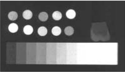

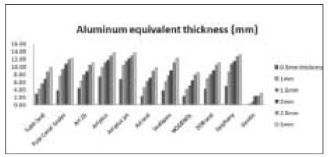
Figure 1
A radiograph showing the radiopacities of each experimental material, dentin and its equivalence to those of the aluminum step wedge
Figure 2
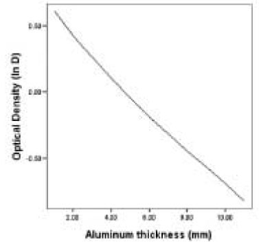
Standard curve for the optical d ensity of the aluminum step wedge at 60 kVp, 0.2 s
Figure 3
Radiopacities expressed in mean equivalent aluminum thickness for experimental materials in comparison with dentin
Figure 1
Figure 2
Figure 3
A comparative study on radiopacity of root canal sealers
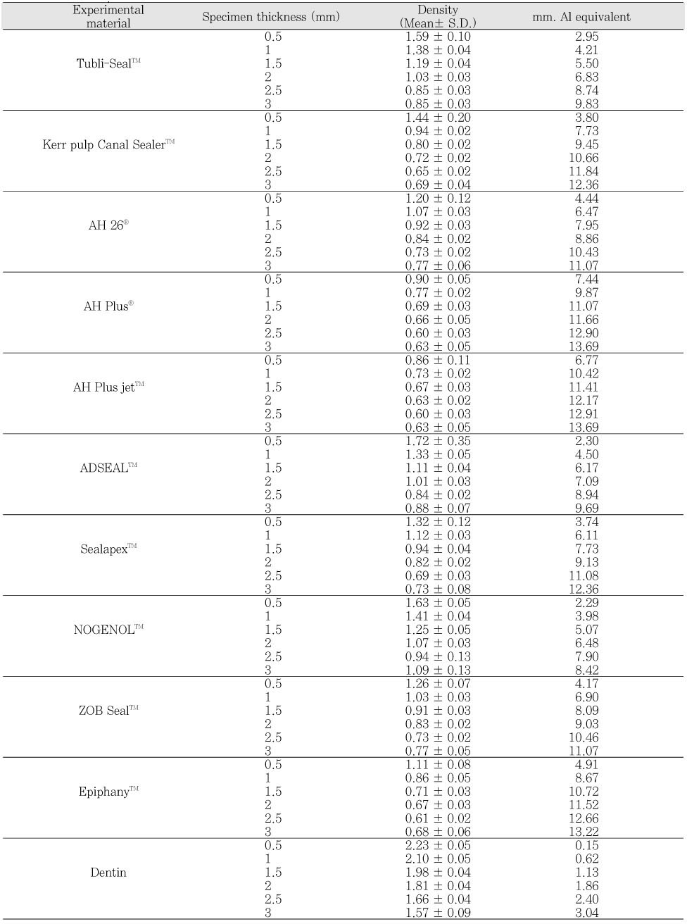
Mean values and standard deviations of the radiopacity values in the terms of equivalent thickness of aluminum for the experimental materials
Table 1
Mean values and standard deviations of the radiopacity values in the terms of equivalent thickness of aluminum for the experimental materials

