Articles
- Page Path
- HOME > Restor Dent Endod > Volume 30(5); 2005 > Article
- Original Article A comparative study of the canal configuration after shaping by protaper rotary and hand files in resin simulated canals
- In-Seok Yang1, In-Chol Kang2, Yun-Chan Hwang1, In-Nam Hwang1, Won-Mann Oh1
-
2005;30(5):-401.
DOI: https://doi.org/10.5395/JKACD.2005.30.5.393
Published online: September 30, 2005
1Department of Conservative Dentistry, School of Dentistry, DSRI, Chonnam National University, Korea.
2Department of Oral Microbiology, School of Dentistry, DSRI, Chonnam National University, Korea.
- Corresponding author: Won-Mann Oh. Department of Conservative Dentistry, School of Dentistry, Chonnam National Universtiy, 8 Hak-dong, Dong-gu, Gwangju, Korea 501-757. Tel: 82-62-220-4431, Fax: 82-62-225-8387, wmoh@chonnam.ac.kr
• Received: February 22, 2005 • Revised: June 2, 2005 • Accepted: June 13, 2005
Copyright © 2005 Korean Academy of Conservative Dentistry
- 724 Views
- 3 Download
Abstract
-
The purpose of this study was to compare the canal configuration after shaping by ProTaper rotary files and ProTaper hand files in resin simulated canals.Forty resin simulated canals with a curvature of J-shape and S-shape were divided into four groups by 10 blocks each. Simulated root canals in resin block were prepared by ProTaper rotary files and ProTaper hand files using a crown-down pressureless technique. All simulated canals were prepared up to size #25 file at end-point of preparation. Pre- and post-instrumentation images were recorded with color scanner. Assessment of canal shape was completed with an image analysis program. Measurements were made at 0, 1, 2, 3, 4, 5, 6 and 7 mm from the apex. At each level, outer canal width, inner canal width, total canal width, and amount of transportation from original axis were recorded. Instrumentation time was recorded. The data were analyzed statistically using independent t-test.The result was that ProTaper hand files cause significantly less canal transportation from original axis of canal body and maintain original canal configuration better than ProTaper rotary files, however ProTaper hand files take more shaping time.
- 1. Schilder H. Cleaning and shaping the root canal. Dent Clin North Am. 1974;18: 269-296.ArticlePubMed
- 2. Schilder H. Filling root canals in three dimensions. Dent Clin North Am. 1967 11;723-744.ArticlePubMed
- 3. Schneider SW. A comparison of canal prepararion in straight and curved root canals. Oral Surg Oral Med Oral Pathol. 1971;32: 271-275.PubMed
- 4. Walia HM, Brantley WA, Gerstein H. An initial investigation of the bending and torsional properties of Nitinol root canal files. J Endod. 1988;14: 346-351.ArticlePubMed
- 5. Zmener O, Balbachan L. Effectiveness of nickel-titanium files for preparing curved root canals. Endod Dent Traumatol. 1995;11: 121-123.ArticlePubMed
- 6. Glossen CR, Haller RH, Dove SB, del Rio CE. A comparison of root canal preparations using Ni-Ti hand, Ni-Ti engine driven, and K-flex endodontic instruments. J Endod. 1995;21: 146-151.ArticlePubMed
- 7. Esposito PT, Cunningham CJ. A comparison of canal preparation with nickel-titanium and stainless steel instruments. J Endod. 1995;21: 173-176.ArticlePubMed
- 8. Yun HH, Kim SK. A comparison of the shaping abilities of 4 nickel-titanium rotary instruments in simulated root canals. Oral Surg Oral Med Oral Pathol Oral Radiol Endod. 2003;95: 228-233.ArticlePubMed
- 9. Hata G, Uemura M, Kato AS, Imura N, Novo NF, Toda T. A comparison of shaping ability using ProFile, GT file, and Flex-R endodontic instruments in simulated canals. J Endod. 2002;28: 316-321.ArticlePubMed
- 10. Bishop K, Dummer PM. A comparison of stainless steel Flexofiles and nickel-titanium NiTiFlex files during the shaping of simulated canals. Int Endod J. 1997;30: 25-34.ArticlePubMed
- 11. Gambill JM, Alder M, del Rio CE. Comparison of nickel-titanium and stainless steel hand-file instrumentation using computed tomography. J Endod. 1996;22: 369-375.ArticlePubMed
- 12. Coleman CL, Svec TA. Analysis of Ni-Ti versus stainless steel instrumentation in resin simulated canals. J Endod. 1997;23: 232-235.ArticlePubMed
- 13. Park H. A comparison of Greater Taper files, ProFiles, and stainless steel files to shape curved root canals. Oral Surg Oral Med Oral Pathol Oral Radiol Endod. 2001;91: 715-718.ArticlePubMed
- 14. Glickman GN, Koch KA. Twenty-first century endodontics. J Am Dent Assoc. 2000;131: 39S-46S.PubMed
- 15. Pruett JP, Clement DJ, Carnes DL Jr. Cyclic fatigue testing of nickel-titanium endodontic instruments. J Endod. 1997;23: 77-85.ArticlePubMed
- 16. Bonetti FI, Miranda ER, de Toledo LR, del Rio CE. Microscopic evaluation of three endodontic files pre and postinstrumentation. J Endod. 1998;24: 461-464.ArticlePubMed
- 17. Calberson FL, Deroose CAJ, Hommez GM, Raes H, De Moor RJ. Shaping ability of GT™ Rotary Files in simulated resin root canals. Int Endod J. 2002;35: 607-614.ArticlePubMed
- 18. Eldeeb ME, Boraas JC. The effect of different files on the preparation shape of severely curved canals. Int Endod J. 1985;18: 1-7.ArticlePubMed
- 19. Lim KC, Webber J. The validity of simulated root canals for the investigation of the prepared root canal shape. Int Endod J. 1985;18: 240-246.ArticlePubMed
- 20. Campos JM, del Rio C. Comparison of mechanical and standard hand instrumentation techniques in curved root canals. J Endod. 1990;16: 230-234.ArticlePubMed
- 21. Hulsmann M, Stryga F. Comparison of root canal preparation using different automated devices and hand instrumentation. J Endod. 1993;19: 141-145.ArticlePubMed
- 22. Christopher JR, Kishor G, Richard TW. Endodontics. 2004;third edition. London, UK: ELSEVIER MOSBY; 153-161.
- 23. Powell SE, Simon JHS, Maze B. A comparison of the effect of modified and nonmodified instrument tips on apical canal configuration. J Endod. 1986;12: 293-300.ArticlePubMed
- 24. Thompson SA, Dummer PM. Shaping ability of Quantec Series 2000 rotary nickel-titanium instruments in simulated root canals: Part 2. Int Endod J. 1998;31: 268-274.ArticlePubMed
- 25. Thompson SA, Dummer PM. Shaping ability of Quantec Series 2000 rotary nickel-titanium instruments in simulated root canals: Part 1. Int Endod J. 1998;31: 259-267.ArticlePubMed
- 26. Griffiths IT, Bryant ST, Dummer PM. Canal shapes produced sequentially during instrumentation with Quantec LX rotary nickel-titanium instruments: a study in simulated canals. Int Endod J. 2000;33: 346-354.ArticlePubMed
- 27. Blum JY, Machtou P, Micallef JP. Location of contact areas on rotary Profile instruments in relationship to the forces developed during mechanical preparation on extracted teeth. Int Endod J. 1999;32: 108-114.PubMed
REFERENCES
Figure 1
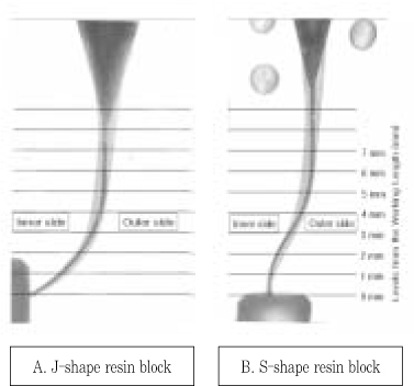
A and B diagrams indicate the points at which the canal widths were measured after superimposition of pre-instrumentaion and post-instrumentaion images.

Figure 2
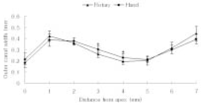
Changes in outer canal width of J-shape resin block after canal shaping by ProTaper rotary files and ProTaper hand files.
*: significant difference (p < 0.05).

Figure 3
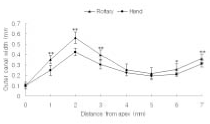
Changes in outer canal width of S-shape resin block after canal shaping by ProTaper rotary files and ProTaper hand files.
**: significant difference (p < 0.01).
*: significant difference (p < 0.05).

Figure 4
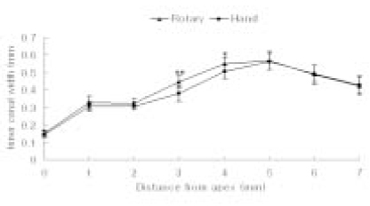
Changes in inner canal width of J-shape resin block after canal shaping by ProTaper rotary files and ProTaper hand files.
**: significant difference (p < 0.01).
*: significant difference (p < 0.05).

Figure 5
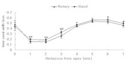
Changes in inner canal width of S-shape resin block after canal shaping by ProTaper rotary files and ProTaper hand files.
**: significant difference (p < 0.01).
*: significant difference (p < 0.05).

Figure 6
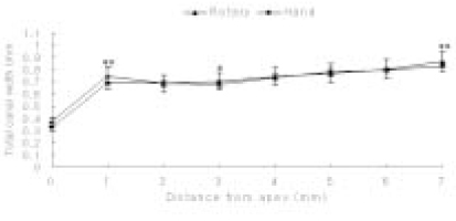
Changes in total canal width of J-shape resin block after canal shaping by ProTaper rotary files and ProTaper hand files.
**: significant difference (p < 0.01).
*: significant difference (p < 0.05).

Figure 7
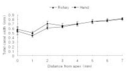
Changes in total canal width of S-shape resin block after canal shaping by ProTaper rotary files and ProTaper hand files.
**: significant difference (p < 0.01).

Figure 8
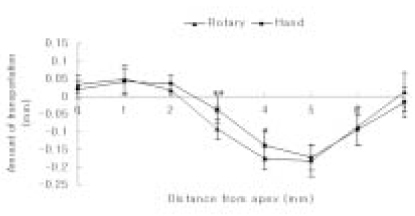
Amounts of transportation from the original axis of J-shape resin block after canal shaping by ProTaper rotary files and ProTaper hand files. Minus values indicate that axis of canal was transported to inner side curvature after canal preparation.
**: significant difference (p < 0.01).
*: significant difference (p < 0.05).

Figure 9
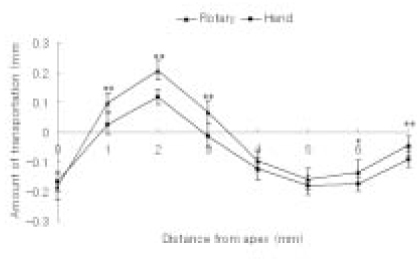
Amounts of transportation from the original axis of S-shape resin block after canal shaping by ProTaper rotary files and ProTaper hand files. Minus values indicate that axis of canal was transported to inner side curvature after canal preparation.
**: significant difference (p < 0.01).
*: significant difference (p < 0.05).

Tables & Figures
REFERENCES
Citations
Citations to this article as recorded by 

A comparative study of the canal configuration after shaping by protaper rotary and hand files in resin simulated canals


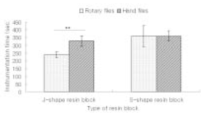
Figure 1
A and B diagrams indicate the points at which the canal widths were measured after superimposition of pre-instrumentaion and post-instrumentaion images.
Figure 2
Changes in outer canal width of J-shape resin block after canal shaping by ProTaper rotary files and ProTaper hand files.
*: significant difference (p < 0.05).
Figure 3
Changes in outer canal width of S-shape resin block after canal shaping by ProTaper rotary files and ProTaper hand files.
**: significant difference (p < 0.01).
*: significant difference (p < 0.05).
Figure 4
Changes in inner canal width of J-shape resin block after canal shaping by ProTaper rotary files and ProTaper hand files.
**: significant difference (p < 0.01).
*: significant difference (p < 0.05).
Figure 5
Changes in inner canal width of S-shape resin block after canal shaping by ProTaper rotary files and ProTaper hand files.
**: significant difference (p < 0.01).
*: significant difference (p < 0.05).
Figure 6
Changes in total canal width of J-shape resin block after canal shaping by ProTaper rotary files and ProTaper hand files.
**: significant difference (p < 0.01).
*: significant difference (p < 0.05).
Figure 7
Changes in total canal width of S-shape resin block after canal shaping by ProTaper rotary files and ProTaper hand files.
**: significant difference (p < 0.01).
Figure 8
Amounts of transportation from the original axis of J-shape resin block after canal shaping by ProTaper rotary files and ProTaper hand files. Minus values indicate that axis of canal was transported to inner side curvature after canal preparation.
**: significant difference (p < 0.01).
*: significant difference (p < 0.05).
Figure 9
Amounts of transportation from the original axis of S-shape resin block after canal shaping by ProTaper rotary files and ProTaper hand files. Minus values indicate that axis of canal was transported to inner side curvature after canal preparation.
**: significant difference (p < 0.01).
*: significant difference (p < 0.05).
Figure 10
Total instrumentation time for the canal preparation at the different type of resin blocks after canal shaping by ProTaper rotary files and ProTaper hand files.
**: significant difference (p < 0.01).
Figure 1
Figure 2
Figure 3
Figure 4
Figure 5
Figure 6
Figure 7
Figure 8
Figure 9
Figure 10
A comparative study of the canal configuration after shaping by protaper rotary and hand files in resin simulated canals

 KACD
KACD

 ePub Link
 Cite
Cite

