
Articles
- Page Path
- HOME > Restor Dent Endod > Volume 32(5); 2007 > Article
- Original Article Influence of the labial surface irregularity on the measurement of the tooth color by spectrometer
- Yong-Jin Choi, Su-Jung Park, Hyun-Gu Cho, Yun-Chan Hwang, Won-Mann Oh, Byung-Ju Park1, In-Nam Hwang
-
2007;32(5):-418.
DOI: https://doi.org/10.5395/JKACD.2007.32.5.411
Published online: September 30, 2007
Department of Conservative Dentistry, School of Dentistry, DSRI, Chonnam National University, Korea.
1Department of Oral Biochemistry, School of Dentistry, DSRI, Chonnam National University, Korea.
- Corresponding Author: In-Nam Hwang. Department of Conservative Dentistry, School of Dentistry, Chonnam National University 5 Hak-dong, Dong-gu, Gwangju, 501-757, Korea. Tel: 82-62-220-4443, Fax: 82-62-225-8387, hinso@jnu.ac.kr
• Received: June 16, 2007 • Revised: July 7, 2007 • Accepted: July 31, 2007
Copyright © 2007 Korean Academy of Conservative Dentistry
- 1,804 Views
- 2 Download
- 1 Crossref
Abstract
-
The most scientific and reliable method for deciding the tooth color is the instrumental measurement. However, such color measuring instrument shows the difference of the measuring value according to the diversified measuring condition.This study was conducted to evaluate what effect of the labial surface irregularity of the tooth to the result of the color measured by spectrometer.11 models of the teeth were made by injecting the A2 shade Luxatemp Automix Plus (DMG, Germany) into the impression acquired from 11 adults. Standard disk samples (15 mm diameter, 7 mm thickness) were made with same material. CIE L*a*b* value was measured at the incisal, central, and gingival area of the central incisor, lateral incisor, canine and first premolar using Specbos 2100 (JETI, Germany) spectrometer. Color difference was calculated between labial surface and standard samples.Among all models of the teeth, L* and b* value showed the reducing tendency as they go toward the gingival area, but a* value showed the increasing tendency.Color difference between model teeth and standard samples showed the most difference at the incisal area, but the gingival area showed the least difference. And the canine showed the least color difference from the comparison of standard sample, and the central incisor showed the highest difference (p < 0.01).Although the visually detectable difference of the measuring value showed notably depending on the type and measured area (p < 0.05), L* and a* value showed notable differences depending more on the measured areas than on the type of the teeth.
- 1. Sproull RC. Color matching in dentistry. Part II: Practical applications of the organization of color. J Prosthet Dent. 1973;29: 556-566.ArticlePubMed
- 2. Miller L. Organizing color in dentistry. J Am Dent Assoc. 1987 12;(Spec No):26E-40E.ArticlePubMed
- 3. Johnston WM, Kao EC. Assessment of appearance match by visual observation and clinical colorimeter. J Dent Res. 1989;68: 819-822.ArticlePubMedPDF
- 4. Hwang IN, Oh WM. Colorimetric analysis of extracted human teeth and five shade guides. J Korean Acad Conserv Dent. 1997;22: 769-781.
- 5. O'Neal SJ, Powell WD. Color discrimination and shade matching ability of third year dental student. J Prosthet Dent. 1984;63: 174.
- 6. Van der Burgt TP, ten Bosch JJ, Borsboom PC, Plasschaert AJ. A new method for matching tooth colors with color standard. J Dent Res. 1985;64: 837-841.ArticlePubMedPDF
- 7. Macentee M, Lakowski R. Instrumental color measurement of vital and extracted teeth. J Oral Rehabil. 1981;8: 203-208.PubMed
- 8. O'Brien WJ, Boenke KM, Groh CL. Coverage error of two shade guide. Int J Prosthodont. 1991;4: 45-50.PubMed
- 9. Hammad IA. Intrarater repeatability of shade selections with two shade guides. J Prosthet Dent. 2003;89: 50-53.ArticlePubMed
- 10. Lemire PA, Burk B. Color in dentistry. 1975;Hartford: CT J.M Ney Co..
- 11. Grajower R, Revah A, Sorin S. Reflectance spectra of natural and acrylic teeth. J Prosthet Dent. 1976;36: 570-579.PubMed
- 12. Goodkind RJ, Schwabacher WB. Use of a fiber-optic colorimeter for in vivo color measurements of 2830 anterior teeth. J Prosthet Dent. 1987;58: 535-542.ArticlePubMed
- 13. Goodkind RJ, Keenan KM, Schwabacher WB. A comparison of Chromascan and spectrophotometric color measurement of 100 natural teeth. J Prosthet Dent. 1985;53: 105-109.PubMed
- 14. Schwabacher WB, Goodkind RJ. Three-dimensional color coordinates of natural teeth compared with three shade guide. J Prosthet Dent. 1990;64: 425-431.PubMed
- 15. Cho KY, Hwang IN, Choi HR, Oh WM. Comparative evaluation of light-cured composite resins based on vita shade by spectrocolorimeter. J Korean Acad Conserv Dent. 1998;23: 424-432.
- 16. Lee MY, Shin DH. New evaluation technique in teeth color using digital camera. J Korean Acad Conserv Dent. 1997;22: 325-333.
- 17. Clifford MS. The Art and Science of Operative Dentistry. 1995;3rd ed. Mosby-Year book.
- 18. Clark EB. The color problem in dentistry. Dent Digest. 1931;499-509.
- 19. Clark EB. An analysis of tooth color. J Am Dent Assoc. 1931;18: 2093-2103.Article
- 20. Clark EB. Tooth color selection. J Am Dent Assoc. 1933;20: 1065-1073.
- 21. Mun EB. The application of color. 2002;Seoul: Kukjebooks; 215-271.
- 22. Park EJ. The basic of color moulding. 1996;2nd ed. Seoul: Mijinsa; 56-194.
- 23. Miller LL. Shade matching. J Esthet Dent. 1993;5: 143-152.ArticlePubMed
- 24. Tung FF, Goldstein GR, Jang S, Hittelman E. The repeatability of an intraoral dental colorimeter. J Prosthet Dent. 2002;88: 585-590.ArticlePubMed
- 25. Barrett AA, Grimaudo NJ, Anuscvice KJ, Yang MCK. Influence of tab and disk design on shade matching of dental porcelain. J Prosthet Dent. 2002;88: 591-597.ArticlePubMed
- 26. Hwang IN, Lee KW. Translucency of light cured composite resins depends on thickness & its influence on color of restorations. J Korean Acad Conserv Dent. 1999;24: 585-603.
- 27. Ruyter IE, Nilner K, Mo¨ller B. Color stability of dental composite resin materials for crown and bridge veneers. Dent Mater. 1987;3: 246-251.ArticlePubMed
- 28. Gross MD, Moser JB. A colorimetric study of coffee and tee staining of four composite resins. J Oral Rehabil. 1977;4: 311-322.PubMed
REFERENCES


Tables & Figures
REFERENCES
Citations
Citations to this article as recorded by 
- A Study on Digital Color Reproduction for Recording Color Appearance of Cultural Heritage
Hyeong Rok Song, Young Hoon Jo
Journal of Conservation Science.2022; 38(2): 154. CrossRef
 ePub Link
 Cite
CiteInfluence of the labial surface irregularity on the measurement of the tooth color by spectrometer
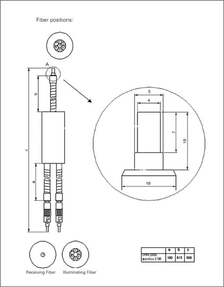
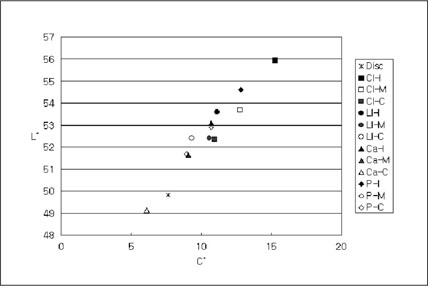
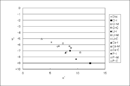
Figure 1
Diagram of the Duplex-Fiber bundle.
Figure 2
L* and C* value of the tested disc samples and tooth models.
CI : Central incisor, LI : Laterial Incisor, Ca : Canine, P : Premolar, I : Incisal, M : Middle, C : Cervical
Figure 3
a* and b* value of the tested disc samples and tooth models.
Figure 1
Figure 2
Figure 3
Influence of the labial surface irregularity on the measurement of the tooth color by spectrometer
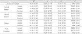
CIE L*a*b* and C* values of disc samples and tooth models
Standard deviations are in parentheses.
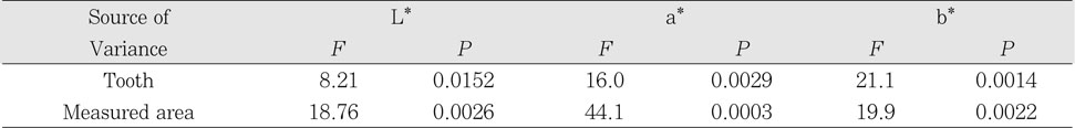
Statistical analysis between tooth and measured area
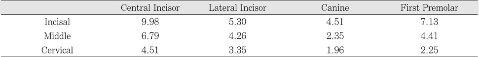
Color difference (ΔE*) between disc sample and tooth models
Color difference among measured area
*There are statistically significant differences among groups (p < 0.01).
Table 1
CIE L*a*b* and C* values of disc samples and tooth models
Standard deviations are in parentheses.
Table 2
Statistical analysis between tooth and measured area
Table 3
Color difference (ΔE*) between disc sample and tooth models
Table 4
Color difference among measured area
*There are statistically significant differences among groups (p < 0.01).

