
Articles
- Page Path
- HOME > Restor Dent Endod > Volume 34(3); 2009 > Article
- Original Article Color difference of the dental composites measured by different color measuring instruments
- Su-Jung Park, Eun-Young Noh, Hyun-Gu Cho, Yun-Chan Hwang, Won-Mann Oh, In-Nam Hwang
-
2009;34(3):-207.
DOI: https://doi.org/10.5395/JKACD.2009.34.3.199
Published online: May 31, 2009
Department of Conservative Dentistry, School of Dentistry, DSRI, Chonnam National University, Korea.
- Corresponding author: In-Nam Hwang. Department of Conservative Dentistry, Chonnam National University School of Dentistry, Yongbong-ro 77, Buk-gu, Gwangju, 500-757, Korea. Tel. 82-62-530-5819, Fax. 82-62-530-5629, hinso@jnu.ac.kr
• Received: March 3, 2009 • Revised: March 26, 2009 • Accepted: April 23, 2009
Copyright © 2009 The Korean Academy of Conservative Dentistry
- 1,550 Views
- 5 Download
- 2 Crossref
Abstract
-
The objective of this study was to evaluate the effect of color measuring instrument by measuring the color of dental composite resins.Nine shade light cured composite resin disks were prepared (diameter : 15 mm, thickness : 4 mm). CIE L*a*b* color scale of each disk was measured with 3 different types of spectrophotometer [MiniScan XE plus (Model 4000S, Hunter Lab, USA), CM-3500d (Minolta, Japan) and Specbos 2100 Miniature VIS Reflection spectrometer (Serial No: 319416, JETI Technishe VIS Instrumentic GmbH, Germany)]. Miniscan XE Plus and CM-3500d using identical measuring geometry with different size of viewing aperture. But Specbos 2100 using different measuring geometry.Within the limitation of this study, there were color difference (ΔE*) from 2.4 to 7.8 between Miniscan XE Plus and CM-3500d, but L*, a*, b* values showed the high correlation. However, there were great color difference (ΔE*) in the extent of about 20 between instruments with the different measuring geometry.Therefore, color scale measured by color measuring instrument should be used as a relative value rather than an absolute value in the field of dentistry.
- 1. Clark EB. An analysis of tooth color. J Am Dent Assoc. 1931;18: 2093-2103.Article
- 2. Clark EB. Tooth color selection. J Am Dent Assoc. 1933;20: 1065-1073.
- 3. Hayashi T. Medical color standard. V. Tooth crown. 1967;Tokyo: Japan Color Research Institute.
- 4. Miller LL. Organizing color in dentistry. J Am Dent Assoc. 1987;Spec No: 12;26E-40E.ArticlePubMed
- 5. Johnston WM, Kao EC. Assessment of appearance match by visual observation and clinical colorimeter. J Dent Res. 1989;68: 819-822.ArticlePubMedPDF
- 6. Hwang IN, Oh OM. Colorimetric analysis of extracted human teeth and five shade guides. J Korean Acad Conserv Dent. 1997;22: 769-781.
- 7. Sproull RC. Color matching in dentistry. Part II: Practical applications of the organization of color. J Prosthet Dent. 1973;29: 556-566.PubMed
- 8. Goodkind RJ, Keenan KM, Schwabacher WB. A comparison of Chromascan and spectrophotometric color measurement of 100 natural teeth. J Prosthet Dent. 1985;53: 105-109.PubMed
- 9. Schwabacher WB, Goodkind RJ. Three-dimensional color coordinates of natural teeth compared with three shade guide. J Prosthet Dent. 1990;64: 425-431.PubMed
- 10. Yeh CL, Powers JM, Miyagawa Y. Color of selected shades of composites by reflection spectrophotometry. J Dent Res. 1982;61: 1176-1179.ArticlePubMedPDF
- 11. Jo KM, Shin DH, Gu NY. Color analysis of the natural teeth with a modified intraoral spectrophotometer. J Korean Acad Conserv Dent. 1998;23: 223-235.
- 12. Lee MY, Shin DH. New evaluation technique in teeth color using digital camera. J Korean Acad Conserv Dent. 1997;23: 325-333.
- 13. Kim HS, Um JM. A study on color differences between composite resins and shade guides. J Korean Acad Conserv Dent. 1996;21: 107-120.
- 14. Jo KH, Hwang IN, Choi HR, Oh OM. Comparative evaluation of light-cured composite resins based on vita shade by spectrocolorimeter. J Korean Acad Conserv Dent. 1998;23: 424-432.
- 15. Hwang IN, Lee KW. Translucency of light cured composite resins depends on thickness & its influence on color of restorations. J Korean Acad Conserv Dent. 1999;24: 604-613.
- 16. Swepston JH, Miller LL. Esthetic matching. J Prosthet Dent. 1985;54: 623-625.ArticlePubMed
- 17. Moon EB. Color System. 2002;Seoul: Gukje; 215-271.
- 18. Park EJ. Basics of Color Moulding. 1996;2th ed. Seoul: Mijinsa; 56-194.
- 19. Park DY. Practical Chromatics. 1992;enlarged edition. Seoul: Bando; 99-120.
- 20. Korean Industrial Standards. KS A 0061.
- 21. Goodkind RJ, Loupe MJ. Teaching of color in predoctoral and postdoctoral dental education in 1988. J Prosthet Dent. 1992;67: 713-717.ArticlePubMed
- 22. Culpepper WD. A comparative study of shade matching procedures. J Prosthet Dent. 1970;24: 166-173.ArticlePubMed
- 23. O'Neal SJ, Powell WD. Color discrimination and shade matching ability of third year dental student. J Dent Res. 1984;63: 174.
- 24. Van der Burgt TP, ten Bosch JJ, Borsboom PC, Plasschaert AJ. A new method for matching tooth colors with color standard. J Dent Res. 1985;64: 837-841.ArticlePubMedPDF
- 25. Gross MD, Moser JB. A colorimetric study of coffee and tee staining of four composite resins. J Oral Rehabil. 1977;4: 311-322.PubMed
- 26. Seghi RR, Hewlett ER, Kim J. Visual and instrumental colorimetricassessments of small color differences on translucent dental porcelain. J Dent Res. 1989;68: 1760-1764.ArticlePubMedPDF
- 27. Bolt RA, Bosch JJ, Coops JC. Influence of window size in small-window color measurement particularly of teeth. Phys Med Biol. 1994;39: 1133-1142.ArticlePubMed
REFERENCES

Figure 2

L* value change of the Z-250 according to the measuring instrument and condition.Miniscan: MiniScan XE Plus
CD: Minolta CD-3500d
spec-O/D: Specbos 2100 with original measuring tip under dark.
spec-O/L: Specbos 2100 with original measuring tip under light.
spec-H/L: Specbos 2100 with handmade measuring tip under light.

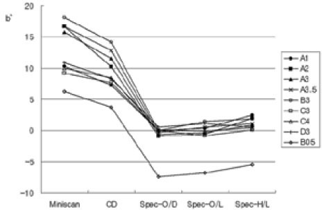
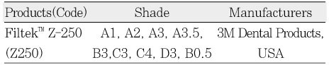
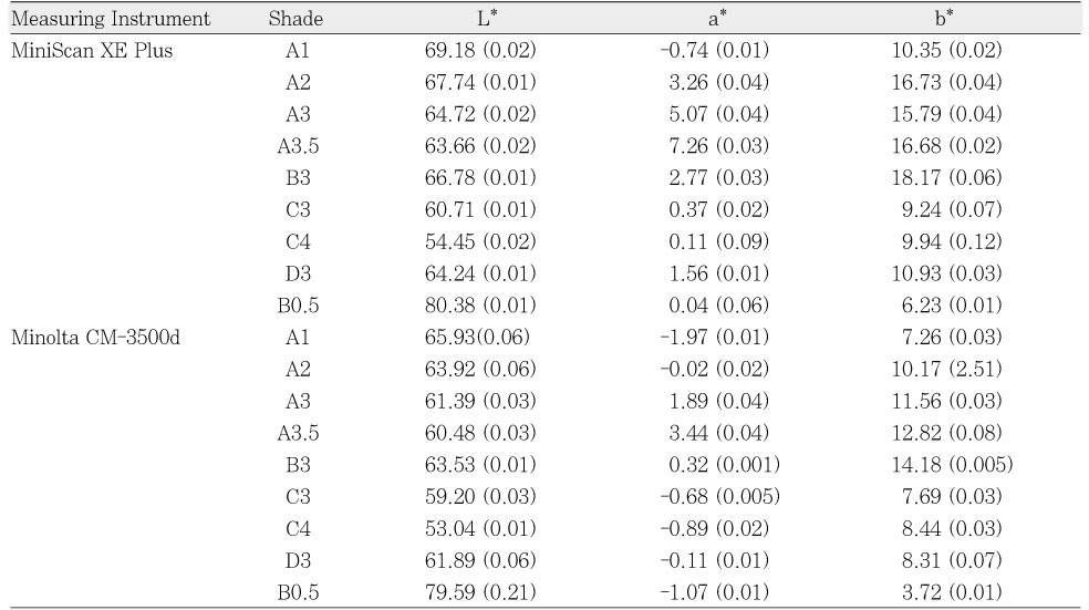
Table 3
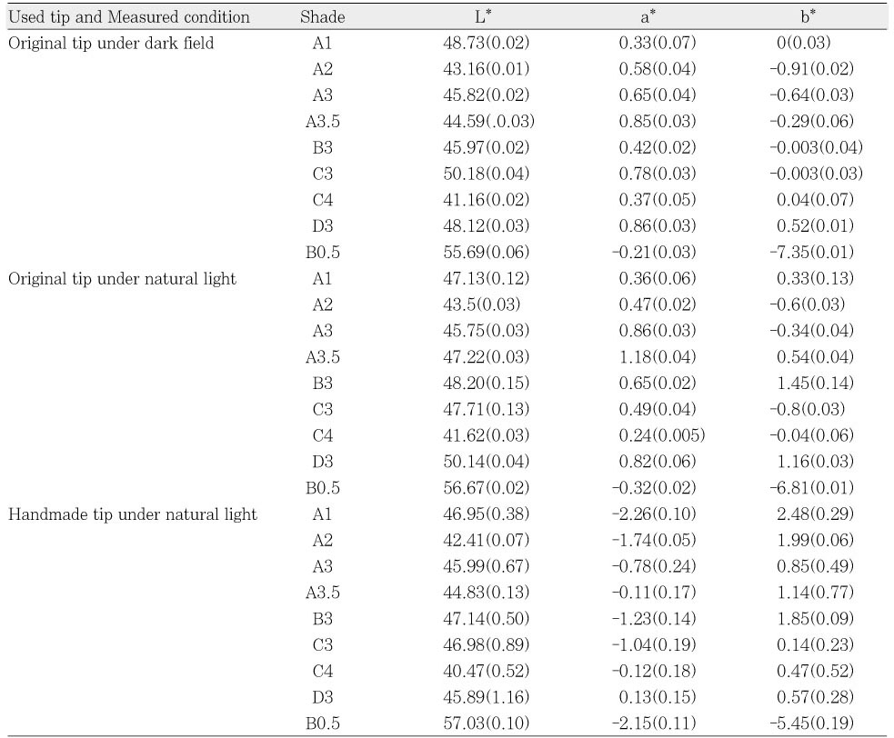
CIE L*a*b* values of Filtek Z-250 measured by Specbos 2100 according to the used tip and measured condition
Table 4
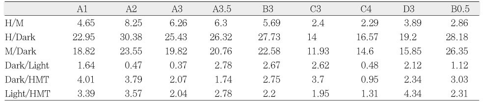
Color difference(ΔE*) among different color measuring instruments in the Z-250
H : MiniScan XE Plus
M : Minolta CD-3500d
Dark : Specbos 2100 with original measuring tip under dark.
Dark : Specbos 2100 with original measuring tip under dark.
Light : Specbos 2100 with original measuring tip under light.
HMT : Specbos 2100 with handmade measuring tip under light.
Tables & Figures
REFERENCES
Citations
Citations to this article as recorded by 
- Color Change in Tooth Induced by Various Calcium Silicate-Based Pulp-Capping Materials
Jiyoon Jeon, Namki Choi, Seonmi Kim
THE JOURNAL OF THE KOREAN ACADEMY OF PEDTATRIC DENTISTRY.2021; 48(3): 280. CrossRef - Effects of the color components of light-cured composite resin before and after polymerization on degree of conversion and flexural strength
Ji-A Yoo, Byeong-Hoon Cho
Journal of Korean Academy of Conservative Dentistry.2011; 36(4): 324. CrossRef
 ePub Link
 Cite
CiteColor difference of the dental composites measured by different color measuring instruments
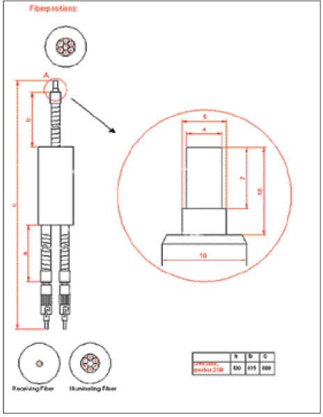
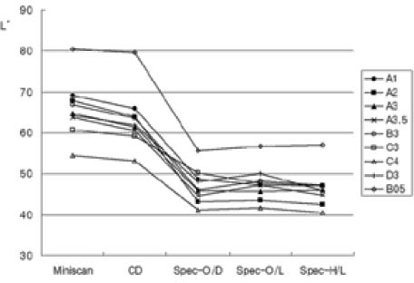
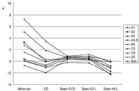

Figure 1
Diagram of the Duplex-Fiber bundle.
Figure 2
L* value change of the Z-250 according to the measuring instrument and condition.Miniscan: MiniScan XE Plus
CD: Minolta CD-3500d
spec-O/D: Specbos 2100 with original measuring tip under dark.
spec-O/L: Specbos 2100 with original measuring tip under light.
spec-H/L: Specbos 2100 with handmade measuring tip under light.
Figure 3
a* value change of the Z-250 according to the measuring instrument and condition.
Figure 4
b* value change of the Z-250 according to the measuring instrument and condition.
Figure 1
Figure 2
Figure 3
Figure 4
Color difference of the dental composites measured by different color measuring instruments
Products, shade, and manufacturers of tested composite resins
CIE L*a*b* values of Filtek Z-250 measured by MiniScan XE Plus and Minolta CM 3500d
Standard deviations are in parentheses.
CIE L*a*b* values of Filtek Z-250 measured by Specbos 2100 according to the used tip and measured condition
Standard deviations are in parentheses
Color difference(ΔE*) among different color measuring instruments in the Z-250
H : MiniScan XE Plus
M : Minolta CD-3500d
Dark : Specbos 2100 with original measuring tip under dark.
Dark : Specbos 2100 with original measuring tip under dark.
Light : Specbos 2100 with original measuring tip under light.
HMT : Specbos 2100 with handmade measuring tip under light.
Table 1
Products, shade, and manufacturers of tested composite resins
Table 2
CIE L*a*b* values of Filtek Z-250 measured by MiniScan XE Plus and Minolta CM 3500d
Standard deviations are in parentheses.
Table 3
CIE L*a*b* values of Filtek Z-250 measured by Specbos 2100 according to the used tip and measured condition
Standard deviations are in parentheses
Table 4
Color difference(ΔE*) among different color measuring instruments in the Z-250
H : MiniScan XE Plus M : Minolta CD-3500d Dark : Specbos 2100 with original measuring tip under dark. Dark : Specbos 2100 with original measuring tip under dark. Light : Specbos 2100 with original measuring tip under light. HMT : Specbos 2100 with handmade measuring tip under light.

