
Search
- Page Path
- HOME > Search
Research Articles
- Effect of sugar and sweetener on the bleachability of coffee and tea-induced stains on composites: an in vitro experimental study
- Nilay Bayraktar, Osman Kerim Arda Karaca, Yunus Ekşılı, Mustafa Furkan Yıldırım, Osman Tolga Harorli
- Restor Dent Endod 2026;51(2):e16. Published online April 1, 2026
- DOI: https://doi.org/10.5395/rde.2026.51.e16
-
 Abstract
 PDF
PDF PubReader
 ePub
ePub - Objectives
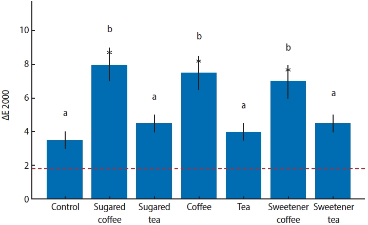
This in vitro study evaluated the effects of various sugary and non-sugary beverages on the color change of a dental composite and the subsequent bleaching efficacy.
Methods
Forty-nine disc-shaped composite samples (Neo Spectra ST, Dentsply Sirona) were split into seven groups at random (n = 7). Distilled water was used to hydrate each sample for 24 hours at 37°C. After 24 hours, the first color measurements (T0) were made by using a clinical spectrophotometer (VITA Easyshade Compact; VITA Zahnfabrik). Color measurements were repeated after 7 days (T1) and 14 days (T2) of immersion in distilled water (control), tea, coffee, sugary tea, sugary coffee, tea with sweetener added, and coffee with sweetener added. After staining for 2 weeks, the specimens were bleached for 6 hours a day for a week using 16% carbamide peroxide (Opalescence Ultradent Products). Color measurements were taken again after bleaching (T3). Using CIEDE2000, color differences (ΔE) were computed. Analysis of variance (ANOVA) and repeated measures ANOVA with a Tukey post hoc test were used to evaluate the data.
Results
After 1 week, coffee-containing solutions produced significantly greater discoloration than the control (p < 0.001). By 2 weeks, tea groups exhibited similar discoloration to coffee groups (p < 0.001). The addition of sugar or sweetener had no significant effect (p > 0.05). Post-bleaching, coffee groups showed lower Whiteness Index values than the control, without statistical significance (p > 0.05).
Conclusions
Coffee and tea markedly stain resin composites, with discoloration persisting post-bleaching, while sugar or sweetener additions exert no significant effect.
- 1,155 View
- 91 Download

- Surface properties and susceptibility to staining of a resin composite after brushing with different whitening toothpastes
- Aline da Silva Barros, Carolina Meneghin Barbosa, Renata Siqueira Scatolin, Waldemir Francisco Vieira Junior, Laura Nobre Ferraz
- Restor Dent Endod 2025;50(1):e6. Published online February 26, 2025
- DOI: https://doi.org/10.5395/rde.2025.50.e6
-
Abstract
PDFPubReaderePub
- Objectives
This study investigated the effects of different whitening toothpaste (WT) on the surface properties and staining susceptibility of a resin composite.
Methods
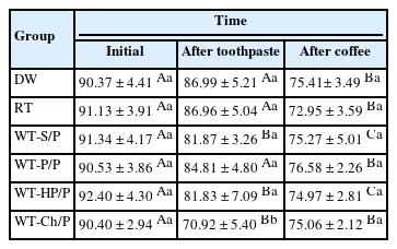
Cylindrical samples were prepared with a micro-hybrid resin composite and were randomized into groups according to the toothpaste (n = 12): distilled water (DW), regular toothpaste (RT), WT with silica + pyrophosphate (WT-S/P), WT with pentaphosphate and pyrophosphate (WT-P/P), WT with hydrogen peroxide and pyrophosphate (WT-HP/P) and WT with charcoal and pyrophosphate (WT-Ch/P). The samples were brushed for 825 cycles in an automatic brushing machine, simulating 30 days of brushing. After that, an immersion in coffee (10 mL/sample) was performed for 30 minutes for 30 days. The analyses of color, surface microhardness (SMH), and surface roughness (Ra) were performed at the initial time, after brushing with toothpaste and after immersion in coffee. The ΔL*, Δa*, Δb*, ΔEab, Δand E00 values were calculated comparing after toothpaste with initial time and after coffee with after toothpaste. Data were analyzed using a mixed linear model for repeated measures (SMH), Kruskal-Wallis, Dunn, Friedman, and Nemenyi tests, with α = 0.05.
Results
For ΔL*, the WT-Ch/P group had the lowest values and differed from the other groups comparing the after toothpaste with the initial time interval (p < 0.001). The WT-Ch/P group had the lowest SMH values in after-toothpaste time (p < 0.001). In after-toothpaste time and after coffee time, the WT-S/P group had the highest Ra values and differed from the groups except the WT-Ch/P group (p < 0.001).
Conclusions
The toothpaste composition affects the surface characteristics and susceptibility to staining of the resin composite. The charcoal-based toothpaste had the worst performance for the color analyses and SMH. -
Citations
Citations to this article as recorded by
- Color Stability and Surface Roughness of Esthetic Resin Composites Following Simulated Toothbrushing with Whitening Toothpastes
Ecehan Kaplan , Ayşe Dündar, Çağatay Barutçugil
Odovtos - International Journal of Dental Sciences.2026; 1(1): 595. CrossRef - Influence of commercial mouth rinses with different formulations on enamel properties during at-home bleaching
Thalita Novello Coelho, Ana Júlia Gil, Marcos Roberto Lima Benati, Carolina Meneghin Barbosa, Tatiane Cristina Dotta, Waldemir Francisco Vieira-Junior, Renata Siqueira Scatolin, Laura Nobre Ferraz
Odontology.2026;[Epub] CrossRef
- Color Stability and Surface Roughness of Esthetic Resin Composites Following Simulated Toothbrushing with Whitening Toothpastes
- 6,644 View
- 199 Download
- 1 Web of Science
- 2 Crossref
- Can discolored dental composites be bleached in depth?
- Luca Giachetti, Daniele Scaminaci Russo, Michele Nieri, Francesca Cinelli
- Restor Dent Endod 2024;49(3):e23. Published online June 11, 2024
- DOI: https://doi.org/10.5395/rde.2024.49.e23
-
Abstract
PDFPubReaderePub
Objectives Previous
in vitro studies determined the whitening effects of bleaching products on stained resin composite surfaces. Thisin vitro study aimed to verify the effectiveness of a whitening system on composite resin previously subjected to pigmentation, specifically examining the depth of whitening effectiveness within the material structure.Materials and Methods A commercially available nano-filled composite resin was used. Specimens were stained using a coffee-based solution and a 10% carbamide peroxide-based gel was employed as the whitening agent. The pigment’s penetration and the effect of the bleaching gel were evaluated by measuring color (CieLab values) from the outer edge to the inner part of the specimens. Color measurements were taken at 14 points, starting from 0.1 mm from the external perimeter up to 3.0 mm.
Results Analysis of variance tests showed a statistically significant difference between the Control Group (CG), Pigmentation Group, and Whitening Group. The whitening agent was effective up to 1.5 mm in depth, with Whiteness index (W) values not statistically different from those of CG up to 0.5 mm in depth.
Conclusions Whitening agents on nano-filled resin composite previously pigmented appear effective in restoring the W to values similar to the original, particularly in the superficial layers of the sample.
-
Citations
Citations to this article as recorded by- Color Stability of Tooth-Colored Restorative Materials After Exposure to Arabic Coffee and Black Tea: A Systematic Review
Abdulrhman Y Alenezi, Abdulwahab M AlEyada, Yousef H Aldhafiri, Mohammed S Alsubaie, Mohammed S Alshahrani, Mahesh Shenoy
Cureus.2025;[Epub] CrossRef - Comparative evaluation to composite resin bleaching using ozone-enhanced low-concentration hydrogen peroxide
Mahmoud K. AL-Omiri, Dania Sa’ed Hussam Abuherra, Khaled M. AL-Omiri, Ali Y. Alsaeed, Mohammad Alamri, Ali M. Alqahtani, Saleh Ali Alqahtani, Ghadeer Saleh Alwadai, Naif Abogazalah, Edward Lynch
Scientific Reports.2025;[Epub] CrossRef - The effects of mechanical and chemical degradation on the surface roughness, gloss, and color stability of bulk-fill resin composites
Merve Nezir, Hanife Altınışık, Esra Özyurt, Naz Bayar, Mediha Büyükgöze Dindar
BMC Oral Health.2025;[Epub] CrossRef
- Color Stability of Tooth-Colored Restorative Materials After Exposure to Arabic Coffee and Black Tea: A Systematic Review
- 5,086 View
- 157 Download
- 2 Web of Science
- 3 Crossref
- Impact of combined at-home bleaching and whitening toothpaste use on the surface and color of a composite resin
- Carolina Meneghin Barbosa, Renata Siqueira Scatolin, Waldemir Francisco Vieira-Junior, Marcia Hiromi Tanaka, Laura Nobre Ferraz
- Restor Dent Endod 2023;48(3):e26. Published online July 26, 2023
- DOI: https://doi.org/10.5395/rde.2023.48.e26
-
Abstract
PDFPubReaderePub
Objective This
in vitro study aimed to evaluate the effects of different whitening toothpastes on a composite resin during at-home bleaching with 10% carbamide peroxide.Materials and Methods Sixty samples (7 mm × 2 mm) were used for color and roughness analyses, while another 60 samples (3 mm × 2 mm) were utilized to assess microhardness. The factors analyzed included toothpaste, for which 5 options with varying active agents were tested (distilled water; conventional toothpaste; whitening toothpaste with abrasive agents; whitening toothpaste with abrasive and chemical agents; and whitening toothpaste with abrasive, chemical, and bleaching agents). Brushing and application of whitening gel were performed for 14 days. Surface microhardness (SMH), surface roughness (Ra), and color (∆L*, ∆a*, ∆b, ∆E*ab, and ∆E00) were analyzed. The Ra and SMH data were analyzed using mixed generalized linear models for repeated measures, while the color results were assessed using the Kruskal-Wallis and Dunn tests.
Results Between the initial and final time points, all groups demonstrated significant increases in Ra and reductions in SMH. No significant differences were found between groups for SMH at the final time point, at which all groups differed from the distilled water group. Conventional toothpaste exhibited the lowest Ra, while whitening toothpaste with abrasive agent had the highest value. No significant differences were observed in ∆L*, ∆a*, and ∆b.
Conclusions While toothpaste composition did not affect the color stability and microhardness of resin composite, combining toothbrushing with whitening toothpaste and at-home bleaching enhanced the change in Ra.
-
Citations
Citations to this article as recorded by- Current evidence on the impact of whitening toothpastes on dental restorative materials: A comprehensive review
Soyeon Kim, Shin Hye Chung, Satoshi Yamaguchi, Taro Arima, Young-Seok Park
Journal of Prosthodontic Research.2026; 70(1): 4. CrossRef - Property changes in resin composite exposed to mouth rinses during 10% carbamide peroxide bleaching
Mariana Ferreira da Silva, Giovana Contin Germinari, Carolina Meneghin Barbosa, Tatiane Cristina Dotta, Renata Siqueira Scatolin, Waldemir Francisco Vieira Júnior, Laura Nobre Ferraz
Brazilian Journal of Oral Sciences.2026; 25: e260366. CrossRef - Influence of commercial mouth rinses with different formulations on enamel properties during at-home bleaching
Thalita Novello Coelho, Ana Júlia Gil, Marcos Roberto Lima Benati, Carolina Meneghin Barbosa, Tatiane Cristina Dotta, Waldemir Francisco Vieira-Junior, Renata Siqueira Scatolin, Laura Nobre Ferraz
Odontology.2026;[Epub] CrossRef - At‐Home and In‐Office Bleaching Protocols on the Color Match of Restorations Made With Single‐Shade Composites
Luciana Vasconcelos Ramos, Dayana Fernandes Rocha Aparicio, André Luis Faria‐e‐Silva, Maíra do Prado, Andréa Vaz Braga Pintor, Marcela Baraúna Magno
Journal of Esthetic and Restorative Dentistry.2025; 37(6): 1567. CrossRef - Surface properties and susceptibility to staining of a resin composite after brushing with different whitening toothpastes
Aline da Silva Barros, Carolina Meneghin Barbosa, Renata Siqueira Scatolin, Waldemir Francisco Vieira Junior, Laura Nobre Ferraz
Restorative Dentistry & Endodontics.2025; 50(1): e6. CrossRef - Dental Care Behaviors and Oral Health Challenges in School-Age Populations
Ahmad Mahmoud Saleh , Aishah Al Daragemeh , Asmaa Morgan Farahat Khatap , Prakash Palanivelu , Arul Vellaiyan , Elturabi Elsayed Ebrahim , Ahmad Rayan , Nermen Abdelftah Mohamed
Salud, Ciencia y Tecnología.2025; 5: 1372. CrossRef - Effect of bleaching and repolishing on whiteness change and staining susceptibility of resin-based materials
Sultan Aktuğ Karademir, Samet Atasoy, Beyza Yılmaz
BMC Oral Health.2024;[Epub] CrossRef - Influence of using different toothpaste during bleaching with violet LED light (405 nm) on the colour and roughness of dental enamel: an in vitro study
Franco Sousa Leticia, Mazzalli Redondo Victor, Ferraz Nobre Laura, Vitti Pino Rafael, Renata Siqueira Scatolin
Lasers in Medical Science.2024;[Epub] CrossRef - Effect of coffee staining and simulated oral hygiene methods on the color and translucency of a nanoceramic resin
Luiz Felipe Schneider, Bruna Mueller, Rubens Nisie Tango, Claudia Angela Maziero Volpato
Journal of Esthetic and Restorative Dentistry.2024; 36(7): 1020. CrossRef
- Current evidence on the impact of whitening toothpastes on dental restorative materials: A comprehensive review
- 6,442 View
- 70 Download
- 8 Web of Science
- 9 Crossref
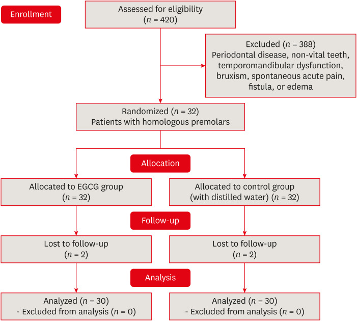
- Epigallocatechin-3-gallate prior to composite resin in abfraction lesions: a split-mouth randomized clinical trial
- Luísa Valente Gotardo Lara Alves, Lisiane Martins Fracasso, Thiago Vinicius Cortez, Aline Evangelista Souza-Gabriel, Silmara Aparecida Milori Corona
- Restor Dent Endod 2023;48(2):e13. Published online March 20, 2023
- DOI: https://doi.org/10.5395/rde.2023.48.e13
-
Abstract
PDFPubReaderePub
Objectives Natural extracts have been investigated as a biomimetic strategy to mechanically strengthen the collagen network and control the biodegradation of extracellular matrix. This study evaluated the effect of epigallocatechin-3-gallate (EGCG) on abfraction lesions prior to the composite resin.
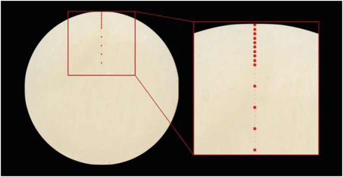
Materials and Methods The sample consisted of 30 patients (aged between 28 and 60 years) with abfraction lesions located in 2 homologous premolars. The teeth were randomly assigned according to dentin treatment: 0.02% EGCG solution or distilled water (control). After enamel acid etching, the solutions were applied immediately for 1 minute. The teeth were restored with Universal Adhesive (3M) and Filtek Z350 XT (3M). Analyzes were done by 2 independent examiners using modified USPHS (retention, secondary caries, marginal adaptation, and postoperative sensitivity) and photographic (color, marginal pigmentation, and anatomical form) criteria at baseline (7 days) and final (18 months). The data analysis used Friedman and Wilcoxon signed-rank tests (α = 0.05).
Results At baseline, all restorations were evaluated as alpha for all criteria. After 18 months, restorations were evaluated as alpha for secondary caries, color, and marginal pigmentation. There was significant difference between baseline and 18 months (
p = 0.009) for marginal adaptation and postoperative sensitivity (p = 0.029), but no significant difference were verified between treatments (p = 0.433). The EGCG group had a restoration retention rate of 93.3%, while the control group had 96.7%.Conclusions The application of EGCG solution on abfraction lesions did not significantly influence the survival of the restorations based on clinical and photographic criteria.
-
Citations
Citations to this article as recorded by- Therapeutic potential of flavonoids in erosive tooth wear management: a scoping review
Gabriel Pereira Nunes, Renata de Oliveira Alves, Geórgia Rondó Peres, Priscila Toninatto Alves de Toledo, Aline Rogéria Freire de Castilho
Clinical Oral Investigations.2025;[Epub] CrossRef
- Therapeutic potential of flavonoids in erosive tooth wear management: a scoping review
- 2,462 View
- 56 Download
- 1 Web of Science
- 1 Crossref
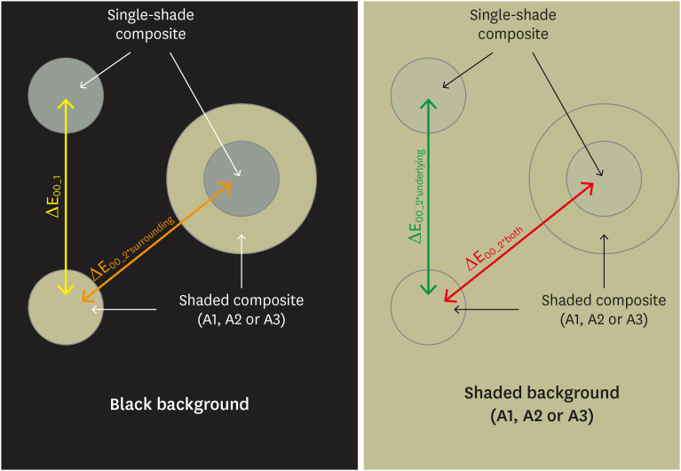
- Effects of surrounding and underlying shades on the color adjustment potential of a single-shade composite used in a thin layer
- Mariana Silva Barros, Paula Fernanda Damasceno Silva, Márcia Luciana Carregosa Santana, Rafaella Mariana Fontes Bragança, André Luis Faria-e-Silva
- Restor Dent Endod 2023;48(1):e7. Published online December 29, 2022
- DOI: https://doi.org/10.5395/rde.2023.48.e7
-
Abstract
PDFPubReaderePub
Objectives This study aimed to evaluate the surrounding and underlying shades’ effect on the color adjustment potential (CAP) of a single-shade composite used in a thin layer.
Materials and Methods Cylinder specimens (1.0 mm thick) were built with the Vittra APS Unique composite, surrounded (dual specimens) or not (simple specimens) by a control composite (shade A1, A2, or A3). Simple specimens were also built only with the control composites. Each specimen’s color was measured against white and black backgrounds or the simple control specimens with a spectrophotometer (CIELAB system). The whiteness index for dentistry (WID) and translucency parameters (TP00) were calculated for simple specimens. Differences (ΔE00) in color between the simple/dual specimens and the controls were calculated. The CAP was calculated based on the ratios between data from simple and dual specimens.
Results The Vittra APS Unique composite showed higher WID and TP00 values than the controls. The highest values of ΔE00 were observed among simple specimens. The color measurements of Vittra APS Unique (simple or dual) against the control specimens presented the lowest color differences. Only surrounding the single-shade composite with a shaded composite barely impacted the ΔE00. The highest CAP values were obtained using a shaded composite under simple or dual specimens.
Conclusions The CAP of Vittra APS Unique was strongly affected by the underlying shade, while surrounding this composite with a shaded one barely affected its color adjustment.
-
Citations
Citations to this article as recorded by- Impact of water absorption on the translucency of single-shade and conventional resin composites: an in vitro comparative study
Ceyda Sari, Elifnur Aydemir Aydın
Odontology.2026;[Epub] CrossRef - Baseline color-matching in anterior non-carious cervical lesions of patients of two single-shade resin composites: a randomized clinical trial
Ayşe Nur Doğan, Soley Arslan
Odontology.2026;[Epub] CrossRef - Evaluation of color blending effect of a single-shade resin composite with hybrid ceramics: an in vitro study
Jongchan Lee, Jinsoo Ahn, Sun-Young Kim
BMC Oral Health.2026;[Epub] CrossRef - Color Change and Compressive Strength of Novel Single-Shade Composite Resins Exposed to Staining Beverages
Juan P. Molina-Gantiva, Midian C. Castillo-Pedraza, Jorge H. Wilches-Visbal
Odovtos - International Journal of Dental Sciences.2026;[Epub] CrossRef - At‐Home and In‐Office Bleaching Protocols on the Color Match of Restorations Made With Single‐Shade Composites
Luciana Vasconcelos Ramos, Dayana Fernandes Rocha Aparicio, André Luis Faria‐e‐Silva, Maíra do Prado, Andréa Vaz Braga Pintor, Marcela Baraúna Magno
Journal of Esthetic and Restorative Dentistry.2025; 37(6): 1567. CrossRef - Evaluation of color matching of three single-shade composites employing simulated 3D printed cavities with different thicknesses using CIELAB and CIEDE2000 color difference formulae
Engin Kariper, Aylin Cilingir
REVIEWS ON ADVANCED MATERIALS SCIENCE.2025;[Epub] CrossRef - Impact of kombucha, coffee, and turmeric beverages on the color stability of a single-shade versus a multi-shade resin-based composite
Hanin E. Yeslam, Abdulaziz F. Bakhsh
PeerJ.2025; 13: e19759. CrossRef - Comparative Study of Esthetic Outcome of Pedo Shades of Composite Resin—A Randomized Controlled Trial: In Vivo and In Vitro Study
Priyanka Raj, Shikha Choubey, Divya Doneria, Diksha Bhat, Shivani Mathur, Shailja Sinha
International Journal of Clinical Pediatric Dentistry.2025; 18(S1): S22. CrossRef - Influence of cavity wall thickness on the color adjustment potential of single-shade resin composites
Fabrício Luscino Alves de Castro, Letícia Brandão Durand
The Journal of the American Dental Association.2024; 155(7): 605. CrossRef - Assessing color mismatch in single-shade composite resins for enamel replacement
Rafaella Mariana Fontes de Bragança, Diana Leyva Del Rio, Luiz Alves Oliveira-Neto, William Michael Johnston
The Journal of Prosthetic Dentistry.2024; 132(3): 613.e1. CrossRef - Color discrepancy of single-shade composites at different distances from the interface measured using cell phone images
Márcia Luciana Carregosa Santana, Gabriella de Jesus Santos Livi, André Luis Faria-e-Silva
Restorative Dentistry & Endodontics.2024;[Epub] CrossRef - Is It Possible for Single-shade Composites to Mimic the Color, Lightness, Chroma, and Hue of Other Single-shade Composites? An In Vitro Study
M Buldur, G Ayan
Operative Dentistry.2024; 49(6): 691. CrossRef - Color evaluation of a one-shade used for restoration of non-carious cervical lesions: an equivalence randomized clinical trial
Michael Willian Favoreto, Amanda de Oliveira de Miranda, Thalita P. Matos, Andrea dos Santos de Castro, Mylena de Abreu Cardoso, Julia Beatriz, Jenny Collantes-Acuña, Alessandra Reis, Alessandro Dourado Loguercio
BMC Oral Health.2024;[Epub] CrossRef - Influence of Thickness on the Translucency Parameter and Whiteness Index of Single-Shade Resin Composites
Ö Yağcı, M Fidan
Operative Dentistry.2024; 49(2): 189. CrossRef - A Comparative Study of the Sensitivity and Specificity of the Ishihara Test With Various Displays
Thomas Klinke, Wolfgang Hannak, Klaus Böning, Holger Jakstat
International Dental Journal.2024; 74(4): 892. CrossRef - Color match evaluation using instrumental method for three single-shade resin composites before and after in-office bleaching
Aylin Cilingir, Engin Kariper
REVIEWS ON ADVANCED MATERIALS SCIENCE.2023;[Epub] CrossRef - The role of interface distance and underlying substrate on the color adjustment potential of single‐shade composites
Gabriella Jesus Santos de Livi, Tauan Rosa Santana, Rafaella Mariana Fontes Bragança, Rosa Maria Viana de Bragança Garcez, André Luis Faria‐e‐Silva
Journal of Esthetic and Restorative Dentistry.2023; 35(8): 1279. CrossRef
- Impact of water absorption on the translucency of single-shade and conventional resin composites: an in vitro comparative study
- 5,923 View
- 132 Download
- 17 Web of Science
- 17 Crossref
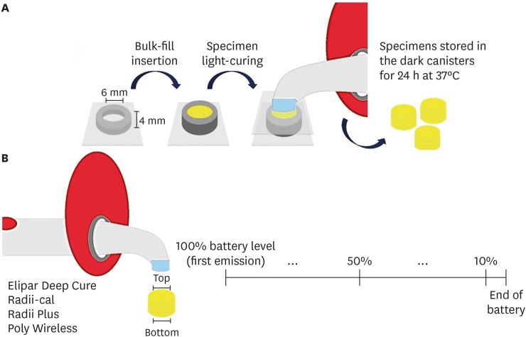
- Relationship between battery level and irradiance of light-curing units and their effects on the hardness of a bulk-fill composite resin
- Fernanda Harumi Oku Prochnow, Patricia Valéria Manozzo Kunz, Gisele Maria Correr, Marina da Rosa Kaizer, Carla Castiglia Gonzaga
- Restor Dent Endod 2022;47(4):e45. Published online November 3, 2022
- DOI: https://doi.org/10.5395/rde.2022.47.e45
-
Abstract
PDFPubReaderePub
Objectives This study evaluated the relationship between the battery charge level and irradiance of light-emitting diode (LED) light-curing units (LCUs) and how these variables influence the Vickers hardness number (VHN) of a bulk-fill resin.
Materials and Methods Four LCUs were evaluated: Radii Plus (SDI), Radii-cal (SDI), Elipar Deep Cure (Filtek Bulk Fill, 3M Oral Care), and Poly Wireless (Kavo Kerr). Irradiance was measured using a radiometer every ten 20-second activations until the battery was discharged. Disks (4 mm thick) of a bulk-fill resin (Filtek Bulk Fill, 3M Oral Care) were prepared, and the VHN was determined on the top and bottom surfaces when light-cured with the LCUs with battery levels at 100%, 50% and 10%. Data were analyzed by 2-way analysis of variance, the Tukey’s test, and Pearson correlations (α = 5%).
Results Elipar Deep Cure and Poly Wireless showed significant differences between the irradiance when the battery was fully charged versus discharged (10% battery level). Significant differences in irradiance were detected among all LCUs, within each battery condition tested. Hardness ratios below 80% were obtained for Radii-cal (10% battery level) and for Poly Wireless (50% and 10% battery levels). The battery level showed moderate and strong, but non-significant, positive correlations with the VHN and irradiance.
Conclusions Although the irradiance was different among LCUs, it decreased in half of the devices along with a reduction in battery level. In addition, the composite resin effectiveness of curing, measured by the hardness ratio, was reduced when the LCUs’ battery was discharged.
-
Citations
Citations to this article as recorded by- Effect of erosive solutions and thermal cycling on the surface properties of universal injectable and regular consistency resin composites
Ahmed Abbas Rhaif, Hoda Saleh Ismail, Tawakol Ahmed Ahmed Enab, Nadia Mohamed Zaghloul
BMC Oral Health.2025;[Epub] CrossRef - Effect of Battery Level During Successive Charging Cycles on the Performance of Certified and Low-cost Uncertified Light-curing Units Available on E-commerce
TS Peres, G Oliveira, SP da Silva Sakamoto, M da Silva Faria, HL Carlo, CJ Soares
Operative Dentistry.2024; 49(6): 673. CrossRef - Influence of Exposure Distance on Light Irradiance of Dental Curing Lamps in Various Operating Modes
Anna Lehmann, Kacper Nijakowski, Marta Mroczyk, Filip Podgórski, Beata Czarnecka, Anna Surdacka
Applied Sciences.2024; 14(21): 9999. CrossRef - ESTADO DA INTENSIDADE LUMINOSA DAS LÂMPADAS DE FOTOPOLIMERIZAÇÃO DAS CLÍNICAS ODONTOLÓGICAS DOS CENTROS DE SAÚDE DA CIDADE DE CUENCA
Milton Alexis Quinchiguano Caraguay, David Ismael Bravo Achundia , Esteban Eduardo Amoroso Calle, Manuel Estuardo Bravo Calderon
RECISATEC - REVISTA CIENTÍFICA SAÚDE E TECNOLOGIA - ISSN 2763-8405.2023; 3(6): e36296. CrossRef
- Effect of erosive solutions and thermal cycling on the surface properties of universal injectable and regular consistency resin composites
- 2,879 View
- 42 Download
- 5 Web of Science
- 4 Crossref
- Surface gloss, gloss retention, and color stability of 2 nano-filled universal resin composites
- Gustavo Fabián Molina, Ricardo Juan Cabral, Ignacio Mazzola, Michael Burrow
- Restor Dent Endod 2022;47(4):e43. Published online October 31, 2022
- DOI: https://doi.org/10.5395/rde.2022.47.e43
-
Abstract
PDFPubReaderePub
Objectives This study compared the surface gloss (SG), gloss retention (GR), and color stability (CS) of 2 universal resin composites after chemical (CA) and mechanical (MA) aging.
Materials and Methods Twenty disc-shaped samples of G-ænial A´Chord (GC-Europe) and Filtek Universal (3M-ESPE) were polished with sequential abrasive papers. For CA, specimens were stored in 1 mL of 75% ethanol for 15 days at 37°C, and readings (SG, GR, and CS) were obtained at baseline and 5, 10, and 15 days. For MA, specimens were subjected to 10,750 simulated brushing cycles. SG and CS were evaluated after every 3,583 cycles. SG was measured with a glossmeter (geometrical configuration: 60°), and values were expressed in gloss units. Color was measured with a spectrophotometer using the CIE-L*a*b* color system. The Student’s
t -test, 1-way analysis of variance, and Scheffé test were used for statistical analysis (α = 0.05).Results G-ænial presented significantly higher SG values than Filtek (
p = 0.02), with GR reductions of 5.2% (CA) and 5.3% (MA) for G-ænial and 7.6% (CA) and 7.2% (MA) for Filtek. The aging protocol had no statistically significant effect on SG or GR (p = 0.25) from baseline to the final readings. G-ænial–MA presented the lowest color difference(∆E = 1.8), and G-ænial–CA and Filtek–CA had the largest changes (∆E = 8.6 and∆E = 11.8, respectively).Conclusion G-ænial presented higher SG values and better CS. Both restorative materials demonstrated acceptable GR and CS. Aging protocols impacted these properties negatively.
-
Citations
Citations to this article as recorded by- Color stability, surface roughness, and surface morphology of universal composites
Mohammad Meniawi, Nazlı Şirinsükan, Esra Can
Odontology.2026; 114(1): 149. CrossRef - RETRACTED: Surface gloss and micro‐CT analysis of additively and subtractively manufactured resin composites and zirconia after simulated tooth brushing with different bristle types and toothpaste formulations: An in vitro study
Ahmet Faruk Ertürk, Rafat Sasany, Seyed Ali Mosaddad, Merve Yelken Kendirci
Journal of Prosthodontics.2026; 35(5): 1. CrossRef - Surface roughness of composite resins subjected to brushing with whitening toothpastes: an in vitro study
Nicolle Madruga Ramos FERREIRA, Vinicius Funghetto LIPPERT, Amanda Baptista da Silva HECK, Ana Maria SPOHR, Marcel Ferreira KUNRATH, Carlos Alberto FELDENS, Paulo Floriani KRAMER
Brazilian Oral Research.2025;[Epub] CrossRef - Evaluation of Color Stability and Surface Abrasion of Nano-modified Glass Ionomer Cement with Dentifrices: An In Vitro Study
Jessy Paulraj, Subhabrata Maiti, Harini Palani
International Journal of Prosthodontics and Restorative Dentistry.2025; 15(1): 10. CrossRef - Security inks with silanized zinc oxide quantum dots and cellulose ethers for the safeguarding of cultural heritage objects
Andrea Louise Matulac, Themis Krasoudaki, Francesca Battaglia, Carlo Spadoni, Martina Piletti, Daniela Iacopino, Rodorico Giorgi
Applied Materials Today.2025; 44: 102718. CrossRef - Gastric acid challenge: Mechanical proficiency and surface gloss of tooth-colored restorative materials
Ozge Gizem Yenidunya, Tugba Misilli, Ebru Yilmaz
BMC Oral Health.2025;[Epub] CrossRef - Impact of in-office bleaching agents on the optical properties of universal resin composites: an in vitro analysis
Esra Özyurt, Merve Nezir, Hanife Altınışık, Mediha Büyükgöze Dindar
BMC Oral Health.2025;[Epub] CrossRef - Aging and Staining Effects on Optical Properties of Flowable Composites
M. M. Sly, Y. Korkmaz‐Ceyhan, F. Dini, R. L. Ocampo Escobedo, E. Abram, R. D. Paravina
Journal of Biomedical Materials Research Part B: Applied Biomaterials.2025;[Epub] CrossRef - Evaluation of The Effect of In-Office Bleaching Agent on Mechanical Properties of Different Single-Shade Resin Composites: An In-Vitro Study
Merve Nezir, Hanife Altınışık, Esra Özyurt
ADO Klinik Bilimler Dergisi.2025; 14(3): 197. CrossRef - Surface gloss changes in 3D-printed resin materials following different polishing procedures and aging protocols
Ilayda Yumak, Hayal Boyacioglu, Lezize Sebnem Turkun
BMC Oral Health.2025;[Epub] CrossRef - The Gloss Retention of Esthetic Restorations Following Simulated Brushing with Charcoal Oral Products: An In-Vitro Study
Fadia Awadalkreem, Nancy S Farghal, Nadin A Abouelhonoud, Raiyan I Khan
The Journal of Contemporary Dental Practice.2024; 25(5): 473. CrossRef - Effect of different finishing and polishing systems on surface properties of universal single shade resin-based composites
Ghada Alharbi, Hend NA Al Nahedh, Loulwa M. Al-Saud, Nourah Shono, Ahmed Maawadh
BMC Oral Health.2024;[Epub] CrossRef - The Effect of Chemical Degradation and Polishing on the Gloss of Composite Dental Materials
Ružica Zovko, Stipo Cvitanović, Mirela Mabić, Zdenko Šarac, Anka Ćorić, Domagoj Glavina, Kristina Goršeta
Materials.2023; 16(10): 3727. CrossRef
- Color stability, surface roughness, and surface morphology of universal composites
- 4,303 View
- 65 Download
- 10 Web of Science
- 13 Crossref
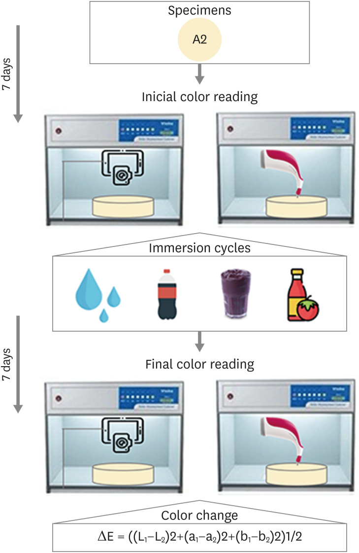
- Comparison of instrumental methods for color change assessment of Giomer resins
- Luiza de Almeida Queiroz Ferreira, Rogéli Tibúrcio Ribeiro da Cunha Peixoto, Cláudia Silami de Magalhães, Tassiana Melo Sá, Monica Yamauti, Francisca Daniele Moreira Jardilino
- Restor Dent Endod 2022;47(1):e8. Published online February 3, 2022
- DOI: https://doi.org/10.5395/rde.2022.47.e8
-
Abstract
PDFPubReaderePub
Objectives The aim of this study was to compare the color change of the Giomer resin composite (Beautifil-Bulk) by using photographs obtained with a smartphone (iPhone 6S) associated with Adobe Photoshop software (digital method), with the spectrophotometric method (Vita Easyshade) after immersion in different pigment solutions.
Materials and Methods Twenty resin composite samples with a diameter of 15.0 mm and thickness of 1.0 mm were confectioned in A2 color (
n = 5). Photographs and initial color readings were performed with a smartphone and spectrophotometer, respectively. Then, samples were randomly divided and subjected to cycles of immersion in distilled water (control), açai, Coke, and tomato sauce, 3 times a day, 20 minutes for 7 days. Later, new photographs and color readings were taken.Results The analysis (2-way analysis of variance, Holm-Sidak,
p < 0.05) demonstrated no statistical difference (p < 0.005) between the methods in all groups. Similar color changes were observed for all pigment solutions when using the spectrophotometric method. For the digital method, all color changes were clinically unacceptable, with distilled water and tomato sauce similar to each other and with statistical differences (p < 0.005) for Coke and açai.Conclusions Only the tomato sauce produced a color change above the acceptability threshold using both methods of color assessment. The spectrophotometric and digital methods produce different patterns of color change. According to our results, the spectrophotometric method is more recommended in color change assessment.
-
Citations
Citations to this article as recorded by- Are Sculptable Bulk‐Fill Composites Susceptible to Color Change: A Systematic Review
Jamieson Wong, Constance Yeo, Michelle The, Filip Taneski, Uros Josic, Lorenzo Breschi, Vesna Miletic
Journal of Esthetic and Restorative Dentistry.2026; 38(1): 70. CrossRef - The effects of mechanical and chemical degradation on the surface roughness, gloss, and color stability of bulk-fill resin composites
Merve Nezir, Hanife Altınışık, Esra Özyurt, Naz Bayar, Mediha Büyükgöze Dindar
BMC Oral Health.2025;[Epub] CrossRef - Color Image Expression through CIE L*a*b* System in Foods
Hyun-Woong Choi, Seong-Eun Park, Hong-Seok Son
Journal of the Korean Society of Food Science and Nutrition.2023; 52(2): 223. CrossRef
- Are Sculptable Bulk‐Fill Composites Susceptible to Color Change: A Systematic Review
- 3,741 View
- 49 Download
- 3 Web of Science
- 3 Crossref
- A 3-year retrospective study of clinical durability of bulk-filled resin composite restorations
- Muhittin Ugurlu, Fatmanur Sari
- Restor Dent Endod 2022;47(1):e5. Published online December 30, 2021
- DOI: https://doi.org/10.5395/rde.2022.47.e5
-
Abstract
PDFPubReaderePub
Objectives This study aimed to assess the clinical longevity of a bulk-fill resin composite in Class II restorations for 3-year.
Materials and Methods Patient record files acquired from the 40 patients who were treated due to needed 2 similar sizes Class II composite restorations were used for this retrospective study. In the experimental cavity, the flowable resin composite SDR was inserted in the dentinal part as a 4 mm intermediate layer. A 2 mm coverage layer with a nano-hybrid resin composite (CeramX) was placed on SDR. The control restoration was performed by an incremental technique of 2 mm using the nano-hybrid resin composite. The restorations were blindly assessed by 2 calibrated examiners using modified United States Public Health Service criteria at baseline and 1, 2, and 3 years. The data were analyzed using non-parametric tests (
p = 0.05).Results Eighty Class II restorations were evaluated. After 3-years, 4 restorations (5%) failed, 1 SDR + CeramX, and 3 CeramX restorations. The annual failure rate (AFR) of the restorations was 1.7%. The SDR + CeramX group revealed an AFR of 0.8%, and the CeramX group an AFR of 2.5% (
p > 0.05). Regarding anatomical form and marginal adaptation, significant alterations were observed in the CeramX group after 3-years (p < 0.05). The changes in the color match were observed in each group over time (p < 0.05).Conclusions The use of SDR demonstrated good clinical durability in deep Class II resin composite restorations.
-
Citations
Citations to this article as recorded by- Bond Strength of Bulk-Fill Resin Repairs: Impact of Surface and Adhesive Protocols
Samuel Eleutério Paiva Sousa, Fiorella Elizabeth Arévalo Tarrillo, Maria Paula Novaes Camargo Manna, Sandra Ribeiro de Barros da Cunha, Maria Ângela Pita Sobral
Odovtos - International Journal of Dental Sciences.2026; 28(2): 204. CrossRef - Evaluation of Surface Roughness and Microhardness of New Generation Bulk-Fill Composites
Zehra SÜSGÜN YILDIRIM, Ezgi SONKAYA, Zeliha Gonca BEK KÜRKLÜ
Cumhuriyet Dental Journal.2023; 26(2): 180. CrossRef - Damping Behaviour and Mechanical Properties of Restorative Materials for Primary Teeth
Thomas Niem, Roland Frankenberger, Stefanie Amend, Bernd Wöstmann, Norbert Krämer
Materials.2022; 15(21): 7698. CrossRef
- Bond Strength of Bulk-Fill Resin Repairs: Impact of Surface and Adhesive Protocols
- 5,034 View
- 45 Download
- 2 Web of Science
- 3 Crossref
- Errors in light-emitting diodes positioning when curing bulk fill and incremental composites: impact on properties after aging
- Abdulrahman A. Balhaddad, Isadora M. Garcia, Haifa Maktabi, Maria Salem Ibrahim, Qoot Alkhubaizi, Howard Strassler, Fabrício M. Collares, Mary Anne S. Melo
- Restor Dent Endod 2021;46(4):e51. Published online September 24, 2021
- DOI: https://doi.org/10.5395/rde.2021.46.e51

-
Abstract
PDFPubReaderePub
Objectives This study aimed to evaluate the effect of improper positioning single-peak and multi-peak lights on color change, microhardness of bottom and top, and surface topography of bulk fill and incremental composites after artificial aging for 1 year.
Materials and Methods Bulk fill and incremental composites were cured using multi-peak and single-peak light-emitting diode (LED) following 4 clinical conditions: (1) optimal condition (no angulation or tip displacement), (2) tip-displacement (2 mm), (3) slight tip angulation (α = 20°) and (4) moderate tip angulation (α = 35°). After 1-year of water aging, the specimens were analyzed for color changes (ΔE), Vickers hardness, surface topography (Ra, Rt, and Rv), and scanning electron microscopy.
Results For samples cured by single-peak LED, the improper positioning significantly increases the color change compared to the optimal position regardless of the type of composite (
p < 0.001). For multi-peak LED, the type of resin composite and the curing condition displayed a significant effect on ΔE (p < 0.001). For both LEDs, the Vickers hardness and bottom/top ratio of Vickers hardness were affected by the type of composite and the curing condition (p < 0.01).Conclusions The bulk fill composite presented greater resistance to wear, higher color stability, and better microhardness than the incremental composite when subjected to improper curing. The multi-peak LED improves curing under improper conditions compared to single-peak LED. Prevention of errors when curing composites requires the attention of all personnel involved in the patient's care once the clinical relevance of the appropriate polymerization reflects on reliable long-term outcomes.
-
Citations
Citations to this article as recorded by- A clinical survey of the output intensity of 50 light-curing units in dental clinics across Davangere and Mangalore region using a spectrometer system
Elizbeth Christy Jose, Sakshi Jha, Prema Shantagouda Biradar, J Arun, TN Nandini, Thushara Mohanan
International Journal of Oral Health Sciences.2025; 15(1): 41. CrossRef - The demineralization resistance and mechanical assessments of different bioactive restorative materials for primary and permanent teeth: an in vitro study
Maria Salem Ibrahim, Fahad Rakad Aldhafeeri, Abdullah Sami Banaemah, Mana S. Alhaider, Yousif A. Al-Dulaijan, Abdulrahman A. Balhaddad
BDJ Open.2024;[Epub] CrossRef - Inorganic Compounds as Remineralizing Fillers in Dental Restorative Materials: Narrative Review
Leena Ibraheem Bin-Jardan, Dalal Ibrahim Almadani, Leen Saleh Almutairi, Hadi A. Almoabid, Mohammed A. Alessa, Khalid S. Almulhim, Rasha N. AlSheikh, Yousif A. Al-Dulaijan, Maria S. Ibrahim, Afnan O. Al-Zain, Abdulrahman A. Balhaddad
International Journal of Molecular Sciences.2023; 24(9): 8295. CrossRef
- A clinical survey of the output intensity of 50 light-curing units in dental clinics across Davangere and Mangalore region using a spectrometer system
- 2,546 View
- 22 Download
- 2 Web of Science
- 3 Crossref
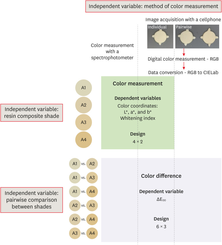
- Color assessment of resin composite by using cellphone images compared with a spectrophotometer
- Rafaella Mariana Fontes de Bragança, Rafael Ratto Moraes, André Luis Faria-e-Silva
- Restor Dent Endod 2021;46(2):e23. Published online April 5, 2021
- DOI: https://doi.org/10.5395/rde.2021.46.e23
-
Abstract
PDFPubReaderePub
Objectives This study assessed the reliability of digital color measurements using images of resin composite specimens captured with a cellphone.
Materials and Methods The reference color of cylindrical specimens built-up with the use of resin composite (shades A1, A2, A3, and A4) was measured with a portable spectrophotometer (CIELab). Images of the specimens were obtained individually or pairwise (compared shades in the same photograph) under standardized parameters. The color of the specimens was measured in the images using RGB system and converted to CIELab system using image processing software. Whiteness index (WID) and color differences (ΔE00) were calculated for each color measurement method. For the cellphone, the ΔE00 was calculated between the pairs of shades in separate images and in the same image. Data were analyzed using 2-way repeated-measures analysis of variance (α = 0.05). Linear regression models were used to predict the reference ΔE00 values of those calculated using color measured in the images.
Results Images captured with the cellphone resulted in different WID values from the spectrophotometer only for shades A3 and A4. No difference to the reference ΔE00 was observed when individual images were used. In general, a similar ranking of ΔE00 among resin composite shades was observed for all methods. Stronger correlation coefficients with the reference ΔE00 were observed using individual than pairwise images.
Conclusions This study showed that the use of cellphone images to measure the color difference seems to be a feasible alternative providing outcomes similar to those obtained with the spectrophotometer.
-
Citations
Citations to this article as recorded by- Reliability of Three Image‐Based Instrumental Color‐Measurement Methods
Eloah Nunes de Almeida, Ido Luiz de Azevedo Feiten, Luis Felipe J. Schneider, Larissa Maria Assad Cavalcante, André Luis Faria‐e‐Silva
Journal of Esthetic and Restorative Dentistry.2026; 38(8): 1598. CrossRef - Evaluation of color stability in single-shade composite resins using spectrophotometer and cross-polarized mobile photography
Hatice Tepe, Ozge Celiksoz, Batu Can Yaman
BMC Oral Health.2025;[Epub] CrossRef - Color discrepancy of single-shade composites at different distances from the interface measured using cell phone images
Márcia Luciana Carregosa Santana, Gabriella de Jesus Santos Livi, André Luis Faria-e-Silva
Restorative Dentistry & Endodontics.2024;[Epub] CrossRef - How the Translucency and Color Stability of Single-Shade Universal Resin Composites Are Affected by Coffee?
Büşra Özdemir, Betül Kübra Kurucu Karadeniz, Seyit Bilal Özdemir, Ömer Akbulut
Current Research in Dental Sciences.2024; 34(4): 270. CrossRef - Color Image Expression through CIE L*a*b* System in Foods
Hyun-Woong Choi, Seong-Eun Park, Hong-Seok Son
Journal of the Korean Society of Food Science and Nutrition.2023; 52(2): 223. CrossRef - Comparative Evaluation of VITA Shade Guide and Various Composite Shades Using Spectrophotometer, Digital Single-lens Reflex, and Cellphone: An In Vitro Study
Aman Verma, Sonali Taneja, Surabhi Ghosh
World Journal of Dentistry.2023; 14(9): 803. CrossRef - Comparison of instrumental methods for color change assessment of Giomer resins
Luiza de Almeida Queiroz Ferreira, Rogéli Tibúrcio Ribeiro da Cunha Peixoto, Cláudia Silami de Magalhães, Tassiana Melo Sá, Monica Yamauti, Francisca Daniele Moreira Jardilino
Restorative Dentistry & Endodontics.2022;[Epub] CrossRef
- Reliability of Three Image‐Based Instrumental Color‐Measurement Methods
- 3,530 View
- 39 Download
- 3 Web of Science
- 7 Crossref
Review Article
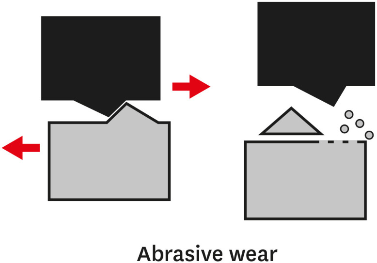
- Wear of contemporary dental composite resin restorations: a literature review
- Dimitrios Dionysopoulos, Olga Gerasimidou
- Restor Dent Endod 2021;46(2):e18. Published online February 25, 2021
- DOI: https://doi.org/10.5395/rde.2021.46.e18
-
Abstract
PDFPubReaderePub
Composite resins are the most commonly used dental restorative materials after minimally invasive dental procedures, and they offer an aesthetically pleasing appearance. An ideal composite restorative material should have wear properties similar to those of tooth tissues. Wear refers to the damaging, gradual loss or deformation of a material at solid surfaces. Depending on the mechanism of action, wear can be categorized as abrasive, adhesive, fatigue, or corrosive. Currently used composite resins cover a wide range of materials with diverse properties, offering dental clinicians multiple choices for anterior and posterior teeth. In order to improve the mechanical properties and the resistance to wear of composite materials, many types of monomers, silane coupling agents, and reinforcing fillers have been developed. Since resistance to wear is an important factor in determining the clinical success of composite resins, the purpose of this literature review was to define what constitutes wear. The discussion focuses on factors that contribute to the extent of wear as well as to the prevention of wear. Finally, the behavior of various types of existing composite materials such as nanohybrid, flowable, and computer-assisted design/computer-assisted manufacturing materials, was investigated, along with the factors that may cause or contribute to their wear.
-
Citations
Citations to this article as recorded by- Comparison of wear behavior of occlusal device materials manufactured by different processes
Catherine Arreaza, Robert R. Seghi, Scott R. Schricker, William M. Johnston, Paola C. Saponaro
The Journal of Prosthetic Dentistry.2026; 135(3): 614.e1. CrossRef - Synergistic Effects of Simulated Energy Drink Exposure and Fatigue Loading on Bioactive and Conventional Resin Composites
Fatin A. Hasanain, Alaa Turkistani
Journal of Functional Biomaterials.2026; 17(1): 29. CrossRef - Direct and Semi-Direct Composite Techniques in Posterior Teeth: A Two-Year Follow-Up Comparative Study
Adriana Saceleanu, Anca Maria Fratila, Vasile Calin Arcas, Cristina Ana-Maria Arcas, Dragos Anton Dadarlat, Laura Stef
Journal of Clinical Medicine.2026; 15(2): 687. CrossRef - Micro-scratch and wear resistance of restorative dental materials: an in vitro study
Valeriya Aleksandrova, Neshka Manchorova, Veselina Todorova, Lyubomir Vangelov, Svetlin Alexandrov
Folia Medica.2026;[Epub] CrossRef - Nanotechnology in contemporary dentistry: a comprehensive review of current clinical applications, bioactive materials, and future perspectives
Murtada A. Ahmed, Samar M. Albuhiri, Manal M. Alanazi, Ameerah A. Albogami, Jawaher N. Alharbi, Eman A. Alruwaili, Maryam A. Shabeen
International Journal Of Community Medicine And Public Health.2026; 13(2): 1044. CrossRef - Advances in Ecofriendly and High-Strength Dental Composites: Structural and Functional Perspectives
Sayem A. Mulla, Amit Patil, Himmat Jaiswal, Bhavani Sangala Nagendra, Ashima Jakhar, Waseem Z. Khan
European Journal of General Dentistry.2026;[Epub] CrossRef - Letter to the Editor regarding, “A digital workflow for tooth-supported complete overdentures with a composite resin injection technique to manage the treatment of a child with ectodermal dysplasia” by Liu et al
Meng Liu, Guoyan Quan
The Journal of Prosthetic Dentistry.2026; 136(1): e475. CrossRef - Two- and three-body wear of BPA-free resin composites
Magdalena A. Osiewicz, Ivana Nedeljkovic, Arie Werner, Albert J. Feilzer, Cornelis J. Kleverlaan
Dental Materials.2026; 42(7): 1172. CrossRef - Evaluation of physical, chemical, and color‐matching properties of monochromatic resin composites: An in vitro study
Ericka dos Santos Lopes, Mayara Evelly Duarte de Lima, Emanuel Ewerton Mendonça Vasconcelos, Natália Gomes de Oliveira, Gabriela Queiroz de Melo Monteiro, Luís Felipe Espíndola‐Castro
European Journal of Oral Sciences.2026;[Epub] CrossRef - Wear resistance, microhardness and compressive strength of high filled flowable composite resins
Seyit Bilal Ozdemir, Busra Ozdemir
Scientific Reports.2026;[Epub] CrossRef - Tribological Performance of CAM-Processed Interim Dental Restoration Materials: Effects of 3D Printing, Milling, and Post-Processing on Wear and Surface Topography
Liliana Porojan, Roxana Diana Vasiliu, Flavia Roxana Bejan, Mihaela Ionela Gherban, Diana Uțu, Anamaria Matichescu
Journal of Functional Biomaterials.2026; 17(3): 136. CrossRef - Effect of acidic exposure on the optical and mechanical properties of esthetic pediatric restorative materials: an in vitro study
Murat Büyüksefil, Sera Şimşek Derelioğlu
Odontology.2026;[Epub] CrossRef - Clinical performance of low-shrinkage giomer compared to nanohybrid resin composite in proximal restorations after one year: a randomized clinical trial
Marwa I. AbdelHafez, Omar Shaalan, Heba Hamza
BDJ Open.2026;[Epub] CrossRef - Surface texture and wear characteristics of micro-hybrid and nano-hybrid resin composites under simulated frictional conditions
Magdalena A. Osiewicz, Cornelis J. Kleverlaan, Arie Werner, Michael Marxer, Dominique Filipec, Magdalena Niemczewska-Wójcik
Scientific Reports.2026;[Epub] CrossRef - Long-term clinical performance of bilaminar veneers combined with the Dahl concept for the management of localized anterior tooth wear: An ambispective cohort study
Edina Lempel, Kinga Dorottya Németh, Erika Katalin Dunavári, Sára Jeges, József Szalma
Dental Materials.2026; 42(9): 1692. CrossRef - Bioactive Fillers in Bulk-Fill Composite Resins: A Comprehensive Review of the Effects on Polymerization Shrinkage Behavior and Mechanical Performance
Vlad Constantin, Ionut Luchian, Ionut Taraboanta, Teona Anamaria Tudorici, Nicoleta Tofan, Florinel Cosmin Bida, Florin Razvan Curca, Dana Gabriela Budala, Dragos Ioan Virvescu, Andrei Georgescu
Materials.2026; 19(11): 2181. CrossRef - Short-term clinical and micromorphological comparison of injectable and paste composites in occlusal cavities
Aslı Ceren Kubilay, Cansu Atalay, Esra Ergin
BMC Oral Health.2026;[Epub] CrossRef - Flexural strength of new and repaired TEGDMA- and HEMA-free resin composites compared to a conventional composite
A.M.O. Dal Piva, M. Khalil, R.O. Pilecco, C.J. Kleverlaan, J.P.M. Tribst
Next Materials.2026; 12: 102355. CrossRef - Long-Term Clinical Performance of Posterior Composite Restorations After Nearly Three Decades: A Clinical Follow-Up Study
Karanvir Singh, Nils Berneburg, Andreas May, Neelam Lingwal, Georgios E. Romanos, Susanne Gerhardt-Szép
Dentistry Journal.2026; 14(6): 356. CrossRef - Longevity of Direct Composite Resin Restorations in Posterior Teeth: A Narrative Review of Survival Rates, Failure Modes, Patient and Operator Related Risk Factors for Direct Resin Restorations
Cathy Mariana Huerta Mauricio, Andrea Yolanda Munsibay Foronda, Antonella Restani, Any Luceli Muñoz Vargas
Journal of Dental Health and Oral Research.2026; 7(2): 1. CrossRef - Effect of three-body wear on the surface properties of different polymer-based CAD/CAM dental restorative and prosthetic materials
Abdallah Fawzy Elsadany, Hesham Samy Borg, Enas A. Elshenawy
BMC Oral Health.2026;[Epub] CrossRef - How surface electronegativity and calcium release in enamel mediate the adsorption and lubrication of salivary proteins: The role of interfacial water
Yue Tang, Lei Lei, Hujun Wang, Haonan Qiu, Jing Zheng, Zhongrong Zhou
Friction.2025; 13(3): 9440912. CrossRef - Effect of SiC particle size and content on the mechanical and tribological properties of porous Si3N4-SiC composites fabricated following a facile low-temperature processing route
Siddharth, Sakshi Tiwari, Pritam Biswas, Nilrudra Mandal, Siddhartha Roy
Ceramics International.2025; 51(14): 19508. CrossRef - Evaluation of Color Stability and Surface Abrasion of Nano-modified Glass Ionomer Cement with Dentifrices: An In Vitro Study
Jessy Paulraj, Subhabrata Maiti, Harini Palani
International Journal of Prosthodontics and Restorative Dentistry.2025; 15(1): 10. CrossRef - Wear resistance of orthodontic attachments: a comparative analysis of different composite resins in clear aligner therapy
Irmak Ocak, Hande Gorucu-Coskuner, Muge Aksu
Clinical Oral Investigations.2025;[Epub] CrossRef - A Review: Resin‐Based Dental Materials and Their Characterization
Arda Bingül, Merve Nezir, Aykan Onur Atilla, Suat Özcan, Zafer Evis
Polymers for Advanced Technologies.2025;[Epub] CrossRef - Biomechanical and Occlusal Factors Influencing the Longevity of Single-Unit Restorations: A Comprehensive Review
Wedad S Alaida, Safa A Gadi, Rokia E Al-Ghannam, Moayad F Alamri, Feras I Mirdad, Ruba M Argaibeh, Bushra A Alqahtani, Abdulrahman M Alqahtani, Abdulelah A Al Jaban, Turki M Alkuraydimi, Abdulrahman S Alamari
Cureus.2025;[Epub] CrossRef - Effect of Bleaching on Surface Roughness of Universal Composite Resins After Chlorhexidine-Induced Staining
Gözde Aksoy Vaizoğlu
Dentistry Journal.2025; 13(7): 277. CrossRef - Wear properties of hybrid antibacterial dental composite with micro-particles of S. persica and hydroxyapatite as fillers
Rihem Chaaben, Ayman Ayedi, Khaled Elleuch
Euro-Mediterranean Journal for Environmental Integration.2025; 10(4): 3055. CrossRef - Comparative Evaluation of Direct and Indirect Composite Restorations in Class II Tooth Preparations - An In vivo Study
Akshun Gupta, Garima Arora, Aprajita Mehta, Satish Sane, Siddhi Nevrekar, Apurva Nagrale
Advances in Human Biology.2025; 15(4): 550. CrossRef - Effect of Thermal Ageing on Flexural Strength and Microhardness of Novel High-Performance Polymer (Nanoksa G-Plus) in Comparison to a Widely Used Bio-HPP/PEEK
Ramy Abdallah Abdelrahim, Ahmed Ali Ezzeldine, Mahmoud Abdellah, SaadEldein Sadeq Elghazawi
Dentistry Journal.2025; 13(8): 370. CrossRef - A comparative 48 month randomized trial of clinical performance and wear of BISGMA based and BISGMA free nanoceramic resin composites
Samah Mohamed Bahig, Heba Helal El Sherbiney, Mohamed Moustafa Zayed, Shereen Hafez Ibrahim
Scientific Reports.2025;[Epub] CrossRef - Bruxism Simulation in Aligner Therapy: Effects on Restored Posterior Teeth
Amelia Anita Boitor (Andreica), Adriana Objelean, Cristina Gasparik, Alexandru Victor Burde, Horațiu Alexandru Colosi, Diana Dudea
Journal of Clinical Medicine.2025; 14(21): 7877. CrossRef - Thermal Effects of Rapid High‐Intensity Light Curing on Bulk‐Fill Resin‐Based Composites: A Systematic Review and Meta‐Analysis
Samille Biasi Miranda, Marina Rodrigues Santi, Giovana Lordsleem de Mendonça, Luiz Antonio Soares Falson, Matheus José Gusmão Simões Barza, Veronica Maria de Sá Rodrigues, Ana Karina Maciel de Andrade, Rodrigo Barros Esteves Lins, Marcos Antonio Japiassú
The Scientific World Journal.2025;[Epub] CrossRef - Effect of Various Toothpaste Tablets on Gloss and Surface Roughness of Resin-based Composite Materials
J Ko, A Tsao, R Kim, C Perry, U Oyoyo, SR Kwon
Operative Dentistry.2024; 49(3): 282. CrossRef - Surface wear of attachments in patients during clear aligner therapy: a prospective clinical study
Qiuying Li, Kai Yang
Progress in Orthodontics.2024;[Epub] CrossRef - Awareness of possible complications associated with direct composite restorations: A multinational survey among dentists from 13 countries with meta-analysis
Anna Lehmann, Kacper Nijakowski, Jakub Jankowski, David Donnermeyer, Paulo J. Palma, Milan Drobac, João Filipe Brochado Martins, Fatma Pertek Hatipoğlu, Indira Tulegenova, Muhammad Qasim Javed, Hamad Mohammad Alharkan, Olga Bekjanova, Sylvia Wyzga, Moataz
Journal of Dentistry.2024; 145: 105009. CrossRef - Evaluation of pre-heated composite resins with soft-start polymerization and conventional composite restorations in class-I carious lesions – A randomized clinical trial
Niral Kotecha, Nimisha C. Shah, Namita N. Gandhi, Priya Porwal, Ajinkya M. Pawar, Novaldy Wahjudianto, Dian Agustin Wahjuningrum, Suraj Arora, Mohmed Isaqali Karobari
Heliyon.2024; 10(10): e30794. CrossRef - Reabilitação estética de dente conóide: relato de caso
Anna Danielle Oliveira dos Santos, Diana Fernandes de Melo , Jorge Alberto Carrazana Moya , Kathleen Rebelo de Sousa , Lizete Karla Filgueiras de Souza, Marcela Lopes Linhares, Márcio Langbeck Castelo Branco , Márcio Lopes Linhares
Revista Clínica de Odontologia.2024; 5(1): 80. CrossRef - Non-collagenous protein analog-induced biomimetic mineralization strategy to restore the dentin interface
Ruhua Chen, Yimeng Xie, Liang Ma, Bing Li, Wei Yao
Biomedical Physics & Engineering Express.2024; 10(6): 062004. CrossRef - Influence of Low pH on the Microhardness and Roughness Surface of Dental Composite—A Preliminary Study
Leszek Szalewski, Dorota Wójcik, Monika Sowa, Vladyslav Vivcharenko, Krzysztof Pałka
Materials.2024; 17(14): 3443. CrossRef - Fabrication of a novel aesthetic orthodontic bracket and evaluation of friction properties between PEEK and stainless steel wires
Jiaqi Wu, Xiujing Wang, Jiuhui Jiang, Yunyang Bai
Technology and Health Care.2024; 32(1): 269. CrossRef - Can wheel polishers improve surface properties and color stability of monochromatic resin composites?
Lezize Sebnem Turkun, Cankut Canevi, Alperen Degirmenci, Hayal Boyacioglu
BMC Oral Health.2024;[Epub] CrossRef - Comparison of volumetric loss and surface roughness of composite dental restorations obtained by additive and subtractive manufacturing methods
Neslihan Güntekin, Ali Rıza Tunçdemir
Heliyon.2024; 10(4): e26269. CrossRef - Comparative evaluation of microhardness of three restorative materials after immersion in chlorhexidine mouthwash: An in vitro study
Shilpa S. Shah, Nishtha K. Patel, Kruti P. Yagnik, Aarshati Vyas, Prerak Doshi, Pooja R. Keshrani
Journal of Conservative Dentistry and Endodontics.2024; 27(5): 520. CrossRef - NON-INTERVENTION VERSUS REPAIR/REPLACEMENT DECISIONS IN POSTERIOR COMPOSITE RESTORATIONS AGED 3-5 YEARS: A RETROSPECTIVE STUDY
Galina Pancu, Andrei Georgescu , Antonia Moldovanu , Angela Ghiorghe , Simona Stoleriu , Irina Nica , Ionut Tărăboanţă , Alexandru Iovan , Sorin Andrian
Romanian Journal of Oral Rehabilitation.2024; 16(2): 186. CrossRef - Effect of tooth brushing simulation on the surface properties of various resin‐matrix computer‐aided design/computer‐aided manufacturing ceramics
Evangelos Ximinis, Dimitrios Dionysopoulos, Constantinos Papadopoulos, Alexandros Tournavitis, Avraam Konstantinidis, Olga Naka
Journal of Esthetic and Restorative Dentistry.2023; 35(6): 937. CrossRef - Effect of toothpaste with different components on toothbrushing wear resistance of micro-hybrid/nano-filled resin composites
Seon-Mi Byeon, Jung-Eun Park, Kyeong-Seon Kim, Tae-Hwan Kim, Chung-Cha Oh, Seung-O Ko3, Min-Ho Lee
Korean Journal of Dental Materials.2023; 50(4): 247. CrossRef - Release Kinetics of Monomers from Dental Composites Containing Fluoride-Doped Calcium Phosphates
Adrián M. Alambiaga-Caravaca, Alicia López-Castellano, Yu Fu Chou, Arlinda Luzi, Juan Manuel Núñez, Avijit Banerjee, María del Mar Jovani Sancho, Salvatore Sauro
Pharmaceutics.2023; 15(7): 1948. CrossRef - Comparative study on the impact-sliding wear behaviour of CAD/CAM resin-ceramic materials and tooth enamel
Chunxiao Jin, Jiuhong Deng, Peiyue Pan, Yuhuan Xiong, Liqing Zhu, Shanshan Gao
Dental Materials.2023; 39(1): 25. CrossRef - The impact of dental varnishes on the immediate surface microhardness and roughness of restorative dental materials: An in vitro study
Jovana Lovric, Milisav Markovic, Marko Bulajic, Sasa Zeljkovic, Jana Ilic, Olivera Dolic
Vojnosanitetski pregled.2023; 80(12): 1022. CrossRef - An In Vitro Study regarding the Wear of Composite Materials Following the Use of Dental Bleaching Protocols
Alexandru Dan Popescu, Mihaela Jana Ţuculină, Lelia Mihaela Gheorghiță, Andrei Osman, Claudiu Nicolicescu, Smaranda Adelina Bugălă, Mihaela Ionescu, Jaqueline Abdul-Razzak, Oana Andreea Diaconu, Bogdan Dimitriu
Journal of Functional Biomaterials.2023; 14(10): 532. CrossRef - Investigation of aging resistance for dental resin composites with and without glass flakes
Dan Feng, Shujun Dong, Zuosen Shi, Zhanchen Cui, Song Zhu
Clinical Oral Investigations.2023; 27(11): 6903. CrossRef - Tribological behavior and wear mechanisms of dental resin composites with different polymeric matrices
Vladja Torno, Paulo Soares
Journal of the Mechanical Behavior of Biomedical Materials.2023; 144: 105962. CrossRef - Performance of two-flux and four-flux models for predicting the spectral reflectance and transmittance factors of flowable dental resin composites
Vincent Duveiller, Raphaël Clerc, Julien Eymard, Jean-Pierre Salomon, Mathieu Hébert
Dental Materials.2023; 39(8): 743. CrossRef - Biocompatibility of bulk-fill resins in vitro
Carla Junqueira, Paulo Mascarenhas, Mariana Avelar, Ana Clara Ribeiro, Isabel Barahona
Clinical Oral Investigations.2023; 27(12): 7851. CrossRef - Optimizing Dental Bond Strength: Insights from Comprehensive Literature Review and Future Implications for Clinical Practice
Yung-Shin Fan-Chiang, Peng-Chen Chou, Yu-Wen Hsiao, Yu-Hsuan Cheng, Yi Huang, Yu-Chieh Chiu, Yu-Ju Lin, Yuichi Mine, Sheng-Wei Feng, I-Ta Lee, Tzu-Yu Peng
Biomedicines.2023; 11(11): 2995. CrossRef - Polymères et résines composites en technique directe
T. Giraud, E. Casazza, B. Ballester, A. Raskin
EMC - Médecine buccale.2023; 16(6): 1. CrossRef - In Vitro Evaluation of the Strength of Dentin Replacement in Complex Posterior Tooth Restoration
Nurhayaty Natsir, Farida Rahim, Juni Jekti Nugroho, Christine Anastasia Rovani, Syamsiah Syam, Muhammad Ruslin, Takashi Saito, Keng-Liang Ou
Applied Sciences.2022; 12(14): 6877. CrossRef - Calcium release-mediated adsorption and lubrication of salivary proteins on resin-based dental composites
Yue Tang, Lei Lei, Dan Yang, Jing Zheng, Qihang Zeng, Heng Xiao, Zhongrong Zhou
Journal of the Mechanical Behavior of Biomedical Materials.2022; 135: 105437. CrossRef - Modifications of Glass Ionomer Cements Using Nanotechnology: Recent Advances
Dimitrios Dionysopoulos, Olga Gerasimidou, Constantinos Papadopoulos
Recent Progress in Materials.2022; 04(02): 1. CrossRef - Microleakage Evaluation in Class V Cavities Restored with Five Different Resin Composites: In vitro Dye Leakage Study
Sahar Bajabaa, Shaza Balbaid, Muruj Taleb, Lujain Islam, Salem Elharazeen, Ebaa Alagha
Clinical, Cosmetic and Investigational Dentistry.2021; Volume 13: 405. CrossRef
- Comparison of wear behavior of occlusal device materials manufactured by different processes
- 15,359 View
- 310 Download
- 56 Web of Science
- 62 Crossref
Research Article
- Effect of dental bleaching on the microhardness and surface roughness of sealed composite resins
- Renan Aparecido Fernandes, Henrico Badaoui Strazzi-Sahyon, Thaís Yumi Umeda Suzuki, André Luiz Fraga Briso, Paulo Henrique dos Santos
- Restor Dent Endod 2020;45(1):e12. Published online January 10, 2020
- DOI: https://doi.org/10.5395/rde.2020.45.e12
-
Abstract
PDFPubReaderePub
Objectives The aim of this
in vitro study was to evaluate the microhardness and surface roughness of composite resins before and after tooth bleaching procedures.Materials and Methods Sixty specimens were prepared of each composite resin (Filtek Supreme XT and Opallis), and BisCover LV surface sealant was applied to half of the specimens. Thirty enamel samples were obtained from the buccal and lingual surfaces of human molars for use as the control group. The surface roughness and microhardness were measured before and after bleaching procedures with 35% hydrogen peroxide or 16% carbamide (
n = 10). Data were analyzed using 1-way analysis of variance and the Fisher test (α = 0.05).Results Neither hydrogen peroxide nor carbamide peroxide treatment significantly altered the hardness of the composite resins, regardless of surface sealant application; however, both treatments significantly decreased the hardness of the tooth samples (
p < 0.05). The bleaching did not cause any change in surface roughness, with the exception of the unsealed Opallis composite resin and dental enamel, both of which displayed an increase in surface roughness after bleaching with carbamide peroxide (p < 0.05).Conclusions The microhardness and surface roughness of enamel and Opallis composite resin were influenced by bleaching procedures.
-
Citations
Citations to this article as recorded by- Property changes in resin composite exposed to mouth rinses during 10% carbamide peroxide bleaching
Mariana Ferreira da Silva, Giovana Contin Germinari, Carolina Meneghin Barbosa, Tatiane Cristina Dotta, Renata Siqueira Scatolin, Waldemir Francisco Vieira Júnior, Laura Nobre Ferraz
Brazilian Journal of Oral Sciences.2026; 25: e260366. CrossRef - Effect of Bleaching Protocols on the Microhardness and Surface Roughness of Composite Resins with Different Filler Architectures: An In Vitro Study
Flor de Maria Celis-Jacinto , Fredy Hugo Cruzado-Oliva, Jorge Wilfredo Vera-Alvarado
Odovtos - International Journal of Dental Sciences.2026;[Epub] CrossRef - Effect of Bleaching on Surface Roughness and Color Parameters of Coffee-Stained Nanohybrid Dental Composites with Different Viscosities
Hetaf S. Redwan, Mohamed A. Hussein, Mohamed M. Abdul-Monem
European Journal of General Dentistry.2025; 14(01): 027. CrossRef - Effect of Staining and External Bleaching on the Color Stability and Surface Roughness of Universal-Shade Resin-Based Composite
AlHanouf AlHabdan, Amal Alsuhaibani, Lama Alomran, Lulwah Almutib
Clinical, Cosmetic and Investigational Dentistry.2025; Volume 17: 1. CrossRef - Comparative Analysis Between Strip and Gels Indicated for at Home Bleaching: Analysis of Color Alteration, Roughness and Microhardness of Dental Enamel
K. M. S. Aidar, L. T. A. Cintra, M. C. B. Ferreira, T. C. Fagundes, L. M. B. Esteves, J. Goto, A. Catelan, A. L. F. Briso
Journal of Esthetic and Restorative Dentistry.2025; 37(6): 1504. CrossRef - Surface properties and susceptibility to staining of a resin composite after brushing with different whitening toothpastes
Aline da Silva Barros, Carolina Meneghin Barbosa, Renata Siqueira Scatolin, Waldemir Francisco Vieira Junior, Laura Nobre Ferraz
Restorative Dentistry & Endodontics.2025; 50(1): e6. CrossRef - Degradation Resistance of Next-Generation Dental Composites Under Bleaching and Immersion: A Multiscale Investigation
Syed Zubairuddin Ahmed, Shahad Al-Qahtani, Naif H. Al-Qahtani, Hussah Al-Mulhim, Maha Al-Qahtani, Ali Albalushi, Sultan Akhtar
Prosthesis.2025; 7(3): 57. CrossRef - Effect of Over-the-Counter Whitening Dentifrices on the Color Stability and Microhardness of Composite Resins
Xinnuo Yu, Maria Pilar Melo, Sofia Folguera, Carmen Llena
Journal of Composites Science.2025; 9(7): 324. CrossRef - From Microstructure to Shade Shift: Confocal and Spectrophotometric Evaluation of Peroxide-Induced Dental Bleaching
Berivan Laura Rebeca Buzatu, Magda Mihaela Luca, Atena Galuscan, Adrian Ovidiu Vaduva, Aurora Doris Fratila, Ramona Dumitrescu, Ruxandra Sava-Rosianu, Octavia Balean, Roxana Buzatu, Daniela Jumanca
Journal of Clinical Medicine.2025; 14(13): 4642. CrossRef - In Vitro Evaluation of Chemical and Microhardness Alterations in Human Enamel Induced by Three Commercial In-Office Bleaching Agents
Berivan Laura Rebeca Buzatu, Atena Galuscan, Ramona Dumitrescu, Roxana Buzatu, Magda Mihaela Luca, Octavia Balean, Gabriela Vlase, Titus Vlase, Iasmina-Mădălina Anghel, Carmen Opris, Bianca Ioana Todor, Mihaela Adina Dumitrache, Daniela Jumanca
Dentistry Journal.2025; 13(8): 357. CrossRef - Effect of Hydrogen Peroxide Bleaching on Color Stability and Microhardness of Alkasite Restorative Materials: An In Vitro Study
Souad A Alfouzan, Rahaf A Alolayan, Asma Munir Khan
Cureus.2025;[Epub] CrossRef - Evaluation of Color Stability and Surface Roughness of Nanohybrid Resin Composites with Different Photoinitiator Systems After Staining and Home/Office Bleaching: An In Vitro Study
Fatma Yılmaz, Buse Kesgin
Meandros Medical And Dental Journal.2025; 26(3): 240. CrossRef - The Effect of Hydrogen Peroxide With Different Concentration on the Color and Surface Microhardness of the Resin Bracket
Song‐Yi Yang
Clinical and Experimental Dental Research.2025;[Epub] CrossRef - Comparative evaluation of different bleaching agents on the color stability, hardness and surface roughness of indirect esthetic restorative materials with different manufacturing methods
Ayse Atay, Defne Canpolat, Soner Sismanoglu, Aslihan Usumez
BMC Oral Health.2025;[Epub] CrossRef - Comparison of Microhardness and Surface Roughness of New Nanofiber Filled Flowable Composite
Rumeysa Hatice ENGINLER OZLEN, Zumrut Ceren OZDUMAN, Burcu OGLAKCI OZKOC, Evrim ELIGUZELOGLU DALKILIC
Bezmialem Science.2024; 12(4): 406. CrossRef - Effect of Bleaching Agents on Composite Resins with and without Bis-GMA: An In Vitro Study
María Melo, Bianca Dumitrache, James Ghilotti, José Luis Sanz, Carmen Llena
Journal of Functional Biomaterials.2024; 15(6): 144. CrossRef - Changes in physical properties of universal composites and CAD/CAM materials after bleaching and antioxidant applications: Scanning electron microscope and atomic force microscope evaluation
Oguz Kaan Tuysuz, Merve Gurses
Microscopy Research and Technique.2024; 87(5): 977. CrossRef - The Effects of Home and Over-The-Counter Whitening Agents on Surface Roughness and Microhardness of High Aesthetic Composites
Elif İpek KILIÇ DÖNMEZ, İhsan HUBBEZOĞLU
Cumhuriyet Dental Journal.2024; 27(1): 30. CrossRef - Effect of carbamide peroxide treatment on the ion release of different dental restorative materials
Merve Nur Yilmaz, Pinar Gul
BMC Oral Health.2024;[Epub] CrossRef - Inorganic Phosphate Effect in a Hydrogen Peroxide-based Bleaching Agent: Physicochemical, Mechanical, and Morphological Properties of Dental Enamel
KG Garcia, GP Nunes, ACB Delbem, PH dos Santos, GLP Fernandes, HF Robles, PBB Lemos, M Danelon
Operative Dentistry.2024; 49(4): 465. CrossRef - Effect of bleaching and repolishing on whiteness change and staining susceptibility of resin-based materials
Sultan Aktuğ Karademir, Samet Atasoy, Beyza Yılmaz
BMC Oral Health.2024;[Epub] CrossRef - Influence of Low pH on the Microhardness and Roughness Surface of Dental Composite—A Preliminary Study
Leszek Szalewski, Dorota Wójcik, Monika Sowa, Vladyslav Vivcharenko, Krzysztof Pałka
Materials.2024; 17(14): 3443. CrossRef - In Vitro Evaluation of the Effectiveness and pH Variation of Dental Bleaching Gels and Their Effect on Enamel Surface Roughness
Federica Veneri, Francesco Cavani, Giovanni Bolelli, Vittorio Checchi, Alessia Bizzi, Giacomo Setti, Luigi Generali
Dentistry Journal.2024; 12(12): 415. CrossRef - Does the combination of whitening toothpaste and hydrogen peroxide bleaching increase the surface roughness and change the morphology of a nanofilled composite?
Cecília Pereira da Silva Braga Tenório, Matheus Kury, Geyse Maria dos Santos Muniz Mota, Cecília Pedroso Turssi, Flávia Lucisano Botelho do Amaral, Vanessa Cavalli
Brazilian Journal of Oral Sciences.2024; 23: e241938. CrossRef - Effect of peroxide‐free and peroxide‐based in‐office bleaching on the surface and mechanical properties of CAD/CAM esthetic restorative materials
Majed M. Alsarani, Aftab Ahmed Khan, Leonel S. J. Bautista, Hanan Alsunbul, Jukka P. Matinlinna
European Journal of Oral Sciences.2024;[Epub] CrossRef - Effect of Repolishing on Color Stability, Translucency, and Surface Roughness of Aged Monochromatic Dental Composites
Mohamed M. Abdul-Monem, Mohamed A. Hussein, Mona G. Abdelrehim
European Journal of General Dentistry.2024; 13(03): 240. CrossRef - Color changes of nanofiller composite resin after glycerin application immersed in turmeric extract
Sukaton, Galih Sampoerno, Widyajeng Ayu Laksmi, Daradhasih Bestari Santiaji
Conservative Dentistry Journal.2023; 13(1): 37. CrossRef - Effects of Dental Bleaching Agents on the Surface Roughness of Dental Restoration Materials
Alexandru Dan Popescu, Mihaela Jana Tuculina, Oana Andreea Diaconu, Lelia Mihaela Gheorghiță, Claudiu Nicolicescu, Cristian Niky Cumpătă, Cristiana Petcu, Jaqueline Abdul-Razzak, Ana Maria Rîcă, Ruxandra Voinea-Georgescu
Medicina.2023; 59(6): 1067. CrossRef - Effect of Bleaching on the Microhardness and Modulus of Elasticity of ACTIVA BioACTIVE – RESTORATIVE: An In Vitro Study
Sushritha Sricharan, Swaroop Hegde, Narmada J., Indiresha H. Narayana, Chatura Mohan, Nithin K. Shetty
Journal of Advanced Oral Research.2023; 14(2): 190. CrossRef - The effect of bleaching on surface roughness and gloss of different CAD/CAM ceramic and hybrid ceramic materials
Ruwaida Z Alshali, Mohammed A AlQahtani, Dalea M Bukhary, Mlak A Alzahrani, Shatha S Alsoraihi, Majed A Alqahtani
Journal of Applied Biomaterials & Functional Materials.2023;[Epub] CrossRef - Effect of bleaching with 15% carbamide peroxide on color stability of microhybrid, nanohybrid, and nanofilled resin composites, each in 3 staining solutions (coffee, cola, red grape juice): A 3-phase study
Azadeh Ghaemi, Sanaz Sharifishoshtari, Mohsen Shahmoradi, Hossein Akbari, Parisa Boostanifard, Sepideh Bagheri, Mohammadreza Shokuhifar, Negin Ashoori, Vahid Rakhshan
Dental Research Journal.2023;[Epub] CrossRef - Micro-Hardness and Surface Roughness of Bulk-Fill Composite Resin: Effect of Surface Sealant Application and Two Bleaching Regimens
Reham Mohamad Attia, Eman Mohamed Sobhy, Mona El Said Abd El Hameed Essa
European Journal of General Dentistry.2023; 12(03): 169. CrossRef - Shear bond strength after using sealant before bonding: a systematic review and meta-analysis of in vitro studies
Jennifer Hoppe, Thomas Lehmann, Christoph-Ludwig Hennig, Ulrike Schulze-Späte, Collin Jacobs
Clinical Oral Investigations.2022; 26(1): 1. CrossRef - Effect of 16% Carbamide Peroxide and Activated-Charcoal-Based Whitening Toothpaste on Enamel Surface Roughness in Bovine Teeth: An In Vitro Study
Jorge Zamudio-Santiago, Marysela Ladera-Castañeda, Flor Santander-Rengifo, Carlos López-Gurreonero, Alberto Cornejo-Pinto, Ali Echavarría-Gálvez, Luis Cervantes-Ganoza, César Cayo-Rojas
Biomedicines.2022; 11(1): 22. CrossRef - Direct dentin bleaching: Would it be possible?
Camila Ferro Clemente, Sibele de Alcântara, Lívia Maria Alves Valentim da Silva, Lara Maria Bueno Esteves, Anderson Catelan, Karen Milaré Seiscento Aidar, Ticiane Cestari Fagundes, André Luiz Fraga Briso
Photodiagnosis and Photodynamic Therapy.2022; 40: 103121. CrossRef - EFFECT OF İN-OFFİCE BLEACHİNG ON THE SURFACE ROUGHNESS OF DİFFERENT COMPOSİTE RESİNS
Seher KAYA, Ozden OZEL BEKTAS
Cumhuriyet Dental Journal.2022; 25(Supplement): 78. CrossRef - Effect of Polishing on the Surface Microhardness of Nanohybrid Composite Resins Subjected to 35% Hydrogen Peroxide
Giovanna Gisella Ramírez-Vargas, Julia Elbia Medina y Mendoza, Ana Sixtina Aliaga-Mariñas, Marysela Irene Ladera-Castañeda, Luis Adolfo Cervantes-Ganoza, César Félix Cayo-Rojas
Journal of International Society of Preventive and Community Dentistry.2021; 11(2): 216. CrossRef - Intrapulpal Concentration of Hydrogen Peroxide of Teeth Restored With Bulk Fill and Conventional Bioactive Composites
DP Silva, BA Resende, M Kury, CB André, CPM Tabchoury, M Giannini, V Cavalli
Operative Dentistry.2021; 46(3): E158. CrossRef - An Environmental Scanning Electron Microscopy Evaluation on Comparison of Three Different Bleaching Agents using the Laser Activated in-Office Bleaching at Different Wavelengths
Shachi Goenka, Sushil Kumar Cirigiri, Kanika Poplai, Baig Mirza Aslam, Shalini Singh, Shweta Gangavane
Journal of Pharmacy and Bioallied Sciences.2021; 13(Suppl 2): S1478. CrossRef - Effects of Artificial Staining and Bleaching Protocols on the Surface Roughness, Color, and Whiteness Changes of an Aged Nanofilled Composite
Geyse Maria dos Santos Muniz Mota, Matheus Kury, Cecília Pereira da Silva Braga Tenório, Flávia Lucisano Botelho do Amaral, Cecília Pedroso Turssi, Vanessa Cavalli
Frontiers in Dental Medicine.2020;[Epub] CrossRef
- Property changes in resin composite exposed to mouth rinses during 10% carbamide peroxide bleaching
- 4,421 View
- 48 Download
- 40 Crossref
Case Report
- Functional and aesthetic rehabilitation in posterior tooth with bulk-fill resin composite and occlusal matrix
- Luciana Fávaro Francisconi-dos-Rios, Johnny Alexandre Oliveira Tavares, Luanderson Oliveira, Jefferson Chaves Moreira, Flavia Pardo Salata Nahsan
- Restor Dent Endod 2020;45(1):e9. Published online January 3, 2020
- DOI: https://doi.org/10.5395/rde.2020.45.e9
-
Abstract
PDFPubReaderePub
The restorative procedure in posterior teeth involves clinical steps related to professional skill, especially when using the incremental technique, which may fail in the long term. A recent alternative is bulk-fill resins, which can reduce polymerization shrinkage, decreasing clinical problems such as marginal leakage, secondary caries, and fracture. This scientific study aims to report a clinical case using bulk-fill resin with an occlusal matrix. As determined in the treatment plan, an acrylic resin matrix was produced to establish an improved oral and aesthetic rehabilitation of the right mandibular first molar, which presented a carious lesion with dentin involvement. The occlusal matrix is a simple technique that maintains the original dental anatomy, showing satisfactory results regarding function and aesthetic rehabilitation.
-
Citations
Citations to this article as recorded by- Mastery of Aesthetic and Functional Restoration of Maxillary Molars Using the Technique of Direct Restoration (Clinical Case)
Yu. Kolenko
SUCHASNA STOMATOLOHIYA.2025; (2): 67. CrossRef - Color stability of bulk‐fill compared to conventional resin‐based composites: A scoping review
Gaetano Paolone, Mauro Mandurino, Nicola Scotti, Giuseppe Cantatore, Markus B. Blatz
Journal of Esthetic and Restorative Dentistry.2023; 35(4): 657. CrossRef - Evaluation of Abfraction Lesions Restored with Three Dental Materials: A Comparative Study
Bogdan Constantin Costăchel, Anamaria Bechir, Alexandru Burcea, Laurența Lelia Mihai, Tudor Ionescu, Olivia Andreea Marcu, Edwin Sever Bechir
Clinics and Practice.2023; 13(5): 1043. CrossRef - Aesthetic restoration of posterior teeth using different occlusal matrix techniques
Elsa Reis Carneiro, Anabela Paula, José Saraiva, Ana Coelho, Inês Amaro, Carlos Miguel Marto, Manuel Marques Ferreira, Eunice Carrilho
British Dental Journal.2021; 231(2): 88. CrossRef - The Effects of Mouthwashes on the Color Stability of Resin-Based Restorative Materials
Ayşe Tuğba Ertürk-Avunduk, Seçkin Aksu, Ebru Delikan
Odovtos - International Journal of Dental Sciences.2020; 23(1): 91. CrossRef
- Mastery of Aesthetic and Functional Restoration of Maxillary Molars Using the Technique of Direct Restoration (Clinical Case)
- 2,421 View
- 28 Download
- 5 Crossref
Research Articles
-
The effect of individualization of fiberglass posts using bulk-fill resin-based composites on cementation: an
in vitro study - Rodrigo Barros Esteves Lins, Jairo Matozinho Cordeiro, Carolina Perez Rangel, Thiago Bessa Marconato Antunes, Luís Roberto Marcondes Martins
- Restor Dent Endod 2019;44(4):e37. Published online October 18, 2019
- DOI: https://doi.org/10.5395/rde.2019.44.e37
-
Abstract
PDFPubReaderePub
Objectives This study evaluated the bond strength of various fiberglass post cementation techniques using different resin-based composites.
Materials and Methods The roots from a total of 100 bovine incisors were randomly assigned to 5 treatment groups: G1, post + Scotchbond Multi-Purpose (SBMP) + RelyX ARC luting agent; G2, relined post (Filtek Z250) + SBMP + RelyX ARC; G3, individualized post (Filtek Z250) + SBMP; G4, individualized post (Filtek Bulk-Fill) + SBMP; G5, individualized post (Filtek Bulk-Fill Flow) + SBMP. The samples were subjected to the push-out (
n = 10) and pull-out (n = 10) bond strength tests. Data from the push-out bond strength test were analyzed using 2-way analysis of variance (ANOVA) with the Bonferronipost hoc test, and data from the pull-out bond strength test were analyzed using 1-way ANOVA.Results The data for push-out bond strength presented higher values for G2 and G5, mainly in the cervical and middle thirds, and the data from the apical third showed a lower mean push-out bond strength in all groups. No significant difference was noted for pull-out bond strength among all groups. The most frequent failure modes observed were adhesive failure between dentine and resin and mixed failure.
Conclusions Fiberglass post cementation using restorative and flowable bulk-fill composites with the individualization technique may be a promising alternative to existing methods of post cementation.
-
Citations
Citations to this article as recorded by- EVALUATION OF PUSH-OUT BOND STRENGTH OF GLASS FIBER POSTS USING DIFFERENT LUTING CEMENTS
Jannah Mohammed, Maha Agha
BULLETIN OF STOMATOLOGY AND MAXILLOFACIAL SURGERY.2025; : 274. CrossRef - EVALUATION OF PUSH-OUT BOND STRENGTH OF GLASS FIBER POSTS USING DIFFERENT LUTING CEMENTS
Jannah Mohammed, Jannah Mohammed
BULLETIN OF STOMATOLOGY AND MAXILLOFACIAL SURGERY.2025; : 274. CrossRef - Effects of a relined fiberglass post with conventional and self-adhesive resin cement
Wilton Lima dos Santos Junior, Marina Rodrigues Santi, Rodrigo Barros Esteves Lins, Luís Roberto Marcondes Martins
Restorative Dentistry & Endodontics.2024;[Epub] CrossRef - Fracture resistance of weakened roots restored with relined or milled CAD-CAM glass fiber posts
Belizane das Graças Oliveira MAIA, Thais da Silva Alves SANTOS, Cláudio Antonio Talge CARVALHO, Francielle Silvestre VERNER, Rafael Binato JUNQUEIRA
Dental Materials Journal.2023; 42(1): 92. CrossRef - Evaluation of pretreatments on intra‐radicular dentin bond strength of self‐adhesive resin cements
Marina Rodrigues Santi, Rodrigo Barros Esteves Lins, Beatriz Ometto Sahadi, Luís Roberto Marcondes Martins, Jorge Rodrigo Soto‐Montero
Journal of Esthetic and Restorative Dentistry.2022; 34(7): 1051. CrossRef - Comparison of the Mechanical Properties and Push-out Bond Strength of Self-adhesive and Conventional Resin Cements on Fiber Post Cementation
MR Santi, RBE Lins, BO Sahadi, JR Soto-Montero, LRM Martins
Operative Dentistry.2022; 47(3): 346. CrossRef - Glass fiber posts
Renata Pereira, Rodrigo Barros Esteves Lins, Victória Castelan Rodrigues, Débora Alves Nunes Leite Lima, Luís Roberto Marcondes Martins, Flávio Henrique Baggio Aguiar
Brazilian Journal of Oral Sciences.2020; 19: e207508. CrossRef
- EVALUATION OF PUSH-OUT BOND STRENGTH OF GLASS FIBER POSTS USING DIFFERENT LUTING CEMENTS
- 2,147 View
- 14 Download
- 7 Crossref
- Finishing and polishing effects of multiblade burs on the surface texture of 5 resin composites: microhardness and roughness testing
- Elodie Ehrmann, Etienne Medioni, Nathalie Brulat-Bouchard
- Restor Dent Endod 2019;44(1):e1. Published online November 26, 2018
- DOI: https://doi.org/10.5395/rde.2019.44.e1

-
Abstract
PDFPubReaderePub
Objectives The aim of this
in vitro study was to test the effect of 2 finishing–polishing sequences (QB, combining a 12/15-fluted finishing bur and an EVO-Light polisher; QWB, adding a 30-fluted polishing bur after the 12/15-fluted finishing bur used in the QB sequence) on 5 nanotech-based resin composites (Filtek Z500, Ceram X Mono, Ceram X Duo, Tetric Evoceram, and Tetric Evoceram Bulk Fill) by comparing their final surface roughness and hardness values to those of a Mylar strip control group (MS).Materials and Methods Twelve specimens of each nanocomposite were prepared in Teflon moulds. The surface of each resin composite was finished with QB (5 samples), QWB (5 samples), or MS (2 samples), and then evaluated (60 samples). Roughness was analysed with an optical profilometer, microhardness was tested with a Vickers indenter, and the surfaces were examined by optical and scanning electron microscopy. Data were analysed using the Kruskal-Wallis test (
p < 0.05) followed by the Dunn test.Results For the hardness and roughness of nanocomposite resin, the QWB sequence was significantly more effective than QB (
p < 0.05). The Filtek Z500 showed significantly harder surfaces regardless of the finishing–polishing sequence (p < 0.05).Conclusions QWB yielded the best values of surface roughness and hardness. The hardness and roughness of the 5 nanocomposites presented less significant differences when QWB was used.
-
Citations
Citations to this article as recorded by- The Influence of Different Polishing Systems on the Gloss and Surface Roughness of Two Distinct Nano‐Filled Composites
Abdulrahman Alghamdi, Hanan Filemban
Journal of Esthetic and Restorative Dentistry.2026; 38(7): 1448. CrossRef - The effect of reduced-step finishing and polishing protocols on roughness, gloss, and surface quality of bulk-fill resin-based composites
Rawan N. AlKahtani, Ghada Alharbi, Hala Aly Maher Ragab, Reem Abdullah AlKhalaf, Abrar Nasser Bin Nooh, Sarah Alghamdi
BMC Oral Health.2026;[Epub] CrossRef - Effect of modeling liquid application on color stability and surface roughness of single-shade composites
Melek Güven Bekdaş, Ihsan Hubbezoglu
BMC Oral Health.2025;[Epub] CrossRef - Effects of different charcoal-containing whitening toothpastes on color and surface roughness of a supra-nanofilled composite resin
Meltem Nermin Polan, Sevil Gurgan
BMC Oral Health.2025;[Epub] CrossRef - Impact of different polishing techniques on surface roughness, gloss, and microhardness of zirconium oxide reinforced flowable bulk-fill resin composite: an in vitro study
Amr Elsayed Elnahas, Mohamed Elshirbeny Elawsya, Abeer ElSayed ElEmbaby
BMC Oral Health.2025;[Epub] CrossRef - Tek Renkli Monokromatik Kompozit Rezinlerle İlgili Bir Durum Değerlendirmesi
Kubra Nur Yeşilova, Sebnem Turkun
Selcuk Dental Journal.2025; 12(2): 331. CrossRef - Effect of different finishing and polishing systems on surface properties of universal single shade resin-based composites
Ghada Alharbi, Hend NA Al Nahedh, Loulwa M. Al-Saud, Nourah Shono, Ahmed Maawadh
BMC Oral Health.2024;[Epub] CrossRef - A comparative study of polishing systems on optical properties and surface roughness of additively manufactured and conventional resin based composites
Ayse Tugba Erturk-Avunduk, Sevim Atılan-Yavuz, Hande Filiz, Esra Cengiz-Yanardag
Scientific Reports.2024;[Epub] CrossRef - Effect of Instrument Lubricant on Mechanical Properties of Restorative Composite
G Pippin, D Tantbirojn, M Wolfgang, JS Nordin, A Versluis
Operative Dentistry.2024; 49(4): 475. CrossRef - An In Vitro Study regarding the Wear of Composite Materials Following the Use of Dental Bleaching Protocols
Alexandru Dan Popescu, Mihaela Jana Ţuculină, Lelia Mihaela Gheorghiță, Andrei Osman, Claudiu Nicolicescu, Smaranda Adelina Bugălă, Mihaela Ionescu, Jaqueline Abdul-Razzak, Oana Andreea Diaconu, Bogdan Dimitriu
Journal of Functional Biomaterials.2023; 14(10): 532. CrossRef - Akıllı Kromatik Teknolojili Kompozit Rezinlerin Farklı pH Değerlerindeki Sıvılarda Bekletilmesi Sonrası Oluşan Yüzey Pürüzlülüğü ve Renk Değişimlerinin Değerlendirilmesi
Fatih ÖZNURHAN, Aylin ÖZEL
Farabi Tıp Dergisi.2023; 2(4): 17. CrossRef - Enamel surface roughness evaluation after debonding and residual resin removal using four different burs
Rapeti Madhu Vanya, Anil Chirla, Uday Kumar Digumarthi, Tarakesh Karri, Bommareddy Radhika, Sanapala Manojna
Journal of Contemporary Orthodontics.2023; 7(3): 173. CrossRef - Finishing and Polishing of Composite Restoration: Assessment of Knowledge, Attitude and Practice Among Various Dental Professionals in India
Sankar Vishwanath, Sadasiva Kadandale, Senthil kumar Kumarappan, Anupama Ramachandran, Manu Unnikrishnan, Honap manjiri Nagesh
Cureus.2022;[Epub] CrossRef - Evaluation of different composite resin finishing and polishing protocols by confocal laser scan microscopy
Kayo Matheus Rodrigues de Souza, Roberto Victor de Melo Silva, Marlon Ferreira Dias, Paulo Cardoso Lins-Filho, Claudio Heliomar Vicente da Silva, Renata Pedrosa Guimarães
Brazilian Journal of Oral Sciences.2022; 21: e225334. CrossRef - Laboratory methods to simulate the mechanical degradation of resin composite restorations
Veronica P. Lima, Jaqueline B. Machado, Yu Zhang, Bas A.C. Loomans, Rafael R. Moraes
Dental Materials.2022; 38(1): 214. CrossRef - FARKLI POLİSAJ SİSTEMLERİNİN POSTERİOR BÖLGEDE KULLANILAN KOMPOZİT REZİNLERİN YÜZEY PÜRÜZLÜLÜĞÜ ÜZERİNE ETKİSİ
Meltem Nermin DURSUN, Cansu ATALAY
Atatürk Üniversitesi Diş Hekimliği Fakültesi Dergisi.2022; : 1. CrossRef - The Effect of Additional Finishing and Polishing Sequences on Hardness and Roughness of Two Different Dental Composites: An In Vitro Study
Kıvanç Dülger
Journal of Advanced Oral Research.2022; 13(2): 216. CrossRef - Effect of immediate and delayed finishing and polishing procedure on Streptococcal mutans adhesion and micro-hardness of composite resin surface: An in-vitro study
Tushar Kanti Majumdar, Moumita Khatua, Paromita Mazumdar, Sayantan Mukherjee
International Dental Journal of Student's Research.2022; 10(1): 5. CrossRef - Comparison of Polishing Systems on the Surface Roughness of Resin Based Composites Containing Different Monomers
Marina Gullo Augusto, Guilherme Schmitt de Andrade, Ingrid Fernandes Mathias-Santamaria, Amanda Maria de Oliveira Dal Piva, João Paulo Mendes Tribst
Journal of Composites Science.2022; 6(5): 146. CrossRef - THE EFFECT OF PH-CYCLING AND TOOTHBRUSHING SIMULATIONS ON SURFACE ROUGHNESS OF BULK-FILL COMPOSITES
Tuğba MİSİLLİ, Nihan GONULOL, Özge Gizem CABADAĞ, Lena ALMASIFAR, Derya DİNÇ
Clinical and Experimental Health Sciences.2021; 11(3): 487. CrossRef - A three-year randomized clinical trial evaluating direct posterior composite restorations placed with three self-etch adhesives
Joseph Sabbagh, Layal El Masri, Jean Claude Fahd, Paul Nahas
Biomaterial Investigations in Dentistry.2021; 8(1): 92. CrossRef - Press-On Force Effect on the Efficiency of Composite Restorations Final Polishing—Preliminary In Vitro Study
Anna Lehmann, Kacper Nijakowski, Natalia Potempa, Paweł Sieradzki, Mateusz Król, Olaf Czyż, Agnieszka Radziszewska, Anna Surdacka
Coatings.2021; 11(6): 705. CrossRef - Surface evaluations of a nanocomposite after different finishing and polishing systems for anterior and posterior restorations
Riccardo Monterubbianesi, Vincenzo Tosco, Giulia Orilisi, Simone Grandini, Giovanna Orsini, Angelo Putignano
Microscopy Research and Technique.2021; 84(12): 2922. CrossRef - Wear, roughness and microhardness analyses of single increment restorative materials submitted to different challenges in vitro
L. C. Oliveira, P. H. dos Santos, F. S. S. Ramos, M. D. Moda, A. L. F. Briso, T. C. Fagundes
European Archives of Paediatric Dentistry.2021; 22(2): 247. CrossRef - Neurotic personality trait as a predictor in the prognosis of composite restorations: A 24-month clinical follow up study
Sulthan Ibrahim Raja Khan, Dinesh Rao, Anupama Ramachandran, Bhaskaran Veni Ashok, Jagan Kumar Baskaradoss
Scientific Reports.2021;[Epub] CrossRef - The Effect of Finishing and Polishing Sequences on The Surface Roughness of Three Different Nanocomposites and Composite/Enamel and Composite/Cementum Interfaces
Ksenia Babina, Maria Polyakova, Inna Sokhova, Vladlena Doroshina, Marianna Arakelyan, Nina Novozhilova
Nanomaterials.2020; 10(7): 1339. CrossRef - Surface Geometry of Four Conventional Nanohybrid Resin‐Based Composites and Four Regular Viscosity Bulk Fill Resin‐Based Composites after Two‐Step Polishing Procedure
Mateusz Granat, Janusz Cieloszyk, Urszula Kowalska, Jadwiga Buczkowska-Radlińska, Ryta Łagocka, Ali Nokhodchi
BioMed Research International.2020;[Epub] CrossRef - Effect of Polishing Systems on Fluoride Release and Surface Roughness of Different Restorative Materials
Soner Şişmanoğlu DDS, PhD, Burak Gümüştaş DDS, PhD, Zuhal Yıldırım-Bilmez DDS, PhD
Odovtos - International Journal of Dental Sciences.2019; 22(1): 81. CrossRef
- The Influence of Different Polishing Systems on the Gloss and Surface Roughness of Two Distinct Nano‐Filled Composites
- 3,240 View
- 27 Download
- 28 Crossref
- Microtensile bond strength of CAD/CAM-fabricated polymer-ceramics to different adhesive resin cements
- Leyla Sadighpour, Farideh Geramipanah, Zahra Ghasri, Mehrnoosh Neshatian
- Restor Dent Endod 2018;43(4):e40. Published online September 3, 2018
- DOI: https://doi.org/10.5395/rde.2018.43.e40
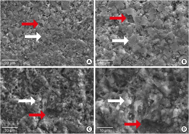
-
Abstract
PDFPubReaderePub
Objectives This study evaluated the microtensile bond strength (µTBS) of polymer-ceramic and indirect composite resin with 3 classes of resin cements.
Materials and Methods Two computer-aided design/computer-aided manufacturing (CAD/CAM)-fabricated polymer-ceramics (Enamic [ENA; Vita] and Lava Ultimate [LAV; 3M ESPE]) and a laboratory indirect composite resin (Gradia [GRA; GC Corp.]) were equally divided into 6 groups (
n = 18) with 3 classes of resin cements: Variolink N (VAR; Vivadent), RelyX U200 (RXU; 3M ESPE), and Panavia F2 (PAN; Kuraray). The μTBS values were compared between groups by 2-way analysis of variance and thepost hoc Tamhane test (α = 0.05).Results Restorative materials and resin cements significantly influenced µTBS (
p < 0.05). In the GRA group, the highest μTBS was found with RXU (27.40 ± 5.39 N) and the lowest with VAR (13.54 ± 6.04 N) (p < 0.05). Similar trends were observed in the ENA group. In the LAV group, the highest μTBS was observed with VAR (27.45 ± 5.84 N) and the lowest with PAN (10.67 ± 4.37 N) (p < 0.05). PAN had comparable results to those of ENA and GRA, whereas the μTBS values were significantly lower with LAV (p = 0.001). The highest bond strength of RXU was found with GRA (27.40 ± 5.39 N,p = 0.001). PAN showed the lowest µTBS with LAV (10.67 ± 4.37 N;p < 0.001).Conclusions When applied according to the manufacturers' recommendations, the µTBS of polymer-ceramic CAD/CAM materials and indirect composites is influenced by the luting cements.
-
Citations
Citations to this article as recorded by- Enhancing severely compromised premolar strength: role of cusp reduction design in CAD/CAM composite restorations
Mohamed F. Haridy, Ahmed Refaat Mohamed, Shehabeldin Saber, Edgar Schafer, Samar Elsayed Swelam, Youssef M. Haridy, Hend S. Ahmed
Odontology.2026; 114(2): 852. CrossRef - Effect of hydrofluoric acid and self-etch ceramic primers on the flexural strength and fatigue resistance of glass ceramics: A systematic review and meta-analysis of in vitro studies
Paulo Matias Moreira, Gabriela Luiza Moreira Carvalho, Rodrigo de Castro Albuquerque, Carolina Bosso André
Japanese Dental Science Review.2024; 60: 198. CrossRef - Light transmittance through resin-matrix composite onlays adhered to resin-matrix cements or flowable composites
Rita Fidalgo-Pereira, Susana O. Catarino, Óscar Carvalho, Nélio Veiga, Orlanda Torres, Annabel Braem, Júlio C.M. Souza
Journal of the Mechanical Behavior of Biomedical Materials.2024; 151: 106353. CrossRef - Effect of thermocycling on the mechanical properties of permanent composite-based CAD-CAM restorative materials produced by additive and subtractive manufacturing techniques
Tuğba Temizci, Hatice Nalan Bozoğulları
BMC Oral Health.2024;[Epub] CrossRef - Effect of different surface treatments on resin-matrix CAD/CAM ceramics bonding to dentin: in vitro study
Hanan Fathy, Hamdi H. Hamama, Noha El-Wassefy, Salah H. Mahmoud
BMC Oral Health.2022;[Epub] CrossRef - Digital image analysis of fluorescence of ceramic veneers with different ceramic materials and resin cements
Jiao ZHANG, Qing YU
Dental Materials Journal.2022; 41(6): 868. CrossRef - Fatigue Behavior of Monolithic Zirconia-Reinforced Lithium Silicate Ceramic Restorations: Effects of Conditionings of the Intaglio Surface and the Resin Cements
F Dalla-Nora, LF Guilardi, CP Zucuni, LF Valandro, MP Rippe
Operative Dentistry.2021; 46(3): 316. CrossRef
- Enhancing severely compromised premolar strength: role of cusp reduction design in CAD/CAM composite restorations
- 2,824 View
- 8 Download
- 7 Crossref
Case Report
- Endocrown restorations for extensively damaged posterior teeth: clinical performance of three cases
- Konstantinos Tzimas, Maria Tsiafitsa, Paris Gerasimou, Effrosyni Tsitrou
- Restor Dent Endod 2018;43(4):e38. Published online August 22, 2018
- DOI: https://doi.org/10.5395/rde.2018.43.e38
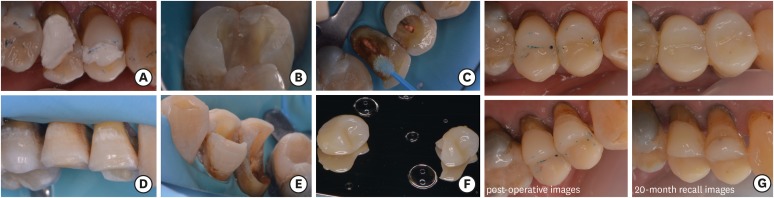
-
Abstract
PDFPubReaderePub
The restoration of endodontically treated teeth (ETT) with more than one cusp missing and thin remaining walls is challenging for the general practitioner. The use of posts combined with full coverage restorations is a well-established approach, yet not following the minimal invasive principles of adhesive dentistry. Endocrowns are indirect monoblock restorations that use the pulp chamber of the ETT for retention. In this study the fabrication of 4 endocrowns and their clinical performance will be discussed. Two clinical cases include computer-aided design/computer-aided manufacturing manufactured molar endocrowns (one feldspathic ceramic and one hybrid composite-ceramic restoration) and the other two are dental laboratory manufactured resin composite premolar endocrown restorations. The modified United States Public Health Service criteria were used to assess the clinical behavior of the restorations at different follow up periods. Endocrown restorations present a satisfactory clinical alternative, either by the use of resin composite or glass ceramic and hybrid materials. Specific guidelines with minimal alterations should be followed for an endocrown restoration to be successful. Due to limited evidence regarding the long term evaluation of this restorative technique, a careful selection of cases should be applied.
-
Citations
Citations to this article as recorded by- Marginal adaptation and fracture resistance of recent 3D printed high-performance polymer and CAD/CAM hybrid ceramic endocrown restorations and designs
Mohamed Atif Elkholy, Amira Mohammad Samy Mostafa, Hoda Gaafar Hassan Hammad
BMC Oral Health.2026;[Epub] CrossRef - Periodontal health of endodontically treated molars restored with composite CAD/CAM endocrowns versus stainless steel crowns in Egyptian children: a randomized controlled trial
Basheer Ali Mabkhot, Sheriene Ezz Eldin Taha, Mostafa Hussein Kamel, Shaimaa Mohamed Sabry
BMC Oral Health.2026;[Epub] CrossRef - Durability and Failure Modes of IPS e.max Endocrown in Endodontically Treated Molars: A 10-Year Clinical Report of Two Cases
Rana Essa Dalol, Jihad Nouman Abou Nassar, Mohammad Y. Hajeer
Cureus.2026;[Epub] CrossRef - Multicenter RCT 6‐Year Survival of Zirconia‐Reinforced Lithium Silicate Endocrowns: 90° Shoulder Versus Butt‐Joint Margins
Ping Xiao, Ziting Zheng, Yuting Zeng, Xiaoyu Lin, Jiayao Mo, Liya Chen, Jilei Wang, Yuan Wu, Zhongchun Tong, Wenjuan Yan
International Endodontic Journal.2026;[Epub] CrossRef - Mechanical performance of endocrown restorations in anterior teeth: A systematic review and network meta-analysis
Julia Fehrenbach, Jéssica Lopes Soares de Soares, João Carlos Silva do Nascimento Foly, Leonardo Lamberti Miotti, Eliseu Aldrighi Münchow
Dental Materials.2025; 41(1): 28. CrossRef - Fracture resistance and mode of failure of modified Polyether-ether-ketone versus lithium disilicate endocrowns
Mohamed G. A. Kharboush, Hesham I. Othman, Mohamed F. Aldamaty, Ahmed M. L. Alameldin
BMC Oral Health.2025;[Epub] CrossRef - Clinical and radiographic assessment of composite CAD/CAM endocrowns and stainless steel crowns for endodontically treated first permanent molars in Egyptian children: randomized controlled pilot study
Basheer Ali Mabkhot, Sheriene Ezz Eldin Taha, Shaimaa Mohamed Sabry
BMC Oral Health.2025;[Epub] CrossRef - The influence of cavity design on the mechanical behavior of endo-crown restorations: an ex-vivo study
Mohamed Gomaa Altamimi, Omaima El Mahallawi, Monika Lukomska-Szymanska, Mohammed Turky
BMC Oral Health.2025;[Epub] CrossRef - Comparison of Microtensile Bonding Strength and Microleakage of Endocrowns Restorations Prepared With Two Different Materials
Emrah Ayna, Burcu Ayman, Cansel Belge
HRU International Journal of Dentistry and Oral Research.2025; 5(2): 66. CrossRef - Exploring the evolution of endocrowns: a bibliometric analysis (2010-2024)
Sanjana Jayakumar Nair, Jinesh Azhuvancheri, Saurabh Kumar Gupta, Gopika Krishnan
International Journal of Research in Medical Sciences.2025; 13(10): 4296. CrossRef - Beyond Traditional Restorations: Management With Endocrown in a Late Adolescent
Abdulaziz Binrayes, Abdullatif A AlGhazzi, Saud M Alotaibi
Cureus.2024;[Epub] CrossRef - Endocrown-retained fixed partial dentures: Revolutionizing tooth restoration or risky business? A finite element study
Nivedha Muthukumar, Parthasarathy Natarajan, Seenivasan Madhan Kumar, Shanmuganathan Natarajan
Journal of Conservative Dentistry and Endodontics.2024; 27(12): 1234. CrossRef - Chinese dentists’ restorative preferences and choices for endodontically treated teeth: a representative survey
Wenhui Li, Ziting Zheng, Yuting Zeng, Zhiyan Zhou, Ping Xiao, Xincen Zhong, Wenjuan Yan
BMC Oral Health.2024;[Epub] CrossRef - PHỤC HÌNH ENDOCROWN TRÊN RĂNG CỐI NHỎ ĐÃ NỘI NHA: BÁO CÁO MỘT CA LÂM SÀNG
Trịnh Minh Trí Trịnh Minh Trí, Lê Võ Thảo Phương Lê Võ Thảo Phương, Nguyễn Tấn Đạt Nguyễn Tấn Đạt, Phạm Nguyên Quân Phạm Nguyên Quân, Văn Hồng Phượng Văn Hồng Phượng
Tạp Chí Khoa Học Trường Đại Học Quốc Tế Hồng Bàng.2024; : 241. CrossRef - Application of one-piece endodontic crowns fabricated with CAD-CAM system to molars
Haruto Hiraba, Kensuke Nishio, Yoshimasa Takeuchi, Takashi Ito, Tetsuo Yamamori, Atsushi Kamimoto
Japanese Dental Science Review.2024; 60: 81. CrossRef - Clinical performance and wear resistance of milled resin composite material versus direct nanohybrid bulk-fill resin composite in the restoration of endodontically treated posterior teeth over 1 year: Randomized clinical trial
Esraa Esmeail H. Elhaddad, Mohamed M. A. Mohsen, Dina Ezz Eldin Mohamed
Journal of Conservative Dentistry and Endodontics.2024; 27(4): 400. CrossRef - Roughness analysis on porcelain sectional surface of porcelain fused to Co-Cr alloy endocrowns
Xuesheng Li
Scientific Reports.2024;[Epub] CrossRef - Minimal İnvaziv Protetik Tedavilerde CAD-CAM Kullanımı: İki Olgu Sunumu
Aynur Beyza Çavuşculu Güdül, Şükriye Ece Geduk, Gaye Sağlam
Journal of International Dental Sciences.2024; 10(3): 167. CrossRef - Comparative Evaluation of Fracture Toughness and Marginal Adaptation of PEEK and Cast Metal Crowns for Restoring Posterior Teeth with Endocrown and Richmond Crown: An In Vitro Study
Lalit Kumar, Komalpreet Kaur, Shefali Singla, Charnpreet Singh, Sunint Singh
International Journal of Prosthodontics and Restorative Dentistry.2024; 14(4): 234. CrossRef - Retrospective study on the evolution of teeth with endodontic treatment in a group of patients from Craiova – Romania
Mihaela-Roxana Boțilă, Mihaela Jana Țuculina , Oana Andreea Diaconu , Mihaela Ionescu , Petre Costin Mărășescu , Luana Corina Lascu , Veronica Mercut
Romanian Journal of Oral Rehabilitation.2024; 16(2): 225. CrossRef - Criterios clínicos y radiológicos de los tratamientos endodónticos para rehabilitación Endocrown: meta análisis
Domenica Camila Astudillo Benavides, Rafael Bernardo Piedra Andrade, Amanda Isabel Pesantez Coronel, Jose Esteban Torrachi Carrasco
Anatomía Digital.2024; 7(4): 81. CrossRef - Three-Dimensional Finite Element Study of Endodontically Treated Maxillary Central Incisors Restored Using Different Post and Crown Materials
Nour Al-Deen Kharboutly, Mirza Allaf, Shaza Kanout
Cureus.2023;[Epub] CrossRef - The impact of extended pulp chamber preparations on the clinical performance of endocrowns in Indian patients: A 1-year observational study
Preethi Duraisamy, Naveen Gopi Chander, Jetty Ramesh Reddy, Muthukumar Balasubramanium
Journal of Oral Biology and Craniofacial Research.2023; 13(5): 616. CrossRef - Endocrowns: Indications, Preparation Techniques, and Material Selection
Dalal S AlDabeeb, Nouf S Alakeel, Raneem M Al jfshar, Thakra K Alkhalid
Cureus.2023;[Epub] CrossRef - Knowledge and Awareness of Dental Practitioners About the Utilization of Endocrown in Post-endodontic Management
Ahmed A Madfa, Moazzy I Almansour, Asma F Alshammari, Nada M. Alenezi, Essa F. Alrashidi, Adel A. Aldhaban, Thoraya Aljohani, Faris A. Alshammari
Cureus.2023;[Epub] CrossRef - Clinical Performance of Two CAD/CAM Fabricated Ceramic Restorations with Different Designs for MIH Rehabilitation: A Randomized Controlled Trial
Ayat G. Montaser, Sara N. Hashem, Menna-Allah S. Ali, Nour Alhoda Fathy, Hebatullah Ahmed Safwat, Alaa M. Eldehna
The Open Dentistry Journal.2023;[Epub] CrossRef - Endocrown as a restorative strategy in endodontically treated teeth: an integrative literature review
Robson de Lima GOMES, Andressa Cristina da Silva QUEIROZ, Viviane Maria Gonçalves de FIGUEIREDO
RGO - Revista Gaúcha de Odontologia.2022;[Epub] CrossRef - ENDOCROWN RESTORATION OF THE ENDODONTICALLY TREATED TEETH BY USING CAD/CAM: CASE SERIES
Begüm ÜNLÜ KURŞUN, Ender AKAN
European Annals of Dental Sciences.2022; 49(Suppl 1): 13. CrossRef - Clinical Evaluation of CAD/CAM Ceramic Endocrown Versus Prefabricated Zirconia Crown in the Restoration of Pulpotomized Primary Molars: A Two-Year Spilt-Mouth Randomized Controlled Trial
Nagwa Mohmmad Ali Khattab, Yasmine Mohamed Farouk El Makawi, Ahmad Abdel Hamid Elheeny
European Journal of Dentistry.2022; 16(03): 627. CrossRef - Hyperplastic Pulpitis Management with Endocrown: A Case Report
Pérez Jardón A, Otero Gayoso N, Otero. Rey E.M, Guerra Caamaño M, Chamorro-Petronacci C.M, Blanco Carrión A, Rivas Mundiña B
The Open Dentistry Journal.2022;[Epub] CrossRef - Influence of inner crown thickness on the bonding strength of porcelain fused to Co-Cr alloy endocrown
Xuesheng Li
Journal of Oral Science.2022; 64(1): 40. CrossRef - Effect of pulp chamber depth on the accuracy of endocrown scans made with different intraoral scanners versus an industrial scanner: An in vitro study
Bahar Gurpinar, Onjen Tak
The Journal of Prosthetic Dentistry.2022; 127(3): 430. CrossRef - Efectividad de las restauraciones en piezas con tratamiento de conducto: Una revisión clínica actual
Guiselle Andrea Verástegui Baldárrago
Revista Odontológica Basadrina.2022; 6(2): 41. CrossRef - “Conservative Bonded Restoration (An Alternative to Full Coverage Crown): A Case Report on Endocrown
Josey Mathew, Liza George, Sinju Paul, Aleesha Joy, Beulah M Bejoy, Sethuparvathi Anitha
Conservative Dentistry and Endodontic Journal.2021; 5(2): 42. CrossRef - Fractography of clinical failures of indirect resin composite endocrown and overlay restorations
Carlo M. Saratti, Giovanni T. Rocca, Stéphane Durual, Ulrich Lohbauer, Jack L. Ferracane, Susanne S. Scherrer
Dental Materials.2021; 37(6): e341. CrossRef - Survival and success of endocrowns: A systematic review and meta-analysis
Raghad A. Al-Dabbagh
The Journal of Prosthetic Dentistry.2021; 125(3): 415.e1. CrossRef - A Thorough Analysis of the Endocrown Restoration: A Literature Review
Dimokritos Papalexopoulos, Theodora-Kalliopi Samartzi, Aspasia Sarafianou
The Journal of Contemporary Dental Practice.2021; 22(4): 422. CrossRef - Full‐Crown Versus Endocrown Approach: A 3D‐Analysis of Both Restorations and the Effect of Ferrule and Restoration Material
João Paulo Mendes Tribst, Amanda Maria de Oliveira Dal Piva, Niek de Jager, Marco Antonio Bottino, Paul de Kok, Cornelis Johannes Kleverlaan
Journal of Prosthodontics.2021; 30(4): 335. CrossRef - Monolithic Endocrown Vs. Hybrid Intraradicular Post/Core/Crown Restorations for Endodontically Treated Teeth; Cross-sectional Study
Mai Soliman, Lamar Alshamrani, Basma Yahya, Ghadah Alajlan, Alhanoof Aldegheishem, Elzahraa Eldwakhly
Saudi Journal of Biological Sciences.2021; 28(11): 6523. CrossRef - Which materials would account for a better mechanical behavior for direct endocrown restorations?
José Augusto Sedrez-Porto, Eliseu Aldrighi Münchow, Maximiliano Sergio Cenci, Tatiana Pereira-Cenci
Journal of the Mechanical Behavior of Biomedical Materials.2020; 103: 103592. CrossRef - Indications and Success Rate of Endo Crowns – A Systematic Review
Shahzeb Hasan Ansari, Abdullah Ahmed Alfaqeeh, Abdullah Al Buryk, Sara Ahmed Alfaqeeh, Abdullatif Yousif A. Almusharraf, Atheer Hussain N. Aljarullah
Journal of Evolution of Medical and Dental Sciences.2020; 9(43): 3247. CrossRef
- Marginal adaptation and fracture resistance of recent 3D printed high-performance polymer and CAD/CAM hybrid ceramic endocrown restorations and designs
- 7,804 View
- 155 Download
- 41 Crossref
Research Articles
- The effect of thermocycling on the degree of conversion and mechanical properties of a microhybrid dental resin composite
- Mehrsima Ghavami-Lahiji, Melika Firouzmanesh, Hossein Bagheri, Tahereh S. Jafarzadeh Kashi, Fateme Razazpour, Marjan Behroozibakhsh
- Restor Dent Endod 2018;43(2):e26. Published online April 26, 2018
- DOI: https://doi.org/10.5395/rde.2018.43.e26
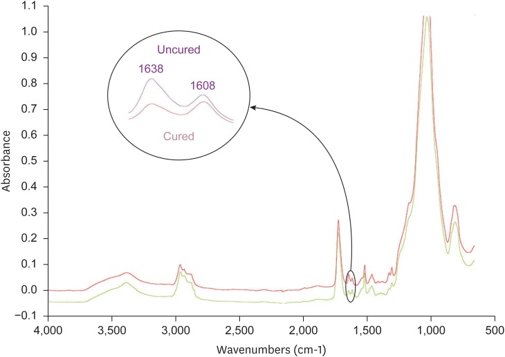
-
Abstract
PDFPubReaderePub
Objective The purpose of this study was to investigate the degree of conversion (DC) and mechanical properties of a microhybrid Filtek Z250 (3M ESPE) resin composite after aging.
Method The specimens were fabricated using circular molds to investigate Vickers microhardness (Vickers hardness number [VHN]) and DC, and were prepared according to ISO 4049 for flexural strength testing. The initial DC (%) of discs was recorded using attenuated total reflectance-Fourier transforming infrared spectroscopy. The initial VHN of the specimens was measured using a microhardness tester under a load of 300 g for 15 seconds and the flexural strength test was carried out with a universal testing machine (crosshead speed, 0.5 mm/min). The specimens were then subjected to thermocycling in 5°C and 55°C water baths. Properties were assessed after 1,000–10,000 cycles of thermocycling. The surfaces were evaluated using scanning electron microscopy (SEM). Data were analyzed using 1-way analysis of variance followed by the Tukey honest significant difference
post hoc test.Results Statistical analysis showed that DC tended to increase up to 4,000 cycles, with no significant changes. VHN and flexural strength values significantly decreased upon thermal cycling when compared to baseline (
p < 0.05). However, there was no significant difference between initial and post-thermocycling VHN results at 1,000 cycles. SEM images after aging showed deteriorative changes in the resin composite surfaces.Conclusions The Z250 microhybrid resin composite showed reduced surface microhardness and flexural strength and increased DC after thermocycling.
-
Citations
Citations to this article as recorded by- Evaluation of the radiopacity of different universal composite resins aged by thermocycling
Dilber Çölkesen, Alican Kuran, Neslihan Tekçe
Odontology.2026; 114(3): 1615. CrossRef - Mechanical, Surface and Physicochemical Properties of Nanozeolite‐Modified 3D Printed Hybrid Ceramics at Varying Concentrations: An In Vitro Study
Ahmed A. Holiel, Yomna M. Ibrahim, Noha Morsy
Journal of Esthetic and Restorative Dentistry.2026; 38(2): 325. CrossRef - Impact of Graphitic Carbon Nitride on Dental Composite’s Mechanical and Antibacterial Properties
Zainab Rafaqat, Saad Liaqat, Ahmed Bari, Warda Khan Yousafzai, Umar Nishan, Sandleen Feroz, Nawshad Muhammad
Journal of Materials Engineering and Performance.2026; 35(18): 18215. CrossRef - Color Stability Under Challenge: Effects of Thermo-Aging and Mouthrinse Exposure on Anterior Teeth and Esthetic Composites
Gökçe Keçeci, Zehra Güner, Süleyman Ziya Şenyurt, Kamile Erciyas
European Journal of Therapeutics.2026; 32(1): 94. CrossRef - Evaluation of surface roughness of PEEK and Laser-Sintered cobalt–chromium subjected to different polishing protocols and thermocycling (an in vitro study)
Marzia Kareem Ahmed, Aras Maruf Rauf
Oxford Open Materials Science.2026;[Epub] CrossRef - Comparative influence of triton and cavity conditioners on the surface characteristics and bond strength of dentin, indicating triton as a cavity conditioner
Lena Bal, Cangül Keskin, Aybüke Karaca Sakallı, Bilge Özcan
Journal of Health Sciences and Medicine.2026; 9(1): 98. CrossRef - The Influence of Thermal and Mechanical Aging on the Flexural Properties of Conventional and 3D-Printed Materials Used in Occlusal Splints Manufacturing
Joanna Smardz, Katarzyna Kresse-Walczak, Heike Meißner, Klaus Böning, Joanna Weżgowiec, Andrzej Małysa, Mieszko Więckiewicz
Materials.2026; 19(2): 421. CrossRef - Failure mechanism analysis and machine learning-based residual strength prediction of CFRP with different ply orientations and loading types under six typical accelerated aging environments
Guofeng Qin, Chenglong Su, Peiwen Mi, Peng Huang, Qiuhan Fan, Ming Li, Wei Tan
Construction and Building Materials.2026; 516: 145630. CrossRef - Discoloration, radiopacity, and push-out bond strength of bismuth-based radiopacifiers in endodontic sealers
Mohammad Ali Saghiri, Mina Shekarian, Amir Abdolmaleki, Michael Conte, Steven M. Morgano
Odontology.2026;[Epub] CrossRef - Color matching and metameric behavior of composite resins applied on anodized titanium surfaces: an in vitro spectrophotometric study
Umur Davut, Ozge Doganay-Ozyilmaz, Ozgun Yusuf Ozyilmaz
BMC Oral Health.2026;[Epub] CrossRef - Effect of an Adhesive-Enhancing Primer on Shear Bond Strength in Repairs of Fresh Resin Composite to Aged Resin Composite: With and Without Saliva Contamination
Pemika Lerttiendamrong, Wisarut Prawatvatchara, Suparaksa Yamockul, Awiruth Klaisiri, Tool Sriamporn, Pakpilai Thiranukoon, Niyom Thamrongananskul
Journal of Composites Science.2026; 10(4): 184. CrossRef - An In Vitro Study on Bond Strength Degradation of Low-Shrinkage Composite Resins to Demineralized Dentin After Thermal Cycling
Maria Cristina Borsatto, Barbara Jarreta, Jaciara Miranda Gomes-Silva, Patricia Gatón-Hernández, Carolina Paes Torres, Rodrigo Galo
Oral.2026; 6(2): 41. CrossRef - Engineered micro-textures enhance adhesion in additively manufactured dental crowns
Shiyao Chen, Andrew B. Cameron, John M. Aarts, Joanne J.E. Choi
Digital Dentistry Journal.2026; 4(1): 100085. CrossRef - Effect of Simulated Toothbrushing on Surface Roughness, Color Stability, and Gloss of Two Single-Shade Composite Resins
Zeynep Hale Keles, Vasfiye Isik, Soner Sismanoglu
Materials.2026; 19(8): 1523. CrossRef - Fracture resistance of nanocomposite 3D-printed resins designated for teeth fabrication: An in vitro analysis before and after thermal aging
Farah Y. Alshaikhnasser, Shaymaa Y. Alboryh, Zainab H. Al Dawood, Rawan F. Almedarham, Fatimah A. Alhassan, Fatimah M. Alatiyyah, Essam A. Nassar, Sultan Akhtar, Soban Q. Khan, Abdulrahman A. Balhaddad, Ahmed Tharwat Al Ammary, Mohammed M. Gad
F1000Research.2026; 14: 894. CrossRef - Flexural strength of new and repaired TEGDMA- and HEMA-free resin composites compared to a conventional composite
A.M.O. Dal Piva, M. Khalil, R.O. Pilecco, C.J. Kleverlaan, J.P.M. Tribst
Next Materials.2026; 12: 102355. CrossRef - Effect of Curing Techniques on Microleakage and Microhardness of Bulk-Fill and Conventional Resin-Based Composites: An In Vitro Study
Ako Abdalrahman Ahmed, Bestoon Mohammed Faraj
Journal of Composites Science.2026; 10(6): 306. CrossRef - Calcium Phosphate Oligomers as Inorganic Glue for Microcrack Repair in Teeth
Yinan Fu, Tianren Liu, Xinyu He, Ruikang Tang, Zhaoming Liu
ACS Applied Materials & Interfaces.2026; 18(25): 34748. CrossRef - Quantitative and Structural Characterization of Streptococcus mutans Biofilms on Dental Restorative Materials Using Scanning Electron Microscopy and Machine Learning
Krishna Kumar Kungumaraj, Sivashankari Rajasekaran, Bao Huynh, Jack Ferracane, Jens Kreth, Ana Paula Fugolin
Journal of Biomedical Materials Research Part B: Applied Biomaterials.2026;[Epub] CrossRef - Clinical Decision‐Making of Repair vs. Replacement of Defective Direct Dental Restorations: A Multinational Cross‐Sectional Study With Meta‐Analysis
Ömer Hatipoğlu, João Filipe Brochado Martins, Mohmed Isaqali Karobari, Nessrin Taha, Thiyezen Abdullah Aldhelai, Daoud M. Ayyad, Ahmed A. Madfa, Benjamin Martin‐Biedma, Rafael Fernández‐Grisales, Bakhyt A. Omarova, Wen Yi Lim, Suha Alfirjani, Kacper Nijak
Journal of Esthetic and Restorative Dentistry.2025; 37(4): 977. CrossRef - An In Vitro Evaluation of Novel Bioactive Liner's Effect on Marginal Adaptation of Class II Composite Restorations: A Scanning Electron Microscope Analysis
Girija S Sajjan, Naveena Ponnada, Praveen Dalavai, Madhu Varma Kanumuri, Venkata Karteek Varma Penmatsa, B V Sindhuja
World Journal of Dentistry.2025; 15(9): 749. CrossRef - Different contemporary resin cements for intracanal luting of glass fiber posts - Bonding and polymerization assessments
Anna Caroliny Detogni, Vitaliano Gomes de Araújo Neto, Caio Felipe de Almeida Nobre, Victor Pinheiro Feitosa, Mário Alexandre Coelho Sinhoreti
International Journal of Adhesion and Adhesives.2025; 138: 103951. CrossRef - Effect of food-simulating liquids and polishing times on the color stability of microhybrid and nanohybrid resin composites
Muhammet Fidan, Nevin Çankaya
Discover Nano.2025;[Epub] CrossRef - Effect of irrigation protocols for post space preparation on the bond of the resin luting agent and post to a hydraulic calcium silicate filled root: An in vitro study
Nuttanun Poeaim, Sirawut Hiran-us, Yanee Tantilertanant
The Journal of Prosthetic Dentistry.2025; 133(4): 1039.e1. CrossRef - Influence of Different Adhesives and Surface Treatments on Shear and Tensile Bond Strength and Microleakage with Micro-CT of Repaired Bulk-Fill Composites
Handan Yıldırım-Işık, Mediha Büyükgöze-Dindar
Polymers.2025; 17(12): 1680. CrossRef - Effect of thermal ageing on physico-mechanical properties and self-healing potential of experimental 3D-printed denture base resin composites
Mariam Raza Syed, Amr Fawzy
Journal of the Mechanical Behavior of Biomedical Materials.2025; 170: 107123. CrossRef - Effects of aging on the physicomechanical, antimicrobial, and cytotoxicity properties of flowable composite resin with strontium-modified phosphate-based glass
Seo-Hyun Kim, Hye-Bin Go, Myung-Jin Lee, Jae-Sung Kwon
Scientific Reports.2025;[Epub] CrossRef - Colour Stability and Optical Properties of Provisional Crowns Fabricated With Milling, 3D Printing, and Direct Technique
Tommaso Rinaldi, Carlos Serrano Granger, Andrea Santamaría Laorden, Jaime Orejas-Perez, Pablo Gómez Cogolludo
International Dental Journal.2025; 75(6): 103932. CrossRef - EVALUATE DEGREE OF CONVERSION OF NEW BIOACTIVE ORTHODONTIC ADHESIVE WITH COLOR CHANGE & FLUORESCENCE PROPERTY
Mohammed Younis, Neam Fakhri Neam Fakhri
BULLETIN OF STOMATOLOGY AND MAXILLOFACIAL SURGERY.2025; : 39. CrossRef - Antibacterial activity and physicochemical properties of light-curable fluoride varnishes containing strontium phosphate-based glass
Na-Yeon Kim, Mi-Sol Ryu, Ji-Min Lee, Soo-Yeon Jeong, Hye-Been Choi, Myung-Jin Lee, Song-Yi Yang
Clinical Oral Investigations.2025;[Epub] CrossRef - Systematic Review of Studies Comparing Microleakage After Restoration With Cention and Conventional Glass Ionomer Cement in Human Extracted Teeth
Rashmi Misra, Mansi Vandekar, Gayatri Pendse, Omkar Bhosale, Pauravi Hegde, Aashaka Vaishnav
Cureus.2025;[Epub] CrossRef - Exploring the sources and routes of micro- and nanoplastics from dental products and materials: their impact on human health - a systematic review
Vidhya Selvaraj, R. Saravanan, N. Raj Vikram, Uma revathi Gopalakrishnan, Ramsamy M
Next Research.2025; 2(4): 100925. CrossRef - Investigation of the mechanical response of MWCNTs infused carbon/glass fiber-based hybrid composites using digital image correlation
Somaiah Chowdary Mallampati, Ujendra Kumar Komal, Paladugu Rakesh, Parthapratim Barman
Construction and Building Materials.2025; 492: 143068. CrossRef - Exploring the Biological and Chemical Properties of Emerging 3D-Printed Dental Resin Composites Compared to Conventional Light-Cured Materials
Nikola Živković, Stefan Vulović, Miloš Lazarević, Anja Baraba, Aleksandar Jakovljević, Mina Perić, Jelena Mitrić, Aleksandra Milić Lemić
Materials.2025; 18(22): 5170. CrossRef - Awareness of possible complications associated with direct composite restorations: A multinational survey among dentists from 13 countries with meta-analysis
Anna Lehmann, Kacper Nijakowski, Jakub Jankowski, David Donnermeyer, Paulo J. Palma, Milan Drobac, João Filipe Brochado Martins, Fatma Pertek Hatipoğlu, Indira Tulegenova, Muhammad Qasim Javed, Hamad Mohammad Alharkan, Olga Bekjanova, Sylvia Wyzga, Moataz
Journal of Dentistry.2024; 145: 105009. CrossRef - Comparative evaluation of bond strength and color stability of polyetheretherketone and zirconia layered with indirect composite before and after thermocycling: An in vitro study
Pooja Singh, Subhabrata Maiti, Amrutha Shenoy
The Journal of Indian Prosthodontic Society.2024; 24(3): 252. CrossRef - Biaxial flexural strength of hydrothermally aged resin-based materials
Rodrigo Ricci Vivan, Mariana Miranda de Toledo Piza, Bruna de Mello Silva, Thalya Fernanda Horsth Maltarollo, Gustavo Sivieri-Araujo, Murilo Priori Alcalde, Marco Antonio Hungaro Duarte, Estevam Augusto Bonfante, Henrico Badaoui Strazzi-Sahyon
Journal of the Mechanical Behavior of Biomedical Materials.2024; 155: 106568. CrossRef - Comparative Strength Study of Indirect Permanent Restorations: 3D-Printed, Milled, and Conventional Dental Composites
João Paulo Mendes Tribst, Adelheid Veerman, Gabriel Kalil Rocha Pereira, Cornelis Johannes Kleverlaan, Amanda Maria de Oliveira Dal Piva
Clinics and Practice.2024; 14(5): 1940. CrossRef - Influencia del termociclado sobre la estabilidad del color de dos resinas compuestas
//Influence of thermocycling on the color stability of two composite resins
Verónica Lucía Ventrera, María Eugenia Alejandra Barrionuevo
Revista de la Asociación Odontológica Argentina.2024; : 1. CrossRef - Efeito do protocolo de polimento e do armazenamento em meio úmido na variação de cor, massa e rugosidade superficial de resinas compostas
Leonardo Cruz Morais, Mateus Victória Gontijo, Gabriela Rodrigues Pires, Victor de Morais Gomes, Milton Carlos Kuga, Francisco Fernando Massola Filho, Amanda Gonçalves Franco, Alberto Nogueira da Gama Antunes
Cuadernos de Educación y Desarrollo.2024; 16(6): e4556. CrossRef - A comparison of the mechanical properties of 3D-printed, milled, and conventional denture base resin materials
Hyeong-Ju YU, You-Jung KANG, Yeseul PARK, Hoon KIM, Jee-Hwan KIM
Dental Materials Journal.2024; 43(6): 813. CrossRef - Effect of aging and fiber‐reinforcement on color stability, translucency, and microhardness of single‐shade resin composites versus multi‐shade resin composite
Muhammet Fidan, Özhan Yağci
Journal of Esthetic and Restorative Dentistry.2024; 36(4): 632. CrossRef - Impact of Artificial Aging on the Physical and Mechanical Characteristics of Denture Base Materials Fabricated via 3D Printing
Ahmed Altarazi, Julfikar Haider, Abdulaziz Alhotan, Nick Silikas, Hugh Devlin, Weihao Yuan
International Journal of Biomaterials.2024;[Epub] CrossRef - Synthesis, monomer conversion, and mechanical properties of polylysine based dental composites
Saadia Bano Lone, Rabia Zeeshan, Hina Khadim, Muhammad Adnan Khan, Abdul Samad Khan, Anila Asif
Journal of the Mechanical Behavior of Biomedical Materials.2024; 151: 106398. CrossRef - Bond strength and surface roughness assessment of novel antimicrobial polymeric coated dental cement
Ghada Naguib, Hisham Mously, Jumana Mazhar, Ibrahim Alkanfari, Abdulelah Binmahfooz, Mohammed Zahran, Mohamed T. Hamed
Discover Nano.2024;[Epub] CrossRef - Evaluation of microhardness, degree of conversion, and abrasion resistance of nanoglass and multiwalled carbon nanotubes reinforced three‐dimensionally printed denture base resins
Pansai Ashraf Mohamed, Yomna Mohamed Ibrahim, Kenda Ibrahim Hisham Hanno, Mohamed Mahmoud Abdul‐Monem
Journal of Prosthodontics.2024;[Epub] CrossRef - Effect of CAD-CAM block thickness and translucency on the polymerization of luting materials
Bengü Doğu Kaya, Selinsu Öztürk, Ayşe Aslı Şenol, Erkut Kahramanoğlu, Pınar Yılmaz Atalı, Bilge Tarçın
BMC Oral Health.2024;[Epub] CrossRef - Simulation of oral environmental conditions through artificial aging of teeth for the assessment of enamel discoloration in orthodontics
Celal Irgın
BMC Oral Health.2024;[Epub] CrossRef - Do universal adhesive systems affect color coordinates and color change of single-shade resin composites compared with a multi-shade composite?
Muhammet FİDAN, Özhan YAĞCI
Dental Materials Journal.2023; 42(6): 886. CrossRef - Fabrication, Evaluation, and Performance Ranking of Tri-calcium Phosphate and Silica Reinforced Dental Resin Composite Materials
Sonu Saini, Anoj Meena, Ramkumar Yadav, Amar Patnaik
Silicon.2023; 15(18): 8045. CrossRef - Can Modification with Urethane Derivatives or the Addition of an Anti-Hydrolysis Agent Influence the Hydrolytic Stability of Resin Dental Composite?
Agata Szczesio-Wlodarczyk, Izabela M. Barszczewska-Rybarek, Marta W. Chrószcz-Porębska, Karolina Kopacz, Jerzy Sokolowski, Kinga Bociong
International Journal of Molecular Sciences.2023; 24(5): 4336. CrossRef - Effect of veneering material type and thickness ratio on flexural strength of bi-layered PEEK restorations before and after thermal cycling
Ahmed Gouda, Ashraf Sherif, Mennatallah Wahba, Tarek Morsi
Clinical Oral Investigations.2023; 27(6): 2629. CrossRef - 3D printed denture base material: The effect of incorporating TiO2 nanoparticles and artificial ageing on the physical and mechanical properties
Ahmed Altarazi, Julfikar Haider, Abdulaziz Alhotan, Nick Silikas, Hugh Devlin
Dental Materials.2023; 39(12): 1122. CrossRef - Influence of silane coupling agent and aging on the repair bond strength of dental composites
Gustavo Jusué-Esparza, José Alejandro Rivera-Gonzaga, Guillermo Grazioli, Ana Josefina Monjarás-Ávila, J. Eliezer Zamarripa-Calderón, Carlos Enrique Cuevas-Suárez
Journal of Adhesion Science and Technology.2023; 37(5): 913. CrossRef - Degree of conversion of light‐polymerized composite resin in implant prosthesis screw access opening
Se‐Hyun Park, Yoon‐Hyuk Huh, Chan‐Jin Park, Lee‐Ra Cho, Kyung‐Ho Ko
Journal of Prosthodontics.2023; 32(9): 829. CrossRef - Investigation of the effect of matrix-interface formed with silane-based coupling agents on physico-chemical behavior and flow distance of dental composites
Zerin Yeşil Acar, Merve Tunç Koçyiğit, Meltem Asiltürk
Journal of Molecular Liquids.2023; 378: 121600. CrossRef - Evaluation of Water Sorption and Solubility of 3D-Printed, CAD/CAM Milled, and PMMA Denture Base Materials Subjected to Artificial Aging
Mariya Dimitrova, Angelina Vlahova, Ilian Hristov, Rada Kazakova, Bozhana Chuchulska, Stoyan Kazakov, Marta Forte, Vanja Granberg, Giuseppe Barile, Saverio Capodiferro, Massimo Corsalini
Journal of Composites Science.2023; 7(8): 339. CrossRef - Effect of thermocycling on internal microhardness of high and low viscosity bulk fill composite resins in class I cavities
Sâmara Luciana de Andrade LIMA, Lais Lemos CABRAL, Natália Russo CARLOS, Saulo André de Andrade LIMA, Kamila Rosamilia KANTOVITZ, Flávia Lucisano Botelho do AMARAL
RGO - Revista Gaúcha de Odontologia.2023;[Epub] CrossRef - Effects of an Acidic Environment on the Strength and Chemical Changes of Resin-based Composites
S Kang, B-H Cho
Operative Dentistry.2023; 48(4): E81. CrossRef - Influence of compressive forces and aging through thermocycling on the strength of mono incremental dental composite resins
Cristian Roberto Sigcho Romero, Henry Fabricio Mejía Mosquera, Sandra Marcela Quisiguiña Guevara, Yudy Jacqueline Alvarado Aguayo
Bionatura.2023; 8(4): 1. CrossRef - Push-out Bond Strength of Two Fiber Posts in Composite Resin Using Different Types of Silanization
RM Novis, BLT Leon, FMG França, CP Turssi, RT Basting, FLB Amaral
Operative Dentistry.2022; 47(2): 173. CrossRef - Penetration and Adaptation of the Highly Viscous Zinc-Reinforced Glass Ionomer Cement on Contaminated Fissures: An In Vitro Study with SEM Analysis
Galiah AlJefri, Sunil Kotha, Muhannad Murad, Reham Aljudaibi, Fatmah Almotawah, Sreekanth Mallineni
International Journal of Environmental Research and Public Health.2022; 19(10): 6291. CrossRef - Surface Characteristics and Color Stability of Dental PEEK Related to Water Saturation and Thermal Cycling
Liliana Porojan, Flavia Roxana Toma, Mihaela Ionela Bîrdeanu, Roxana Diana Vasiliu, Ion-Dragoș Uțu, Anamaria Matichescu
Polymers.2022; 14(11): 2144. CrossRef - Effects of aging and light-curing unit type on the volume and internal porosity of bulk-fill resin composite restoration
Afnan O. Al-Zain, Elaf A. Alboloshi, Walaa A. Amir, Maryam A. Alghilan, Eliseu A. Münchow
The Saudi Dental Journal.2022; 34(3): 243. CrossRef - An Evaluation of the Hydrolytic Stability of Selected Experimental Dental Matrices and Composites
Agata Szczesio-Wlodarczyk, Karolina Kopacz, Malgorzata Iwona Szynkowska-Jozwik, Jerzy Sokolowski, Kinga Bociong
Materials.2022; 15(14): 5055. CrossRef - Comparison of the Mechanical Properties and Push-out Bond Strength of Self-adhesive and Conventional Resin Cements on Fiber Post Cementation
MR Santi, RBE Lins, BO Sahadi, JR Soto-Montero, LRM Martins
Operative Dentistry.2022; 47(3): 346. CrossRef - Effect of Different Polymerization Times on Color Change, Translucency Parameter, and Surface Hardness of Bulk-Fill Resin Composites
HY Gonder, M Fidan
Nigerian Journal of Clinical Practice.2022; 25(10): 1751. CrossRef - Surface degradation and biofilm formation on hybrid and nanohybrid composites after immersion in different liquids
Gabriela Escamilla-Gómez, Octavio Sánchez-Vargas, Diana M. Escobar-García, Amaury Pozos-Guillén, Norma V. Zavala-Alonso, Mariana Gutiérrez-Sánchez, José E. Pérez-López, Gregorio Sánchez-Balderas, Gabriel F. Romo-Ramírez, Marine Ortiz-Magdaleno
Journal of Oral Science.2022; 64(4): 263. CrossRef - Effects of Different Adhesive Systems and Orthodontic Bracket Material on Enamel Surface Discoloration: An In Vitro Study
Ali Alqerban, Doaa R. M. Ahmed, Ali S. Aljhani, Dalal Almadhi, Amjad AlShahrani, Hussah AlAdwene, Abdulaziz Samran
Applied Sciences.2022; 12(24): 12885. CrossRef - Effects of Immediate Coating on Unset Composite with Different Bonding Agents to Surface Hardness
Nantawan Krajangta, Supissara Ninbanjong, Sunisa Khosook, Kanjana Chaitontuak, Awiruth Klaisiri
European Journal of Dentistry.2022; 16(04): 828. CrossRef - Rational durability of optical properties of chameleon effect of Omnichroma and Essentia composite thermocycled in black dark drinks (in vitro study)
Bassma Abdelhamed, Asmaa Abdel-Hakeem Metwally, Heba A. Shalaby
Bulletin of the National Research Centre.2022;[Epub] CrossRef - Comparative Evaluation of Shear Bond Strength of Nanohybrid Composite Restoration After the Placement of Flowable Compomer and Composite Using the Snowplow Technique
Meghna Dugar, Anuja Ikhar, Pradnya Nikhade, Manoj Chandak, Nidhi Motwani
Cureus.2022;[Epub] CrossRef - The First Step in Standardizing an Artificial Aging Protocol for Dental Composites—Evaluation of Basic Protocols
Agata Szczesio-Wlodarczyk, Magdalena Fronczek, Katarzyna Ranoszek-Soliwoda, Jarosław Grobelny, Jerzy Sokolowski, Kinga Bociong
Molecules.2022; 27(11): 3511. CrossRef - Effect of Different Surface Treatments on the Long‐Term Repair Bond Strength of Aged Methacrylate‐Based Resin Composite Restorations: A Systematic Review and Network Meta‐analysis
Mahdi Hadilou, Amirmohammad Dolatabadi, Morteza Ghojazadeh, Hossein Hosseinifard, Parnian Alizadeh Oskuee, Fatemeh Pournaghi Azar, Victor Feitosa
BioMed Research International.2022;[Epub] CrossRef - Edge chipping resistance of veneering composite resins
Parissa Nassary Zadeh, Bogna Stawarczyk, Rüdiger Hampe, Anja Liebermann, Felicitas Mayinger
Journal of the Mechanical Behavior of Biomedical Materials.2021; 116: 104349. CrossRef - The effect of radiation exposure and storage time on the degree of conversion and flexural strength of different resin composites
Ragia M. Taher, Lamiaa M. Moharam, Amin E. Amin, Mohamed H. Zaazou, Farid S. El-Askary, Mokhtar N. Ibrahim
Bulletin of the National Research Centre.2021;[Epub] CrossRef - Fracture Load of CAD/CAM Fabricated Cantilever Implant-Supported Zirconia Framework: An In Vitro Study
Ibraheem F. Alshiddi, Syed Rashid Habib, Muhammad Sohail Zafar, Salwa Bajunaid, Nawaf Labban, Mohammed Alsarhan
Molecules.2021; 26(8): 2259. CrossRef - A numerical, theoretical and experimental study of the effect of thermocycling on the matrix-filler interface of dental restorative materials
Yoan Boussès, Nathalie Brulat-Bouchard, Pierre-Olivier Bouchard, Yannick Tillier
Dental Materials.2021; 37(5): 772. CrossRef - Impact of polymerization and storage on the degree of conversion and mechanical properties of veneering resin composites
Felicitas MAYINGER, Marcel REYMUS, Anja LIEBERMANN, Marc RICHTER, Patrick KUBRYK, Henning GROẞEKAPPENBERG, Bogna STAWARCZYK
Dental Materials Journal.2021; 40(2): 487. CrossRef - Intrapulpal Concentration of Hydrogen Peroxide of Teeth Restored With Bulk Fill and Conventional Bioactive Composites
DP Silva, BA Resende, M Kury, CB André, CPM Tabchoury, M Giannini, V Cavalli
Operative Dentistry.2021; 46(3): E158. CrossRef - Silane content influences physicochemical properties in nanostructured model composites
Larissa Maria Cavalcante, Lucielle Guimarães Ferraz, Karinne Bueno Antunes, Isadora Martini Garcia, Luis Felipe Jochims Schneider, Fabrício Mezzomo Collares
Dental Materials.2021; 37(2): e85. CrossRef - AĞIZ GARGARALARI VE ANTİSEPTİKLERİNİN FARKLI KOMPOZİT REZİNLERİN RENK STABİLİTESİNE ETKİSİ
Turan Emre KUZU, Özcan KARATAŞ
Atatürk Üniversitesi Diş Hekimliği Fakültesi Dergisi.2021; : 1. CrossRef - Evaluation of Immediate and Delayed Microleakage of Class V Cavities Restored with Chitosan-incorporated Composite Resins: An In Vitro Study
Arpita Deb, Veena Pai, Roopa R Nadig
International Journal of Clinical Pediatric Dentistry.2021; 14(5): 621. CrossRef - Influence of Diode Laser for the Treatment of Dentin Hypersensitivity on Microleakage of Cervical Restorations
Doaa R. M. Ahmed, Diana G. Shaath, Jomana B. Alakeel, Abdulaziz A. Samran, Mona Abbassy
BioMed Research International.2021;[Epub] CrossRef - Ageing of Dental Composites Based on Methacrylate Resins—A Critical Review of the Causes and Method of Assessment
Agata Szczesio-Wlodarczyk, Jerzy Sokolowski, Joanna Kleczewska, Kinga Bociong
Polymers.2020; 12(4): 882. CrossRef - Flexural strength and surface microhardness of materials used for temporary dental disocclusion submitted to thermal cycling: An in vitro study
Tamires Borges de Lima, José Guilherme Neves, Ana Paula Terossi de Godoi, Ana Rosa Costa, Viviane Veroni Degan, Américo Bortolazzo Correr, Heloisa Cristina Valdrighi
International Orthodontics.2020; 18(3): 519. CrossRef - Evaluation of the repair capacities and color stabilities of a resin nanoceramic and hybrid CAD/CAM blocks
Hasibe Sevilay Bahadır, Yusuf Bayraktar
The Journal of Advanced Prosthodontics.2020; 12(3): 140. CrossRef - Effect of Different Surface Treatments of Resin Relined Fiber Posts Cemented With Self-adhesive Resin Cement on Push-out and Microtensile Bond Strength Tests
RV Machry, PE Fontana, TC Bohrer, LF Valandro, OB Kaizer
Operative Dentistry.2020; 45(4): E185. CrossRef - Influences of Successive Exposure to Bleaching and Fluoride Preparations on the Surface Hardness and Roughness of the Aged Resin Composite Restoratives
Khalid M. Abdelaziz, Shugufta Mir, Shafait Ullah Khateeb, Suheel M. Baba, Saud S. Alshahrani, Eman A. Alshahrani, Zahra A. Alsafi
Medicina.2020; 56(9): 476. CrossRef - Fracture Resistance of Lithıum Disilicate, Indirect Resin Composite and Zirconıa by Using Dual Cure Resin Cements
Mohammed BADWAN, Erkut KAHRAMANOĞLU
Clinical and Experimental Health Sciences.2020; 10(4): 435. CrossRef - Effect of Stress-decreasing Resin Thickness as an Intermediate Layer on Fracture Resistance of Class II Composite Restoration: An In Vitro Study
Dennis Dennis, Arwin Leonardy, Trimurni Abidin
World Journal of Dentistry.2020; 11(2): 91. CrossRef - Effect of Thermocycling on Biaxial Flexural Strength of CAD/CAM, Bulk Fill, and Conventional Resin Composite Materials
EB Benalcázar Jalkh, CM Machado, M Gianinni, I Beltramini, MMT Piza, PG Coelho, R Hirata, EA Bonfante
Operative Dentistry.2019; 44(5): E254. CrossRef - Mechanical properties of hybrid computer-aided design/computer-aided manufacturing (CAD/CAM) materials after aging treatments
Hae-Yong Jeong, Hae-Hyoung Lee, Yu-Sung Choi
Ceramics International.2018; 44(16): 19217. CrossRef
- Evaluation of the radiopacity of different universal composite resins aged by thermocycling
- 11,132 View
- 81 Download
- 93 Crossref
- Comparison of light-transmittance in dental tissues and dental composite restorations using incremental layering build-up with varying enamel resin layer thickness
- Rodrigo Rocha Maia, Dayane Oliveira, Tracy D'Antonio, Fang Qian, Frederick Skiff
- Restor Dent Endod 2018;43(2):e22. Published online April 16, 2018
- DOI: https://doi.org/10.5395/rde.2018.43.e22
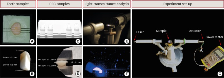
-
Abstract
PDFPubReaderePub
Objectives To evaluate and compare light-transmittance in dental tissues and dental composite restorations using the incremental double-layer technique with varying layer thickness.
Materials and Methods B1-colored natural teeth slabs were compared to dental restoration build-ups with A2D and B1E-colored nanofilled, supra-nanofilled, microfilled, and microhybrid composites. The enamel layer varied from 0.3, 0.5, or 1.2 mm thick, and the dentin layer was varied to provide a standardized 3.7 mm overall sample thickness (
n = 10). All increments were light-cured to 16 J/cm2 with a multi-wave LED (Valo, Ultradent). Using a spectrophotometer, the samples were irradiated by an RGB laser beam. A voltmeter recorded the light output signal to calculate the light-transmittance through the specimens. The data were analyzed using 1-way analysis of variance followed by thepost hoc Tukey's test (p = 0.05).Results Mean light-transmittance observed at thicker final layers of enamel were significantly lower than those observed at thinner final layers. Within 1.2 mm final enamel resin layer (FERL) thickness, all composites were similar to the dental tissues, with exception of the nanofilled composite. However, within 0.5 mm FERL thickness, only the supra-nanofilled composite showed no difference from the dental tissues. Within 0.3 mm FERL thickness, none of the composites were similar to the dental tissues.
Conclusions The supra-nanofilled composite had the most similar light-transmittance pattern when compared to the natural teeth. However, for other composites, thicker FERL have a greater chance to match the light-transmittance of natural dental tissues.
-
Citations
Citations to this article as recorded by- Impact of water absorption on the translucency of single-shade and conventional resin composites: an in vitro comparative study
Ceyda Sari, Elifnur Aydemir Aydın
Odontology.2026;[Epub] CrossRef - 3-year randomized clinical trial to evaluate the performance of posterior composite restorations lined with ion-releasing materials
Basma Ahmed, Ramy Ahmed Wafaie, Hamdi H. Hamama, Salah Hasab Mahmoud
Scientific Reports.2024;[Epub] CrossRef - Investigation on the Biaxial Flexural Strength of Universal Shade Resin-Based Composites
Keiko Sakuma, Taku Horie, Takafumi Kishimoto, Mayumi Maesako, Shigetaka Tomoda, Morioki Fujitani, Akimasa Tsujimoto
Polymers.2024; 16(13): 1853. CrossRef - Fabrication of color-graded feldspathic dental prosthetics for aesthetic and restorative dentistry
Imam Akbar Sutejo, Jeehwan Kim, Sinuo Zhang, Chang Woo Gal, Yeong-Jin Choi, Honghyun Park, Hui-suk Yun
Dental Materials.2023; 39(6): 568. CrossRef - Spectrophotometric evaluation of restorative composite shades and their match with a classical shade guide
Rafael Melara, Luciana Mendonça, Fábio Herrmann Coelho-de-Souza, Juliana Nunes Rolla, Luciano de Souza Gonçalves
Restorative Dentistry & Endodontics.2021;[Epub] CrossRef - In vitro wear of dual‐cured bulkfill composites and flowable bulkfill composites
Jean‐François Roulet, Snigdha Gummadi, Hind S. Hussein, Nader Abdulhameed, Chiayi Shen
Journal of Esthetic and Restorative Dentistry.2020; 32(5): 512. CrossRef
- Impact of water absorption on the translucency of single-shade and conventional resin composites: an in vitro comparative study
- 2,422 View
- 19 Download
- 6 Crossref
Case Report
- Management of dental erosion induced by gastro-esophageal reflux disorder with direct composite veneering aided by a flexible splint matrix
- Sherin Jose Chockattu, Byathnal Suryakant Deepak, Anubhav Sood, Nandini T. Niranjan, Arun Jayasheel, Mallikarjun K. Goud
- Restor Dent Endod 2018;43(1):e13. Published online February 6, 2018
- DOI: https://doi.org/10.5395/rde.2018.43.e13
-
Abstract
PDFPubReaderePub
Dental erosion is frequently overlooked in clinical practice. The management of erosion-induced damage to the dentition is often delayed, such that extensive occlusal rehabilitation is required. These cases can be diagnosed by a careful clinical examination and a thorough review of the patient's medical history and/or lifestyle habits. This case report presents the diagnosis, categorization, and management of a case of gastro-esophageal reflux disease-induced palatal erosion of the maxillary teeth. The early management of such cases is of utmost importance to delay or prevent the progression of damage both to the dentition and to occlusal stability. Non-invasive adhesively bonded restorations aid in achieving this goal.
-
Citations
Citations to this article as recorded by- Effect of Acidic Media on Surface Topography and Color Stability of Two Different Glass Ceramics
Fatma Makkeyah, Nesrine A. Elsahn, Mahmoud M. Bakr, Mahmoud Al Ankily
European Journal of Dentistry.2025; 19(01): 173. CrossRef - Mechanical Performance and Surface Roughness of Lithium Disilicate and Zirconia-Reinforced Lithium Silicate Ceramics Before and After Exposure to Acidic Challenge
Ahmed Elsherbini, Salma M. Fathy, Walid Al-Zordk, Mutlu Özcan, Amal A. Sakrana
Dentistry Journal.2025; 13(3): 117. CrossRef - Biomechanical reinforcement by CAD-CAM materials affects stress distributions of posterior composite bridges: 3D finite element analysis.
Alaaeldin Elraggal, Islam M. Abdelraheem, David C. Watts, Sandipan Roy, Vamsi Krishna Dommeti, Abdulrahman Alshabib, Khaled Abid Althaqafi, Rania R. Afifi
Dental Materials.2024; 40(5): 869. CrossRef - Surface Properties and Wear Resistance of Injectable and Computer-Aided Design/Computer Aided Manufacturing–Milled Resin Composite Thin Occlusal Veneers
Nesrine A. Elsahn, Hatem M. El-Damanhoury, Zainab Shirazi, Abdul Rahman M. Saleh
European Journal of Dentistry.2023; 17(03): 663. CrossRef - Effect of acidic media on flexural strength and fatigue of CAD-CAM dental materials
Alaaeldin Elraggal, Rania. R Afifi, Rasha A. Alamoush, Islam Abdel Raheem, David C. Watts
Dental Materials.2023; 39(1): 57. CrossRef - Three-year Follow-up of Conservative Direct Composite Veneers on Eroded Teeth
RQ Ramos, NF Coelho, GC Lopes
Operative Dentistry.2022; 47(2): 131. CrossRef - The effects of intrinsic and extrinsic acids on nanofilled and bulk fill resin composites: Roughness, surface hardness, and scanning electron microscopy analysis
Milena F. Alencar, Mirella T. Pereira, Maria D. R. De‐Moraes, Sérgio L. Santiago, Vanara F. Passos
Microscopy Research and Technique.2020; 83(2): 202. CrossRef
- Effect of Acidic Media on Surface Topography and Color Stability of Two Different Glass Ceramics
- 3,222 View
- 28 Download
- 7 Crossref
Research Article
- Effect of water storage on flexural strength of silorane and methacrylate-based composite resins
- Narges Panahandeh, Hassan Torabzadeh, Hani Naderi, Seyedeh Mahsa Sheikh-Al-Eslamian
- Restor Dent Endod 2017;42(4):309-315. Published online November 6, 2017
- DOI: https://doi.org/10.5395/rde.2017.42.4.309
-
Abstract
PDFPubReaderePub
Objectives This study assessed the effect of water storage on the flexural strength (FS) of low shrinkage composites.
Materials and Methods A total of 165 bar-shaped specimens (2 × 2 × 25 mm) were fabricated of 2 low shrinkage composites (Filtek P90 [3M ESPE], GC Kalore [GC International]) and a conventional methacrylate-based composite (Filtek Z250 [3M ESPE]). The specimens were subjected to 3-point bending test at 6 time intervals, namely: immediately after curing, at 24 hours, 1 week, 1 month, 6 months, and 1 year following storage in wet and dry conditions. The FS of the specimens were measured by applying compressive load at a crosshead speed of 1.0 mm/min. Data was analyzed using 3-way analysis of variance (ANOVA) and Tukey's test.
Results Three-way ANOVA revealed significant interactions between time, type of composite, and storage condition (
p = 0.001). Tukey's multiple comparison test revealed significant reductions in FS of all composites after 6 months and 1 year of storage in distilled water compared to dry condition.Conclusions Filtek P90 showed the highest and GC Kalore showed the lowest FS after 1 year storage in distilled water. The immediate high strength of Filtek Z250 significantly decreased at 1 year and its final value was lower than that of Filtek P90.
-
Citations
Citations to this article as recorded by- Graphene–Catechol Dental Sealant: Antibacterial and Mechanical Evaluation
Renata Pereira, Flávio H. B. Aguiar, Rodrigo B. E. Lins, Maria C. A. J. Mainairdi, Bruna G. Silva, Marcela A. Ferretti, Klaus Rischka
Advanced Engineering Materials.2025;[Epub] CrossRef - Effects of Bio-Aging on Mechanical Properties and Microbial Behavior of Different Resin Composites
Yuke Shou, Lanzhi Deng, Xiaoyu Huang, Xinyu Peng, Xinxuan Zhou, Zheng Wang, Yannan Huang, Bina Yang, Haohao Wang, Min Zhang, Lei Cheng
Biomolecules.2023; 13(7): 1125. CrossRef - Changes in color and contrast ratio of resin composites after curing and storage in water
Marlus da Silva Pedrosa, Fernando Neves Nogueira, Vitor de Oliveira Baldo, Igor Studart Medeiros
The Saudi Dental Journal.2021; 33(8): 1160. CrossRef - Ageing of Dental Composites Based on Methacrylate Resins—A Critical Review of the Causes and Method of Assessment
Agata Szczesio-Wlodarczyk, Jerzy Sokolowski, Joanna Kleczewska, Kinga Bociong
Polymers.2020; 12(4): 882. CrossRef - Color stability of nanohybrid composite resins in drinks
Juliana Jendiroba Faraoni, Isabela Barbosa Quero, Lívia Semedo Schiavuzzo, Regina Guenka Palma-Dibb
Brazilian Journal of Oral Sciences.2019; 18: e191601. CrossRef - Mechanical Degradation of Different Classes of Composite Resins Aged in Water, Air, and Oil
Weber Adad Ricci, Priscila Alfano, Saulo Pamato, Carlos Alberto dos Santos Cruz, Jefferson Ricardo Pereira
BioMed Research International.2019; 2019: 1. CrossRef - Effects of water and microbial-based aging on the performance of three dental restorative materials
Xinxuan Zhou, Suping Wang, Xian Peng, Yao Hu, Biao Ren, Mingyun Li, Liying Hao, Mingye Feng, Lei Cheng, Xuedong Zhou
Journal of the Mechanical Behavior of Biomedical Materials.2018; 80: 42. CrossRef
- Graphene–Catechol Dental Sealant: Antibacterial and Mechanical Evaluation
- 2,458 View
- 7 Download
- 7 Crossref
Review Article
- Survival rates against fracture of endodontically treated posterior teeth restored with full-coverage crowns or resin composite restorations: a systematic review
- Warattama Suksaphar, Danuchit Banomyong, Titalee Jirathanyanatt, Yaowaluk Ngoenwiwatkul
- Restor Dent Endod 2017;42(3):157-167. Published online July 31, 2017
- DOI: https://doi.org/10.5395/rde.2017.42.3.157
-
Abstract
PDFPubReaderePub
This systematic review aims to summarize the current clinical studies that investigated survival rates against fracture of endodontically treated posterior teeth restored with crowns or resin composite restorations. Literature search were performed using keywords. Publications from 1980 to 2016 were searched in PubMed, ScienceDirect, Web of Science, MEDLINE, and SCOPUS. Included studies were selected based on inclusion and exclusion criteria. Three clinical studies were included: 1 randomized controlled trial and 1 prospective and 1 retrospective cohort studies. Pooled survival rates ranged from 94%–100% and 91.9%–100% for crowns and resin composite, respectively. The majority of teeth had no more than 3 surface loss of tooth structure. The studies included were heterogeneous, and were not appropriate for further meta-analysis. Current evidence suggested that the survival rates against the fracture of endodontically treated posterior teeth restored with crowns or resin composites were not significantly different in the teeth with minimum to moderate loss of tooth structure.
-
Citations
Citations to this article as recorded by- Clinical Outcomes and Complication Rates of Crown Restorations with Various Endodontic Posts: A Retrospective Analysis
Ali Alenezi, Hanin Alsalhi
Journal of Functional Biomaterials.2026; 17(2): 84. CrossRef - Restoration of the fractured or heavily restored premolar
Chris Redman, Ewen McColl, Graeme Bryce
Dental Update.2026; 53(5): 323. CrossRef - Comparison of fracture resistance among restorative techniques for endodontically treated mandibular premolars: an in vitro study
Dinie Qurratuaini Zulkifly, Mohd Haikal Muhamad Halil, Ubadah Zamry, Siti Nabilah Huda Mohamad Nor
The Saudi Dental Journal.2026;[Epub] CrossRef - Effect of using different materials and restorative techniques on cuspal deflection and microleakage in endodontically treated teeth
Ceyda Sari, Oya Bala, Sinem Akgul, Cemile Kedici Alp
BMC Oral Health.2025;[Epub] CrossRef - Direct restorations versus full crowns in endodontically treated molar teeth: A three-year randomized clinical trial
Motasum Abu-Awwad, Ruba Halasa, Laila Haikal, Ahmad El-Ma'aita, Mohammad Hammad, Haralampos Petridis
Journal of Dentistry.2025; 156: 105699. CrossRef - Is the use of an intraradicular post essential for reducing failures in restoring endodontically treated teeth? A systematic review and meta-analysis
Jacqueline Salomão Jardim, Vinicius de Menezes Félix Ferreira, Hiskell Francine Fernandes e Oliveira, Daniele Sorgatto Faé, Cleidiel Aparecido Araujo Lemos
Journal of Dentistry.2025; 159: 105739. CrossRef - Systematic Reviews Comparing Direct and Indirect Restorations: An Umbrella Review That Examines Restoration Type and Confidence in Results
Mona Kimmel, Clovis Mariano Faggion
Clinical and Experimental Dental Research.2025;[Epub] CrossRef - Knowledge, Awareness, and Perceptions on Root Canal Treatment among Patients Reporting with Dental Pain to Conservative Dentistry and Endodontics Department: An Institution-based Survey
Abdu Semeer Palottil, Moopil Midhun Mohanan, N. T. Nishad, S. Jayasree
Journal of Primary Care Dentistry and Oral Health.2025; 6(2): 99. CrossRef - One-year clinical performance of restorations with and without a bulk-fill flowable base in endodontically treated premolars: a pilot randomized controlled trial
Brenda Leyton, Jullyana Dezanetti, Rodrigo Rached, Sérgio Ignácio, Evelise Souza
BMC Oral Health.2025;[Epub] CrossRef - One-piece endodontic crown fixed partial denture: Is it possible?
João Paulo M. Tribst, Amanda Maria de O. Dal Piva, Joris Muris, Cornelis J. Kleverlaan, Albert J. Feilzer
The Journal of Prosthetic Dentistry.2024; 131(6): 1118. CrossRef - Survival Rate Against Fracture of Endodontically Treated Premolars Restored with Crowns and Resin Composites: A Retrospective Study
Enas Khamakhim, Farida Alsayeh
AlQalam Journal of Medical and Applied Sciences.2024; : 398. CrossRef - Knowledge and Awareness of Root Canal Treatment among Patients in Tripoli: A Survey-Based Study
Sumaya Aghila
AlQalam Journal of Medical and Applied Sciences.2024; : 532. CrossRef - Clinical performance of polyethylenefiber reinforced resin composite restorations in endodontically treated teeth: (a randomized controlled clinical trial)
Ahmed Abdelsattar Metwaly, Amira Farid Elzoghby, Rawda Hesham Abd ElAziz
BMC Oral Health.2024;[Epub] CrossRef - Direct Versus Indirect Treatment Options of Endodontically Treated Posterior Teeth: A Narrative Review
Mai M Alhamdan, Rodina F Aljamaan, Munira M Abuthnain, Shahd A Alsumikhi, Ghada S Alqahtani, Reem A Alkharaiyef
Cureus.2024;[Epub] CrossRef - Single crown vs. composite for glass fiber post-retained restorations: An 8-year randomized clinical trial
Victório Poletto-Neto, Luiz Alexandre Chisini, Wietske Fokkinga, Cees Kreulen, Bas Loomans, Maximiliano Sérgio Cenci, Tatiana Pereira-Cenci
Journal of Dentistry.2024; 142: 104837. CrossRef - Factors influencing the clinical performance of the restoration of endodontically treated teeth: An assessment of systematic reviews of clinical studies
Lara Dotto, Luiza Paloma S. Girotto, Yara Teresinha Correa Silva Sousa, Gabriel Kalil Rocha Pereira, Ataís Bacchi, Rafael Sarkis-Onofre
The Journal of Prosthetic Dentistry.2024; 131(6): 1043. CrossRef - Influence of technical quality and coronal restoration on periapical health of root canal treatment performed by Malaysian undergraduate students
Norazlina Mohammad, Faizah Abdul Fatah, Azlan Jaafar, Siti Hajar Omar, Aimi Amalina Ahmad, Abdul Azim Asy Abdul Aziz, Aws Hashim Ali Al-Kadhim
Saudi Endodontic Journal.2023; 13(1): 63. CrossRef - The success rate of indirect adhesive restorations in the distal dentition fabricated with chairside CAD/CAM system
Marek Šupler, Andrej Jenča, Michal Straka, Juraj Deglovič, Janka Jenčová
Stomatológ.2023; 33(2): 10. CrossRef - A Comparative Study of the Marginal Fit of Endocrowns Fabricated From Three Different Computer-Aided Design/Computer-Aided Manufacturing (CAD/CAM) Ceramic Materials: An In Vitro Study
Esraa Attar, Shatha Alshali, Tariq Abuhaimed
Cureus.2023;[Epub] CrossRef - Evaluation of titanium mesh and fibers in reinforcing endodontically treated molars: An in vitro study
Hemalatha Hiremath, Devansh Verma, Sheetal Khandelwal, AishwaryaSingh Solanki, Sonam Patidar
Journal of Conservative Dentistry.2022; 25(2): 189. CrossRef - Effect of surface treatment, ferrule height, and luting agent type on pull-out bond strength of monolithic zirconia endocrowns
Emine B. Buyukerkmen, Durmuş A. Bozkurt, Arslan Terlemez
Journal of Oral Science.2022; 64(4): 279. CrossRef - An Umbrella Review of Systematic Reviews and Meta‐Analyses Evaluating the Success Rate of Prosthetic Restorations on Endodontically Treated Teeth
Amirhossein Fathi, Behnaz Ebadian, Sara Nasrollahi Dezaki, Nahal Mardasi, Ramin Mosharraf, Sabire Isler, Shiva Sadat Tabatabaei, Stefano Pagano
International Journal of Dentistry.2022;[Epub] CrossRef - Survival and success of endocrowns: A systematic review and meta-analysis
Raghad A. Al-Dabbagh
The Journal of Prosthetic Dentistry.2021; 125(3): 415.e1. CrossRef - Fracture strength of non-invasively reinforced MOD cavities on endodontically treated teeth
René Daher, Stefano Ardu, Enrico Di Bella, Giovanni T. Rocca, Albert J. Feilzer, Ivo Krejci
Odontology.2021; 109(2): 368. CrossRef - Retrospective study of fracture survival in endodontically treated molars: the effect of single-unit crowns versus direct-resin composite restorations
Kanet Chotvorrarak, Warattama Suksaphar, Danuchit Banomyong
Restorative Dentistry & Endodontics.2021;[Epub] CrossRef - An insight into patient's perceptions regarding root canal treatment: A questionnaire-based survey
Ramta Bansal, Aditya Jain
Journal of Family Medicine and Primary Care.2020; 9(2): 1020. CrossRef - Endodontically treated posterior teeth restored with or without crown restorations: A 5‐year retrospective study of survival rates from fracture
Titalee Jirathanyanatt, Warattama Suksaphar, Danuchit Banomyong, Yaowaluk Ngoenwiwatkul
Journal of Investigative and Clinical Dentistry.2019;[Epub] CrossRef - Fracture resistance, gap and void formation in root‐filled mandibular molars restored with bulk‐fill resin composites and glass‐ionomer cement base
Nathamon Thongbai‐on, Kanet Chotvorrarak, Danuchit Banomyong, Michael F. Burrow, Sittichoke Osiri, Nattha Pattaravisitsate
Journal of Investigative and Clinical Dentistry.2019;[Epub] CrossRef - Current options concerning the endodontically-treated teeth restoration with the adhesive approach
Marco Aurélio de Carvalho, Priscilla Cardoso Lazari, Marco Gresnigt, Altair Antoninha Del Bel Cury, Pascal Magne
Brazilian Oral Research.2018;[Epub] CrossRef
- Clinical Outcomes and Complication Rates of Crown Restorations with Various Endodontic Posts: A Retrospective Analysis
- 7,131 View
- 85 Download
- 29 Crossref
Research Articles
-
In vitro study ofStreptococcus mutans adhesion on composite resin coated with three surface sealants - Da Hye Kim, Tae-Yub Kwon
- Restor Dent Endod 2017;42(1):39-47. Published online December 30, 2016
- DOI: https://doi.org/10.5395/rde.2017.42.1.39
-
Abstract
PDFPubReaderePub
Objectives Although the coating of surface sealants to dental composite resin may potentially reduce bacterial adhesion, there seems to be little information regarding this issue. This preliminary
in vitro study investigated the adhesion ofStreptococcus mutans (S. mutans ) on the dental composite resins coated with three commercial surface sealants.Materials and Methods Composite resin (Filtek Z250) discs (8 mm in diameter, 1 mm in thickness) were fabricated in a mold covered with a Mylar strip (control). In group PoGo, the surfaces were polished with PoGo. In groups PS, OG, and FP, the surfaces polished with PoGo were coated with the corresponding surface sealants (PermaSeal, PS; OptiGuard, OG; Fortify Plus, FP). The surfaces of the materials and
S. mutans cells were characterized by various methods.S. mutans adhesion to the surfaces was quantitatively evaluated using flow cytometry (n = 9).Results Group OG achieved the lowest water contact angle among all groups tested (
p < 0.001). The cell surface ofS. mutans tested showed hydrophobic characteristics. Group PoGo exhibited the greatest bacterial adhesion among all groups tested (p < 0.001). The sealant-coated groups showed statistically similar (groups PS and FP,p > 0.05) or significantly lower (group OG,p < 0.001) bacterial adhesion when compared with the control group.Conclusions The application of the surface sealants significantly reduced
S. mutans adhesion to the composite resin polished with the PoGo.-
Citations
Citations to this article as recorded by- Magnesium oxide nanoparticles-modified 3D-printed denture tooth resin: Interfacial antibiofilm activity against Streptococcus mutans biofilms and material performance in vitro
Qi Xue, Wenshun Wang, Chongyi Wang, Ximei Xiao, Yongliang Wang, Meiwen Cao, Jing Fu
Colloids and Surfaces A: Physicochemical and Engineering Aspects.2026; 732: 139162. CrossRef - Comparative Analysis of Streptococcus mutans and Lactobacillus spp. Colonization on Stainless Steel versus Zirconia Crowns in Root Canal-Treated Teeth
Bahni S Pathak, Alpana Talukdar, Indrani Barman, Shivansh Aggrohiya , Upasana Barman , Samar Khan, Seema Gupta
Cureus.2025;[Epub] CrossRef - Comparative assessment of clinical performance and Streptococcus mutans adherence in primary molars restored with Bioflx and Zirconia crowns: A split-mouth pilot study
Apoorva Goswami, Hemalatha Ramkumar, Shankar Paulindraraj, Trophimus Gnanabagyan Jayakaran, Kalpana Hari Krishnan, Aroonika Bedre
Journal of Indian Society of Pedodontics and Preventive Dentistry.2025; 43(4): 575. CrossRef - Polychromatic Composite and Resin Infiltration Restorations in the Esthetic Zone: A Five-year Clinical Report
K Karimi, NG Fischer, CA Jurado, J Villalobos-Tinoco, A Tsujimoto
Operative Dentistry.2023; 48(3): 245. CrossRef - Antibacterial Effect of Zirconia Nanoparticles on Polyethyl Methacrylate Resin for Provisional Crowns
Hee-Seon Kim, Woohyung Jang, Yeong-Gwan Im, Hyun-Pil Lim
International Journal of Nanomedicine.2022; Volume 17: 6551. CrossRef - Do the differences in organic contents of composite resins affect surface roughness and Streptococcus mutans adhesion?
Duygu Hisarbeyli, Nazmiye Dönmez, Nursen Topçuoğlu
Journal of Adhesion Science and Technology.2022; 36(9): 921. CrossRef - The Effect of Liquid Rubber Addition on the Physicochemical Properties, Cytotoxicity, and Ability to Inhibit Biofilm Formation of Dental Composites
Krzysztof Pałka, Małgorzata Miazga-Karska, Joanna Pawłat, Joanna Kleczewska, Agata Przekora
Materials.2021; 14(7): 1704. CrossRef - PLGA nanoparticles loaded with quaternary ammonium silane and riboflavin for potential applications in adhesive dentistry
Umer Daood, Meera Priyadarshini Balasankar, Marrwa A. Ibrahim, Mallikarjuna Rao Pichika, Kit-Kay Mak, Amr S. Fawzy
International Journal of Adhesion and Adhesives.2021; 105: 102797. CrossRef - Effect of Washing Condition on the Fracture Strength, and the Degree of Conversion of 3D Printing Resin
Woohyung Jang, Gyeong-Soo Kook, Jin-Ho Kang, Yeseul Kim, Yina Yun, Seon-Ki Lee, Sang-Won Park, Hyun-Pil Lim, Kwi-Dug Yun, Chan Park
Applied Sciences.2021; 11(24): 11676. CrossRef - Effect of surface sealant on surface roughness and bacterial adhesion of bulk-fill composites
Gunce Ozan, Meltem Mert Eren, Cansu Vatansever, Ugur Erdemir
Polymers and Polymer Composites.2021; 29(9_suppl): S475. CrossRef - Evaluation of adhesion of Streptococcus mutans, plaque accumulation on zirconia and stainless steel crowns, and surrounding gingival inflammation in primary molars: randomized controlled trial
Mebin George Mathew, S. R. Samuel, Ashu Jagdish Soni, Korishettar Basavaraj Roopa
Clinical Oral Investigations.2020; 24(9): 3275. CrossRef - Influence of resin-coating agents on the roughness and color of composite resins
Fabio A.P. Rizzante, Juliana S.F. Bombonatti, Layla Vasconcelos, Thiago S. Porto, Sorin Teich, Rafael F.L. Mondelli
The Journal of Prosthetic Dentistry.2019; 122(3): 332.e1. CrossRef - ANALYSIS OF THE CLINICAL EFFICIENCY OF RESTORATIVE FILLING MATERIALS
Valentina I. Kolodkina
Kuban Scientific Medical Bulletin.2019; 26(2): 64. CrossRef - Inhibitory effect of Bacillus velezensis on biofilm formation by Streptococcus mutans
Yesol Yoo, Dong-Ho Seo, Hyunjin Lee, Eui-Sang Cho, Nho-Eul Song, Tea Gyu Nam, Young-Do Nam, Myung-Ji Seo
Journal of Biotechnology.2019; 298: 57. CrossRef - Evaluation of Surface Roughness and <i>Streptococcus mutans</i> Adhesion to Bulk-Fill Resin Composites Polished with Different Systems
Wafaa E. Soliman, Ashraf I. Ali, Walid F. Elkhatib
Advances in Microbiology.2019; 09(01): 87. CrossRef - Effectiveness of the DHMAI monomer in the development of an antibacterial dental composite
Fatima Zohra Cherchali, Mohamed Mouzali, Jean Bernard Tommasino, Dominique Decoret, Nina Attik, Hazem Aboulleil, Dominique Seux, Brigitte Grosgogeat
Dental Materials.2017; 33(12): 1381. CrossRef
- Magnesium oxide nanoparticles-modified 3D-printed denture tooth resin: Interfacial antibiofilm activity against Streptococcus mutans biofilms and material performance in vitro
- 3,370 View
- 15 Download
- 16 Crossref
- Effect of immersion into solutions at various pH on the color stability of composite resins with different shades
- Ji-Deok Moon, Eun-Mi Seon, Sung-Ae Son, Kyoung-Hwa Jung, Yong-Hoon Kwon, Jeong-Kil Park
- Restor Dent Endod 2015;40(4):270-276. Published online August 28, 2015
- DOI: https://doi.org/10.5395/rde.2015.40.4.270
-
Abstract
PDFPubReaderePub
Objectives This study examined the color changes of a resin composite with different shades upon exposure to water with different pH.
Materials and Methods Nanohybrid resin composites (Filtek Z350XT, 3M ESPE) with four different shades (A2, A3, B1, and B2) were immersed in water with three different pH (pH 3, 6, and 9) for 14 day. The CIE
L*a*b* color coordinates of the specimens were evaluated before and after immersion in the solutions. The color difference (ΔE* ) and the translucency parameter (TP ) were calculated using the color coordinates.Results ΔE* ranged from 0.33 to 1.58, and the values were affected significantly by the pH. The specimens immersed in a pH 6 solution showed the highestΔE* values (0.87 - 1.58). The specimens with a B1 shade showed the lowestΔE* change compared to the other shades.TP ranged from 7.01 to 9.46 depending on the pH and resin shade. TheTP difference between before and after immersion in the pH solutions was less than 1.0.Conclusions The resulting change of color of the tested specimens did not appear to be clinically problematic because the color difference was < 1.6 in the acidic, neutral, and alkaline solutions regardless of the resin shade, i.e., the color change was imperceptible.
-
Citations
Citations to this article as recorded by- Effect of Acidic Environment on the Mechanical Strength and Surface Properties of Three-Dimensional-Printed Denture Base Resin
Eun-Ju Kim, Se-Eun Kim, Ye-Jin Kim, Ji-Eun Kim, Yun-Yeong Hwang, Hye-Bin Go, Song-Yi Yang
International Dental Journal.2026; 76(5): 109757. CrossRef - Effect of mouth rinses on roughness and optical properties of restorative materials for oral rehabilitation
Laura Firmo de Carvalho, Edmara T. P. Bergamo, Ernesto B. Benalcázar-Jalkh, Tiago M. B. Campos, Abbas Zahoui, Elisa De Souza Fermino, Ana Clara Mota de Oliveira, Ana Carolina Magalhães, Estevam A. Bonfante, Fábio José B. Bezerra, Larissa M.M. Alves
Biomaterial Investigations in Dentistry.2025; 12: 25. CrossRef - Effect of Printing Layer Thickness on the Color Stability and Surface Roughness of Three-dimensional Printed Resin Material Immersed in Different Aging Media: An In Vitro Study
Vilas Rajguru, Smita Khalikar, Sonali Mahajan, Gopika Gopan, Siddhi D Bhatawadekar, Kishor Mahale
International Journal of Prosthodontics and Restorative Dentistry.2025; 15(1): 36. CrossRef - Colour Retaining Ability of Three Restorative Materials Used in Pediatric Dentistry with the Use of Health Drinks and Beverages - An In Vitro Study
Arnab Mondal, Swati Singh, Seema Qamar, Barun Dasgupta, Shovan Roy
Indian Journal of Dental Research.2025; 36(1): 89. CrossRef - Effects of different antiviral mouthwashes on the surface roughness, hardness, and color stability of composite CAD/CAM materials
Ahmet Hazar, Ecehan Hazar
Journal of Applied Biomaterials & Functional Materials.2024;[Epub] CrossRef - Flexural Properties of Contemporary Bioactive Restorative Materials: Effect of Environmental pH
JEX Ong, AU Yap, A Abdul Aziz, NA Yahya
Operative Dentistry.2023; 48(1): 90. CrossRef - Effect of Mouthwashes for COVID-19 Prevention on Surface Changes of Resin Composites
Saijai Tanthanuch, Boonlert Kukiattrakoon, Chailuck Naiyanart, Tanyanat Promtong, Panuwit Yothinwatthanabamrung, Suttida Pumpua
International Dental Journal.2023; 73(4): 511. CrossRef - Can Modification with Urethane Derivatives or the Addition of an Anti-Hydrolysis Agent Influence the Hydrolytic Stability of Resin Dental Composite?
Agata Szczesio-Wlodarczyk, Izabela M. Barszczewska-Rybarek, Marta W. Chrószcz-Porębska, Karolina Kopacz, Jerzy Sokolowski, Kinga Bociong
International Journal of Molecular Sciences.2023; 24(5): 4336. CrossRef - Understanding the Mechanical, Surface, and Color Behavior of Oral Bioactive Prosthetic Polymers under Biodegradation Processes
Cristina B. Neves, Joana Costa, Jaime Portugal, Ana F. Bettencourt
Polymers.2023; 15(11): 2549. CrossRef - An Evaluation of the Hydrolytic Stability of Selected Experimental Dental Matrices and Composites
Agata Szczesio-Wlodarczyk, Karolina Kopacz, Malgorzata Iwona Szynkowska-Jozwik, Jerzy Sokolowski, Kinga Bociong
Materials.2022; 15(14): 5055. CrossRef - Evaluation of the Color Stability, Water Sorption, and Solubility of Current Resin Composites
Wenkai Huang, Ling Ren, Yuyao Cheng, Minghua Xu, Wenji Luo, Desong Zhan, Hidehiko Sano, Jiale Fu
Materials.2022; 15(19): 6710. CrossRef - Comparative Evaluation of Surface Roughness of different Composites and its effect on Colour Stability of the Restoration
Nalini B, Srinivas Kumar Ch, Narsimha Rao VV
Research Journal of Pharmacy and Technology.2022; : 3854. CrossRef - Comparison of instrumental methods for color change assessment of Giomer resins
Luiza de Almeida Queiroz Ferreira, Rogéli Tibúrcio Ribeiro da Cunha Peixoto, Cláudia Silami de Magalhães, Tassiana Melo Sá, Monica Yamauti, Francisca Daniele Moreira Jardilino
Restorative Dentistry & Endodontics.2022;[Epub] CrossRef - Effect of hydrothermal aging on the microhardness of high- and low-viscosity conventional and additively manufactured polymers
Nadin Al-Haj Husain, Albert J. Feilzer, Cornelis J. Kleverlaan, Samir Abou-Ayash, Mutlu Özcan
The Journal of Prosthetic Dentistry.2022; 128(4): 822.e1. CrossRef - KARANFİL VE YEŞİL ÇAYIN RESTORATİF MATERYALLERİN YÜZEY VE OPTİK ÖZELLİKLERİ ÜZERİNE ETKİSİ: İN VİTRO ÇALIŞMA
Zeynep BUKET KAYNAR, Tolgahan DOĞAN, Nazmiye DÖNMEZ, Mağrur KAZAK
Atatürk Üniversitesi Diş Hekimliği Fakültesi Dergisi.2022; : 1. CrossRef - Effects of Red Dragon Fruit on Color Stability of Self-Adhesive Flowable Composite
Rosalina Tjandrawinata, Brigitta Heidy, Octarina
Applied Mechanics and Materials.2022; 910: 1. CrossRef - Effect of different cement systems and aging on the bond strength of chairside CAD-CAM ceramics
Seda Ustun, Elif Aydogan Ayaz
The Journal of Prosthetic Dentistry.2021; 125(2): 334. CrossRef - Influence of Selected Restorative Materials on the Environmental pH: In Vitro Comparative Study
Anna Lehmann, Kacper Nijakowski, Michalina Nowakowska, Patryk Woś, Maria Misiaszek, Anna Surdacka
Applied Sciences.2021; 11(24): 11975. CrossRef - Color stability of an artificially aged nanofilled composite resin post-cured with different techniques
Lais Sampaio Souza, Tais Rocha Donato, Gabriela Alves Cerqueira, Andrea Nobrega Cavalcanti, Paula Mathias
Journal of Dental Research, Dental Clinics, Dental Prospects.2021; 15(1): 53. CrossRef - Ageing of Dental Composites Based on Methacrylate Resins—A Critical Review of the Causes and Method of Assessment
Agata Szczesio-Wlodarczyk, Jerzy Sokolowski, Joanna Kleczewska, Kinga Bociong
Polymers.2020; 12(4): 882. CrossRef - Use of Resin Solvent as a Facilitator for Removal of Resin Composite Restoratives by Influencing their Mechanical Properties: Is this Possible?
Inas A Elghandour
Journal of Operative Dentistry & Endodontics.2019; 4(1): 1. CrossRef - Effect of powder-liquid ratios and powder colors on color stability of 4-META / MMA-TBB resin after immersion in coffee.
Yoorina Choi, Seo-Jin Jang, Su-Jung Park
Korean Journal of Dental Materials.2018; 45(3): 187. CrossRef - O uso do gel de glicerina melhora a estabilidade de cor de resinas compostas?
Marcus Vinicius Loureiro BERTOLO, Mário Alexandre Coelho SINHORETI, Julia Puppin RONTANI, Pedro Paulo Albuquerque Cavalcanti de ALBUQUERQUE, Luis Felipe Jochims SCHNEIDER
Revista de Odontologia da UNESP.2018; 47(4): 256. CrossRef - In situ evaluation of color stability and hardness' decrease of resin‐based composites
Juliana Silva Ribeiro, Sonia Luque Peralta, Vinícius Esteves Salgado, Rafael Guerra Lund
Journal of Esthetic and Restorative Dentistry.2017; 29(5): 356. CrossRef - Discoloration Effects of Traditional Turkish Beverages on different Composite Restoratives
Serdar Baglar, Erol Keskin, Tahir Orun, Abdulhamit Es
The Journal of Contemporary Dental Practice.2017; 18(2): 83. CrossRef - The effect of red and white wine on color changes of nanofilled and nanohybrid resin composites
Saijai Tanthanuch, Boonlert Kukiattrakoon, Thanwalee Peerasukprasert, Nilobon Chanmanee, Parnchanok Chaisomboonphun, Apisara Rodklai
Restorative Dentistry & Endodontics.2016; 41(2): 130. CrossRef
- Effect of Acidic Environment on the Mechanical Strength and Surface Properties of Three-Dimensional-Printed Denture Base Resin
- 2,509 View
- 16 Download
- 26 Crossref
- Effect of organic acids in dental biofilm on microhardness of a silorane-based composite
- Sedighe Sadat Hashemikamangar, Seyed Jalal Pourhashemi, Mohammad Talebi, Nazanin Kiomarsi, Mohammad Javad Kharazifard
- Restor Dent Endod 2015;40(3):188-194. Published online June 2, 2015
- DOI: https://doi.org/10.5395/rde.2015.40.3.188
-
Abstract
PDFPubReaderePub
Objectives This study evaluated the effect of lactic acid and acetic acid on the microhardness of a silorane-based composite compared to two methacrylate-based composite resins.
Materials and Methods Thirty disc-shaped specimens each were fabricated of Filtek P90, Filtek Z250 and Filtek Z350XT. After measuring of Vickers microhardness, they were randomly divided into 3 subgroups (
n = 10) and immersed in lactic acid, acetic acid or distilled water. Microhardness was measured after 48 hr and 7 day of immersion. Data were analyzed using repeated measures ANOVA (p < 0.05). The surfaces of two additional specimens were evaluated using a scanning electron microscope (SEM) before and after immersion.Results All groups showed a reduction in microhardness after 7 day of immersion (
p < 0.001). At baseline and 7 day, the microhardness of Z250 was the greatest, followed by Z350 and P90 (p < 0.001). At 48 hr, the microhardness values of Z250 and Z350 were greater than P90 (p < 0.001 for both), but those of Z250 and Z350 were not significantly different (p = 0.095). Also, the effect of storage media on microhardness was not significant at baseline, but significant at 48 hr and after 7 day (p = 0.001 andp < 0.001, respectively). Lactic acid had the greatest effect.Conclusions The microhardness of composites decreased after 7 day of immersion. The microhardness of P90 was lower than that of other composites. Lactic acid caused a greater reduction in microhardness compared to other solutions.
-
Citations
Citations to this article as recorded by- Effect of hydroelectrolytic beverages on the roughness and microhardness of bulk fill resin composites
Renata Siqueira Scatolin, Caio Castro Grigoletto, Laura Nobre Ferraz, Rafael Pino Vitti
Brazilian Journal of Oral Sciences.2025; 24: e254003. CrossRef - Investigating the effect of three carbonated drinks on tooth enamel roughness and microhybrid composite
Sara Akbari Fard, Saeed Nemati Anaraki, Haleh Kazemi -Yazdi, Mahsa Qenaat
journal of research in dental sciences.2024; 21(3): 174. CrossRef - Evaluating the effect of natural, industrial juices and beverage on orthodontic bonding composite (in-vitro study)
Rusal S Ahmed, Alan I Saleem
Journal of Baghdad College of Dentistry.2023; 35(3): 10. CrossRef - Stoichiometric models of sucrose and glucose fermentation by oral streptococci: Implications for free acid formation and enamel demineralization
Marzieh Mansouri, Evan P. O'Brien, Karabi Mondal, Chien-Chia Chen, James L. Drummond, Luke Hanley, Karl J. Rockne
Dental Materials.2023; 39(4): 351. CrossRef - Effect of mouthwashes on the microhardness of aesthetic composite restorative materials
Noura Abdulaziz Alessa
Anales del Sistema Sanitario de Navarra.2023;[Epub] CrossRef - Evaluation of the Effect of Natural and Industrial Orange Juices and Beverage on Surface Roughness of Orthodontic Bonding Composite
Rusal Saad Ahmed, Alan Issa Saleem
Dental Hypotheses.2022; 13(3): 107. CrossRef - Effects of particle distribution and calculation method on results of nano-indentation technique in heterogeneous nanocomposites-experimental and numerical approaches
M. Heidari, A. Karimzadeh, M.R. Ayatollahi, M.Y. Yahya
International Journal of Solids and Structures.2021; 225: 111054. CrossRef - New Resin-Based Bulk-Fill Composites: in vitro Evaluation of Micro-Hardness and Depth of Cure as Infection Risk Indexes
Marco Colombo, Simone Gallo, Claudio Poggio, Vittorio Ricaldone, Carla Renata Arciola, Andrea Scribante
Materials.2020; 13(6): 1308. CrossRef - Tribological Behavior of Restorative Dental Microcomposites After Exposure to Mouth Acids
A. C. Branco, J. Brito, M. Codorniz, M. Steinhausen, F. Martins, J. Reis, P. Maurício, R. Colaço, A. P. Serro
Tribology Letters.2019;[Epub] CrossRef - Vickers Micro-Hardness of New Restorative CAD/CAM Dental Materials: Evaluation and Comparison after Exposure to Acidic Drink
Marco Colombo, Claudio Poggio, Alessandro Lasagna, Marco Chiesa, Andrea Scribante
Materials.2019; 12(8): 1246. CrossRef - 30 Months Clinical Evaluation of Posterior Composite Resin Restorations
Serdar Akarsu, Hüseyin Özgür Özdemir
The Journal of Dentists.2018; 6: 6. CrossRef - Survival and Associated Risk Factors of Selective Caries Removal Treatments in Primary Teeth: A Retrospective Study in a High Caries Risk Population
Ximena C. Melgar, Niek J.M. Opdam, Marcos Britto Correa, Renata Franzon, Flávio Fernando Demarco, Fernando B. Araujo, Luciano Casagrande
Caries Research.2017; 51(5): 466. CrossRef
- Effect of hydroelectrolytic beverages on the roughness and microhardness of bulk fill resin composites
- 2,318 View
- 9 Download
- 12 Crossref
- The effect of resin thickness on polymerization characteristics of silorane-based composite resin
- Sung-Ae Son, Hyoung-Mee Roh, Bock Hur, Yong-Hoon Kwon, Jeong-Kil Park
- Restor Dent Endod 2014;39(4):310-318. Published online September 5, 2014
- DOI: https://doi.org/10.5395/rde.2014.39.4.310
-
Abstract
PDFPubReaderePub
Objectives This study examined the influence of the resin thickness on the polymerization of silorane- and methacrylate-based composites.
Materials and Methods One silorane-based (Filtek P90, 3M ESPE) and two methacrylate-based (Filtek Z250 and Z350, 3M ESPE) composite resins were used. The number of photons were detected using a photodiode detector at the different thicknesses (thickness, 1, 2 and 3 mm) specimens. The microhardness of the top and bottom surfaces was measured (
n = 15) using a Vickers hardness with 200 gf load and 15 sec dwell time conditions. The degree of conversion (DC) of the specimens was determined using Fourier transform infrared spectroscopy (FTIR). Scratched powder of each top and bottom surface of the specimen dissolved in ethanol for transmission FTIR spectroscopy. The refractive index was measured using a Abbe-type refractometer. To measure the polymerization shrinkage, a linometer was used. The results were analyzed using two-way ANOVA and Tukey's test atp < 0.05 level.Results The silorane-based resin composite showed the lowest filler content and light attenuation among the specimens. P90 showed the highest values in the DC and the lowest microhardness at all depth. In the polymerization shrinkage, P90 showed a significantly lower shrinkage than the rest two resin products (
p < 0.05). P90 showed a significantly lower refractive index than the remaining two resin products (p < 0.05).Conclusions DC, microhardness, polymerization rate and refractive index linearly decreased as specimen thickness linearly increased. P90 showed much less polymerization shrinkage compared to other specimens. P90, even though achieved the highest DC, showed the lowest microhardness and refractive index.
-
Citations
Citations to this article as recorded by- Effect of Different Coring Thicknesses on Tooth Movement During Processing of Complete Dentures: An In Vitro Study
Akshaya Kadunganari, Nandakumar Karamannattil, Aysha Mohamed Ali KP, Neethu Niduvote Poyil, Priyanka K, Shahnaz Shahnawaz
Cureus.2026;[Epub] CrossRef - A Year-Long Comparison of Dentin Bond Strength Using the Co-Curing Technique and Conventional Adhesive Application
Josipa Vukelja Bosnić, Eva Klarić, Ivan Sever, Zrinka Tarle
Journal of Composites Science.2025; 9(3): 131. CrossRef - Chameleon Effect of Universal Shade Composite Polymers in Repairing CAD/CAM Lithium Disilicate
Gaetano Paolone, Giacomo Collivasone, Niccolò De Masi, Alicia Heinichen, Katia Greco, Enrico Gherlone, Giuseppe Cantatore
Materials.2025; 18(13): 3020. CrossRef - The influence of inorganic fillers on the light transmission through resin-matrix composites during the light-curing procedure: an integrative review
Rita Fidalgo-Pereira, Daniela Carpio, Orlanda Torres, Oscar Carvalho, Filipe Silva, Bruno Henriques, Mutlu Özcan, Júlio C. M. Souza
Clinical Oral Investigations.2022; 26(9): 5575. CrossRef - Conversion, Polymerization Shrinkage, Heat Generation, and Depth of Cure of Novel Dental Composites
Saad Liaqat, Humaira Jabeen
Pakistan BioMedical Journal.2022;[Epub] CrossRef - Effect of Polymerization on the Color of Resin Composites
B Korkut, G Dokumacigil, N Murat, PY Atali, B Tarcin, GB Gocmen
Operative Dentistry.2022; 47(5): 514. CrossRef - Shrinkage Stress and Temperature Variation in Resin Composites Cured via Different Photoactivation Methods: Insights for Standardisation of the Photopolymerisation
Guilherme dos Santos Sousa, Gabriel Felipe Guimarães, Edilmar Marcelino, José Eduardo Petit Rodokas, Arilson José de Oliveira Júnior, Ivana Cesarino, Alcides Lopes Leão, Carla dos Santos Riccardi, Mohammad Arjmand, Rafael Plana Simões
Polymers.2021; 13(13): 2065. CrossRef - Effect of the incorporation of silica blow spun nanofibers containing silver nanoparticles (SiO2/Ag) on the mechanical, physicochemical, and biological properties of a low-viscosity bulk-fill composite resin
Soraya Salmanzadeh Ardestani, Roberta Ferreti Bonan, Mariaugusta Ferreira Mota, Rosiane Maria da Costa Farias, Romualdo Rodrigues Menezes, Paulo Rogério Ferreti Bonan, Panmella Pereira Maciel, Flávia Maria de Moraes Ramos-Perez, André Ulisses Dantas Batis
Dental Materials.2021; 37(10): 1615. CrossRef - Light-Curing Units, Photoinitiators System, and Monomers on Physico-Mechanical Properties of Experimental Composite Resins
Gustavo Furlan da Silva Prezotto, Weverteon Soares de Lima, Rafael Pino Vitti, Ariel Farias da Silva, Mário Alexandre Coelho Sinhoreti, William Cunha Brandt
Matéria (Rio de Janeiro).2020;[Epub] CrossRef - Influence of Different Cordless Light-emitting-diode Units and Battery Levels on Chemical, Mechanical, and Physical Properties of Composite Resin
IO Cardoso, AC Machado, DNR Teixeira, FC Basílio, A Marletta, PV Soares
Operative Dentistry.2020; 45(4): 377. CrossRef - Shrinkage in composites: An enigma
Dhakshinamoorthy Malarvizhi, Arumugam Karthick, NewBegin Selvakumar Gold Pearlin Mary, Alagarsamy Venkatesh
Journal of International Oral Health.2019; 11(5): 244. CrossRef - Development and status of resin composite as dental restorative materials
Xinxuan Zhou, Xiaoyu Huang, Mingyun Li, Xian Peng, Suping Wang, Xuedong Zhou, Lei Cheng
Journal of Applied Polymer Science.2019;[Epub] CrossRef - Effect of the Time of Salivary Contamination during Light Curing on Degree of Conversion and Microhardness of a Restorative Composite Resin
Rasoul Sahebalam, Alireza Boruziniat, Fahimeh Mohammadzadeh, Abdolrasoul Rangrazi
Biomimetics.2018; 3(3): 23. CrossRef - LIGHT POLYMERIZATION OF PHOTO-CURED COMPOSITE MATERIALS: MODERN APPROACHES AND APPLICATION PECULIARITIES
O. A. Udod, V. H. Tsentilo, O. M. Adamenko
Bulletin of Problems Biology and Medicine.2018; 2(4): 72. CrossRef - Resistencia a la compresión del ionómero de vidrio y de la resina compuesta. Estudio in vitro
Sara Blanco Lerech, Sebastián Frías Tarón, Arnulfo Tarón Dunoyer, José María Bustillo Arrieta, Antonio Díaz Caballero
Revista Odontológica Mexicana.2017; 21(2): 109. CrossRef - Compressive strength of glass ionomer and composite resin. In vitro study
Sara Blanco Lerech, Sebastián Frías Tarón, Arnulfo Tarón Dunoyer, José María Bustillo Arrieta, Antonio Díaz Caballero
Revista Odontológica Mexicana.2017; 21(2): e107. CrossRef - Influência de três modos de fotopolimerização sobre a microdureza de três resinas compostas
Andréa Cristina Schneider, Márcio José Mendonça, Roberta Bento Rodrigues, Priscilla do Monte Ribeiro Busato, Veridiana Camilotti
Polímeros.2016; 26(spe): 37. CrossRef - Vickers microhardness comparison of 4 composite resins with different types of filler.
Rene García-Contreras, Rogelio Scougall-Vilchis, Laura Acosta-Torres, Concepción Arenas-Arrocena, Rigoberto García-Garduño, Javier de la Fuente-Hernández
Journal Oral Of Research.2015; 4(5): 313. CrossRef
- Effect of Different Coring Thicknesses on Tooth Movement During Processing of Complete Dentures: An In Vitro Study
- 2,644 View
- 11 Download
- 18 Crossref
- Effect of 38% carbamide peroxide on the microleakage of silorane-based versus methacrylate-based composite restorations
- Sedighe Sadat Hashemi Kamangar, Maryam Ghavam, Nazanin Mahinfar, Seyed Jalal Pourhashemi
- Restor Dent Endod 2014;39(3):172-179. Published online May 13, 2014
- DOI: https://doi.org/10.5395/rde.2014.39.3.172
-
Abstract
PDFPubReaderePub
Objectives This study aimed to assess the effect of 38% carbamide peroxide on the microleakage of class V cavities restored with either a silorane-based composite or two methacrylate-based composites.
Materials and Methods A total of 96 class V cavities were prepared on the buccal surface of extracted human teeth with both enamel and dentin margins and were randomly assigned into three groups of Filtek P90 (3M-ESPE) + P90 system adhesive (3M-ESPE)(group A), Filtek Z250 (3M-ESPE) + Adper Prompt L-Pop (3M-ESPE)(group B) and Filtek Z350XT (3M-ESPE) + Adper Prompt L-Pop (group C). Half of the teeth were randomly underwent bleaching (38% carbamide peroxide, Day White, Discus Dental, applying for 15 min, twice a day for 14 day) while the remaining half (control) were not bleached. Dye penetration was measured following immersion in basic fuchsine. Data were statistically analyzed using Kruskal-Wallis and Mann-Whitney U tests at a level of 0.05.
Results No significant differences were found between composites in the control groups in enamel (
p = 0.171) or dentin (p = 0.094) margins. After bleaching, microleakage of Z250 (in enamel [p = 0.867] or dentin [p = 0.590] margins) and Z350 (in enamel [p = 0.445] or dentin [p = 0.591] margins) did not change significantly, but the microleakage of P90 significantly increased in both enamel (p = 0.042) and dentin (p = 0.002) margins.Conclusions No significant differences were noted between the bleached and control subgroups of two methacrylate-based composites in enamel or dentin margins. Microleakage of silorane-based composite significantly increased after bleaching.
- 1,785 View
- 4 Download
-
Antibacterial properties of composite resins incorporating silver and zinc oxide nanoparticles on
Streptococcus mutans andLactobacillus - Shahin Kasraei, Lida Sami, Sareh Hendi, Mohammad-Yousef AliKhani, Loghman Rezaei-Soufi, Zahra Khamverdi
- Restor Dent Endod 2014;39(2):109-114. Published online March 21, 2014
- DOI: https://doi.org/10.5395/rde.2014.39.2.109
-
Abstract
PDFPubReaderePub
Objectives Recurrent caries was partly ascribed to lack of antibacterial properties in composite resin. Silver and zinc nanoparticles are considered to be broad-spectrum antibacterial agents. The aim of the present study was to evaluate the antibacterial properties of composite resins containing 1% silver and zinc-oxide nanoparticles on
Streptococcus mutans andLactobacillus .Materials and Methods Ninety discoid tablets containing 0%, 1% nano-silver and 1% nano zinc-oxide particles were prepared from flowable composite resin (
n = 30). The antibacterial properties of composite resin discs were evaluated by direct contact test. Diluted solutions ofStreptococcus mutans (PTCC 1683) andLactobacillus (PTCC 1643) were prepared. 0.01 mL of each bacterial species was separately placed on the discs. The discs were transferred to liquid culture media and were incubated at 37℃ for 8 hr. 0.01 mL of each solution was cultured on blood agar and the colonies were counted. Data was analyzed with Kruskall-Wallis and Mann-WhitneyU tests.Results Composites containing nano zinc-oxide particles or silver nanoparticles exhibited higher antibacterial activity against
Streptococcus mutans andLactobacillus compared to the control group (p < 0.05). The effect of zinc-oxide onStreptococcus mutans was significantly higher than that of silver (p < 0.05). There were no significant differences in the antibacterial activity againstLactobacillus between composites containing silver nanoparticles and those containing zinc-oxide nanoparticles.Conclusions Composite resins containing silver or zinc-oxide nanoparticles exhibited antibacterial activity against
Streptococcus mutans andLactobacillus .-
Citations
Citations to this article as recorded by- Fabricated modified compomer bearing CF/SBA-15 nanomaterials: Physicochemical and antibacterial properties
Fatma Nur Kızılay, Mustafa Aydınbelge, Sezer Demirbuğa, Kevser Kolçakoğlu, Nilay Ildız, Serkan Dayan
Dental Materials.2026; 42(3): 451. CrossRef - Physicochemical and antibacterial evaluation of novel nano α-TCP–AgNPs biocomposites for direct pulp-capping applications
Selviana Wulansari, Hendra Dian Adhita Dharsono, Nasrul Wathoni, Rosalina Tjandrawinata, Arief Cahyanto, Moehamad Orliando Roeslan
Frontiers in Oral Health.2026;[Epub] CrossRef - Dental plaque biofilm-targeting composite nanomaterials: advances and outlook
Jiaxuan Zhao, Shengyuan Huang, Yongjia Yang, Jilei Wang, Qianqian Guo, Yueming Xu, Bingyin Jiang, Jiang Lin
Biomaterials Science.2026; 14(4): 952. CrossRef - Next-Gen Restorative Materials to Revolutionise Smiles
John Yun Niu, Kelsey Xingyun Ge, Iris Xiaoxue Yin, Olivia Lili Zhang, Irene Shuping Zhao, Chun Hung Chu
Bioengineering.2026; 13(2): 143. CrossRef - Antibacterial activity and cytotoxicity of tricalcium silicate-based cements with different antibacterial additives
Reda Banon, Luc Martens, Peter De Coster, Jakob van Acker, Jerina Boelens, Elanagai Rathinam, Sivaprakash Rajasekharan
Scientific Reports.2026;[Epub] CrossRef - Clinical Roles of Nanoparticles in Orthodontic Bonding Materials
Maria Arampatzi, Ellas Spyratou, Iosif Sifakakis, Efstathios P. Efstathopoulos
Applied Sciences.2026; 16(4): 1996. CrossRef - A one-step additive for dental 3D-printed PMMA resins: Green-synthesized AgNP-doped PVP nanofibers confer mechanical, antifungal, and aesthetic advantages
Meryem Erdogdu (Meryem Erdoğdu), Ali Riza Tuncdemir (Ali Rıza Tunçdemir), Fatih Erci
Dental Materials.2026;[Epub] CrossRef - Safety and Efficacy of Emerging Dental Technologies
Parvati Iyer, Marco C. Bottino, Isaac J. de Souza Araújo, Tasneem Alluhaidan, Masoumah Qaw, Mary Anne S. Melo, Maria Kunstadter, Richard B. Price, Cristiane Maucoski, Daniel Labrie, Huzefa S. Talib, Thomas G. Wiedemann, Robert S. Glickman, Somyung Ji, Lad
Journal of the California Dental Association.2026;[Epub] CrossRef - The effect of photoinitiator systems on resin-based composite containing ZnO-nanoparticles
Abdulaziz Alayed, Nikolaos Silikas, David C. Watts
Dental Materials.2025; 41(2): 220. CrossRef - Synthesis and evaluation of antibacterial and antioxidant effects of propolis nanoparticles and cinnamon nanostructures in preventive dentistry: Experimental and theoretical approaches
Faeze Hamze, Mahnaz Amiri, Zeinab Sadat Islami, Tayebeh Shamspur, Razieh Razavi, Payam Khazaeli
Phytochemical Analysis.2025; 36(8): 2236. CrossRef - Synthesis of boron nitride@copper oxide‐based light‐curing resin composites: Investigating mechanical and antibacterial properties
Shuya Li, Dawei Liu, Zegang Shi, Wenyi Yu, Tingting Yang, Yufeng Bai, Tianlu He, Tai Peng
Polymer Composites.2025; 46(3): 2073. CrossRef - Long-lasting antimicrobial effect of multipurpose ZnO nanoparticle-loaded dental resins enhanced by blue light photodynamic therapy
Maria Luisa Leite, Patricia Comeau, Ala Zaghwan, Ya Shen, Adriana Pigozzo Manso
Dental Materials.2025; 41(3): 347. CrossRef - Use of Antimicrobial Nanoparticles for the Management of Dental Diseases
Iris Xiaoxue Yin, Anjaneyulu Udduttulla, Veena Wenqing Xu, Kitty Jieyi Chen, Monica Yuqing Zhang, Chun Hung Chu
Nanomaterials.2025; 15(3): 209. CrossRef - Assessment of cytotoxicity of clear aligners coated with zinc oxide nanoparticles
Indu Ravi, Vignesh Kailasam
Journal of Oral Biology and Craniofacial Research.2025; 15(2): 262. CrossRef - Emerging developments in plant-based metal nanomaterials for diverse versatile applications - A review
Garima Rana, Vivek Kumar Dhiman, Syed Kashif Ali, Ankush Chauhan, Majid S. Jabir, Suresh Ghotekar
Results in Chemistry.2025; 15: 102231. CrossRef - An in vitro comparative evaluation of silver and chitosan nanoparticles on shear bond strength of nanohybrid composite using different adhesion protocols
Roopadevi Garlapati, Nagesh Bolla, Mayana Aameena Banu, Anila Bandlapally Sreenivasa Guptha, Niharika Halder, Ram Chowdary Basam
Journal of Conservative Dentistry and Endodontics.2025; 28(6): 522. CrossRef - Assessment of the Antimicrobial Properties of Mesoporous Zinc Oxide Nanoparticles Against Streptococcus mutans: An In Vitro Investigation
Zahra Jowkar, Shima Askarzadeh, Seyed Ahmadreza Hamidi, Zahra Fattah, Ali Moaddeli, Hannah Wesley
International Journal of Dentistry.2025;[Epub] CrossRef - The role of theranostic nanoparticles in dental infectious diseases: A review
Mitra Rostami, Pouria Farahani, Moslem Karimzadeh, Samar Esmaelian, Abbas Fadel Hussein, Kamyar Nasiri, Hareth A. Alrikabi, Naghmeh Shenasa
Journal of Drug Delivery Science and Technology.2025; 112: 107223. CrossRef - Effect on hygroscopic characteristics of n‐ZnO additions to resin composite
Abdulaziz Alayed, Nikolaos Silikas, David C. Watts
European Journal of Oral Sciences.2025;[Epub] CrossRef - Applications of Nanoparticles in Endodontics as an Antibacterial Agent: A Mini-review
Mina Saliminasab
Advances in Applied NanoBio-Technologies.2025; 6(2): 46. CrossRef - Effect of Nanohydroxyapatite and Silver Nanoparticle Incorporation on the Flexural Strength of Resin Composites
Marzie Moradaian, Maryam Saadat, Shahab Agharezaei, Zahra Khorshidi Asl, Baisakhi Banerjee
BioMed Research International.2025;[Epub] CrossRef - An In Vitro Comparative Evaluation of Microleakage and Microhardness of Omnichroma and Silver Nanoparticles- incorporated Omnichroma
Mrithyunjay Satish Mendon, Suma Sogi, Gulbar Shah, Gagandeep Bhagat, Simran Gupta
International Journal of Clinical Pediatric Dentistry.2025; 18(10): 1181. CrossRef - EVOLUTION OF DENTAL IMPLANT AND IMPLANT SURFACE TREATMENTS- A NARRATIVE REVIEW
Wamiq Fareed, Hossam Mossa, Medhat Mohamed, Malik Almutairi, Rashed Alfehaid, Yousef Ahmad
BULLETIN OF STOMATOLOGY AND MAXILLOFACIAL SURGERY.2025; : 303. CrossRef - Comprehensive review on zinc oxide nanoparticle production and the associated antibacterial mechanisms and therapeutic potential
Aeshah M. Mohammed, Mohammed Mohammed, Jawad K. Oleiwi, Falah H. Ihmedee, Tijjani Adam, Bashir O. Betar, Subash C.B. Gopinath
Nano Trends.2025; 11: 100145. CrossRef - Evaluation of antibacterial efficacy and surface roughness of orthodontic brackets coated with silver–copper hybrid or zinc oxide nanoparticles: An in-vitro study
Aseem Sharma, Tanushree Sharma, Nambi. Rammohan. Shrinivaasan, Geetika Tomer, Nisha Gupta, Pramada Kishore, Prashant Babaji, Azhar Mohammed, Ananya Neralla
Journal of Orthodontic Science.2025;[Epub] CrossRef - In vitro evaluation of the antimicrobial efficacy, antibiofilm effect, and resistance to biodegradation of a novel composite against Streptococcus mutans
S. Pallavi, A. Devadathan, Lizymol Philipose Pampadykandathil, Vibha Chandrababu, N J Nagaraj, Arvind Kumar Alexander
Journal of Conservative Dentistry and Endodontics.2025; 28(10): 1001. CrossRef - Cymbopogon citratus essential oil infused zinc oxide nanoparticles for eco-friendly anticariogenic action
Preeti Pallavi, Saswat Aryan, Pragnya Paramita Sahoo, Adyasha Anapurba Sahoo, Sangeeta Raut
Clinical Oral Investigations.2025;[Epub] CrossRef - Computational Insights Into Antimicrobial Peptide‐Enhanced Dental Resin Composites: Targeting Porphyromonas gingivalis Heme‐Binding Proteins and Biofilms
Ravinder S. Saini, Doni Dermawan, Abdulkhaliq Ali F. Alshadidi, Rayan Ibrahim H. Binduhayyim, Rajesh Vyas, Fahad Hussain Alhamoudi, Sunil Kumar Vaddamanu, Mohamed Saheer Kuruniyan, Lujain Ibrahim N. Aldosari, Artak Heboyan
MicrobiologyOpen.2025;[Epub] CrossRef - The formation of cariogenic plaque to contemporary adhesive restorative materials: an in vitro study
Anna Lehrkinder, Olivia Rydholm, Anna Wänström, Keisuke Nakamura, Ulf Örtengren
Odontology.2024; 112(4): 1090. CrossRef - The Impact of Incorporating Five Different Boron Materials into a Dental Composite on Its Mechanical Properties
Mehmet Kutluhan Ucuk, Musa Kazim Ucuncu
Applied Sciences.2024; 14(3): 1054. CrossRef - Albumin nanoparticles are a promising drug delivery system in dentistry
Mohammad Kiarashi, Saman Yasamineh
BioMedical Engineering OnLine.2024;[Epub] CrossRef - The Effects of Three Antibacterial Nanoparticle Coatings on the Surface Characteristics of Stainless Steel
Ahmed Al-Mayali, Ammar Kadhum, Thair Alzubaydi
Metals.2024; 14(8): 853. CrossRef - Bioresponsive nanotechnology in pediatric dental drug delivery
Seyed Ebrahim Alavi, Lieba Malik, Raghad Matti, Farah Al-Najafi, Hasan Ebrahimi Shahmabadi, Lavanya A. Sharma
Journal of Drug Delivery Science and Technology.2024; 93: 105436. CrossRef - Visible light-activated curcumin-doped zinc oxide nanoparticles integrated into orthodontic adhesive on Micro-tensile bond strength, degree of conversion, and antibacterial effectiveness against Staphylococcus Aureus. An investigation using scanning elect
Abdullah A. Alnazeh, Muhammad Abdullah Kamran, Salem Almoammar, Mohammed Mohsen Al Jearah, Muhammad Qasim, Ibrahim Alshahrani
Journal of Photochemistry and Photobiology B: Biology.2024; 253: 112888. CrossRef - Antimicrobial activity of PMMA enriched with nano-clay loaded with metronidazole and chlorhexidine
Eduardo Buozi Moffa, Samuel Santana Malheiros, Larissa Tavares Sampaio Silva, Delcio Ildefonso Branco, Regis Cléo Fernandes Grassia Junior, William Cunha Brandt, Flavia Goncalves, Valentim Adelino Ricardo Barao, Letícia Cristina Cidreira Boaro
Brazilian Oral Research.2024;[Epub] CrossRef - Navigating Antibacterial Frontiers: A Panoramic Exploration of Antibacterial Landscapes, Resistance Mechanisms, and Emerging Therapeutic Strategies
Krittika Ralhan, Kavita A. Iyer, Leilani Lotti Diaz, Robert Bird, Ankush Maind, Qiongqiong Angela Zhou
ACS Infectious Diseases.2024; 10(5): 1483. CrossRef - Use of nanotechnology-based nanomaterial as a substitute for antibiotics in monogastric animals
Abdul Qadeer, Aamir Khan, Noor Muhammad Khan, Abdul Wajid, Kaleem Ullah, Sylvie Skalickova, Pompido Chilala, Petr Slama, Pavel Horky, Mohammed S. Alqahtani, Maha Awjan Alreshidi
Heliyon.2024; 10(11): e31728. CrossRef - Local and systemic adverse effects of nanoparticles incorporated in dental materials- a critical review
Harini Karunakaran, Jogikalmat Krithikadatta, Mukesh Doble
The Saudi Dental Journal.2024; 36(1): 158. CrossRef - Determining the cytotoxicity of the Minimum Inhibitory Concentration (MIC) of silver and zinc oxide nanoparticles in ESBL and carbapenemase producing Proteus mirabilis isolated from clinical samples in Shiraz, Southwest Iran
Farshad Kakian, Esmaeil Mirzaei, Afagh Moattari, Sara Takallu, Abdollah Bazargani
BMC Research Notes.2024;[Epub] CrossRef - New insights into nanotherapeutics for periodontitis: a triple concerto of antimicrobial activity, immunomodulation and periodontium regeneration
Jiaxin Li, Yuxiao Wang, Maomao Tang, Chengdong Zhang, Yachen Fei, Meng Li, Mengjie Li, Shuangying Gui, Jian Guo
Journal of Nanobiotechnology.2024;[Epub] CrossRef - The effect of silver and calcium fluoride nanoparticles on antibacterial activity of composite resin against Streptococcus mutans: An in vitro study
Mehdi Fathi, Zahra Hosseinali, Tina Molaei, Somayeh Hekmatfar
Dental Research Journal.2024;[Epub] CrossRef - Flowable resin-based composites modified with chlorhexidine-loaded mesoporous silica nanoparticles induce superior antibiofilm properties
Barsha Shrestha, Sultan Aati, Sheetal Maria Rajan, Amr Fawzy
Journal of Nanoparticle Research.2024;[Epub] CrossRef - A Temporary Acrylic Soft Denture Lining Material Enriched with Silver-Releasing Filler-Cytotoxicity, Mechanical and Antifungal Properties
Grzegorz Chladek, Igor Kalamarz, Wojciech Pakieła, Izabela Barszczewska-Rybarek, Zenon Czuba, Anna Mertas
Materials.2024; 17(4): 902. CrossRef - The Antibacterial Properties of a Reinforced Zinc Oxide Eugenol Combined with Cloisite 5A Nanoclay: An In-Vitro Study
Bahareh Nazemisalman, Shaghayegh Niaz, Shayan Darvish, Ayda Notash, Ali Ramazani, Ionut Luchian
Journal of Functional Biomaterials.2024; 15(7): 198. CrossRef - Assessing the physico-mechanical, anti-bacterial, and anti-demineralization properties of orthodontic resin composite containing different concentrations of photoactivated zinc oxide nanoparticles on Streptococcus mutans biofilm around ceramic and metal o
Yasamin Babaee Hemmati, Rashin Bahrami, Maryam Pourhajibagher
International Orthodontics.2024; 22(4): 100901. CrossRef - Biosynthesis of a Novel Composite Resin Incorporating Gamma Radiation Synthesized Pomegranate Extract–Coated Zinc Oxide Nanoparticles and In Vitro Assessment Against Streptococcus mutans Causing Dental Caries
Amany Badr El-Deen Abd El-Aziz, Mehreshan El-Mokadem, Hoda Hassan Abo-Ghalia, Zakaria Ahmed Mattar, Abdelrazq Ibrahim Sallam
BioNanoScience.2024; 14(5): 5017. CrossRef - Global trend and hotspot of resin materials for dental caries repair: a bibliometric analysis
Baodi Han, Lian Wang
Frontiers in Materials.2024;[Epub] CrossRef - Studying the Application of Nanoparticles in Orthodontics: A Review Study
Wojciech Dobrzynski, Maria Szymonowicz, Rafal J. Wiglusz, Zbigniew Rybak, Anna Zawadzka-Knefel, Mateusz Janecki, Adam Lubojanski, Karolina Kurek, Maciej Dobrzynski, Wojciech Zakrzewski
Annals of Dental Specialty.2024; 12(1): 57. CrossRef - Comparative evaluation of zinc oxide nanoparticles (ZnONPs): Photocatalysis, antibacterial, toxicity and genotoxicity
Olcay Gençyılmaz, Fahriye Zemheri Navruz, Sinan İnce, Abdulsattar Ali Abbas, Abdullah Hüssein Salim Salim
Journal of Photochemistry and Photobiology A: Chemistry.2024; 456: 115847. CrossRef - Advancements in Nanoparticle-Based Strategies for Enhanced Antibacterial Interventions
Madineh Moradialvand, Nastaran Asri, Mahtab Jahdkaran, Maryam Beladi, Hamidreza Houri
Cell Biochemistry and Biophysics.2024; 82(4): 3071. CrossRef - Performance evaluation of carbon quantum dots impregnated glass ionomer cement to avoid peri-implant disease
Febina Josephraj, Ashwin Kumar N, Vidyashree Nandini V, Sujatha S, Varshini Karthik
Biomedical Materials.2024; 19(3): 035040. CrossRef - Recent advances in nanomaterial-based biosensor for periodontitis detection
Mohammad Hosseini Hooshiar, Masoud Amiri Moghaddam, Mohammad Kiarashi, Athraa Y. Al-Hijazi, Abbas Fadel Hussein, Hareth A.Alrikabi, Sara Salari, Samar Esmaelian, Hassan Mesgari, Saman Yasamineh
Journal of Biological Engineering.2024;[Epub] CrossRef - Comparison of the antibacterial properties of Resin cements with and without the addition of nanoparticles: a systematic review
Ravinder Saini, Sunil Kumar Vaddamanu, Masroor Ahmed Kanji, Syed Altafuddin Quadri, Saeed Awod Bin Hassan, Sukumaran Anil, Deepti Shrivastava, Kumar Chandan Srivastava
BMC Oral Health.2024;[Epub] CrossRef - Nanotecnologia aplicada a Biomateriais em técnicas preventivas e restauradoras
Lucas Mateus Do Nascimento, Ricardo Felipe Ferreira Da Silva
Revista Sociedade Científica.2024; 7(1): 2326. CrossRef - Comparative Evaluation of Penetration of Various Nano-sized Intra-canal Medicaments: An In Vitro Confocal Laser Scanning Microscopic Study
Mounika Veeraiyan, Chikine Yashas Chandhar, Deepa Mastammanavar, Kantheti Kavya, Deepa Jarupula, Gangishetti Sairam
Journal of Pharmacy and Bioallied Sciences.2024; 16(Suppl 2): S1690. CrossRef - Preparation and characterization of nano silver antibacterial gel for gynecolog
Qiuqun Xiao, Jingnan Zhu, Tao Fang, Ruyi Peng, Jiayi Chen, Kailan Liu, Yanshi Ceng, Meng Yuan, Yunrui Hu
Ferroelectrics.2024; 618(13-14): 2249. CrossRef - Synthesis and characterization of mesoporous zinc oxide nanoparticles and evaluation of their biocompatibility in L929 fibroblasts
Zahra Jowkar, Ali Moaddeli, Fereshteh Shafiei, Tara Tadayon, Seyed Ahmadreza Hamidi
Clinical and Experimental Dental Research.2024;[Epub] CrossRef - Impact of Nanoparticles on Dental Composites: A Systematic Review and Meta-Analysis
Dhruv Ahuja, M. R. Akhila, Ashish Kumar Singh, Puneet Batra
Journal of International Oral Health.2024; 16(6): 439. CrossRef - Nanotechnology in Orthodontics: Current Applications and Future Perspectives
Wojciech Dobrzynski, Maria Szymonowicz, Rafal J. Wiglusz, Zbigniew Rybak, Anna Zawadzka-Knefel, Mateusz Janecki, Adam Lubojanski, Karolina Kurek, Maciej Dobrzynski, Wojciech Zakrzewski
Asian Journal of Periodontics and Orthodontics.2024; 4(1): 24. CrossRef - Inorganic Compounds as Remineralizing Fillers in Dental Restorative Materials: Narrative Review
Leena Ibraheem Bin-Jardan, Dalal Ibrahim Almadani, Leen Saleh Almutairi, Hadi A. Almoabid, Mohammed A. Alessa, Khalid S. Almulhim, Rasha N. AlSheikh, Yousif A. Al-Dulaijan, Maria S. Ibrahim, Afnan O. Al-Zain, Abdulrahman A. Balhaddad
International Journal of Molecular Sciences.2023; 24(9): 8295. CrossRef - Antimicrobial Efficacy of Various Nanoparticles on Addition to Orthodontic
Materials- A Systematic Review and Meta-Analysis
Crystal Runa Soans, Deesha Kumari, Shalin Shersha, Rahila Mansoor, M.S. Ravi
Nanoscience & Nanotechnology-Asia.2023;[Epub] CrossRef - Influence of inorganic nanoparticles on dental materials’ mechanical properties. A narrative review
Ghada Naguib, Abdulrahman A. Maghrabi, Abdulghani I. Mira, Hisham A. Mously, Maher Hajjaj, Mohamed T. Hamed
BMC Oral Health.2023;[Epub] CrossRef - Application of Nanomaterials in Restorative Dentistry
Rutvik Mandhalkar, Priyanka Paul, Amit Reche
Cureus.2023;[Epub] CrossRef - The germicidal effect, biosafety and mechanical properties of antibacterial resin composite in cavity filling
Jiamu Ren, Xinwei Guo
Heliyon.2023; 9(9): e19078. CrossRef - Influence of the Loading with Newly Green Silver Nanoparticles Synthesized Using Equisetum sylvaticum on the Antibacterial Activity and Surface Hardness of a Composite Resin
Ionuț Tărăboanță, Ana Flavia Burlec, Simona Stoleriu, Andreia Corciovă, Adrian Fifere, Denisa Batir-Marin, Monica Hăncianu, Cornelia Mircea, Irina Nica, Andra Claudia Tărăboanță-Gamen, Sorin Andrian
Journal of Functional Biomaterials.2023; 14(8): 402. CrossRef - Antimicrobial and Cytotoxic Activity of Ocimum tenuiflorum and Stevia rebaudiana-Mediated Silver Nanoparticles – An In vitro Study
Indumathy Pandiyan, Meignana Indiran Arumugham, Sri Sakthi Doraikannan, Pradeep Kumar Rathinavelu, Jayashri Prabakar, S. Rajeshkumar
Contemporary Clinical Dentistry.2023; 14(2): 109. CrossRef - Comparison of Antibacterial Properties of an Orthodontic Composite Containing Silver and Amor-phous Tricalcium Phosphate Nanoparticles against Streptococcus mutans: An In Vitro Study
Zahra Tavakolinejad, Mahmood Sheikh Fathollahi, farzaneh Mirzaei, Farzaneh Mirzaei, Elham Mirzaei
Journal of Research in Dental and Maxillofacial Sciences.2023; 8(4): 257. CrossRef - Effect of incorporating silica-hydroxyapatite-silver hybrid nanoparticles into the resin-modified glass ionomer on the adhesive remnant index score and shear bond strength of orthodontic metal brackets: An in vitro study
Nazila Biglar, Elahe Chaychi Raghimi, Somayeh Sadighian, Farzaneh Karamitanha, Elham Zajkani, Azin Nourian
International Orthodontics.2023; 21(3): 100761. CrossRef - Method development for the intraoral release of nanoparticles from dental restorative materials
Laura Kleinvogel, Gregor Wemken, Cosima Reidelbach, Manuel Garcia-Käufer, Kirstin Vach, Elmar Hellwig, Benedikt C. Spies, Olga Polydorou
Dental Materials.2023; 39(8): 693. CrossRef - Recent advancements in blended and reinforced polymeric systems as bioscaffolds
Jasmin Joseph, Ramesh Parameswaran, Unnikrishnan Gopalakrishna Panicker
International Journal of Polymeric Materials and Polymeric Biomaterials.2023; 72(11): 834. CrossRef - Antibacterial and Antibiofilm Efficacy of Copper-Doped Phosphate Glass on Pathogenic Bacteria
Sunaina Shetty, Priyadharshini Sekar, Raghavendra M. Shetty, Ensanya Ali Abou Neel
Molecules.2023; 28(7): 3179. CrossRef - Effect of Zinc Oxide Incorporation on the Antibacterial, Physicochemical, and Mechanical Properties of Pit and Fissure Sealants
Ji-Won Choi, Song-Yi Yang
Polymers.2023; 15(3): 529. CrossRef - Novel bioactive dental restorations to inhibit secondary caries in enamel and dentin under oral biofilms
Wen Zhou, Hong Chen, Michael D. Weir, Thomas W. Oates, Xuedong Zhou, Suping Wang, Lei Cheng, Hockin H.K. Xu
Journal of Dentistry.2023; 133: 104497. CrossRef - Antimicrobial properties of glass-ionomer cement incorporated with zinc oxide nanoparticles against mutans streptococci and lactobacilli under orthodontic bands: An in vivo split-mouth study
Maryam Shirazi, Fatemeh Fotoohi Qazvini, Saeed Mohamadrezaie
Dental Research Journal.2023;[Epub] CrossRef - Evaluation of the cell viability and antimicrobial effects of orthodontic bands coated with silver or zinc oxide nanoparticles: An in vitro study
Rashin Bahrami, Maryam Pourhajibagher, Alireza Badiei, Reza Masaeli, Behrad Tanbakuchi
Korean Journal of Orthodontics.2023; 53(1): 16. CrossRef - Evaluation and comparison of the effect of incorporating zinc oxide and titanium dioxide nanoparticles on the bond strength and microleakage of two orthodontic fixed retainer adhesives
Leila Jazi, Ahmad Sodagar, Sepehr Sobhani Kazemi, Amirhossein Mirhashemi
Journal of the World Federation of Orthodontists.2023; 12(1): 22. CrossRef - Bioactive Materials for Caries Management: A Literature Review
Olivia Lili Zhang, John Yun Niu, Iris Xiaoxue Yin, Ollie Yiru Yu, May Lei Mei, Chun Hung Chu
Dentistry Journal.2023; 11(3): 59. CrossRef - Antimicrobial efficacy of zinc oxide nanoparticle-coated aligners on Streptococcus mutans and Candida albicans
Prathima Anita, Haritha Pottipalli Sathyanarayana, Kennedy Kumar, Krishnapriya Ramanathan, Vignesh Kailasam
American Journal of Orthodontics and Dentofacial Orthopedics.2023; 163(3): 338. CrossRef - The Impact of Adhesive-Containing Nanoparticles of ZrO2and TiO2 on Antimicrobial Effectiveness, the Strength of Bonding, and the Extent of Microleakage in Dentin Affected by Caries
Fayez Hussain Niazi, Shadi El Bahra, Nisren Ansary, Zeeshan Qamar, Hajar Albahkaly, Badr Bamousa, Ahlam Smran, Ahmed Al Ahmari, Saleh Wael S. Al-Akki, Abdulaziz Samran
Journal of Biomaterials and Tissue Engineering.2023; 13(9): 946. CrossRef - Comparitive evaluation of antimicrobial effectiveness of silver oxide coatings on different types of ceramic brackets against Streptococcus mutans
S. V. Ramesh Goud, K. Raja Sigamani, Bhaskar, Kurinchi Kumaran, Mohammed Arafat, S.N Reddy Duvvuri
The Journal of Dental Panacea.2022; 4(2): 75. CrossRef - Evaluation of antibacterial and mechanical features of dental adhesives containing colloidal gold nanoparticles
Sara Dadkan, Mehrdad Khakbiz, Lida Ghazanfari, Meizi Chen, Ki-Bum Lee
Journal of Molecular Liquids.2022; 365: 119824. CrossRef - Preparation of electrospun silver/poly(vinyl alcohol) fibrous membranes and characterization of the effect of sterilization processes on the antibacterial activity
Wen-Cheng Chen, Chia-Ying Ko, Kai-Chi Chang, Chih-Hua Chen, Dan-Jae Lin
Journal of Industrial Textiles.2022; 51(4_suppl): 7205S. CrossRef - Nanomaterial-Based Zinc Ion Interference Therapy to Combat Bacterial Infections
Yongbin Wei, Jiaming Wang, Sixuan Wu, Ruixue Zhou, Kaixiang Zhang, Zhenzhong Zhang, Junjie Liu, Shangshang Qin, Jinjin Shi
Frontiers in Immunology.2022;[Epub] CrossRef - Innovative Investigation of Zinc Oxide Nanoparticles Used in Dentistry
Ajay Kumar Tiwari, Saket Jha, Abhimanyu Kumar Singh, Sheo Kumar Mishra, Ashok Kumar Pathak, Rudra Prakash Ojha, Raghvendra Singh Yadav, Anupam Dikshit
Crystals.2022; 12(8): 1063. CrossRef - Efficient Route for the Preparation of Composite Resin Incorporating Silver Nanoparticles with Enhanced Antibacterial Properties
Drake Beery, Mohammad Abdul Mottaleb, Mohammed J. Meziani, James Campbell, Isabella Pires Miranda, Michael Bellamy
Nanomaterials.2022; 12(3): 471. CrossRef - Antibacterial performance of composite containing quaternary ammonium silica (QASi) filler – A preliminary study
Michal Dekel-Steinkeller, Ervin I. Weiss, Trudi Lev-Dor Samovici, Itzhak Abramovitz
Journal of Dentistry.2022; 123: 104209. CrossRef - Therapeutic Applications of Antimicrobial Silver-Based Biomaterials in Dentistry
Qiyu Wang, Yu Zhang, Qiang Li, Li Chen, Hui Liu, Meng Ding, Heng Dong, Yongbin Mou
International Journal of Nanomedicine.2022; Volume 17: 443. CrossRef - In-vitro Comparative Assessment of Antibacterial and Anti-adherent Effect of Two Types of Surface Modificants on Stainless Steel Orthodontic Brackets Against Streptococcus mutans
Mrunmaye Math, Alok G. Shah, Parag Gangurde, Anita G. Karandikar, Anjali Gheware, Bhagyashree S. Jadhav
Journal of Indian Orthodontic Society.2022; 56(3): 282. CrossRef - Dental Composites with Magnesium Doped Zinc Oxide Nanoparticles Prevent Secondary Caries in the Alloxan-Induced Diabetic Model
Tahreem Tanweer, Nosheen Fatima Rana, Iqra Saleem, Iqra Shafique, Sultan M. Alshahrani, Hanadi A. Almukhlifi, Amenah S. Alotaibi, Sohad Abdulkaleg Alshareef, Farid Menaa
International Journal of Molecular Sciences.2022; 23(24): 15926. CrossRef - Effect of Different Concentrations of Silver Nanoparticles on the Quality of the Chemical Bond of Glass Ionomer Cement Dentine in Primary Teeth
Faisal Mohammed Abed, Sunil Babu Kotha, Haneen AlShukairi, Fatmah Nasser Almotawah, Rwan Abdulali Alabdulaly, Sreekanth Kumar Mallineni
Frontiers in Bioengineering and Biotechnology.2022;[Epub] CrossRef - Antibacterial Activity of Dental Composite with Ciprofloxacin Loaded Silver Nanoparticles
Wafa Arif, Nosheen Rana, Iqra Saleem, Tahreem Tanweer, Muhammad Khan, Sohad Alshareef, Huda Sheikh, Fatima Alaryani, Manal AL-Kattan, Hanan Alatawi, Farid Menaa, Aroosa Nadeem
Molecules.2022; 27(21): 7182. CrossRef - Experimental composite containing silicon dioxide-coated silver nanoparticles for orthodontic bonding: Antimicrobial activity and shear bond strength
Rogéria Christina de Oliveira AGUIAR, Larissa Pereira NUNES, Eduardo Silva BATISTA, Marina Mariante VIANA, Marcela Charantola RODRIGUES, Bruno BUENO-SILVA, Marina Guimarães ROSCOE
Dental Press Journal of Orthodontics.2022;[Epub] CrossRef - DEAE-Dextran Coated AgNPs: A Highly Blendable Nanofiller Enhances Compressive Strength of Dental Resin Composites
Shabia Azhar, Nosheen Fatima Rana, Amer Sohail Kashif, Tahreem Tanweer, Iqra Shafique, Farid Menaa
Polymers.2022; 14(15): 3143. CrossRef - Synthesis of a Novel, Biocompatible and Bacteriostatic Borosiloxane Composition with Silver Oxide Nanoparticles
Denis N. Chausov, Veronika V. Smirnova, Dmitriy E. Burmistrov, Ruslan M. Sarimov, Alexander D. Kurilov, Maxim E. Astashev, Oleg V. Uvarov, Mikhail V. Dubinin, Valery A. Kozlov, Maria V. Vedunova, Maksim B. Rebezov, Anastasia A. Semenova, Andrey B. Lisitsy
Materials.2022; 15(2): 527. CrossRef - Mechanical Properties, Biocompatibility and Anti-Bacterial Adhesion Property Evaluation of Silicone-Containing Resin Composite with Different Formulae
Muzi Liao, Hui Tong, Xiangya Huang, Fang Liu, Jingwei He, Sui Mai
Journal of Renewable Materials.2022; 10(12): 3201. CrossRef - Current trends and future perspectives on dental nanomaterials – An overview of nanotechnology strategies in dentistry
Vidhya Rekha Umapathy, Prabhu Manickam Natarajan, C. SumathiJones, Bhuminathan Swamikannu, W.M.S. Johnson, V. Alagarsamy, Ashequr Rahman Milon
Journal of King Saud University - Science.2022; 34(7): 102231. CrossRef - Boron nitride nanosheets modified with zinc oxide nanoparticles as novel fillers of dental resin composite
Ameenah Saad Alansy, Thekra Ali Saeed, Reem Al-Attab, Yuqing Guo, Yanwei Yang, Bin Liu, Zengjie Fan
Dental Materials.2022; 38(10): e266. CrossRef - Zinc Oxide Nanoparticles: A Review on Its Applications in Dentistry
C Pushpalatha, Jithya Suresh, VS Gayathri, SV Sowmya, Dominic Augustine, Ahmed Alamoudi, Bassam Zidane, Nassreen Hassan Mohammad Albar, Shankargouda Patil
Frontiers in Bioengineering and Biotechnology.2022;[Epub] CrossRef - Evaluation of Mechanical Properties and Antibacterial Activity of Nano Titania-Enriched Alkasite Restorative Material: An In Vitro Study
Neven S. Aref, Reham M. Abdallah
The Open Dentistry Journal.2022;[Epub] CrossRef - Exploring the Physicochemical, Mechanical, and Photocatalytic Antibacterial Properties of a Methacrylate-Based Dental Material Loaded with ZnO Nanoparticles
Patricia Comeau, Julia Burgess, Niknaz Malekafzali, Maria Luisa Leite, Aidan Lee, Adriana Manso
Materials.2022; 15(14): 5075. CrossRef - Mechanical characterization and adhesive properties of a dental adhesive modified with a polymer antibiotic conjugate
Camila Sabatini, Russell J. Aguilar, Ziwen Zhang, Steven Makowka, Abhishek Kumar, Megan M. Jones, Michelle B. Visser, Mark Swihart, Chong Cheng
Journal of the Mechanical Behavior of Biomedical Materials.2022; 129: 105153. CrossRef - Development of Direct Immobilization Technique of Ag Nanoparticles on Resin Substrates Imparting High Antibacterial and Antiviral Activities
Satoshi Seino, Yuji Ohkubo, Tomonari Magara, Hiroki Enomoto, Eri Nakajima, Tomoki Nishida, Yasuo Imoto, Takashi Nakagawa
Nanomaterials.2022; 12(17): 3046. CrossRef - Nanotechnology interventions as a putative tool for the treatment of dental afflictions
Pooja Jain, Uzma Farooq, Nazia Hassan, Mohammed Albratty, Md. Shamsher Alam, Hafiz A. Makeen, Mohd. Aamir Mirza, Zeenat Iqbal
Nanotechnology Reviews.2022; 11(1): 1935. CrossRef - Impact of Different Antibacterial Substances in Dental Composite Materials: A Comprehensive Review
Badr Soliman AlHussain, Abdulrahman Abdulaziz AlFayez, Abdullah Abdulrahman AlDuhaymi, Essam Abdulaziz AlMulhim, Mohammad Yahya Assiri, Shahzeb Hasan Ansari
International Journal of Dental Research and Allied Sciences.2022; 2(1): 1. CrossRef - Antibacterial Dental Resin Composites: A Narrative Review
Ameenah Saad Alansy, Thekra Ali Saeed, Yuqing Guo, Yanwei Yang, Bin Liu, Zengjie Fan
Open Journal of Stomatology.2022; 12(05): 147. CrossRef - Chlorhexidine in operative dentistry - A review
TanviSanjay Satpute, SanjyotA Mulay
Journal of the International Clinical Dental Research Organization.2021; 13(2): 80. CrossRef - Nanotechnology-based materials as emerging trends for dental applications
Tejas Barot, Deepak Rawtani, Pratik Kulkarni
REVIEWS ON ADVANCED MATERIALS SCIENCE.2021; 60(1): 173. CrossRef - A Brief Review on the Evolution of Metallic Dental Implants: History, Design, and Application
Sumanth Ratna Kandavalli, Qingge Wang, Mahmoud Ebrahimi, Ceren Gode, Faramarz Djavanroodi, Shokouh Attarilar, Shifeng Liu
Frontiers in Materials.2021;[Epub] CrossRef - Inhibition of Streptococcus mutans, antioxidant property and cytotoxicity of novel nano-zinc oxide varnish
Manali Deb Barma, Indumathy Muthupandiyan, Srinivasan Raj Samuel, Bennett T. Amaechi
Archives of Oral Biology.2021; 126: 105132. CrossRef - Nanomaterials Application in Orthodontics
Wojciech Zakrzewski, Maciej Dobrzynski, Wojciech Dobrzynski, Anna Zawadzka-Knefel, Mateusz Janecki, Karolina Kurek, Adam Lubojanski, Maria Szymonowicz, Zbigniew Rybak, Rafal J. Wiglusz
Nanomaterials.2021; 11(2): 337. CrossRef - Mechanical properties of new denture base material modified with gold nanoparticles
Adamović Tijana, Veselinović Valentina, Trtić Nataša, Hadži-Mihailović Miloš, Gotovac Atlagić Suzana, Balaban Milica, Hattori Yoshiyuki, Sugiyama Hironori, Andrej Ivanič, Rudolf Rebeka
Journal of Prosthodontic Research.2021; 65(2): 155. CrossRef - Dynamics of different ion release from denture-base acrylic resins and their mechanical properties after the addition of bioactive materials
Zbigniew Raszewski
The Saudi Dental Journal.2021; 33(8): 1071. CrossRef - Genotoxic assay of silver and zinc oxide nanoparticles synthesized by leaf extract of Garcinia livingstonei T. Anderson: A comparative study
AzharuddinB Daphedar, SiddappaB Kakkalameli, Govindappa Melappa, TarikereChandrashekharappa Taranath, Chandrashekar Srinivasa, Chandan Shivamallu, Asad Syed, Najat Marraiki, AbdallahM Elgorban, Ravindra Veerapur, SharangoudaS Patil, ShivaPrasad Kollur
Pharmacognosy Magazine.2021; 17(5): 114. CrossRef - Nanostructures as Targeted Therapeutics for Combating Oral Bacterial Diseases
Shima Afrasiabi, Nasim Chiniforush, Hamid Reza Barikani, Alireza Partoazar, Ramin Goudarzi
Biomedicines.2021; 9(10): 1435. CrossRef - Well-Orientation Strategy Biosynthesis of Cefuroxime-Silver Nanoantibiotic for Reinforced Biodentine™ and Its Dental Application against Streptococcus mutans
Sanaa M. F. Gad El-Rab, Amal A. Ashour, Sakeenabi Basha, Amal Ahmed Alyamani, Nayef H. Felemban, Enas Tawfik Enan
Molecules.2021; 26(22): 6832. CrossRef - Application of Selected Nanomaterials and Ozone in Modern Clinical Dentistry
Adam Lubojanski, Maciej Dobrzynski, Nicole Nowak, Justyna Rewak-Soroczynska, Klaudia Sztyler, Wojciech Zakrzewski, Wojciech Dobrzynski, Maria Szymonowicz, Zbigniew Rybak, Katarzyna Wiglusz, Rafal J. Wiglusz
Nanomaterials.2021; 11(2): 259. CrossRef - Nanomaterials as drug delivery systems with antibacterial properties: current trends and future priorities
Khatereh Khorsandi, Reza Hosseinzadeh, Homa Sadat Esfahani, Saeedeh Keyvani-Ghamsari, Saeed Ur Rahman
Expert Review of Anti-infective Therapy.2021; 19(10): 1299. CrossRef - Synthesis and antibacterial activity of polymer–antibiotic conjugates incorporated into a resin-based dental adhesive
Ziwen Zhang, Megan M. Jones, Camila Sabatini, Stephen T. Vanyo, Ming Yang, Abhishek Kumar, Yancheng Jiang, Mark T. Swihart, Michelle B. Visser, Chong Cheng
Biomaterials Science.2021; 9(6): 2043. CrossRef - Effect Of Various Antibacterial Materials In Dental Composites: A Systematic Review
Abdulrahman Abdulaziz Alfayez, Abdullah Abdulrahman Alduhaymi, Essam Abdulaziz Almulhim, Mohammad Yahya Assiri, Shahzeb Hasan Ansari
Annals of Dental Specialty.2021; 9(3): 39. CrossRef - Low-temperature flow-synthesis-assisted urethane-grafted zinc oxide-based dental composites: physical, mechanical, and antibacterial responses
Jaffar Hussain Bukhari, Abdul Samad Khan, Kashif Ijaz, Shahreen Zahid, Aqif Anwar Chaudhry, Muhammad Kaleem
Journal of Materials Science: Materials in Medicine.2021;[Epub] CrossRef - Review of nano‐technology applications in resin‐based restorative materials
Natalia Almeida Bastos, Sandro Basso Bitencourt, Emerson Alves Martins, Grace Mendonca De Souza
Journal of Esthetic and Restorative Dentistry.2021; 33(4): 567. CrossRef - Minimally Invasive Therapies for the Management of Dental Caries—A Literature Review
Hetal Desai, Cameron Stewart, Yoav Finer
Dentistry Journal.2021; 9(12): 147. CrossRef - In-vitro Cytotoxicity Evaluation of Green Synthesized Gold Nanoparticles and Its Indigenous Mouthwash
Lichi. A. Solanki, KK Shantha Sundari, S Rajeshkumar
Journal of Pure and Applied Microbiology.2021; 15(2): 735. CrossRef - Antibacterial response of oral microcosm biofilm to nano-zinc oxide in adhesive resin
Isadora Martini Garcia, AbdulRahman A. Balhaddad, Maria S. Ibrahim, Michael D. Weir, Hockin H.K. Xu, Fabrício Mezzomo Collares, Mary Anne S. Melo
Dental Materials.2021; 37(3): e182. CrossRef - Toxic mechanisms of Roth801, Canals, microparticles and nanoparticles of ZnO on MG-63 osteoblasts
Mei-Chi Chang, Chia-Mei Tang, Yu-Heng Lin, Hsin-Cheng Liu, Tong-Mei Wang, Wen-Chien Lan, Ru-Hsiu Cheng, Yan-Ru Lin, Hsiao-Hua Chang, Jiiang-Huei Jeng
Materials Science and Engineering: C.2021; 119: 111635. CrossRef - Initial Mechanical Stabilization of Conventional Glass Ionomer Cements with Different Active Principles
Caroline Santos Ribeiro, Mayra Manoella Perez, Pablo Lenin Benitez-Sellan, Renata de Oliveira Guaré, Eduardo Bresciani, Michele Baffi Diniz
Pesquisa Brasileira em Odontopediatria e Clínica Integrada.2021;[Epub] CrossRef - Zinc oxide nanoparticles inhibit bacterial biofilm formation via altering cell membrane permeability
Tanvir Kaur, Chayanika Putatunda, Ashish Vyas, Gaurav Kumar
Preparative Biochemistry & Biotechnology.2021; 51(4): 309. CrossRef - Biocompatibility, mechanical, and bonding properties of a dental adhesive modified with antibacterial monomer and cross-linker
Hoda Moussa, Megan M. Jones, Ningbo Huo, Runsheng Zhang, Mayuresh Keskar, Michelle B. Visser, Mark T. Swihart, Chong Cheng, Camila Sabatini
Clinical Oral Investigations.2021; 25(5): 2877. CrossRef - The Benefits of Smart Nanoparticles in Dental Applications
Silvia Vasiliu, Stefania Racovita, Ionela Aurica Gugoasa, Maria-Andreea Lungan, Marcel Popa, Jacques Desbrieres
International Journal of Molecular Sciences.2021; 22(5): 2585. CrossRef - Microshear Bond Strength of Nanoparticle-Incorporated Conventional and Resin-Modified Glass Ionomer to Caries-Affected Dentin
Zahra Fattah, Zahra Jowkar, Safoora Rezaeian, Lucas da Fonseca Roberti Garcia
International Journal of Dentistry.2021; 2021: 1. CrossRef - An Insight in to Various Metallic Oxide Nanoparticles as Antimicrobials and Their Applications in Dentistry
Hema Kanathila, Ashwin M. Pangi, Suvidha Patil, Shilpa Shirlal, Rahul Jaiswal
Journal of Evolution of Medical and Dental Sciences.2021; 10(33): 2803. CrossRef - Innovative Strategies in Minimally Invasive Dentistry for Caries Management: A Literature Synthesis
Mariana Vega, Hugo Fuentes, Valeria Cordero, Luis Silva, Regina Cabrera
International Journal of Dental Research and Allied Sciences.2021; 1(2): 66. CrossRef - Nanoparticle technology and its implications in endodontics: a review
Natasha Raura, Anirudh Garg, Arpit Arora, M. Roma
Biomaterials Research.2020;[Epub] CrossRef Use of Silver Nanomaterials for Caries Prevention: A Concise Review
Iris Xiaoxue Yin, Irene Shuping Zhao, May Lei Mei, Quanli Li, Ollie Yiru Yu, Chun Hung Chu
International Journal of Nanomedicine.2020; Volume 15: 3181. CrossRef- Study on a novel antibacterial light-cured resin composite containing nano-MgO
Zhongyuan Wu, Haiping Xu, Wei Xie, Meimei Wang, Cunjin Wang, Cheng Gao, Fang Gu, Jie Liu, Jing Fu
Colloids and Surfaces B: Biointerfaces.2020; 188: 110774. CrossRef - Effect of Addition of Nano-TiO2, Nano-SiO2, and a Combination of Both, on Antimicrobial Activity of an Orthodontic Composite
Abbas Bahador, Mohammad J Kharazifard, Nazanin Kiomarsi, Paniz Zamani, Sedighe S Hashemikamangar, Maryam Pourhajibagher
The Journal of Contemporary Dental Practice.2020; 21(8): 857. CrossRef - Biomimetic Aspects of Oral and Dentofacial Regeneration
Akshaya Upadhyay, Sangeeth Pillai, Parisa Khayambashi, Hisham Sabri, Kyungjun T. Lee, Maryam Tarar, Stephanie Zhou, Ingrid Harb, Simon D. Tran
Biomimetics.2020; 5(4): 51. CrossRef Dental Materials Incorporated with Nanometals and Their Effect on the Bacterial Growth of Staphylococcus aureus
Rabeah Yousef Rawashdeh, Reyad Sawafta, Hanan I Malkawi
International Journal of Nanomedicine.2020; Volume 15: 4325. CrossRef- Does the Addition of Zinc Oxide Nanoparticles Improve the Antibacterial Properties of Direct Dental Composite Resins? A Systematic Review
Divya Arun, Dulanja Adikari Mudiyanselage, Rumana Gulam Mohamed, Michael Liddell, Nur Mohammad Monsur Hassan, Dileep Sharma
Materials.2020; 14(1): 40. CrossRef - Effect of zinc oxide nanoparticles on broilers’ performance and health status
Usama T. Mahmoud, Hosnia S. Abdel-Mohsein, Manal A. M. Mahmoud, Omar A. Amen, Rasha I. M. Hassan, Ashraf M. Abd-El-Malek, Sohair M. M. Rageb, Hanan S. A. Waly, Aly A. Othman, Mohamed A. Osman
Tropical Animal Health and Production.2020; 52(4): 2043. CrossRef - The potential anti‐infective applications of metal oxide nanoparticles: A systematic review
Yasmin Abo‐zeid, Gareth R. Williams
WIREs Nanomedicine and Nanobiotechnology.2020;[Epub] CrossRef - ZnO Modulates Swine Gut Microbiota and Improves Growth Performance of Nursery Pigs When Combined with Peptide Cocktail
Xiaoyuan Wei, Tsungcheng Tsai, Joshua Knapp, Kristopher Bottoms, Feilong Deng, Robert Story, Charles Maxwell, Jiangchao Zhao
Microorganisms.2020; 8(2): 146. CrossRef - Biogenic synthesis and antimicrobial activity of silica-coated silver nanoparticles for esthetic dental applications
Marcela Charantola Rodrigues, Wallace Rosado Rolim, Marina Mariante Viana, Thaís Rodrigues Souza, Flavia Gonçalves, Caio Junji Tanaka, Bruno Bueno-Silva, Amedea Barozzi Seabra
Journal of Dentistry.2020; 96: 103327. CrossRef - Advances of Anti-Caries Nanomaterials
Hui Chen, Lisha Gu, Binyou Liao, Xuedong Zhou, Lei Cheng, Biao Ren
Molecules.2020; 25(21): 5047. CrossRef - Antibacterial Properties of Nanoparticles in Dental Restorative Materials. A Systematic Review and Meta-Analysis
Elena Ferrando-Magraner, Carlos Bellot-Arcís, Vanessa Paredes-Gallardo, José Manuel Almerich-Silla, Verónica García-Sanz, Mercedes Fernández-Alonso, José María Montiel-Company
Medicina.2020; 56(2): 55. CrossRef - Polymeric and inorganic nanoscopical antimicrobial fillers in dentistry
Pooyan Makvandi, Jun Ting Gu, Ehsan Nazarzadeh Zare, Behnaz Ashtari, Arash Moeini, Franklin R. Tay, Li-na Niu
Acta Biomaterialia.2020; 101: 69. CrossRef - The effect of incorporation Nano Cinnamon powder on the shear bond of the orthodontic composite (an in vitro study)
Saba N. Yaseen, Amer A. Taqa, Ali R. Al-Khatib
Journal of Oral Biology and Craniofacial Research.2020; 10(2): 128. CrossRef - Synthesis of an anti-cariogenic experimental dental composite containing novel drug-decorated copper particles
Mehwish Pasha, Nawshad Muhammad, Maleeha Nayyer, Jaffar Hussain Bokhari, Hina Ashraf, Sher Zaman Safi, Muhammad Kaleem
Materials Science and Engineering: C.2020; 114: 111040. CrossRef The Effects of Silver, Zinc Oxide, and Titanium Dioxide Nanoparticles Used as Dentin Pretreatments on the Microshear Bond Strength of a Conventional Glass Ionomer Cement to Dentin
Zahra Jowkar, Zahra Fattah, Saeedreza Ghanbarian, Fereshteh Shafiei
International Journal of Nanomedicine.2020; Volume 15: 4755. CrossRef- Metal‐Based Nanomaterials in Biomedical Applications: Antimicrobial Activity and Cytotoxicity Aspects
Pooyan Makvandi, Chen‐yu Wang, Ehsan Nazarzadeh Zare, Assunta Borzacchiello, Li‐na Niu, Franklin R. Tay
Advanced Functional Materials.2020;[Epub] CrossRef - Metal Oxide Nanoparticles as Biomedical Materials
Maria P. Nikolova, Murthy S. Chavali
Biomimetics.2020; 5(2): 27. CrossRef - Comparison of antibacterial effects of orthodontic composites containing different nanoparticles on Streptococcus mutans at different times
Soghra Yassaei, Ali Nasr, Hengameh Zandi, Mohammad Nima Motallaei
Dental Press Journal of Orthodontics.2020; 25(2): 52. CrossRef - Nanoparticles as Anti-Microbial, Anti-Inflammatory, and Remineralizing Agents in Oral Care Cosmetics: A Review of the Current Situation
Florence Carrouel, Stephane Viennot, Livia Ottolenghi, Cedric Gaillard, Denis Bourgeois
Nanomaterials.2020; 10(1): 140. CrossRef - Nanometals in Dentistry: Applications and Toxicological Implications—a Systematic Review
Rupali Agnihotri, Sumit Gaur, Sacharia Albin
Biological Trace Element Research.2020; 197(1): 70. CrossRef - Ex vivo investigation on internal tunnel approach/internal resin infiltration and external nanosilver-modified resin infiltration of proximal caries exceeding into dentin
Andrej M. Kielbassa, Marlene R. Leimer, Jens Hartmann, Stephan Harm, Markus Pasztorek, Ina B. Ulrich, Yogendra Kumar Mishra
PLOS ONE.2020; 15(1): e0228249. CrossRef - Nanocomposites based on low density polyethylene filled with carbon nanotubes prepared by high energy ball milling and their potential antibacterial activity
Erika Benigno, Miguel A Lorente, Dania Olmos, Gustavo González‐Gaitano, Javier González‐Benito
Polymer International.2019; 68(6): 1155. CrossRef - Titanium dioxide and modified titanium dioxide by silver nanoparticles as an anti biofilm filler content for composite resins
Hércules Bezerra Dias, Maria Inês Basso Bernardi, Taís Maria Bauab, Antônio Carlos Hernandes, Alessandra Nara de Souza Rastelli
Dental Materials.2019; 35(2): e36. CrossRef - Synthesis, characterization and application of Ag doped ZnO nanoparticles in a composite resin
Hércules Bezerra Dias, Maria Inês Basso Bernardi, Valéria Spolon Marangoni, Adilson César de Abreu Bernardi, Alessandra Nara de Souza Rastelli, Antônio Carlos Hernandes
Materials Science and Engineering: C.2019; 96: 391. CrossRef - The effect of silver nanoparticles incorporation in the self-etch adhesive system on its antibacterial activity and degree of conversion: an in-vitro study
Heba F. Mohammed, Mona I. Riad
F1000Research.2019; 8: 244. CrossRef - Influence of silver nanoparticles on resin-dentin bond strength durability in a self-etch and an etch-and-rinse adhesive system
Zahra Jowkar, Fereshteh Shafiei, Elham Asadmanesh, Fatemeh Koohpeima
Restorative Dentistry & Endodontics.2019;[Epub] CrossRef - Development and status of resin composite as dental restorative materials
Xinxuan Zhou, Xiaoyu Huang, Mingyun Li, Xian Peng, Suping Wang, Xuedong Zhou, Lei Cheng
Journal of Applied Polymer Science.2019;[Epub] CrossRef - Influence of silver nanoparticle solution on the mechanical properties of resin cements and intrarradicular dentin
Thaís Yumi Umeda Suzuki, Juno Gallego, Wirley Gonçalves Assunção, André Luiz Fraga Briso, Paulo Henrique dos Santos, Chun-Pin Lin
PLOS ONE.2019; 14(6): e0217750. CrossRef - Physical properties and cytotoxicity of antimicrobial dental resin adhesives containing dimethacrylate oligomers of Ciprofloxacin and Metronidazole
Yasaman Delaviz, Timothy W. Liu, Ashley R. Deonarain, Yoav Finer, Babak Shokati, J. Paul Santerre
Dental Materials.2019; 35(2): 229. CrossRef - Application of Antimicrobial Nanoparticles in Dentistry
Wenjing Song, Shaohua Ge
Molecules.2019; 24(6): 1033. CrossRef - Evaluation of antibacterial and antifungal properties of a tissue conditioner used in complete dentures after incorporation of ZnO‒Ag nanoparticles
Seyed Amin Mousavi, Reza Ghotaslou, Abolfazl Akbarzadeh, Niloufar Azima, Ali Aeinfar, Azin Khorramdel
Journal of Dental Research, Dental Clinics, Dental Prospects.2019; 13(1): 11. CrossRef - Antibacterial and Mechanical Properties of Pit and Fissure Sealants Containing Zinc Oxide and Calcium Fluoride Nanoparticles
Dara Lakshmi Swetha, C. Vinay, K. S. Uloopi, Kakarla Sri RojaRamya, Rayala Chandrasekhar
Contemporary Clinical Dentistry.2019; 10(3): 477. CrossRef - Addition of antibacterial agent effect on color stability of composites after immersion of different beverages
Makbule T. Tuncdemir, Nilgun Gulbahce
Journal of Esthetic and Restorative Dentistry.2019; 31(5): 508. CrossRef - Toward dental caries: Exploring nanoparticle-based platforms and calcium phosphate compounds for dental restorative materials
Abdulrahman A. Balhaddad, Anmar A. Kansara, Denise Hidan, Michael D. Weir, Hockin H.K. Xu, Mary Anne S. Melo
Bioactive Materials.2019; 4: 43. CrossRef - Antibacterial effects of polymeric PolymP-n Active nanoparticles. An in vitro biofilm study
M.C. Sánchez, M. Toledano-Osorio, J. Bueno, E. Figuero, M. Toledano, A.L. Medina-Castillo, R. Osorio, D. Herrera, M. Sanz
Dental Materials.2019; 35(1): 156. CrossRef - Potencial antimicrobiano de diferentes retentores intrarradiculares frente a Enterococcus faecalis: uma avaliação in vitro
Nicole Hoffmann FINGER, Marília PAULUS, Alexandra Flávia GAZZONI
Revista de Odontologia da UNESP.2019;[Epub] CrossRef - Nanomedicine for anticancer and antimicrobial treatment: an overview
Shatavari Kulshrestha, Asad U. Khan
IET Nanobiotechnology.2018; 12(8): 1009. CrossRef - Effect of Silver Nanoparticles, Zinc Oxide Nanoparticles and Titanium Dioxide Nanoparticles on Microshear Bond Strength to Enamel and Dentin
Fatemeh Koohpeima, Zahra Jowkar, Nazbanoo Farpour, Mohammad J Mokhtari, Fereshteh Shafiei
The Journal of Contemporary Dental Practice.2018; 19(11): 1405. CrossRef - Studies on the Curing Efficiency and Mechanical Properties of Bis-GMA and TEGDMA Nanocomposites Containing Silver Nanoparticles
Izabela Barszczewska-Rybarek, Grzegorz Chladek
International Journal of Molecular Sciences.2018; 19(12): 3937. CrossRef - Properties of Experimental Dental Composites Containing Antibacterial Silver-Releasing Filler
Robert Stencel, Jacek Kasperski, Wojciech Pakieła, Anna Mertas, Elżbieta Bobela, Izabela Barszczewska-Rybarek, Grzegorz Chladek
Materials.2018; 11(6): 1031. CrossRef - Evaluation of antibacterial properties on polysulfone composite membranes using synthesized biogenic silver nanoparticles with Ulva compressa (L.) Kütz. and Cladophora glomerata (L.) Kütz. extracts
Fozia T. Minhas, Gulsin Arslan, I. Hilal Gubbuk, Cengiz Akkoz, Betul Yılmaz Ozturk, Baran Asıkkutlu, Ugur Arslan, Mustafa Ersoz
International Journal of Biological Macromolecules.2018; 107: 157. CrossRef - Human In Situ Study of the effect of Bis(2-Methacryloyloxyethyl) Dimethylammonium Bromide Immobilized in Dental Composite on Controlling Mature Cariogenic Biofilm
Mary Anne S. Melo, Michael D. Weir, Vanara F. Passos, Juliana P. M. Rolim, Christopher D. Lynch, Lidiany K. A. Rodrigues, Hockin H. K. Xu
International Journal of Molecular Sciences.2018; 19(11): 3443. CrossRef - A systematic review on antibacterial activity of zinc against Streptococcus mutans
Manal Mohamed Almoudi, Alaa Sabah Hussein, Mohamed Ibrahim Abu Hassan, Nurhayati Mohamad Zain
The Saudi Dental Journal.2018; 30(4): 283. CrossRef - Antibacterial activity against Streptococcus mutans and diametrical tensile strength of an interim cement modified with zinc oxide nanoparticles and terpenes: An in vitro study
Verónica Andrade, Alejandra Martínez, Ninón Rojas, Helia Bello-Toledo, Paulo Flores, Gabriela Sánchez-Sanhueza, Alfonso Catalán
The Journal of Prosthetic Dentistry.2018; 119(5): 862.e1. CrossRef - Developing a New Generation of Therapeutic Dental Polymers to Inhibit Oral Biofilms and Protect Teeth
Ke Zhang, Bashayer Baras, Christopher Lynch, Michael Weir, Mary Melo, Yuncong Li, Mark Reynolds, Yuxing Bai, Lin Wang, Suping Wang, Hockin Xu
Materials.2018; 11(9): 1747. CrossRef - Do quaternary ammonium monomers induce drug resistance in cariogenic, endodontic and periodontal bacterial species?
Suping Wang, Haohao Wang, Biao Ren, Hao Li, Michael D. Weir, Xuedong Zhou, Thomas W. Oates, Lei Cheng, Hockin H.K. Xu
Dental Materials.2017; 33(10): 1127. CrossRef - Antimicrobial nanomaterials against biofilms: an alternative strategy
Chunhua Liu, Jing Guo, Xiaoqing Yan, Yongbing Tang, Asit Mazumder, Shikai Wu, Yan Liang
Environmental Reviews.2017; 25(2): 225. CrossRef - Metal oxide nanoparticles as antimicrobial agents: a promise for the future
Azhwar Raghunath, Ekambaram Perumal
International Journal of Antimicrobial Agents.2017; 49(2): 137. CrossRef - Antimicrobial Polymers in the Nano-World
Marta Álvarez-Paino, Alexandra Muñoz-Bonilla, Marta Fernández-García
Nanomaterials.2017; 7(2): 48. CrossRef - Biosynthesis of Silver Nanoparticles on Orthodontic Elastomeric Modules: Evaluation of Mechanical and Antibacterial Properties
Alma Hernández-Gómora, Edith Lara-Carrillo, Julio Robles-Navarro, Rogelio Scougall-Vilchis, Susana Hernández-López, Carlo Medina-Solís, Raúl Morales-Luckie
Molecules.2017; 22(9): 1407. CrossRef - Zinc Oxide Nanoparticles Influence Microflora in Ileal Digesta and Correlate Well with Blood Metabolites
Yanni Feng, Lingjiang Min, Weidong Zhang, Jing Liu, Zhumei Hou, Meiqiang Chu, Lan Li, Wei Shen, Yong Zhao, Hongfu Zhang
Frontiers in Microbiology.2017;[Epub] CrossRef - Antibacterial activity of glass ionomer cement modified by zinc oxide nanoparticles
Patrícia Petromilli Nordi Sasso Garcia, Mariana Florian Bell Cardia, Renata Serignoli Francisconi, Lívia Nordi Dovigo, Denise Madalena Palomari Spolidório, Alessandra Nara de Souza Rastelli, Ana Carolina Botta
Microscopy Research and Technique.2017; 80(5): 456. CrossRef - Effects on cytotoxicity and antibacterial properties of the incorporations of silver nanoparticles into the surface coating of dental alloys
Xiao-ting Shen, Yan-zhen Zhang, Fang Xiao, Jing Zhu, Xiao-dong Zheng
Journal of Zhejiang University-SCIENCE B.2017; 18(7): 615. CrossRef - Nanosilver coated orthodontic brackets:in vivoantibacterial properties and ion release
Gamze Metin-Gürsoy, Lale Taner, Gülçin Akca
The European Journal of Orthodontics.2017; 39(1): 9. CrossRef - Prenatal Exposure to Nanosized Zinc Oxide in Rats: Neurotoxicity and Postnatal Impaired Learning and Memory Ability
Feng Xiaoli, Wu Junrong, Lai Xuan, Zhang Yanli, Wei Limin, Liu Jia, Shao Longquan
Nanomedicine.2017; 12(7): 777. CrossRef - Heat-Polymerized Resin Containing Dimethylaminododecyl Methacrylate Inhibits Candida albicans Biofilm
Hui Chen, Qi Han, Xuedong Zhou, Keke Zhang, Suping Wang, Hockin Xu, Michael Weir, Mingye Feng, Mingyun Li, Xian Peng, Biao Ren, Lei Cheng
Materials.2017; 10(4): 431. CrossRef - Zinc oxide 3D microstructures as an antimicrobial filler content for composite resins
Hércules Bezerra Dias, Maria Inês Basso Bernardi, Matheus Aparecido dos Santos Ramos, Tamara Carolina Trevisan, Taís Maria Bauab, Antônio Carlos Hernandes, Alessandra Nara de Souza Rastelli
Microscopy Research and Technique.2017; 80(6): 634. CrossRef - Polymer-Ceramic Bionanocomposites for Dental Application
Jung-Hwan Lee, Hae-Won Kim, Seog-Jin Seo
Journal of Nanomaterials.2016; 2016: 1. CrossRef - Biocompatibility of nanosilver-coated orthodontic brackets: an in vivo study
Gamze Metin-Gürsoy, Lale Taner, Emre Barış
Progress in Orthodontics.2016;[Epub] CrossRef - Restorative materials containing antimicrobial agents: is there evidence for their antimicrobial and anticaries effects? A systematic review
GS do Amaral, T Negrini, M Maltz, RA Arthur
Australian Dental Journal.2016; 61(1): 6. CrossRef - Evaluation of Bond Strength and Microleakage of a Novel Metal-titanate Antibacterial Agent
S Deng, KH Chung, DCN Chan, C Spiekerman
Operative Dentistry.2016; 41(3): E48. CrossRef - Investigation of optical, structural, morphological and antimicrobial properties of carboxymethyl cellulose capped Ag-ZnO nanocomposites prepared by chemical and mechanical methods
Magdalena-Valentina Lungu, Eugeniu Vasile, Mariana Lucaci, Delia Pătroi, Natalia Mihăilescu, Florentina Grigore, Virgil Marinescu, Alexandra Brătulescu, Sorina Mitrea, Arcadie Sobetkii, Arcadii A. Sobetkii, Marcela Popa, Mariana-Carmen Chifiriuc
Materials Characterization.2016; 120: 69. CrossRef - Substantivity of Ag–Ca–Si mesoporous nanoparticles on dentin and its ability to inhibit Enterococcus faecalis
Wei Fan, Yujie Wu, Tengjiao Ma, Yanyun Li, Bing Fan
Journal of Materials Science: Materials in Medicine.2016;[Epub] CrossRef - Nanomaterials for Tissue Engineering In Dentistry
Manila Chieruzzi, Stefano Pagano, Silvia Moretti, Roberto Pinna, Egle Milia, Luigi Torre, Stefano Eramo
Nanomaterials.2016; 6(7): 134. CrossRef - Alternative Antimicrobial Approach: Nano-Antimicrobial Materials
Nurit Beyth, Yael Houri-Haddad, Avi Domb, Wahid Khan, Ronen Hazan
Evidence-Based Complementary and Alternative Medicine.2015; 2015: 1. CrossRef - Advances in Dental Materials through Nanotechnology: Facts, Perspectives and Toxicological Aspects
Gislaine C. Padovani, Victor P. Feitosa, Salvatore Sauro, Franklin R. Tay, Gabriela Durán, Amauri J. Paula, Nelson Durán
Trends in Biotechnology.2015; 33(11): 621. CrossRef - Antimicrobial properties of conventional restorative filling materials and advances in antimicrobial properties of composite resins and glass ionomer cements—A literature review
Cher Farrugia, Josette Camilleri
Dental Materials.2015; 31(4): e89. CrossRef - Biofilm and Dental Biomaterials
Marit Øilo, Vidar Bakken
Materials.2015; 8(6): 2887. CrossRef - Antibiofilm efficacy of silver nanoparticles as a vehicle for calcium hydroxide medicament against Enterococcus faecalis
Farzaneh Afkhami, Seyyed Jalal Pourhashemi, Mona Sadegh, Yasaman Salehi, Mohammad Javad Kharrazi Fard
Journal of Dentistry.2015; 43(12): 1573. CrossRef - Physical Properties of an Ag-Doped Bioactive Flowable Composite Resin
Hiba Kattan, Xanthippi Chatzistavrou, James Boynton, Joseph Dennison, Peter Yaman, Petros Papagerakis
Materials.2015; 8(8): 4668. CrossRef - Effect of sterilization techniques prior to antimicrobial testing on physical properties of dental restorative materials
Cher Farrugia, Glenn Cassar, Vasilis Valdramidis, Josette Camilleri
Journal of Dentistry.2015; 43(6): 703. CrossRef - The effects of gold coated and uncoated zinc oxide nanohexagons on the photophysicochemical properties of the low symmetry zinc phthalocyanine
Sarah D'Souza, Racheal Ogbodu, Tebello Nyokong
Journal of Molecular Structure.2015; 1099: 551. CrossRef - In Vitro Activity of Curcumin and Silver Nanoparticles Against Blastocystis hominis
Mona Abdel-Fattah Ahmed, Khadiga Ahmed Ismail, Sabah Abd-El-Ghany Ahmed, Ayman Nabil Ibrahim, Yousry Mahmoud Gohar
Infectious Diseases in Clinical Practice.2015; 23(3): 135. CrossRef - Development and characterization of novel ZnO-loaded electrospun membranes for periodontal regeneration
Eliseu A. Münchow, Maria Tereza P. Albuquerque, Bianca Zero, Krzysztof Kamocki, Evandro Piva, Richard L. Gregory, Marco C. Bottino
Dental Materials.2015; 31(9): 1038. CrossRef - Review of Nanomaterials in Dentistry: Interactions with the Oral Microenvironment, Clinical Applications, Hazards, and Benefits
Alexandros Besinis, Tracy De Peralta, Christopher J. Tredwin, Richard D. Handy
ACS Nano.2015; 9(3): 2255. CrossRef - Study on the Bactriostasis of Nano-Silver against Penicillium
Lu Qiu, Mei Hua Xie, Jia Yan Lv, Shu Guo Fan, Jian Hui Gao
Advanced Materials Research.2014; 1051: 62. CrossRef - Antibacterial Effects of Silver Nanoparticles Produced by Satureja hortensis Extract on Isolated Bacillus cereus from Soil of Sistan Plain
Ebrahim Shirmohammadi, Saeide Saeidi, Taher Mohasseli, Ali Rahimian Boogar
International Journal of Infection.2014;[Epub] CrossRef - Strategies to Improve Biocompatibility of Dental Materials
Gottfried Schmalz
Current Oral Health Reports.2014; 1(4): 222. CrossRef
- Fabricated modified compomer bearing CF/SBA-15 nanomaterials: Physicochemical and antibacterial properties
- 8,098 View
- 72 Download
- 212 Crossref
- Bond strength of self-adhesive resin cements to composite submitted to different surface pretreatments
- Victor Hugo dos Santos, Sandro Griza, Rafael Ratto de Moraes, André Luis Faria-e-Silva
- Restor Dent Endod 2014;39(1):12-16. Published online January 20, 2014
- DOI: https://doi.org/10.5395/rde.2014.39.1.12
-
Abstract
PDFPubReaderePub
Objectives Extensively destroyed teeth are commonly restored with composite resin before cavity preparation for indirect restorations. The longevity of the restoration can be related to the proper bonding of the resin cement to the composite. This study aimed to evaluate the microshear bond strength of two self-adhesive resin cements to composite resin.
Materials and Methods Composite discs were subject to one of six different surface pretreatments: none (control), 35% phosphoric acid etching for 30 seconds (PA), application of silane (silane), PA + silane, PA + adhesive, or PA + silane + adhesive (
n = 6). A silicone mold containing a cylindrical orifice (1 mm2 diameter) was placed over the composite resin. RelyX Unicem (3M ESPE) or BisCem (Bisco Inc.) self-adhesive resin cement was inserted into the orifices and light-cured. Self-adhesive cement cylinders were submitted to shear loading. Data were analyzed by two-way ANOVA and Tukey's test (p < 0.05).Results Independent of the cement used, the PA + Silane + Adhesive group showed higher microshear bond strength than those of the PA and PA + Silane groups. There was no difference among the other treatments. Unicem presented higher bond strength than BisCem for all experimental conditions.
Conclusions Pretreatments of the composite resin surface might have an effect on the bond strength of self-adhesive resin cements to this substrate.
-
Citations
Citations to this article as recorded by- Hydraulic fast‐setting calcium silicate cement for crown cementation
Atefeh Nemati Karimooy, Bahram Ranjkesh, Dirk Leonhardt, Henrik Løvschall
Journal of Prosthodontics.2026;[Epub] CrossRef - Dental cements in prosthodontics: A review and comparative analysis of modern materials
Nataša Janković, Milena Kostić, Aleksandra Milovanović, Erkin Crnišanin
Balkan Journal of Dental Medicine.2026; 30(1): 16. CrossRef - An Innovative Method of Permanent Retention on Veneered Crowns
Yugandhar Garlapati, Sampath Krishna Veni, Jashva Vamsi Kogila, Polisetty Siva Krishna, K. N. Anand Kumar
Journal of Indian Orthodontic Society.2025; 59(3): 279. CrossRef - Effect of Cement Type on Marginal Microleakage of Zirconia Crowns with or without Cervical Margin Relocation: An In Vitro Study
RI Farah
Operative Dentistry.2025; 50(2): 194. CrossRef - Challenges faced when masking a single discoloured tooth - Part 2: indirect restoration procedures
May Aljanahi, Argwan Alhussin, Haitham Elbishari
British Dental Journal.2025; 239(1): 25. CrossRef - Influence of mechanochemical treatment and oxygen inhibited layer on the adhesion of self-adhesive resin cement to bulk-fill composite resin
Sreya Dutta, Samikhya Priyadarsani Sahu, Anushka Arora, Srikant Natarajan, Abhishek Parolia, Manuel Thomas
Cumhuriyet Dental Journal.2024; 27(2): 79. CrossRef - Substrate Rigidity Effect on CAD/CAM Restorations at Different Thicknesses
César Rogério Pucci, Ana Paula Valente Pinho Mafetano, Alexandre Luiz Souto Borges, Guilherme Schmitt de Andrade, Amanda Maria de Oliveira Dal Piva, Cornelis J. Kleverlaan, João Paulo Mendes Tribst
European Journal of Dentistry.2023; 17(04): 1020. CrossRef - Microgap Formation between a Dental Resin-Matrix Computer-Aided Design/Computer-Aided Manufacturing Ceramic Restorative and Dentin after Various Surface Treatments and Artificial Aging
Alexandros Galanopoulos, Dimitrios Dionysopoulos, Constantinos Papadopoulos, Petros Mourouzis, Kosmas Tolidis
Applied Sciences.2023; 13(4): 2335. CrossRef - Dental Luting Cements: An Updated Comprehensive Review
Artak Heboyan, Anna Vardanyan, Mohmed Isaqali Karobari, Anand Marya, Tatevik Avagyan, Hamid Tebyaniyan, Mohammed Mustafa, Dinesh Rokaya, Anna Avetisyan
Molecules.2023; 28(4): 1619. CrossRef - Effect of full-step versus simplified resin cement luting strategies on the push-out bond strength of indirect resin composite restorations bonded to dentin
Bianca Cristina Dantas da Silva, Isabelle Helena Gurgel de Carvalho, Taciana Emília Leite Vila-Nova, Gabriela Monteiro de Araújo, Boniek Castillo Dutra Borges, Marília Regalado Galvão Rabelo Caldas, Isauremi Vieira de Assunção, Mutlu Özcan, Rodrigo Othávi
Journal of Adhesion Science and Technology.2023; 37(24): 3552. CrossRef - Effect of various polymerization protocols on the cytotoxicity of conventional and self-adhesive resin-based luting cements
Ece Irem Oguz, Ufuk Hasanreisoglu, Sadullah Uctasli, Mutlu Özcan, Mehmet Kiyan
Clinical Oral Investigations.2020; 24(3): 1161. CrossRef - Repair bond strength of resin composite to three aged CAD/CAM blocks using different repair systems
Pinar Gul, Latife Altınok-Uygun
The Journal of Advanced Prosthodontics.2020; 12(3): 131. CrossRef - Evaluation of the Surface Characteristics of Dental CAD/CAM Materials after Different Surface Treatments
Konstantinos Papadopoulos, Kimon Pahinis, Kyriaki Saltidou, Dimitrios Dionysopoulos, Effrosyni Tsitrou
Materials.2020; 13(4): 981. CrossRef - Adhesive Systems Used in Indirect Restorations Cementation: Review of the Literature
Cristian Abad-Coronel, Belén Naranjo, Pamela Valdiviezo
Dentistry Journal.2019; 7(3): 71. CrossRef - Effects of different etching methods and bonding procedures on shear bond strength of orthodontic metal brackets applied to different CAD/CAM ceramic materials
S. Kutalmış Buyuk, Ahmet Serkan Kucukekenci
The Angle Orthodontist.2018; 88(2): 221. CrossRef - Ceramic repairs with resins: silanization protocols
Teresa Cristina Vasconcelos dos Santos
Journal of Dental Health, Oral Disorders & Therapy.2018;[Epub] CrossRef - Influence of different surface treatments on bond strength of novel CAD/CAM restorative materials to resin cement
Meltem Bektaş Kömürcüoğlu, Elçin Sağırkaya, Ayça Tulga
The Journal of Advanced Prosthodontics.2017; 9(6): 439. CrossRef - Adhesive bonding to polymer infiltrated ceramic
Judith SCHWENTER, Fredy SCHMIDLI, Roland WEIGER, Jens FISCHER
Dental Materials Journal.2016; 35(5): 796. CrossRef - Orthodontic bracket bonding to glazed full-contour zirconia
Ji-Young Kwak, Hyo-Kyung Jung, Il-Kyung Choi, Tae-Yub Kwon
Restorative Dentistry & Endodontics.2016; 41(2): 106. CrossRef - Effect of Silanization on Microtensile Bond Strength of Different Resin Cements to a Lithium Disilicate Glass Ceramic
Cristina Parise Gré, Renan C de Ré Silveira, Shizuma Shibata, Carlo TR Lago, Luiz CC Vieira
The Journal of Contemporary Dental Practice.2016; 17(2): 149. CrossRef - Effects of air abrasion with alumina or glass beads on surface characteristics of CAD/CAM composite materials and the bond strength of resin cements
ARAO Nobuaki, YOSHIDA Keiichi, SAWASE Takashi
Journal of Applied Oral Science.2015; 23(6): 629. CrossRef - Resin cement to indirect composite resin bonding: Effect of various surface treatments
Omer Kirmali, Cagatay Barutcugil, Osman Harorli, Alper Kapdan, Kursat Er
Scanning.2015; 37(2): 89. CrossRef - Impact of different adhesives on work of adhesion between CAD/CAM polymers and resin composite cements
Christine Keul, Manuel Müller-Hahl, Marlis Eichberger, Anja Liebermann, Malgorzata Roos, Daniel Edelhoff, Bogna Stawarczyk
Journal of Dentistry.2014; 42(9): 1105. CrossRef - Effect of Plasma Deposition Using Low-Power/Non-thermal Atmospheric Pressure Plasma on Promoting Adhesion of Composite Resin to Enamel
Geum-Jun Han, Jae-Hoon Kim, Sung-No Chung, Bae-Hyeock Chun, Chang-Keun Kim, Byeong-Hoon Cho
Plasma Chemistry and Plasma Processing.2014; 34(4): 933. CrossRef - Bonding efficacy of a self-adhesive resin cement to enamel and dentin
Linhu Wang, Haixing Xu, Songyang Li, Bin Shi, Rong Li, Mingfu Ye, Jing Yang
Journal of Wuhan University of Technology-Mater. Sci. Ed..2014; 29(6): 1307. CrossRef
- Hydraulic fast‐setting calcium silicate cement for crown cementation
- 2,553 View
- 8 Download
- 25 Crossref
-
Inhibitory effect on
Streptococcus mutans and mechanical properties of the chitosan containing composite resin - Ji-Sun Kim, Dong-Hoon Shin
- Restor Dent Endod 2013;38(1):36-42. Published online February 26, 2013
- DOI: https://doi.org/10.5395/rde.2013.38.1.36
-
Abstract
PDFPubReaderePub
Objectives This study evaluated the antibacterial effect and mechanical properties of composite resins (LCR, MCR, HCR) incorporating chitosan with three different molecular weights (L, Low; M, Medium; H, High).
Materials and Methods Streptococcus (S). mutans 100 mL and each chitosan powder were inoculated in sterilized 10 mL Brain-Heart Infusion (BHI) solution, and was centrifuged for 12 hr. Absorbance of the supernatent was measured at OD660 to estimate the antibacterial activities of chitosan. AfterS. mutans was inoculated in the disc shaped chitosan-containing composite resins, the disc was cleansed with BHI and diluted with serial dilution method.S. mutans was spread onMitis-salivarius bacitracin agar. After then, colony forming unit (CFU) was measured to verify the inhibitory effect onS. mutans biofilm. To ascertain the effect on the mechanical properties of composite resin, 3-point bending and Vickers hardness tests were done after 1 and 3 wk water storage, respectively. Using 2-way analysis of variance (ANOVA) and Scheffe test, statistical analysis was done with 95% significance level.Results All chitosan powder showed inhibition effect against
S. mutans . CFU number in chitosan-containing composite resins was smaller than that of control resin without chitosan. The chitosan containing composite resins did not show any significant difference in flexural strength and Vickers hardness in comparison with the control resin. However, the composite resin, MCR showed a slightly decreased flexural strength and the maximum load than those of control and the other composite resins HCR and LCR.Conclusions LCR and HCR would be recommended as a feasible antibacterial restorative due to its antibacterial nature and mechanical properties.
-
Citations
Citations to this article as recorded by- Antibacterial activity, cytotoxicity, and microshear bond strength of an experimental adhesive system containing chitosan-based silver oxide particles
Hamideh Sadat Mohammadipour, Alireza Boruziniat, Seyedeh Azam Hoseini, Hosein Bagheri, Navid Ramezanian, Abbas Tanhaieian, Solmaz Pourgonabadi, Arsalan Shahri
Odontology.2026; 114(2): 494. CrossRef - Antibacterial efficacy of chitosan-coated thermoplastic orthodontic aligners against oral pathogens: An in vivo study – a randomized controlled trial
K. Gobinath, S. Saravana Kumar, Prema Anbarasu, R. Kanmani, Varalakshmi Raja Kuppusamy, R. Arunkumar
APOS Trends in Orthodontics.2026; 0: 1. CrossRef - Physicochemical and antibacterial properties of composite resin incorporating glycidyl methacrylate-chitosan derivative
Haiying Zhang, Shen Zhao, Yaoxin Wang, Mangnan Liu, Luxuan Wang, Chen Zhang, Benxiang Hou
Materials Research Express.2026; 13(8): 085401. CrossRef - Dental Resin Composites Modified with Chitosan: A Systematic Review
Wojciech Dobrzyński, Paweł J. Piszko, Jan Kiryk, Sylwia Kiryk, Mateusz Michalak, Agnieszka Kotela, Julia Kensy, Witold Świenc, Natalia Grychowska, Jacek Matys, Maciej Dobrzyński
Marine Drugs.2025; 23(5): 199. CrossRef - Bactericidal Effects of Ultraviolet-C Light-Emitting Diode Prototype Device Through Thin Optical Fiber
Mi-Jeong Jeon, Yu-Sung Choi, Deog-Gyu Seo
Applied Sciences.2025; 15(8): 4504. CrossRef - Synthesis and evaluation of a novel antibacterial nanocomposite resin restorative material
Rasha Abd El Rahman El Naggar, Manal A. ElEbiary, ElRefaie Kenawy, Gehan A. Elolimy
Tanta Dental Journal.2025; 22(3): 439. CrossRef - The impact of chitosan in experimental resin with different photoinitiator systems
Isaías Donizeti Silva, Letícia Cristina Cidreira Boaro, Bruno Vilela Muniz, Karina Cogo-Muller, Flávia Gonçalves, William Cunha Brandt
Journal of the Mechanical Behavior of Biomedical Materials.2024; 150: 106323. CrossRef - The effect of adding chitosan nanoparticles on different properties of the adhesive and high-filled composite resin
Mahan Masoumi, Sara Valizadeh, Ricardo M. Carvalho, Alireza Akbari Moghaddam, Safoura Ghodsi
International Journal of Adhesion and Adhesives.2024; 134: 103766. CrossRef - Prospective and applications of bacterial nanocellulose in dentistry
Yasmin Alimardani, Esmaeel Mirzakhani, Fereshteh Ansari, Hadi Pourjafar, Nadia Sadeghi
Cellulose.2024; 31(13): 7819. CrossRef - BNN/TiO2 nanocomposite system–modified dental flow resins and the mechanism of the enhancement of mechanical and antibacterial properties
Xinzi Kong, Qize Han, Axue Jiang, Yurui Wang, Ruizhi Li, Yuting Wang, Shengjie Xiao, Rong Wei, Yu Ma
Biomaterials Science.2023; 11(8): 2775. CrossRef - Influence of the Loading with Newly Green Silver Nanoparticles Synthesized Using Equisetum sylvaticum on the Antibacterial Activity and Surface Hardness of a Composite Resin
Ionuț Tărăboanță, Ana Flavia Burlec, Simona Stoleriu, Andreia Corciovă, Adrian Fifere, Denisa Batir-Marin, Monica Hăncianu, Cornelia Mircea, Irina Nica, Andra Claudia Tărăboanță-Gamen, Sorin Andrian
Journal of Functional Biomaterials.2023; 14(8): 402. CrossRef - The Impact of Adding Chitosan Nanoparticles on Biofilm Formation, Cytotoxicity, and Certain Physical and Mechanical Aspects of Directly Printed Orthodontic Clear Aligners
Botan Barzan Taher, Tara Ali Rasheed
Nanomaterials.2023; 13(19): 2649. CrossRef - Synthesis of Submicrometric Chitosan Particles Loaded with Calcium Phosphate for Biomedical Applications
Diana Pereira Lopes, Selma Regina Muniz Freitas, Carina Baptiston Tanaka, Giovanne Delechiave, Lucia Nobuco Takamori Kikuchi, Roberto R. Braga, Jamie J. Kruzic, Maria Stella Moreira, Leticia Cristina Cidreira Boaro, Luiz Henrique Catalani, Flávia Gonçalve
AAPS PharmSciTech.2023;[Epub] CrossRef - Biodegradable Nonwoven Materials with Antipathogenic Layer
Longina Madej-Kiełbik, Karolina Gzyra-Jagieła, Jagoda Jóźwik-Pruska, Maria Wiśniewskia-Wrona, Marzena Dymel
Environments.2022; 9(7): 79. CrossRef - Comparative evaluation of eighth-generation bonding agent modified with 7% arginine and 0.12% chitosan for antibacterial property and microtensile bond strength
HimaliRajan Desai, SanjyotA Mulay, RonitR Shinde, PradeepK Shetty, SoumyaS Shetty
Journal of Conservative Dentistry.2022; 25(4): 440. CrossRef - Effects of the crosslinking of chitosan/DCPA particles in the antimicrobial and mechanical properties of dental restorative composites
Lucia Nobuco Takamori Kikuchi, Selma Regina Muniz Freitas, Aldo Ferreira Amorim, Giovanne Delechiave, Luiz Henrique Catalani, Roberto Ruggiero Braga, Maria Stella Moreira, Leticia Cristina Cidreira Boaro, Flávia Gonçalves
Dental Materials.2022; 38(9): 1482. CrossRef - Antimicrobial activity of lactoferrin-chitosan-gellan nanoparticles and their influence on strawberry preservation
Larissa G.R. Duarte, Carolina S.F. Picone
Food Research International.2022; 159: 111586. CrossRef - Polyphenol-Enriched Extract of Lacquer Sap Used as a Dentine Primer with Benefits of Improving Collagen Cross-Linking and Antibacterial Functions
Ying Zhao, Xi He, Han Wang, Huimin Wang, Zuosen Shi, Song Zhu, Zhanchen Cui
ACS Biomaterials Science & Engineering.2022; 8(9): 3741. CrossRef - Evaluation of the changes in physical properties and mineral content of enamel exposed to radiation after treating with remineralization agent
Merve Pelin Dur, Neslihan Celik, Nilgun Seven
Clinical Oral Investigations.2022; 26(9): 5673. CrossRef - Evaluation of Immediate and Delayed Microleakage of Class V Cavities Restored with Chitosan-incorporated Composite Resins: An In Vitro Study
Arpita Deb, Veena Pai, Roopa R Nadig
International Journal of Clinical Pediatric Dentistry.2021; 14(5): 621. CrossRef - Evaluation of Microleakage of Micro Hybrid Composite Resins versus Chitosan-Incorporated Composite Resins When Restored in Class V Cavities Using Total Etch and Self-Etch Adhesives
Arpita Deb, Veena Pai, Aesha Akhtar, Roopa R. Nadig
Contemporary Clinical Dentistry.2021; 12(4): 346. CrossRef - Nanomaterials Application in Orthodontics
Wojciech Zakrzewski, Maciej Dobrzynski, Wojciech Dobrzynski, Anna Zawadzka-Knefel, Mateusz Janecki, Karolina Kurek, Adam Lubojanski, Maria Szymonowicz, Zbigniew Rybak, Rafal J. Wiglusz
Nanomaterials.2021; 11(2): 337. CrossRef - Antibacterial Effect on Enterococcus Faecalis and Physical Properties of Chitosan Added Calcium Hydroxide Canal Filling Material
Sol Song, Yu-Jin Kim, Jung-Hwan Lee, Joonhaeng Lee, Jisun Shin, Jongbin Kim
THE JOURNAL OF THE KOREAN ACADEMY OF PEDTATRIC DENTISTRY.2021; 48(2): 198. CrossRef - Antibacterial and Bonding Properties of Universal Adhesive Dental Polymers Doped with Pyrogallol
Naji Kharouf, Ammar Eid, Louis Hardan, Rim Bourgi, Youri Arntz, Hamdi Jmal, Federico Foschi, Salvatore Sauro, Vincent Ball, Youssef Haikel, Davide Mancino
Polymers.2021; 13(10): 1538. CrossRef - Efficacy of chitosan-based chewing gum on reducing salivary S. mutans counts and salivary pH: a randomised clinical trial
Zahra Khamverdi, Fatemeh Farhadian, Salman Khazaei, Maryam Adabi
Acta Odontologica Scandinavica.2021; 79(4): 268. CrossRef - Effect of antiseptic gels in the microbiologic colonization of the suture threads after oral surgery
Samuel Rodríguez Zorrilla, Andrés Blanco Carrión, Abel García García, Pablo Galindo Moreno, Xabier Marichalar Mendía, Rafael Seoane Prado, Antonio J. Pérez Estévez, Mario Pérez-Sayáns
Scientific Reports.2020;[Epub] CrossRef - Evaluating antibacterial and surface mechanical properties of chitosan modified dental resin composites
Shahid Ali, Laila Sangi, Naresh Kumar, Bharat Kumar, Zohaib Khurshid, Muhammad S. Zafar
Technology and Health Care.2020; 28(2): 165. CrossRef - Comparison of antibacterial effects of orthodontic composites containing different nanoparticles on Streptococcus mutans at different times
Soghra Yassaei, Ali Nasr, Hengameh Zandi, Mohammad Nima Motallaei
Dental Press Journal of Orthodontics.2020; 25(2): 52. CrossRef - Development of novel dental restorative composites with dibasic calcium phosphate loaded chitosan fillers
Carina B. Tanaka, Diana P. Lopes, Lucia N.T. Kikuchi, Maria Stella Moreira, Luiz H. Catalani, Roberto R. Braga, Jamie J. Kruzic, Flávia Gonçalves
Dental Materials.2020; 36(4): 551. CrossRef - Effect of iodonium salt and chitosan on the physical and antibacterial properties of experimental infiltrants
Mariana Dias FLOR-RIBEIRO, Talita Signoreti GRAZIANO, Flávio Henrique Baggio AGUIAR, Rafael Nóbrega STIPP, Giselle Maria MARCHI
Brazilian Oral Research.2019;[Epub] CrossRef - Chitosan/Fluoride Nanoparticles for Preventing Dental Caries
Niousha Ebrahimi, Ali Asghar Soleimani, Jamal Rashidiani, Beheshteh Malekafzali, Fatemeh Abedini, Hossein Hosseinkhani
Current Dentistry.2019; 1(1): 61. CrossRef - Physical and chemical properties of model composites containing quaternary ammonium methacrylates
Marina Lermenn Vidal, Guilherme Ferreira Rego, Gil Mendes Viana, Lucio Mendes Cabral, Juliana Primo Basílio Souza, Nick Silikas, Luis Felipe Schneider, Larissa Maria Cavalcante
Dental Materials.2018; 34(1): 143. CrossRef - Chitosan—PRP nanosphere as a growth factors slow releasing device with superior antibacterial capability
Radyum Ikono, Etik Mardliyati, Iis Tentia Agustin, Muhammad Mufarrij Fuad Ulfi, Dimas Andrianto, Uswatun Hasanah, Boy Muchlis Bachtiar, Nofa Mardianingsih, Endang Winiati Bachtiar, Nurwenda Novan Maulana, Nurul Taufiqu Rochman, Li Xianqi, Hideaki Kagami,
Biomedical Physics & Engineering Express.2018; 4(4): 045026. CrossRef - The Antibacterial Effect of Two Cavity Disinfectants against One of Cariogenic Pathogen: An In vitro Comparative Study
Hanaa M. Elgamily, Hoda S. El-Sayed, Ali Abdelnabi
Contemporary Clinical Dentistry.2018; 9(3): 457. CrossRef - Chitosan-Properties and Applications in Dentistry
Kmiec M
Advances in Tissue Engineering & Regenerative Medicine: Open Access.2017;[Epub] CrossRef - Analysis of the shelf life of chitosan stored in different types of packaging, using colorimetry and dentin microhardness
Antonio Miranda da Cruz-Filho, Angelo Rafael de Vito Bordin, Luis Eduardo Souza-Flamini, Débora Fernandes da Costa Guedes, Paulo César Saquy, Ricardo Gariba Silva, Jesus Djalma Pécora
Restorative Dentistry & Endodontics.2017; 42(2): 87. CrossRef - Restorative materials containing antimicrobial agents: is there evidence for their antimicrobial and anticaries effects? A systematic review
GS do Amaral, T Negrini, M Maltz, RA Arthur
Australian Dental Journal.2016; 61(1): 6. CrossRef - Antibacterial capacity of cavity disinfectants against Streptococcus mutans and their effects on shear bond strength of a self-etch adhesive
Han-Sol CHA, Dong-Hoon SHIN
Dental Materials Journal.2016; 35(1): 147. CrossRef - Antibacterial Effect and Physical-Mechanical Properties of Temporary Restorative Material Containing Antibacterial Agents
Amanda Mahammad Mushashe, Carla Castiglia Gonzaga, Paulo Henrique Tomazinho, Leonardo Fernandes da Cunha, Denise Piotto Leonardi, Janes Francio Pissaia, Gisele Maria Correr
International Scholarly Research Notices.2015; 2015: 1. CrossRef - Antimicrobial properties of conventional restorative filling materials and advances in antimicrobial properties of composite resins and glass ionomer cements—A literature review
Cher Farrugia, Josette Camilleri
Dental Materials.2015; 31(4): e89. CrossRef - Antibacterial effect of self-etching adhesive systems onStreptococcus mutans
Seung-Ryong Kim, Dong-Hoon Shin
Restorative Dentistry & Endodontics.2014; 39(1): 32. CrossRef - Dental materials with antibiofilm properties
Zhejun Wang, Ya Shen, Markus Haapasalo
Dental Materials.2014; 30(2): e1. CrossRef - Antibacterial properties of composite resins incorporating silver and zinc oxide nanoparticles onStreptococcus mutansandLactobacillus
Shahin Kasraei, Lida Sami, Sareh Hendi, Mohammad-Yousef AliKhani, Loghman Rezaei-Soufi, Zahra Khamverdi
Restorative Dentistry & Endodontics.2014; 39(2): 109. CrossRef - Antibakterielle fyllinger - hvor står vi i dag?
Nils Jacobsen
Den norske tannlegeforenings Tidende.2014; 124(8): 616. CrossRef
- Antibacterial activity, cytotoxicity, and microshear bond strength of an experimental adhesive system containing chitosan-based silver oxide particles
- 2,780 View
- 15 Download
- 44 Crossref
- Colorimetric comparison of single layered dental composite with double layered dental composite
- Young-Sang Song, Ja-Hyun Kim, Bin-Na Lee, Ji-Hyun Jang, Hoon-Sang Chang, Yun-Chan Hwang, Won-Mann Oh, In-Nam Hwang
- Restor Dent Endod 2012;37(2):84-89. Published online May 18, 2012
- DOI: https://doi.org/10.5395/rde.2012.37.2.84
-
Abstract
PDFPubReaderePub
Objectives This study analyzed the difference in color caused by different thickness in enamel layer of composite resins when applied with single and layering placement technique, and evaluated if the results agreed with the shade guide from the manufacturers to verify reliability of the color matching process of the manufacturers.
Materials and Methods For single composite resin samples, 6 mm diameter and 4 mm thickness cylindrical samples were fabricated using Ceram-X mono (DENTSPLY DeTrey) and CIE L*a*b* values were measured with spectrophotometer. Same process was done for layering composite resin samples, making 3 dentinal shade samples, 4 mm thickness, for each shade using Ceram-X duo (DENTSPLY DeTrey) and enamel shade resins were layered in 2 mm thickness and CIE L*a*b* values were measured. These samples were ground to 0.2 mm thickness each time, and CIE L*a*b* values were measured to 1 mm thickness of enamel shade resin.
Results Color difference (ΔE*) between single and layering composite resin was 1.37 minimum and 10.53 maximum when layering thicknesses were between 1 mm and 2 mm and 6 out of 10 same shade groups suggested by manufacturer showed remarkable color difference at any thickness (ΔE* > 3.3).
Conclusion When using Ceram-X mono and duo for composite resin restoration, following the manufacturer's instructions for choosing the shade is not appropriate, and more accurate information for Ceram-X duo is needed on the variation and expression of the shades depending on the thickness of the enamel.
-
Citations
Citations to this article as recorded by- Improvement of mechanical strength and water repellency of Hanji (traditional Korean paper) through acetylation in supercritical CO2
Seungmok Shin, Hwi-Sung Lee, Hee Suk Woo, Mulugeta G. Aregay, Tae Jun Yoon, Youn-Woo Lee
The Journal of Supercritical Fluids.2022; 190: 105735. CrossRef - Color Change in Tooth Induced by Various Calcium Silicate-Based Pulp-Capping Materials
Jiyoon Jeon, Namki Choi, Seonmi Kim
THE JOURNAL OF THE KOREAN ACADEMY OF PEDTATRIC DENTISTRY.2021; 48(3): 280. CrossRef
- Improvement of mechanical strength and water repellency of Hanji (traditional Korean paper) through acetylation in supercritical CO2
- 1,674 View
- 5 Download
- 2 Crossref
Basic Research
- Effects of the color components of light-cured composite resin before and after polymerization on degree of conversion and flexural strength
- Ji-A Yoo, Byeong-Hoon Cho
- J Korean Acad Conserv Dent 2011;36(4):324-335. Published online July 31, 2011
- DOI: https://doi.org/10.5395/JKACD.2011.36.4.324
-
Abstract
PDFPubReaderePub
Objectives This study investigated the effects of the color components of light-cured composite resin before and after polymerization on degree of conversion (DC) and biaxial flexural strength (FS).
Materials and Methods Four enamel shades (A1, A2, A3, A4) and two dentin shades (A2O, A3O) of Premisa (Kerr Co.) and Denfil (Vericom Co.) were evaluated on their CIE L*, a*, b* color components using the spectrophotometer before curing, after curing and at 7 day. The DC of same specimens were measured with Near-infrared spectrometer (Nexus, Thermo Nicolet Co.) at 2 hr after cure and at 7 day. Finally, the FS was obtained after all the other measurements were completed at 7 day. The correlations between each color component and DC and FS were evaluated.
Results The light-curing of composite resin resulted in color changes of Premisa in red-blue direction and Denfil in green-blue direction. The DC and FS were affected by product, time and shade (3-way ANOVA,
p < 0.05) and product and shade (2-way ANOVA,p < 0.05), respectively. Premisa only showed a significant correlation between the DC and CIE a* component - before and after polymerization (Pearson product moment correlation,p < 0.05). The FS of Premisa showed significant negative correlations with CIE a* and CIE b* components.Conclusions The DC and FS of the light-curing composite resin were affected by the color components of the material before and after polymerization.
-
Citations
Citations to this article as recorded by- Evaluation of Color Stability according to Shade of Temporary Crown Resin Using Digital Spectrophotometer: In Vitro Study
Hye-min Ku, Mi-Kyoung Jun
Journal of Dental Hygiene Science.2022; 22(3): 139. CrossRef - The properties of UDMA dental composite resin with novel photosensitizers
Gum Ju Sun
Journal of Korean Acedemy of Dental Technology.2013; 35(3): 209. CrossRef
- Evaluation of Color Stability according to Shade of Temporary Crown Resin Using Digital Spectrophotometer: In Vitro Study
- 2,007 View
- 4 Download
- 2 Crossref
Case Report
- Treatment of a lateral incisor anatomically complicated with palatogingival groove
- Moon-Sun Choi, Se-Hee Park, Kyung-Mo Cho, Jin-Woo Kim
- J Korean Acad Conserv Dent 2011;36(3):238-242. Published online May 31, 2011
- DOI: https://doi.org/10.5395/JKACD.2011.36.3.238
-
Abstract
PDFPubReaderePub
Objectives Palatogingival groove is a developmental anomaly that starts near the cingulum of the tooth and runs down the cementoenamel junction in apical direction, terminating at various depths along the roots. While frequently associated with periodontal pockets and bone loss, pulpal necrosis of these teeth may precipitate a combined endodontic-periodontal lesion. This case presents a case of a lateral incisor anatomically complicated with palatogingival groove.
Methods Two patients with lesion associated with the palatogingival groove were chosen for this report. Palatogingival grooves were treated with different restoration materials with endodontic treatment.
Conclusions Maxillary lateral incisor with a palatogingival groove may occur the periodontal disease with pulpal involvement. Elimination of groove may facilitate the periodontal re-attachment and prevent the recurrence.
-
Citations
Citations to this article as recorded by- Endodontic treatment of maxillary lateral incisors with anatomical variations
Moon-Hwan Lee, Jung-Hong Ha, Myoung-Uk Jin, Young-Kyung Kim, Sung-Kyo Kim
Restorative Dentistry & Endodontics.2013; 38(4): 253. CrossRef
- Endodontic treatment of maxillary lateral incisors with anatomical variations
- 1,770 View
- 2 Download
- 1 Crossref
Basic Researchs
- Effect of surface treatments of fiber posts on bond strength to composite resin cores
- Hye-Jo Keum, Hyun-Mi Yoo
- J Korean Acad Conserv Dent 2010;35(3):173-179. Published online May 31, 2010
- DOI: https://doi.org/10.5395/JKACD.2010.35.3.173
-
Abstract
PDFPubReaderePub
The purpose of the present study was to compare the influence of post-surface treatment with silane, hydrogen peroxide, hydrofluoric acid or sandblasting and to investigate the effect of silane in combination of the other treatments on the microtensile bond strength between fiber posts and composite resins for core build-up. Thirty-two glass-fiber posts (FRC Postec Plus, Ivoclar Vivadent, Schaan, Liechtenstein) were divided into eight groups according to the different surface pretreatments performed: silane application (S); immersion in 28% hydrogen peroxide (HP); immersion in hydrogen peroxide followed by application of silane (HP-S); immersion in 4% hydrofluoric acid gel (HF); immersion in hydrofluoric acid gel followed by application of silane (HF-S); sandblasting with aluminum oxide particles (SB); sandblasting followed by application of silane (SB-S). In control group, no surface treatment was performed. The composite resin (Tetric Flow, Ivoclar Vivadent, Schaan, Liechtenstein) was applied onto the posts to produce the composite cylinder specimen. It was sectioned into sticks to measure the microtensile bond strength. The data was analyzed with one-way ANOVA and LSD test for post hoc comparison (p < 0.05). Post pretreatment with sandblasting enhanced the interfacial strength between the fiber posts and core materials. Moreover, sand-blasting followed by application of silane appears to be the most effective method that can improve the clinical performance of glass fiber posts.
-
Citations
Citations to this article as recorded by- Preparation and properties of glass fiber-reinforced endodontic (root canal therapy) posts
Jae-Yong Son, Kyoung-Ja Kim, Kyoung-Hun Kim, Joo-Seok Park, Kwang-Bo Shim
Journal of the Korean Crystal Growth and Crystal Technology.2015; 25(3): 105. CrossRef - Fracture resistance of upper central incisors restored with different posts and cores
Maryam Rezaei Dastjerdi, Kamran Amirian Chaijan, Saeid Tavanafar
Restorative Dentistry & Endodontics.2015; 40(3): 229. CrossRef - Effect of surface treatment of FRC-Post on bonding strength to resin cements
Chan-Hyun Park, Se-Hee Park, Jin-Woo Kim, Kyung-Mo Cho
Journal of Korean Academy of Conservative Dentistry.2011; 36(2): 125. CrossRef
- Preparation and properties of glass fiber-reinforced endodontic (root canal therapy) posts
- 2,010 View
- 6 Download
- 3 Crossref
- Influence of rebonding procedures on microleakage of composite resin restorations
- Mi-Ae Lee, Duck-Kyu Seo, Ho-Hyun Son, Byeong-Hoon Cho
- J Korean Acad Conserv Dent 2010;35(3):164-172. Published online May 31, 2010
- DOI: https://doi.org/10.5395/JKACD.2010.35.3.164
-
Abstract
PDFPubReaderePub
During a composite resin restoration, an anticipating contraction gap is usually tried to seal with low-viscosity resin after successive polishing, etching, rinsing and drying steps, which as a whole is called rebonding procedure. However, the gap might already have been filled with water or debris before applying the sealing resin. We hypothesized that microleakage would decrease if the rebonding agent was applied before the polishing step, i.e., immediately after curing composite resin. On the buccal and lingual surfaces of 35 extracted human molar teeth, class V cavities were prepared withthe occlusal margin in enamel and the gingival margin in dentin. They were restored with a hybrid composite resin Z250 (3M ESPE, USA) using an adhesive AdperTM Single Bond 2 (3M ESPE). As rebonding agents, BisCover LV (Bisco, USA), ScotchBond Multi-Purpose adhesive (3M ESPE) and an experimental adhesive were applied on the restoration margins before polishing step or after successive polishing and etching steps. The infiltration depth of 2% methylene blue into the margin was measured using an optical stereomicroscope. The correlation between viscosity of rebonding agents and mciroleakage was also evaluated. There were no statistically significant differences in the microleakage within the rebonding procedures, within the rebonding agents, and within the margins. However, when the restorations were not rebonded, the microleakage at gingival margin was significantly higher than those groups rebonded with 3 agents (p < 0.05). The difference was not observed at the occlusal margin. No significant correlation was found between viscosity of rebonding agents and microleakage, except very weak correlation in case of rebonding after polishing and etching at gingival margin (r = -0.326, p = 0.041).
-
Citations
Citations to this article as recorded by- Antibacterial effect of self-etching adhesive systems onStreptococcus mutans
Seung-Ryong Kim, Dong-Hoon Shin
Restorative Dentistry & Endodontics.2014; 39(1): 32. CrossRef
- Antibacterial effect of self-etching adhesive systems onStreptococcus mutans
- 1,920 View
- 8 Download
- 1 Crossref
- The effect of Er,Cr:YSGG irradiation on microtensile bond strength of composite resin restoration
- Jeong-Hye Son, Hyeon-Cheol Kim, Bock Hur, Jeong-Kil Park
- J Korean Acad Conserv Dent 2010;35(2):134-142. Published online March 31, 2010
- DOI: https://doi.org/10.5395/JKACD.2010.35.2.134
-
Abstract
PDFPubReaderePub
The purpose of this study was to evaluate the effect of Er,Cr:YSGG laser irradiation with hypersensitivity mode on microtensile bond strength of composite resin. Twenty extracted permanent molars were randomly assigned to six groups, according to the irradiation of Er,Cr:YSGG laser, adhesive system (Optibond FL or Clearfil SE bond) and application time of etchant (15 sec or 20 sec). Then composite resin was build up on each conditioned surface. The restored teeth were stored in distilled water at room temperature for 24 h and twelve specimens for each group were prepared. All specimens were subjected to microtensile bond strength and the fracture modes were evaluated. Also, the prepared dentin surface and laser irradiated dentin surface were examined under SEM.
The results were as follows:
The microtensile bond strength of laser irradiated group was lower than that of no laser irradiated group.
Regardless of laser irradiation, the microtensile bond strength of Optibond FL was higher than that of Clearfil SE bond. And the microtensile bond strength of 20 sec etching group was higher than that of 15 sec etching group when using Optibond FL.
The SEM image of laser irradiated dentin surface showed prominent peritubular dentin, opened dentinal tubules and no smear layer.
-
Citations
Citations to this article as recorded by- Enamel pretreatment with Er:YAG laser: effects on the microleakage of fissure sealant in fluorosed teeth
Mahtab Memarpour, Nasrin Kianimanesh, Bahareh Shayeghi
Restorative Dentistry & Endodontics.2014; 39(3): 180. CrossRef
- Enamel pretreatment with Er:YAG laser: effects on the microleakage of fissure sealant in fluorosed teeth
- 2,249 View
- 7 Download
- 1 Crossref
- Microtensile bond strength of self-etching and self-adhesive resin cements to dentin and indirect composite resin
- Jae-Gu Park, Young-Gon Cho, Il-Sin Kim
- J Korean Acad Conserv Dent 2010;35(2):106-115. Published online March 31, 2010
- DOI: https://doi.org/10.5395/JKACD.2010.35.2.106
-
Abstract
PDFPubReaderePub
The purpose of this study was to evaluate the microtensile bond strength (µTBS), failure modes and bonding interfaces of self-etching and three self-adhesive resin cements to dentin and indirect composite resin.
Cylindrical composite blocks (Tescera, Bisco Inc.) were luted with resin cements (PA: Panavia F 2.0, Kuraray Medical Inc., RE: RelyX Unicem Clicker, 3M ESPE., MA: Maxem, Kerr Co., BI: BisCem, Bisco Inc.) on the prepared occlusal dentin surfaces of 20 extracted molars. After storage in distilled water for 24 h, 1.0 mm × 1.0 mm composite-dentin beams were prepared. µTBS was tested at a cross-head speed of 0.5 mm/min. Data were analyzed with one-way ANOVA and Tukey's HSD test. Dentin sides of all fractured specimens and interfaces of resin cements-dentin or resin cements-composite were examined at FE-SEM (Field Emission-Scanning Electron Microscope).
In conclusion, PA and RE showed higher bond strength and closer adaptation than MA and BI when indirect composite blocks were luted to dentin using a self-etching and three self-adhesive resin cements.
- 1,594 View
- 6 Download
Short Communication
- Development of anticariogenic composite resin
- Jeong-Won Park
- J Korean Acad Conserv Dent 2010;35(2):65-68. Published online March 31, 2010
- DOI: https://doi.org/10.5395/JKACD.2010.35.2.065
-
Abstract
PDFPubReaderePub
Due to the improvement of the composite resin and esthetic desire of the patient, amalgam restoration has been replaced by composite resin. However, still there are many unsolved problems, for example, technique sensitivity, polymerization shrinkage stress and limited mechanical properties. These factors results in fracture of the restoration and secondary caries of the tooth. Also the use of the dental bonding system should be used for the retention of the restoration. In this paper, I want to talk about the present and the future of the remineralizing component released from dental composite resin to overcome the secondary caries and there possibility in the clinical use.
- 1,140 View
- 4 Download
Basic Research
- The effect of the amount of interdental spacing on the stress distribution in maxillary central incisors restored with porcelain laminate veneer and composite resin: A 3D-finite element analysis
- Junbae Hong, Seung-Min Tak, Seung-Ho Baek, Byeong-Hoon Cho
- J Korean Acad Conserv Dent 2010;35(1):30-39. Published online January 31, 2010
- DOI: https://doi.org/10.5395/JKACD.2010.35.1.030
-
Abstract
PDFPubReaderePub
This study evaluated the influence of the type of restoration and the amount of interdental spacing on the stress distribution in maxillary central incisors restored by means of porcelain laminate veneers and direct composite resin restorations.
Three-dimensional finite element models were fabricated to represent different types of restorations. Four clinical situations were considered. Type I, closing diastema using composite resin. Labial border of composite resin was extended just enough to cover the interdental space; Type II, closing diastema using composite resin without reduction of labial surface. Labial border of composite resin was extended distally to cover the half of the total labial surface; Type III, closing diastema using composite resin with reduction of labial surface. Labial border of the preparation and restored composite resin was extended distally two-thirds of the total labial surface; Type IV, closing diastema using porcelain laminate veneer with a feathered-edge preparation technique. Four different interdental spaces (1.0, 2.0, 3.0, 4.0 mm) were applied for each type of restorations.
For all types of restoration, adding the width of free extension of the porcelain laminate veneer and composite resin increased the stress occurred at the bonding layer. The maximum stress values observed at the bonding layer of Type IV were higher than that of Type I, II and III. However, the increasing rate of maximum stress value of Type IV was lower than that of Type I, II and III.
-
Citations
Citations to this article as recorded by- Revamping the Peg Smile: An Art of Rehabilitation of Peg Laterals with Ceramic Veneers and Composite Restorations—A Case Report
Mahendran Kavitha, Ramdhas Annapurani, Pasupathy Shakunthala, Jayavel Nandhakumar
Journal of Operative Dentistry & Endodontics.2022; 6(2): 69. CrossRef - Minimally Invasive Diastema Restoration with Prefabricated Sectional Veneers
Claudio Novelli, Andrea Scribante
Dentistry Journal.2020; 8(2): 60. CrossRef
- Revamping the Peg Smile: An Art of Rehabilitation of Peg Laterals with Ceramic Veneers and Composite Restorations—A Case Report
- 1,953 View
- 4 Download
- 2 Crossref
Original Articles
- The effects of dentin bonding agent thickness on stress distribution of composite-tooth interface : Finite element method
- Sang-Il Park, Yemi Kim, Byoung-Duk Roh
- J Korean Acad Conserv Dent 2009;34(5):442-449. Published online September 30, 2009
- DOI: https://doi.org/10.5395/JKACD.2009.34.5.442
-
Abstract
PDFPubReaderePub
The aim of this study was to examine that thick dentin bonding agent application or low modulus composite restoration could reduce stresses on dentin bonding agent layer.
A mandibular first premolar with abfraction lesion was modeled by finite element method. The lesion was restored by different composite resins with variable dentin bonding agent thickness (50µm, 100µm, 150µm). 170N of occlusal loading was applied buccally or lingually. Von Mises stress on dentin bonding agent layer were measured.
When thickness of dentin bonding agent was increased von Mises stresses at dentin bonding agent were decreased in both composites. Lower elastic modulus composite restoration showed decreased von Mises stresses. On root dentin margin more stresses were generated than enamel margin.
For occlusal stress relief at dentin boning agent layer to applicate thick dentin bonding agent or to choose low elastic modulus composite is recommended.
-
Citations
Citations to this article as recorded by- Effect of Minimal and Non-Preparation Techniques and Universal Adhesive Systems on the Fracture Resistance of Direct Composite Veneers
Suzan Cangül, Özkan Adıgüzel, Begüm Evran, Ayşe Günay, Ezgi Sonkaya, Makbule Taşyürek
HRU International Journal of Dentistry and Oral Research.2026; 6(1): 20. CrossRef - Influence of application methods of one-step self-etching adhesives on microtensile bond strength
Chul-Kyu Choi, Sung-Ae Son, Jin-Hee Ha, Bock Hur, Hyeon-Cheol Kim, Yong-Hun Kwon, Jeong-Kil Park
Journal of Korean Academy of Conservative Dentistry.2011; 36(3): 203. CrossRef
- Effect of Minimal and Non-Preparation Techniques and Universal Adhesive Systems on the Fracture Resistance of Direct Composite Veneers
- 1,946 View
- 2 Download
- 2 Crossref
- Effect of an intermediate bonding resin and flowable resin on the compatibility of two-step total etching adhesives with a self-curing composite resin
- Sook-Kyung Choi, Ji-Wan Yum, Hyeon-Cheol Kim, Bock Hur, Jeong-Kil Park
- J Korean Acad Conserv Dent 2009;34(5):397-405. Published online September 30, 2009
- DOI: https://doi.org/10.5395/JKACD.2009.34.5.397
-
Abstract
PDFPubReaderePub
This study compared the effect of an activator, intermediate bonding resin and low-viscosity flowable resin on the microtensile bond strength of a self-curing composite resin used with two-step total etching adhesives. Twenty extracted permanent molars were used. The teeth were assigned randomly to nine groups (n=10) according to the adhesive system and application of additional methods (activator, intermediate adhesive, flowable resin). The bonding agents and additional applications of each group were applied to the dentin surfaces. Self-curing composite resin buildups were made for each tooth to form a core, 5mm in height. The restored teeth were then stored in distilled water at room temperature for 24h before sectioning. The microtensile bond strength of all specimens was examined. The data was analyzed statistically by one-way ANOVA and a Scheffe's test. The application of an intermediate bonding resin (Optibond FL adhesive) and low-viscosity flowable resin (Tetric N-flow) produced higher bond strength than that with the activator in all groups. Regardless of the method selected, Optibond solo plus produced the lowest µTBS to dentin. The failure modes of the tested dentin bonding agents were mostly adhesive failure but there were some cases showed cohesive failure in the resin.
- 1,956 View
- 11 Download
- Fracture resistance of crown-root fractured teeth repaired with dual-cured composite resin and horizontal posts
- Seok-Woo Chang, Yong-Keun Lee, Seung-Hyun Kyung, Hyun-Mi Yoo, Tae-Seok Oh, Dong-Sung Park
- J Korean Acad Conserv Dent 2009;34(5):383-389. Published online September 30, 2009
- DOI: https://doi.org/10.5395/JKACD.2009.34.5.383
-
Abstract
PDFPubReaderePub
The purpose of this study was to investigate the fracture resistance of crown-root fractured teeth repaired with dual-cured composite resin and horizontal posts. 48 extracted human premolars were assigned to control group and three experimental groups. Complete crown-root fractures were experimentally induced in all control and experimental teeth. In the control group, the teeth (n=12) were bonded with resin cement and endodontically treated. Thereafter, the access cavities were sealed with dual-cured composite resin. In composite resin core - post group (n=12), the teeth were endodontically treated and access cavities were sealed with dual-cured composite resin. In addition, the fractured segments in this group were fixed using horizontal posts. In composite resin core group (n=12), the teeth were endodontically treated and the access cavities were filled with dual-cured composite resin without horizontal posts. In bonded amalgam group (n=12), the teeth were endodontically treated and the access cavities were sealed with bonded amalgam. Experimental complete crown-root fractures were induced again on repaired control and experimental teeth. The ratio of fracture resistance to original fracture resistance was analyzed with Kruskal-Wallis test. The results showed that teeth in control and composite resin core - post group showed significantly higher resistance to re-fracture than those in amalgam core group (
p < 0.05). The resistance to refracture was high in the order of composite resin - post group, control group, composite resin group and bonded amalgam group. Within the scope of this study, the use of horizontal post could be beneficial in increasing the fracture resistance of previously fractured teeth.
- 1,846 View
- 5 Download
- Microleakage of the experimental composite resin with three component photoinitiator systems
- Ji-Hoon Kim, Dong-Hoon Shin
- J Korean Acad Conserv Dent 2009;34(4):333-339. Published online July 31, 2009
- DOI: https://doi.org/10.5395/JKACD.2009.34.4.333
-
Abstract
PDFPubReaderePub
This study was done to determine if there is any difference in microleakage between experimental composite resins, in which various proportions of three component photoinitiators (Camphoroquinone, OPPI, Amine) were included.
Four kinds of experimental composite resin were made by mixing 3.2% silanated barium glass (78 wt.%, average size; 1 µm) with each monomer system including variously proportioned photoinitiator systems used for photoinitiating BisGMA/BisEMA/TEGDMA monomer blend (37.5:37.5:25 wt.%). The weight percentage of each component were as follows (in sequence Camphoroquinone, OPPI, Amine): Group A - 0.5%, 0%, 1% / Group B - 2%, 0.2%, 2% / Group C - 0.2%, 1%, 0.2% / Group D - 1%, 1%, 2%.
Each composite resin was used as a filling material for round class V cavities (diameter: 2/3 of mesiodistal width; depth: 1.5 mm) made on extracted human premolars and they were polymerized using curing light unit (XL 2500, 3M ESPE) for 40 s with an intensity of 600 mW/cm2. Teeth were thermocycled five-hundred times between 50℃ and 550℃ for 30s at each temperature.
Electrical conductivity (µA) was recorded two times (just after thermocycling and after three-month storage in saline solution) by electrochemical method.
Microleakage scores of each group according to evaluation time were as follows [Group: at first record / at second record; unit (µA)]: A: 3.80 (0.69) / 13.22 (4.48), B: 3.42 (1.33) / 18.84 (5.53), C: 4.18 (2.55) / 28.08 (7.75), D: 4.12 (1.86) / 7.41 (3.41).
Just after thermocycling, there was no difference in microleakage between groups, however, group C showed the largest score after three-month storage. Although there seems to be no difference in microleakage between groups just after thermocycling, composite resin with highly concentrated initiation system or classical design (Camphoroquinone and Amine system) would be more desirable for minimizing microleakage after three-month storage.
-
Citations
Citations to this article as recorded by- Comparison of polymerization shrinkage of dual-cure core build-up resin according to shade and curing mode
Yoorina Choi, Karl Lee, Hoon-Sang Chang
Oral Biology Research.2019; 43(4): 243. CrossRef - Optimal combination of 3-component photoinitiation system to increase the degree of conversion of resin monomers
Chang-Gyu Kim, Ho-Jin Moon, Dong-Hoon Shin
Journal of Korean Academy of Conservative Dentistry.2011; 36(4): 313. CrossRef
- Comparison of polymerization shrinkage of dual-cure core build-up resin according to shade and curing mode
- 1,480 View
- 1 Download
- 2 Crossref
- Effect of a desensitizer on dentinal bond strength in cementation of composite resin inlay
- Sae-Hee Han, Young-Gon Cho
- J Korean Acad Conserv Dent 2009;34(3):223-231. Published online May 31, 2009
- DOI: https://doi.org/10.5395/JKACD.2009.34.3.223
-
Abstract
PDFPubReaderePub
The purpose of this study was to evaluate the effect of a desensitizer on dentinal bond strength in cementation of composite resin inlay. Fifty four molar teeth were exposed the occlusal dentin. Class I inlay cavities were prepared and randomly divided into six groups. Control group ; no agent, Group 1 ; Isodan, Group 2 ; One-step, Group 3 ; All-Bond SE, Group 4 ; Isodan + One-step, Group 5 ; Isodan + All-Bond SE.
Desensitizing agent and dentin bonding agents were applied immediately after the completion of the preparations. Impressions were then made. The composite resin inlays (Tescera, Bisco) were fabricated according to the manufacturers' guidelines. Cementation procedures followed a standard protocol by using resin cement (Bis-Cem, Bisco). Specimens were stored in distilled water at 37℃ for 24 hours.
All specimens were sectioned to obtained sticks with 1.0 × 1.0 mm2 cross sectional area. The microtensile bond strength (µTBS) was tested at crosshead speed of 1 mm/min. The data was analyzed using oneway ANOVA and Tukey's test. Scanning electron microscopy analysis was made to examine the details of the bonding interface.
1. Group 1 showed significantly lower µTBS than other groups (p<0.05).
2. There was no significant difference between the µTBS of Group 3 and Group 5.
3. The µTBS of Group 4 showed significantly lower than that of Group 2 (p<0.05).
In conclusion, a desensitizer (Isodan) might have an adverse effect on the bond strength of composite resin inlay to dentin.
-
Citations
Citations to this article as recorded by- Microtensile bond strength of self-etching and self-adhesive resin cements to dentin and indirect composite resin
Jae-Gu Park, Young-Gon Cho, Il-Sin Kim
Journal of Korean Academy of Conservative Dentistry.2010; 35(2): 106. CrossRef
- Microtensile bond strength of self-etching and self-adhesive resin cements to dentin and indirect composite resin
- 1,790 View
- 4 Download
- 1 Crossref
- A survey on the use of composite resin in Class II restoration in Korea
- Dong-Ho Shin, Se-Eun Park, In-Seok Yang, Juhea Chang, In-Bog Lee, Byeong-Hoon Cho, Ho-Hyun Son
- J Korean Acad Conserv Dent 2009;34(2):87-94. Published online March 31, 2009
- DOI: https://doi.org/10.5395/JKACD.2009.34.2.087
-
Abstract
PDFPubReaderePub
The purpose of this study was to assess the current materials, methods and difficulties according to the year of licence and educational background of Korean dentists in Class II direct composite resin restorations.
Total 17 questions were included in the questionnaire. Questions were broadly divided into two parts; first, operator's information, and second, the materials and methods used in Class II posterior composite restoration. The questionnaire was sent to dentists enrolled in Korean Dental Association via e-mail. Total 12,193 e-mails were distributed to dentists, 2,612 e-mails were opened, and 840 mails (32.2%) were received from respondents. The data was statically analyzed by chi-square test using SPSS(v. 12.0.1, SPSS Inc, Chicago, IL, USA).
Male dentists among respondents was 79%. 60.3% of the respondents acquired their licences recently (1998-2007), and 77% practiced in private offices. 83.4% have acquired their knowledge through school lectures, conferences and seminars.
For the Class II restorations, gold inlays were preferred by 65.7% of respondents, while direct composite resin restorations were used by 12.1% amalgam users were only 4.4% of respondents.
For the restorative technique, 74.4% of respondents didn't use rubber dam as needed. For the matrix, mylar strip (53.4%), metal matrix (33.8%) and Palodent system (6.5%) were used. 99.6% of respondents restored the Class II cavity by incremental layering.
Obtaining of the tight interproximal contact was considered as the most difficult procedure (57.2%) followed by field isolation (21%).
Among various bonding systems, 22.6% of respondents preferred SE Bond and 20.2% used Single Bond. Z-250 was used most frequently among a variety of composite resins.
-
Citations
Citations to this article as recorded by- A review of dental antibacterial agents and antibacterial modification of composite resins and dentin adhesives
Hojin Moon
Korean Journal of Dental Materials.2024; 51(4): 189. CrossRef - Comparison of operative techniques between female and male dentists in class 2 and class 5 resin composite restorations
Juhea Chang, Hae-Young Kim, Ho-Hyun Son
Journal of Korean Academy of Conservative Dentistry.2010; 35(2): 116. CrossRef
- A review of dental antibacterial agents and antibacterial modification of composite resins and dentin adhesives
- 1,973 View
- 5 Download
- 2 Crossref
- Effect of pre-heating on some physical properties of composite resin
- Myoung Uk Jin, Sung Kyo Kim
- J Korean Acad Conserv Dent 2009;34(1):30-37. Published online January 31, 2009
- DOI: https://doi.org/10.5395/JKACD.2009.34.1.030
-
Abstract
PDFPubReaderePub
The purpose of this study was to evaluate the effect of pre-heating on some physical properties of composite resin.
Eighty extracted, noncarious human molars were used in the present study. Four different temperatures of composite resin were used: 4℃, 17℃, 48℃, and 56℃. The 4℃ and 17℃ values represented the refrigerator storage temperature and room temperature respectively. For 48℃ and 56℃, composite resin was heated to the temperatures. As physical properties of composite resin, shear bond strength, microhardness, and degree of conversion were measured. The data for each group were subjected to one-way ANOVAs followed by the Tukey's HSD test at 95% confidence level.
Both in enamel and dentin, among composite resin of 4℃, 17℃, 48℃, and 56℃, the pre-heated composite resin up to 56℃ revealed the highest shear bond strength, and pre-heated composite resin to the higher temperature revealed higher shear bond strength.
Microhardness value was also higher with composite resin of higher temperature.
Degree of conversion was also higher with composite resin of the higher temperature.
In this study, it seems that pre-heating composite resin up to the higher temperature may show higher shear bond strength, higher microhardness value, and higher degree of conversion. Therefore, when using composite resin in the clinic, preheating the composite resin could be recommended to have enhanced physical properties of it.
-
Citations
Citations to this article as recorded by- Polymerization of a resin composite luting agent: effects of temperature, preheating duration, and light-curing duration
Ahmad Alsahli, Syed Saad B. Qasim, Dalal AlMatar, Shaikha AlSanousi, Mirza Rustum Baig
Frontiers in Oral Health.2026;[Epub] CrossRef - Effect of Thermocycling on the Microhardness of Pre-Heated and Non-Heated Zirconium Composite Resin
P. Saloni, Kavitha Sankaran, S. Balaji Ganesh, S. Jayalakshmi, V. Vishnu Priya, R. Gayathri
Journal of International Oral Health.2025; 17(4): 304. CrossRef - Evaluation of Shear Bond Strength of Lithium Disilicate Veneers Using Pre-heated Resin Composite With Two Conventional Resin Cements: An In Vitro Study
Ghalia Akyle, Hassan Achour
Cureus.2024;[Epub] CrossRef - The different effects of preheating and heat treatment on the surface microhardness of nanohybrid resin composite
Brelian Elok Septyarini, Irfan Dwiandhono, Dian N. Agus Imam
Dental Journal.2020; 53(1): 6. CrossRef
- Polymerization of a resin composite luting agent: effects of temperature, preheating duration, and light-curing duration
- 3,485 View
- 20 Download
- 4 Crossref
- The Effect of Temporary Filling Materials on The Adhesion between Dentin Adhesive-coated Surface and Resin Inlay
- Tae-Gun Kim, Kwang-Won Lee, Mi-Kyung Yu
- J Korean Acad Conserv Dent 2008;33(6):553-559. Published online November 30, 2008
- DOI: https://doi.org/10.5395/JKACD.2008.33.6.553
-
Abstract
PDFPubReaderePub
The purpose of this research was to compare the microtensile bond strength of resin coated surface and resin inlay according temporary filling materials prior to applying self-adhesive resin cement. Caviton(GC, Japan), Provifil(Promedica, Neumunster, Germany), Provifil(Promedica, Neumunster, Germany) & petrolatum, and Eugenol-based cement, Tembond(Kerr, Orange CA, USA) were used as temporary filling materials. After fabrication of Tescera(Bisco, Schamburg IL, USA), it was bonded with a self-adhesive resin cement, Rely X unicem(3M, St. Paul. Minn, USA). After this procedure, the microtensile bond strength was measured and it was analyzed through one-way ANOVA and Duncan test(p<0.05).
Caviton(GC, Tokyo, Japan) showed statistical difference except for the control(group I) and the saliva(group II)(p<0.05). Provifil(group IV), Provifil & petroneum(group V), Tembond(group VI) had lower microtensile bond strength.
-
Citations
Citations to this article as recorded by- Shear bond strength of a self-adhesive resin cement to resin-coated dentin
Jee-Youn Hong, Cheol-Woo Park, Jeong-Uk Heo, Min-Ki Bang, Jae-Jun Ryu
The Journal of Korean Academy of Prosthodontics.2013; 51(1): 27. CrossRef - Microtensile bond strength of resin inlay bonded to dentin treated with various temporary filling materials
Tae-Woo Kim, Bin-Na Lee, Young-Jung Choi, So-Young Yang, Hoon-Sang Chang, Yun-Chan Hwang, In-Nam Hwang, Won-Mann Oh
Journal of Korean Academy of Conservative Dentistry.2011; 36(5): 419. CrossRef
- Shear bond strength of a self-adhesive resin cement to resin-coated dentin
- 1,729 View
- 9 Download
- 2 Crossref
- The effect of different curing modes on composite resin/dentin bond strength in class icavities
- Shin-young Baek, Young-Gon Cho, Byeong-Choon Song
- J Korean Acad Conserv Dent 2008;33(5):428-434. Published online September 30, 2008
- DOI: https://doi.org/10.5395/JKACD.2008.33.5.428
-
Abstract
PDFPubReaderePub
The purpose of this study was to compare the microtensile bond strength in Class I cavities associated with different light curing modes of same light energy density.
Occlusal enamel was removed to expose a flat dentin surface and twenty box-shaped Class I cavities were prepared in dentin. Single Bond (3M Dental product) was applied and Z 250 was inserted using bulk technique. The composite was light-cured using one of four techniques; pulse delay (PD group), soft-start (SS group), pulse cure (PC group) and standard continuous cure (CC group). The light-curing unit capable of adjusting time and intensity (VIP, Bisco Dental product) was selected and the light energy density for all curing modes was fixed at 16 J/cm2. After storage for 24 hours, specimens were sectioned into beams with a rectangular cross-sectional area of approximately 1 mm2. Microtensile bond strength (µTBS) test was performed using a universal testing machine (EZ Test, Shimadzu Co.). The results were analyzed using oneway ANOVA and Tukey's test at significance level 0.05. The µTBS of PD group and SS group was higher than that of PC group and CC group.
Within the limitations of this in vitro study, modification of curing modes such as pulse delay and soft start polymerization can improve resin/dentin bond strength in Class I cavities by controlling polymerization velocity of composite resin.
- 1,367 View
- 5 Download
 First
First Prev
Prev

