Warning: mkdir(): Permission denied in /home/virtual/lib/view_data.php on line 81

Warning: fopen(upload/ip_log/ip_log_2024-12.txt): failed to open stream: No such file or directory in /home/virtual/lib/view_data.php on line 83

Warning: fwrite() expects parameter 1 to be resource, boolean given in /home/virtual/lib/view_data.php on line 84
Functional and aesthetic rehabilitation in posterior tooth with bulk-fill resin composite and occlusal matrix
Skip Navigation
Skip to contents

Restor Dent Endod : Restorative Dentistry & Endodontics

OPEN ACCESS

Articles

Page Path
HOME > Restor Dent Endod > Volume 45(1); 2020 > Article
Case Report Functional and aesthetic rehabilitation in posterior tooth with bulk-fill resin composite and occlusal matrix
Luciana Fávaro Francisconi-dos-Rios1orcid, Johnny Alexandre Oliveira Tavares2orcid, Luanderson Oliveira3orcid, Jefferson Chaves Moreira3orcid, Flavia Pardo Salata Nahsan2orcid

DOI: https://doi.org/10.5395/rde.2020.45.e9
Published online: January 3, 2020

1Department of Operative Dentistry, School of Dentistry, University of São Paulo, São Paulo, SP, Brazil.

2Program in Dentistry, Department of Dentistry, Federal University of Sergipe, Aracaju, SE, Brazil.

3Department of Dentistry, Federal University of Sergipe, Aracaju, SE, Brazil.

Correspondence to Flavia Pardo Salata Nahsan, DDS, MSC, PhD. Adjunct Professor, Program in Dentistry, Federal University of Sergipe, Rua Claudio Batista, s/n, Aracaju, SE 49060-100, Brazil. flavia_odonto@hotmail.com
• Received: May 18, 2019   • Revised: October 20, 2019   • Accepted: November 21, 2019

Copyright © 2020. The Korean Academy of Conservative Dentistry

This is an Open Access article distributed under the terms of the Creative Commons Attribution Non-Commercial License (https://creativecommons.org/licenses/by-nc/4.0/) which permits unrestricted non-commercial use, distribution, and reproduction in any medium, provided the original work is properly cited.

  • 19 Views
  • 0 Download
prev next
  • The restorative procedure in posterior teeth involves clinical steps related to professional skill, especially when using the incremental technique, which may fail in the long term. A recent alternative is bulk-fill resins, which can reduce polymerization shrinkage, decreasing clinical problems such as marginal leakage, secondary caries, and fracture. This scientific study aims to report a clinical case using bulk-fill resin with an occlusal matrix. As determined in the treatment plan, an acrylic resin matrix was produced to establish an improved oral and aesthetic rehabilitation of the right mandibular first molar, which presented a carious lesion with dentin involvement. The occlusal matrix is a simple technique that maintains the original dental anatomy, showing satisfactory results regarding function and aesthetic rehabilitation.
The use of composite resin in restorative procedures is the first choice for posterior teeth affected mainly by carious lesions, with a global prevalence of 35%, and it may affect enamel and dentin [1] or cause fractures [2,3,4].
The incremental restorative technique is most commonly used, and it consists of placing a maximum amount of 2 mm of composite resin in the cavity to reduce polymerization shrinkage and consequently factor C [5]. Thus, a higher clinical success would be guaranteed, because if polymerization shrinkage is not minimized, an increase in postoperative sensitivity, marginal leakage, and secondary caries may occur [6]. Moreover, the incremental technique structure depends on operator skill, and it may compromise occlusion, function, and aesthetics if not performed well.
Thus, bulk-fill resin composites were developed with low polymerization shrinkage stress, low and high viscosity, and greater polymerization depth, with the potential to restore the cavity with an increment from 4 to 5 mm [5,7,8]. These resins have polymerization mediators that reduce shrinkage stress without reducing the degree of conversion of monomers into polymers [9,10,11] with a good amount of inorganic load, thus presenting good mechanical properties.
The use of such a technique of a single increment of composite resin presents a shorter clinical time for restorative treatment due to its simplicity and readiness [5,12]. Additionally, considering that bulk-fill resins allow larger increments, the use of occlusal matrices produced before cavity preparation is possible and it optimizes time for the clinician to restore function and aesthetics of the tooth in a rapid and simple manner. Besides presenting mechanical properties similar to hybrid resins [13], another advantage is the reduction of failures, because the cavity is filled only once [14] with less or none shrinkage. This case reports a clinical case using bulk-fill resin with the occlusal matrix technique.
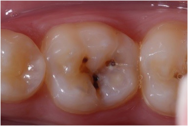
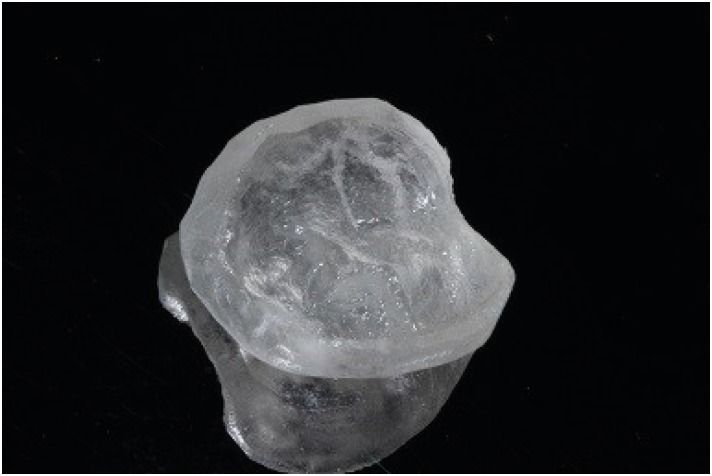
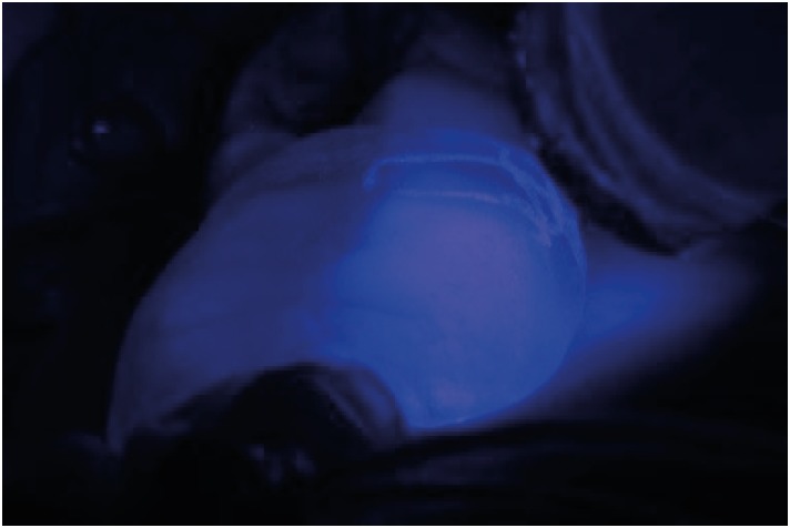
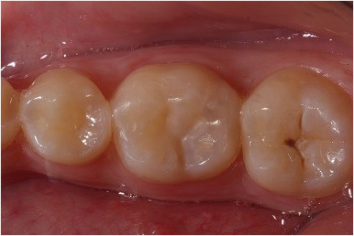
The patient sought the Department of Dentistry of the Federal University of Sergipe (Brazil) for a general oral health assessment. The clinical examination showed a carious lesion on the right mandibular first molar, and the restorative treatment plan was determined based on the clinical and radiographic findings (Figure 1). In order to establish an improved oral and aesthetic rehabilitation, an acrylic resin matrix was produced due to the preservation of the dental anatomy itself (Figure 2). For such production, an insulator was applied with the help of a microbrush on the tooth surface. After manipulating the powder/liquid of the acrylic resin (Jet Classico, Sao Paulo, SP, Brazil) in a paladon recipient (Golgran, Sao Caetano do Sul, SP, Brazil) with a spatula, the resin was applied on the occlusal surface of the tooth. After fixation, the resin was removed and maintained in water to prevent distortion, while the other procedures were performed.
Figure 1

Occlusal view of carious lesion.

rde-45-e9-g001.jpg
Figure 2

Matrix resin to be placed in occlusal tooth face.

rde-45-e9-g002.jpg
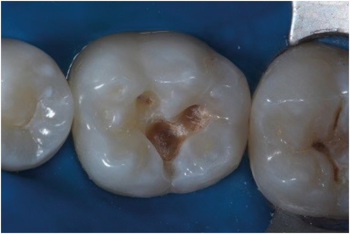
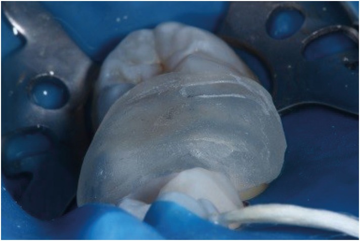
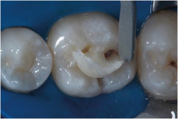
Initially, the verification of occlusal contacts was performed, as well as prophylaxis with pumice stone and water, at low rotation with Robbinson brush (Microdont, Sao Paulo, SP, Brazil). The enamel was accessed with a high-speed 1011 spherical diamond bur (KG Sorensen, São Paulo, SP, Brazil) and the carious tissue was removed with a low-speed #4 (KG Sorensen) spherical carbide bur (Figure 3). Continuing with the restorative process, etching was performed with 37% phosphoric acid (Condac – FGM, Joinville, SC, Brazil) for 30 seconds in enamel and 15 seconds in dentin followed by cleaning with water for double the time. The dentin was dried with sterile absorbent paper, and the application and photopolymerization of the adhesive system were done (Single Bond; 3M ESPE, Campinas, SP, Brazil) using LED light (Radii-Cal; SDI, Victoria, Australia). The acrylic resin matrix was removed from the recipient with water and dried with sterile gauze and air blast. A single increment of bulk-fill resin composite (Aura Bulk Fill; SDI) was placed in the cavity with a resin spatula, and the matrix was positioned on the tooth with light pressure, followed by a 20-second polymerization with and without the matrix (Figures 4, 5 and 6). After assessing the occlusal contact, the excesses were removed with finishing points and burs. Twenty-four hours after completing the restoration, it was finished and polished with rubbers Optimize (TDV, Pomerode, SC, Brazil) (Figure 7).
Figure 3

Class I cavity prepared with spherical bur.

rde-45-e9-g003.jpg
Figure 4

After phosphoric acid and adhesive layer placed, Bulk Fill composite resin as placed in the cavity with one increment.
rde-45-e9-g004.jpg
Figure 5

Matrix placed in the occlusal surface.

rde-45-e9-g005.jpg
Figure 6

Photopolymerization of the composite resin using LED light.

rde-45-e9-g006.jpg
Figure 7

Composite Bulk Fill Restoration placed after finishing and polishing procedures.

rde-45-e9-g007.jpg
Carious lesions are treated clinically every day with restorative materials, mostly of direct insertion. The restorative material mostly used in the last years has been composite resin. Along with the material, the execution of the technique induces negative factors such as the adaptation that may lead to failures, leakage, and secondary caries lesion [6,14]. The present clinical case showed the restoration in a posterior molar with a bulk-fill resin composite associated with the use of an occlusal matrix, aiming to optimize clinical time without the need for inserting composite resin increments.
Using the occlusal matrix may require a longer time prior to cavity preparation, but it allows restoring the original dental anatomy in cases of tooth wear [15] and carious lesions. Such a matrix may be produced with acrylic resin, bis-acrylic composite (usually used do to mock up in anterior tooth), and even with gingival protection resin for bleaching. The latter case, however, requires special care for proper polymerization, considering such resin does not allow the passage of light. Additionally, the occlusal matrix may be used with conventional resins—in which the matrix is inserted only in the last increment—or with bulk-fill resins.
The main advantage of associating this technique with the bulk-fill resin is to perform a satisfactory rehabilitation in a simple manner, as well as the good color camouflage of the restorative material, which shows satisfactory aesthetic results. Despite the advantages of bulk-fill resins, the systematic review performed by Veloso et al. [16] showed that the longevity of restorations is similar when compared with the incremental technique, but it requires a longer follow-up time. In addition, when a matrix with a less flowable bulk-fill resin is used, bubbles can appear, such as an incremental filling.
The use of such resins allows increments from 4 to 5 mm due to the high translucency, which allows proper polymerization and satisfactory aesthetic results due to the increased amount of light reflexion quality [17]. An effective polymerization requires a light with adequate wavelength, prolonged exposure time, and reduced distance between the light and the composite resin [7,13]. Photoinitiators were added to prevent energy reduction for each millimeter of tooth distance, due to the size of the increment placed, improving effectiveness, and promoting a synergistic effect to camphorquinone [13]. These resins present inorganic-organic hybrid polymers that form, through hydrolysis and alkoxides, an inorganic Si-O-Si network modified selectively by incorporating organic groups connected by covalent bond, thus presenting lower polymerization shrinkage [18].
The literature shows studies [14,19,20] comparing the performance of conventional and bulk-fill resins. A laboratory study assessing internal adaptation and the aging effect on it showed that composite resin along with the incremental technique presents an improved adaptation [14]. In another laboratory study comparing microleakage through silver nitrate absorption, between conventional composite and bulk-fill resins, the conventional one presented a lower risk of leakage [20]. Kruly et al. [19] showed in a systematic review that the performance of the clinical restorative agent of bulk-fill and conventional resins is similar, while the success of long-term restorations requires more research to confirm the actual advantage of using the bulk-fill resin.
The incremental technique, which consists of placing a 2-mm increment, may present some flaws, especially regarding the possibility of leaving empty spaces [18]. This technique requires a longer clinical time from the operator to reduce factor C [5]. Moreover, using conventional resins to restore deep cavities may potentially form air bubbles [5].
The case report addresses the use of a resin launched in the market, aiming to reduce the clinical failures that may occur with the use of the incremental technique, as well as the shorter clinical time required for performing restorations. In this context, producing the occlusal matrix shows satisfactory results from the clinical point of view, because of the shorter time of execution and functional rehabilitation, especially by maintaining original anatomy and aesthetics.
The application of minimally invasive dentistry concepts based on the literature by associating the bulk-fill resin with the production of an occlusal matrix allows replicating the original dental anatomy, obtaining satisfactory aesthetic and functional results.

Conflict of Interest: No potential conflict of interest relevant to this article was reported.

Author Contributions:

  • Conceptualization: Francisconi-dos-Rios LF, Nahsan FPS.

  • Data curation: Oliveira L.

  • Formal analysis: Francisconi-dos-Rios LF, Nahsan FPS.

  • Investigation: Tavares JAO, Oliveira L.

  • Methodology: Tavares JAO, Moreira JC.

  • Project administration: Tavares JAO.

  • Resources: Oliveira L, Nahsan FPS.

  • Supervision: Francisconi-dos-Rios LF, Nahsan FPS.

  • Writing - original draft: Francisconi-dos-Rios LF, Tavares JAO, Oliveira L, Moreira JC, Nahsan FPS.

  • Writing - review & editing: Francisconi-dos-Rios LF, Moreira JC, Nahsan FPS.

  • 1. Richards D. Oral diseases affect some 3.9 billion people. Evid Based Dent 2013;14:35. ArticlePubMedPDF
  • 2. Demarco FF, Corrêa MB, Cenci MS, Moraes RR, Opdam NJ. Longevity of posterior composite restorations: not only a matter of materials. Dent Mater 2012;28:87-101. ArticlePubMed
  • 3. Laegreid T, Gjerdet NR, Johansson A, Johansson AK. Clinical decision making on extensive molar restorations. Oper Dent 2014;39:E231-E240. ArticlePubMedPDF
  • 4. Yazici AR, Antonson SA, Kutuk ZB, Ergin E. Thirty-six-month clinical comparison of bulk fill and nanofill composite restorations. Oper Dent 2017;42:478-485. PubMed
  • 5. Costa T, Rezende M, Sakamoto A, Bittencourt B, Dalzochio P, Loguercio AD, Reis A. Influence of adhesive type and placement technique on postoperative sensitivity in posterior composite restorations. Oper Dent 2017;42:143-154. ArticlePubMedPDF
  • 6. van Dijken JW, Pallesen U. A randomized controlled three year evaluation of “bulk-filled” posterior resin restorations based on stress decreasing resin technology. Dent Mater 2014;30:e245-e251. ArticlePubMed
  • 7. Margeas RC. Bulk-fill materials: simplify restorations, reduce chairtime. Compend Contin Educ Dent 2015;36:e1-e4. PubMed
  • 8. Zorzin J, Maier E, Harre S, Fey T, Belli R, Lohbauer U, Petschelt A, Taschner M. Bulk-fill resin composites: polymerization properties and extended light curing. Dent Mater 2015;31:293-301. PubMed
  • 9. Ilie N, Bucuta S, Draenert M. Bulk-fill resin-based composites: an in vitro assessment of their mechanical performance. Oper Dent 2013;38:618-625. ArticlePubMedPDF
  • 10. Alshali RZ, Silikas N, Satterthwaite JD. Degree of conversion of bulk-fill compared to conventional resin-composites at two time intervals. Dent Mater 2013;29:e213-e217. ArticlePubMed
  • 11. El-Damanhoury H, Platt J. Polymerization shrinkage stress kinetics and related properties of bulk-fill resin composites. Oper Dent 2014;39:374-382. ArticlePubMedPDF
  • 12. Olegário IC, Hesse D, Bönecker M, Imparato JC, Braga MM, Mendes FM, Raggio DP. Effectiveness of conventional treatment using bulk-fill composite resin versus Atraumatic Restorative Treatments in primary and permanent dentition: a pragmatic randomized clinical trial. BMC Oral Health 2016;17:34. PubMedPMC
  • 13. Braz R, Mergulhão VA, Oliveira LR, Alves MS, Canto CA. Flared roots reinforced with bulk-fill flowable composite-case report. Oper Dent 2018;43:225-231. ArticlePubMedPDF
  • 14. Alqudaihi FS, Cook NB, Diefenderfer KE, Bottino MC, Platt JÁ. Comparison of internal adaptation of bulk-fill and increment-fill resin composite materials. Oper Dent 2019;44:E32-E44. ArticlePubMedPDF
  • 15. Ammannato R, Rondoni D, Ferraris F. Update on the ‘index technique’ in worn dentition: a no-prep restorative approach with a digital workflow. Int J Esthet Dent 2018;13:516-537. PubMed
  • 16. Veloso SR, Lemos CA, de Moraes SL, do Egito Vasconcelos BC, Pellizzer EP, de Melo Monteiro GQ. Clinical performance of bulk-fill and conventional resin composite restorations in posterior teeth: a systematic review and meta-analysis. Clin Oral Investig 2019;23:221-233.PubMed
  • 17. Maghaireh GA, Price RB, Abdo N, Taha NA, Alzraikat H. Effect of thickness on light transmission and Vickers hardness of five bulk-fill resin-based composites using polywave and single-peak light-emitting diode curing lights. Oper Dent 2019;44:96-107. ArticlePubMedPDF
  • 18. Tauböck TT, Jäger F, Attin T. Polymerization shrinkage and shrinkage force kinetics of high- and low-viscosity dimethacrylate- and ormocer-based bulk-fill resin composites. Odontology 2019;107:103-110. ArticlePubMedPDF
  • 19. Kruly PC, Giannini M, Pascotto RC, Tokubo LM, Suga US, Marques AC, Terada RS. Meta-analysis of the clinical behavior of posterior direct resin restorations: low polymerization shrinkage resin in comparison to methacrylate composite resin. PLoS One 2018;13:e0191942. ArticlePubMedPMC
  • 20. AlSagob EI, Bardwell DN, Ali AO, Khayat SG, Stark PC. Comparison of microleakage between bulk-fill flowable and nanofilled resin-based composites. Interv Med Appl Sci 2018;10:102-109. PubMedPMC

Tables & Figures

REFERENCES

    Citations

    Citations to this article as recorded by  

      • ePub LinkePub Link
      • Cite
        CITE
        export Copy Download
        Close
        Download Citation
        Download a citation file in RIS format that can be imported by all major citation management software, including EndNote, ProCite, RefWorks, and Reference Manager.

        Format:
        • RIS — For EndNote, ProCite, RefWorks, and most other reference management software
        • BibTeX — For JabRef, BibDesk, and other BibTeX-specific software
        Include:
        • Citation for the content below
        Functional and aesthetic rehabilitation in posterior tooth with bulk-fill resin composite and occlusal matrix
        Restor Dent Endod. 2020;45(1):e9  Published online January 3, 2020
        Close
      • XML DownloadXML Download
      Figure
      • 0
      • 1
      • 2
      • 3
      • 4
      • 5
      • 6
      Functional and aesthetic rehabilitation in posterior tooth with bulk-fill resin composite and occlusal matrix
      Image Image Image Image Image Image Image
      Figure 1 Occlusal view of carious lesion.
      Figure 2 Matrix resin to be placed in occlusal tooth face.
      Figure 3 Class I cavity prepared with spherical bur.
      Figure 4 After phosphoric acid and adhesive layer placed, Bulk Fill composite resin as placed in the cavity with one increment.
      Figure 5 Matrix placed in the occlusal surface.
      Figure 6 Photopolymerization of the composite resin using LED light.
      Figure 7 Composite Bulk Fill Restoration placed after finishing and polishing procedures.
      Functional and aesthetic rehabilitation in posterior tooth with bulk-fill resin composite and occlusal matrix

      Restor Dent Endod : Restorative Dentistry & Endodontics
      Close layer
      TOP