-
Influence of adjacent restorative material and distance on the accuracy of inlay cavity impressions with intraoral scanner: an in vitro study
-
So-Yeon Lee, Sung-Ae Son, Jae-Hoon Kim, Deog-Gyu Seo, Jeong-Kil Park
-
Restor Dent Endod 2026;51(1):e6. Published online January 23, 2026
-
DOI: https://doi.org/10.5395/rde.2026.51.e6
-
-
 Abstract Abstract
 PDF PDF PubReader PubReader ePub ePub
- Objectives
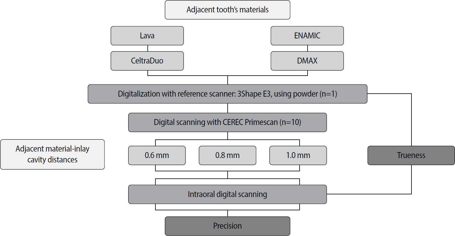
This study aimed to evaluate the influence of adjacent restorative material and interproximal distance on the accuracy of digital impressions of inlay cavities obtained using an intraoral scanner.
Methods
A disto-occlusal inlay cavity was prepared on a mandibular right first molar model, and digital scans were performed using a CEREC Primescan (Dentsply Sirona). The adjacent restorative materials used were Lava (3M ESPE), ENAMIC (VITA Zahnfabrik), Celtra Duo (Dentsply Sirona), and DMAX (DMAX), and the interproximal distances were set to 0.6 mm, 0.8 mm, and 1.0 mm. The obtained scan data were analyzed using GOM Inspect software (GOM GmbH).
Results
Trueness, maximum positive and negative deviations, and precision were significantly influenced by both the adjacent restorative material and the interproximal distance, while their interaction showed a significant effect only on precision. Celtra Duo demonstrated the highest trueness, with mean deviation values decreasing from 7.8 μm at a 0.6 mm interproximal distance to 7.3 μm at 1.0 mm. ENAMIC showed the best precision, presenting mean deviations of 2.6 μm at 0.6 mm, 2.9 μm at 0.8 mm, and 2.4 μm at 1.0 mm. A narrow interproximal distance of 0.6 mm resulted in lower trueness, measured at 8.3 μm, and the highest precision deviation of 3.4 μm. In contrast, an interproximal distance of 1.0 mm yielded improved scan accuracy, with increased trueness and reduced precision variation.
Conclusions
Digital impression accuracy of inlay cavities was influenced by adjacent restorative material and interproximal distance, suggesting clinical consideration is needed in CAD/CAM workflows to optimize restoration fit.
-
Citations
Citations to this article as recorded by  - 3D-SCANNING IN PROSTHETIC DENTISTRY: ADVANTAGES, DISADVANTAGES, AND DEVELOPMENT PROSPECTS
V. S. Kuz, O. I. Teslenko, H. M. Kuz, H. M. Balia, Yu. S. Lunkova, O. V. Shemetov, I. M. Martynenko
Bulletin of Problems Biology and Medicine.2026; 1(1): 98. CrossRef
-
1,800
View
-
105
Download
-
1
Crossref
-
Effect of immersion into solutions at various pH on the color stability of composite resins with different shades
-
Ji-Deok Moon, Eun-Mi Seon, Sung-Ae Son, Kyoung-Hwa Jung, Yong-Hoon Kwon, Jeong-Kil Park
-
Restor Dent Endod 2015;40(4):270-276. Published online August 28, 2015
-
DOI: https://doi.org/10.5395/rde.2015.40.4.270
-
-
Abstract
PDFPubReaderePub
- Objectives
This study examined the color changes of a resin composite with different shades upon exposure to water with different pH. Materials and MethodsNanohybrid resin composites (Filtek Z350XT, 3M ESPE) with four different shades (A2, A3, B1, and B2) were immersed in water with three different pH (pH 3, 6, and 9) for 14 day. The CIE L*a*b* color coordinates of the specimens were evaluated before and after immersion in the solutions. The color difference (ΔE*) and the translucency parameter (TP) were calculated using the color coordinates. ResultsΔE* ranged from 0.33 to 1.58, and the values were affected significantly by the pH. The specimens immersed in a pH 6 solution showed the highest ΔE* values (0.87 - 1.58). The specimens with a B1 shade showed the lowest ΔE* change compared to the other shades. TP ranged from 7.01 to 9.46 depending on the pH and resin shade. The TP difference between before and after immersion in the pH solutions was less than 1.0. ConclusionsThe resulting change of color of the tested specimens did not appear to be clinically problematic because the color difference was < 1.6 in the acidic, neutral, and alkaline solutions regardless of the resin shade, i.e., the color change was imperceptible.
-
Citations
Citations to this article as recorded by - Effect of mouth rinses on roughness and optical properties of restorative materials for oral rehabilitation
Laura Firmo de Carvalho, Edmara T. P. Bergamo, Ernesto B. Benalcázar-Jalkh, Tiago M. B. Campos, Abbas Zahoui, Elisa De Souza Fermino, Ana Clara Mota de Oliveira, Ana Carolina Magalhães, Estevam A. Bonfante, Fábio José B. Bezerra, Larissa M.M. Alves
Biomaterial Investigations in Dentistry.2025; 12: 25. CrossRef - Effect of Printing Layer Thickness on the Color Stability and Surface Roughness of Three-dimensional Printed Resin Material Immersed in Different Aging Media: An In Vitro Study
Vilas Rajguru, Smita Khalikar, Sonali Mahajan, Gopika Gopan, Siddhi D Bhatawadekar, Kishor Mahale
International Journal of Prosthodontics and Restorative Dentistry.2025; 15(1): 36. CrossRef - Colour Retaining Ability of Three Restorative Materials Used in Pediatric Dentistry with the Use of Health Drinks and Beverages - An In Vitro Study
Arnab Mondal, Swati Singh, Seema Qamar, Barun Dasgupta, Shovan Roy
Indian Journal of Dental Research.2025; 36(1): 89. CrossRef - Effects of different antiviral mouthwashes on the surface roughness, hardness, and color stability of composite CAD/CAM materials
Ahmet Hazar, Ecehan Hazar
Journal of Applied Biomaterials & Functional Materials.2024;[Epub] CrossRef - Flexural Properties of Contemporary Bioactive Restorative Materials: Effect of Environmental pH
JEX Ong, AU Yap, A Abdul Aziz, NA Yahya
Operative Dentistry.2023; 48(1): 90. CrossRef - Effect of Mouthwashes for COVID-19 Prevention on Surface Changes of Resin Composites
Saijai Tanthanuch, Boonlert Kukiattrakoon, Chailuck Naiyanart, Tanyanat Promtong, Panuwit Yothinwatthanabamrung, Suttida Pumpua
International Dental Journal.2023; 73(4): 511. CrossRef - Can Modification with Urethane Derivatives or the Addition of an Anti-Hydrolysis Agent Influence the Hydrolytic Stability of Resin Dental Composite?
Agata Szczesio-Wlodarczyk, Izabela M. Barszczewska-Rybarek, Marta W. Chrószcz-Porębska, Karolina Kopacz, Jerzy Sokolowski, Kinga Bociong
International Journal of Molecular Sciences.2023; 24(5): 4336. CrossRef - Understanding the Mechanical, Surface, and Color Behavior of Oral Bioactive Prosthetic Polymers under Biodegradation Processes
Cristina B. Neves, Joana Costa, Jaime Portugal, Ana F. Bettencourt
Polymers.2023; 15(11): 2549. CrossRef - An Evaluation of the Hydrolytic Stability of Selected Experimental Dental Matrices and Composites
Agata Szczesio-Wlodarczyk, Karolina Kopacz, Malgorzata Iwona Szynkowska-Jozwik, Jerzy Sokolowski, Kinga Bociong
Materials.2022; 15(14): 5055. CrossRef - Evaluation of the Color Stability, Water Sorption, and Solubility of Current Resin Composites
Wenkai Huang, Ling Ren, Yuyao Cheng, Minghua Xu, Wenji Luo, Desong Zhan, Hidehiko Sano, Jiale Fu
Materials.2022; 15(19): 6710. CrossRef - Comparative Evaluation of Surface Roughness of different Composites and its effect on Colour Stability of the Restoration
Nalini B, Srinivas Kumar Ch, Narsimha Rao VV
Research Journal of Pharmacy and Technology.2022; : 3854. CrossRef - Comparison of instrumental methods for color change assessment of Giomer resins
Luiza de Almeida Queiroz Ferreira, Rogéli Tibúrcio Ribeiro da Cunha Peixoto, Cláudia Silami de Magalhães, Tassiana Melo Sá, Monica Yamauti, Francisca Daniele Moreira Jardilino
Restorative Dentistry & Endodontics.2022;[Epub] CrossRef - Effect of hydrothermal aging on the microhardness of high- and low-viscosity conventional and additively manufactured polymers
Nadin Al-Haj Husain, Albert J. Feilzer, Cornelis J. Kleverlaan, Samir Abou-Ayash, Mutlu Özcan
The Journal of Prosthetic Dentistry.2022; 128(4): 822.e1. CrossRef - KARANFİL VE YEŞİL ÇAYIN RESTORATİF MATERYALLERİN YÜZEY VE OPTİK ÖZELLİKLERİ ÜZERİNE ETKİSİ: İN VİTRO ÇALIŞMA
Zeynep BUKET KAYNAR, Tolgahan DOĞAN, Nazmiye DÖNMEZ, Mağrur KAZAK
Atatürk Üniversitesi Diş Hekimliği Fakültesi Dergisi.2022; : 1. CrossRef - Effects of Red Dragon Fruit on Color Stability of Self-Adhesive Flowable Composite
Rosalina Tjandrawinata, Brigitta Heidy, Octarina
Applied Mechanics and Materials.2022; 910: 1. CrossRef - Effect of different cement systems and aging on the bond strength of chairside CAD-CAM ceramics
Seda Ustun, Elif Aydogan Ayaz
The Journal of Prosthetic Dentistry.2021; 125(2): 334. CrossRef - Influence of Selected Restorative Materials on the Environmental pH: In Vitro Comparative Study
Anna Lehmann, Kacper Nijakowski, Michalina Nowakowska, Patryk Woś, Maria Misiaszek, Anna Surdacka
Applied Sciences.2021; 11(24): 11975. CrossRef - Color stability of an artificially aged nanofilled composite resin post-cured with different techniques
Lais Sampaio Souza, Tais Rocha Donato, Gabriela Alves Cerqueira, Andrea Nobrega Cavalcanti, Paula Mathias
Journal of Dental Research, Dental Clinics, Dental Prospects.2021; 15(1): 53. CrossRef - Ageing of Dental Composites Based on Methacrylate Resins—A Critical Review of the Causes and Method of Assessment
Agata Szczesio-Wlodarczyk, Jerzy Sokolowski, Joanna Kleczewska, Kinga Bociong
Polymers.2020; 12(4): 882. CrossRef - Use of Resin Solvent as a Facilitator for Removal of Resin Composite Restoratives by Influencing their Mechanical Properties: Is this Possible?
Inas A Elghandour
Journal of Operative Dentistry & Endodontics.2019; 4(1): 1. CrossRef - Effect of powder-liquid ratios and powder colors on color stability of 4-META / MMA-TBB resin after immersion in coffee.
Yoorina Choi, Seo-Jin Jang, Su-Jung Park
Korean Journal of Dental Materials.2018; 45(3): 187. CrossRef - O uso do gel de glicerina melhora a estabilidade de cor de resinas compostas?
Marcus Vinicius Loureiro BERTOLO, Mário Alexandre Coelho SINHORETI, Julia Puppin RONTANI, Pedro Paulo Albuquerque Cavalcanti de ALBUQUERQUE, Luis Felipe Jochims SCHNEIDER
Revista de Odontologia da UNESP.2018; 47(4): 256. CrossRef - In situ evaluation of color stability and hardness' decrease of resin‐based composites
Juliana Silva Ribeiro, Sonia Luque Peralta, Vinícius Esteves Salgado, Rafael Guerra Lund
Journal of Esthetic and Restorative Dentistry.2017; 29(5): 356. CrossRef - Discoloration Effects of Traditional Turkish Beverages on different Composite Restoratives
Serdar Baglar, Erol Keskin, Tahir Orun, Abdulhamit Es
The Journal of Contemporary Dental Practice.2017; 18(2): 83. CrossRef - The effect of red and white wine on color changes of nanofilled and nanohybrid resin composites
Saijai Tanthanuch, Boonlert Kukiattrakoon, Thanwalee Peerasukprasert, Nilobon Chanmanee, Parnchanok Chaisomboonphun, Apisara Rodklai
Restorative Dentistry & Endodontics.2016; 41(2): 130. CrossRef
-
2,304
View
-
16
Download
-
25
Crossref
-
Effect of resin thickness on the microhardness and optical properties of bulk-fill resin composites
-
Eun-Ha Kim, Kyoung-Hwa Jung, Sung-Ae Son, Bock Hur, Yong-Hoon Kwon, Jeong-Kil Park
-
Restor Dent Endod 2015;40(2):128-135. Published online January 13, 2015
-
DOI: https://doi.org/10.5395/rde.2015.40.2.128
-
-
Abstract
PDFPubReaderePub
- Objectives
This study evaluated the effects of the resin thickness on the microhardness and optical properties of bulk-fill resin composites. MethodsFour bulk-fill (Venus Bulk Fill, Heraeus Kulzer; SDR, Dentsply Caulk; Tetric N-Ceram Bulk Fill, Ivoclar vivadent; SonicFill, Kerr) and two regular resin composites (Charisma flow, Heraeus Kulzer; Tetric N-Ceram, Ivoclar vivadent) were used. Sixty acrylic cylindrical molds were prepared for each thickness (2, 3 and 4 mm). The molds were divided into six groups for resin composites. The microhardness was measured on the top and bottom surfaces, and the colors were measured using Commission Internationale d'Eclairage (CIE) L*a*b* system. Color differences according to the thickness and translucency parameters and the correlations between the microhardness and translucency parameter were analyzed. The microhardness and color differences were analyzed by ANOVA and Scheffe's post hoc test, and a student t-test, respectively. The level of significance was set to α = 0.05. ResultsThe microhardness decreased with increasing resin thickness. The bulk-fill resin composites showed a bottom/top hardness ratio of almost 80% or more in 4 mm thick specimens. The highest translucency parameter was observed in Venus Bulk Fill. All resin composites used in this study except for Venus Bulk Fill showed linear correlations between the microhardness and translucency parameter according to the thickness. ConclusionsWithin the limitations of this study, the bulk-fill resin composites used in this study can be placed and cured properly in the 4 mm bulk.
-
Citations
Citations to this article as recorded by
-
3,075
View
-
24
Download
-
68
Crossref
-
Effect of additional etching and ethanol-wet bonding on the dentin bond strength of one-step self-etch adhesives
-
Joonghee Ahn, Kyoung-Hwa Jung, Sung-Ae Son, Bock Hur, Yong-Hoon Kwon, Jeong-Kil Park
-
Restor Dent Endod 2015;40(1):68-74. Published online November 18, 2014
-
DOI: https://doi.org/10.5395/rde.2015.40.1.68
-
-
Abstract
PDFPubReaderePub
- Objectives
This study examined the effects of additional acid etching on the dentin bond strength of one-step self-etch adhesives with different compositions and pH. The effect of ethanol wetting on etched dentin bond strength of self-etch adhesives was also evaluated. Materials and MethodsForty-two human permanent molars were classified into 21 groups according to the adhesive types (Clearfil SE Bond [SE, control]; G-aenial Bond [GB]; Xeno V [XV]; Beauti Bond [BB]; Adper Easy Bond [AE]; Single Bond Universal [SU]; All Bond Universal [AU]), and the dentin conditioning methods. Composite resins were placed on the dentin surfaces, and the teeth were sectioned. The microtensile bond strength was measured, and the failure mode of the fractured specimens was examined. The data were analyzed statistically using two-way ANOVA and Duncan's post hoc test. ResultsIn GB, XV and SE (pH ≤ 2), the bond strength was decreased significantly when the dentin was etched (p < 0.05). In BB, AE and SU (pH 2.4 - 2.7), additional etching did not affect the bond strength (p > 0.05). In AU (pH = 3.2), additional etching increased the bond strength significantly (p < 0.05). When adhesives were applied to the acid etched dentin with ethanol-wet bonding, the bond strength was significantly higher than that of the no ethanol-wet bonding groups, and the incidence of cohesive failure was increased. ConclusionsThe effect of additional acid etching on the dentin bond strength was influenced by the pH of one-step self-etch adhesives. Ethanol wetting on etched dentin could create a stronger bonding performance of one-step self-etch adhesives for acid etched dentin.
-
Citations
Citations to this article as recorded by - Evaluation of bonding effectiveness of universal adhesives to dentin: A Bayesian network meta-analysis review of in vitro studies
Fatih Bedir, Günes Bulut Eyüboglu, Tugba Serin Kalay, Muhammet Karadas
International Journal of Adhesion and Adhesives.2026; 149: 104330. CrossRef - Effect of aging on microshearing bond strength of different adhesive systems
Cansu Dağdelen Ahısha, Mine Betül Üçtaşlı
BMC Oral Health.2026;[Epub] CrossRef - Influence of Different Application Modes of a Universal Adhesive System on the Bond Strength of Bulk‐Fill Composite Resin to Enamel and Dentin in Primary Teeth
Ali Nozari, Maryam Pakniyat Jahromi, Farnaz Haji Abbas Oghli, Zahra Jowkar, Seyed Ahmadreza Hamidi
Clinical and Experimental Dental Research.2024;[Epub] CrossRef - Effect of a novel pretreatment on the microtensile bond strength of universal adhesives with dentin
Yixiang Pan, Jiajia Xu, Xue Cai, Xiaodong Li, Xiaoyan Wang
Journal of Dental Sciences.2023; 18(3): 1148. CrossRef - Microfluidic Organ-on-A-chip: A Guide to Biomaterial Choice and Fabrication
Uyen M. N. Cao, Yuli Zhang, Julie Chen, Darren Sayson, Sangeeth Pillai, Simon D. Tran
International Journal of Molecular Sciences.2023; 24(4): 3232. CrossRef - Effect of phytic acid on bond strength and interfacial integrity of universal adhesive to deep dentin
Ahmed Mostafa Attia, Ahmed Fawzy Abo-Elezz, Rehab Khalil Safy
Brazilian Dental Journal.2022; 33(5): 116. CrossRef - Microtensile Bond Strength of Total-Etch and Self-Etch Universal Adhesives Containing 10-MDP: A Systematic Review
I. Hisham Ismail, N.A. Abdul Razak, N.D. Mohd Ramzi, M.Y.P. Mohd Yusof
The Journal of Dentists.2022; 10: 12. CrossRef - Biomodification of dentin collagen by primers with crosslinking reagents using ethanol wet bonding technique
Talita Arrais Daniel Mendes, Samuel Chillavert Dias Pascoal, Marcelo Victor Sidou Lemos, Sérgio Lima Santiago, Juliano Sartori Mendonça
International Journal of Adhesion and Adhesives.2022; 119: 103254. CrossRef - Is the presence of 10-MDP associated to higher bonding performance for self-etching adhesive systems? A meta-analysis of in vitro studies
Julia Fehrenbach, Cristina Pereira Isolan, Eliseu Aldrighi Münchow
Dental Materials.2021; 37(10): 1463. CrossRef - The effect of additional chlorhexidine and/or ethanol on the bond strength of universal adhesives
Zeynep Buket Kaynar, Magrur Kazak, Nazmiye Donmez, Evrim Eliguzeloglu Dalkilic
Journal of Adhesion Science and Technology.2021; 35(4): 375. CrossRef - Evaluation of the Effect of Cold Plasma Treatment on the Microshear Bond Strength of Composite Resin Restorations to Dentin using Different Adhesive Systems and the Effect of Thermocycling
Sara Valizadeh, Elham Farhadi, Aida Moradi, Sedighe S. Hashemikamangar
The Open Dentistry Journal.2021; 15(1): 734. CrossRef - Bond Strength of Universal Adhesives to Dentin: A Systematic Review and Meta-Analysis
Louis Hardan, Rim Bourgi, Naji Kharouf, Davide Mancino, Maciej Zarow, Natalia Jakubowicz, Youssef Haikel, Carlos Enrique Cuevas-Suárez
Polymers.2021; 13(5): 814. CrossRef - Effects of simplified ethanol–wet bonding and hydrophobic coating on resin–dentin bonding properties
Xia Wang, He Li, Liang Chen, Yue Wang, Jianfei Bai, Defei Wang, Hong Liu
Journal of Adhesion Science and Technology.2021; 35(9): 913. CrossRef - Effect of dentin biomodification techniques on the stability of the bonded interface
Nida Mehmood, Rajni Nagpal, UdaiPratap Singh, Meenal Agarwal
Journal of Conservative Dentistry.2021; 24(3): 265. CrossRef - Assessment of nanohardness, elastic modulus, and nanoleakage of the adhesive interface using the ethanol-wet-bonding technique
Mauricio Yugo Souza, Jéssica Lopes Andrade, Taciana Marco Ferraz Caneppele, Eduardo Bresciani
International Journal of Adhesion and Adhesives.2020; 99: 102572. CrossRef - The improvement of biocompatibility of adhesives
Cigdem Atalayin, Huseyin Tezel, Zeynep Ergucu, Nimet Unlu, Guliz Armagan, Taner Dagci, Timur Kose
Clinical Oral Investigations.2019; 23(8): 3213. CrossRef - Comparison of the micro-tensile bond strengths of four different universal adhesives to caries-affected dentin after ER:YAG laser irradiation
Nazmiye DÖNMEZ, Ayça Sarıalioğlu GÜNGÖR, Barış KARABULUT, Şeyda Hergüner SİSO
Dental Materials Journal.2019; 38(2): 218. CrossRef - Six-month performance of restorations produced with the ethanol-wet-bonding technique: a randomized trial
Maurício Yugo de SOUZA, Ana Luiza Barbosa JUREMA, Taciana Marco Ferraz CANEPPELE, Eduardo BRESCIANI
Brazilian Oral Research.2019;[Epub] CrossRef - Influence of ethanol-wet dentin, adhesive mode of application, and aging on bond strength of universal adhesive
Mauricio Yugo de SOUZA, Rebeca DI NICOLÓ, Eduardo BRESCIANI
Brazilian Oral Research.2018;[Epub] CrossRef - Effects of light curing modes and ethanol-wet bonding on dentin bonding properties
Mu-zi Li, Jin-rui Wang, Hong Liu, Xia Wang, Kang Gan, Xiu-ju Liu, De-li Niu, Xiao-qing Song
Journal of Zhejiang University-SCIENCE B.2016; 17(9): 703. CrossRef - Effect of an Er,Cr:YSGG laser preparation on dentin bond strength of a universal adhesive
A. Rüya Yazici, Emel Karaman, Duygu Tuncer, Gizem Berk, Atilla Ertan
Journal of Adhesion Science and Technology.2016; 30(22): 2477. CrossRef - The effect of saliva decontamination procedures on dentin bond strength after universal adhesive curing
Jayang Kim, Sungok Hong, Yoorina Choi, Sujung Park
Restorative Dentistry & Endodontics.2015; 40(4): 299. CrossRef
-
2,787
View
-
7
Download
-
22
Crossref
-
The effect of resin thickness on polymerization characteristics of silorane-based composite resin
-
Sung-Ae Son, Hyoung-Mee Roh, Bock Hur, Yong-Hoon Kwon, Jeong-Kil Park
-
Restor Dent Endod 2014;39(4):310-318. Published online September 5, 2014
-
DOI: https://doi.org/10.5395/rde.2014.39.4.310
-
-
Abstract
PDFPubReaderePub
- Objectives
This study examined the influence of the resin thickness on the polymerization of silorane- and methacrylate-based composites. Materials and MethodsOne silorane-based (Filtek P90, 3M ESPE) and two methacrylate-based (Filtek Z250 and Z350, 3M ESPE) composite resins were used. The number of photons were detected using a photodiode detector at the different thicknesses (thickness, 1, 2 and 3 mm) specimens. The microhardness of the top and bottom surfaces was measured (n = 15) using a Vickers hardness with 200 gf load and 15 sec dwell time conditions. The degree of conversion (DC) of the specimens was determined using Fourier transform infrared spectroscopy (FTIR). Scratched powder of each top and bottom surface of the specimen dissolved in ethanol for transmission FTIR spectroscopy. The refractive index was measured using a Abbe-type refractometer. To measure the polymerization shrinkage, a linometer was used. The results were analyzed using two-way ANOVA and Tukey's test at p < 0.05 level. ResultsThe silorane-based resin composite showed the lowest filler content and light attenuation among the specimens. P90 showed the highest values in the DC and the lowest microhardness at all depth. In the polymerization shrinkage, P90 showed a significantly lower shrinkage than the rest two resin products (p < 0.05). P90 showed a significantly lower refractive index than the remaining two resin products (p < 0.05). ConclusionsDC, microhardness, polymerization rate and refractive index linearly decreased as specimen thickness linearly increased. P90 showed much less polymerization shrinkage compared to other specimens. P90, even though achieved the highest DC, showed the lowest microhardness and refractive index.
-
Citations
Citations to this article as recorded by - Effect of Different Coring Thicknesses on Tooth Movement During Processing of Complete Dentures: An In Vitro Study
Akshaya Kadunganari, Nandakumar Karamannattil, Aysha Mohamed Ali KP, Neethu Niduvote Poyil, Priyanka K, Shahnaz Shahnawaz
Cureus.2026;[Epub] CrossRef - A Year-Long Comparison of Dentin Bond Strength Using the Co-Curing Technique and Conventional Adhesive Application
Josipa Vukelja Bosnić, Eva Klarić, Ivan Sever, Zrinka Tarle
Journal of Composites Science.2025; 9(3): 131. CrossRef - Chameleon Effect of Universal Shade Composite Polymers in Repairing CAD/CAM Lithium Disilicate
Gaetano Paolone, Giacomo Collivasone, Niccolò De Masi, Alicia Heinichen, Katia Greco, Enrico Gherlone, Giuseppe Cantatore
Materials.2025; 18(13): 3020. CrossRef - The influence of inorganic fillers on the light transmission through resin-matrix composites during the light-curing procedure: an integrative review
Rita Fidalgo-Pereira, Daniela Carpio, Orlanda Torres, Oscar Carvalho, Filipe Silva, Bruno Henriques, Mutlu Özcan, Júlio C. M. Souza
Clinical Oral Investigations.2022; 26(9): 5575. CrossRef - Conversion, Polymerization Shrinkage, Heat Generation, and Depth of Cure of Novel Dental Composites
Saad Liaqat, Humaira Jabeen
Pakistan BioMedical Journal.2022;[Epub] CrossRef - Effect of Polymerization on the Color of Resin Composites
B Korkut, G Dokumacigil, N Murat, PY Atali, B Tarcin, GB Gocmen
Operative Dentistry.2022; 47(5): 514. CrossRef - Shrinkage Stress and Temperature Variation in Resin Composites Cured via Different Photoactivation Methods: Insights for Standardisation of the Photopolymerisation
Guilherme dos Santos Sousa, Gabriel Felipe Guimarães, Edilmar Marcelino, José Eduardo Petit Rodokas, Arilson José de Oliveira Júnior, Ivana Cesarino, Alcides Lopes Leão, Carla dos Santos Riccardi, Mohammad Arjmand, Rafael Plana Simões
Polymers.2021; 13(13): 2065. CrossRef - Effect of the incorporation of silica blow spun nanofibers containing silver nanoparticles (SiO2/Ag) on the mechanical, physicochemical, and biological properties of a low-viscosity bulk-fill composite resin
Soraya Salmanzadeh Ardestani, Roberta Ferreti Bonan, Mariaugusta Ferreira Mota, Rosiane Maria da Costa Farias, Romualdo Rodrigues Menezes, Paulo Rogério Ferreti Bonan, Panmella Pereira Maciel, Flávia Maria de Moraes Ramos-Perez, André Ulisses Dantas Batis
Dental Materials.2021; 37(10): 1615. CrossRef - Light-Curing Units, Photoinitiators System, and Monomers on Physico-Mechanical Properties of Experimental Composite Resins
Gustavo Furlan da Silva Prezotto, Weverteon Soares de Lima, Rafael Pino Vitti, Ariel Farias da Silva, Mário Alexandre Coelho Sinhoreti, William Cunha Brandt
Matéria (Rio de Janeiro).2020;[Epub] CrossRef - Influence of Different Cordless Light-emitting-diode Units and Battery Levels on Chemical, Mechanical, and Physical Properties of Composite Resin
IO Cardoso, AC Machado, DNR Teixeira, FC Basílio, A Marletta, PV Soares
Operative Dentistry.2020; 45(4): 377. CrossRef - Shrinkage in composites: An enigma
Dhakshinamoorthy Malarvizhi, Arumugam Karthick, NewBegin Selvakumar Gold Pearlin Mary, Alagarsamy Venkatesh
Journal of International Oral Health.2019; 11(5): 244. CrossRef - Development and status of resin composite as dental restorative materials
Xinxuan Zhou, Xiaoyu Huang, Mingyun Li, Xian Peng, Suping Wang, Xuedong Zhou, Lei Cheng
Journal of Applied Polymer Science.2019;[Epub] CrossRef - Effect of the Time of Salivary Contamination during Light Curing on Degree of Conversion and Microhardness of a Restorative Composite Resin
Rasoul Sahebalam, Alireza Boruziniat, Fahimeh Mohammadzadeh, Abdolrasoul Rangrazi
Biomimetics.2018; 3(3): 23. CrossRef - LIGHT POLYMERIZATION OF PHOTO-CURED COMPOSITE MATERIALS: MODERN APPROACHES AND APPLICATION PECULIARITIES
O. A. Udod, V. H. Tsentilo, O. M. Adamenko
Bulletin of Problems Biology and Medicine.2018; 2(4): 72. CrossRef - Resistencia a la compresión del ionómero de vidrio y de la resina compuesta. Estudio in vitro
Sara Blanco Lerech, Sebastián Frías Tarón, Arnulfo Tarón Dunoyer, José María Bustillo Arrieta, Antonio Díaz Caballero
Revista Odontológica Mexicana.2017; 21(2): 109. CrossRef - Compressive strength of glass ionomer and composite resin. In vitro study
Sara Blanco Lerech, Sebastián Frías Tarón, Arnulfo Tarón Dunoyer, José María Bustillo Arrieta, Antonio Díaz Caballero
Revista Odontológica Mexicana.2017; 21(2): e107. CrossRef - Influência de três modos de fotopolimerização sobre a microdureza de três resinas compostas
Andréa Cristina Schneider, Márcio José Mendonça, Roberta Bento Rodrigues, Priscilla do Monte Ribeiro Busato, Veridiana Camilotti
Polímeros.2016; 26(spe): 37. CrossRef - Vickers microhardness comparison of 4 composite resins with different types of filler.
Rene García-Contreras, Rogelio Scougall-Vilchis, Laura Acosta-Torres, Concepción Arenas-Arrocena, Rigoberto García-Garduño, Javier de la Fuente-Hernández
Journal Oral Of Research.2015; 4(5): 313. CrossRef
-
2,422
View
-
11
Download
-
18
Crossref
-
Influence of application methods of one-step self-etching adhesives on microtensile bond strength
-
Chul-Kyu Choi, Sung-Ae Son, Jin-Hee Ha, Bock Hur, Hyeon-Cheol Kim, Yong-Hun Kwon, Jeong-Kil Park
-
J Korean Acad Conserv Dent 2011;36(3):203-210. Published online May 31, 2011
-
DOI: https://doi.org/10.5395/JKACD.2011.36.3.203
-
-
Abstract
PDFPubReaderePub
-
Objectives
The purpose of this study was to evaluate the effect of various application methods of one-step self-etch adhesives to microtensile resin-dentin bond strength.
Materials and Methods
Thirty-six extracted human molars were used. The teeth were assigned randomly to twelve groups (n = 15), according to the three different adhesive systems (Clearfil Tri-S Bond, Adper Prompt L-Pop, G-Bond) and application methods. The adhesive systems were applied on the dentin as follows: 1) The single coating, 2) The double coating, 3) Manual agitation, 4) Ultrasonic agitation. Following the adhesive application, light-cure composite resin was constructed. The restored teeth were stored in distilled water at room temperature for 24 hours, and prepared 15 specimens per groups. Then microtensile bond strength was measured and the failure mode was examined.
Results
Manual agitation and ultrasonic agitation of adhesive significantly increased the microtensile bond strength than single coating and double coating did. Double coating of adhesive significantly increased the microtensile bond strength than single coating did and there was no significant difference between the manual agitation and ultrasonic agitation group. There was significant difference in microtensile bonding strength among all adhesives and Clearfil Tri-S Bond showed the highest bond strength.
Conclusions
In one-step self-etching adhesives, there was significant difference according to application methods and type of adhesives. No matter of the material, the manual or ultrasonic agitation of the adhesive showed significantly higher microtensile bond strength.
-
Citations
Citations to this article as recorded by - Effect of Baicalein on Bond Strength of Indirect Ceramic Restoration
Nuray Zulkadir Ergin, Aslı Seçilmiş
Süleyman Demirel Üniversitesi Sağlık Bilimleri Dergisi.2025; 16(3): 356. CrossRef - The Classification and Selection of Adhesive Agents; an Overview for the General Dentist
Naji Ziad Arandi
Clinical, Cosmetic and Investigational Dentistry.2023; Volume 15: 165. CrossRef
-
2,595
View
-
14
Download
-
2
Crossref
-
Effect of the difference in spectral outputs of the single and dual-peak LEDs on the microhardness and the color stability of resin composites
-
Hye-Jung Park, Sung-Ae Son, Bock Hur, Hyeon-Cheol Kim, Yong-Hoon Kwon, Jeong-Kil Park
-
J Korean Acad Conserv Dent 2011;36(2):108-113. Published online March 31, 2011
-
DOI: https://doi.org/10.5395/JKACD.2011.36.2.108
-
-
Abstract
PDFPubReaderePub
-
Objectives
To determine the effect of the spectral output of single and dual-peak light emitting diode (LED) curing lights on the microhardness and color stability of commercial resin composites formulated with camphorquinone and alternative photoinitiators in combination.
Materials and Methods
Three light-polymerized resin composites (Z100 (3M ESPE), Tetric Ceram (Ivoclar Vivadent) and Aelite LS Posterior (Bisco)) with different photoinitiator systems were used. The resin composites were packed into a Teflon mold (8 mm diameter and 2 mm thickness) on a cover glass. After packing the composites, they were light cured with single-peak and dual-peak LEDs. The Knoop microhardness (KHN) and color difference (ΔE) for 30 days were measured. The data was analyzed statistically using a student's t-test (p < 0.05).
Results
All resin composites showed improved microhardness when a third-generation dual-peak LED light was used. The color stability was also higher for all resin composites with dual-peak LEDs. However, there was a significant difference only for Aelite LS Posterior.
Conclusions
The dual-peak LEDs have a beneficial effect on the microhardness and color stability of resin composites formulated with a combination of camphorquinone and alternative photoinitiators.
-
Citations
Citations to this article as recorded by - Effect of irradiance from curing units on the microhardness of composite - a systematic review
Neenu Francis, Rakesh R. Rajan, Vijay Kumar, Anju Varughese, Vineetha Karuveetil, C. M. Sapna
Evidence-Based Dentistry.2022;[Epub] CrossRef - Micro-computed tomography evaluation of volumetric polymerization shrinkage and degree of conversion of composites cured by various light power outputs
Pablo J. ATRIA, Camila S. SAMPAIO, Eduardo CÁCERES, Jessica FERNÁNDEZ, Andre F. REIS, Marcelo GIANNINI, Paulo G. COELHO, Ronaldo HIRATA
Dental Materials Journal.2018; 37(1): 33. CrossRef - Effect of the irradiance distribution from light curing units on the local micro-hardness of the surface of dental resins
Thomas Haenel, Berenika Hausnerová, Johannes Steinhaus, Richard B.T. Price, Braden Sullivan, Bernhard Moeginger
Dental Materials.2015; 31(2): 93. CrossRef - 1,3-Butadiene as an Adhesion Promoter Between Composite Resin and Dental Ceramic in a Dielectric Barrier Discharge Jet
Geum-Jun Han, Sung-No Chung, Bae-Hyeock Chun, Chang-Keun Kim, Kyu Hwan Oh, Byeong-Hoon Cho
Plasma Chemistry and Plasma Processing.2013; 33(2): 539. CrossRef - Optimal combination of 3-component photoinitiation system to increase the degree of conversion of resin monomers
Chang-Gyu Kim, Ho-Jin Moon, Dong-Hoon Shin
Journal of Korean Academy of Conservative Dentistry.2011; 36(4): 313. CrossRef
-
1,797
View
-
3
Download
-
5
Crossref
-
Management of white spots: resin infiltration technique and microabrasion
-
Jeong-Hye Son, Bock Hur, Hyeon-Cheol Kim, Jeong-Kil Park
-
J Korean Acad Conserv Dent 2011;36(1):66-71. Published online January 31, 2011
-
DOI: https://doi.org/10.5395/JKACD.2011.36.1.66
-
-
Abstract
PDFPubReaderePub
This case report compared the effectiveness of resin infiltration technique (Icon, DMG) with microabrasion (Opalustre, Ultradent Products, Inc.) in management of white spot lesions. It demonstrates that although neither microabrasion nor resin infiltration technique can remove white spot lesions completely, resin infiltration technique seems to be more effective than microabrasion. Therefore resin infiltration technique can be chosen preferentially for management of white spot lesions and caution should be taken for case selection. -
Citations
Citations to this article as recorded by - Clinical Effectiveness of Resin Infiltration and Fluoride Varnish for Managing White Spot Lesions (International Caries Detection and Assessment System (ICDAS) ≤ 2) During Multibracket Orthodontic Treatment: A Systematic Review and Meta-Analysis
Vilius Kosys, Gabija Streimikyte, Giedre Trakiniene
Cureus.2026;[Epub] CrossRef - Resin Infiltration for the Esthetic Improvement of Dental Fluorosis and White Spots: A Case Report
Sumayyah L Alkhudhayri, Shahad L Alhassani, Nada A AbdelAleem
Cureus.2024;[Epub] CrossRef - White spot lesions in fixed orthodontic treatment: Etiology, pathophysiology, diagnosis, treatment, and future research perspectives
Suma Shankarappa, Jerusha Titus Burk, Pradeep Subbaiah, Raghunath Nagasundara Rao, Vidya Gowdappa Doddawad
Journal of Orthodontic Science.2024;[Epub] CrossRef - White Spot Lesions in Fixed Orthodontics: A Literature Review on Etiology, Prevention, and Treatment
Deem Al-Blaihed, Omar El Meligy, Khlood Baghlaf, Rabab A Aljawi, Shahad Abudawood
Cureus.2024;[Epub] CrossRef - Effectiveness of low-viscosity resin infiltration (Icon) on color change of enamel white spot lesions: 1-year follow-up clinical study
Mohamed. H. Zaazou, Reham S. Saleh, Shahinaz N. Hassan, Ali Abdelnabi, Zeinab M. Zaki, Tamer M. Hamdy, Dalia Y. Zaki, Lamiaa M. Moharam
Bulletin of the National Research Centre.2024;[Epub] CrossRef - Individual tooth segmentation in human teeth images using pseudo edge-region obtained by deep neural networks
Seongeun Kim, Chang-Ock Lee
Signal Processing: Image Communication.2024; 120: 117076. CrossRef - Surface topography and spectrophotometric assessment of white spot lesions restored with nano-hydroxyapatite-containing universal adhesive resin: an in-vitro study
Neven S. Aref, Rahaf M. Alsdrani
BMC Oral Health.2023;[Epub] CrossRef - Infiltrating Resins, Noninvasive Treatment of White Spot Lesions: A Case Report
Rubén Darío Miranda-Carreño, Jacqueline Adelina Rodríguez-Chávez, Abigailt Flores-Ledesma
Journal of Oral Health and Community Dentistry.2023; 17(2): 75. CrossRef - Management of Turner's Hypoplasia Using Resin Infiltration: A Case Report
Dhruvi Solanki, Punit Fulzele, Nilima Thosar, Unnati Shirbhate
Cureus.2023;[Epub] CrossRef - Colour Parameters and Changes of Tea-Stained Resin Composite Exposed to Whitening Pen (In Vitro Study)
Abdulaziz Alhotan, Rasha M. Abdelraouf, Saleh Alhijji, Merry Angelyn Tan De Vera, Aref Sufyan, Jukka P. Matinlinna, Tamer M. Hamdy
Polymers.2023; 15(14): 3068. CrossRef - Effect of Resin Infiltration and Microabrasion on the Microhardness of the Artificial White Spot Lesions (An in Vitro Study)
Reem Majeed H.J. Al-Mamoori, Aseel Haidar M.J. Al Haidar
Journal of Baghdad College of Dentistry.2022; 34(1): 44. CrossRef - Effect of sodium fluoride plus tricalcium phosphate with and without CO2 laser on remineralization of white spot lesions
Nouran M. Eissa, Eman M. Elshourbagy, Nahla E. Gomaa
Heliyon.2022; 8(10): e10752. CrossRef - Efficacy of Resin Infiltration and Fluoride Casein Phosphopeptide Amorphous Calcium Phosphate Varnish on Non-cavitated Active White Spot Lesions in Children: A Randomized Clinical Trial
Mohit Dhamija, Rish Tyagi, Namita Kalra, Amit Khatri
Pesquisa Brasileira em Odontopediatria e Clínica Integrada.2022;[Epub] CrossRef - Individual Tooth Segmentation in Human Teeth Images Using Pseudo Edge-Region Obtained by Deep Neural Networks
Seongeun Kim, Chang-Ock Lee
SSRN Electronic Journal .2022;[Epub] CrossRef - In Vitro, Influence Of In-Office Dental Whitening On The Color Of Teeth Treated With Resin Infiltration
Basil Almutairi, Mohammed Al-Refai, Bander AL-Meshary, Abdulrahman Al-Asim, Fahad Al-Sharidah, Abdullah Alshehri
Annals of Dental Specialty.2021; 9(4): 6. CrossRef - Evaluation of color changes of white spot lesions treated with three different treatment approaches: an in-vitro study
Shaza M. Hammad, Noha A. El-Wassefy, Mohamed A. Alsayed
Dental Press Journal of Orthodontics.2020; 25(1): 26. CrossRef - Spectrophotometric Evaluation of Color Change in Tooth Enamel Defects Using Resin Infiltrate: An In Vivo Study
Anil Gupta, Shikha Dogra, Sakshi Joshi, Vimanyu Kataria, Jyotika Saini, Monika Nagpal, Payal Narula
International Journal of Clinical Pediatric Dentistry.2020; 13(2): 150. CrossRef - Erosion Infiltration in the Management of Molar‐Incisor Hypomineralization (MIH) Defects
Rym Mabrouk, Souha Yahia, Afef Oueslati, Nadia Frih, Yuk Kwan Chen
Case Reports in Dentistry.2020;[Epub] CrossRef - Pediatric Dentists’ Educational Experiences, Attitudes, and Professional Behavior Concerning Resin Infiltration: Implications for Dental Education
Michael Jordan Halcomb, Marita R. Inglehart, Elisabeta Karl
Journal of Dental Education.2020; 84(3): 290. CrossRef - Esthetic improvements of postorthodontic white-spot lesions treated with resin infiltration and microabrasion: A split-mouth, randomized clinical trial
Xi Gu, Lin Yang, Deqin Yang, Yuan Gao, Xiaolei Duan, Xin Zhu, He Yuan, Jiyao Li
The Angle Orthodontist.2019; 89(3): 372. CrossRef - Effect of resin infiltration on the color and microhardness of bleached white‐spot lesions in bovine enamel (an in vitro study)
Sidika Aynur Horuztepe, Meserret Baseren
Journal of Esthetic and Restorative Dentistry.2017; 29(5): 378. CrossRef - Effect of Resin Infiltration on Artificial Caries: Anin vitroEvaluation of Resin Penetration and Microhardness
Deepesh Prajapati, Rashmi Nayak, Deepika Pai, Nagraj Upadhya, Vipin K Bhaskar, Pujan Kamath
International Journal of Clinical Pediatric Dentistry.2017; 10(3): 250. CrossRef - Application of quantitative light-induced fluorescence to determine the depth of demineralization of dental fluorosis in enamel microabrasion: a case report
Tae-Young Park, Han-Sol Choi, Hee-Won Ku, Hyun-Su Kim, Yoo-Jin Lee, Jeong-Bum Min
Restorative Dentistry & Endodontics.2016; 41(3): 225. CrossRef - Non-destructive management of white spot lesions by using tooth jewelry
Hee-Jin Kim, Lorena Karanxha, Su-Jung Park
Restorative Dentistry & Endodontics.2012; 37(4): 236. CrossRef - Color and hardness changes in artificial white spot lesions after resin infiltration
Ji-Hoon Kim, Ho-Hyun Son, Juhea Chang
Restorative Dentistry & Endodontics.2012; 37(2): 90. CrossRef
-
5,963
View
-
53
Download
-
25
Crossref
-
Physical properties of novel composite using Portland cement for retro-filling material
-
Sang-Jin Lee, Ok-In Cho, Jiwan Yum, Jeong-Kil Park, Bock Hur, Hyeon-Cheol Kim
-
J Korean Acad Conserv Dent 2010;35(6):445-452. Published online November 30, 2010
-
DOI: https://doi.org/10.5395/JKACD.2010.35.6.445
-
-
Abstract
PDFPubReaderePub
-
Objectives
The aim of this study was to compare apical sealing ability and physical properties of MTA, MTA - AH-plus mixture (AMTA) and experimental Portland cement - Epoxy resin mixture (EPPC) for a development of a novel retro-filling material.
Materials and Methods
Forty-nine extracted roots were instrumented and filled with gutta-percha. Apical root was resected at 3 mm and the retro-filling cavity was prepared for 3 mm depth. Roots were randomly divided into 3 groups of 15 roots each. The retro-filling was done using MTA, AMTA, and EPPC as the groups divided. Four roots were used as control groups. After setting in humid condition for 24 hours, the roots were immersed in 1% methylene blue dye solution for 72 hours to test the apical leakage. After immersion, the roots were vertically sectioned and photos were taken to evaluate microleakage. Setting times were measured with Vicat apparatus and digital radiographs were taken to evaluate aluminum equivalent thickness using aluminum step wedge. The results of microleakage and setting time were compared between groups using one-way ANOVA and Scheffe's post-hoc comparison at the significance level of 95%.
Results
AMTA and EPPC showed less microleakage than MTA group (p < 0.05). AMTA showed the highest radio-opacity than other groups and the novel EPPC showed 5 mm aluminum thickness radio-opacity. EPPC showed the shortest initial and final setting times than other groups while the MTA showed the longest (p < 0.05).
Conclusions
Under the condition of this study, the novel composite using Portland cement-Epoxy resin mixture may useful for retro-filling with the properties of favorable leakage resistance, radio-opacity and short setting time.
-
Citations
Citations to this article as recorded by - Comparison of Setting Time, Compressive Strength, Solubility, and pH of Four Kinds of MTA
Jing-Ling Che, Jae-Hwan Kim, Seon-Mi Kim, Nam-ki Choi, Hyun-Joo Moon, Moon-Jin Hwang, Ho-Jun Song, Yeong-Joon Park
Korean Journal of Dental Materials.2016; 43(1): 61. CrossRef - Biological Effects and Washout Resistance of a Newly Developed Fast-setting Pozzolan Cement
Yoorina Choi, Su-Jung Park, Seoung-Hoon Lee, Yun-Chan Hwang, Mi-Kyung Yu, Kyung-San Min
Journal of Endodontics.2013; 39(4): 467. CrossRef
-
1,638
View
-
2
Download
-
2
Crossref
-
Effect of 2% chlorhexidine application on microtensile bond strength of resin composite to dentin using one-step self-etch adhesives
-
Soon-Ham Jang, Bock Hur, Hyeon-Cheol Kim, Yong-Hun Kwon, Jeong-Kil Park
-
J Korean Acad Conserv Dent 2010;35(6):486-491. Published online November 30, 2010
-
DOI: https://doi.org/10.5395/JKACD.2010.35.6.486
-
-
Abstract
PDFPubReaderePub
-
Objectives
This study examined the effect of 2% chlorhexidine on the µTBS of a direct composite restoration using one-step self-etch adhesives on human dentin.
Materials and Methods
Twenty-four extracted permanent molars were used. The teeth were assigned randomly to six groups (n = 10), according to the adhesive system and application of chlorhexidine. With or without the application of chlorhexidine, each adhesive system was applied to the dentin surface. After the bonding procedure, light-cure composite resin buildups were produced. The restored teeth were stored in distilled water at room temperature for 24 hours, and then cut and glued to the jig of the microtensile testing machine. A tensile load was applied until the specimen failed. The failure mode was examined using an operating microscope. The data was analyzed statistically using one-way ANOVA, Student's t-test (p < 0.05) and Scheffé's test.
Results
Regardless of the application of chlorhexidine, the Clearfil S3 Bond showed the highest µTBS, followed by G-Bond and Xeno V. Adhesive failure was the main failure mode of the dentin bonding agents tested with some samples showing cohesive failure.
Conclusions
The application of 2% chlorhexidine did not affect the µTBS of the resin composite to the dentin using a one-step self-etch adhesive.
-
Citations
Citations to this article as recorded by - Effects of Collagen Cross-Linking Agents on Dentin-Composite Interface Strength and Morphology
Faisal Ali bin Abbooud AlQhtani, Zuhair Motlak Alkahtani, Muhammad Abdullah Kamran, Ahmed Ali M. Albariqi, Khalid M. Abdelaziz
Calcified Tissue International.2026;[Epub] CrossRef
-
1,987
View
-
6
Download
-
1
Crossref
-
Comparison of push-out bond strength of post according to cement application methods
-
Seo-Ryeong Kim, Jiwan Yum, Jeong-Kil Park, Bock Hur, Hyeon-Cheol Kim
-
J Korean Acad Conserv Dent 2010;35(6):479-485. Published online November 30, 2010
-
DOI: https://doi.org/10.5395/JKACD.2010.35.6.479
-
-
Abstract
PDFPubReaderePub
-
Objectives
The aim of this study was to compare the push-out bond strengths of resin cement/fiber post systems to post space dentin using different application methods of resin cement.
Materials and Methods
Thirty extracted human premolars were selected and randomly divided into 3 groups according to the technique used to place the cement into root canal: using lentulo-spiral instrument (group Lentulo), applying the cement onto the post surface (group Direct), and injecting the material using a specific elongation tip (group Elongation tip). After shaping and filling of the root canal, post space was drilled using Rely-X post drill. Rely-X fiber post was seated using Rely-X Unicem and resin cement was light polymerized. The root specimens were embedded in an acrylic resin and the specimens were sectioned perpendicularly to the long axis using a low-speed saw. Three slices per each root containing cross-sections of coronal, middle and apical part of the bonded fiber posts were obtained by sectioning. The push-out bond strength was measured using Universal Testing Machine. Specimens after bond failure were examined using operating microscope to evaluate the failure modes.
Results
Push-out bond strengths were statistically influenced by the root regions. Group using the elongation tip showed significantly higher bond strength than other ways. Most failures occurred at the cement/dentin interface or in a mixed mode.
Conclusions
The use of an elongation tip seems to reduce the number of imperfections within the self-adhesive cement interface compared to the techniques such as direct applying with the post and lentulo-spiral technique.
-
Citations
Citations to this article as recorded by - Photodynamic therapy and other pretreatment methods on epoxy-based glass fiber post on the push-out bond strength to radicular dentin
Abdulaziz A. Al-Kheraif, Badreldin A. Mohamed, Aref Othman Hasan Sufyan, Aftab Ahmed Khan, Darshan Devang Divakar
Photodiagnosis and Photodynamic Therapy.2021; 36: 102526. CrossRef - Micro-computed tomography analysis of gap and void formation in different prefabricated fiber post cementation materials and techniques
Aws ArRejaie, Saleh A. Alsuliman, Mohammed O. Aljohani, Hesham A. Altamimi, Emad Alshwaimi, Ahmad M. Al-Thobity
The Saudi Dental Journal.2019; 31(2): 236. CrossRef - Micro-computerized tomography analysis of cement voids and pull-out strength of glass fiber posts luted with self-adhesive and glass-ionomer cements in the root canal
Serkan Sarıdağ, Dilek Helvacıoğlu-Yiğit, Mutlu Özcan, Egemen Avcu, Güllü Kızıltaş
Journal of Adhesion Science and Technology.2016; 30(14): 1585. CrossRef - Pull-out bond strength of a self-adhesive resin cement to NaOCl-treated root dentin: effect of antioxidizing agents
Maryam Khoroushi, Marzieh Kachuei
Restorative Dentistry & Endodontics.2014; 39(2): 95. CrossRef - Retentive strength of different intracanal posts in restorations of anterior primary teeth: anin vitrostudy
Mahtab Memarpour, Fereshteh Shafiei, Maryam Abbaszadeh
Restorative Dentistry & Endodontics.2013; 38(4): 215. CrossRef - Effects of dentin moisture on the push-out bond strength of a fiber post luted with different self-adhesive resin cements
Sevinç Aktemur Türker, Emel Uzunoğlu, Zeliha Yılmaz
Restorative Dentistry & Endodontics.2013; 38(4): 234. CrossRef
-
1,717
View
-
3
Download
-
6
Crossref
-
The effect of Er,Cr:YSGG irradiation on microtensile bond strength of composite resin restoration
-
Jeong-Hye Son, Hyeon-Cheol Kim, Bock Hur, Jeong-Kil Park
-
J Korean Acad Conserv Dent 2010;35(2):134-142. Published online March 31, 2010
-
DOI: https://doi.org/10.5395/JKACD.2010.35.2.134
-
-
Abstract
PDFPubReaderePub
The purpose of this study was to evaluate the effect of Er,Cr:YSGG laser irradiation with hypersensitivity mode on microtensile bond strength of composite resin. Twenty extracted permanent molars were randomly assigned to six groups, according to the irradiation of Er,Cr:YSGG laser, adhesive system (Optibond FL or Clearfil SE bond) and application time of etchant (15 sec or 20 sec). Then composite resin was build up on each conditioned surface. The restored teeth were stored in distilled water at room temperature for 24 h and twelve specimens for each group were prepared. All specimens were subjected to microtensile bond strength and the fracture modes were evaluated. Also, the prepared dentin surface and laser irradiated dentin surface were examined under SEM.
The results were as follows:
The microtensile bond strength of laser irradiated group was lower than that of no laser irradiated group.
Regardless of laser irradiation, the microtensile bond strength of Optibond FL was higher than that of Clearfil SE bond. And the microtensile bond strength of 20 sec etching group was higher than that of 15 sec etching group when using Optibond FL.
The SEM image of laser irradiated dentin surface showed prominent peritubular dentin, opened dentinal tubules and no smear layer.
-
Citations
Citations to this article as recorded by - Enamel pretreatment with Er:YAG laser: effects on the microleakage of fissure sealant in fluorosed teeth
Mahtab Memarpour, Nasrin Kianimanesh, Bahareh Shayeghi
Restorative Dentistry & Endodontics.2014; 39(3): 180. CrossRef
-
2,010
View
-
7
Download
-
1
Crossref
-
Effect of an intermediate bonding resin and flowable resin on the compatibility of two-step total etching adhesives with a self-curing composite resin
-
Sook-Kyung Choi, Ji-Wan Yum, Hyeon-Cheol Kim, Bock Hur, Jeong-Kil Park
-
J Korean Acad Conserv Dent 2009;34(5):397-405. Published online September 30, 2009
-
DOI: https://doi.org/10.5395/JKACD.2009.34.5.397
-
-
Abstract
PDFPubReaderePub
This study compared the effect of an activator, intermediate bonding resin and low-viscosity flowable resin on the microtensile bond strength of a self-curing composite resin used with two-step total etching adhesives. Twenty extracted permanent molars were used. The teeth were assigned randomly to nine groups (n=10) according to the adhesive system and application of additional methods (activator, intermediate adhesive, flowable resin). The bonding agents and additional applications of each group were applied to the dentin surfaces. Self-curing composite resin buildups were made for each tooth to form a core, 5mm in height. The restored teeth were then stored in distilled water at room temperature for 24h before sectioning. The microtensile bond strength of all specimens was examined. The data was analyzed statistically by one-way ANOVA and a Scheffe's test. The application of an intermediate bonding resin (Optibond FL adhesive) and low-viscosity flowable resin (Tetric N-flow) produced higher bond strength than that with the activator in all groups. Regardless of the method selected, Optibond solo plus produced the lowest µTBS to dentin. The failure modes of the tested dentin bonding agents were mostly adhesive failure but there were some cases showed cohesive failure in the resin.
-
Effects of condensation techniques and canal sizes on the microleakage of orthograde MTA apical plug in simulated canals
-
Deuk-Lim Nam, Jeong-Kil Park, Bock Hur, Hyeon-Cheol Kim
-
J Korean Acad Conserv Dent 2009;34(3):208-214. Published online May 31, 2009
-
DOI: https://doi.org/10.5395/JKACD.2009.34.3.208
-
-
Abstract
PDFPubReaderePub
The purpose of this study was to compare the dye leakage of MTA (mineral trioxide aggregate) apical plug produced by two orthograde placement techniques (hand condensation technique and ultrasonically assisted hand condensation technique).
To simulate straight canal, 60 transparent acrylic blocks with straight canal were fabricated. These transparent acrylic blocks were divided into 2 groups (Group C; hand condensation technique (HC) and Group U; ultrasonically assisted hand condensation technique (UAHC)) of 30 blocks with each MTA application method. Each group was divided into 2 subgroups (n = 15) with different canal size of #70 (subgroup C70 and subgroup U70) and #120 (subgroup C120 and subgroup U120). After apical plug was created, a wet paper point was placed over the MTA plug and specimen was kept in a humid condition at room temperature to allow MTA to set. After 24 hours, remaining canal space was backfilled using Obtura II. All specimens were transferred to floral form socked by 0.2% rhodamine B solution and stored in 100% humidity at room temperature. After 48 hours, resin block specimens were washed and scanned using a scanner. The maximum length of microleakage was measured from the scanned images of four surfaces of each resin block using Photoshop 6.0.
Statistical analysis was performed with Mann-Whitney U test. Group U of UAHC had significantly lower leakage than Group C of HC in #70-size canal (subgroup U70) (p < 0.05).
-
Stress distribution for NiTi files of triangular based and rectangular based cross-sections using 3-dimensional finite element analysis
-
Hyun-Ju Kim, Chan-Joo Lee, Byung-Min Kim, Jeong-Kil Park, Bock Hur, Hyeon-Cheol Kim
-
J Korean Acad Conserv Dent 2009;34(1):1-7. Published online January 31, 2009
-
DOI: https://doi.org/10.5395/JKACD.2009.34.1.001
-
-
Abstract
PDFPubReaderePub
The purpose of this study was to compare the stress distributions of NiTi rotary instruments based on their cross-sectional geometries of triangular shape-based cross-sectional design, S-shaped cross-sectional design and modified rectangular shape-based one using 3D FE models.
NiTi rotary files of S-shaped and modified rectangular design of cross-section such as Mtwo or NRT showed larger stress change while file rotation during simulated shaping.
The stress of files with rectangular cross-section design such as Mtwo, NRT was distributed as an intermittent pattern along the long axis of file. On the other hand, the stress of files with triangular cross-section design was distributed continuously.
When the residual stresses which could increase the risk of file fatigue fracture were analyzed after their withdrawal, the NRT and Mtwo model also presented higher residual stresses.
From this result, it can be inferred that S-shaped and modified rectangular shape-based files were more susceptible to file fracture than the files having triangular shape-based one. -
Citations
Citations to this article as recorded by - Ex-Vivo Comparison of Torsional Stress on Nickel–Titanium Instruments Activated by Continuous Rotation or Adaptive Motion
Joo Yeong Lee, Sang Won Kwak, Jung-Hong Ha, Hyeon-Cheol Kim
Materials.2020; 13(8): 1900. CrossRef - Autogenous teeth used for bone grafting: a comparison with traditional grafting materials
Young-Kyun Kim, Su-Gwan Kim, Pil-Young Yun, In-Sung Yeo, Seung-Chan Jin, Ji-Su Oh, Heung-Joong Kim, Sun-Kyoung Yu, Sook-Young Lee, Jae-Sung Kim, In-Woong Um, Mi-Ae Jeong, Gyung-Wook Kim
Oral Surgery, Oral Medicine, Oral Pathology and Oral Radiology.2014; 117(1): e39. CrossRef - Analysis of crystalline structure of autogenous tooth bone graft material: X-Ray diffraction analysis
Gyung-Wook Kim, In-Sung Yeo, Su-Gwan Kim, In-Woong Um, Young-Kyun Kim
Journal of the Korean Association of Oral and Maxillofacial Surgeons.2011; 37(3): 225. CrossRef
-
1,837
View
-
9
Download
-
3
Crossref
-
Stress distribution of endodontically treated maxillary second premolars restored with different methods: Three-dimensional finite element analysis
-
Dong-Yeol Lim, Hyeon-Cheol Kim, Bock Hur, Kwang-Hoon Kim, Kwon Son, Jeong-Kil Park
-
J Korean Acad Conserv Dent 2009;34(1):69-79. Published online January 31, 2009
-
DOI: https://doi.org/10.5395/JKACD.2009.34.1.069
-
-
Abstract
PDFPubReaderePub
The purpose of this study was to evaluate the influence of elastic modulus of restorative materials and the number of interfaces of post and core systems on the stress distribution of three differently restored endodontically treated maxillary second premolars using 3D FE analysis. Model 1, 2 was restored with a stainless steel or glass fiber post and direct composite resin. A PFG or a sintered alumina crown was considered. Model 3 was restored by EndoCrown. An oblique 500 N was applied on the buccal (Load A) and palatal (Load B) cusp. The von Mises stresses in the coronal and root structure of each model were analyzed using ANSYS. The elastic modulus of the definitive restorations rather than the type of post and core system was the primary factor that influenced the stress distribution of endodontically treated maxillary premolars. The stress concentration at the coronal structure could be lowered through the use of definitive restoration of high elastic modulus. The stress concentration at the root structure could be lowered through the use of definitive restoration of low elastic modulus. -
Citations
Citations to this article as recorded by - How loss of tooth structure impacts the biomechanical behavior of a single-rooted maxillary premolar: FEA
Roaa Abdelwahab Abdelfattah, Nawar Naguib Nawar, Engy M. Kataia, Shehabeldin Mohamed Saber
Odontology.2024; 112(1): 279. CrossRef - Effect of Proximal Caries-driven Access on the Biomechanical Behavior of Endodontically Treated Maxillary Premolars
Nawar Naguib Nawar, Roaa Abdelwahab Abdelfattah, Mohamed Kataia, Shehabeldin Mohamed Saber, Engy Medhat Kataia, Hyeon-Cheol Kim
Journal of Endodontics.2023; 49(10): 1337. CrossRef - Survival and success of endocrowns: A systematic review and meta-analysis
Raghad A. Al-Dabbagh
The Journal of Prosthetic Dentistry.2021; 125(3): 415.e1. CrossRef - Influence of Cavity Design on Stress Distribution in Second Premolar Tooth Using Finite Element Analysis
Z. Parlar, E.U. Gökçek, K. Yildirim, A. Kahyaoglu
Acta Physica Polonica A.2017; 132(3-II): 949. CrossRef - Influence of post types and sizes on fracture resistance in the immature tooth model
Jong-Hyun Kim, Sung-Ho Park, Jeong-Won Park, Il-Young Jung
Journal of Korean Academy of Conservative Dentistry.2010; 35(4): 257. CrossRef
-
2,286
View
-
21
Download
-
5
Crossref
-
Effect of restoration type on the stress distribution of endodontically treated maxillary premolars; Three-dimensional finite element study
-
Heun-Sook Jung, Hyeon-Cheol Kim, Bock Hur, Kwang-Hoon Kim, Kwon Son, Jeong-Kil Park
-
J Korean Acad Conserv Dent 2009;34(1):8-19. Published online January 31, 2009
-
DOI: https://doi.org/10.5395/JKACD.2009.34.1.008
-
-
Abstract
PDFPubReaderePub
The purpose of this study was to investigate the effects of four restorative materials under various occlusal loading conditions on the stress distribution at the CEJ of buccal, palatal surface and central groove of occlusal surface of endodontically treated maxillary second premolar, using a 3D finte element analysis.
A 3D finite element model of human maxillary second premolar was endodontically treated. After endodontic treatment, access cavity was filled with Amalgam, resin, ceramic or gold of different mechanical properties. A static 500N forces were applied at the buccal (Load-1) and palatal cusp (Load-2) and a static 170N forces were applied at the mesial marginal ridge and palatal cusp simultaneously as centric occlusion (Load-3). Under 3-type Loading condition, the value of tensile stress was analyzed after 4-type restoration at the CEJ of buccal and palatal surface and central groove of occlusal surface
Excessive high tensile stresses were observed along the palatal CEJ in Load-1 case and buccal CEJ in Load-2 in all of the restorations. There was no difference in magnitude of stress in relation to the type of restorations. Heavy tensile stress concentrations were observed around the loading point and along the central groove of occlusal surface in all of the restorations. There was slight difference in magnitude of stress between different types of restorations. High tensile stress concentrations around the loading points were observed and there was no difference in magnitude of stress between different types of restorations in Load-3.
-
The effect of various bonding systems on the microtensile bond strength of immediate and delayed dentin sealing
-
Jin-hee Ha, Hyeon-Cheol Kim, Bock Hur, Jeong-Kil Park
-
J Korean Acad Conserv Dent 2008;33(6):526-536. Published online November 30, 2008
-
DOI: https://doi.org/10.5395/JKACD.2008.33.6.526
-
-
Abstract
PDFPubReaderePub
The purpose of this study was to compare the effect of various dentin bonding systems on microtensile bond strength of immediate dentin sealing (IDS) and delayed dentin sealing (DDS). Eighteen extracted permanent molars were used in this study. The teeth for DDS group were restored with a provisional restorations, and immersed in saline solution for 1 week, and divided into 3 subgroups according to various dentin bonding adhesives; SB subgroup (3 step total-etch adhesive), SE subgroup (2 step self-etch adhesive), XE subgroup (1 step self-etch adhesive). In IDS group, the teeth were divided into 3 subgroups, and applied with bonding adhesives as in DDS group. The teeth were restored with provisional restorations, and immersed in saline solution for 1 week. Indirect composite disc was cemented with resin cement, and all specimens were subjected to microtensile bond strength. The data were statistically analyzed with one-way ANOVA and Student t-test.
The results were as follows:
The IDS group showed significantly higher µTBS than DDS group in 3 step total-etch and 2 step self-etch adhesive (p < 0.05).
In IDS and DDS group, 3 step total-etch adhesive showed the highest µTBS value, followed by 2 step self-etch, and 1 step self-etch adhesive. In IDS group, the µTBS value for 1 step self-etch adhesive was significantly different from those of the other subgroups (p < 0.05), and in DDS group, there were statistical differences in all subgroup (p < 0.05).
Failure modes of tested dentin bonding adhesives were mostly mixed failure and only 1 step self-etch adhesive showed adhesive failure.
-
Citations
Citations to this article as recorded by - The effect of Er,Cr:YSGG irradiation on microtensile bond strength of composite resin restoration
Jeong-Hye Son, Hyeon-Cheol Kim, Bock Hur, Jeong-Kil Park
Journal of Korean Academy of Conservative Dentistry.2010; 35(2): 134. CrossRef
-
2,223
View
-
9
Download
-
1
Crossref
-
Stress analysis of maxillary premolars with composite resin restoration of notch-shaped class V cavity and access cavity; Three-dimensional finite element study
-
Seon-Hwa Lee, Hyeon-Cheol Kim, Bock Hur, Kwang-Hoon Kim, Kwon Son, Jeong-Kil Park
-
J Korean Acad Conserv Dent 2008;33(6):570-579. Published online November 30, 2008
-
DOI: https://doi.org/10.5395/JKACD.2008.33.6.570
-
-
Abstract
PDFPubReaderePub
The purpose of this study was to investigate the distribution of tensile stress of canal obturated maxillary second premolar with access cavity and notch-shaped class V cavity restored with composite resin using a 3D finite element analysis.
The tested groups were classified as 8 situations by only access cavity or access cavity with notch-shaped class VS cavity (S or N), loading condition (L1 or L2), and with or without glass ionomer cement base (R1 or R2). A static load of 500 N was applied at buccal and palatal cusps. Notch-shaped cavity and access cavity were filled microhybrid composite resin (Z100) with or without GIC base (Fuji II LC). The tensile stresses presented in the buccal cervical area, palatal cervical area and occlusal surface were analyzed using ANSYS.
Tensile stress distributions were similar regardless of base. When the load was applied on the buccal cusp, excessive high tensile stress was concentrated around the loading point and along the central groove of occlusal surface. The tensile stress values of the tooth with class V cavity were slightly higher than that of the tooth without class V cavity. When the load was applied the palatal cusp, excessive high tensile stress was concentrated around the loading point and along the central groove of occlusal surface. The tensile stress values of the tooth without class V cavity were slightly higher than that of the tooth with class V cavity.
-
EFFECT OF THE ADDITIONAL ETCHING PROCEDURE ON PUSH-OUT BOND STRENGTH OF ONE-STEP RESIN CEMENT
-
Soon-Il Kang, Jeong-Kil Park, Bock Hur, Hyeon-Cheol Kim
-
J Korean Acad Conserv Dent 2008;33(5):443-451. Published online January 14, 2008
-
DOI: https://doi.org/10.5395/JKACD.2008.33.5.443
-
-
Abstract
PDFPubReaderePub
- Abstract
The purpose of this study was to evaluate the effect of additional etching procedure prior to Maxcem resin cement application in indirect restoration cementation using push-out bonding strength.
One hundred and two extracted human molars were used to make indirect resin restorations of gold inlay and Synfony. These restorations were cemented using Maxcem and Variolink II. Additional etching procedures were done for one group with Maxcem. Three groups have 17 specimens in both restoration types. Push-out bond strength was measured using multi-purpose tester and calculated for bonding strength per sqaure-millimeter area. The mean bonding strength values were compared using SPSS 12.0K program for one-way ANOVA and Scheffe's Test with 95% significance.
Under the condition of this study, the additional etching procedure prior to usage of Maxcem resulted in reduced bond strength for both of restoration types.
-
Citations
Citations to this article as recorded by - Shear bond strength of a self-adhesive resin cement to resin-coated dentin
Jee-Youn Hong, Cheol-Woo Park, Jeong-Uk Heo, Min-Ki Bang, Jae-Jun Ryu
The Journal of Korean Academy of Prosthodontics.2013; 51(1): 27. CrossRef - Effect of dentin surface wetness on tensile bond strength of self adhesive resin cements
Sung-Young Yoon, Se-Hee Park, Jin-Woo Kim, Kyung-Mo Cho
Journal of Korean Academy of Conservative Dentistry.2009; 34(2): 113. CrossRef
-
1,441
View
-
1
Download
-
2
Crossref
-
Stress distribution of three NiTi rotary files under bending and torsional conditions using 3-dimensional finite element analysis
-
Tae-Oh Kim, Chan-Joo Lee, Byung-Min Kim, Jeong-Kil Park, Bock Hur, Hyeon-Cheol Kim
-
J Korean Acad Conserv Dent 2008;33(4):323-331. Published online July 31, 2008
-
DOI: https://doi.org/10.5395/JKACD.2008.33.4.323
-
-
Abstract
PDFPubReaderePub
Flexibility and fracture properties determine the performance of NiTi rotary instruments. The purpose of this study was to evaluate how geometrical differences between three NiTi instruments affect the deformation and stress distributions under bending and torsional conditions using finite element analysis.
Three NiTi files (ProFile .06 / #30, F3 of ProTaper and ProTaper Universal) were scanned using a Micro-CT. The obtained structural geometries were meshed with linear, eight-noded hexahedral elements. The mechanical behavior (deformation and von Mises equivalent stress) of the three endodontic instruments were analyzed under four bending and rotational conditions using ABAQUS finite element analysis software. The nonlinear mechanical behavior of the NiTi was taken into account.
The U-shaped cross sectional geometry of ProFile showed the highest flexibility of the three file models. The ProTaper, which has a convex triangular cross-section, was the most stiff file model. For the same deflection, the ProTaper required more force to reach the same deflection as the other models, and needed more torque than other models for the same amount of rotation. The highest von Mises stress value was found at the groove area in the cross-section of the ProTaper Universal.
Under torsion, all files showed highest stresses at their groove area. The ProFile showed highest von Mises stress value under the same torsional moment while the ProTaper Universal showed the highest value under same rotational angle. -
Citations
Citations to this article as recorded by - Effect of internal stress on cyclic fatigue failure in .06 taper ProFile
Hye-Rim Jung, Jin-Woo Kim, Kyung-Mo Cho, Se-Hee Park
Restorative Dentistry & Endodontics.2012; 37(2): 79. CrossRef
-
1,908
View
-
15
Download
-
1
Crossref
-
Comparative analysis of various corrosive environmental conditions for NiTi rotary files
-
Ji-Wan Yum, Jeong-Kil Park, Bock Hur, Hyeon-Cheol Kim
-
J Korean Acad Conserv Dent 2008;33(4):377-388. Published online July 31, 2008
-
DOI: https://doi.org/10.5395/JKACD.2008.33.4.377
-
-
Abstract
PDFPubReaderePub
The aim of the present study is to compare the corrosion tendency using two kinds of NiTi files in the various environmental conditions through the visual examination and electrochemical analysis. ProTaper Universal S2, 21 mm (Dentsply Maillefer, Ballaigues, Switzerland) and Hero 642, 0.06 tapers, size 25, 21 mm (Micromega, Besancon, France) rotary instruments were tested. The instruments were randomly divided into eighteen groups (n = 5) by the immersion temperature, the type of solution, the brand of NiTi rotary instrument and the presence of mechanical loading. Each file was examined at various magnifications using Scanning Electron Microscope (JEOL, Akishima, Tokyo, Japan) equipped with energy dispersive X-ray microanalysis (EDX). EDX was used to determine the components of the endodontic file alloy in corroded and noncorroded areas. The corrosion resistance of unused and used NiTi files after repeated uses in the human teeth was evaluated electrochemically by potentiodynamic polarization test using a potentiostat (Applied Corrosion Monitoring, Cark-in-Cartmel, UK).
Solution temperature and chloride ion concentration may affect on passivity of NiTi files. Under the conditions of this in vitro study, the corrosion resistance is slightly increased after clinical use. -
Citations
Citations to this article as recorded by - A novel approach to assess surface roughness and EDX profiling of blue rotary NiTi files following dynamic immersion in various hypochlorite concentrations
Hebatullah Ahmad Safwat, Nesreen Y. Mohammed, Asmaa Abd El-Hady
BMC Oral Health.2025;[Epub] CrossRef - Comparing Cyclic Fatigue Resistance and Free Recovery Transformation Temperature of NiTi Endodontic Single-File Systems Using a Novel Testing Setup
Emad Youssef, Holger Jungbluth, Søren Jepsen, Manfred Gruener, Christoph Bourauel
Materials.2024; 17(3): 566. CrossRef - The Determination of the Corrosion Rates of Rotary Ni-Ti Instruments in Various Irrigation Solutions
Tolga Özcan, Bade Sonat, Meltem Dartar Öztan, Fatma Basmaci, Umut Aksoy
Cyprus Journal of Medical Sciences.2023; 8(2): 136. CrossRef - A Nonlinear Probabilistic Pitting Corrosion Model of Ni–Ti Alloy Immersed in Shallow Seawater
Špiro Ivošević, Gyöngyi Vastag, Nataša Kovač, Peter Majerič, Rebeka Rudolf
Micromachines.2022; 13(7): 1031. CrossRef - Corrosion resistance assessment of nickel-titanium endodontic files with and without heat treatment
Tatiana Dias Costa, Elison da Fonseca e Silva, Paula Liparini Caetano, Marcio José da Silva Campos, Leandro Marques Resende, André Guimarães Machado, Antônio Márcio Resende do Carmo
Restorative Dentistry & Endodontics.2021;[Epub] CrossRef - Effect of Sodium Hypochlorite, EDTA, and Chitosan Solution on Corrosion and Quantity of Extruded Nickel Ions Using Two Rotary Instruments (In Vitro)
Eltica Oktavia, Trimurni Abidin
World Journal of Dentistry.2019; 10(3): 207. CrossRef
-
2,302
View
-
9
Download
-
6
Crossref
-
The influence of combining composite resins with different elastic modulus on the stress distribution of Class V restoration: a three-dimensional finite element study
-
Jeong-Kil Park, Bock Hur, Sung-Kyo Kim
-
J Korean Acad Conserv Dent 2008;33(3):184-197. Published online May 31, 2008
-
DOI: https://doi.org/10.5395/JKACD.2008.33.3.184
-
-
Abstract
PDFPubReaderePub
This study was to investigate the influence of combining composite resins with different elastic modulus, and occlusal loading condition on the stress distribution of restored notch-shaped non-carious cervical lesion using 3D finite element (FE) analysis.
The extracted maxillary second premolar was scanned serially with Micro-CT. The 3D images were processed by 3D-DOCTOR. ANSYS was used to mesh and analyze 3D FE model. A notch-shaped cavity was modeled and filled with hybrid, flowable resin or a combination of both. After restoration, a static load of 500N was applied in a point-load condition at buccal cusp and palatal cusp. The stress data were analyzed using analysis of principal stress.
Results showed that combining method such that apex was restored by material with high elastic modulus and the occlusal and cervical cavosurface margin by small amount of material with low elastic modulus was the most profitable method in the view of tensile stress that was considered as the dominant factor jeopardizing the restoration durability and promoting the lesion progression. -
Citations
Citations to this article as recorded by - Effect of restoration type on the stress distribution of endodontically treated maxillary premolars; Three-dimensional finite element study
Heun-Sook Jung, Hyeon-Cheol Kim, Bock Hur, Kwang-Hoon Kim, Kwon Son, Jeong-Kil Park
Journal of Korean Academy of Conservative Dentistry.2009; 34(1): 8. CrossRef - Stress distribution of endodontically treated maxillary second premolars restored with different methods: Three-dimensional finite element analysis
Dong-Yeol Lim, Hyeon-Cheol Kim, Bock Hur, Kwang-Hoon Kim, Kwon Son, Jeong-Kil Park
Journal of Korean Academy of Conservative Dentistry.2009; 34(1): 69. CrossRef - Finite element analysis of maxillary central incisors restored with various post-and-core applications
MinSeock Seo, WonJun Shon, WooCheol Lee, Hyun-Mi Yoo, Byeong-Hoon Cho, Seung-Ho Baek
Journal of Korean Academy of Conservative Dentistry.2009; 34(4): 324. CrossRef - Stress analysis of maxillary premolars with composite resin restoration of notch-shaped class V cavity and access cavity; Three-dimensional finite element study
Seon-Hwa Lee, Hyeon-Cheol Kim, Bock Hur, Kwang-Hoon Kim, Kwon Son, Jeong-Kil Park
Journal of Korean Academy of Conservative Dentistry.2008; 33(6): 570. CrossRef
-
2,012
View
-
4
Download
-
4
Crossref
-
The influence of occlusal loads on stress distribution of cervical composite resin restorations: A three-dimensional finite element study
-
Chan-Seok Park, Bock Hur, Hyeon-Cheol Kim, Kwang-Hoon Kim, Kwon Son, Jeong-Kil Park
-
J Korean Acad Conserv Dent 2008;33(3):246-257. Published online May 31, 2008
-
DOI: https://doi.org/10.5395/JKACD.2008.33.3.246
-
-
Abstract
PDFPubReaderePub
The purpose of this study was to investigate the influence of various occlusal loading sites and directions on the stress distribution of the cervical composite resin restorations of maxillary second premolar, using 3 dimensional (3D) finite element (FE) analysis. Extracted maxillary second premolar was scanned serially with Micro-CT (SkyScan1072; SkyScan, Aartselaar, Belgium). The 3D images were processed by 3D-DOCTOR (Able Software Co., Lexington, MA, USA). HyperMesh (Altair Engineering, Inc., Troy, USA) and ANSYS (Swanson Analysis Systems, Inc., Houston, USA) was used to mesh and analyze 3D FE model. Notch shaped cavity was filled with hybrid (Z100, 3M Dental Products, St. Paul, MN, USA) or flowable resin (Tetric Flow, Vivadent Ets., FL-9494-Schaan, Liechtenstein) and each restoration was simulated with adhesive layer thickness (40 µm). A static load of 200 N was applied on the three points of the buccal incline of the palatal cusp and oriented in 20° increments, from vertical (long axis of the tooth) to oblique 40° direction towards the buccal. The maximum principal stresses in the occlusal and cervical cavosurface margin and vertical section of buccal surfaces of notch-shaped class V cavity were analyzed using ANSYS. As the angle of loading direction increased, tensile stress increased. Loading site had little effect on it. Under same loading condition, Tetric Flow showed relatively lower stress than Z100 overall, except both point angles. Loading direction and the elastic modulus of restorative material seem to be important factor on the cervical restoration. -
Citations
Citations to this article as recorded by - Impact of bonding protocols and physical-mechanical properties of composite on durability and failure rate of fixed orthodontic retainers: a systematic review of laboratory studies
Maciej Jedliński, Marta Mazur, Claudia Salerno, Katarzyna Grocholewicz, Joanna Janiszewska-Olszowska
BMC Oral Health.2025;[Epub] CrossRef - Finite element analysis of maxillary central incisors restored with various post-and-core applications
MinSeock Seo, WonJun Shon, WooCheol Lee, Hyun-Mi Yoo, Byeong-Hoon Cho, Seung-Ho Baek
Journal of Korean Academy of Conservative Dentistry.2009; 34(4): 324. CrossRef
-
2,531
View
-
14
Download
-
2
Crossref
-
The effect of different bonding systems on shear bond strength of repaired composite resin
-
Eun-Mi Seon, Hyeon-Cheol Kim, Bock Hur, Jeong-Kil Park
-
J Korean Acad Conserv Dent 2008;33(2):125-132. Published online March 31, 2008
-
DOI: https://doi.org/10.5395/JKACD.2008.33.2.125
-
-
Abstract
PDFPubReaderePub
The purpose of this study is to compare the shear bond strength of repaired composite resin with different bonding agents and evaluate the effect of bonding agents on composite repair strength. Forty composite specimens (Z-250) were prepared and aged for 1week by thermocycling between 5 and 55℃ with a dwell time of 30s. After air abrasion with 50 µm aluminum oxide, following different bonding agents were applied (n = 10); SB group: Scotchbond multipurpose adhesive (3 step Total-Etch system); SE group: Clearfil SE bond (2 step Self-Etch system); XP group: XP bond (2 step Total-Etch system); XE group: XenoIII (1 step Self-Etch system). After bonding procedure was completed, new composite resin (Z-250) was applied to the mold and cured. For control group, 10 specimens were prepared. Seven days after repair, shear bond strength was measured. Data was statistically analyzed using one-way ANOVA and Tukey's test (p < 0.05). The means and standard deviations of shear bond strength (MPa ± S.D.) per group were as follows: SB group: 17.06; SE group: 19.10; XP group: 14.44; XE group: 13.57; Control Group: 19.40. No significant difference found in each group. Within the limit of this study, it was concluded that the different type of bonding system was not affect on the shear bond strength of repaired composite resin. -
Citations
Citations to this article as recorded by - Smart Biomaterials: An Evolving Paradigm in Dentistry
Harsha P Rathi, Manoj Chandak, Amit Reche, Abhilasha Dass, Swayangprabha Sarangi, Samiksha R Thawri
Cureus.2023;[Epub] CrossRef
-
2,029
View
-
9
Download
-
1
Crossref
-
Stress distribution of Class V composite resin restorations: A three-dimensional finite element study
-
Jeong-Kil Park, Bock Hur, Sung-Kyo Kim
-
J Korean Acad Conserv Dent 2008;33(1):28-38. Published online January 31, 2008
-
DOI: https://doi.org/10.5395/JKACD.2008.33.1.028
-
-
Abstract
PDFPubReaderePub
This study was to investigate the influence of composite resins with different elastic modulus, cavity modification and occlusal loading condition on the stress distribution of restored notch-shaped noncarious cervical lesion using 3-dimensional (3D) finite element (FE) analysis.
The extracted maxillary second premolar was scanned serially with Micro-CT. The 3D images were processed by 3D-DOCTOR. ANSYS was used to mesh and analyze 3D FE model. A notch-shaped cavity and a modified cavity with a rounded apex were modeled. Unmodified and modified cavities were filled with hybrid or flowable resin. After restoration, a static load of 500N was applied in a point-load condition at buccal cusp and palatal cusp. The stress data were analyzed using analysis of principal stress.
The results were as follows:
In the unrestored cavity, the stresses were highly concentrated at mesial CEJ and lesion apex and the peak stress was observed at the mesial point angle under both loading conditions.
After restoration of the cavity, stresses were significantly reduced at the lesion apex, however cervical cavosurface margin, stresses were more increased than before restoration under both loading conditions.
When restoring the notch-shaped lesion, material with high elastic modulus worked well at the lesion apex and material with low elastic modulus worked well at the cervical cavosurface margin.
Cavity modification the rounding apex did not reduce compressive stress, but tensile stress was reduced.
-
Citations
Citations to this article as recorded by - Effect of Loading and Restoration on the Biomechanical Behavior of Premolars with Simulated Abfraction Lesions
Deepa N Thangaraj, Sebeena Mathew, Karthick Kumaravadivel, Kerena Joseline, Boopathi Thangavel, Manimaran Sekar
World Journal of Dentistry.2025; 15(12): 1076. CrossRef - A Comparative Evaluation of Fiber-Reinforced Composite and Conventional Composite Restorations in Non-Carious Cervical Lesions: A Randomized Clinical Trial
Vinutha Manjunath, Aparna Nair, Vidya G. Doddawad
Journal of International Oral Health.2025; 17(2): 145. CrossRef - Numerical investigation of stress concentration at the tip of the V-notch partially filled with material
V.P. Matveenko, A.Y. Fedorov, E.B. Galkina
Computational Continuum Mechanics.2022; 15(3): 333. CrossRef - Clinical Evaluation of Bioactive Restorative Material versus Resin Modified Glass Ionomer in Cervical Restorations: A Randomized Controlled Clinical Trial
Yehia Hafez Yehia, Amir Hafez Ibrahim, Eman Abou-auf, Amira Farid Elzogbhi
Open Access Macedonian Journal of Medical Sciences.2022; 10(D): 33. CrossRef - Effects of Root Maturation and Thickness Variation in Coronal Mineral Trioxide Aggregate Plugs Under Traumatic Load on Stress Distribution in Regenerative Endodontic Procedures: A 3-dimensional Finite Element Analysis Study
Akif Demirel, Tuğba Bezgin, Şaziye Sarı
Journal of Endodontics.2021; 47(3): 492. CrossRef - Finite element analysis of V-shaped tooth defects filled with universal nanohybrid composite using incremental technique
Tsanka Dikova, Tihomir Vasilev, Vesela Hristova, Vladimir Panov
Journal of the Mechanical Behavior of Biomedical Materials.2021; 118: 104425. CrossRef - Are Increased Masticatory Forces Risk for Primary 2nd Molars without Successors? A 3D FEA Study
Akif Demirel, Şaziye Sarı
Journal of Clinical Pediatric Dentistry.2019; 43(1): 64. CrossRef - Finite element analysis of maxillary central incisors restored with various post-and-core applications
MinSeock Seo, WonJun Shon, WooCheol Lee, Hyun-Mi Yoo, Byeong-Hoon Cho, Seung-Ho Baek
Journal of Korean Academy of Conservative Dentistry.2009; 34(4): 324. CrossRef - The influence of combining composite resins with different elastic modulus on the stress distribution of Class V restoration: a three-dimensional finite element study
Jeong-Kil Park, Bock Hur, Sung-Kyo Kim
Journal of Korean Academy of Conservative Dentistry.2008; 33(3): 184. CrossRef - Stress analysis of maxillary premolars with composite resin restoration of notch-shaped class V cavity and access cavity; Three-dimensional finite element study
Seon-Hwa Lee, Hyeon-Cheol Kim, Bock Hur, Kwang-Hoon Kim, Kwon Son, Jeong-Kil Park
Journal of Korean Academy of Conservative Dentistry.2008; 33(6): 570. CrossRef
-
2,598
View
-
15
Download
-
10
Crossref
-
Comparison of apical sealing efficacies using different plugging depth in continuous wave of obturation technique
-
Sang-Jin Lee, Jeong-Kil Park, Bock Hur, Hyeon-Cheol Kim
-
J Korean Acad Conserv Dent 2007;32(6):491-497. Published online November 30, 2007
-
DOI: https://doi.org/10.5395/JKACD.2007.32.6.491
-
-
Abstract
PDFPubReaderePub
The purpose of this study was to compare apical sealing ability of continuous wave canal filling technique according to various heat source plugging depths.
Eighty one extracted human premolars with straight root were cleaned and shaped to size 35 using .06 taper rotary NiTi file. After cleansing and shaping, the teeth were divided into 5 groups following the heat source probing depths from the apex; 3, 4, 5, 6 and 7 mm. All specimens were filled using E&Q plus with #35 / .06 tapered gutta-percha cone. The positive control teeth were not filled. All teeth were coated with nail varnish except the apical 1 mm around the apical foramen. Negative control teeth were completely sealed include the apical foramen. All specimens were immersed in 1% methylene blue solution for 72 hours. Then the specimens were sectioned horizontally at 1, 2 and 3 mm from the root apex. Each sectioned surface was photographed using a digital camera attached to the stereomicroscope at 12.5 × 2.5 fold magnification. All points at 1, 2 and 3 mm were summed as final score of one specimen. Statistical analysis of the collected data was performed.
Under the condition of this study, there was no significant difference between the heat source plugging depths of 3, 4, 5, 6 and 7 mm in apical sealing ability. All of apical heat source plugging depth from 3 to 7 mm including Buchanan's protocol -from 5 to 7 mm- seems to be acceptable in clinical application.
-
Comparison of apical sealing ability of continuous wave of obturation technique using EndoTwinn and System B
-
Hyun-Ju Shin, Jeong-Kil Park, Bock Hur, Hyeon-Cheol Kim
-
J Korean Acad Conserv Dent 2007;32(6):522-529. Published online November 30, 2007
-
DOI: https://doi.org/10.5395/JKACD.2007.32.6.522
-
-
Abstract
PDFPubReaderePub
The purpose of this study was to compare the apical leakage of the root canal filled with the System B and the EndoTwinn (the combined application of heat and ultrasonic vibration).
Sixty extracted premolars with straight root were cleaned and shaped to size 35. Group SB was obturated using System B and Group ET was filled with EndoTwinn. A size 35 of 0.06 tapered gutta-percha and Adseal were used and the plugger which could be introduced to 4 mm short of working length was selected in the obturation procedure. As the positive control, Group PC was not filled. In Group SB, ET and PC, all external surfaces of each tooth were coated with nail varnish leaving only 1 mm area around the apical foramen. In the negative control of Group NSB and Group NET, all of external tooth surface including apical foramen was coated with the nail varnish. The specimens were immersed in methylene blue dye solution for 2 days. Then the specimens were sectioned at each 1 mm from apex to 5 mm level. The final score of one specimen was given by summing up of the points at all levels.
The dye leakage of Group ET was significantly less than that observed in Group SB (p < 0.05). And the frequency of gutta-percha pulling out from root canal when the plugger was removed was more often with the System B than with EndoTwinn but there was no significant difference.
-
Comparison of shaping ability using various Nickel-Titanium rotary files and hybrid technique
-
Jung-Won Kim, Jeong-Kil Park, Bock Hur, Hyeon-Cheol Kim
-
J Korean Acad Conserv Dent 2007;32(6):530-541. Published online November 30, 2007
-
DOI: https://doi.org/10.5395/JKACD.2007.32.6.530
-
-
Abstract
PDFPubReaderePub
Currently, various Nickel-Titanium rotary files are used in endodontic treatment, but there is no one perfect system that can be applied to any clinical situation. Therefore, the combined uses of various file systems which can emphasize the advantages of each system are introduced as hybrid instrumentation.
The ProTaper system is efficient in body shaping and apical pre-enlargement but is reported to have more possibility of transportation and produce more aberrations and deformation in more or less severe curved canals. Recently, new ProTaper system (ProTaper Universal) with different configuration and cross-sectional design to overcome the week points of ProTaper have been marketed.
The purpose of this study was to compare and evaluate the shaping abilities of ProTaper, ProTaper Universal system, and two hybrid methods using S-series of ProTaper Universal and Hero Shaper or ProFile.
The time lapses for instrumentation were measured and the used files were inspected for distortion. The pre- and post-instrumented root canals were scanned and superimposed to evaluate the aberrations and reduction of root canal curvature and change of radius of canal curvature. The increased canal width and apical centering ratio were calculated at 1, 2, 3, 4 and 5 mm levels from apical foramen.
Under the conditions of this study, the ProTaper Universal seems to have better shaping ability than ProTaper in terms of instrumented width and instrumentation time. It may be suggested that the ProTaper Universal system is efficient as much as hybrid instrumentation using ProTaper and other constant-tapered NiTi file systems in highly experienced operators. -
Citations
Citations to this article as recorded by - A survey of experience-based preference of Nickel-Titanium rotary files and incidence of fracture among general dentists
WooCheol Lee, Minju Song, Euiseong Kim, Hyojin Lee, Hyeon-Cheol Kim
Restorative Dentistry & Endodontics.2012; 37(4): 201. CrossRef - Mechanical and geometric features of endodontic instruments and its clinical effect
Hyeon-Cheol Kim
Journal of Korean Academy of Conservative Dentistry.2011; 36(1): 1. CrossRef
-
1,916
View
-
5
Download
-
2
Crossref
-
Surface roughness and color stability of various composite resins
-
Sung-Yi Lee, Hyeon-Cheol Kim, Bock Hur, Jeong-Kil Park
-
J Korean Acad Conserv Dent 2007;32(6):542-549. Published online November 30, 2007
-
DOI: https://doi.org/10.5395/JKACD.2007.32.6.542
-
-
Abstract
PDFPubReaderePub
The purpose of this study was to evaluate the difference in the surface roughness after polishing and to evaluate the difference in color stability after immersion in a dye solution among four types of composite resin materials. Four light-polymerized composite resins (Shade A2) with different sized filler content (a nanofilled, a hybrid, a microfilled, a flowble) were used. Average surface roughness (Ra) was measured with a surface roughness tester (Surftest Formtracer) before and after polishing with aluminum oxide abrasive discs (Super-Snap). Color of specimens before and after staining with 2% methylene blue solution were measured using spectrophotometer (CM-3700d) with SCI geometries. The results of Ra and ΔE were analyzed by one-way analysis of variance (ANOVA), a Scheffe multiple comparison test and Student t-test (p = 0.05). After polishing, Ra values were decreased regardless of type of composite resins. In surface roughness after polishing and color stability after staining, nanofilled composite resin was not different with other composite resins except flowable resins. -
Citations
Citations to this article as recorded by - Effect of contemporary polishing systems on hardness and roughness of one-shaded dental composites
Kivanc Dulger, Gencaga Purcek
Journal of the Australian Ceramic Society.2025; 61(3): 841. CrossRef - Physicomechanical properties and polymerization shrinkage of the newly developed radiopaque flowable composite derived from rice husk
Nor Ain Fatihah Azlisham, Yanti Johari, Dasmawati Mohamad, Mohd Firdaus Yhaya, Zuliani Mahmood
Polymer Composites.2025; 46(7): 5924. CrossRef - Highly Filled Flowable Composite Resins as Sole Restorative Materials: A Systematic Review
Konstantinos Tzimas, Eftychia Pappa, Maria Fostiropoulou, Efstratios Papazoglou, Christos Rahiotis
Materials.2025; 18(14): 3370. CrossRef - Effect of immersion and thermocycling in different beverages on the surface roughness of single- and multi-shade resin composites
Aiah A. El-Rashidy, Omar Shaalan, Rasha M. Abdelraouf, Nour A. Habib
BMC Oral Health.2023;[Epub] CrossRef - Degree of conversion and physicomechanical properties of newly developed flowable composite derived from rice husk using urethane dimethacrylate monomer
Nor Ain Fatihah Azlisham, Yanti Johari, Dasmawati Mohamad, Mohd Firdaus Yhaya, Zuliani Mahmood
Proceedings of the Institution of Mechanical Engineers, Part H: Journal of Engineering in Medicine.2023; 237(12): 1339. CrossRef - Translucency and Color Stability of a Simplified Shade Nanohybrid Composite after Ultrasonic Scaling and Air-Powder Polishing
Ksenia Babina, Maria Polyakova, Inna Sokhova, Vladlena Doroshina, Alexandr Zaytsev, Elena E. Nikonova, Gleb S. Budylin, Evgeny A. Shirshin, Christian Tantardini, Nina Novozhilova
Nanomaterials.2022; 12(24): 4465. CrossRef - Surface properties and color stability of dental flowable composites influenced by simulated toothbrushing
Guangyun LAI, Liya ZHAO, Jun WANG, Karl-Heinz KUNZELMANN
Dental Materials Journal.2018; 37(5): 717. CrossRef - Topography and surface roughness of fluid resins used as bioprotectors of mini-implants
Rogério Lacerda-Santos, Mirella de Fátima Liberato de Moura, Fabíola Galbiatti Carvalho, Hugo Lemes Carlo, Matheus Melo Pithon, Bruno Alessandro Silva Guedes de Lima, Tibério Andrade dos Passos
Applied Adhesion Science.2014;[Epub] CrossRef
-
3,317
View
-
25
Download
-
8
Crossref
-
Comparison of shaping ability between single length technique and crown-down technique using Mtwo rotary file
-
Yoo-Kyoung Lim, Jeong-Kil Park, Bock Hur, Hyeon-Cheol Kim
-
J Korean Acad Conserv Dent 2007;32(4):385-396. Published online July 31, 2007
-
DOI: https://doi.org/10.5395/JKACD.2007.32.4.385
-
-
Abstract
PDFPubReaderePub
The aims of this study were to compare the shaping effect and safety between single length technique recommended by manufacturer and crown-down technique using Mtwo rotary file and to present a modified method in use of Mtwo file.
Sixty simulated root canal resin blocks were used. The canals were divided into three groups according to instrument and the manner of using methods. Each group had 20 specimens. Group MT was instrumented with single length technique of Mtwo, group MC was instrumented with crowndown technique of Mtwo and group PT was instrumented with crown-down technique of ProTaper. All of the rotary files used in this study were operated by an electric motor. The scanned canal images of before and after preparation were superimposed. These superimposed images were evaluated at apical 1 to 8 mm levels. Angle changes were calculated. The preparation time, weight loss, instrument failure and binding, canal aberrations, and centering ratio were measured. Statistical analysis of the three experimental groups was performed with ANOVA and Duncan's multiple range tests for post-hoc comparison and Fisher's exact test was done for the frequency comparison.
In total preparation time, group MT and group MC were less than group PT. In the aberrations, group MT had more elbows than those of group MC and group PT. The binding of group MC was least and group MT was less than group PT (P < 0.05).
Under the condition of this study, crown-down technique using Mtwo rotary file is better and safer method than single length technique recommended by the manufacturer.
-
The influence of composite resin restoration on the stress distribution of notch shaped noncarious cervical lesion; A three dimensional finite element analysis study
-
Chae-Kyung Lee, Jeong-Kil Park, Hyeon-Cheol Kim, Sung-Gwan Woo, Kwang-Hoon Kim, Kwon Son, Bock Hur
-
J Korean Acad Conserv Dent 2007;32(1):69-79. Published online January 31, 2007
-
DOI: https://doi.org/10.5395/JKACD.2007.32.1.069
-
-
Abstract
PDFPubReaderePub
The purpose of this study was to investigate the effects of composite resin restorations on the stress distribution of notch shaped noncarious cervical lesion using three-dimensional (3D) finite element analysis (FEA).
Extracted maxillary second premolar was scanned serially with Micro-CT (SkyScan1072; SkyScan, Aartselaar, Belgium). The 3D images were processed by 3D-DOCTOR (Able Software Co., Lexington, MA, USA). ANSYS (Swanson Analysis Systems, Inc., Houston, USA) was used to mesh and analyze 3D FE model. Notch shaped cavity was filled with hybrid or flowable resin and each restoration was simulated with adhesive layer thickness (40 µM). A static load of 500 N was applied on a point load condition at buccal cusp (loading A) and palatal cusp (loading B). The principal stresses in the lesion apex (internal line angle of cavity) and middle vertical wall were analyzed using ANSYS.
The results were as follows
1. Under loading A, compressive stress is created in the unrestored and restored cavity. Under loading B, tensile stress is created. And the peak stress concentration is seen at near mesial corner of the cavity under each load condition.
2. Compared to the unrestored cavity, the principal stresses at the cemeto-enamel junction (CEJ) and internal line angle of the cavity were more reduced in the restored cavity on both load conditions.
3. In teeth restored with hybrid composite, the principal stresses at the CEJ and internal line angle of the cavity were more reduced than flowable resin. -
Citations
Citations to this article as recorded by - The influence of occlusal loads on stress distribution of cervical composite resin restorations: A three-dimensional finite element study
Chan-Seok Park, Bock Hur, Hyeon-Cheol Kim, Kwang-Hoon Kim, Kwon Son, Jeong-Kil Park
Journal of Korean Academy of Conservative Dentistry.2008; 33(3): 246. CrossRef
-
1,954
View
-
3
Download
-
1
Crossref
-
The effect of adhesive thickness on microtensile bond strength to the cavity wall
-
Hwa-Eon Lee, Hyeon-Cheol Kim, Bock Hur, Jeong-Kil Park
-
J Korean Acad Conserv Dent 2007;32(1):9-18. Published online January 31, 2007
-
DOI: https://doi.org/10.5395/JKACD.2007.32.1.009
-
-
Abstract
PDFPubReaderePub
The purposes of this study were to examine the variability of adhesive thickness on the different site of the cavity wall when used total-etch system without filler and simplified self-etch system with filler and to evaluate the relationship between variable adhesive thickness and microtensile bond strength to the cavity wall.
A class I cavity in six human molars was prepared to expose all dentinal walls. Three teeth were bonded with a filled adhesive, Clearfil™ SE bond and the other three teeth were bonded with unfilled adhesives, Scotchbond™ Multi Purpose. Morphology and thickness of adhesive layer were examined using fluorescence microscope. Bonding agent thickness was measured at three points along the axial cavity wall, edge of cavity margin (rim), halfway down each cavity wall (hlf), internal angle of the cavity (ang). After reproducing the adhesive thickness at rim, hlf and ang, micro-tensile bond strength were evaluated.
For both bonding agents, adhesive thickness of ang was significantly thicker than that of rim and hlf (P < 0.05). As reproduced the adhesive thickness, microtensile bond strength was increased as adhesive thickness was increased in two bonding agents.
Adhesive thickness of internal angle of the cavity was significantly thicker than that of the cavity margin and the halfway cavity wall for both bonding agents. Microtensile bond strength of the thick adhesive layer at the internal angle of the cavity was higher than that of the thin adhesive layer at the cavity margin and the halfway cavity in the two bonding systems. -
Citations
Citations to this article as recorded by - Bond strength of a 3-step total-etch bonding system to dentine – An improved approach
H. Hassan Elnadif, W. Palin, M.A. Hadis, B.W. Darvell
Dental Materials.2025; 41(5): 483. CrossRef - Evaluation of bonding effectiveness of a self-etch and an etch-and-rinse adhesive resin to un-treated and Er:Yag laser treated dentin using mini-interfacial fracture toughness test
Marjan Behroozibakhsh, Sotoudeh Davaie, Abbas Monzavi, Tayebeh Abazari, Sima Shahabi, Maryam Pirmoradian
Journal of Adhesion Science and Technology.2019; 33(11): 1201. CrossRef - The effect of Er,Cr:YSGG irradiation on microtensile bond strength of composite resin restoration
Jeong-Hye Son, Hyeon-Cheol Kim, Bock Hur, Jeong-Kil Park
Journal of Korean Academy of Conservative Dentistry.2010; 35(2): 134. CrossRef
-
2,370
View
-
7
Download
-
3
Crossref
-
Effects of occlusal load on the cervical stress distribution: A three-dimensional finite element study
-
Hyeong-Mo Lee, Bock Hur, Hyeon-Cheol Kim, Sung-Gwan Woo, Kwang-Hoon Kim, Kwon Son, Jeong-Kil Park
-
J Korean Acad Conserv Dent 2006;31(6):427-436. Published online November 30, 2006
-
DOI: https://doi.org/10.5395/JKACD.2006.31.6.427
-
-
Abstract
PDFPubReaderePub
The objective of this study was to investigate the effects of various occlusal loads on the stress distribution of the buccal cervical region of a normal maxillary second premolar, using a three dimensional finite element analysis (3D FEA).
After 3D FE modeling of maxillary second premolar, a static load of 500N of three load cases was applied. Stress analysis was performed using ANSYS (Swanson Analysis Systems, Inc., Houston, USA). The maximum principal stresses and minimum principal stresses were sampled at thirteen nodal points in the buccal cervical enamel for each four horizontal planes, 1.0 mm above CEJ, 0.5 mm above CEJ, CEJ, 0.5 mm under CEJ.
The results were as follows
1. The peak stress was seen at the cervical enamel surface of the mesiobuccal line angle area, asymmetrically.
2. The values of compressive stresses were within the range of the failure stress of enamel. But the values of tensile stresses exceeded the range of the failure stress of enamel.
3. The tensile stresses from the perpendicular load at the buccal incline of palatal cusp may be shown to be the primary etiological factors of the NCCLs. -
Citations
Citations to this article as recorded by - Biomechanical and Occlusal Factors Influencing the Longevity of Single-Unit Restorations: A Comprehensive Review
Wedad S Alaida, Safa A Gadi, Rokia E Al-Ghannam, Moayad F Alamri, Feras I Mirdad, Ruba M Argaibeh, Bushra A Alqahtani, Abdulrahman M Alqahtani, Abdulelah A Al Jaban, Turki M Alkuraydimi, Abdulrahman S Alamari
Cureus.2025;[Epub] CrossRef - Effect of restoration type on the stress distribution of endodontically treated maxillary premolars; Three-dimensional finite element study
Heun-Sook Jung, Hyeon-Cheol Kim, Bock Hur, Kwang-Hoon Kim, Kwon Son, Jeong-Kil Park
Journal of Korean Academy of Conservative Dentistry.2009; 34(1): 8. CrossRef - Stress distribution of endodontically treated maxillary second premolars restored with different methods: Three-dimensional finite element analysis
Dong-Yeol Lim, Hyeon-Cheol Kim, Bock Hur, Kwang-Hoon Kim, Kwon Son, Jeong-Kil Park
Journal of Korean Academy of Conservative Dentistry.2009; 34(1): 69. CrossRef
-
2,425
View
-
7
Download
-
3
Crossref
-
Effects of one or two applications of all-in-one adhesive on microtensile bond strength to unground enamel
-
Chang-Yong Son, Hyeon-Cheol Kim, Bock Hur, Jeong-Kil Park
-
J Korean Acad Conserv Dent 2006;31(6):445-451. Published online November 30, 2006
-
DOI: https://doi.org/10.5395/JKACD.2006.31.6.445
-
-
Abstract
PDFPubReaderePub
The purposes of this study were to compare the effects of one or two applications of all-in-one adhesives on microtensile bond strengths (µTBS) to unground enamel and to investigate the morphological changes in enamel surfaces treated with these adhesives using a scanning electron microscopy (SEM).
Twenty-five noncarious, unrestored human mandibular molars were used. The unground enamel surfaces were cleansed with pumice. The following adhesives were applied to lingual, mid-coronal surfaces according to manufacture's directions; Clearfil SE bond in SE group, Adper Prompt L-Pop™1 coat in LP1 group, 2 coats in LP2 group, Xeno® III 1 coat in XN1 group, and 2 coats in XN2 group. After application of the adhesives, a hybrid light-activated resin composite was built up on the unground enamel. Each tooth was sectioned to make a cross-sectional area of approximately 1.0 mm2 for each stick. The microtensile bond strength was determined. Each specimen was observed under SEM to examine the morphological changes. Data were analyzed with one-way ANOVA.
The results of this study were as follows;
1. The microtensile bond strength values were; SE (19.77±2.44 MPa), LP1 (13.88±3.67 MPa), LP2 (14.50±2.52 MPa), XN1 (14.42±2.51 MPa) and XN2 (15.28±2.79 MPa). SE was significantly higher than the other groups in bond strength (p < 0.05). All groups except SE were not significantly different in bond strength (p < 0.05).
2. All groups were characterized as shallow and irregular etching patterns. -
Citations
Citations to this article as recorded by - The effect of various bonding systems on the microtensile bond strength of immediate and delayed dentin sealing
Jin-hee Ha, Hyeon-Cheol Kim, Bock Hur, Jeong-Kil Park
Journal of Korean Academy of Conservative Dentistry.2008; 33(6): 526. CrossRef
-
1,844
View
-
0
Download
-
1
Crossref
-
Comparison of screw-in effect of three NiTi file systems used by undergraduates
-
Seung-Hei Oh, Jeong-Kil Park, Bock Hur, Hyeon-Cheol Kim
-
J Korean Acad Conserv Dent 2006;31(6):477-484. Published online November 30, 2006
-
DOI: https://doi.org/10.5395/JKACD.2006.31.6.477
-
-
Abstract
PDFPubReaderePub
The purposes of this study were to compare the apical terminus width of simulated curved root canal prepared with three NiTi file systems used by undergraduates for evaluation the effects of flute angle and pitch or radial land on reducing screw-in effect and to determine more safe NiTi file system for inexperienced operators.
Fifty inexperienced undergraduate students prepared 150 simulated curved root canals in resin blocks with three NiTi file systems ; ProFile®, Hero Shaper®, K3™. The electric motor set at a speed of 300 rpm and torque of 30 in a 16 : 1 reduction handpiece was used. The simulated root canal was prepared to ISO #25 sizes with each file system. The scanned images of pre- and post-instrumented canal of resin block were superimposed. To evaluate the screw-in effect of three NiTi file systems, apical terminus width of root canal was measured from superimposed images and statistical analysis was performed.
There were significant differences in three NiTi file systems. ProFile® had significantly smaller width than Hero Shaper® and K3™ (P < 0.05), but no significant difference was observed between K3™ and Hero Shaper®.
Under the condition of this study, active file system (Hero Shaper®, K3™) with variable pitch and helical angle had more screw-in effect than passive file system (ProFile®) with constant pitch and helical angle. It seems that the radial lands play more important role in reducing screw-in effect. -
Citations
Citations to this article as recorded by - Mechanical and geometric features of endodontic instruments and its clinical effect
Hyeon-Cheol Kim
Journal of Korean Academy of Conservative Dentistry.2011; 36(1): 1. CrossRef - Comparison of screw-in effect for several nickel-titanium rotary instruments in simulated resin root canal
Jung-Hong Ha, Myoung-Uk Jin, Young-Kyung Kim, Sung-Kyo Kim
Journal of Korean Academy of Conservative Dentistry.2010; 35(4): 267. CrossRef - Influence of taper on the screw-in effect of nickel-titanium rotary files in simulated resin root canal
Hye-Jin Sung, Jung-Hong Ha, Sung-Kyo Kim
Journal of Korean Academy of Conservative Dentistry.2010; 35(5): 380. CrossRef - Influence of root canal curvature on the screw-in effect of nickel-titanium rotary files in simulated resin root canal
Ji-Young Son, Jung-Hong Ha, Young-Kyung Kim
Journal of Korean Academy of Conservative Dentistry.2010; 35(5): 374. CrossRef
-
2,010
View
-
2
Download
-
4
Crossref
-
Effects of occlusal load on the stress distribution of four cavity configurations of noncarious cervical lesions: A three-dimensional finite element analysis study
-
Sang-Je Jeon, Jeong-Kil Park, Hyeon-Cheol Kim, Sung-Gwan Woo, Kwang-Hoon Kim, Kwon Son, Bock Hur
-
J Korean Acad Conserv Dent 2006;31(5):359-370. Published online January 14, 2006
-
DOI: https://doi.org/10.5395/JKACD.2006.31.5.359
-
-
Abstract
PDFPubReaderePub
- Abstract
The objective of this study was to investigate the effect of excessive occlusal loading on stress distribution on four type of cervical lesion, using a three dimensional finite element analysis (3D FEA).
The extracted maxillary second premolar was scanned serially with Micro-CT. The 3D images were processed by 3D-DOCTOR. ANSYS was used to mesh and analyze 3D FE model. Four different lesion configurations representative of the various types observed clinically for teeth were studied. A static point load of 500N was applied to the buccal and lingual cusp (Load A and B). The principal stresses in lesion apex, and vertical sectioned margin of cervical wall were analyzed.
The results were as follows
The patterns of stress distribution were similar but the magnitude was different in four types of lesion.
The peak stress was observed at mesial corner and also stresses concentrated at lesion apex.
The compressive stress under load A and the tensile stress under load B were dominant stress.
Under the load, lesion can be increased and harmful to tooth structure unless restored.
-
In vitro comparison of measurement accuracy in pre-enlarged and enlarged canals with four apex locators
-
Sang-Yup Sung, Jeong-Kil Park, Bock Hur, Hyeon-Cheol Kim
-
J Korean Acad Conserv Dent 2006;31(5):371-377. Published online September 30, 2006
-
DOI: https://doi.org/10.5395/JKACD.2006.31.5.371
-
-
Abstract
PDFPubReaderePub
The purposes of this study were to assess the accuracy of measurements in pre-enlarged canals with small instruments and to compare the accuracies, in enlarged canal, with small size instruments and instruments that match the actual canal diameter using Root ZX, Bingo1020, SmarPex, and e-Magic Finder. Ten extracted teeth were embedded in an alginate model made for testing apex locators. A size 10 file was placed into the root canal until the tip of the file reached the plane of the major diameter of the foramen under a dental operating microscope at the 25 × magnification. The measurement was done with digital caliper and defined as actual length. Electronic length measurement with a size 10 file in pre-enlarged canal was done by reading the index indicating Apex of each device to gain a definite value. After completion of canal enlargement to a size 45 file, each difference between actual length and electric measurement value with a size 10 and 40 files in enlarged canal was recorded as L10 and L40. The one-way ANOVA and Scheffe's multiple range tests were computed for analyze the differences among the four apex locators in the same group. The Student's t-test between L10 and L40 of each locator was done. The accuracies of electronic measurements were significantly different among the 4 devices. The file size made no difference on the accuracy of electronic measurement in enlarged canal with same device. The e-Magic Finder was the most accurate device among the 4 apex locators used in this study. -
Citations
Citations to this article as recorded by - Working Length Determination Using Two Fifth-Generation Apex Locators With Radiovisiography: An In vivo Study
H. K. Shwetha, C. N. Vijay Kumar, Arun J. Kumar, Ashwini Bhaskar, M Kavyashree, S Amrita
Journal of Pharmacy and Bioallied Sciences.2025; 17(2): 69. CrossRef
-
1,708
View
-
1
Download
-
1
Crossref
-
Comparison of shaping ability of rotary Ni-Ti file systems used by undergraduates
-
Mun-Seong Kang, Hyeon-Cheol Kim, Bock Hur, Jeong-Kil Park
-
J Korean Acad Conserv Dent 2006;31(1):1-10. Published online January 31, 2006
-
DOI: https://doi.org/10.5395/JKACD.2006.31.1.001
-
-
Abstract
PDFPubReaderePub
The purpose of this study was to compare the shaping ability of three Ni-Ti file systems used by dental students or the experts and consequently to aid in choosing a proper systems for educational courses of dental students and beginners.
Fifty students and ten dentists who have clinical experience over two years prepared 180 simulated root canals in resin blocks with three Ni-Ti systems; ProFile® (PF), HeroShaper® (HS), K3TM (K3).
After preparation, the Ni-Ti files were evaluated for distortion and canal preparation time was recorded. The images of pre- and post-instrumented canals were scanned and superimposed. Amounts of increased canal widths, deviation, and centering ratio were calculated at apical 1, 3 and 5 mm levels and statistical analysis was performed.
The results were as follows:
HS showed the shortest preparation time and instrumented canal width in K3 was significantly larger than other groups (P < 0.05).
At 1 and 3 mm levels, all groups had outward deviation. In student group, at the 1 mm level, PF had the least deviation (P < 0.05).
In the centering ratio, the PF had the best centering ability compared to the others at 5 mm level. At 1 and 3 mm levels, HS and PF had better abilities than K3. Student group had better ratio than the expert at 3 mm level with PF (P < 0.05).
Based on the results, it is surmised that the ProFile® is the safest and most ideal instrument for students and beginners. -
Citations
Citations to this article as recorded by - A comparative study of root canal shaping using protaper universal and protaper next rotary files in preclinical dental education
Gül Çelik, Feyza Özdemir Kısacık, Emir Faruk Yılmaz, Arife Mersinlioğlu, İhsan Furkan Ertuğrul, Hikmet Orhan
PeerJ.2019; 7: e7419. CrossRef
-
1,575
View
-
0
Download
-
1
Crossref
-
Comparison of shaping ability between various hybrid instrumentation methods with ProTaper
-
Eun-Sook Hong, Jeong-Kil Park, Bock Hur, Hyeon-Cheol Kim
-
J Korean Acad Conserv Dent 2006;31(1):11-19. Published online January 31, 2006
-
DOI: https://doi.org/10.5395/JKACD.2006.31.1.011
-
-
Abstract
PDFPubReaderePub
The purpose of this study was to compare and evaluate the shaping abilities of various hybrid instrumentation method using constant tapered file systems with ProTaper® S1 and the difference between experts and inexperienced clinicians in use of NiTi file.
Three hybrid methods used in this study were composed of ProTaper® S1 and K-Flexofile® (group S), ProTaper® S1 and HeroShaper® (group H), and ProTaper® S1 and ProFile® (group P), respectively. The ProTaper®-alone method (group C) was introduced as a control group.
After canal preparation, the lapse of time was recorded. The images of pre- and post-operative canal were scanned and superimposed. Amounts of instrumented canal widths and centering ratio were measured at apical 1, 2 and 3 mm levels and statistical analysis was performed.
In this study, both of the group C and S took more time to prepare canals than other groups. Inexperienced operators required more time for the entire preparation with the groups C and H than the experienced (p < 0.05). And the centering ratio of group P were preferable to ProTaper®-alone method or the hybrid technique using stainless steel files. As such, within experienced operators, group H also showed better results in addition to the group P.
Under these condition, the hybrid methods of each the ProFile® system and HeroShaper® with ProTaper® are recommendable comparative to ProTaper®-alone method. According to the results, the hybrid instrumentation method is a more appropriate method of canal preparation than single file system for narrow or curved canals. -
Citations
Citations to this article as recorded by - Mechanical and geometric features of endodontic instruments and its clinical effect
Hyeon-Cheol Kim
Journal of Korean Academy of Conservative Dentistry.2011; 36(1): 1. CrossRef - Comparison of Forces Generated During Root Canal Shaping and Residual Stresses of Three Nickel–Titanium Rotary Files by Using a Three-Dimensional Finite-element Analysis
Hyeon-Cheol Kim, Gary Shun-Pan Cheung, Chan-Joo Lee, Byung-Min Kim, Jeong-Kil Park, Soon-Il Kang
Journal of Endodontics.2008; 34(6): 743. CrossRef - Comparison of shaping ability between single length technique and crown-down technique using Mtwo rotary file
Yoo-Kyoung Lim, Jeong-Kil Park, Bock Hur, Hyeon-Cheol Kim
Journal of Korean Academy of Conservative Dentistry.2007; 32(4): 385. CrossRef - Comparison of shaping ability using various Nickel-Titanium rotary files and hybrid technique
Jung-Won Kim, Jeong-Kil Park, Bock Hur, Hyeon-Cheol Kim
Journal of Korean Academy of Conservative Dentistry.2007; 32(6): 530. CrossRef
-
2,097
View
-
4
Download
-
4
Crossref
-
The effect of restorative materials on the stress distribution of class V composite resin restorations - a 3D finite element investigation
-
Hyoung-Ryoul Ahn, Hyeon-Cheol Kim, Bock Huh, Jeong-Kil Park
-
J Korean Acad Conserv Dent 2006;31(1):20-29. Published online January 31, 2006
-
DOI: https://doi.org/10.5395/JKACD.2006.31.1.020
-
-
Abstract
PDFPubReaderePub
The purpose of this study was to analyze the stress distribution aspect of unrestored and restored combined shape (wedge shape occlusally and saucer shape gingivally) class V cavity, which found frequently in clinical cases.
A maxillary second premolar restored with a combined shape class V composite restorations were modeled using the three dimensional finite element method. Static occlusal load of 170 N was applied on lingual incline of buccal cusp at the angle of 45° with the longitudinal axis of the tooth. And three dimensional finite element analysis was taken by ANSYS (Version 6.0, Swanson Analysis System Co., Houston, U.S.A) program which represent the stress distribution on unrestored and restored cavity wall and margin.
The conclusions were as follows.
Compared to the unrestored cavity, Von Mises stress at the cementoenamel junction and line angle of the cavity base were reduced and in restored cavity.
Von Mises stress at the occlusal and cervical cavity margin and wall were increased in restored cavity in comparison with the unrestored cavity.
In the hybrid and hybrid/flowable composite resin restoration, Von Mises stress at the cementoenamel junction and line angle of the cavity base were reduced more than in the flowable restoration.
In the hybrid and hybrid/flowable composite resin restoration, Von Mises stress at the occlusal and cervical cavity margin and wall were increased more than in the flowable restoration.
-
Citations
Citations to this article as recorded by - Comparison of marginal microleakage between low and high flowable resins in class V cavity
Sang-Bae Bae, Young-Gon Cho, Myeong-Seon Lee
Journal of Korean Academy of Conservative Dentistry.2009; 34(6): 477. CrossRef
-
1,673
View
-
7
Download
-
1
Crossref
-
Step by step analysis of root canal instrumentation with ProTaper®
-
Mi-Hee Kim, Bock Huh, Hyeon-Cheol Kim, Jeong-Kil Park
-
J Korean Acad Conserv Dent 2006;31(1):50-57. Published online January 31, 2006
-
DOI: https://doi.org/10.5395/JKACD.2006.31.1.050
-
-
Abstract
PDFPubReaderePub
The purpose of this study was to investigate influence of each file step of ProTaper® system on canal transportation.
Twenty simulated canals were prepared with either engine-driven ProTaper® or manual ProTaper®. Group R-resin blocks were instrumented with rotary ProTaper® and group M-resin blocks were instrumented with manual ProTaper®. Pre-operative resin blocks and post-operative resin blocks after each file step preparation were scanned. Original canal image and the image after using each file step were superimposed for calculation of centering ratio. The image after using each file step and image after using previous file step were superimposed for calculation of the amount of deviation. Measurements were taken horizontally at five different levels (1, 2, 3, 4 and 5 mm) from the level of apical foramen.
In rotary ProTaper® instrumentation group, centering ratio and the amount of deviation of each step at all levels were not significantly different (p > 0.05). In manual ProTaper® instrumentation group, centering ratio and the amount of deviation of each step at all levels except of 1 mm were not significantly different (p > 0.05). At the level of 1 mm, F2 file step had significantly large centering ratio and the amount of deviation (p < 0.05).
Under the condition of this study, F2 file step of manual ProTaper® tended to transport the apical part of the canals than that of rotary ProTaper®.
-
The effect of different flute design and torque-controlled motor on the shaping ability of simulated resin root canals
-
Hyoung-Mee Roh, Bock Huh, Hyeon-Cheol Kim, Jeong-Kil Park
-
J Korean Acad Conserv Dent 2005;30(6):486-492. Published online November 30, 2005
-
DOI: https://doi.org/10.5395/JKACD.2005.30.6.486
-
-
Abstract
PDFPubReaderePub
The purpose of this study was to compare the shaping ability of the two different Ni-Ti file systems and the two different engine systems in simulated canals.
A total of four groups of each 10 were tested. Each group was instrumented with HeroShaper®and Endo-Mate2® (Group HE), HeroShaper® and Tecnika® (Group HT), ProFile® and Endo-Mate2® (Group PE), and ProFile® and Tecnika® (Group PT).
Canal preparation time was recorded. The images of pre- and post-instrumented root canals were scanned and superimposed. The amounts of increased width and centering ratio were measured and calculated at apical 1, 3 and 5 mm levels.
These data were statistically analyzed with one-way ANOVA and Duncan's multiple range test
The results of this study were as follows;
1. Canal preparation time of HT group was the shortest (p < 0.05).
2. The amount of increased canal width in HE group was significantly larger than PT group at apical 1 mm level (p < 0.05). At apical 3 mm level, PT group was significantly smaller than other groups (p < 0.05). At apical 5 mm level, PE group was significantly larger than PT group (p < 0.05).
3. The amount of centering ratio in HE group was significantly larger than other groups (p < 0.05). At apical 5 mm level, HT group was significantly larger than PE group and PT group (p < 0.05).
Under the condition of this study, torque-controlled endodontic motor is safer than no torque controlled motor, especially when the active file is used.
-
Relative efficacy of three Ni-Ti file systems used by undergraduates
-
Hyeon-Cheol Kim, Jeong-Kil Park, Bock Hur
-
J Korean Acad Conserv Dent 2005;30(1):38-48. Published online January 31, 2005
-
DOI: https://doi.org/10.5395/JKACD.2005.30.1.038
-
-
Abstract
PDFPubReaderePub
The purpose of this study was to compare and evaluate the shaping ability of the three different Ni-Ti file systems used by undergraduate students.
Fifty undergraduate students prepared 150 simulated curved root canals in resin blocks with three Ni-Ti file systems - ProFile® (PF), Manual ProTaper® (MPT), Rotary ProTape® (RPT). Every student prepared 3 simulated root canals with each system respectively. After root canal preparation, the Ni-Ti files were evaluated for distortion or breakage. Assessments were made according to the presence of various types of canal aberrations. The pre- and post-instrumented canal images were attained and superimposed. The instrumented root canal width were measured and calculated for the net transportation (deviation) and the centering ratio.
Under the condition of this study, both ProTaper® systems allowed significantly more removal of root canal wall than the ProFile® system. In the important other aspects such as the centering ratio, there was no significant differences between the systems. Novice dental students were able to prepare curved root canals with any kinds of Ni-Ti file systems with little aberration and great conservation of tooth structure. Students want to learn effective methods and at the same time simple rotary procedures. The rotary ProTaper® systems were one of the most compatible to these students from the point of view of cutting ability. The ProFile® system was also compatible in safe and gentle shaping. -
Citations
Citations to this article as recorded by - Prognostic factors influencing clinical outcome of nonsurgical endodontic treatment
Seonah Kim
Journal of Korean Academy of Conservative Dentistry.2010; 35(6): 436. CrossRef - Stress distribution of three NiTi rotary files under bending and torsional conditions using a mathematic analysis
T. O. Kim, G. S. P. Cheung, J. M. Lee, B. M. Kim, B. Hur, H. C. Kim
International Endodontic Journal.2009; 42(1): 14. CrossRef - Stress distribution of three NiTi rotary files under bending and torsional conditions using 3-dimensional finite element analysis
Tae-Oh Kim, Chan-Joo Lee, Byung-Min Kim, Jeong-Kil Park, Bock Hur, Hyeon-Cheol Kim
Journal of Korean Academy of Conservative Dentistry.2008; 33(4): 323. CrossRef - Comparison of shaping ability between single length technique and crown-down technique using Mtwo rotary file
Yoo-Kyoung Lim, Jeong-Kil Park, Bock Hur, Hyeon-Cheol Kim
Journal of Korean Academy of Conservative Dentistry.2007; 32(4): 385. CrossRef - Comparison of shaping ability using various Nickel-Titanium rotary files and hybrid technique
Jung-Won Kim, Jeong-Kil Park, Bock Hur, Hyeon-Cheol Kim
Journal of Korean Academy of Conservative Dentistry.2007; 32(6): 530. CrossRef - Comparison of shaping ability of rotary Ni-Ti file systems used by undergraduates
Mun-Seong Kang, Hyeon-Cheol Kim, Bock Hur, Jeong-Kil Park
Journal of Korean Academy of Conservative Dentistry.2006; 31(1): 1. CrossRef - Comparison of shaping ability between various hybrid instrumentation methods with ProTaper
Eun-Sook Hong, Jeong-Kil Park, Bock Hur, Hyeon-Cheol Kim
Journal of Korean Academy of Conservative Dentistry.2006; 31(1): 11. CrossRef
-
1,599
View
-
0
Download
-
7
Crossref
-
Microtensile bond strength of all-in-one adhesive to caries-affected dentin
-
Ji-Deok Moon, Jeong-Kil Park, Bock Hur, Hyeon-Cheol Kim
-
J Korean Acad Conserv Dent 2005;30(1):49-57. Published online January 31, 2005
-
DOI: https://doi.org/10.5395/JKACD.2005.30.1.049
-
-
Abstract
PDFPubReaderePub
The purpose of this study was to evaluate the effect of multiple application of all-in-one dentin adhesive system on microtensile bond strength to caries-affected dentin.
Twenty one extracted human molars with occlusal caries extending into mid-dentin were prepared by grinding the occlusal surface flat. The carious lesions were excavated with the aid of caries detector dye. The following adhesives were applied to caries-affected dentin according to manufacturer's directions; Scotchbond™ Multi-Purpose in SM group, Adper Prompt L-Pop™ 1 coat in LP1 group, 2 coats in LP2 group, 3 coats in LP3 group, Xeno® III 1 coat in XN1 group, 2 coats in XN2 group, and 3 coats in XN3 group. After application of the adhesives, a cylinder of resin-based composite was built up on the occlusal surface. Each tooth was sectioned vertically to obtain the 1 × 1 mm2 sticks. The microtensile bond strength was determined. Each specimen was observed under SEM to examine the failure mode. Data were analyzed with one-way ANOVA.
The results of this study were as follows;
1. The microtensile bond strength values were; SM (14.38 ± 2.01 MPa), LP1 (9.15 ± 1.81 MPa), LP2 (14.08 ± 1.75 MPa), LP3 (14.06 ± 1.45 MPa), XN1 (13.65 ± 1.95 MPa), XN2 (13.98 ± 1.60 MPa), XN3 (13.88 ± 1.66 MPa). LP1 was significantly lower than the other groups in bond strength (p < 0.05). All groups except LP1 were not significantly different in bond strength (p > 0.05).
2. In LP1, there were a higher number of specimens showing adhesive failure. Most specimens of all groups except LP1 showed mixed failure.
-
A comparison of shaping ability of the three ProTaper® instrumentation techniques in simulated canals
-
So-Youn Kim, Jeong-Kil Park, Bock Hur, Hyeon-Cheol Kim
-
J Korean Acad Conserv Dent 2005;30(1):58-65. Published online January 31, 2005
-
DOI: https://doi.org/10.5395/JKACD.2005.30.1.058
-
-
Abstract
PDFPubReaderePub
The purpose of this study was to compare the shaping ability of the three ProTaper® instrumentation techniques in simulated canals.
Thirty resin blocks were divided into 3 groups with 10 canals each. Each group was instrumented with manual ProTaper® (Group M), rotary ProTaper® (Group R), and hybrid technique (Group H). Canal preparation time was recorded. The images of pre- and post-instrumented root canals were scanned and superimposed. The amounts of canal deviation, total canal width, inner canal width, outer canal width and centering ratio were measured at apical 1, 2, 3, 4, 5 and 6 mm levels.
1. Canal preparation time was the shortest in R group (p < 0.05).
2. The amounts of total canal width in R group was generally larger than the other groups, but no significant differences were observed except at the 1, 3 mm levels (p > 0.05).
3. The amounts of inner canal width in R group was larger than M group at the 1 mm level and H group was larger than R group at the 6 mm level (p < 0.05). The amounts of outer canal width in R group was larger than H group only at the 1 mm level (p < 0.05).
4. The direction of canal deviation in H, R group at the 1, 2, 3 mm levels was outward and that in M group at the 1, 2 mm levels was inward. The amounts of canal deviation in H group was larger than R group at the 6 mm level (p < 0.05).
5. The amounts of centering ratio in H group was larger than R group at the 6 mm level (p < 0.05).
-
The effect of hybrid layer thickness on microtensile bond strength of three-step and self-etching dentin adhesive systems
-
Hye-Jung Lee, Jeong-Kil Park, Bock Hur
-
J Korean Acad Conserv Dent 2003;28(6):491-497. Published online November 30, 2003
-
DOI: https://doi.org/10.5395/JKACD.2003.28.6.491
-
-
Abstract
PDFPubReaderePub
The purpose of this study was to evaluate the correlation between hybrid layer thickness and bond strength using confocal laser scanning microscope and microtensile bond strength test of two adhesive systems.
The dentin surface of human molars, sectioned to remove the enamel from the occlusal surface. Either Scotchbond Multi-Purpose(3M Dental Product, St. Paul, MN, U.S.A) or Clearfil SE Bond(Kuraray, Osaka, Japan) was bonded to the surface, and covered with resin-composite. The resin-bonded teeth were serially sliced perpendicular to the adhesive interface to measure the hybrid layer thickness by confocal laser scanning microscope. The specimen were trimmed to give a bonded cross-sectional surface area of 1mm2, then the micro-tensile bone test was performed at a crosshead speed of 1.0 mm/min. All fractured surfaces were also observed by stereomicroscope.
There was no significant differences in bond strengths the materials(p>0.05). However, the hybrid layers of three-step dentin adhesive system, SM, had significantly thicker than self-etching adhesive system, CS(p<0.05). Pearson's correlation coefficient showed no correlation between hybrid layer thickness and bond strengths(p>0.05). Bond strengths of dentin adhesive systems were not dependent on the thickness of hybrid layer. -
Citations
Citations to this article as recorded by - Techniques for the restorative management of localized and generalized tooth wear
Alex Milosevic
Dental Update.2023; 50(10): 842. CrossRef
-
1,824
View
-
5
Download
-
1
Crossref
-
Rewetting effect of water-based primer on the air-dried dentin
-
Ki-Young Kim, Jeong-Kil Park, Bock Hur
-
J Korean Acad Conserv Dent 2003;28(6):498-503. Published online November 30, 2003
-
DOI: https://doi.org/10.5395/JKACD.2003.28.6.498
-
-
Abstract
PDFPubReaderePub
The purpose of this study was to evaluate the rewetting effect of water-based primer on the air-dried dentin. In this in vitro study, freshly extracted non-caries human molars and three-step adhesive system(SBMP) were used.
Freshly extracted non-caries human molars and three-step adhesive system(SBMP) were used. Flat occlusal dentin surface were prepared using low-speed diamond saw. Prepared teeth were randomly divided into three groups. Group 1.(W); etched(35% phosphoric acid for 15s) and blot-dried, Group 2.(5D); 5s air-dried, Group 3.(30D); 30s air-dried. To obtain color contrast in CLSM observation, primer was mixed with rhodamine B and bonding resin was mixed with fluorescein. Microscopic sample of each group were obtained after longitudinal section. Morphological investigation of resin-dentin interface and thickness of hybrid layer measurement using CLSM were done. Microtensile bond strength for each specimen was measured. Specimen were observed under microscope to examine the failure patterns of interface between resin and dentin.
The results of this study were as follows:
The results(mean) of Thickness of hybrid layer were W;19.67, 5D;20.9, 30D;10µm. Only 30D had statistically significant differences to W and 5D(P<0.05).
The results(mean) of Microtensile bond strength were W;16.02, 5D;14.69, 30D;11.14MPa. Only 30D had statistically significant differences to W and 5D(P<0.05).
There were positive correlation between Thickness of hybrid layer and microtensile bond strength(P<0.05).
|