Abstract
-
Objectives
The aim of this study was to compare the push-out bond strengths of resin cement/fiber post systems to post space dentin using different application methods of resin cement.
-
Materials and Methods
Thirty extracted human premolars were selected and randomly divided into 3 groups according to the technique used to place the cement into root canal: using lentulo-spiral instrument (group Lentulo), applying the cement onto the post surface (group Direct), and injecting the material using a specific elongation tip (group Elongation tip). After shaping and filling of the root canal, post space was drilled using Rely-X post drill. Rely-X fiber post was seated using Rely-X Unicem and resin cement was light polymerized. The root specimens were embedded in an acrylic resin and the specimens were sectioned perpendicularly to the long axis using a low-speed saw. Three slices per each root containing cross-sections of coronal, middle and apical part of the bonded fiber posts were obtained by sectioning. The push-out bond strength was measured using Universal Testing Machine. Specimens after bond failure were examined using operating microscope to evaluate the failure modes.
-
Results
Push-out bond strengths were statistically influenced by the root regions. Group using the elongation tip showed significantly higher bond strength than other ways. Most failures occurred at the cement/dentin interface or in a mixed mode.
-
Conclusions
The use of an elongation tip seems to reduce the number of imperfections within the self-adhesive cement interface compared to the techniques such as direct applying with the post and lentulo-spiral technique.
-
Keywords: Elongation tip; Post; Push-out bond strength; Resin cement
Introduction
The clinical success of a restorative procedure of endodontically treated teeth depends on the cementation used to create a link between the restoration and the tooth. The retention of a post is a major factor influencing the survival of the restoration. Most clinical failures involving endodontically treated teeth reconstructed with posts are due to cementation failure of posts, whereas root fractures are the most serious type of failure.
1-
3 To achieve adequate retention, posts are bonded to root canal with cement. Therefore the cement with the greatest retention should give the best performance.
Nowadays, the restoration of endodontically treated teeth is based on the use of materials with a modulus of elasticity similar to that of dentine (18.6 GPa). Fiber posts, resin cements and some composite resins all have this characteristic.
4 With these materials, a mechanically homogeneous unit can be created. And also these materials are easy to use and have the advantage of reducing fracture risk.
Although several kinds of dental cements have been used for post-cementation, the self-adhesive resin cements are widely promoted to simplify the dentin bonding procedure and to reduce the time taken to bond resins to dentin. Especially, for the bonding a glass fiber-reinforced post, a simplified dual-cure self-adhesive resin luting cement usually does not require pre-treatment of the root canals.
Retention of fiber posts within root canals is affected by several factors: type of post, its adaptation into the post space, type of adhesive and operative procedures. Furthermore, in case of using the resin cement, the curing methods, viscosity, and flow-ability of the cement may have an effect on the post retention.
5,
6 The distribution of resin cement into the post space during the luting procedure and the anatomical and histological characteristics of the root dentine seemed to influence bond strength between resin luting agent and root canal regions.
7
Classically, when zinc phosphate cement was used to cement post, it was easily accomplished with a lentulo-spiral, or loading the cement into the canal with a small-tipped syringe, or coating the post itself. With resin cements, however, the use of a lentulo-spiral is typically contraindicated because of the potential risk to accelerate the set of the cement and thus inadequate seating of the post. Coating the post and hoping it carries the cement into the canal may be unpredictable as it depends on the viscosity of the cement.
Thus, the viscosity of resin cement and the application method used to place it into the post space seemed to be important factors that may affect the complete setting of fiber posts and, consequently, influence bond strength values of the post-adhesive cement complex. Nevertheless, a lot of controversial results have been reported about the role of the application methods of dual-cure resin cements to the root canal and their effect on retentive strength of fiber post.
8
Therefore, the aim of this study was to evaluate the bond strengths of resin cement/fiber post systems to post space dentin by using different application methods of resin cement.
Materials and Methods
Tooth preparation
Thirty extracted human mandibular premolars were selected. External debris was removed by ultrasonic scaler (Piezon instrument, EMS, Switzerland). The inclusion criteria were as follows: straight roots; absence of root decay, defects, cracks, and/or previous endodontic treatment; and root length of at least 14 mm. Selected specimens were stored in saline. Crown surfaces of each tooth were sectioned above the cementoenamel junction perpendicular to their long axis to obtain 14 mm long roots.
The working length was established 1 mm short of the apex. Instrumentation of the root canals was performed with a crown-down technique, using ProTaper (S1 and S2) and ProFile nickel-titanium rotary instruments (Dentsply Maillefer, Ballaigues, Switzerland). All canals were prepared to ISO size 30, 0.06 taper. Each canal was irrigated with 5% sodium hypochlorite and 17% EDTA, dried with paper points (Meta biomed Inc., Cheongju, Korea) and obturated with gutta-percha (Meta biomed Inc.) and AD Seal (Meta biomed Inc.). Down-packing was performed using the continuous wave warm vertical compaction technique (Duo alpha; B&L Co., Seoul, Korea), and backfilling was performed with Duo beta (B&L Co.).
Post Cementation
After 24 hours, the gutta-percha was removed from the coronal and middle thirds of each root by #2 Gates-glidden drill (MANI, Tochigi, Japan). Post spaces were prepared to a depth of 10 mm measured from the sectioned surfaces. Apical gutta-percha of 4 mm was left to preserve the apical seal. A post space was then prepared with size #2 Rely-X post drill (3M ESPE, St. Paul, MN, USA), and the same size of a tapered Rely-X glass fiber post (3M ESPE) were chosen (
Figure 1). Prepared post-space was rinsed with 5% NaOCl. A final irrigation was accomplished with distilled water, and post spaces were dried with paper points. Each group was randomly divided into 3 subgroups (
n = 10) according to the technique used to place the cement into root canal: using a #30 lentulo-spiral instrument (Dentsply Maillefer, Ballaigues, Switzerland) for 3 seconds before the post seating (group Lentulo), applying the cement onto the post surface (group Direct), and injecting the material using a specific elongation tip (3M ESPE) (group Elongation tip) (
Figure 1). Resin cement was applied to the whole surface of post for the group direct, while for the group elongation, cement was injected to the depth of post hole using the elongation tip. All the posts were then seated to full depth in the prepared spaces using finger pressure. Excess of the cement was immediately removed with a small brush. Then the resin cements were light polymerized for 40 seconds. Thirty minutes after the cementation procedures, all root specimens were stored at 100% humid condition at room temperature for 1 week.
The root specimens were embedded in an autopolymerizing acrylic resin (Tokuso curefast; Tokuyama, Tokyo, Japan). After setting the acrylic resin mold, the specimens were sectioned perpendicularly to the long axis using a low-speed saw (Accutom-50; Struers, Rødovre, Denmark) under water coolant. Three slices per each root (
Figure 2), containing cross-sections of coronal, middle and apical part of the bonded fiber posts, were obtained by sectioning. The thickness of sectioned slice specimens were 2.0 ± 0.1 mm.
8 Each slice was marked on its apical side with an indelible marker and individually stored at 100% humid condition. The push-out test was performed by applying a compressive load to the apical aspect of each slice via a 0.7 mm diameter cylindrical punch mounted on a Universal Testing Machine (R and B, Daejeon, Korea). A punch pin was positioned to contact only the post, without pressing the surrounding cement and/or root canal walls. The load was applied apico-coronally on the apical surface of the slices with a crosshead speed of 1.0 mm/min. Maximum failure load values were recorded (N) and converted into MPa, considering the bonding area (mm
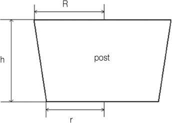
2) of the post segments. Post diameters were measured using a digital micro-caliper (MITUTOYO, Kawasaki, Japan), and the total bonding area for each post segment was calculated by using the formula: π(R + r)[(h
2 + (R - r)
2]
0.5, where π= 3.14, R represents the coronal post radius (mm), r the apical post radius (mm), and h is the thickness of the slice (mm) (
Figure 3).
Fractured test specimens were examined using operating microscope (OPMI pico; Carl zeiss, Obercohen, Germany) under 25 times magnification and the failure mode was classified into four types
7: (1) adhesive between post and resin cement (no cement visible around the post); (2) mixed, with resin cement covering of the post surface; (3) adhesive between resin cement and root canal (post enveloped by resin cement); (4) cohesive in dentine.
For the comparison of push-out bond strength among the application techniques and root level, two-way ANOVA was performed and Tukey's test was used for post-hoc multiple comparison using SPSS 12.0 software (SPSS Inc., Chicago, IL, USA). One-way ANOVA was also performed using the average data to compare the technique. The level of significance was set at p < 0.05.
Results
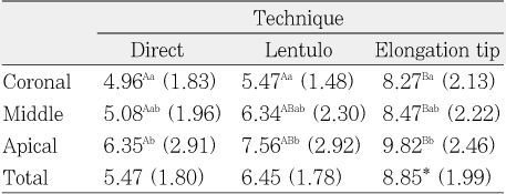
Two-way analysis of variance displayed that bond strengths were statistically influenced by root level (
p < 0.05). Apical bond strength was significantly higher than cervical dentin, but not than middle dentin. Luting application technique significantly affected bond strength values (
p < 0.05). 'Elongation tip' group showed significantly higher push-out bond strength compared with the other techniques (
Table 1).
The failure modes for different resin cement application methods are presented in
Table 2. Microscopic evaluation for the failure modes demonstrated that most failures occurred in an adhesive mode at the cement/dentin interface or in a mixed mode.
Discussion
In present study, the push-out bond strength was compared according to the cement application methods and post space level. Post retention has been measured with microtensile and push-out tests.
9-
11 It has been suggested that, because of the small size of specimens, a microtensile test permits a uniform stress distribution along the bonded interface.
12 However, Goracci et al. revealed that a push-out test is a more reliable method for determining bond strengths between fiber posts and post-space dentin because of the high number of premature failures occurring during specimen preparation and large data distribution spread associated with microtensile testing.
13
In addition, this study used three sequential series of samples for one post to reduce the sample error which may have a big deviation for one sample at a certain level of root. Thus the sequential three samples were tested for the push-out tests.
14 This method also provided the information for the bonding strength differences according to the root dentin level.
In this study, the push-out bond strengths were statistically influenced by the root levels (
p < 0.05). Present result confirmed previous studies
13,
15 that observed influence of root canal region on fiber post retention. Gaston et al. reported higher bond strengths in the apical third than in the parts of the root canal.
9 On the other hands, several studies have shown that the bond strength of resin cements to root canals is affective in the cervical third but weak in the apical third.
16 Regardless of the bond strength in apical regions, the frictional retention in these areas may contribute to the dislocation resistance of the fiber post.
17,
18
On the other hand, post retention to root canal dentine seemed to vary with luting cement application technique. Various in vitro researches revealed controversial results concerning bond strength values of different luting technique to fiber posts and root canal dentin.
19 Thus, in present study, the push-out bond strength were compared according to the cement delivery ways of direct application with post, lentulo-spiral application, and a special elongation tip for the cement delivery.
It was previously reported that the application of the resin luting agent with a lentulo-spiral instrument permits a favorable distribution of luting cement throughout the post space and a formation of uniform, continuous cement layer.
20 However, if a dual-cure resin was selected, the major recommendation is to avoid early set of the cement before complete post seating.
21 For dual-curing composite resin luting systems, the use of a lentulo-spiral drill is not recommended by the manufacturers, since the increased input energy may cause premature set of luting composite. Therefore, some manufacturers have been suggested, dual-cure resin cement should be taken into the root canal by applying a thin layer of cement over the post before setting it in position. However, clinicians would have a difficulty to get a uniform cement application within the post space with only the direct application on the post surface. Furthermore the round cross section of a post also may have a difficulty to match the irregular canal area efficiently with the direct cement applying method. Actually, some sectioned specimens in the direct applying group showed a vacant space within the cement area and/or adhesion layer. These voids could impede an appropriate cementation of the post, and may cause its debonding.
22,
23 Thus, the injection technique with specific syringes was introduced most recently and used for application of the resin cements seems to be preferred as an effective method for reducing voids and bubbles within the luting cement
4 and increasing homogeneous dispersion of the luting cement. The result of present study that the elongation tip showed significantly higher push-out bond strength than other ways was consistent with these points.
These findings are in accordance to those reported by Ronny et al.
14 The authors showed that the use of a flexible root-canal-shaped application aid as the elongation tip is superior compared to the conventional application technique in terms of root canal-post-cement interface homogeneity. By use of this application aid only ~4% of inhomogeneities for the complete post length within the cement interface of the adhesively luted fiber post were recorded, compared to 19% for the conventional application procedure.
Analyses of failure mode showed that most of failures occurred at the cement/dentin interface (adhesive failure) or in a mixed mode. It might be caused from the lower bond strength between cement and dentin than that between post and cement. It seemed that fact that the Rely-X Unicem as a kind of self adhesive system did not make a hybrid layer
24 for better bonding would make the lower bond strength than the strength between cement and post which has better condition for bonding with the silanization claimed by the manufacturer.
In addition to the self adhesive cement, as a future research, it is also needed to compare the bonding strengths of many other resin cements with varying pre-treatment methods according to various cement application method.
The use of an elongation tip reduces the number of imperfections within the self-adhesive cement interface compared to the conventional application techniques such as direct applying with the post and lentulo-spiral technique. Thus it is recommended to use an elongation tip which may bring favorable bond strength for the post cementation using resin cement.
-
This work was supported for two years by Pusan National University Research Grant.
REFERENCES
- 1. Axelsson P, Lindhe J, Nystrom B. On the prevention of caries and periodontal disease. Results of a 15-year longitudinal study in adults. J Clin Periodontol. 1991;18: 182-189.PubMed
- 2. Testori T, Badino M, Castagnola M. Vertical root fractures in endodontically treated teeth: a clinical survey of 36 cases. J Endod. 1993;19: 87-91.ArticlePubMed
- 3. Bergman B, Lundquist P, Sjogren U, Sundquist G. Restorative and endodontic results after treatment with cast posts and cores. J Prosthet Dent. 1989;61: 10-15.ArticlePubMed
- 4. Boschian Pest L, Cavalli G, Bertani P, Gagliani M. Adhesive post-endodontic restorations with fiber posts: push-out tests and SEM observations. Dent Mater. 2002;18: 596-602.ArticlePubMed
- 5. Kim MH, Kim HJ, Cho YG. Effect of curing methods of resin cements on bond strength and adhesive interface of post. J Korean Acad Conserv Dent. 2009;34: 103-112.Article
- 6. Kim JW, Yu MK, Lee SJ, Lee KW. Microtensile bonding of resin fiber reinforced post to radicular dentin using resin cement. J Korean Acad Conserv Dent. 2003;28: 80-87.Article
- 7. D'Arcangelo C, D'Amario M, Vadini M, Zazzeroni S, De Angelis F, Caputi S. An evaluation of luting agent application technique effect on fibre post retention. J Dent. 2008;36: 235-240.ArticlePubMed
- 8. D'Arcangelo C, D'Amario M, De Angelis F, Zazzeroni S, Vadini M, Caputi S. Effect of Application Technique of Luting Agent on the Retention of Three Types of Fiber-reinforced Post Systems. J Endod. 2007;33: 1378-1382.ArticlePubMed
- 9. Gaston BA, West LA, Liewehr FR, Fernandes C, Pashley DH. Evaluation of regional bond strength of resin cement to endodontic surfaces. J Endod. 2001;27: 321-324.ArticlePubMed
- 10. Foxton RM, Nakajima M, Tagami J, Miura H. Bonding of photo and dual cure adhesives to root canal dentin. Oper Dent. 2003;28: 543-551.PubMed
- 11. Kurtz JS, Perdigao J, Geraldeli S, Hodges JS, Bowles WR. Bond strengths of toothcolored posts, effect of sealer, dentin adhesive, and root region. Am J Dent. 2003;16 Spec No: 31A-36A.PubMed
- 12. Cardoso PE, Sadek FT, Goracci C, Ferrari M. Adhesion testing with the microtensile method: effects of dental substrate and adhesive system on bond strength measurement. J Adhes Dent. 2002;4: 291-297.PubMed
- 13. Goracci C, Tavares AU, Monticelli F, et al. The adhesion between fiber posts and root canal walls: comparison between microtensile and push-out bond strengths measurements. Eur J Oral Sci. 2004;112: 353-361.PubMed
- 14. Watzke R, Blunck U, Frankenberger R, Naumann M. Interface homogeneity of adhesively luted glass fiber posts. Dent Mater. 2008;24: 1512-1517.ArticlePubMed
- 15. Foxton RM, Nakajima M, Tagami J, Miura H. Adhesion to root canal dentine using one and two-step adhesives with dual-cure composite core materials. J Oral Rehabil. 2005;32: 97-104.ArticlePubMed
- 16. D'Arcangelo C, Cinelli M, De Angelis F, et al. The effect of resin cement film thickness on the pullout strength of a fiber reinforced post system. J Prosthet Dent. 2007;98: 193-198.ArticlePubMed
- 17. Goracci C, Fabianelli A, Sadek FT, et al. The contribution of friction to the dislocation resistance of bonded fiber posts. J Endod. 2005;31: 608-612.ArticlePubMed
- 18. Faria-e-Silva AL, Reis AF, Martins LR. The effects of different luting procedures in the push-out bond strength of fibre posts to the root canal. Braz J Oral Sci. 2008;27: 1653-1666.
- 19. Bitter K, Meyer-Lueckel H, Priehn K, Kanjuparambil JP, Neumann K, Kielbassa AM. Effects of luting agent and thermocycling on bond strengths to root canal dentine. Int Endod J. 2006;39: 809-818.ArticlePubMed
- 20. Akgungor G, Akkayan B. Influence of dentin bonding agents and polymerization modes on the bond strength between translucent fiber posts and three dentin regions within a post space. J Prosthet Dent. 2006;95: 368-378.ArticlePubMed
- 21. Schwartz RS, Robbins JW. Post placement and restoration of endodontically treated teeth: a literature review. J Endod. 2004;30: 289-301.ArticlePubMed
- 22. Ferrari M, Vichi A, Grandini S, Goracci C. Efficacy of a selfcuring adhesive/resin cement system on luting glass-fiber posts into root canals: an SEM investigation. Int J Prosthodont. 2001;14: 543-549.PubMed
- 23. Lee HA, Cho YG. Comparison of bond strength of a fiber post cemented with various resin cements. J Korean Acad Conserv Dent. 2008;33: 499-506.Article
- 24. De Munck J, Vargas M, Van Launduyt K, Hikita K, Lambrechts P, Van Meerbeek B. Bonding of an auto-adhesive luting material to enamel and dentin. Dent Mater. 2004;20: 963-971.ArticlePubMed
Figure 1Rely-X post drill and Rely-X fiber post (left), Rely-X Unicem with an elongation tip is ready to inject into the canal (right).

Figure 2Experimental design and preparation of specimens low-speed saw was used to prepare 3 sections through luted post specimen, each 2 mm thick (left). Push-out bond strength testing was performed using universal testing machine (right).

Figure 3Bonding area of post/cement interface was measured using formula of conical frustum: top (R) and bottom radius (r) of post along with height of specimen (h): total bonding area = π(R + r)[(h2 + (R - r)2]0.5

Table 1Mean push-out bond strength (MPa) and standard deviation (S.D.) for experimental groups

Table 2Failure modes of experimental groups


 KACD
KACD

 ePub Link
 Cite
Cite

