
Articles
- Page Path
- HOME > Restor Dent Endod > Volume 31(5); 2006 > Article
- Original Article Effects of occlusal load on the stress distribution of four cavity configurations of noncarious cervical lesions: A three-dimensional finite element analysis study
- Sang-Je Jeon1, Jeong-Kil Park1, Hyeon-Cheol Kim1, Sung-Gwan Woo2, Kwang-Hoon Kim2, Kwon Son2, Bock Hur1,*
-
J Korean Acad Conserv Dent 2006;31(5):-370.
DOI: https://doi.org/10.5395/JKACD.2006.31.5.359
Published online: January 14, 2006
1Department of Conservative dentistry, College of Dentistry, Pusan National University
2Department of Mechanical design engineering, College of Engineering, Pusan National University
- *Corresponding Author: Bock Hur, Department of Conservative Dentistry, College of Dentistry, Pusan National University, 1-10, Ami-dong, Seo-gu, Busan, 602-739, Korea, Tel: 82-51-240-7455, E-mail: bhur@pusan.ac.kr
• Received: July 4, 2006 • Revised: July 28, 2006 • Accepted: September 4, 2006
Copyright © 2006 The Korean Academy of Conservative Dentistry
This is an Open Access article distributed under the terms of the Creative Commons Attribution Non-Commercial License (http://creativecommons.org/licenses/by-nc/3.0) which permits unrestricted non-commercial use, distribution, and reproduction in any medium, provided the original work is properly cited.
- 1,512 Views
- 1 Download
Abstract
-
The objective of this study was to investigate the effect of excessive occlusal loading on stress distribution on four type of cervical lesion, using a three dimensional finite element analysis (3D FEA).The extracted maxillary second premolar was scanned serially with Micro-CT. The 3D images were processed by 3D-DOCTOR. ANSYS was used to mesh and analyze 3D FE model. Four different lesion configurations representative of the various types observed clinically for teeth were studied. A static point load of 500N was applied to the buccal and lingual cusp (Load A and B). The principal stresses in lesion apex, and vertical sectioned margin of cervical wall were analyzed.The results were as follows
The patterns of stress distribution were similar but the magnitude was different in four types of lesion.
The peak stress was observed at mesial corner and also stresses concentrated at lesion apex.
The compressive stress under load A and the tensile stress under load B were dominant stress.
Under the load, lesion can be increased and harmful to tooth structure unless restored.


Figure 3.
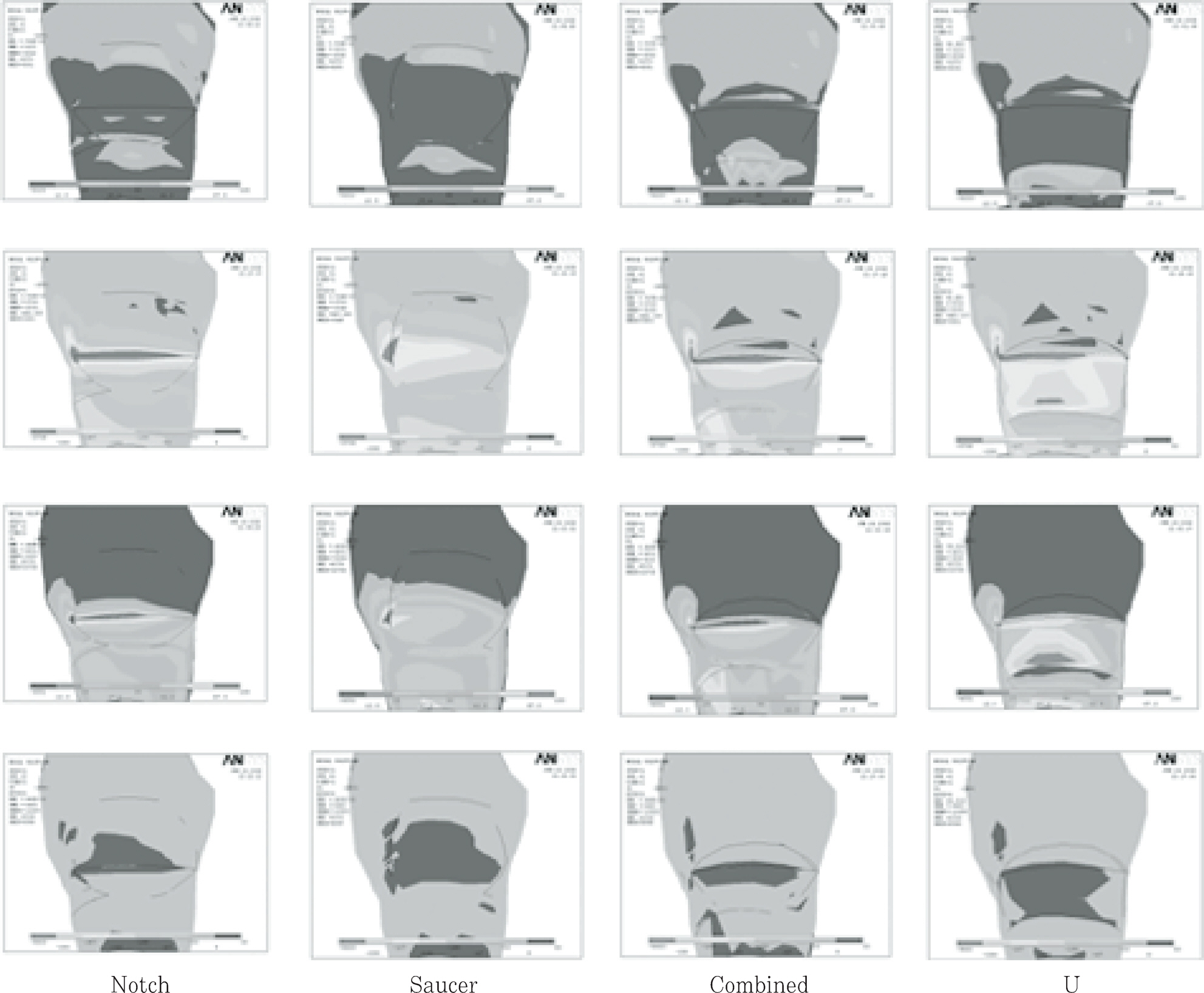
The Principal stress distribution of four cavities under Load A and B(Upper 2 rows: maximal and minimal principal stress under load A, Lower 2 rows; maximal and minimal principal stress under load B. From left line, notch-shape showed, followed by saucer, combined, U-shape lesion)
Figure 4.
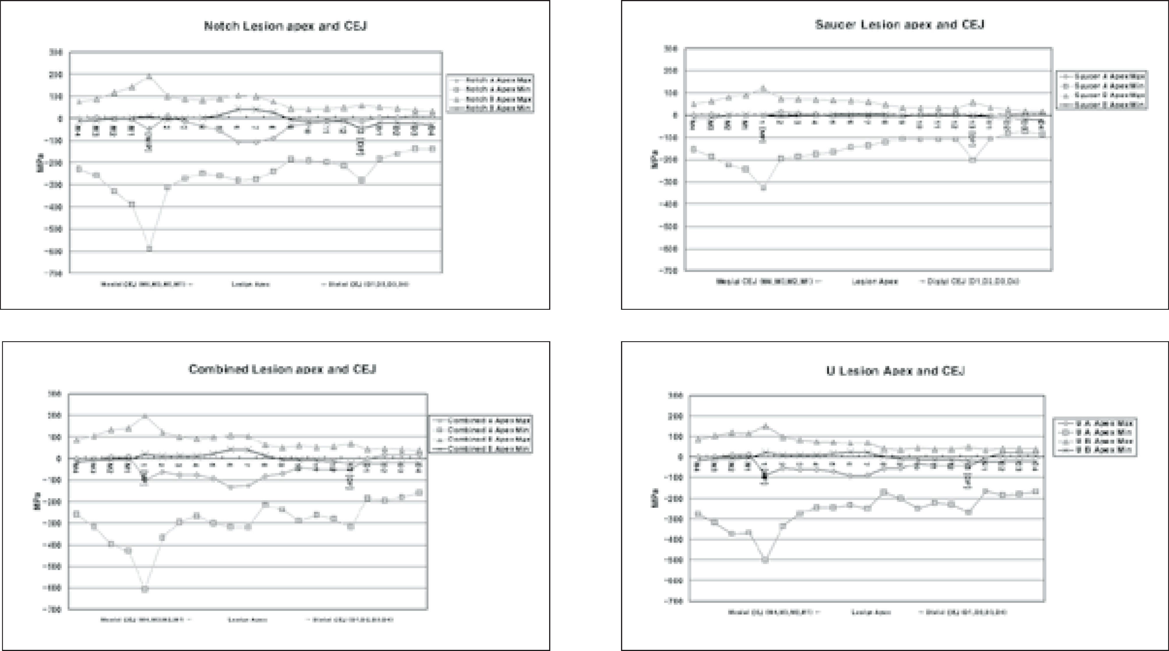
Principal stresses of lesion apex of 4 shape lesion.
(M1, M2, M3, M4: nodes near mesial point angle, D1, D2, D3, D4: nodes near distal point angle. If the value of one principal stress of an element was positive, the element was determined to be in the tensile condition, if it was negative, the element was determined to be in the compressive condition).
Figure 5.
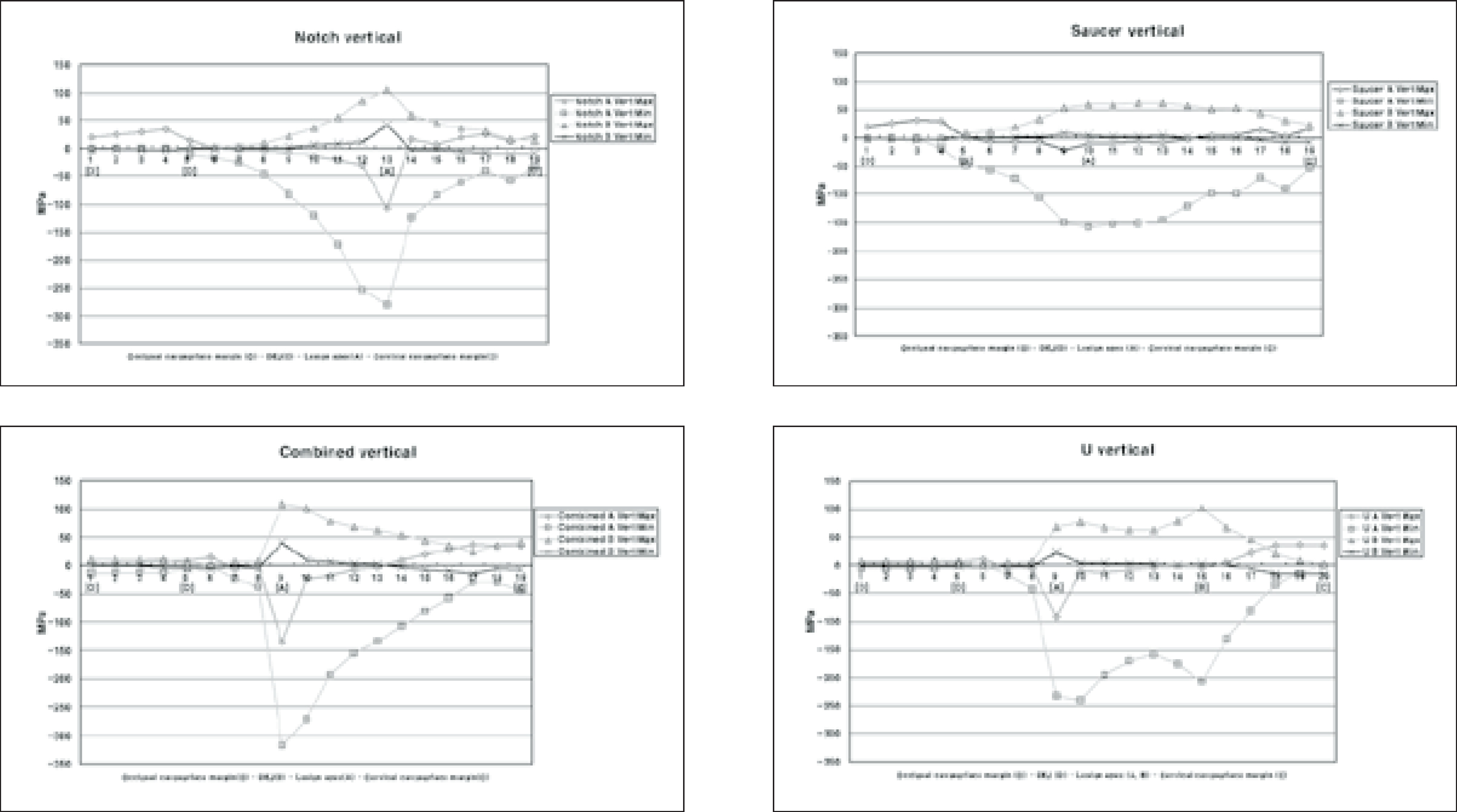
Vertical distribution of principal stresses at cavity wall in 4 shape lesion. (If the value of one principal stress of an element was positive, the element was determined to be in the tensile condition, if it was negative, the element was determined to be in the compressive condition).
Table 1.
Numbers of nodes and elements for each cavity
| Model | Notch | Saucer | Combined | U |
|---|---|---|---|---|
| Node | 18245 | 18236 | 18434 | 18709 |
| Element | 16668 | 16668 | 16780 | 17056 |
Table 2.
Mechanical properties of the tooth used in the study
Table 3.
Mechanical properties of teeth (MPa)*
| Compressive strength of enamel | 277 – 384 |
|---|---|
| Compressive strength of dentin | 249 – 347 |
| Tensile strength of enamel | 10 – 24 |
| Tensile strength of dentin | 32 – 103 |
| Tensile strength of DEJ | 52 |
Table 4.
Peak stresses of lesion apex of four cavities
| Load A | Load B | ||||||||
|---|---|---|---|---|---|---|---|---|---|
| 1st Peak | 2nd Peak | 1st Peak | 2nd Peak | ||||||
| MPa | Node | MPa | Node | MPa | Node | MPa | Node | ||
| Notch | Max | 12.4 | 6 | 10.9 | 20 | 193* | 1 | 104.3* | 5 |
| Min | -558* | 1 | -279* | 13 | 40.2 | 6 | NS | NS | |
| Saucer | Max | 14.6 | 1 | 8.8 | 16 | 123.0* | 1 | 55.8* | 13 |
| Min | -330.0* | 1 | -203.3 | 13 | NS | NS | NS | NS | |
| Combined | Max | 10.0 | M1 | 18.3 | D3 | 195.5* | 1 | 108.3* | 6 |
| Min | -606.7* | 1 | -320.7* | 7 | NS | NS | NS | NS | |
| U | Max | 17.8 | D3 | 13.9 | M1 | 147.9* | 1 | 71.1* | 7 |
| Min | -500.9* | 1 | -268.7* | 13 | NS | NS | NS | NS | |
Table 5.
Peak stresses values of inner cavity wall vertically
| Load A | Load B | ||||||||
|---|---|---|---|---|---|---|---|---|---|
| 1st Peak | 2nd Peak | 1st Peak | 2nd Peak | ||||||
| MPa | Node | MPa | Node | MPa | Node | MPa | Node | ||
| Notch | Max | 33.7* | 4 | 25.5 | 17 | 104.3* | 13 | NS | NS |
| Min | -280.4* | 13 | -58.8 | 18 | 40.2 | 13 | NS | NS | |
| Saucer | Max | 30.8 | 3 | 15.2 | 17 | 61.9* | 13 | 53.0* | 16 |
| Min | -157.1 | 10 | -98.1 | 16 | 7.8 | 9 | 4.4 | 13 | |
| Combined | Max | 36.8 | 17 | 14.7 | 6 | 108.3* | 9 | 42.0* | 19 |
| Min | -317.7* | 1 | -44.2 | 19 | 41.1 | 9 | NS | NS | |
| U | Max | 36.01 | 19 | 11.3 | 6 | 100.7* | 15 | 76.6* | 9 |
| Min | -240.5 | 10 | -207.3 | 15 | 22.7 | 9 | 2.7 | 15 | |
- 1. James D, Linda C, Levitch . How dentists classified and treated non-carious cervical lesions. J Am Dent Assoc 124(5):46-54. 1993.ArticlePubMed
- 2. Bradley T, William B Hancock. Examining the prevalence and characteristics of abfraction like cervical lesions in a population of U.S. veterans. J Am Dent Assoc 132(12):1694-1701. 2001.ArticlePubMed
- 3. Lee WC, Eakle WS. Stress-induced cervical lesions: Review of advances on the past 10 years. J Prosthet Dent 75:487-494. 1996.PubMed
- 4. Grippo JO. Abfractions: A new classification of hard tissue lesions of teeth. J Esthet Dent 3(1):14-19. 1991.ArticlePubMed
- 5. Gallien GS, Kaplan I, Owen BM. A review of noncari-ous dental cervical lesions. Compend Contin Educ Dent 15(11):1366-1372. 1994.
- 6. Grippo JO, Schreiner S. Attrition, abrasion, corrosion and abfraction revisited-A new perspective on tooth surface lesion. J Am Dent Assoc 135(8):1109-1117. 2004.PubMed
- 7. Rees JS, Hammadeh M. Undermining of enamel as a mechanism of abfraction lesion formation: a finite element study. Eur J Oral Sci 112:347-352. 2004.ArticlePubMed
- 8. Radentz WH, Barnes GP, Cutright DE. A survey of factors possibly associated with cervical abrasion of tooth surface. J Periodontol 47:148-154. 1976.PubMed
- 9. Kahn F, Young WG, Shahabi S, Daley TJ. Dental cervical lesions associated with occlusal erosion and attrition. Aust Dent J 44:176-186. 1999.ArticlePubMed
- 10. Levitch LC, Bader JD, Shugars DA, Heymann HO. Non-carious cervical lesions. J Dent 22:195-207. 1994.ArticlePubMed
- 11. Leinfelder KF. Restoration of abfracted lesions. Compend Contin Educ Dent 15(4):1396-1400. 1994.
- 12. Park Jk, Hur B, Lee HJ. The effect of configuration on marginal leakage of class 5 restoration. J Kor Acad Cons Dent 26(2):162-170. 2001.
- 13. Kuroe T, Itoh H, Caputo AA, Konuma M. Biomechanics of cervical tooth structure lesions and their restoration. Quint Int 31(4):267-274. 2000.
- 14. Grippo JO. Noncarious cervical lesions: The decision to ignore or restore. J Esthet Dent 4:118-132. 1992.Article
- 15. Litonjua LA, Andreana S, Patra AK, Cohen RE. An assessment of stress analyses in the theory of abfraction. Biomed Mater Eng 14:311-321. 2004.PubMed
- 16. Rees JS, Jacobsen PH. The effect of cuspal flexure on a buccal Class V restoration: a finite element study. J Dent 26(4):361-367. 1998.ArticlePubMed
- 17. Lindehe J, Karring T. Textbook of Clinical Periodontology. 2nd edition. Munksgaard; Copenhagen: p. p19-69. 1989.
- 18. Schroeder HE, Page RC. Periodontal Diseases. 2nd edition. Lea & Fabiger; Philadelphia: p. p 3-52. 1990.
- 19. Rubin C, Krishnamurthy N, Capilouto E, Yi H. Stress analysis of the human tooth using a three-dimensional finite element model. J Dent Res 62:82-86. 1983.ArticlePubMedPDF
- 20. Katona TR, Winkler MM. Stress analysis of a bulk-filled class V light-cured composite restoration. J Dent Res 73(8):1470-1477. 1994.ArticlePubMedPDF
- 21. Geramy A, Sharafoddin F. Abfraction: 3D analysis by means of the finite method. Quint Int 34(7):526-533. 2003.
- 22. AW TC, Lepe X, Johnson GH, Mancl L. Characteristics of noncarious cervical lesions: A clinical investigation. J Am Dent Assoc 133(6):725-733. 2002.PubMed
- 23. Widmalm SE, Ericsson SG. Maximal bite force with centric and eccentric load. J Oral Rehabil 9:445-450. 1982.ArticlePubMed
- 24. Gibbs CH, Mahan PE, Lundeen HC, Brehnan K, Walsh EK, Holbrook WB. Occlusal forces during chewing and swallowing as measured by sound transmission. J Prosthet Dent 46:443-449. 1981.ArticlePubMed
- 25. Caputo AA, Standlee JP. Biomechanics in Clinical Dentistry. Chicago Quintessence Int 21-27. 1987.
- 26. Ziemiecki TL, Dennison JB, Charbeneau GT. Clinical evaluation of cervical composite resin restorations placed without retention. Oper Dent 12:27-33. 1987.PubMed
- 27. Whitehead SA, Wilson NHF, Watts DC. Demonstration of “Vertical Barreling“using profilometry. Eur J Prosthodont Restor Dent 7(4):131-134. 1999.PubMed
- 28. Browning WD, Brackett WW, Gilpatrick RO. Two-year clinical comparison of a microfilled and hybrid resin based composite in noncarious class V lesions. Oper Dent 25:46-50. 2000.PubMed
- 29. Kubo S, Yokota H, Sata Y, Hayashi Y. The effect of flexural load cycling on the microleakage of cervical resin composites. Oper Dent 26:451-459. 2001.PubMed
REFERENCES
Tables & Figures
REFERENCES
Citations
Citations to this article as recorded by 
 ePub Link
 Cite
CiteEffects of occlusal load on the stress distribution of four cavity configurations of noncarious cervical lesions: A three-dimensional finite element analysis study


Figure 1. Four types of lesion configurations.
Figure 2. Design of loading conditions.
Figure 3. The Principal stress distribution of four cavities under Load A and B(Upper 2 rows: maximal and minimal principal stress under load A, Lower 2 rows; maximal and minimal principal stress under load B. From left line, notch-shape showed, followed by saucer, combined, U-shape lesion)
Figure 4. Principal stresses of lesion apex of 4 shape lesion.
Figure 5. Vertical distribution of principal stresses at cavity wall in 4 shape lesion. (If the value of one principal stress of an element was positive, the element was determined to be in the tensile condition, if it was negative, the element was determined to be in the compressive condition).
Figure 1.
Figure 2.
Figure 3.
Figure 4.
Figure 5.
Effects of occlusal load on the stress distribution of four cavity configurations of noncarious cervical lesions: A three-dimensional finite element analysis study
| Model | Notch | Saucer | Combined | U |
|---|---|---|---|---|
| Node | 18245 | 18236 | 18434 | 18709 |
| Element | 16668 | 16668 | 16780 | 17056 |
| Materials | Mechanical properties Young's modulus (MPa) | Poisson's ratio (υ) |
|---|---|---|
| Enamel | 84000 a | 0.33 a |
| Dentin | 18000 a | 0.31 a |
| Periodontal ligament | 0.667 b | 0.49 b |
| Cancellous bone | 13700 b | 0.38 b |
| Cortical bone | 34000 b | 0.26 b |
| Compressive strength of enamel | 277 – 384 |
|---|---|
| Compressive strength of dentin | 249 – 347 |
| Tensile strength of enamel | 10 – 24 |
| Tensile strength of dentin | 32 – 103 |
| Tensile strength of DEJ | 52 |
| Load A | Load B | ||||||||
|---|---|---|---|---|---|---|---|---|---|
| 1st Peak | 2nd Peak | 1st Peak | 2nd Peak | ||||||
| MPa | Node | MPa | Node | MPa | Node | MPa | Node | ||
| Notch | Max | 12.4 | 6 | 10.9 | 20 | 193 |
1 | 104.3 |
5 |
| Min | -558 |
1 | -279 |
13 | 40.2 | 6 | NS | NS | |
| Saucer | Max | 14.6 | 1 | 8.8 | 16 | 123.0 |
1 | 55.8 |
13 |
| Min | -330.0 |
1 | -203.3 | 13 | NS | NS | NS | NS | |
| Combined | Max | 10.0 | M1 | 18.3 | D3 | 195.5 |
1 | 108.3 |
6 |
| Min | -606.7 |
1 | -320.7 |
7 | NS | NS | NS | NS | |
| U | Max | 17.8 | D3 | 13.9 | M1 | 147.9 |
1 | 71.1 |
7 |
| Min | -500.9 |
1 | -268.7 |
13 | NS | NS | NS | NS | |
| Load A | Load B | ||||||||
|---|---|---|---|---|---|---|---|---|---|
| 1st Peak | 2nd Peak | 1st Peak | 2nd Peak | ||||||
| MPa | Node | MPa | Node | MPa | Node | MPa | Node | ||
| Notch | Max | 33.7 |
4 | 25.5 | 17 | 104.3 |
13 | NS | NS |
| Min | -280.4 |
13 | -58.8 | 18 | 40.2 | 13 | NS | NS | |
| Saucer | Max | 30.8 | 3 | 15.2 | 17 | 61.9 |
13 | 53.0 |
16 |
| Min | -157.1 | 10 | -98.1 | 16 | 7.8 | 9 | 4.4 | 13 | |
| Combined | Max | 36.8 | 17 | 14.7 | 6 | 108.3 |
9 | 42.0 |
19 |
| Min | -317.7 |
1 | -44.2 | 19 | 41.1 | 9 | NS | NS | |
| U | Max | 36.01 | 19 | 11.3 | 6 | 100.7 |
15 | 76.6 |
9 |
| Min | -240.5 | 10 | -207.3 | 15 | 22.7 | 9 | 2.7 | 15 | |
Table 1. Numbers of nodes and elements for each cavity
Table 2. Mechanical properties of the tooth used in the study
a: Katona
Table 3. Mechanical properties of teeth (MPa)*
: Litonjua
Table 4. Peak stresses of lesion apex of four cavities
: Excessive stresses over the failure range. NS: Not significant
Table 5. Peak stresses values of inner cavity wall vertically
: Excessive stresses over the failure range. NS: Not significant

