
Search
- Page Path
- HOME > Search
Research Article
- Difference in light transmittance and depth of cure of flowable composite depending on tooth thickness: an in vitro experimental study
- Seong-Pyo Bae, Myung-Jin Lee, Kyung-San Min, Mi-Kyung Yu, Kwang-Won Lee
- Restor Dent Endod 2025;50(4):e39. Published online November 28, 2025
- DOI: https://doi.org/10.5395/rde.2025.50.e39
-
 Abstract
 PDF
PDF PubReader
 ePub
ePub - Objectives
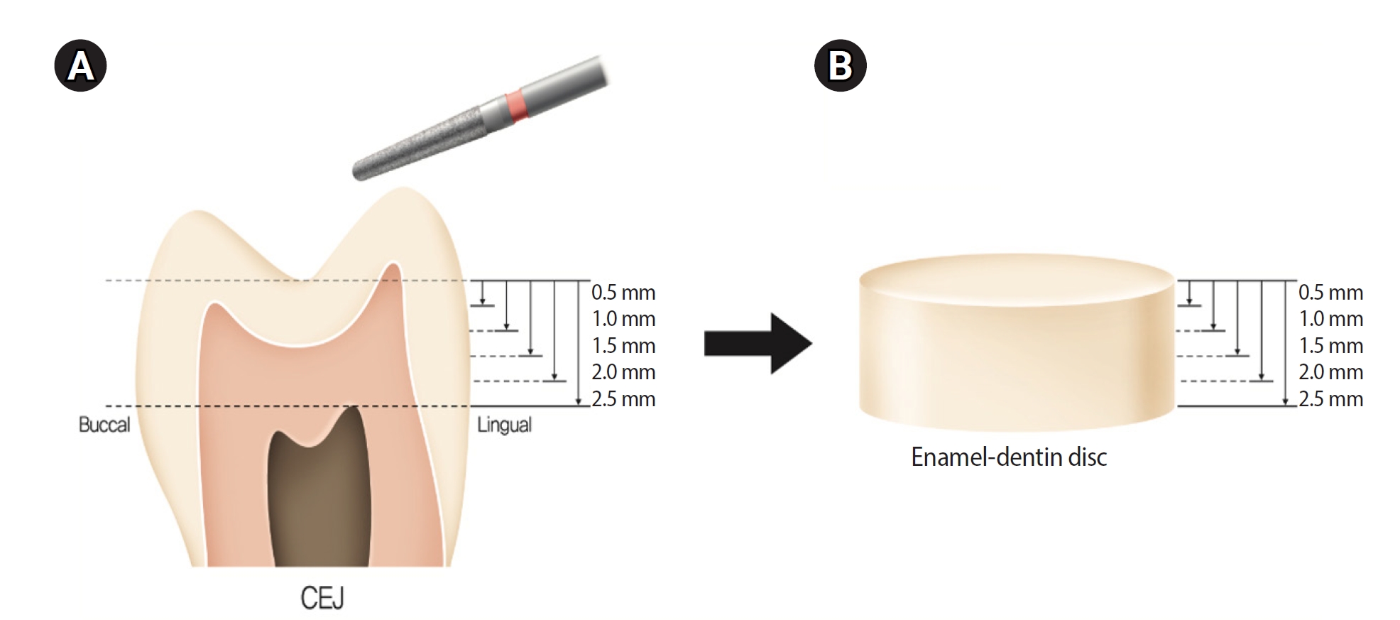
This study aimed to quantify light attenuation through varying tooth thicknesses and its impact on the depth of cure of composite resin.
Methods
Twenty extracted premolars were used to create enamel-dentin discs that were sanded progressively in 0.5 mm increments from 2.5 mm to 0.5 mm. Light irradiance was measured with and without tooth specimens to evaluate light transmittance. Resin was cured beneath different thicknesses, and the depth of cure was assessed using the Vickers hardness test.
Results
The results demonstrated that light transmittance significantly decreased as tooth thickness increased (p < 0.01), leading to reduced resin polymerization. In the 2.0-mm and 2.5-mm tooth thickness groups, the depth of cure was significantly lower than in the control group without tooth specimens (p < 0.05).
Conclusions
Ultimately, for tooth structures exceeding 2 mm, self-cure or dual-cure resin polymerization is thought to be more efficient than light polymerization.
- 1,877 View
- 143 Download

Case Report
- Surgical management of maxillary sinusitis of endodontic origin after reestablishing maxillary sinus floor healing through a nonsurgical approach: a case report
- Eun-Sook Kang, Min-Kyeong Kim, Mi-Kyung Yu, Kyung-San Min
- Restor Dent Endod 2025;50(2):e12. Published online April 8, 2025
- DOI: https://doi.org/10.5395/rde.2025.50.e12
-
Abstract
PDFPubReaderePub
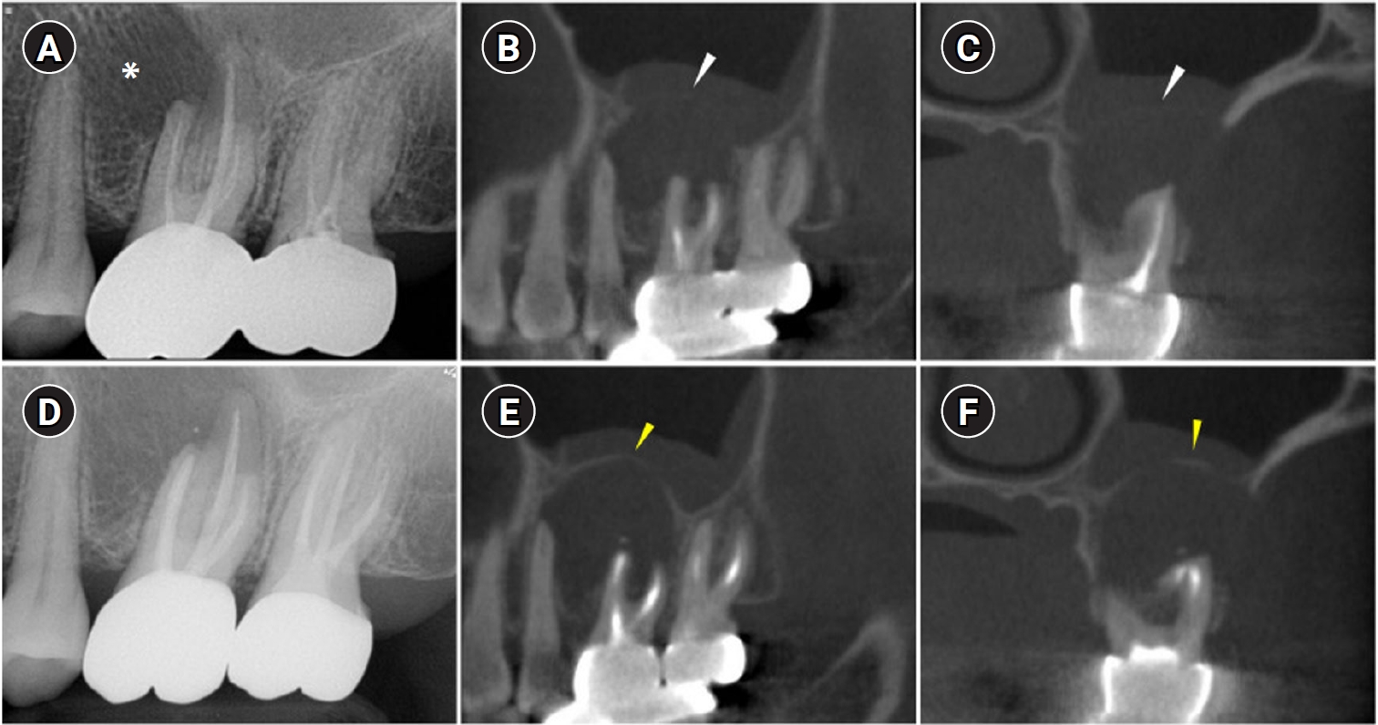
- When root canal infections breach the maxillary sinus floor (MSF), maxillary sinusitis of endodontic origin (MSEO) can result. This case illustrates the surgical management of MSEO following the nonsurgical reestablishment of the MSF. A 55-year-old woman presented with left facial pain and was diagnosed with MSEO originating from the left upper first molar. Despite undergoing nonsurgical root canal treatment, there was no evidence of bony healing after 6 months. However, cone-beam computed tomographic (CBCT) scans revealed the reestablishment of MSF. Subsequently, surgical intervention was carried out using a dental operating microscope. Two years after surgery, CBCT images indicated that the mucosal edema had resolved, and the MSF was well reestablished. Preserving the MSF is crucial for the success of endodontic surgery. When MSEO is present, the integrity of the MSF must be assessed to determine appropriate treatment options.
- 5,128 View
- 233 Download
Research Articles
- Push-out bond strength and intratubular biomineralization of a hydraulic root-end filling material premixed with dimethyl sulfoxide as a vehicle
- Ju-Ha Park, Hee-Jin Kim, Kwang-Won Lee, Mi-Kyung Yu, Kyung-San Min
- Restor Dent Endod 2023;48(1):e8. Published online January 20, 2023
- DOI: https://doi.org/10.5395/rde.2023.48.e8
-
Abstract
PDFPubReaderePub
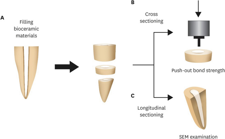
Objectives This study was designed to evaluate the parameters of bonding performance to root dentin, including push-out bond strength and dentinal tubular biomineralization, of a hydraulic bioceramic root-end filling material premixed with dimethyl sulfoxide (Endocem MTA Premixed) in comparison to a conventional powder-liquid–type cement (ProRoot MTA).
Materials and Methods The root canal of a single-rooted premolar was filled with either ProRoot MTA or Endocem MTA Premixed (
n = 15). A slice of dentin was obtained from each root. Using the sliced specimen, the push-out bond strength was measured, and the failure pattern was observed under a stereomicroscope. The apical segment was divided into halves; the split surface was observed under a scanning electron microscope, and intratubular biomineralization was examined by observing the precipitates formed in the dentinal tubule. Then, the chemical characteristics of the precipitates were evaluated with energy-dispersive X-ray spectroscopic (EDS) analysis. The data were analyzed using the Student’st -test followed by the Mann-WhitneyU test (p < 0.05).Results No significant difference was found between the 2 tested groups in push-out bond strength, and cohesive failure was the predominant failure type. In both groups, flake-shaped precipitates were observed along dentinal tubules. The EDS analysis indicated that the mass percentage of calcium and phosphorus in the precipitate was similar to that found in hydroxyapatite.
Conclusions Regarding bonding to root dentin, Endocem MTA Premixed may have potential for use as an acceptable root-end filling material.
-
Citations
Citations to this article as recorded by
- Comparison of intratubular biomineralization between in vivo and in vitro conditions
Sieun Nam, Yeon-Jee Yoo, Mi-Kyung Yu, Kyung-San Min
Journal of Oral Science.2026; 68(1): 30. CrossRef - Effectiveness of Sectioning Method and Filling Materials on Roughness and Cell Attachments in Root Resection Procedure
Tarek Ashi, Naji Kharouf, Olivier Etienne, Bérangère Cournault, Pierre Klienkoff, Varvara Gribova, Youssef Haikel
European Journal of Dentistry.2025; 19(01): 240. CrossRef - Bond Strength and Adhesive Interface Quality of New Pre‐Mixed Bioceramic Root Canal Sealer
Gustavo Creazzo, Bruna Monteiro de Barros Ciribelli Alves, Helena Cristina de Assis, Karen Gisselle Garay Villamayor, Manoel Damião de Sousa‐Neto, Jardel Francisco Mazzi‐Chaves, Fabiane Carneiro Lopes‐Olhê
Microscopy Research and Technique.2025; 88(7): 1989. CrossRef - Evaluation of clinical and radiographic outcome of premixed injectable mineral trioxide aggregate and conventional mineral trioxide aggregate as pulpotomy medicaments in primary molars – A split-mouth randomized control trial
U. S. Aiswarya, Sharan S. Sargod, Sundeep K. Hegde, H. T. Ajay Rao, Nanditha Hegde
Journal of Indian Society of Pedodontics and Preventive Dentistry.2025; 43(4): 559. CrossRef - Evaluation of the root dentin bond strength and intratubular biomineralization of a premixed calcium aluminate-based hydraulic bioceramic endodontic sealer
Yu-Na Lee, Min-Kyeong Kim, Hee-Jin Kim, Mi-Kyung Yu, Kwang-Won Lee, Kyung-San Min
Journal of Oral Science.2024; 66(2): 96. CrossRef - Removal efficiency of a fast setting pozzalan-based bioactive cement: a micro CT study
Feyza Çetinkaya, Ahter Şanal Çıkman, Ali Keleş, Banu Arıcıoğlu
BMC Oral Health.2024;[Epub] CrossRef - Antibacterial Activity and Sustained Effectiveness of Calcium Silicate-Based Cement as a Root-End Filling Material against Enterococcus faecalis
Seong-Hee Moon, Seong-Jin Shin, Seunghan Oh, Ji-Myung Bae
Materials.2023; 16(18): 6124. CrossRef
- Comparison of intratubular biomineralization between in vivo and in vitro conditions
- 3,920 View
- 106 Download
- 7 Web of Science
- 7 Crossref
- Change of phase transformation and bond strength of Y-TZP with various hydrofluoric acid etching
- Mi-Kyung Yu, Eun-Jin Oh, Myung-Jin Lim, Kwang-Won Lee
- Restor Dent Endod 2021;46(4):e54. Published online October 20, 2021
- DOI: https://doi.org/10.5395/rde.2021.46.e54
-
Abstract
PDFPubReaderePub
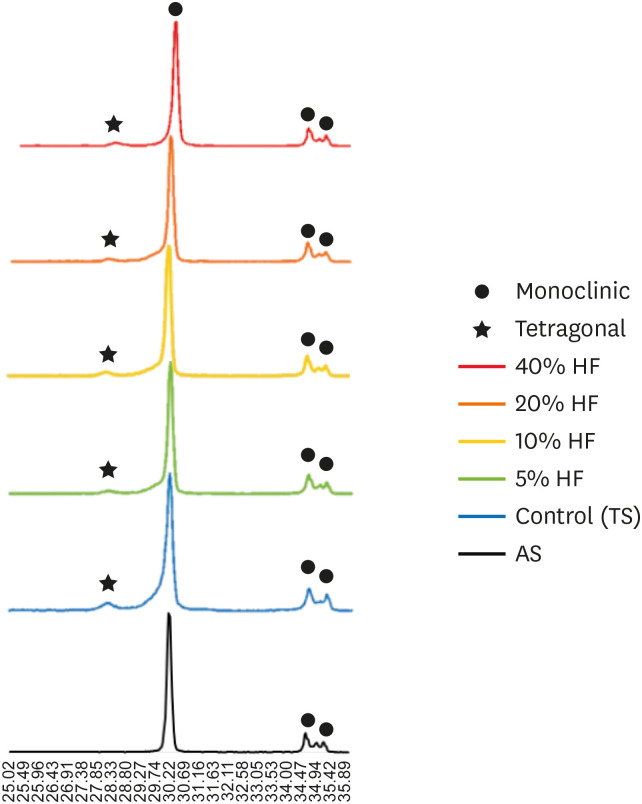
Objectives The purpose of this study was to quantify phase transformation after hydrofluoric acid (HF) etching at various concentrations on the surface of yttria-stabilized tetragonal zirconia polycrystal (Y-TZP), and to evaluate changes in bonding strength before and after thermal cycling.
Materials and Methods A group whose Y-TZP surface was treated with tribochemical silica abrasion (TS) was used as the control. Y-TZP specimens from each experimental group were etched with 5%, 10%, 20%, and 40% HF solutions at room temperature for 10 minutes. First, to quantify the phase transformation, Y-TZP specimens (
n = 5) treated with TS, 5%, 10%, 20% and 40% HF solutions were subjected to X-ray diffraction. Second, to evaluate the change in bond strength before and after thermal cycling, zirconia primer and MDP-containing resin cement were sequentially applied to the Y-TZP specimen. After 5,000 thermal cycles for half of the Y-TZP specimens, shear bond strength was measured for all experimental groups (n = 10).Results The monoclinic phase content in the 40% HF-treated group was higher than that of the 5%, 10%, and 20% HF-treated groups, but lower than that of TS-treated group (
p < 0.05). The 40% HF-treated group showed significantly higher bonding strength than the TS, 5%, and 10% HF-treated groups, even after thermal cycling (p < 0.05).Conclusions Through this experiment, the group treated with SiO2 containing air-borne abrasion on the Y-TZP surface showed higher phase transformation and higher reduction in bonding strength after thermal cycling compared to the group treated with high concentration HF.
-
Citations
Citations to this article as recorded by- Phase transition regulation and enhancement of optical properties in YPO4:Eu3+ through the influence of alkali metal ions
Junwei Zhan, Liusai Yang, Yaoxian Zhu, Yifan Zhu, Jianlei Liu, Siyan Peng, Jianping Zou
Journal of Molecular Structure.2026; 1352: 144420. CrossRef - Etchability of zirconia ceramics and its effect on adhesion: A systematic review and meta-analysis
Anina Sieber, Luiza Freitas Brum Souza, Tan Fırat Eyüboğlu, Mutlu Özcan
International Journal of Adhesion and Adhesives.2026; 148: 104303. CrossRef - Effect of BaTiO3 particle size and distribution on the BaTiO3-3Y-TZP interface reactivity of bioactive ceramic composites
João Pinto, Michael Gasik, Óscar Carvalho, Filipe S. Silva
Ceramics International.2026; 52(12): 18587. CrossRef - Improving the Clinical Performance of Dental Implants Through Advanced Surface Treatments: The Case of Ti and ZrO2 Coatings
Mohamed Aissi, Qanita Tayyaba, Azzedine Er-Ramly, Hendra Hermawan, Nadia Merzouk
Metals.2025; 15(3): 320. CrossRef - Enhancing the bonding of zirconia to resin by constructing a graded zirconia-glass composite surface
Zhiqi Yan, Jiale Li, Jing Chen, Zhe Zhao, Fan Li, Ling Zhang, Jihua Chen, Fu Wang
Surfaces and Interfaces.2025; 64: 106374. CrossRef - Surface property changes observed in zirconia during etching with high-concentration hydrofluoric acid over various immersion times
Ga-Eul YOU, Myung-Jin LIM, Kyung-San MIN, Mi-Kyung YU, Kwang-Won LEE
Dental Materials Journal.2024; 43(1): 52. CrossRef - Effect of surface treatments on the bond strength for different generation of zirconia CAD/CAM blocks
Man-Jong Cho, Sunwoong Song, Shin Hye Chung, Young-Seok Park, Bum-Soon Lim
Korean Journal of Dental Materials.2024; 51(3): 157. CrossRef - Is zirconia surface etching a viable alternative to airborne particle abrasion? A systematic review and meta-analysis of in vitro studies
Carlo D'Alessandro, Uros Josic, Claudia Mazzitelli, Tatjana Maravic, Laurel Graham, Carlo Barausse, Annalisa Mazzoni, Lorenzo Breschi, Markus B. Blatz
Journal of Dentistry.2024; 151: 105394. CrossRef - Exploring Zirconia Adhesion: Pre and Postsintering Physical Surface Treatment, Chemical Treatment, and Cement Interactions
Flávia Gonçalves, Mirko Dennys Ayala-Perez, Francisco Carlos dos Santos Reis, Walter Gomes Miranda-Júnior, Letícia Cristina Cidreira Boaro, Heng Bo Jiang
BioMed Research International.2024;[Epub] CrossRef - 3Y-TZP electrostatic painting to increase bond strength to dentin and dental prostheses
Alessandro Brito Thomaz, Carlos Nelson Elias, Heraldo Elias Salomão dos Santos, Celso Renato de Souza Resende, Claudinei dos Santos
Journal of Materials Research and Technology.2023; 26: 9063. CrossRef - Effect of surface topography and wettability on shear bond strength of Y-TZP ceramic
Suriyakul Wongsue, Ornnicha Thanatvarakorn, Taweesak Prasansuttiporn, Piyarat Nimmanpipug, Thanapat Sastraruji, Keiichi Hosaka, Richard M. Foxton, Masatoshi Nakajima
Scientific Reports.2023;[Epub] CrossRef - Adhesive Cementation of Zirconia Based Ceramics-Surface Modification Methods Literature Review
Magdalena Szawioła-Kirejczyk, Karolina Chmura, Krzysztof Gronkiewicz, Andrzej Gala, Jolanta E. Loster, Wojciech Ryniewicz
Coatings.2022; 12(8): 1067. CrossRef - Y-TZP Physicochemical Properties Conditioned with ZrO2 and SiO2 Nanofilms and Bond Strength to Dual Resin Cement
Ricardo Faria Ribeiro, Danilo Flamini Oliveira, Camila Bussola Tovani, Ana Paula Ramos, Ana Flavia Sanches Borges, Adriana Claudia Lapria Faria, Rossana Pereira de Almeida, Renata Cristina Silveira Rodrigues
Materials.2022; 15(22): 7905. CrossRef - Enhanced osteogenic activity of titania-modified zirconia implant by ultraviolet irradiation
Shuang Tang, Yan Wang, Zhenyu Zong, Ning Ding, Zutai Zhang
Frontiers in Bioengineering and Biotechnology.2022;[Epub] CrossRef
- Phase transition regulation and enhancement of optical properties in YPO4:Eu3+ through the influence of alkali metal ions
- 2,816 View
- 26 Download
- 15 Web of Science
- 14 Crossref
- Effect of hydrofluoric acid-based etchant at an elevated temperature on the bond strength and surface topography of Y-TZP ceramics
- Mi-Kyung Yu, Myung-Jin Lim, Noo-Ri Na, Kwang-Won Lee
- Restor Dent Endod 2020;45(1):e6. Published online December 3, 2019
- DOI: https://doi.org/10.5395/rde.2020.45.e6
-
Abstract
PDFPubReaderePub
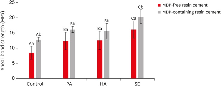
Objectives This study investigated the effects of a hydrofluoric acid (HA; solution of hydrogen fluoride [HF] in water)-based smart etching (SE) solution at an elevated temperature on yttria-stabilized tetragonal zirconia polycrystal (Y-TZP) ceramics in terms of bond strength and morphological changes.
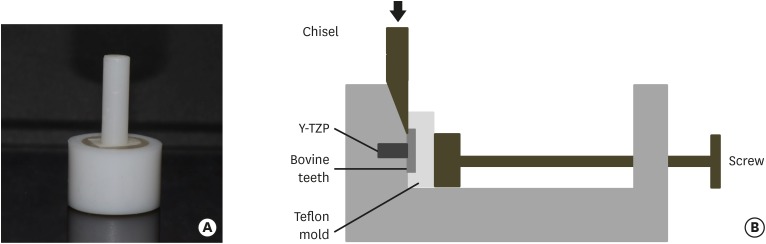
Materials and Methods Eighty sintered Y-TZP specimens were prepared for shear bond strength (SBS) testing. The bonding surface of the Y-TZP specimens was treated with 37% phosphoric acid etching at 20°C–25°C, 4% HA etching at 20°C–25°C, or HA-based SE at 70°C–80°C. In all groups, zirconia primers were applied to the bonding surface of Y-TZP. For each group, 2 types of resin cement (with or without methacryloyloxydecyl dihydrogen phosphate [MDP]) were used. SBS testing was performed. Topographic changes of the etched Y-TZP surface were analyzed using scanning electron microscopy and atomic force microscopy. The results were analyzed and compared using 2-way analysis of variance.
Results Regardless of the type of resin cement, the highest bond strength was measured in the SE group, with significant differences compared to the other groups (
p < 0.05). In all groups, MDP-containing resin cement yielded significantly higher bond strength values than MDP-free resin cement (p < 0.05). It was also shown that the Y-TZP surface was etched by the SE solution, causing a large change in the surface topography.Conclusions Bond strength significantly improved when a heated HA-based SE solution was applied to the Y-TZP surface, and the etched Y-TZP surface was more irregular and had higher surface roughness.
-
Citations
Citations to this article as recorded by- Etchability of zirconia ceramics and its effect on adhesion: A systematic review and meta-analysis
Anina Sieber, Luiza Freitas Brum Souza, Tan Fırat Eyüboğlu, Mutlu Özcan
International Journal of Adhesion and Adhesives.2026; 148: 104303. CrossRef - Evaluation of Different Surface Roughening Techniques on Clear Aligner Attachments Bonded to Monolithic Zirconia: In Vitro Study
Nehal F Albelasy, Ahmad M Hafez, Abdullah S Alhunayni
The Journal of Contemporary Dental Practice.2025; 25(12): 1104. CrossRef - Effect of Acid Surface Treatments on the Shear Bond Strength of Metal Bracket to Zirconia Ceramics
Punchanit Wongrachit, Bancha Samruajbenjakun, Boonlert Kukiattrakoon, Tanapat Jearanai, Supontep Teerakanok, Pannapat Chanmanee
Ceramics.2024; 7(2): 689. CrossRef - Exploring Zirconia Adhesion: Pre and Postsintering Physical Surface Treatment, Chemical Treatment, and Cement Interactions
Flávia Gonçalves, Mirko Dennys Ayala-Perez, Francisco Carlos dos Santos Reis, Walter Gomes Miranda-Júnior, Letícia Cristina Cidreira Boaro, Heng Bo Jiang
BioMed Research International.2024;[Epub] CrossRef - Evaluation of zirconia surfaces and shear bond strength after acid–etching with ultrasonic vibration
Xiaozhen Zhang, Hepeng Nie, Jiaxin Lv, Shanshan Yuan, Juan Wang, Kunzhan Cai, Jin Wu, Qingqing Zhang, Chunbo Tang
Materials Research Express.2024; 11(2): 025401. CrossRef - Effects of Surface-Etching Systems on the Shear Bond Strength of Dual-Polymerized Resin Cement and Zirconia
Sang-Hyun Kim, Kyung Chul Oh, Hong-Seok Moon
Materials.2024; 17(13): 3096. CrossRef - Zirconia bond strength durability following artificial aging: A systematic review and meta-analysis of in vitro studies
Athanasios E. Rigos, Katia Sarafidou, Eleana Kontonasaki
Japanese Dental Science Review.2023; 59: 138. CrossRef - Y-TZP Physicochemical Properties Conditioned with ZrO2 and SiO2 Nanofilms and Bond Strength to Dual Resin Cement
Ricardo Faria Ribeiro, Danilo Flamini Oliveira, Camila Bussola Tovani, Ana Paula Ramos, Ana Flavia Sanches Borges, Adriana Claudia Lapria Faria, Rossana Pereira de Almeida, Renata Cristina Silveira Rodrigues
Materials.2022; 15(22): 7905. CrossRef - Effect of the nanofilm-coated zirconia ceramic on resin cement bond strength
Viviane Maria Gonçalves de Figueiredo, Alecsandro de Moura Silva, Marcos Massi, Argemiro Soares da Silva Sobrinho, José Renato Cavalcanti de Queiroz, João Paulo Barros Machado, Renata Falchete do Prado, Lafayette Nogueira Junior
Journal of Dental Research, Dental Clinics, Dental Prospects.2022; 16(3): 170. CrossRef - Change of phase transformation and bond strength of Y-TZP with various hydrofluoric acid etching
Mi-Kyung Yu, Eun-Jin Oh, Myung-Jin Lim, Kwang-Won Lee
Restorative Dentistry & Endodontics.2021;[Epub] CrossRef - Changes in Bond Strength and Topography for Y-TZP Etched with Hydrofluoric Acid Depending on Concentration and Temperature Conditions
Hyo-Eun Kim, Myung-Jin Lim, Mi-Kyung Yu, Kwang-Won Lee
Medicina.2020; 56(11): 568. CrossRef - Do different sintering conditions influence bond strength between the resin cements and a currently used esthetic zirconia?
Fatma Ayse Sanal, Hamiyet Kilinc
Journal of Adhesion Science and Technology.2020; 34(16): 1809. CrossRef
- Etchability of zirconia ceramics and its effect on adhesion: A systematic review and meta-analysis
- 2,801 View
- 13 Download
- 12 Crossref
Case Report
- Surgical management of an accessory canal in a maxillary premolar: a case report
- Hee-Jin Kim, Mi-Kyung Yu, Kwang-Won Lee, Kyung-San Min
- Restor Dent Endod 2019;44(3):e30. Published online July 29, 2019
- DOI: https://doi.org/10.5395/rde.2019.44.e30
-
Abstract
PDFPubReaderePub
We report the surgical endodontic treatment of a maxillary first premolar with a lateral lesion that originated from an accessory canal. Although lesions originating from accessory canals frequently heal with simple conventional endodontic therapy, some lesions may need additional and different treatment. In the present case, conventional root canal retreatment led to incomplete healing with the need for further treatment (
i.e. , surgery). Surgical endodontic management with a fast-setting calcium silicate cement was performed on the accessory canal using a dental operating microscope. At the patient's 9-month recall visit, the lesion was resolved upon radiography.-
Citations
Citations to this article as recorded by- Predictive analysis of root canal morphology in relation to root canal treatment failures: a retrospective study
Mohmed Isaqali Karobari, Vishnu Priya Veeraraghavan, P. J. Nagarathna, Sudhir Rama Varma, Jayaraj Kodangattil Narayanan, Santosh R. Patil
Frontiers in Dental Medicine.2025;[Epub] CrossRef - Endodontic management of internal replacement resorption of two maxillary central incisors with the aid of cone-beam computed tomography as the diagnostic tool: a case report and review of literature
Fatemeh Eskandari, Safoora Sahebi, Negar Ghorbani Jahandizi, Hossein Mofidi
Journal of Medical Case Reports.2025;[Epub] CrossRef - The Impact of the Preferred Reporting Items for Case Reports in Endodontics (PRICE) 2020 Guidelines on the Reporting of Endodontic Case Reports
Sofian Youssef, Phillip Tomson, Amir Reza Akbari, Natalie Archer, Fayjel Shah, Jasmeet Heran, Sunmeet Kandhari, Sandeep Pai, Shivakar Mehrotra, Joanna M Batt
Cureus.2023;[Epub] CrossRef - Main and Accessory Canal Filling Quality of a Premixed Calcium Silicate Endodontic Sealer According to Different Obturation Techniques
Su-Yeon Ko, Hae Won Choi, E-Deun Jeong, Vinicius Rosa, Yun-Chan Hwang, Mi-Kyung Yu, Kyung-San Min
Materials.2020; 13(19): 4389. CrossRef
- Predictive analysis of root canal morphology in relation to root canal treatment failures: a retrospective study
- 2,191 View
- 20 Download
- 4 Crossref
Research Articles
- The effect of continuous application of MDP-containing primer and luting resin cement on bond strength to tribochemical silica-coated Y-TZP
- Myung-Jin Lim, Mi-Kyung Yu, Kwang-Won Lee
- Restor Dent Endod 2018;43(2):e19. Published online April 3, 2018
- DOI: https://doi.org/10.5395/rde.2018.43.e19
-
Abstract
PDFPubReaderePub
Objectives This study investigated the effect of continuous application of 10-methacryloyloxydecyldihydrogen phosphate (MDP)-containing primer and luting resin cement on bond strength to tribochemical silica-coated yttria-stabilized tetragonal zirconia polycrystal (Y-TZP).
Materials and Methods Forty bovine teeth and Y-TZP specimens were prepared. The dentin specimens were embedded in molds, with one side of the dentin exposed for cementation with the zirconia specimen. The Y-TZP specimen was prepared in the form of a cylinder with a diameter of 3 mm and a height of 10 mm. The bonding surface of the Y-TZP specimen was sandblasted with silica-coated aluminium oxide particles. The forty tribochemical silica-coated Y-TZP specimens were cemented to the bovine dentin (4 groups;
n = 10) with either an MDP-free primer or an MDP-containing primer and either an MDP-free resin cement or an MDP-containing resin cement. After a shear bond strength (SBS) test, the data were analyzed using 1-way analysis of variance and the Tukey test (α = 0.05).Results The group with MDP-free primer and resin cement showed significantly lower SBS values than the MDP-containing groups (
p < 0.05). Among the MDP-containing groups, the group with MDP-containing primer and resin cement showed significantly higher SBS values than the other groups (p < 0.05).Conclusions The combination of MDP-containing primer and luting cement following tribochemical silica coating to Y-TZP was the best choice among the alternatives tested in this study.
-
Citations
Citations to this article as recorded by- In Vitro Evaluation of the Performance of Self-Adhesive Resin Cements on Zirconia
Jiyoung Kwon, Hosung Lee, Hyun-Jung Kim, Kyoung-Kyu Choi
Journal of Functional Biomaterials.2026; 17(2): 70. CrossRef - Selective Infiltration Etching as a surface modification for high-translucency zirconia: Comparative analysis of roughness, composition, and bond strength
Bushra Alakeel, Ibrahim Alghoraibi, issam jamous
F1000Research.2026; 15: 562. CrossRef - Improving Zirconia–Resin Cement Bonding Through Laser Surface Texturing: A Comparative Study
Ji-Young Yoon
Prosthesis.2025; 7(1): 19. CrossRef - Challenges faced when masking a single discoloured tooth - Part 2: indirect restoration procedures
May Aljanahi, Argwan Alhussin, Haitham Elbishari
British Dental Journal.2025; 239(1): 25. CrossRef - Enhancing dental porcelain repair strength: the impact of chairside plasma surface treatment—an in vitro study
Mehmet Köse, Özlem Çölgeçen
BMC Oral Health.2025;[Epub] CrossRef - Effect of Laser Surface Texturing Using a Surface Transition Machine on Bonding Strength to Zirconia Ceramic
YongWoo Choi, Jongbin Kim, Mi Ran Han, Jisun Shin, Joonhaeng Lee, Jongsoo Kim
THE JOURNAL OF THE KOREAN ACADEMY OF PEDTATRIC DENTISTRY.2025; 52(3): 302. CrossRef - The effect of restorative material selection and cementation procedures on the durability of endocrowns in the anterior teeth: an in-vitro study
Nehal Samra, Manal M Madina, Salwa Abd El-Raof El-Negoly, Lamia Dawood
BMC Oral Health.2024;[Epub] CrossRef - Effects of primer components of silane and 10-methacryloyloxydecyl dihydrogen phosphate on resin bonding to tribochemical silica-coated highly translucent zirconia
Fumika Tsuda, Keiichi Yoshida, Takashi Sawase
Clinical Oral Investigations.2024;[Epub] CrossRef - Zirconia Crowns with Porcelain Veneers for Optimal Esthetics in Children Using CAD/CAM Technology:
A Case Report
P Bakhtiary, P Aref
Journal of Research in Dental and Maxillofacial Sciences.2022; 7(3): 168. CrossRef - A novel porous silica-zirconia coating for improving bond performance of dental zirconia
Zhiwei Su, Mingxing Li, Ling Zhang, Chaoyang Wang, Leiqing Zhang, Jingqiu Xu, Baiping Fu
Journal of Zhejiang University-SCIENCE B.2021; 22(3): 214. CrossRef - Change of phase transformation and bond strength of Y-TZP with various hydrofluoric acid etching
Mi-Kyung Yu, Eun-Jin Oh, Myung-Jin Lim, Kwang-Won Lee
Restorative Dentistry & Endodontics.2021;[Epub] CrossRef - Effect of hydrofluoric acid-based etchant at an elevated temperature on the bond strength and surface topography of Y-TZP ceramics
Mi-Kyung Yu, Myung-Jin Lim, Noo-Ri Na, Kwang-Won Lee
Restorative Dentistry & Endodontics.2020;[Epub] CrossRef - Computer-aided Design and Computer-aided Manufacturing Restorations with Minimal Invasive Approaches
Emine Mustafaoğlu, Özge Ünal, Bora Bağış
International Journal of Prosthodontics and Restorative Dentistry.2020; 10(1): 39. CrossRef - Changes in Bond Strength and Topography for Y-TZP Etched with Hydrofluoric Acid Depending on Concentration and Temperature Conditions
Hyo-Eun Kim, Myung-Jin Lim, Mi-Kyung Yu, Kwang-Won Lee
Medicina.2020; 56(11): 568. CrossRef - Effects of MDP‑based primers on shear bond strength between resin cement and zirconia
Xin Yue, Xiaoyan Hou, Jing Gao, Pingping Bao, Jing Shen
Experimental and Therapeutic Medicine.2019;[Epub] CrossRef
- In Vitro Evaluation of the Performance of Self-Adhesive Resin Cements on Zirconia
- 3,016 View
- 16 Download
- 15 Crossref
-
Removal efficacy and cytotoxicity of a calcium hydroxide paste using
N -2-methyl-pyrrolidone as a vehicle - Myung-Jin Lim, Hyun-Jin Jang, Mi-Kyung Yu, Kwang-Won Lee, Kyung-San Min
- Restor Dent Endod 2017;42(4):290-300. Published online October 20, 2017
- DOI: https://doi.org/10.5395/rde.2017.42.4.290
-
Abstract
PDFPubReaderePub
Objectives This study investigated the removal efficacy and cytotoxicity of a newly developed calcium hydroxide paste (cleaniCal, Maruchi) using
N -2-methyl-pyrrolidone (NMP) as a vehicle in comparison with ApexCal (Ivoclar Vivadent) and Calcipex II (Nishika), which use different vehicles such as polyethylene glycol and propylene glycol, respectively.Materials and Methods Thirty maxillary premolars with oval-shaped canals were divided into 3 groups and the teeth were filled with one of the pastes. After removal of the paste, micro-computed tomographic (μ-CT) imaging was obtained to assess the volume of residual paste in the root canal of each tooth. The teeth were then split longitudinally and the area of the paste-coated surface was evaluated by stereomicroscopy. The cytotoxicity of each product was assessed using an agar overlay assay. The effect of each vehicle on cell viability was evaluated using the 3-(4,5-dimethylthiazol-2-yl)-2,5-diphenyltetrazolium bromide (MTT) assay. The data were analyzed using one-way analysis of variance and Tukey's tests to detect any significance (
p < 0.05).Results In the μ-CT and stereomicroscopic analysis, cleaniCal exhibited less remnants of medicament than ApexCal and Calcipex. cleaniCal showed a higher cytotoxicity than the other pastes in the agar overlay assay. Furthermore, NMP exhibited lower cell viability compared to the other vehicles.
Conclusions cleaniCal showed better removal efficacy compared to the other products. However, clinicians should be aware of the higher cytotoxicity of the NMP-based material and consider its possible adverse effects on periradicular tissue when it is overfilled.
-
Citations
Citations to this article as recorded by- Removal of Calcium Hydroxide Paste Leaked Into the Maxillary Sinus
Dohee Kim, Young Kim, Jeong Joon Han
Ear, Nose & Throat Journal.2025;[Epub] CrossRef - Synergistic effects of reduced graphene oxide on the antibacterial activity of calcium hydroxide-based intracanal medicaments containing different vehicles
Mi-Ah Kim, Min-Kyeong Kim, Eun-Sook Kang, Kyung-San Min
Journal of Oral Science.2025; 67(1): 35. CrossRef - Lipoteichoic Acid from Lacticaseibacillus rhamnosus GG as a Novel Intracanal Medicament Targeting Enterococcus faecalis Biofilm Formation
Ji-Young Yoon, Somin Park, Dongwook Lee, Ok-Jin Park, WooCheol Lee, Seung Hyun Han
Journal of Microbiology.2024; 62(10): 897. CrossRef - Rheological properties and handling characteristics of four injectable calcium hydroxide pastes
Min-Jung KIM, In-Bog LEE
Dental Materials Journal.2024; 43(6): 796. CrossRef - Role of vehicles on antimicrobial efficacy of calcium hydroxide
Dikshya Purohit, Shronika, Pradyumna Misra, Gaurav Jain, Preeti Shukla
Asian Journal of Oral Health and Allied Sciences.2023; 13: 9. CrossRef - Conservative Management of Molar Incisor Hypomineralization Using Biomimetic Material in a 9-Year-Old Boy
Sahili Mungekar-Markandey, Ashwin Jawdekar
Journal of Dental Research and Review.2022; 9(4): 320. CrossRef - Sonic irrigation for removal of calcium hydroxide in the apical root canal: A micro-CT and light-coupled tracking analysis
Wonjoon Moon, Shin Hye Chung, Juhea Chang, Zhaoqiang Zhang
PLOS ONE.2022; 17(6): e0268791. CrossRef - Effect of N-2-methyl-pyrrolidone on Enterococcus faecalis biofilms
Mi-Ah KIM, Prasanna NEELAKANTAN, Kyung-San MIN
Dental Materials Journal.2022; 41(5): 774. CrossRef - Characterization, Antimicrobial Effects, and Cytocompatibility of a Root Canal Sealer Produced by Pozzolan Reaction between Calcium Hydroxide and Silica
Mi-Ah Kim, Vinicius Rosa, Prasanna Neelakantan, Yun-Chan Hwang, Kyung-San Min
Materials.2021; 14(11): 2863. CrossRef - Synthesis, structure, and theoretical studies of a calcium complex of a unique dianion derived from 1-methylpyrrolidin-2-one
Ray J. Butcher, Andrew P. Purdy, Paul A. Brown, Daniel Gunlycke
Acta Crystallographica Section E Crystallographic Communications.2021; 77(1): 70. CrossRef - Effect of a calcium hydroxide-based intracanal medicament containing N-2-methyl pyrrolidone as a vehicle against Enterococcus faecalis biofilm
Taegun KIM, Mi-Ah KIM, Yun-Chan HWANG, Vinicius ROSA, Massimo DEL FABBRO, Kyung-San MIN
Journal of Applied Oral Science.2020;[Epub] CrossRef
- Removal of Calcium Hydroxide Paste Leaked Into the Maxillary Sinus
- 2,809 View
- 16 Download
- 11 Crossref
Review Article
- Recognition and management of palatogingival groove for tooth survival: a literature review
- Hee-Jin Kim, Yoorina Choi, Mi-Kyung Yu, Kwang-Won Lee, Kyung-San Min
- Restor Dent Endod 2017;42(2):77-86. Published online April 12, 2017
- DOI: https://doi.org/10.5395/rde.2017.42.2.77
-
Abstract
PDFPubReaderePub
Palatogingival groove (PGG) is an anomaly in the maxillary anterior teeth, often accompanied by the area of bony destruction adjacent to the teeth with no carious or traumatic history. The hidden trap in the tooth can harbor plaque and bacteria, resulting in periodontal destruction with or without pulpal pathologic change. Related diseases can involve periodontal destruction, combined endodontic-periodontal lesions, or separate endodontic and periodontal lesions. Disease severity and prognosis related to PGG depend on several factors, including location, range, depth, and type of the groove. Several materials have been used and recommended for cases of extensive periodontal destruction from PGG to remove and block the inflammatory source and recover the health of surrounding periodontal tissues. Even in cases of severe periodontal destruction, several studies have reported favorable treatment outcomes with proper management. With new options in diagnosis and treatment, clinicians need a detailed understanding of the characteristics, treatment, and prognosis of PGG to successfully manage the condition.
-
Citations
Citations to this article as recorded by- Prevalence of Palatal Grooves on Maxillary Anterior Teeth Using Cone-beam Computed Tomography: A Systematic Review and Meta-Analysis
Oscar Lozano González, Marco Felipe Salas Orozco, Nuria Patiño Marín, Paul V. Abbott, Marc Garcia-Font, Francesc Abella Sans
Journal of Endodontics.2026; 52(1): 14. CrossRef - Endodontic bioceramics: current and futurity aspects
Roma M, Karthik Shetty, Laxmish Mallya, Krishna Prasad Shetty
Frontiers in Oral Health.2026;[Epub] CrossRef - A Unified Deep Learning Framework for Visual Diagnosis of Palatal Radicular Grooves in CBCT Scans: A Multicenter Validation Study
Qikui Zhu, Weitao Fu, Yeyu Lin, Jiaxing Li, Wenhui Tang, Ying Zhang, Rui Zhang, Guanfan Lu, Yao Lin, Jing Shen, Zhuan Bian, Liuyan Meng
Journal of Endodontics.2026;[Epub] CrossRef - Endodontic and Periodontal Treatment of a Two‐Rooted Maxillary Lateral Incisor With a Type III Palatoradicular Groove: A Case Report With 2‐Year Follow‐Up
Katsuhiro Takeda, Tomoya Naruse, Yohei Takahashi, Reina Kawai, Kimiaki Yuhi, Hideki Shiba, Barbara Lapinska
Case Reports in Dentistry.2026;[Epub] CrossRef - Morphological analysis of palatogingival grooves in an Iraqi population: a retrospective cone-beam computed tomography study
O. B. Taha, N. S. Irhayyim, H. Y. Mohammed, M. Z. AL-Rawas, M.A. A. Naw, J. Y. Abdullah, M. I. Karobari
Endodontics Today.2026;[Epub] CrossRef - Three-year follow-up case report: root canal treatment combined with intentional replantation for treating type III palatogingival groove in a maxillary lateral incisor
Jixu Jia, Miao Cheng, Sumeng Shi, Yanchun Qiao
Frontiers in Oral Health.2025;[Epub] CrossRef - Prevalence of palatogingival groove and its association with periapical lesions and periodontal bone loss: a cone beam computed tomography study
Dilan Pelin Yildirim, Selin Goker Kamali
BMC Oral Health.2025;[Epub] CrossRef - Evaluation of Morphology and Prevalence of Palatoradicular Grooves on Affected Maxillary Anterior Teeth Using Cone-Beam Computed Tomography: An Institutional Retrospective Study
Dilara Baştuğ, Leyla Benan Ayrancı
Applied Sciences.2025; 15(14): 8031. CrossRef - Sulco palato-gengival e suas consequências: Revisão de literatura
Marielli de Paula Gonçalves, Maria Júlia Ribeiro Chalita Vieira, Mikaelly Kawany Martins da Silva, Fabiana Tavares Lunardi Palhari, Maria Isabel Gonçalves Fialho
Research, Society and Development.2025; 14(8): e5014849388. CrossRef - Credibility of Intentional Reimplantation Techniques for Periodontally Compromised Teeth: A Report of Two Cases
Satarupa Suklabaidya, Ilakiya Mathi, Kennedy Babu, Gandhimadhi D, Manoj Margabandhu
Cureus.2025;[Epub] CrossRef - Prevalence of Palatal Radicular Groove in upper Lateral Incisors: A CBCT study at Isfahan Azad dental school
Amirreza Zefreh, Azadeh Torkzadeh, Hajar Shekarchizadeh, Maryam Zare Jahromi, Rojin Ardalani
Contemporary Orofacial Science.2025;[Epub] CrossRef - A classification of radicular grooves from the perspective of periodontology
Huxiao Li, Zhaowei Tai, Jiachen Dong, Zhongchen Song
BMC Oral Health.2025;[Epub] CrossRef - Advancements in Root Canal Therapy: Translational Innovations and the Role of Nanoparticles in Endodontic Treatment
Noha M. Badawi, Mohamed M. Kataia, Hadeel A. Mousa, Mozhgan Afshari
Journal of Nanotechnology.2025;[Epub] CrossRef - Cone-beam computed tomographic evaluation to estimate the prevalence of palatogingival groove in the maxillary anterior teeth and its radiographic characteristics: An institutional retrospective study
Mousumi Biswas, Dibyendu Mazumdar, Binayak Saha, Siddhi Agarwala, Kallol Kumar Saha, Kuntal Chowdhury
Journal of Conservative Dentistry and Endodontics.2024; 27(3): 233. CrossRef - A Three-Dimensional Assessment of a Type I Shallow Palatogingival Groove by Cone Beam Computed Tomography: A Case Report
Ramachandra Reddy Gowda Venkatesha, Karthik Rajaram Mohan, Saramma Mathew Fenn, Sabitha Gokulraj, Kumar Appusamy
Cureus.2024;[Epub] CrossRef - Diagnostic Approaches of Palatogingival Groove: A Systematic Review
Greta Venskutė
Journal of Dental Health and Oral Research.2024; : 1. CrossRef - Palatal groove associated with periodontal lesions: a systematic review illustrated by a decisional tree for management
Yvan Gaudex, Vianney Gandillot, Isabelle Fontanille, Philippe Bouchard, Stephane Kerner, Maria Clotilde Carra
BMC Oral Health.2024;[Epub] CrossRef - Palatogingival Groove: The Known–unknown Devourer
Sumedha Gupta, Sandeep Tandon, Ambika S Rathore, Rinku Mathur, Tripti S Rai, Kanchan Kumari Dhaker
International Journal of Clinical Pediatric Dentistry.2024; 17(S1): S95. CrossRef - Nomogram to predict radicular grooves in maxillary lateral incisors in preoperative orthodontic population
Xiuneng Zhou, Jie Deng, Nianke Liu, Chunhui Yang, Shiyu Li, Yaling Song
Clinical Oral Investigations.2024;[Epub] CrossRef - Management of Palatogingival Groove in Maxillary Lateral Incisor: A Report of a Rare Case With a Brief Review of Literature
Irfan Ansari, Sanjay Miglani, Vijay Yadav, Shamimul Hasan
Cureus.2023;[Epub] CrossRef - Prevalence of palatogingival groove affecting maxillary anterior teeth in Saudi subpopulation: A cone-beam computed tomographic study with literature review
Ali Ibrahim Aljuailan, Roqayah Aljuailan, Rahul N. Gaikwad, Shaul Hameed Kolarkodi, Nasser Rufaydan Alamri
The Saudi Dental Journal.2023; 35(8): 1039. CrossRef - Bioceramics in Endodontics: Updates and Future Perspectives
Xu Dong, Xin Xu
Bioengineering.2023; 10(3): 354. CrossRef - Interdisciplinary approach for diagnosis and management of the tooth with type III palatogingival groove
Harakh Chand Baranwal, Jyoti Yadav
Saudi Endodontic Journal.2023; 13(2): 211. CrossRef - Progress in Diagnosis and Treatment of Palatogingival Groove
倩 郑
Advances in Clinical Medicine.2022; 12(04): 2723. CrossRef - Palatogingival grooves associated with periodontal bone Loss of maxillary incisors in a Chinese population
Rui Zhang, Jie Xiong, Markus Haapasalo, Ya Shen, Liuyan Meng
Australian Endodontic Journal.2022; 48(2): 313. CrossRef - Surgical management of lateral lesions with intentional replantation in single-rooted mandibular first premolars with radicular groove
Ya-Hsin Yu, Minje Kim, Samuel Kratchman, Bekir Karabucak
The Journal of the American Dental Association.2022; 153(4): 371. CrossRef - Management of the palato-radicular groove with a periodontal regenerative procedure and prosthodontic treatment: A case report
Dan-Hua Ling, Wei-Ping Shi, Yan-Hong Wang, Dan-Ping Lai, Yan-Zhen Zhang
World Journal of Clinical Cases.2022; 10(17): 5732. CrossRef - Combined Periodontal and Endodontic Management of Palatal Radicular Groove with Platelet‐Rich Fibrin and Biodentine®
Arjun Hari Rijal, Bhageshwar Dhami, Pratistha Ghimire, Konstantinos Michalakis
Case Reports in Dentistry.2022;[Epub] CrossRef - Intentional replantation combined root resection therapy for the treatment of type III radicular groove with two roots: A case report
Dan Tan, Shi-Ting Li, Hao Feng, Zhong-Chao Wang, Cai Wen, Min-Hai Nie
World Journal of Clinical Cases.2022; 10(20): 6991. CrossRef - DENTAL DEFECTS WITH SUBGINGIVAL EXTENSION: A RESTORATIVE CONUNDRUM
Seema Yadav
INTERNATIONAL JOURNAL OF SCIENTIFIC RESEARCH.2021; : 20. CrossRef - Misdiagnosis or Missed Diagnosis? Cone-Beam Computed Tomography-Aided Multidisciplinary Management of Maxillary Central Incisor with Palatogingival Groove
R. Kurinji Amalavathy, K.M. Vidya, Sonali Nabil Sarooshi, Hrudi Sundar Sahoo
Indian Journal of Dental Sciences.2021; 13(1): 46. CrossRef - Root and Root Canal Morphology: Study Methods and Classifications
Duaa M Shihab , Anas F Mahdee
Journal of Baghdad College of Dentistry.2021; 33(4): 11. CrossRef - Prevalence and radiological characteristics of palatogingival groove: A retrospective cone-beam computed tomography study in an Indian cohort
MS Lekshmi, Sheetal Sharma, ShaliniR Gupta, Sidhartha Sharma, Vijay Kumar, Amrita Chawla, Ajay Logani
Journal of Conservative Dentistry.2021; 24(4): 359. CrossRef - Successful Multidisciplinary Management of an Endodontic‐Periodontal Lesion Associated With a Palato‐Radicular Groove: A Case Report
Diksha Katwal, Jennifer K. Fiorica, Jane Bleuel, Stephen J. Clark
Clinical Advances in Periodontics.2020; 10(2): 88. CrossRef - Anatomical, microbiological, and genetic considerations in treatment of Chinese periodontal patients
Edwin X. J. Goh, Marianne M. A. Ong
Journal of Investigative and Clinical Dentistry.2019;[Epub] CrossRef - A new system for classifying tooth, root and canal anomalies
H. M. A. Ahmed, P. M. H. Dummer
International Endodontic Journal.2018; 51(4): 389. CrossRef
- Prevalence of Palatal Grooves on Maxillary Anterior Teeth Using Cone-beam Computed Tomography: A Systematic Review and Meta-Analysis
- 10,454 View
- 221 Download
- 36 Crossref
Research Article
-
In vitro evaluation of a newly produced resin-based endodontic sealer - Yoo-Seok Song, Yoorina Choi, Myung-Jin Lim, Mi-Kyung Yu, Chan-Ui Hong, Kwang-Won Lee, Kyung-San Min
- Restor Dent Endod 2016;41(3):189-195. Published online July 26, 2016
- DOI: https://doi.org/10.5395/rde.2016.41.3.189
-
Abstract
PDFPubReaderePub
Objectives A variety of root canal sealers were recently launched to the market. This study evaluated physicochemical properties, biocompatibility, and sealing ability of a newly launched resin-based sealer (Dia-Proseal, Diadent) compared to the existing root canal sealers (AHplus, Dentsply DeTrey and ADseal, Metabiomed).
Materials and Methods The physicochemical properties of the tested sealers including pH, solubility, dimensional change, and radiopacity were evaluated. Biocompatibility was measured using the 3-(4,5-dimethylthiazol-2-yl)-2,5-diphenyltetrazolium bromide (MTT) assay. For microleakage test, single-rooted teeth were instrumented, and obturated with gutta-percha and one of the sealers (
n = 10). After immersion in 1% methylene blue solution for 2 weeks, the specimens were split longitudinally. Then, the maximum length of staining was measured. Statistical analysis was performed by one-way analysis of variance followed by Tukey test (p = 0.05).Results Dia-Proseal showed the highest pH value among the tested sealers (
p < 0.05). ADseal showed higher dimensional change compared to AHplus and Dia-Proseal (p < 0.05). The solubility values of AHplus and Dia-Proseal were similar, whereas ADseal had the lowest solubility value (p < 0.05). The flow values of sealer in increasing order were AHplus, DiaProseal, and ADseal (p < 0.05). The radiopacity of AHplus was higher than those of ADseal and Dia-Proseal (p < 0.05). The cell viability of the tested materials was statistically similar throughout the experimental period. There were no significant differences in microleakage values among the tested samples.Conclusions The present study indicates that Dia-Proseal has acceptable physicochemical properties, biocompatibility, and sealing ability.
-
Citations
Citations to this article as recorded by- Comparative analysis between resin-based root canal sealer and recent bioceramic-based root canal sealers using MicroCT, film thickness, and solubility
Amira Galal Ismail, Manar M. Galal, Tamer M. Hamdy
Journal of Oral Biology and Craniofacial Research.2026; 16(2): 101400. CrossRef - Comparison of Apical Sealing Ability of Different Endodontic Sealers – An In Vitro Study
Supriya Patil, Rahul Singh, B Jyothi Lekshmi, Sameer Ahmed Khan, H Shalini, Prashanth Kumar Katta
Journal of Pharmacy and Bioallied Sciences.2025; 17(Suppl 1): S513. CrossRef - Comparative evaluation of ICON resin infiltration and bioactive glass adhesive for managing initial caries lesions using quantitative light-induced fluorescence: a randomized clinical trial
Zakereyya S.M. Albashaireh, Susan N. Al-Khateeb, Malak K. Altallaq
Journal of Dentistry.2025; 159: 105853. CrossRef - An In Vitro Comparison of Epoxy Resin Sealer Removal During Endodontic Retreatment
Prashant A Bondarde, Aditi S Patkar, Aishwarya R Pawar, Rukmini Pande, Akshata Deshpande, Rachana S Agrawal, Seema Gupta
Cureus.2025;[Epub] CrossRef - Stereomicroscopic evaluation of sealing ability of four different root canal sealers: an in-vitro study
Sonam Sah, Panna Mangat, Ajay Kumar, Neha Sah, Ganiga Channaiah Shivakumar, Marco Di Blasio, Gabriele Cervino, Giuseppe Minervini
BMC Oral Health.2024;[Epub] CrossRef - Physicochemical properties of AH plus bioceramic sealer, Bio-C Sealer, and ADseal root canal sealer
Tamer M. Hamdy, Manar M. Galal, Amira Galal Ismail, Shehabeldin Saber
Head & Face Medicine.2024;[Epub] CrossRef - Biological investigation of resinous endodontic sealers containing calcium hydroxide
Carlos Roberto Emerenciano Bueno, Francine Benetti, Marina Tolomei Sandoval Cury, Ana Maria Veiga Vasques, Leopoldo Cosme-Silva, Índia Olinta de Azevedo Queiroz, Ana Cláudia Rodrigues da Silva, Rogério de Castilho Jacinto, Luciano Tavares Angelo Cintra, E
PLOS ONE.2023; 18(7): e0287890. CrossRef - Comparison of the apical seal obtained by Adseal, Proseal, and AH26 sealers in root canal obturation with lateral compaction technique
Akam Saeidi, Romina Hajipour, Elham Mahmoudi, Farideh Feizi, Soraya Khafri
Dental Research Journal.2023;[Epub] CrossRef - Evaluation of Cytotoxicity of Calcium Silicate-based Mineral Trioxide Aggregate Sealers: A Systematic Review of In Vitro Studies
Nezar Boreak, Mazen Ahmed Qadi, Faisal Hadi Khormi, Luay Mutaen Faqiri, Sadeem Omar Zaylai, Yaser Ali Jad, Bassam Ali Hamdi, Asayil Juraybi
The Journal of Contemporary Dental Practice.2023; 24(8): 610. CrossRef - Comparative evaluation of push-out bond strength of bioceramic and epoxy sealers after using various final irrigants: An in vitro study
Chandrasekhar Veeramachaneni, Swathi Aravelli, Sreeja Dundigalla
Journal of Conservative Dentistry.2022; 25(2): 145. CrossRef - Comparative Evaluation of Root Reinforcement Using MTA-based, Epoxy Resin-based, and Silicone-based Endodontic Sealers in Canals Instrumented with Single-file Rotary System: An In Vitro Study
Reshma Rajasekhar, Varsha Maria Sebastian, Farhat Nasreen, Pramod Junjanna, Azeem Hassan, Venkidesh Hari Maratt
The Journal of Contemporary Dental Practice.2022; 22(10): 1098. CrossRef - The Short-Term Antibacterial Activity of Three Selected Endodontic Sealers against Enterococcus faecalis Bacterial Culture
Matej Rosa, Yuliya Morozova, Roman Moštěk, Pavel Holík, Lucia Somolová, Barbora Novotná, Soňa Zábojníková, Kateřina Bogdanová, Kateřina Langová, Iva Voborná, Lenka Pospíšilová, Josef Paul Kovařík
Life.2022; 12(2): 158. CrossRef - Antimicrobial potential of AH Plus supplemented with bismuth lipophilic nanoparticles on E. faecalis isolated from clinical isolates
Jesús Alejandro Torres-Betancourt, Rene Hernandez-Delgadillo, Jorge Jaime Flores-Treviño, Juan Manuel Solís-Soto, Nayely Pineda-Aguilar, Maria Argelia Akemi Nakagoshi-Cepeda, Rosa Isela Sánchez-Nájera, Shankararaman Chellam, Claudio Cabral-Romero
Journal of Applied Biomaterials & Functional Materials.2022;[Epub] CrossRef - A micro-computed tomographic study using a novel test model to assess the filling ability and volumetric changes of bioceramic root repair materials
Fernanda Ferrari Esteves Torres, Jader Camilo Pinto, Gabriella Oliveira Figueira, Juliane Maria Guerreiro-Tanomaru, Mario Tanomaru-Filho
Restorative Dentistry & Endodontics.2021;[Epub] CrossRef - Energy-Dispersive X-Ray Spectrometry Analysis and Radiopacity of Five Different Root Canal Sealers
Gözde Kandemir Demirci, Mehmet Emin Kaval, Seniha Miçooğulları Kurt, Burcu Serefoglu, Pelin Güneri, Michael Hülsmann, Mehmet Kemal Caliskan
Brazilian Dental Journal.2021; 32(5): 1. CrossRef - Ultrasonic vibration and thermo‐hydrodynamic technique for filling root canals: Technical overview and a case series
Yong‐Sik Cho
International Endodontic Journal.2021; 54(9): 1668. CrossRef - Physicochemical Properties of Two Generations of MTA-Based Root Canal Sealers
Sawsan Abu Zeid, Hadeel Yaseen Edrees, Abeer Abdulaziz Mokeem Saleh, Osama S. Alothmani
Materials.2021; 14(20): 5911. CrossRef - Micro-computed tomographic evaluation of a new system for root canal filling using calcium silicate-based root canal sealers
Mario Tanomaru-Filho, Fernanda Ferrari Esteves Torres, Jader Camilo Pinto, Airton Oliveira Santos-Junior, Karina Ines Medina Carita Tavares, Juliane Maria Guerreiro-Tanomaru
Restorative Dentistry & Endodontics.2020;[Epub] CrossRef - Radiopacity of endodontic materials using two models for conversion to millimeters of aluminum
Victor Manuel OCHOA-RODRÍGUEZ, Jorge Homero WILCHES-VISBAL, Barbara ROMA, Hernán COAGUILA-LLERENA, Mário TANOMARU-FILHO, Andréa GONÇALVES, Rubens SPIN-NETO, Gisele FARIA
Brazilian Oral Research.2020;[Epub] CrossRef - Flow characteristics and alkalinity of novel bioceramic root canal sealers
Anastasios Katakidis, Konstantinos Sidiropoulos, Elisabeth Koulaouzidou, Christos Gogos, Nikolaos Economides
Restorative Dentistry & Endodontics.2020;[Epub] CrossRef - Micro-computed tomographic evaluation of the flow and filling ability of endodontic materials using different test models
Fernanda Ferrari Esteves Torres, Juliane Maria Guerreiro-Tanomaru, Gisselle Moraima Chavez-Andrade, Jader Camilo Pinto, Fábio Luiz Camargo Villela Berbert, Mario Tanomaru-Filho
Restorative Dentistry & Endodontics.2020;[Epub] CrossRef - SELECTED PROPERTIES OF CONTEMPORARY ENDODONTIC SEALERS: PART 1
M Rosa, Y Morozova, R Moštěk, A Jusku, V Kováčová, L Somolová, I Voborná, T Kovalský
Česká stomatologie a praktické zubní lékařství.2020; 120(4): 107. CrossRef - Calcium phosphates as fillers for methacrylate-based sealer
Flávia Veronezi Rostirolla, Vicente Castelo Branco Leitune, Fabio Rocha Bohns, Fernando Freitas Portella, Susana Maria Werner Samuel, Fabrício Mezzomo Collares
Clinical Oral Investigations.2019; 23(12): 4417. CrossRef - Do in vitro solubility studies on endodontic sealers demonstrate a high level of evidence? A systematic review
Ankur Razdan, Ana Raquel Benetti, Lars Bjørndal
Acta Odontologica Scandinavica.2019; 77(4): 253. CrossRef - Physicochemical properties of two epoxy resin-based sealants: Topseal® and Adseal™. a comparative study
Julio César Cardona-Hidalgo, José Manuel González-Carreño, Julio César Avendaño-Rueda
Revista Facultad de Odontología.2019;[Epub] CrossRef - In Vitro Comparison of Biocompatibility of Calcium Silicate-Based Root Canal Sealers
Ju Kyung Lee, Sunil Kim, Sukjoon Lee, Hyeon-Cheol Kim, Euiseong Kim
Materials.2019; 12(15): 2411. CrossRef - Physicochemical Properties of Epoxy Resin-Based and Bioceramic-Based Root Canal Sealers
Ju Kyung Lee, Sang Won Kwak, Jung-Hong Ha, WooCheol Lee, Hyeon-Cheol Kim
Bioinorganic Chemistry and Applications.2017; 2017: 1. CrossRef
- Comparative analysis between resin-based root canal sealer and recent bioceramic-based root canal sealers using MicroCT, film thickness, and solubility
- 2,579 View
- 21 Download
- 27 Crossref
Case Report
- Reattachment of a fractured fragment with relined fiber post using indirect technique: a case report
- Eun-Soo Kim, Kyung-San Min, Mi-Kyung Yu, Kwang-Won Lee
- Restor Dent Endod 2014;39(4):324-328. Published online September 5, 2014
- DOI: https://doi.org/10.5395/rde.2014.39.4.324
-
Abstract
PDFPubReaderePub
Although fiber-reinforced posts have been widely used, they sometimes fail to obtain sufficient retention because of an extremely large canal space. To address this, several techniques have been introduced including relining of the fiber-reinforced posts. Here, we used a relined glass-fiber post to increase retention and fitness to the root canal in a crown reattachment case. The relining procedure was performed by using an indirect method on the working cast. This case also highlights the esthetic concerns regarding dehydration of the attached crown fragment.
-
Citations
Citations to this article as recorded by- Clinical Outcomes of Nonmetallic Customized Post-and-Core Systems: A Systematic Review
Jonathan Jun Xian Yuen, Yew Hin Beh, Zhi Kuan Saw, Hock Siang Chua
Journal of Endodontics.2026; 52(4): 525. CrossRef
- Clinical Outcomes of Nonmetallic Customized Post-and-Core Systems: A Systematic Review
- 1,871 View
- 15 Download
- 1 Crossref
Research Article
- Washout resistance of fast-setting pozzolan cement under various root canal irrigants
- Ga-Yeon Jang, Su-Jung Park, Seok-Mo Heo, Mi-Kyung Yu, Kwang-Won Lee, Kyung-San Min
- Restor Dent Endod 2013;38(4):248-252. Published online November 12, 2013
- DOI: https://doi.org/10.5395/rde.2013.38.4.248
-
Abstract
PDFPubReaderePub
Objectives Fast-setting pozzolan cement (Endocem, Maruchi) was recently developed. The aim of this study was to investigate the effects of various root canal irrigants on the washout of Endocem in comparison to the previously marketed mineral trioxide aggregate (ProRoot; Dentsply) in a furcal perforation model.
Materials and Methods ProRoot and Endocem were placed into acrylic molds on moist Oasis. Each mold was then immediately exposed to either physiologic saline, 2.5% sodium hypochlorite (NaOCl), or 2% chlorhexidine (CHX) under gentle shaking for five minutes. Washout testing was performed by scoring scanning electron microscope (SEM) images.
Results Endocem exhibited higher washout resistance compared to ProRoot, especially in the NaOCl group.
Conclusions These results suggest that Endocem can be considered a useful repair material for furcal perforation, especially in a single-visit scenario.
-
Citations
Citations to this article as recorded by- Harnessing and Optimizing α-TCP for Oral Tissue Engineering and Regenerative Dentistry
Wenbo Du, Yadong Guo, Janak Lal Pathak, Chen Xiaoshi, Hanfu Su, Liping Wang
International Dental Journal.2026; 76(1): 109288. CrossRef - The Washout Resistance of Bioactive Root-End Filling Materials—A Systematic Review
Joanna Falkowska-Ostrowska, Włodzimierz Dura
Journal of Clinical Medicine.2025; 14(7): 2446. CrossRef - Effect of Vital Pulp Therapy Biomaterials on Tooth Discolouration: A Review of the Literature
Maedeh Gilvari Sarshari, Kiana Shakeri, Ardavan Parhizkar, Naresh Kasoju
International Journal of Biomaterials.2025;[Epub] CrossRef - Effects of L-ascorbic acid as an additive for improving the physical properties and setting behavior of fabricated calcium silicate cement
Yun-Jeong Park, Hyeon Seo, Yo-Han Song, Weon-Young Choi, Alphonse Umugire, Heejoo Ryu, Ho-Jun Song, Yeong-Joon Park
Journal of Dental Sciences.2025;[Epub] CrossRef - Stereomicroscopic Evaluation of Sealing Ability of Three Different Furcal Perforation Repair Materials: An In vitro Study
Sriparna De, N Sathyajith Naik, Shivangi Sharma, Pallavi Vashisth, Rasleen Dua, Priya Maheshwari
Contemporary Clinical Dentistry.2024; 15(4): 259. CrossRef - Chemical and physical properties of radiopaque Portland cement formulation with reduced particle size
Hoda Mohamed ELNAWAWY, Muralithran Govindan KUTTY, Noor Azlin YAHYA, Noor Hayaty ABU KASIM, Paul Roy COOPER, Josette CAMILLERI, Hany Mohamed Aly AHMED
Dental Materials Journal.2024; 43(5): 672. CrossRef - The Washout Resistance of Bioactive Root-End Filling Materials
Joanna Falkowska, Tomasz Chady, Włodzimierz Dura, Agnieszka Droździk, Małgorzata Tomasik, Ewa Marek, Krzysztof Safranow, Mariusz Lipski
Materials.2023; 16(17): 5757. CrossRef - Effects of fast- and slow-setting calcium silicate–based root-end filling materials on the outcome of endodontic microsurgery: a retrospective study up to 6 years
Dohyun Kim, Hyunjung Lee, Minsun Chung, Sunil Kim, Minju Song, Euiseong Kim
Clinical Oral Investigations.2020; 24(1): 247. CrossRef - Novel anti-biofouling bioactive calcium silicate-based cement containing 2-methacryloyloxyethyl phosphorylcholine
Jae-Sung Kwon, Myung-Jin Lee, Ji-Young Kim, Dohyun Kim, Jeong-Hyun Ryu, Sungil Jang, Kwang-Mahn Kim, Chung-Ju Hwang, Sung-Hwan Choi, Jinkee Hong
PLOS ONE.2019; 14(1): e0211007. CrossRef - Surface and vertical dimensional changes of mineral trioxide aggregate and biodentine in different environmental conditions
Hacer Aksel, Selen Küçükkaya Eren, Sevinc Askerbeyli Õrs, Eda Karaismailoğlu
Journal of Applied Oral Science.2018;[Epub] CrossRef - Push-out Bond Strength of Fast-setting Mineral Trioxide Aggregate and Pozzolan-based Cements: ENDOCEM MTA and ENDOCEM Zr
Emmanuel João Nogueira Leal Silva, Nancy Kudsi Carvalho, Marta Reis da Costa Labanca Guberman, Marina Prado, Plinio Mendes Senna, Erick M. Souza, Gustavo De-Deus
Journal of Endodontics.2017; 43(5): 801. CrossRef - Dynamic intratubular biomineralization following root canal obturation with pozzolan‐based mineral trioxide aggregate sealer cement
Yeon‐Jee Yoo, Seung‐Ho Baek, Kee‐Yeon Kum, Won‐Jun Shon, Kyung‐Mi Woo, WooCheol Lee
Scanning.2016; 38(1): 50. CrossRef - Comparison of Setting Time, Compressive Strength, Solubility, and pH of Four Kinds of MTA
Jing-Ling Che, Jae-Hwan Kim, Seon-Mi Kim, Nam-ki Choi, Hyun-Joo Moon, Moon-Jin Hwang, Ho-Jun Song, Yeong-Joon Park
Korean Journal of Dental Materials.2016; 43(1): 61. CrossRef - Management of Idiopathic External Root Resorption: A case report
Yoorina Choi, Hyeon-Ha Kim, Su-Jung Park
The Korean Journal of Oral and Maxillofacial Pathology.2016; 40(5): 865. CrossRef - A Randomized Controlled Study of the Use of ProRoot Mineral Trioxide Aggregate and Endocem as Direct Pulp Capping Materials: 3-month versus 1-year Outcomes
Youngjune Jang, Minju Song, Il-Sang Yoo, Yunjung Song, Byoung-Duck Roh, Euiseong Kim
Journal of Endodontics.2015; 41(8): 1201. CrossRef - Odontogenic effects of a fast-setting calcium-silicate cement containing zirconium oxide
Kyoung-A KIM, Yeon-Mi YANG, Young-Sun KWON, Yun-Chan HWANG, Mi-Kyung YU, Kyung-San MIN
Dental Materials Journal.2015; 34(4): 432. CrossRef - D90: The Strongest Contributor to Setting Time in Mineral Trioxide Aggregate and Portland Cement
William N. Ha, Dale P. Bentz, Bill Kahler, Laurence J. Walsh
Journal of Endodontics.2015; 41(7): 1146. CrossRef - A Randomized Controlled Study of the Use of ProRoot Mineral Trioxide Aggregate and Endocem as Direct Pulp Capping Materials
Minju Song, Minji Kang, Hyeon-Cheol Kim, Euiseong Kim
Journal of Endodontics.2015; 41(1): 11. CrossRef - Physical properties and biological/odontogenic effects of an experimentally developed fast-setting α-tricalcium phosphate-based pulp capping material
Jun-Bong Lee, Su-Jung Park, Hyun-Ha Kim, Young-Sun Kwon, Kwang-Won Lee, Kyung-San Min
BMC Oral Health.2014;[Epub] CrossRef - Effect of Mineral Trioxide Aggregate Surface Treatments on Morphology and Bond Strength to Composite Resin
Joo-Hee Shin, Ji-Hyun Jang, Sang Hyuk Park, Euiseong Kim
Journal of Endodontics.2014; 40(8): 1210. CrossRef
- Harnessing and Optimizing α-TCP for Oral Tissue Engineering and Regenerative Dentistry
- 2,263 View
- 7 Download
- 20 Crossref
Basic Researchs
- Effect of adhesive hydrophobicity on microtensile bond strength of low-shrinkage silorane resin to dentin
- So-Yeun Cho, Hyun-Young Kang, Kyoung-A Kim, Mi-Kyung Yu, Kwang-Won Lee
- J Korean Acad Conserv Dent 2011;36(4):280-289. Published online July 31, 2011
- DOI: https://doi.org/10.5395/JKACD.2011.36.4.280
-
Abstract
PDFPubReaderePub
Objectives The purpose of this study was to evaluate µTBS (microtensile bond strength) of current dentin bonding adhesives which have different hydrophobicity with low-shrinkage silorane resin.
Materials and Methods Thirty-six human third molars were used. Middle dentin was exposed. The teeth were randomly assigned to nine experimental groups: Silorane self-etch adhesives (SS), SS + phosphoric acid etching (SS + pa), Adper easy bond (AE), AE + Silorane system bonding (AE + SSb), Clearfil SE bond (CSE), CSE + SSb, All-Bond 2 (AB2), AB2 + SSb, All-Bond 3 (AB3). After adhesive's were applied, the clinical crowns were restored with Filtek LS (3M ESPE). The 0.8 mm × 0.8 mm sticks were submitted to a tensile load using a Micro Tensile Tester (Bisco Inc.). Water sorption was measured to estimate hydrophobicity adhesives.
Results µTBS of silorane resin to 5 adhesives: SS, 23.2 MPa; CSE, 19.4 MPa; AB3, 30.3 MPa; AB2 and AE, no bond. Additional layering of SSb: CSE + SSb, 26.2 MPa; AB2 + SSb, 33.9 MPa; AE + SSb, no bond. High value of µTBS was related to cohesive failure. SS showed the lowest water sorption. AE showed the highest solubility.
Conclusions The hydrophobicity of adhesive increased, and silorane resin bond-strength was also increased. Additional hydrophobic adhesive layer did not increase the bond-strength to silorane resin except AB2 + SSb. All-Bond 3 showed similar µTBS & water sorption with SS. By these facts, we could reach a conclusion that All-Bond 3 is a competitive adhesive which can replace the Silorane adhesive system.
-
Citations
Citations to this article as recorded by- Microtensile bond strength of silorane-based composite specific adhesive system using different bonding strategies
Laura Alves Bastos, Ana Beatriz Silva Sousa, Brahim Drubi-Filho, Fernanda de Carvalho Panzeri Pires-de-Souza, Lucas da Fonseca Roberti Garcia
Restorative Dentistry & Endodontics.2015; 40(1): 23. CrossRef
- Microtensile bond strength of silorane-based composite specific adhesive system using different bonding strategies
- 1,949 View
- 1 Download
- 1 Crossref
- Effect of moisture on sealing ability of root canal filling with different types of sealer through the glucose penetration model
- Jin-Ah Jang, Hee-Lyang Kim, Mi-Ja Her, Kwang-Won Lee, Mi-Kyung Yu
- J Korean Acad Conserv Dent 2010;35(5):335-343. Published online September 30, 2010
- DOI: https://doi.org/10.5395/JKACD.2010.35.5.335
-
Abstract
PDFPubReaderePub
Objectives To compared the effect of different levels of moisture of root canal on the sealing ability after filling with four different types of sealer.
Materials and Methods Single-rooted teeth (
n = 90) instrumented to and apical size of 0.06 / 45 were randomly assigned to 12 experimental groups (n = 7 per group), positive/negative control groups (n = 3 per group). The teeth of the experimental groups (a. DRY; b. PAPER POINT DRY; c. WET) were obturated with sealer (Group 1-3: Sealapex; Group 4-6: AH plus; Group 7-9: Tubuli-seal; Group 10-12: EndoRez) and warm vertical compaction method. After 7 days in 37℃, 100% humidity, the coronal-to-apical microleakage was evaluated quantitatively using a glucose leakage model. The leaked glucose concentration was measured with spectrophotometer at 1, 3, 7, 14, 21, and 30 days. Data were recorded ad mmol/L and statistically analysed with the two-way ANOVA and Duncan test (p = 0.05).Results Throughout the experimental period Tubuli-seal/WET (Group 9) showed the highest mean cumulative glucose penetration (178.75 mmol/L), whereas AH plus/DRY (Group 4) had the least (20.78 mmol/L).
Conclusions The results of this study demonstrated that the moisture condition of root canals at the time of obturation and the type of sealer that was used had a significant effect on leakage and sealing ability. Thus drying procedure according to sealer types is a critical step and should not be missed in endodontic treatment.
-
Citations
Citations to this article as recorded by- Effect of Root Dentin Moisture on the Apical Sealing Ability of Root Canal Sealers: In vitro Study
Zahraa Khalil Alani, Manal Hussain Abd-alla
Al-Rafidain Journal of Medical Sciences ( ISSN 2789-3219 ).2025; 8(2): 122. CrossRef - Effect of different root canal drying techniques on the push-out bond strength of ceraseal sealer – An in vitro study
R. Patil Pooja, D. N. Nirupama, Nainan Thomas Mohan, R. Vijay, Helen Thomas, P. K. Sneha
Journal of Conservative Dentistry and Endodontics.2025; 28(7): 642. CrossRef - Comparison of push‐out bond strength of endodontic sealers after root canal drying with different techniques
Ahmadreza Sarrafan, Ali Soleymani, Tasnim Bagheri Chenari, Seyedali Seyedmajidi
Clinical and Experimental Dental Research.2023; 9(2): 314. CrossRef - Quantitative microleakage analysis of root canal filling materials in single‐rooted canals
Sin‐Young Kim, Kyung‐Jae Kim, Young‐Ah Yi, Deog‐Gyu Seo
Scanning.2015; 37(4): 237. CrossRef - Evaluation of softening ability of Xylene & Endosolv-R on three different epoxy resin based sealers within 1 to 2 minutes - anin vitrostudy
Pratima Ramakrishna Shenoi, Gautam Pyarelal Badole, Rajiv Tarachand Khode
Restorative Dentistry & Endodontics.2014; 39(1): 17. CrossRef
- Effect of Root Dentin Moisture on the Apical Sealing Ability of Root Canal Sealers: In vitro Study
- 4,245 View
- 24 Download
- 5 Crossref
Original Articles
- Changes in µ-TBS to pulp chamber dentin after the application of NaOCl & reversal effect by using sodium ascorbate
- Su-Mi Kwon, Tae-Gun Kim, Mi-Kyung Yu, Kwang-Won Lee
- J Korean Acad Conserv Dent 2009;34(6):515-525. Published online November 30, 2009
- DOI: https://doi.org/10.5395/JKACD.2009.34.6.515
-
Abstract
PDFPubReaderePub
Clinical suggestion for the limitation of application time of NaOCl solution is needed to avoid large reductions in resin-dentin bond strength. The aim of this study was to measure the change of µ-tensile bond strength after the various application time of 5.25% NaOCl solution to pulp chamber dentin in endodontic access cavity, and to evaluate the effect of 10% sodium ascorbate application for 10 min on bond strength after the treatment of 5.25% NaOCl solution. In this experiment, there were no statistical differences(p>0.05) in bond strengths between upper chamber dentin and lower chamber dentin. NaOCl-treated group for 20 min did not show any significant decrease(p>0.05) in bond strength than non-treated control group. In contrast to that, bond strengths of NaOCl-treated groups for 40 & 80 min were significantly lower(p<0.05) than that of non-treated control group.
10% sodium ascorbate retreated group for 10 min after 5.25% NaOCl application for 40 min to chamber dentin showed the recovery of bond strength significantly. However, the bond strength of sodium ascorbate retreated group after 5.25% NaOCl application for 80 min was still significantly lower(p<0.05) compared to the non-treated control group, which means the reductions in resin-dentin bond strength were not fully reversed. On the contrary, sodium ascorbate retreated group after 5.25% NaOCl application for 5 min showed significantly higher(p<0.05) bond strength compared to the control group, which demonstrates its superior recovery effect. In SEM exminations of specimens retreated with 10% sodium ascorbate after NaOCl application for 40 & 80 min showed that resin tags were formed clearly and densely, but weakly in density and homogeneity of individual resin tag compared to the control specimen.
-
Citations
Citations to this article as recorded by- Influence of Sodium Hypochorite & EDTA on the Microtensile Bond Strength of Ethanol Wet Bonding
Deok-Joong Kim, Yong-Beom Song, Sang-Hee Park, Hyoung-Sun Kim, Hye-Yoon Lee, Mi-Kyung Yu, Kwang-Won Lee
Journal of Dental Rehabilitation and Applied Science.2013; 29(1): 37. CrossRef
- Influence of Sodium Hypochorite & EDTA on the Microtensile Bond Strength of Ethanol Wet Bonding
- 1,591 View
- 4 Download
- 1 Crossref
- Effect of soft chelating irrigation on the sealing ability of GP/AH Plus root fillings
- Yi-Suk Yu, Tae-Gun Kim, Kwang-Won Lee, Mi-Kyung Yu
- J Korean Acad Conserv Dent 2009;34(6):484-490. Published online November 30, 2009
- DOI: https://doi.org/10.5395/JKACD.2009.34.6.484
-
Abstract
PDFPubReaderePub
The purpose of this study was to evaluate the effect of soft chelating irrigant on the sealing ability of root fillings by using a glucose leakage test.
A total of 45 single-rooted teeth were selected for the study. The teeth were decoronated leaving a total length of 13mm. The root canals prepared using K3 NiTi rotary instruments to an apical dimension of size 45(0.06 taper). The specimens were then randomly divided into 3 experimental groups of 13 roots each and 2 control groups of 3 roots each. Specimen in each group were prepared with different irrigation protocols : group 1, 2.5% NaOCl; group 2, 2.5% NaOCl and 17% EDTA; group 3, 2.5% NaOCl and 15% HEBP. The root canals were filled with gutta-percha and AH Plus sealer using lateral condensation. After 7 days in 37℃, 100% humidity, the coronal-to-apical microleakage was evaluated quantitatively using a glucose leakage model. The leaked glucose concentration was measured with spectrophotometry at 1, 4, 7, 14, 21 and 28 days.
There was a tendency of increase in leakage in all experimental groups during experimental period. HEBP-treated dentin showed no significant difference with EDTA-treated dentin during experimental period. From the 21th day onward, HEBP-treated dentin showed significantly lower leakage than smear-covered dentin. HEBP-treated dentin displayed a similar sealing pattern to EDTA-treated dentin and a better sealing ability than smear-covered dentin. Consequently, a soft chelator(HEBP) could be considered as the possible alternative to EDTA.
-
Citations
Citations to this article as recorded by- Effect of moisture on sealing ability of root canal filling with different types of sealer through the glucose penetration model
Jin-Ah Jang, Hee-Lyang Kim, Mi-Ja Her, Kwang-Won Lee, Mi-Kyung Yu
Journal of Korean Academy of Conservative Dentistry.2010; 35(5): 335. CrossRef
- Effect of moisture on sealing ability of root canal filling with different types of sealer through the glucose penetration model
- 1,585 View
- 3 Download
- 1 Crossref
- The Effect of Temporary Filling Materials on The Adhesion between Dentin Adhesive-coated Surface and Resin Inlay
- Tae-Gun Kim, Kwang-Won Lee, Mi-Kyung Yu
- J Korean Acad Conserv Dent 2008;33(6):553-559. Published online November 30, 2008
- DOI: https://doi.org/10.5395/JKACD.2008.33.6.553
-
Abstract
PDFPubReaderePub
The purpose of this research was to compare the microtensile bond strength of resin coated surface and resin inlay according temporary filling materials prior to applying self-adhesive resin cement. Caviton(GC, Japan), Provifil(Promedica, Neumunster, Germany), Provifil(Promedica, Neumunster, Germany) & petrolatum, and Eugenol-based cement, Tembond(Kerr, Orange CA, USA) were used as temporary filling materials. After fabrication of Tescera(Bisco, Schamburg IL, USA), it was bonded with a self-adhesive resin cement, Rely X unicem(3M, St. Paul. Minn, USA). After this procedure, the microtensile bond strength was measured and it was analyzed through one-way ANOVA and Duncan test(p<0.05).
Caviton(GC, Tokyo, Japan) showed statistical difference except for the control(group I) and the saliva(group II)(p<0.05). Provifil(group IV), Provifil & petroneum(group V), Tembond(group VI) had lower microtensile bond strength.
-
Citations
Citations to this article as recorded by- Shear bond strength of a self-adhesive resin cement to resin-coated dentin
Jee-Youn Hong, Cheol-Woo Park, Jeong-Uk Heo, Min-Ki Bang, Jae-Jun Ryu
The Journal of Korean Academy of Prosthodontics.2013; 51(1): 27. CrossRef - Microtensile bond strength of resin inlay bonded to dentin treated with various temporary filling materials
Tae-Woo Kim, Bin-Na Lee, Young-Jung Choi, So-Young Yang, Hoon-Sang Chang, Yun-Chan Hwang, In-Nam Hwang, Won-Mann Oh
Journal of Korean Academy of Conservative Dentistry.2011; 36(5): 419. CrossRef
- Shear bond strength of a self-adhesive resin cement to resin-coated dentin
- 1,583 View
- 9 Download
- 2 Crossref
- Influence of Sodium Ascorbate on Microtensile Bond Strengths to Pulp Chamber Dentin treated with NaOCl
- Soo-Yeon Jeon, Kwang-Won Lee, Mi-Kyung Yu
- J Korean Acad Conserv Dent 2008;33(6):545-552. Published online November 30, 2008
- DOI: https://doi.org/10.5395/JKACD.2008.33.6.545
-
Abstract
PDFPubReaderePub
The purpose of this study was to evaluate the influence of sodium ascorbate on microtensile bond strengths of total-etching adhesive system to pulp chamber dentin treated with NaOCl.
Pulp chambers of extracted human non-caries permanent molars were treated as follows: group 1, with 0.9% NaCl; group 2, with 5.25% NaOCl; group 3, with 5.25% NaOCl and 10% sodium ascorbate for 1min; group 4, with 5.25% NaOCl and 10% sodium ascorbate for 1 min and 10ml of water; group 5, with 5.25% NaOCl and 10% sodium ascorbate for 5 min; group 6, with 5.25% NaOCl and 10% sodium ascorbate for 5 min and 10ml of water; group 7, with 5.25% NaOCl and 10% sodium ascorbate for 10 min; group 8, with 5.25% NaOCl and 10% sodium ascorbate for 10 min and 10ml of water. Treated specimens were dried, bonded with a total-etching adhesive system (Single bond), restored with a composite resin(Z250) and kept for 24h at 100% humidity to measure the microtensile bond strength.
NaOCl-treated group (group 2) demonstrated significantly lower strength than the other groups. No significant difference in microtensile bond strengths was found between NaCl-treated group (group 1) and sodium ascorbate-treated groups (group 3-8). The results of this study indicated that dentin treated with NaOCl reduced the microtensile bond strength of Single bond. Application of 10% sodium ascorbate restored the bond strength of Single bond on NaOCl-treated dentin. Application time of sodium ascorbate did not have a significant effect.
-
Citations
Citations to this article as recorded by- Influence of Sodium Hypochorite & EDTA on the Microtensile Bond Strength of Ethanol Wet Bonding
Deok-Joong Kim, Yong-Beom Song, Sang-Hee Park, Hyoung-Sun Kim, Hye-Yoon Lee, Mi-Kyung Yu, Kwang-Won Lee
Journal of Dental Rehabilitation and Applied Science.2013; 29(1): 37. CrossRef - Changes in µ-TBS to pulp chamber dentin after the application of NaOCl & reversal effect by using sodium ascorbate
Su-Mi Kwon, Tae-Gun Kim, Mi-Kyung Yu, Kwang-Won Lee
Journal of Korean Academy of Conservative Dentistry.2009; 34(6): 515. CrossRef
- Influence of Sodium Hypochorite & EDTA on the Microtensile Bond Strength of Ethanol Wet Bonding
- 1,628 View
- 1 Download
- 2 Crossref
- Effect of canal tapering in teeth of various apical size & cross-sectional configuration on microleakage
- Jung-Hee Kim, Kyung-Ha Lee, Se-Joon Lee, Mi-Kyung Yu, Kwang-Won Lee
- J Korean Acad Conserv Dent 2005;30(2):95-101. Published online March 31, 2005
- DOI: https://doi.org/10.5395/JKACD.2005.30.2.095
-
Abstract
PDFPubReaderePub
The aim of this study was to evaluate the microleakage of teeth according to root canal preparation with & without apical enlargement in various size of apical foramen. 60 extracted one canal roots were cross-cutted at 5 mm from root apex and divided into two groups according to their apical foramen size of large (L) and small (S). Each group was subdivided into two groups accordance with their cross-sectional configuration at 5 mm from apex, round (R) and ovoid (O); SR Group, SO Group, LR Group, LO Group. Each group was shaped in .02 taper by Quantec series Nickel-Titanium (NiTi) rotary file, obturated by lateral condensation method. Leakage was measured using a fluid transport model under 40 cmH2O pressure. After the leakage test, blocks which had showed the leakage retreated with .04 taper and .06 taper and evaluated the degree of fluid filtration in each group. The data was analysed statistically using chi-square test and fisher's exact test.
The results obtained were as follows:
1. Significant difference in leakage was found in groups which had different apical foramen size in .02 taper instrumentation (p < 0.05), but not in .04 taper instrumentation (p > 0.05).
2. The difference in microleakage according to the shape of canal was not evident at 5 mm from apex (p > 0.05).
3. There was correlation between .02 taper instrumentation and .04 taper instrumentation in LR group , LO group (p < 0.05).
- 1,318 View
- 1 Download
-
An
in-vitro evaluation of sealer placement methods in simulated root canal extensions - Sung-Young Kim, Mi-Jeong Lee, Jang-Won Moon, Se-Joon Lee, Mi-Kyung Yu
- J Korean Acad Conserv Dent 2005;30(1):31-37. Published online January 31, 2005
- DOI: https://doi.org/10.5395/JKACD.2005.30.1.031
-
Abstract
PDFPubReaderePub
The aim of this study was to evaluate the effectiveness of sealer placement in simulated root canal extensions. Forty resin blocks were attained from the Endo-training Bloc. In each block, the simulated root canal was made with #20, 08taper GT file. After each block was longitudinally split into two halves, a standardized groove was prepared on one canal wall of two halves to simulate the canal extensions with various irregularities. The two halves of each block were assembled and all simulated root canals were obturated by single cone method with AH26 sealer. Four different methods of sealer placement were used: group A, #20 K-file; group B, ultrasonic file; group C, lentulo spiral; group D, EZ-Fill bi-directional spiral. All obturated blocks were stored in 100% humidity at 37℃ for 1 week. Using a low speed saw, each block was sectioned horizontally. Images of the sections were taken using a stereomicroscope at × 30 magnification and a digital camera. The amount of the sealer in the groove was evaluated using a scoring system, a higher score indicated better sealing effectiveness. The data was statistically analysed by Fisher's Exact Test.
The sealing score was the lowest, specially at the middle area of canal extensions in group A, and that was statistically significant difference from other groups. In conclusion, the ultrasonic file, lentulo spiral and EZ-Fill bi-directional spiral were effective methods of sealer placement in simulated canal extensions. The K file was the least effective method, specially at the middle area of canal extensions.
-
Citations
Citations to this article as recorded by- Comparison of apical sealing efficacies using different plugging depth in continuous wave of obturation technique
Sang-Jin Lee, Jeong-Kil Park, Bock Hur, Hyeon-Cheol Kim
Journal of Korean Academy of Conservative Dentistry.2007; 32(6): 491. CrossRef
- Comparison of apical sealing efficacies using different plugging depth in continuous wave of obturation technique
- 1,606 View
- 4 Download
- 1 Crossref
- Surface roughness and microleakage of class V composite restorations : Effect of surface sealing
- Min-Jeong Kim, Mi-Jeong Lee, Mi-Kyung Yu, Soo-Joung Park, Kwang-Won Lee
- J Korean Acad Conserv Dent 2005;30(1):22-30. Published online January 31, 2005
- DOI: https://doi.org/10.5395/JKACD.2005.30.1.022
-
Abstract
PDFPubReaderePub
The purpose of this study was to compare the effect of surface sealing materials on microleakage and surface roughness in Class V composite restorations.
Twenty five standardized Class V cavity preparations were made on the facial surface of human premolars and were randomly assigned to 5 groups. The teeth were restored with Z-250 after applying Single Bond. Following 7 days storage in distilled water at 37℃, the restorations were sealed as following systems : No sealing ; Single Bond Adhesive ; Biscover ; Fortify ; Optiguard. Then, toothbrush abrasion test was conducted using a wear testing machine.
Surface roughness was measured by means of profilometer before and after toothbrushing and the results were statistically analysed by using a paired t-test and ANOVA. The bonded interfaces and the changes of surface roughness were examined by SEM.
For microleakage test, specimens were stained in a 2% methylene blue solution, then longitudinally sectioned and analyzed for leakage at occlusal and cervical interfaces using stereomicroscope. The results were statistically analysed by using a Kruskal-Wallis and Mann-Whitney U test.
Surface roughness was increasing in all groups after toothbrushing, but no statistically significant differences. In SEM observation, surface sealant was partially retained and partially detached in bonded interfaces. Especially, microgap was identified in cervical margins. In microleakage test, there was better seal in the enamel region and a significant difference between groups at occlusal margin. Control group and Single Bond group had significantly better marginal seal at enamel margin than cervical margin.
-
Citations
Citations to this article as recorded by- Microleakage of 2-step adhesive systems in diamond-prepared cavity
Myung-Goo Lee, Kwon-Hwan Cho, Young-Gon Cho
Journal of Korean Academy of Conservative Dentistry.2007; 32(5): 437. CrossRef
- Microleakage of 2-step adhesive systems in diamond-prepared cavity
- 1,397 View
- 1 Download
- 1 Crossref
- THE CHANGE OF ADAPTABILITY CHANGE IN ADHESIVE SYSTEMS TO DENTIN SUBSTRTE ACCORDING TO STORAGE TIME
- Young-Gon Cho, Il-Hwan Ban, Mi-Kyung Yu
- J Korean Acad Conserv Dent 2005;30(3):204-214. Published online January 14, 2005
- DOI: https://doi.org/10.5395/JKACD.2005.30.3.204
-
Abstract
PDFPubReaderePub
ABSTRACT This study compared the microtensile bond strength (μTBS) and microscopic change of two 2-step and two 1-step self-etching adhesives to dentin according to storage times in distilled water.
Occlusal dentin was exposed in 48 human molars. They were divided to four groups by different adhesives: SE Bond group (Clearfil SE Bond), AdheSE group (AdheSE), Adper group (Adper Prompt L-Pop), and Xeno group (Xeno III). Each group was stored in 37℃ distilled water for 1, 15, and 30 days.
Resin-bonded specimens were sectioned into beams and subjected to μTBS testing with a crosshead speed of 1 mm/minute. For SEM observation, one specimen was selected and sectioned in each group after each stroage time. Resin-dentin interface was observed under FE-SEM.
In all storage times, mean μTBS of SE group was significantly higher than those of other groups (p < 0.05). There was no significant difference between mean μTBS of SE group and AdheSE group among all storage times, but significant difference between 1- and 30-day storage in mean μTBS of Adper group and Xeno group (p < 0.05). For 1-and 15-day storage, all groups showed the close adaptation between resin-dentin interfaces. For 30-day storage, resin-dentin interfaces showed wide gap in Adper group and separate pattern in Xeno III group.
-
Citations
Citations to this article as recorded by- Effect of a desensitizer on dentinal bond strength in cementation of composite resin inlay
Sae-Hee Han, Young-Gon Cho
Journal of Korean Academy of Conservative Dentistry.2009; 34(3): 223. CrossRef - The effect of thermocycling on the durability of dentin adhesive systems
Young-Hoon Moon, Jong-Ryul Kim, Kyung-Kyu Choi, Sang-Jin Park
Journal of Korean Academy of Conservative Dentistry.2007; 32(3): 222. CrossRef
- Effect of a desensitizer on dentinal bond strength in cementation of composite resin inlay
- 1,524 View
- 0 Download
- 2 Crossref
- Effect of a desensitizer on microtensile bond strength of different adhesives
- Sung-Yeon Hwang, Kyung-Ha Lee, Mi-Kyung Yu, Kwang-Won Lee
- J Korean Acad Conserv Dent 2003;28(5):378-384. Published online September 30, 2003
- DOI: https://doi.org/10.5395/JKACD.2003.28.5.378
-
Abstract
PDFPubReaderePub
This study evaluated the influence of a desensitizer(MS coat) on microtensile bond strength of different adhesives: a three-step adhesive(All-Bond 2), a two-step adhesive(Single Bond), a one-step adhesive(One-up Bond F).
Non-caries extracted human molars were used. Dentin surface was obtained by horizontal section on midportion of crown using a water-cooled low speed diamond saw. Teeth were randomly divided into 6 group. AMO(MS coat + All Bond)-, SMO(MS coat + Single Bond)- and OMO(MS coat + One-up Bond F)-dentin surface were treated with 17% EDTA before bonded adhesive. AMX-, SMX- and OMX-dentin surface were bonded with All-Bond 2, Single Bond and One-up Bond F, respectively, with no previous treatment with MS coat and 17% EDTA. About 1cm high resin composite(Z-250™) were incrementally build-up on the treated surface. The specimens for the microtensile test were serially sectioned perpendicular to the adhesive layer to obtain 0.7×0.7 mm sticks. 30 sticks were prepared from each group.
After that, tensile bond strength for each stick was measured with Microtensile Tester at a 1mm/min crosshead speed. Fractured dentin surfaces were observed under the SEM. The results were statistically analysed by using a One-way ANOVA and Tukey's test(p<0.05).
Value in MPa were: AMO-44.35±13.21; SMO-39.35±13.32; OMO-31.07±10.25; AMX-49.22±16.38; SMX-56.02±13.35; OMX-72.93±16.19. Application of MS coat reduced microtensile bond strengths of both Single Bond and One-up Bond F, whereas microtensile bond strengths of All-Bond 2 were not affected significantly.
- 1,240 View
- 2 Download
- Measurement of adhesion of root canal sealer to dentine and gutta-percha
- Mi-Ja Her, Mi-Kyung Yu, Se-Joon Lee, Kwang-Won Lee
- J Korean Acad Conserv Dent 2003;28(1):89-99. Published online January 31, 2003
- DOI: https://doi.org/10.5395/JKACD.2003.28.1.089
-
Abstract
PDFPubReaderePub
The purpose of this study was to investigate the bonding of resin-based root canal sealer, AH26 when the sealer was applied as a thin layer between dentine and gutta-percha surface.
In this study, forty non-caries extracted human molars and resin-based root canal sealer(AH 26, DeTrey/Dentsply, Germany) were used. Disks of gutta-percha, 6mm in diameter·6mm thick (Diadent/Dentsply, Korea) for thermoplastic obturation were used and dentin surfaces were treated with 2% NaOCl(Group 1) or 2%NaOCl+17% EDTA(Group 3). Disks of gutta-percha, 6mm in diameter·6mm thick (Diadent/Dentsply, Korea) for conventional obturation were used and dentin surface were treated with 2% NaOCl(Group 2) or 2%NaOCl+17% EDTA(Group 4). Enamel was removed by a horizontal section 1mm below the deepest portion of the central occlusal groove by using a water-cooled low speed diamond saw. A second horizontal section was done around cementoenamel junction. Exposed dentin surface was cut to approximately 8×8 mm rectangular shape and was ground against 320, 400, 600 grade silicon carbide abrasive paper serially. After grinding, the dentine surface were soaked in a solution of 2% NaOCl for 30 minutes and twenty of specimens were treated with 17% EDTA solution for 1 minute. The treated specimens were washed and dried. Root canal sealer, AH26 was prepared according to the manufacture's instructions. The Gutta-percha and dentin surface were coated with a thin layer of the freshly mixed sealer. The specimens were left overnight at room temperature. After their initial set, they were transferred to an incubator at 37℃ for 72 h. After 72 hours, resin blocks were made. The resin block was serially sectioned vertically into stick of 1·1mm. Twenty sticks were prepared from each group.
After that, tensile bond strength for each stick was measured with Microtensile Tester. Failure patterns of the specimens at the interface between gutta-percha and dentin were observed under the SEM(×1000) and Stereomicroscope (LEICA M420, Meyer Inst., TX U.S.A) at 1.25 ×25 magnification. The results were statistically analysed by using a One-way ANOVA and Tukey's test.
The results were as follows;
1. Tensile bond strengths(mean±SD) were expressed with ascending order as follows: Group 1, 3.09±1.05MPa ; Group 2, 6.23±1.16MPa ; Group 3, 7.12±1.07MPa ; Group 4, 10.32±2.06MPa.
2. Tensile bond strengths of the group 2 and 4 used disks of gutta-percha for conventional obturation were significantly higher than that of the group 1 and 3 used for thermoplastic obturation. (p < 0.05)
3. Tensile bond strengths of the group 3 and 4 treated with 2% NaOCl+17% EDTA were significantly higher than that of the group 1 and 2 treated with 2% NaOCl. (p < 0.05)
4. In analysis of failure patterns at the interface between sealer and gutta-percha, there were observed 49 (61%)cases of adhesive failure patterns and 31(39%) cases of mixed failures patterns.
- 1,293 View
- 1 Download
- Microtensile bonding of resin fiber reinforced post to radicular dentin using resin cement
- Jin-Woo Kim, Mi-Kyung Yu, Se-Joon Lee, Kwang-Won Lee
- J Korean Acad Conserv Dent 2003;28(1):80-88. Published online January 31, 2003
- DOI: https://doi.org/10.5395/JKACD.2003.28.1.080
-
Abstract
PDFPubReaderePub
Object The purpose of this study were to evaluate the microtensile bond strength of resin fiber reinforced post to radicular dentin using resin cement according to various dentin surface treatment and to observe the interface between post and root dentin under SEM.
Material and Method A total 16 extracted human single rooted teeth were used.
A lingual access was made using a #245 carbide bur in a high-speed handpiece with copious air water spray. The post space was mechanically enlarged using H-file(up to #60) and Gates Glidden bures(#3). This was followed by refining of the canal space using the calbrating drill set provided in ER Dentinpost(GEBR, BRASSELER GmbH&Co. KG).
The 16 teeth were randomly distributed into 4 group of 4 teeth.
Group 1 teeth had their post space prepared using 10% phosphoric acid as root canal surface treatment agent during 20s. The canal was then rinsed with saline and dried with paper point.
Group 2 teeth had their post space prepared using 3% NaOCl as root canal surface treatment agent during 30min. The canal was then rinsed with saline and dried with paper point.
Group 3 teeth had their post space prepared using 17% EDTA as root canal surface treatment agent during 1min. The canal was then rinsed with saline and dried with paper point.
Group 4 teeth had their post space prepared using 17% EDTA as root canal surface treatment agent during 1min. After rinsing with saline, the canal was rinced 10ml of 3% NaOCl for 30min.
After drying with paper point, the post(ER Dentinpost, GEBR, BRASSELER GmbH&Co. KG) was placed in the treated canals using resin cement. Once the canal was filled with resin cement(Super bond C&B sunmedical co. Ltd.), a lentulo was inserted to the depth of the canal to ensure proper coating of the root canal wall.
After 24 hours, acrylic resin blocks(10·10·50mm) were made. The resin block was serially sectioned vertically into stick of 1·1mm. Twenty sticks were prepared from each group. After that, tensile bond strengths for each stick was measured with Microtensile Tester.
Failure pattern of the specimen at the interface between post and dentin were observed under SEM.
Results 1. Tensile bond strengths(meen±SD) were expressed with ascending order as follows ; group 4, 12.52±6.60 ; group 1, 7.63±5.83 ; group 2, 4.13±2.31 ; group 3, 3.31±1.44.
2. Tensile bond strengths of Group 4 treated with 17% EDTA +3%NaOCl were significant higher than those of group 1, 2 and 3 (p<0.05).
3. Tensile bond strengths of Group 1 treated with 10% phosphoric acid were significant higher than those of group 2 (p<0.05). Tensile bond strengths of Group 4 treated with 17% EDTA +3% NaOCl was significant higher than those of other groups.
-
Citations
Citations to this article as recorded by- Comparison of push-out bond strength of post according to cement application methods
Seo-Ryeong Kim, Jiwan Yum, Jeong-Kil Park, Bock Hur, Hyeon-Cheol Kim
Journal of Korean Academy of Conservative Dentistry.2010; 35(6): 479. CrossRef - Influence of post types and sizes on fracture resistance in the immature tooth model
Jong-Hyun Kim, Sung-Ho Park, Jeong-Won Park, Il-Young Jung
Journal of Korean Academy of Conservative Dentistry.2010; 35(4): 257. CrossRef - Comparison of bond strength of a fiber post cemented with various resin cements
Hyun-A Lee, Young-Gon Cho
Journal of Korean Academy of Conservative Dentistry.2008; 33(6): 499. CrossRef
- Comparison of push-out bond strength of post according to cement application methods
- 1,447 View
- 0 Download
- 3 Crossref
- COMPARISON OF CANAL SHAPING METHODS WITH GT™ ROTARY FILE AND CONDENSATION METHODS
- Yu-Mi Kang, Jeong-Hee Jin, Mi-Kyung Yu, Se-Joon Lee, Kwang-Won Lee
- J Korean Acad Conserv Dent 2002;27(5):521-529. Published online January 14, 2002
- DOI: https://doi.org/10.5395/JKACD.2002.27.5.521
-
Abstract
PDFPubReaderePub
ABSTRACT The purpose of this study was to compare the shaping time of two shaping methods and the leakage of three different obturation techniques. Ninty three canaled human molar teeth were used, which were randomly divided into two groups of forty teeth each and ten control teeth. After working length determination, the one group was prepared crown-down technique using rotary root canal instruments of GT rotary files .12/20, .10/20, .08/20 and .06/20 taper(Maillefer Instrument SA. Switzerland). The other group was instrumented with Gates Glidden burs(#1, #2, and #3) to coronal preparation and GT rotary files .08/20 and .06/20 taper to apical preparation. Shaping time was measured.
After root canals were instrumented, they were divided to three subgroups and obturated as follows: Subgroup 1, obturated with single cone method : Subgroup 2, obturated with lateral condensation : Subgroup 3, obturated with continuous wave technique. Three subgroups were obturated using non-standardized gutta-percha cone(Diadent, Korea, .06 or .08 taper) and AH-26(Dentsply DeTrey, Germany) as a root canal cement.
Ten unobturated teeth served as positive and negative controls. After immersion in 2% methylene blue solution for 1 month, the teeth were washed during 24h. The teeth were demineralized in 10% nitric acid and dehydrated by immersion in 80, 90 and 100% ethyl alcohol. The teeth were finally cleared and stored in 100%methylsalicylate, and apical dye penetration was evaluated under stereomicroscope(Leica M420, LC, U.S.A)at ×8.75 magnification.
Liner measurement of dye penetration was assessed with the use of digitalized image analysing system (analySIS, GmbH, Germany). The data were analysed statistically using independent T-test and Two-way ANOVA and Tukey test. The result were as follows:
In canal prepared with GT™rotary file, shaphing time taked more than the group of using Gates Glidden drill to coronal preparation without statistical significance (p>0.05).
The group of single cone obturation using canal preparation of GT™ rotary files showed significantly more apical leakage than those of lateral condensation and continuous wave technique regardless of shaping method (p<0.05).
The group of single cone obturation using canal preparation of GT™ rotary files and Gates Glidden drill showed significantly more apical leakage than those of continuous wave technique regardless of shaping method (p<0.05).
Regardless of shaping method, The group of continuous wave obturation showed less apical leakage than those of lateral condensation without statistical significance (p>0.05).
The group of single cone obturation using canal preparation of GT™ rotary files and Gates Glidden drill showed more apical leakage than the group of lateral condensation using same shaping method without statistical significance (p>0.05).
- 1,216 View
- 0 Download
 First
First Prev
Prev

