Warning: mkdir(): Permission denied in /home/virtual/lib/view_data.php on line 81

Warning: fopen(upload/ip_log/ip_log_2024-12.txt): failed to open stream: No such file or directory in /home/virtual/lib/view_data.php on line 83

Warning: fwrite() expects parameter 1 to be resource, boolean given in /home/virtual/lib/view_data.php on line 84
Push-out bond strength and intratubular biomineralization of a hydraulic root-end filling material premixed with dimethyl sulfoxide as a vehicle
Skip Navigation
Skip to contents

Restor Dent Endod : Restorative Dentistry & Endodontics

OPEN ACCESS

Articles

Page Path
HOME > Restor Dent Endod > Volume 48(1); 2023 > Article
Research Article Push-out bond strength and intratubular biomineralization of a hydraulic root-end filling material premixed with dimethyl sulfoxide as a vehicle
Ju-Ha Park1orcid, Hee-Jin Kim2orcid, Kwang-Won Lee1,3,4orcid, Mi-Kyung Yu1,3,4orcid, Kyung-San Min1,3,4orcid

DOI: https://doi.org/10.5395/rde.2023.48.e8
Published online: January 20, 2023

1Department of Conservative Dentistry, School of Dentistry, Jeonbuk National University, Jeonju, Korea.

2Department of Dentistry, College of Medicine, Kosin University, Busan, Korea.

3Research Institute of Clinical Medicine of Jeonbuk National University, Jeonju, Korea.

4Biomedical Research Institute of Jeonbuk National University Hospital, Jeonju, Korea.

Correspondence to Mi-Kyung Yu, DDS, PhD. Department of Conservative Dentistry, School of Dentistry, Jeonbuk National University, 567 Baekje-daero, Deokjin-gu, Jeonju 54896, Korea. mkyou102@jbnu.ac.kr
Correspondence to Kyung-San Min, DDS, PhD. Department of Conservative Dentistry, School of Dentistry, Jeonbuk National University, 567 Baekje-daero, Deokjin-gu, Jeonju 54896, Korea. endomin@gmail.com
• Received: October 21, 2022   • Revised: November 30, 2022   • Accepted: December 12, 2022

Copyright © 2023. The Korean Academy of Conservative Dentistry

This is an Open Access article distributed under the terms of the Creative Commons Attribution Non-Commercial License (https://creativecommons.org/licenses/by-nc/4.0/) which permits unrestricted non-commercial use, distribution, and reproduction in any medium, provided the original work is properly cited.

  • 28 Views
  • 1 Download
  • 4 Web of Science
  • 3 Scopus
prev next
  • Objectives
    This study was designed to evaluate the parameters of bonding performance to root dentin, including push-out bond strength and dentinal tubular biomineralization, of a hydraulic bioceramic root-end filling material premixed with dimethyl sulfoxide (Endocem MTA Premixed) in comparison to a conventional powder-liquid–type cement (ProRoot MTA).
  • Materials and Methods
    The root canal of a single-rooted premolar was filled with either ProRoot MTA or Endocem MTA Premixed (n = 15). A slice of dentin was obtained from each root. Using the sliced specimen, the push-out bond strength was measured, and the failure pattern was observed under a stereomicroscope. The apical segment was divided into halves; the split surface was observed under a scanning electron microscope, and intratubular biomineralization was examined by observing the precipitates formed in the dentinal tubule. Then, the chemical characteristics of the precipitates were evaluated with energy-dispersive X-ray spectroscopic (EDS) analysis. The data were analyzed using the Student’s t-test followed by the Mann-Whitney U test (p < 0.05).
  • Results
    No significant difference was found between the 2 tested groups in push-out bond strength, and cohesive failure was the predominant failure type. In both groups, flake-shaped precipitates were observed along dentinal tubules. The EDS analysis indicated that the mass percentage of calcium and phosphorus in the precipitate was similar to that found in hydroxyapatite.
  • Conclusions
    Regarding bonding to root dentin, Endocem MTA Premixed may have potential for use as an acceptable root-end filling material.
The purpose of a root-end filling is to establish a seal between the root canal space and the periradicular tissues [1]. Hydraulic bioceramic cements have been used successfully in root-end fillings due to their favorable sealing ability [2]. Mineral trioxide aggregate (MTA) (ProRoot MTA, Dentsply, Tulsa, OK, USA) was the first type of hydraulic bioceramic material used for root-end filling. Originally, MTA was formulated from commercial Portland cement, which is mainly composed of calcium silicates, combined with bismuth oxide powder for radiopacity. However, the chemical composition of MTA was changed to improve clinical performance, and many derivatives have been introduced. These types of materials are now called hydraulic bioceramic materials instead of MTA for the appropriateness of nomenclature.
Despite its favorable sealing ability, hydraulic bioceramic material presents less-than-ideal working properties, since the cement resulting from the mixture of powder with liquid is difficult to handle [3]. Various approaches have been attempted to achieve convenience of manipulation and delivery. As a result, a method of mixing the powder with a non-aqueous vehicle to produce an injectable, ready-to-use form was developed. This process results in greater plasticity and higher flow, as well as improved handling characteristics compared to conventional bioceramic materials. However, little information exists regarding the effect of the vehicle on the bonding of the material to dentin. Recently, an injectable hydraulic bioceramic cement premixed with dimethyl sulfoxide (DMSO) was developed (Endocem MTA Premixed, Maruchi, Wonju, Korea). In the present study, we aimed to evaluate the bonding ability to root canal dentin of the DMSO-containing root-end filling material in comparison to a widely used conventional powder-liquid cement (ProRoot MTA). To evaluate the bonding, we investigated push-out bond strength and biomineralization in the dentinal tubules. The null hypothesis was that no significant difference exists between the tested materials.
Preparation of the specimens
A total of 30 freshly extracted single-rooted mandibular human premolars with oval root canals were obtained after the study was approved by the institutional review board of Kosin University Gospel Hospital, Busan, Korea (KUGH-022-08-033). Teeth presenting dental caries, cracks, and fractures were excluded. After cleansing of visible blood and gross debris, radiographs were taken to ensure a similar buccolingual dimension of the root canals. The teeth were stored in a sodium hypochlorite (NaOCl) solution diluted 1:10 with distilled water until the time of experimental usage.
To obtain standardized root lengths, the roots were sectioned 12 mm from the apex with a diamond bar under water cooling. The root canals were instrumented using a rotary instrument system (ProTaper Next, Sybron Endo, Orange, CA, USA) up to size F3. During instrumentation, the root canals were irrigated with 2 mL of 1% NaOCl using a 27-gauge side-vented needle. The smear layer formed in the root canal wall was removed using 1 mL of 17% ethylenediaminetetraacetic acid for 2 minutes. Finally, the root canals were irrigated with 2 mL of distilled water and dried with paper points.
The root canal specimens were randomly divided into 2 groups (n = 15), which were then treated with different filling materials. In the first group, each root canal was filled with ProRoot MTA. After the powder was mixed with the liquid, the mixture was inserted into an MTA carrier (MAP system, Dentsply-Maillefer, Ballaigues, Switzerland). Then, the material was placed in the root canal and compacted apically with plugger and paper points. In the other group, the root canal was filled with Endocem MTA Premixed. The material was injected into the canal and apically placed using paper points. The filling procedure was performed until the canal was filled up to 3 mm from the orifice. Then, the orifice was restored with a temporary filling material (Caviton, GC, Tokyo, Japan). The specimens were stored in phosphate-buffered saline (PBS; HyClone Laboratories Inc., Logan, UT, USA) for 30 days to facilitate biomineralization.
Push-out bond strength
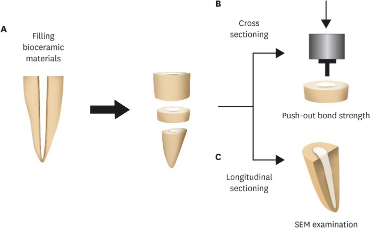
The specimens were embedded in a chemically cured acrylic resin (Ortho-Jet Acrylic, Lang Dental MFG, Wheeling, IL, USA) in a custom-made mold. The mold was placed perpendicular to the tooth surface. Using a low-speed micro-saw, each embedded tooth specimen was cut into a slice of 2.0 ± 0.2 mm in thickness to obtain a specimen for the push-out bond strength test (Figure 1A). A universal testing machine (Z020, Zwick Roell, Ulm, Germany) was used with a 500-N load cell at a crosshead speed of 0.5 mm/min to apply push-out force in the apico-coronal direction (Figure 1B). The maximum failure load was recorded in newtons (N) and then converted to megapascals (MPa) by applying the following formula:
(1)
Push-out Bond Strength (MPa) = Maximum Load (N)/Adhesion Area of Root Filling (A) (mm2) ----------
Figure 1

Illustration of the experimental procedure. (A) The tooth filled with the tested materials was sectioned horizontally to obtain a sliced specimen and an apical segment. (B) Push-out bond strength was measured with the sliced specimen using a universal testing machine. (C) The apical segment was sectioned longitudinally, and the intratubular biomineralization was observed under scanning electron microscopy.
SEM, scanning electron microscope.
rde-48-e8-g001.jpg
The adhesion area of the root canal filling was calculated using the following equation:
(2)
A=π(r1+r2)×r1-r22+h22 ----------
where r1 is the smaller radius of the root canal diameter (mm), r2 is the larger radius, h represents the thickness of the root section (mm), and π is the mathematical constant pi, approximated as 3.14.
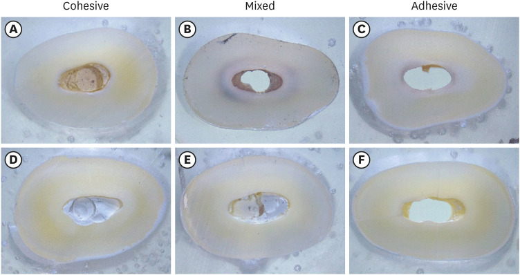
The sample was observed under a stereomicroscope (MZ16FA, Leica, Wetzlar, Germany) at ×30 magnification to determine the failure mode. Failures were categorized as cohesive (dentin walls totally covered with sealer), adhesive (no sealer visible on dentin walls), or mixed (a combination of cohesive and adhesive failure).
Intratubular biomineralization
The apical segment was cut perpendicular to the long axis of the root with a disc until the cut was 1 mm short of the filling material. Then, the specimen was split in half. The split surface was observed under a scanning electron microscope (SU8230, Hitachi, Tokyo, Japan), and the intratubular biomineralization was examined (Figure 1C). Also, the chemical characteristics of the precipitates formed in the dentinal tubule were evaluated with energy-dispersive X-ray spectroscopic (EDS) analysis.
Statistical analysis
Power analysis was applied with the t-test (effect size = 0.8, power = 0.55) to determine the required sample size using G*Power 3.1 software (University of Düsseldorf, Düsseldorf, Germany). The Shapiro-Wilk test was used to determine normality of the distribution prior to the statistical analysis. Then, the data were analyzed using the Student’s t-test followed by the Mann-Whitney U test using SPSS version 23.0 (IBM Corp., Armonk, NY, USA). A p value < 0.05 was considered indicative of statistical significance.
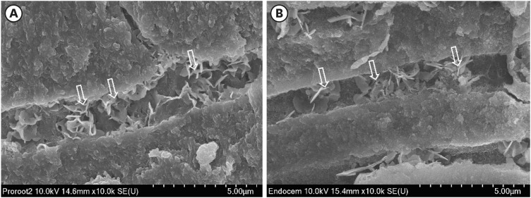
No significant difference was observed between the 2 tested groups in push-out bond strength (p > 0.05) (Figure 2A). Cohesive failure was predominant in both groups, followed by mixed and adhesive failures (Figures 2B and 3). In both groups, small flake-shaped precipitates were observed along dentinal tubules (Figure 4). As shown by the EDS analysis of the crystal seeds visible in the tubules, the mass percentage of calcium and phosphorus was around 2:1, similar to hydroxyapatite (Figure 5).
Figure 2

Push-out bond strength and the failure patterns of the tested materials. (A) Bar chart showing the mean bond strength of the 2 tested material groups. (B) Failure mode distribution according to filling material.
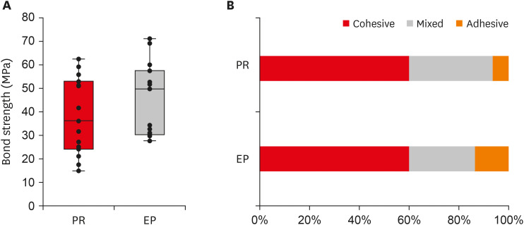
PR, ProRoot MTA; EP, Endocem MTA Premixed.
rde-48-e8-g002.jpg
Figure 3

Failure mode analysis using a stereomicroscope at ×30 magnification. (A-C) Representative images of the ProRoot MTA group. (D-F) Representative images of the Endocem MTA Premixed group.
rde-48-e8-g003.jpg
Figure 4

Representative scanning electron microscopy images of intratubular biomineralization. (A) ProRoot MTA-filled root canal. (B) Endocem MTA Premixed-filled root canal. Arrows indicate the flake-shaped intratubular precipitates.
rde-48-e8-g004.jpg
Figure 5

EDS analysis of the chemical characteristics of intratubular precipitate. (A) A scanning electron microscope image showing the precipitate (white cross). (B) Representative graph of EDS analysis of the precipitate. (C) Semiquantitative chemical composition showing the calcium/phosphorus ratio of the crystalline area denoted with a white cross.

EDS, energy-dispersive X-ray spectroscopic.
rde-48-e8-g005.jpg
In this study, we investigated the interfacial adaptation of Endocem MTA Premixed, a newly developed premixed hydraulic bioceramic material, to the root canal dentin in comparison to the conventional material ProRoot MTA. Interfacial adaptation to root canal dentin is a principal characteristic of root-end filling material because it is thought to prevent reinfection and entomb any remaining microorganisms in root canals.
First, we measured the push-out bond strength to quantify the interfacial adaptation values of the cements to the root canal dentin. Endocem MTA Premixed was found to have a similar value to ProRoot MTA. Notably, ProRoot MTA has been considered a gold standard for root-end filling and is usually adopted as the control material when interfacial adaptation is evaluated [4,5,6]. Furthermore, push-out bond strength is a widely used methodology to evaluate the adaptation of root-end filling material [7,8]. Therefore, this result suggests that Endocem MTA Premixed is comparable to ProRoot MTA regarding interfacial adaptation and can be considered to show adequate performance in root-end filling.
In the present study, bond failures observed in the 2 experimental groups were predominantly found within the filling material (the cohesive type). In both groups, more than half of the specimens (9 of 15) exhibited cohesive failure. This finding contrasts with several previous studies in which bond failures were usually at the root-end filling material-dentin interface (adhesive or mixed failures) [9,10,11,12,13]. The failure mode may be affected by the thickness of the specimen slice. When the thickness of the slice ranges from 1 to 1.5 mm, adhesive or mixed failures occur, mainly because the mechanical strength of the cement exceeds the bond strength. On the contrary, in this study, the thickness of the slice was 2 mm, and the cohesive failure pattern was predominant over the adhesive type. Similarly, in studies involving 2-mm or thicker specimens, cohesive failure was frequently found [14]. In this respect, the difference in the thickness of root slices between the present study and previous studies may explain the different modes of bond failure. The thickness of the specimen should be considered in failure mode analysis.
The effect of liquid vehicles on the bond strength of hydraulic bioceramic materials is still under debate. Salem Milani et al. [15] reported that mixing with propylene glycol enhanced the strength of bonding of MTA Angelus (Angelus, Londrina, Brazil) to dentin. In contrast, Al-Hiyasat and Yousef [14] reported that a premixed putty-type material (TotalFill FS, FKG Dentaire, La Chaux-de-Fonds, Switzerland) exhibited lower bond strength than Biodentine and ProRoot MTA. Furthermore, Retana-Lobo et al. [16] found that a powder-liquid calcium silicate cement (BioRoot RCS, Septodont, Saint-Maur-des-Fossés, France) presented a higher push-out bond strength than a premixed sealer (EndoSequence BC Sealer, Brasseler, Savannah, GA, USA). The presence of vehicles might interfere with the direct interaction between the cement and the root dentin. Endocem MTA Premixed is also mixed with a non-aqueous vehicle, DMSO, which has been demonstrated to improve the adhesive bonding to coronal dentin and to decrease leakage by increasing dentin wettability [17,18]. Interestingly, Lindblad et al. [19] recently reported that final irrigation with DMSO resulted in a significantly higher push-out strength of hydraulic calcium silicate cement. Also, in the DMSO irrigation group, the cohesive failure mode was more common than in the control group. In the present study, Endocem MTA Premixed exhibited similar push-out bond strength to ProRoot MTA (p > 0.05), perhaps due to the presence of DMSO, which increased the dentin wettability and provided sufficient opportunity for the hydraulic cement to interact with the root dentin.
Hydraulic bioceramic material is believed to be bioactive, forming hydroxyapatite crystals inside the dentinal tubules [20] in a process called biomineralization. Yoo et al. [21] reported intratubular biomineralization in root canals filled with bioceramic material after 8 years of clinical maintenance. Furthermore, this phenomenon can be promoted by treating the root canal with PBS in vitro [22]. To that end, we immersed the specimens in PBS for 30 days. In the present study, in both groups, small flake-shaped crystallized precipitates were observed along dentinal tubules up to around 300 μm. We also performed an EDS analysis to identify the chemical characteristics of the precipitates. According to the elemental analysis, the crystalline mass contained calcium and phosphorus at a mass percentage ratio of around 2:1, similar to the ratio found in hydroxyapatite. Yoo et al. [23] similarly found that the precipitates in their study had calcium/phosphorus ratios similar to hydroxyapatite and claimed that these may be favorable biomineralization patterns for the sealing of the root canal system. Reyes-Carmona et al. [24] also reported that the biomineralization induced by MTA enhanced the push-out strength of hydraulic cement. Leaving other issues aside, this study shows that Endocem MTA Premixed can induce biomineralization, and it suggests that further investigations are required on the effect of biomineralization in dentinal tubules on the bonding ability of hydraulic bioceramic materials.
This study showed that Endocem MTA Premixed, a newly developed premixed bioceramic root-end filling material, showed similar push-out bond strength to the widely used material ProRoot MTA. Consequently, the null hypothesis was accepted. Endocem MTA Premixed also exhibited biomineralization in dentinal tubules. Considering its ease of use and enhanced handling characteristics, Endocem MTA Premixed has potential for use as an acceptable root-end filling material for endodontic surgery.

Funding: This research was supported by a grant of the Korea Medical Device Development Fund grant funded by the Korea government (the Ministry of Science and ICT, the Ministry of Trade, Industry and Energy, the Ministry of Health & Welfare, Republic of Korea, the Ministry of Food and Drug Safety) (Project Number: RS-2020-KD000045).

Conflict of Interest: No potential conflict of interest relevant to this article was reported.

Author Contributions:

  • Conceptualization: Park JH, Yu MK, Min KS.

  • Data curation: Park JH, Yu MK.

  • Formal analysis: Min KS.

  • Funding acquisition: Min KS.

  • Investigation: Park JH, Kim HJ.

  • Methodology: Park JH, Yu MK, Min KS.

  • Project administration: Yu MK, Min KS.

  • Resources: Yu MK, Min KS.

  • Software: Min KS.

  • Supervision: Lee KW.

  • Validation: Lee KW, Min KS.

  • Visualization: Park JH, Min KS.

  • Writing - original draft: Park JH, Min KS.

  • Writing - review & editing: Kim HJ, Lee KW, Yu MK.

  • 1. Saxena P, Gupta SK, Newaskar V. Biocompatibility of root-end filling materials: recent update. Restor Dent Endod 2013;38:119-127. PubMedPMC
  • 2. Bernabé PF, Gomes-Filho JE, Bernabé DG, Nery MJ, Otoboni-Filho JA, Dezan E Jr, Cintra LT. Sealing ability of MTA used as a root end filling material: effect of the sonic and ultrasonic condensation. Braz Dent J 2013;24:107-110. PubMed
  • 3. Torabinejad M, Hong CU, McDonald F, Pitt Ford TR. Physical and chemical properties of a new root-end filling material. J Endod 1995;21:349-353. ArticlePubMed
  • 4. Alsubait SA, Hashem Q, AlHargan N, AlMohimeed K, Alkahtani A. Comparative evaluation of push-out bond strength of ProRoot MTA, bioaggregate and Biodentine. J Contemp Dent Pract 2014;15:336-340. PubMed
  • 5. Nagas E, Cehreli ZC, Uyanik MO, Vallittu PK, Lassila LV. Effect of several intracanal medicaments on the push-out bond strength of ProRoot MTA and Biodentine. Int Endod J 2016;49:184-188. PubMed
  • 6. Buldur B, Oznurhan F, Kaptan A. The effect of different chelating agents on the push-out bond strength of ProRoot MTA and EndoSequence root repair material. Eur Oral Res 2019;53:88-93. PubMedPMC
  • 7. Saghiri MA, Shokouhinejad N, Lotfi M, Aminsobhani M, Saghiri AM. Push-out bond strength of mineral trioxide aggregate in the presence of alkaline pH. J Endod 2010;36:1856-1859. ArticlePubMed
  • 8. Vivan RR, Guerreiro-Tanomaru JM, Bosso-Martelo R, Costa BC, Duarte MA, Tanomaru-Filho M. Push-out bond strength of root-end filling materials. Braz Dent J 2016;27:332-335. PubMed
  • 9. Shokouhinejad N, Razmi H, Fekrazad R, Asgary S, Neshati A, Assadian H, Kheirieh S. Push-out bond strength of two root-end filling materials in root-end cavities prepared by Er,Cr:YSGG laser or ultrasonic technique. Aust Endod J 2012;38:113-117. PubMed
  • 10. Ashofteh Yazdi K, Bolhari B, Sabetmoghaddam T, Meraji N, Kharazifard MJ. Effect of blood exposure on push-out bond strength of four calcium silicate based cements. Iran Endod J 2017;12:196-200. PubMedPMC
  • 11. Tuncel B, Nagas E, Cehreli Z, Uyanik O, Vallittu P, Lassila L. Effect of endodontic chelating solutions on the bond strength of endodontic sealers. Braz Oral Res 2015;29:S1806-83242015000100256.ArticlePubMed
  • 12. Araújo CC, Brito-Júnior M, Faria-E-Silva AL, Pereira RD, Silva-Sousa YT, Cruz-Filho AM, Sousa-Neto MD. Root filling bond strength using reciprocating file-matched single-cones with different sealers. Braz Oral Res 2016;30:S1806-83242016000100251.PubMed
  • 13. Prado MC, Martiniano K, Pereira AC, Cortellazzi KL, Marciano MA, Abuna G, de-Jesus-Soares A. Do intracanal medications used in regenerative endodontics affect the bond strength of powder-to-liquid and ready-to-use cervical sealing materials? J Conserv Dent 2021;24:464-469. PubMed
  • 14. Al-Hiyasat AS, Yousef WA. Push-out bond strength of calcium silicate-based cements in the presence or absence of a smear layer. Int J Dent 2022;2022:7724384. PubMedPMC
  • 15. Salem Milani A, Froughreyhani M, Charchi Aghdam S, Pournaghiazar F, Asghari Jafarabadi M. Mixing with propylene glycol enhances the bond strength of mineral trioxide aggregate to dentin. J Endod 2013;39:1452-1455. ArticlePubMed
  • 16. Retana-Lobo C, Tanomaru-Filho M, Guerreiro-Tanomaru JM, Benavides-García M, Hernández-Meza E, Reyes-Carmona J. Push-out bond strength, characterization, and ion release of premixed and powder-liquid bioceramic sealers with or without gutta-percha. Scanning 2021;2021:6617930. PubMedPMC
  • 17. Salim Al-Ani AA, Mutluay M, Stape THS, Tjäderhane L, Tezvergil-Mutluay A. Effect of various dimethyl sulfoxide concentrations on the durability of dentin bonding and hybrid layer quality. Dent Mater J 2018;37:501-505. PubMed
  • 18. Tjäderhane L, Mehtälä P, Scaffa P, Vidal C, Pääkkönen V, Breschi L, Hebling J, Tay FR, Nascimento FD, Pashley DH, Carrilho MR. The effect of dimethyl sulfoxide (DMSO) on dentin bonding and nanoleakage of etch-and-rinse adhesives. Dent Mater 2013;29:1055-1062. PubMed
  • 19. Lindblad RM, Lassila LVJ, Vallittu PK, Tjäderhane L. The effect of chlorhexidine and dimethyl sulfoxide on long-term sealing ability of two calcium silicate cements in root canal. Dent Mater 2021;37:328-335. ArticlePubMed
  • 20. Reyes-Carmona JF, Felippe MS, Felippe WT. Biomineralization ability and interaction of mineral trioxide aggregate and white Portland cement with dentin in a phosphate-containing fluid. J Endod 2009;35:731-736. PubMed
  • 21. Yoo YJ, Lee YS, Yoo JS, Perinpanayagam H, Yoo CS, Kang HS, Oh S, Chang SW, Kum KY. Intratubular biomineralization in a root canal filled with calcium-enriched material over 8 years. Materials (Basel) 2017;10:1388. PubMedPMC
  • 22. Reyes-Carmona JF, Felippe MS, Felippe WT. A phosphate-buffered saline intracanal dressing improves the biomineralization ability of mineral trioxide aggregate apical plugs. J Endod 2010;36:1648-1652. PubMed
  • 23. Yoo YJ, Baek SH, Kum KY, Shon WJ, Woo KM, Lee W. Dynamic intratubular biomineralization following root canal obturation with pozzolan-based mineral trioxide aggregate sealer cement. Scanning 2016;38:50-56. ArticlePubMedPDF
  • 24. Reyes-Carmona JF, Felippe MS, Felippe WT. The biomineralization ability of mineral trioxide aggregate and Portland cement on dentin enhances the push-out strength. J Endod 2010;36:286-291. PubMed

Tables & Figures

REFERENCES

    Citations

    Citations to this article as recorded by  

      • ePub LinkePub Link
      • Cite
        CITE
        export Copy Download
        Close
        Download Citation
        Download a citation file in RIS format that can be imported by all major citation management software, including EndNote, ProCite, RefWorks, and Reference Manager.

        Format:
        • RIS — For EndNote, ProCite, RefWorks, and most other reference management software
        • BibTeX — For JabRef, BibDesk, and other BibTeX-specific software
        Include:
        • Citation for the content below
        Push-out bond strength and intratubular biomineralization of a hydraulic root-end filling material premixed with dimethyl sulfoxide as a vehicle
        Restor Dent Endod. 2023;48(1):e8  Published online January 20, 2023
        Close
      • XML DownloadXML Download
      Figure
      • 0
      • 1
      • 2
      • 3
      • 4
      Push-out bond strength and intratubular biomineralization of a hydraulic root-end filling material premixed with dimethyl sulfoxide as a vehicle
      Image Image Image Image Image
      Figure 1 Illustration of the experimental procedure. (A) The tooth filled with the tested materials was sectioned horizontally to obtain a sliced specimen and an apical segment. (B) Push-out bond strength was measured with the sliced specimen using a universal testing machine. (C) The apical segment was sectioned longitudinally, and the intratubular biomineralization was observed under scanning electron microscopy.SEM, scanning electron microscope.
      Figure 2 Push-out bond strength and the failure patterns of the tested materials. (A) Bar chart showing the mean bond strength of the 2 tested material groups. (B) Failure mode distribution according to filling material.PR, ProRoot MTA; EP, Endocem MTA Premixed.
      Figure 3 Failure mode analysis using a stereomicroscope at ×30 magnification. (A-C) Representative images of the ProRoot MTA group. (D-F) Representative images of the Endocem MTA Premixed group.
      Figure 4 Representative scanning electron microscopy images of intratubular biomineralization. (A) ProRoot MTA-filled root canal. (B) Endocem MTA Premixed-filled root canal. Arrows indicate the flake-shaped intratubular precipitates.
      Figure 5 EDS analysis of the chemical characteristics of intratubular precipitate. (A) A scanning electron microscope image showing the precipitate (white cross). (B) Representative graph of EDS analysis of the precipitate. (C) Semiquantitative chemical composition showing the calcium/phosphorus ratio of the crystalline area denoted with a white cross.EDS, energy-dispersive X-ray spectroscopic.
      Push-out bond strength and intratubular biomineralization of a hydraulic root-end filling material premixed with dimethyl sulfoxide as a vehicle

      Restor Dent Endod : Restorative Dentistry & Endodontics
      Close layer
      TOP