
Search
- Page Path
- HOME > Search
Research Article
- Effects of dentin surface preparations on bonding of self-etching adhesives under simulated pulpal pressure
- Chantima Siriporananon, Pisol Senawongse, Vanthana Sattabanasuk, Natchalee Srimaneekarn, Hidehiko Sano, Pipop Saikaew
- Restor Dent Endod 2022;47(1):e4. Published online December 28, 2021
- DOI: https://doi.org/10.5395/rde.2022.47.e4
-
 Abstract
 PDF
PDF PubReader
 ePub
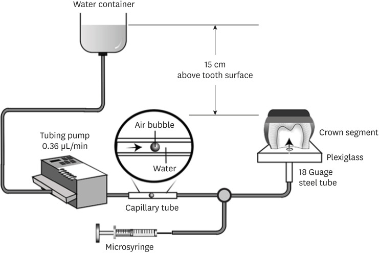
ePub Objectives This study evaluated the effects of different smear layer preparations on the dentin permeability and microtensile bond strength (µTBS) of 2 self-etching adhesives (Clearfil SE Bond [CSE] and Clearfil Tri-S Bond Universal [CTS]) under dynamic pulpal pressure.
Materials and Methods Human third molars were cut into crown segments. The dentin surfaces were prepared using 4 armamentaria: 600-grit SiC paper, coarse diamond burs, superfine diamond burs, and carbide burs. The pulp chamber of each crown segment was connected to a dynamic intra-pulpal pressure simulation apparatus, and the permeability test was done under a pressure of 15 cmH2O. The relative permeability (%P) was evaluated on the smear layer-covered and bonded dentin surfaces. The teeth were bonded to either of the adhesives under pulpal pressure simulation, and cut into sticks after 24 hours water storage for the µTBS test. The resin-dentin interface and nanoleakage observations were performed using a scanning electron microscope. Statistical comparisons were done using analysis of variance and
post hoc tests.Results Only the method of surface preparation had a significant effect on permeability (
p < 0.05). The smear layers created by the carbide and superfine diamond burs yielded the lowest permeability. CSE demonstrated a higher µTBS, with these values in the superfine diamond and carbide bur groups being the highest. Microscopic evaluation of the resin-dentin interface revealed nanoleakage in the coarse diamond bur and SiC paper groups for both adhesives.Conclusions Superfine diamond and carbide burs can be recommended for dentin preparation with the use of 2-step CSE.
-
Citations
Citations to this article as recorded by
- Effect of smear layer pretreatment with EDTA and sodium hypochlorite on the dentin bond durability of universal adhesives
Thanawat Ruaydee, Chantida Pawaputanon Na Mahasarakham, Vanthana Sattabanasuk, Pipop Saikaew
Frontiers in Dental Medicine.2026;[Epub] CrossRef - Determination of marginal permeability of restorations in the cervical region using a universal adhesive system: a randomized controlled open-label laboratory study
Svetlana N. Razumova, Anzhela S. Brago, Oxana R. Ruda, Artur G. Talandis, Lamara M. Khaskhanova, Ruzanna M. Bragunova, Bohdan O. Pecherskyi
Russian Journal of Dentistry.2026; 30(2): 113. CrossRef - Catechol–Phosphonate–Augmented Universal Adhesive for Hydrolysis-Resistant Dentin Bonds: A µTBS and Spectroscopic Study
Rabeia J. Khalil, Suha K. Ibrahim, Athraa H. Madhat, Ali H. Tawfieq
European Journal of Dentistry.2026;[Epub] CrossRef - The effect of different adhesive strategies and diamond burs on dentin bond strength of universal resin cements
Chavakorn Atsavathavornset, Pipop Saikaew, Choltacha Harnirattisai, Hidehiko Sano
Clinical Oral Investigations.2025;[Epub] CrossRef - Universal adhesive systems in dentistry: A narrative review
Svetlana N. Razumova, Anzhela S. Brago, Oxana R. Ruda, Zoya A. Guryeva, Elvira V. Adzhieva
Russian Journal of Dentistry.2024; 28(5): 512. CrossRef - Delayed light activation of resin composite affects the bond strength of adhesives under dynamic simulated pulpal pressure
Nattaporn Sukprasert, Choltacha Harnirattisai, Pisol Senawongse, Hidehiko Sano, Pipop Saikaew
Clinical Oral Investigations.2022; 26(11): 6743. CrossRef
- Effect of smear layer pretreatment with EDTA and sodium hypochlorite on the dentin bond durability of universal adhesives
- 4,325 View
- 65 Download
- 4 Web of Science
- 6 Crossref

Case Report
- Chair-side CAD/CAM fabrication of a single-retainer resin bonded fixed dental prosthesis: a case report
- Carlos Alberto Jurado, Akimasa Tsujimoto, Hidehiko Watanabe, Jose Villalobos-Tinoco, Jorge Luis Garaicoa, Mark David Markham, Wayne Walter Barkmeier, Mark Andrew Latta
- Restor Dent Endod 2020;45(2):e15. Published online February 6, 2020
- DOI: https://doi.org/10.5395/rde.2020.45.e15
-
Abstract
PDFPubReaderePub
This clinical report describes designing and fabricating a single-retainer resin-bonded fixed dental prosthesis with a chair-side computer-aided design/computer-aided manufacturing system. The whole procedure, from tooth extraction to final placement of the prosthesis, was completed in one day, and a single clinic visit. No clinical complications were found at the 2-year follow-up after placement of the restoration, and satisfactory functional and esthetic results were achieved.
-
Citations
Citations to this article as recorded by- Optimizing Hard and Soft‐Tissue Esthetics With Anterior Cantilever Zirconia Ceramic Resin‐Bonded Fixed Dental Prostheses
Markus B. Blatz, Tony Rotondo, Szabi Hant, Lea S. Prott
Journal of Esthetic and Restorative Dentistry.2026; 38(3): 491. CrossRef - Fracture Resistance of CAD/CAM Resin-Matrix Ceramic Overlays and Full-Coverage Crowns for Maxillary Premolars
Ali Abulkasim Mohamed, Brian Morrow, Stella Mireles, Carlos A. Jurado, Mark A. Antal, Silvia Rojas-Rueda, Hamid Nurrohman, Franklin Garcia-Godoy
Biomimetics.2026; 11(5): 291. CrossRef - Single-Retained Lithium Disilicate “Maryland” with Pontic-Derived Stamp Technique for Anterior Symmetry: A Case Report
Pier Edoardo Maltagliati, Ahmed Deraz, Alberto Maltagliati, Giovanni Messina, Yashar Imenpour, Stefano Benedicenti
Dentistry Journal.2026; 14(7): 427. CrossRef - Using the foundation restoration as a blueprint: An uncomplicated approach to retrofitting crowns to existing removable partial dentures using CAD-CAM technology
Jae-Hoon Lee, Juliana Pfeffer, Carlos A. Jurado, Francisco X. Azpiazu-Flores
The Journal of Prosthetic Dentistry.2025; 133(6): 1416. CrossRef - Fracture load of chairside CAD‐CAM veneers fabricated with pre‐and fully crystalized lithium disilicate ceramics
Carlos A. Jurado, Jacquelyn S. Yeh, Cristina M. P. Vidal, Seok‐Hwan Cho, Salahaldeen Abuhammoud
Journal of Prosthodontics.2025; 34(4): 429. CrossRef - Clinical Efficacy of Anterior Ceramic Materials in Resin-Bonded Fixed Dental Prostheses with Different Bridge Designs—A Systematic Review and Meta-Analysis
Nutsongsak Panyasuksri, Pattarika Angkasith, Apichai Yavirach, Pisaisit Chaijareenont, Surasak Saokaew, Sukrit Kanchanasurakit
Prosthesis.2025; 7(2): 41. CrossRef - Full Zirconia Resin‐Bonded Fixed Dental Prosthesis: A Clinical Report
Majda Lemrichi, Amal El Yamani
Clinical Case Reports.2025;[Epub] CrossRef - The Influence of Thickness on Light Transmission for Pre- and Fully Crystallized Chairside CAD/CAM Lithium Disilicate Ceramics
Franciele Floriani, Salahaldeen Abuhammoud, Silvia Rojas-Rueda, Amit Unnadkat, Nicholas G. Fischer, Chin-Chuan Fu, Carlos A. Jurado
Materials.2024; 17(9): 2045. CrossRef - Microstructural and flexural strength of various CAD‐CAM lithium disilicate ceramics
Joissi Ferrari Zaniboni, Amanda Soares Silva, Aryvelto Miranda Silva, João Felipe Besegato, Oscar Fernando Muñoz‐Chávez, Edson Alves de Campos
Journal of Prosthodontics.2024;[Epub] CrossRef - Fracture resistance of zirconia surveyed crowns with four different occlusal rest seat designs
Carlos Alberto Jurado, Akram Sayed Ahmed, Nathaniel C. Lawson, Francisco X. Azpiazu‐Flores, Conley Green, Seok‐Hwan Cho
Journal of Prosthodontics.2024; 33(5): 484. CrossRef - Effect of incisal preparation design on the fracture strength of monolithic zirconia‐reinforced lithium silicate laminate veneers
Carlos A. Jurado, Ramtin Sadid‐Zadeh, Hidehiko Watanabe, Craig E. Robbins, Kelvin I. Afrashtehfar, Nicholas G. Fischer, Damian J. Lee
Journal of Prosthodontics.2024; 33(3): 281. CrossRef - Fracture resistance of CAD/CAM provisional crowns with two different designs: an in vitro study
Salwa Mekled, Mark Iskander, Belinda Rodriguez, Paige Hodges, Jasleen Bhogal, Joan Adechoubou, Geraldine Weinstein
Exploration of Medicine.2024;[Epub] CrossRef - Evaluation of Glazing and Polishing Systems for Novel Chairside CAD/CAM Lithium Disilicate and Virgilite Crowns
CA Jurado, K Arndt, FX Azpiazu-Flores, F Faddoul, R França, NG Fischer, H Watanabe
Operative Dentistry.2023; 48(6): 689. CrossRef - Traditional versus conservative endodontic access impact on fracture resistance of chairside CAD‐CAM lithium disilicate anterior crowns: An in vitro study
Carlos A. Jurado, Clarisa Amarillas‐Gastelum, Bruna Santos Honório Tonin, Gentry Nielson, Kelvin I. Afrashtehfar, Nicholas G. Fischer
Journal of Prosthodontics.2023; 32(8): 728. CrossRef - Digital Full-Mouth Reconstruction Assisted by Facial and Intraoral Scanners: A Case Report and Technique Description
Jorge Garaicoa, Carlos A. Jurado, Kelvin I. Afrashtehfar, Abdulaziz Alhotan, Nicholas G. Fischer
Applied Sciences.2023; 13(3): 1917. CrossRef - Students’ perception of digital waxing software for dental anatomy education
Amira Elgreatly, Ahmed Mahrous, Wendy A. Clark, Ingeborg J. De Kok, Fang Qian, Akimasa Tsujimoto
Journal of Oral Science.2022; 64(2): 178. CrossRef - Effectiveness of Different Polishing Kits for Chairside CAD/CAM Provisional Restorative Materials
CA Jurado, WW Barkmeier, A Alshabib, SS Alresayes, C-C Fu, EC Teixeira, AG Baruth, A Tsujimoto
Operative Dentistry.2022; 47(6): 670. CrossRef - Fatigue bond strength of dental adhesive systems: Historical background of test methodology, clinical considerations and future perspectives
Akimasa Tsujimoto, Wayne W. Barkmeier, Erica C. Teixeira, Toshiki Takamizawa, Masashi Miyazaki, Mark A. Latta
Japanese Dental Science Review.2022; 58: 193. CrossRef - Diagnostic Mock-Up as a Surgical Reduction Guide for Crown Lengthening: Technique Description and Case Report
Carlos A. Jurado, Venkata Parachuru, Jose Villalobos Tinoco, Gerardo Guzman-Perez, Akimasa Tsujimoto, Ramya Javvadi, Kelvin I. Afrashtehfar
Medicina.2022; 58(10): 1360. CrossRef - Color stability of fully- and pre-crystalized chair-side CAD-CAM lithium disilicate restorations after required and additional sintering processes
Carlos Alberto Jurado, Tamer El-Gendy, Jared Hyer, Akimasa Tsujimoto
The Journal of Advanced Prosthodontics.2022; 14(1): 56. CrossRef - Comparison of Fracture Resistance for Chairside CAD/CAM Lithium Disilicate Crowns and Overlays with Different Designs
Carlos Alberto Jurado, Zinaida Kaleinikova, Akimasa Tsujimoto, Daniel Alberto Cortés Treviño, Robert R. Seghi, Damian J. Lee
Journal of Prosthodontics.2022; 31(4): 341. CrossRef - Light Transmission for a Novel Chairside CAD/CAM Lithium Disilicate Ceramic
Carlos A Jurado, Akimasa Tsujimoto, Clarisa Amarillas-Gastelum, Saad Alresayes, Kennedee French, Hamid Nurrohman
The Journal of Contemporary Dental Practice.2022; 22(12): 1365. CrossRef - Intraoral Scanning with Rubber Dam Isolation in Place for Fabrication of a Chairside Computer-assisted Design and Computer-assisted Manufacture Ceramic Restoration
Rachel Lederman, Jeffrey Cohen, Akimasa Tsujimoto
The Journal of Contemporary Dental Practice.2021; 22(8): 943. CrossRef
- Optimizing Hard and Soft‐Tissue Esthetics With Anterior Cantilever Zirconia Ceramic Resin‐Bonded Fixed Dental Prostheses
- 2,871 View
- 29 Download
- 23 Crossref
Research Article
-
Antibacterial effect of self-etching adhesive systems on
Streptococcus mutans - Seung-Ryong Kim, Dong-Hoon Shin
- Restor Dent Endod 2014;39(1):32-38. Published online January 20, 2014
- DOI: https://doi.org/10.5395/rde.2014.39.1.32
-
Abstract
PDFPubReaderePub
Objectives In this study, we evaluated the antibacterial activity of self-etching adhesive systems against
Streptococcus mutans using the agar diffusion method.Materials and Methods Three 2-step systems, Clearfil SE Bond (SE, Kuraray), Contax (CT, DMG), and Unifil Bond (UnB, GC), and three 1-step systems, Easy Bond (EB, 3M ESPE), U-Bond (UB, Vericom), and All Bond SE (AB, BISCO) were used. 0.12% chlorhexidine (CHX, Bukwang) and 37% phosphoric acid gel (PA, Vericom) were used as positive controls.
Results The antibacterial activity of CHX and PA was stronger than that of the other groups, except SE. After light activation, the inhibition zone was reduced in the case of all 2-step systems except CT. However, all 1-step systems did not exhibit any inhibition zone upon the light activation.
Conclusions SE may be better than CT or UnB among the 2-step systems with respect to antibacterial activity, however, 1-step systems do not exhibit any antibacterial activity after light curing.
-
Citations
Citations to this article as recorded by- Comparative Evaluation of Antibacterial Activity of Three Universal Bonding Agents Against Streptococcus mutans on Demineralized Dentin: An In Vitro Study
Kirti Rathee, Charu Dayal, Reena Rani, A Anukriti, Anjum Zia, Ankita Sundan, Seema Gupta
Cureus.2026;[Epub] CrossRef - Incorporation of chlorhexidine in self-adhesive resin cements
Idris M. MEHDAWI, Ranna KITAGAWA, Haruaki KITAGAWA, Satoshi YAMAGUCHI, Nanako HIROSE, Tomoki KOHNO, Satoshi IMAZATO
Dental Materials Journal.2022; 41(5): 675. CrossRef - Antibacterial and Bonding Properties of Universal Adhesive Dental Polymers Doped with Pyrogallol
Naji Kharouf, Ammar Eid, Louis Hardan, Rim Bourgi, Youri Arntz, Hamdi Jmal, Federico Foschi, Salvatore Sauro, Vincent Ball, Youssef Haikel, Davide Mancino
Polymers.2021; 13(10): 1538. CrossRef - Influence of Protease Inhibitors on Bond Degradation of Self-Etch Adhesive Systems to Caries-Affected Dentin: An <i>in Vitro</i> Study
Diana Roberta Pereira Grandizoli, Sérgio Luiz Pinheiro
Advances in Biological Chemistry.2018; 08(01): 15. CrossRef - Epigallocatechin-3-gallate and Epigallocatechin-3-O-(3-O-methyl)-gallate Enhance the Bonding Stability of an Etch-and-Rinse Adhesive to Dentin
Hao-Han Yu, Ling Zhang, Fan Yu, Fang Li, Zheng-Ya Liu, Ji-Hua Chen
Materials.2017; 10(2): 183. CrossRef - An In vitro Assessment of Antibacterial Activity of Three Self-etching Primers Against Oral Microflora
Sneha Dipak Shinde, Vikram Pai, R. Vijay Naik
APOS Trends in Orthodontics.2017; 7: 181. CrossRef - Functional Dental Restorative Materials That Hinder Oral Biofilm
Hércules Bezerra Dias, Victor Trassi Fernandes da Silva Souza, Rafael Amorim Martins, Ana Carolina Bosco Mendes, Monica Irma Aparecida Valdeci de Souza, Ângela Cristina Cilense Zuanon, Alessandra Nara de Souza Rastelli
Current Oral Health Reports.2017; 4(1): 22. CrossRef - In vitroantibacterial activity of various adhesive materials against oral streptococci
Emre Ozel, Fetiye Kolayli, Elif Bahar Tuna, Doganhan Er
Biotechnology & Biotechnological Equipment.2016; 30(1): 121. CrossRef - A systematic review about antibacterial monomers used in dental adhesive systems: Current status and further prospects
Alexandra Rubin Cocco, Wellington Luiz de Oliveira da Rosa, Adriana Fernandes da Silva, Rafael Guerra Lund, Evandro Piva
Dental Materials.2015; 31(11): 1345. CrossRef
- Comparative Evaluation of Antibacterial Activity of Three Universal Bonding Agents Against Streptococcus mutans on Demineralized Dentin: An In Vitro Study
- 2,156 View
- 6 Download
- 9 Crossref
Case Report
- Considerations during crown reattachment procedure over the pulpal exposure: case report
- Bona Kim, Yoon Lee, Min-Ju Song, Su-Jung Shin, Jeong-Won Park
- Restor Dent Endod 2012;37(4):240-244. Published online November 21, 2012
- DOI: https://doi.org/10.5395/rde.2012.37.4.240
-
Abstract
PDFPubReaderePub
Crown reattachment is the most conservative treatment which can be used to restore fractured tooth, presumably with sufficient strength, while maintaining original contour, incisal translucency, and reducing chair time and cost.
However, in case of crown fracture with pin-point pulp exposure, we should cautiously minimize the irritation to the pulp and consider pre-treatment pulpal status, choice of pulp capping materials, choice of bonding system and treatment sequence during crown reattachment procedures. This case reports the considerations while crown reattachment with direct pulp capping using calcium hydroxide (Dycal, Dentsply Caulk).
-
Citations
Citations to this article as recorded by- A Conservative Approach to the Management of a Dental Trauma for Immediate Natural Esthetics
Pallav Mahesh Patni, Pradeep Jain, Mona Jain Patni
Archives of Trauma Research.2016;[Epub] CrossRef
- A Conservative Approach to the Management of a Dental Trauma for Immediate Natural Esthetics
- 2,358 View
- 9 Download
- 1 Crossref
Research Articles
- Effect of moisture and drying time on the bond strength of the one-step self-etching adhesive system
- Yoon Lee, Jeong-Won Park
- Restor Dent Endod 2012;37(3):155-159. Published online August 29, 2012
- DOI: https://doi.org/10.5395/rde.2012.37.3.155
-
Abstract
PDFPubReaderePub
Objectives To investigate the effect of dentin moisture degree and air-drying time on dentin-bond strength of two different one-step self-etching adhesive systems.
Materials and Methods Twenty-four human third molars were used for microtensile bond strength testing of G-Bond and Clearfil S3 Bond. The dentin surface was either blot-dried or air-dried before applying these adhesive agents. After application of the adhesive agent, three different air drying times were evaluated: 1, 5, and 10 sec. Composite resin was build up to 4 mm thickness and light cured for 40 sec with 2 separate layers. Then the tooth was sectioned and trimmed to measure the microtensile bond strength using a universal testing machine. The measured bond strengths were analyzed with three-way ANOVA and regression analysis was done (
p = 0.05).Results All three factors, materials, dentin wetness and air drying time, showed significant effect on the microtensile bond strength. Clearfil S3 Bond, dry dentin surface and 10 sec air drying time showed higher bond strength.
Conclusions Within the limitation of this experiment, air drying time after the application of the one-step self-etching adhesive agent was the most significant factor affecting the bond strength, followed by the material difference and dentin moisture before applying the adhesive agent.
-
Citations
Citations to this article as recorded by- An in vitro study on comparative evaluation of shear bond strength of bioactive composite to tooth structure with various dentin conditioning agents
Priyanka Pokkula, Shaik Mohammed Asif, Abdullah Alqarni, Shahabe Saquib Abullais, Shaik Mohamed Shamsudeen, Syed M Yassin, Abosofyan S. Atta, Wahaj Ahmad Khan
AIP Advances.2025;[Epub] CrossRef - The Influence of Drying Time, Application Mode, and Agitation on the Dentin Bond Strength of a Novel Mesoporous Bioactive Glass-Containing Universal Dentin Adhesive
Jiyoung Kwon, Jungwon Kim, Dongseok Choi, Duck-Su Kim
Journal of Functional Biomaterials.2025; 16(7): 247. CrossRef - The occluding effects of layered calcium phosphate and cyanoacrylate on dentinal tubules: a SEM study
Özge Uzuner Bilgiç, Sühan Gürbüz, Altan Dogan
BMC Oral Health.2025;[Epub] CrossRef - Shear bond strengths of two newly marketed self‐adhesive resin cements to different substrates: A light and scanning electron microscopy evaluation
Cansu Atalay, Uzay Koc Vural, Ivana Miletic, Sevil Gurgan
Microscopy Research and Technique.2022; 85(5): 1694. CrossRef - The effect of curing mode of dual-cure resin cements on bonding performance of universal adhesives to enamel, dentin and various restorative materials
Erick LUZ MADRIGAL, Antonin TICHY, Keiichi HOSAKA, Masaomi IKEDA, Masatoshi NAKAJIMA, Junji TAGAMI
Dental Materials Journal.2021; 40(2): 446. CrossRef - Effect of adhesive air-drying time on bond strength to dentin: A systematic review and meta-analysis
Mohamed M. Awad, Ali Alrahlah, Jukka P. Matinlinna, Hamdi Hosni Hamama
International Journal of Adhesion and Adhesives.2019; 90: 154. CrossRef - Effect of pre-curing of two universal adhesives on the shear bond strength of resin cement to zirconia
Ga-Eun Son, Tae-Yub Kwon, Young Kyung Kim
Korean Journal of Dental Materials.2019; 46(1): 21. CrossRef - Bonding effectiveness of different dentin conditions on etch-and-rinse mode of two universal adhesives: the confocal laser scanning and shear bond strength
Jounghyun Lee, Ka-Young Cho, Jin-Young Kim, Sungho Park, Byoung-Duck Roh, Yooseok shin
Journal of Adhesion Science and Technology.2017; 31(9): 933. CrossRef - Effect of dentin dehydration and composite resin polymerization mode on bond strength of two self-etch adhesives
Pooran Samimi, Mehdi Alizadeh, Farinaz Shirban, Amin Davoodi, Maryam Khoroushi
Contemporary Clinical Dentistry.2016; 7(1): 16. CrossRef - Effect of different air-drying time on the microleakage of single-step self-etch adhesives
Horieh Moosavi, Maryam Forghani, Esmatsadat Managhebi
Restorative Dentistry & Endodontics.2013; 38(2): 73. CrossRef
- An in vitro study on comparative evaluation of shear bond strength of bioactive composite to tooth structure with various dentin conditioning agents
- 2,249 View
- 11 Download
- 10 Crossref
- Effect of different chlorhexidine application times on microtensile bond strength to dentin in Class I cavities
- Hyun-Jung Kang, Ho-Jin Moon, Dong-Hoon Shin
- Restor Dent Endod 2012;37(1):9-15. Published online March 2, 2012
- DOI: https://doi.org/10.5395/rde.2012.37.1.9
-
Abstract
PDFPubReaderePub
Objectives This study evaluated the effect of 2% chlorhexidine digluconate (CHX) with different application times on microtensile bonds strength (MTBS) to dentin in class I cavities and intended to search for ideal application time for a simplified bonding protocol.
Materials and Methods Flat dentinal surfaces with class I cavities (4 mm × 4 mm × 2 mm) in 40 molar teeth were bonded with etch-and-rinse adhesive system, Adper Single Bond 2 (3M ESPE) after: (1) etching only as a control group; (2) etching + CHX 5 sec + rinsing; (3) etching + CHX 15 sec + rinsing; (4) etching + CHX 30 sec + rinsing; and (5) etching + CHX 60 sec + rinsing. Resin composite was built-up with Z-250 (3M ESPE) using a bulk method and polymerized for 40 sec. For each condition, half of the specimens were immediately submitted to MTBS test and the rest of them were assigned to thermocycling of 10,000 cycles between 5℃ and 55℃ before testing. The data were analyzed using two-way ANOVA, at a significance level of 95%.
Results There was no significant difference in bond strength between CHX pre-treated group and control group at the immediate testing period. After thermocycling, all groups showed reduced bond strength irrespective of the CHX use. However, groups treated with CHX maintained significantly higher MTBS than control group (
p < 0.05). In addition, CHX application time did not have any significant influence on the bond strength among groups treated with CHX.Conclusion Application of 2% CHX for a short time period (5 sec) after etching with 37% phosphoric acid may be sufficient to preserve dentin bond strength.
-
Citations
Citations to this article as recorded by- Evaluation of Fracture Resistance of Tooth Fragment Reattached with Cavity Disinfectants in Primary and Permanent Teeth: An In Vitro Study
Komal P Bhosale, Vishnu R Chamarthi, Dhanraj Kalaivanan, Santham Krishnamoorthy, Sumaiyya Saleem, Santhosh Priya AKR, Sai SK Kothimbakkam
The Journal of Contemporary Dental Practice.2026; 27(3): 269. CrossRef - Effect of nonthermal atmospheric plasma application at different time intervals on the dentinal shear bond strength pretreated with 2% chlorhexidine as cavity disinfectant: An in vitro study
Roopadevi Garlapati, Nagesh Bolla, Gali Praveen Kumar, Mayana Aameena Banu, Bandlapally Sreenivasa Guptha Anila, Shaik Afreen Kamal
Journal of Conservative Dentistry and Endodontics.2024; 27(7): 769. CrossRef - Comparative evaluation ofEmblica officinalisas an etchant and an MMP inhibitor with orthophosphoric acid and chlorhexidine on the microshear bond strength of composite resin: anex vivostudy
Divya Sangeetha Rajkumar, Annapoorna Ballagere Mariswamy
Restorative Dentistry & Endodontics.2021;[Epub] CrossRef - Effect of Cavity Disinfectants on Adhesion to Primary Teeth—A Systematic Review
Ana Coelho, Inês Amaro, Ana Apolónio, Anabela Paula, José Saraiva, Manuel Marques Ferreira, Carlos Miguel Marto, Eunice Carrilho
International Journal of Molecular Sciences.2021; 22(9): 4398. CrossRef - Effect of Different Matrix Metalloproteinase Inhibitors on Shear Bond Strength of Composite Attached to Primary Teeth Dentin
Najmeh Mohammadi, Zahra Parsaie, Dana Jafarpour, Fatemeh Bizolm
European Journal of General Dentistry.2020; 9(03): 147. CrossRef
- Evaluation of Fracture Resistance of Tooth Fragment Reattached with Cavity Disinfectants in Primary and Permanent Teeth: An In Vitro Study
- 2,698 View
- 6 Download
- 5 Crossref
Basic Researchs
- Microshear bond strength of a self-etching primer adhesive to enamel according to the type of bur
- Jin-Ho Jeong, Young-Gon Cho, Myung-Seon Lee
- J Korean Acad Conserv Dent 2011;36(6):477-482. Published online November 30, 2011
- DOI: https://doi.org/10.5395/JKACD.2011.36.6.477
-
Abstract
PDFPubReaderePub
Objectives The purpose of this study was to compare the microshear bond strength (uSBS) to enamel prepared with different burs and to determine what type of bur were chosen when a self-etching primer adhesive was used.
Materials and Methods Enamel of forty-two human molars were used. They were divided into one of six groups (n = 7), Group 1, coarse (125 - 150 µm) diamond bur; Group 2, standard (106 - 125 µm) diamond bur; Group 3, fine (53 - 63 µm) diamond bur; Group 4, extrafine (20 - 30 µm) diamond bur; Group 5, plain-cut carbide bur (no. 245); Group 6, cross-cut carbide bur (no. 557). Clearfil SE Bond and Clearfil AP-X (Kuraray Medical Inc.) was bonded to enamel surface. The bonded specimens were subjected to uSBS testing.
Results The uSBS of Group 4 was the highest among groups and it was significantly higher than that of Groups 1, 2, 3, and 6 (
p < 0.05), but it was not significantly different from that of Group 5.Conclusions Different burs used on enamel surface affected the microshear bond strengths of a self-etching primer adhesive to the enamel surface. In the case of Clearfil SE Bond, extrafine diamond and plain-cut carbide bur are recommended for bonding to enamel.
-
Citations
Citations to this article as recorded by- Sixty-month follow up of three different universal adhesives used with a highly-filled flowable resin composite in the restoration of non-carious cervical lesion
Fatma Dilsad Oz, Canan Ozturk, Reza Soleimani, Sevil Gurgan
Clinical Oral Investigations.2022; 26(8): 5377. CrossRef
- Sixty-month follow up of three different universal adhesives used with a highly-filled flowable resin composite in the restoration of non-carious cervical lesion
- 1,611 View
- 5 Download
- 1 Crossref
- The effects of total-etch, wet-bonding, and light-curing of adhesive on the apical seal of a resin-based root canal filling system
- Won-Il Ryu, Won-Jun Shon, Seung-Ho Baek, In-Han Lee, Byeong-Hoon Cho
- J Korean Acad Conserv Dent 2011;36(5):385-396. Published online September 30, 2011
- DOI: https://doi.org/10.5395/JKACD.2011.36.5.385
-
Abstract
PDFPubReaderePub
Objectives This study evaluated the effects of adhesion variables such as the priming concepts of canal wall and the curing modes of adhesives on the sealing ability of a resin-based root canal filling system.
Materials and Methods Apical microleakage of the Resilon-RealSeal systems filled with 3 different combinations of adhesion variables was compared with the conventional gutta-percha filling using a dye penetration method. Experimental groups were SEDC, Resilon (Resilon Research LLC) filling with self-etch RealSeal (SybronEndo) primer and dual-cure RealSeal sealer; NELC, Resilon filling with no etching, Scotchbond Multi-Purpose (3M ESPE) primer application and light-curing adhesive; and TELC, Resilon filling with Scotchbond Multi-Purpose primer and adhesive used under total etch / wet bonding and light-cure protocols. GPCS, gutta-percha filling with conventional AH26 plus sealer, was the control group.
Results The median longitudinal dye penetration length of TELC was significantly shorter than those of GPCS and SEDC (Kruskal-Wallis test,
p < 0.05). In the cross-sectional microleakage scores, TELC showed significant differences from other groups at 2 to 5 mm from the apical foramen (Kruskal-Wallis test,p < 0.05).Conclusions When a resin-based root canal filling material was used, compared to the self-etching primer and the dual-cure sealer, the total etch/wet-bonding with primer and light-curing of adhesive showed improved apical sealing and was highly recommended.
- 1,844 View
- 1 Download
- Influence of application methods of one-step self-etching adhesives on microtensile bond strength
- Chul-Kyu Choi, Sung-Ae Son, Jin-Hee Ha, Bock Hur, Hyeon-Cheol Kim, Yong-Hun Kwon, Jeong-Kil Park
- J Korean Acad Conserv Dent 2011;36(3):203-210. Published online May 31, 2011
- DOI: https://doi.org/10.5395/JKACD.2011.36.3.203
-
Abstract
PDFPubReaderePub
Objectives The purpose of this study was to evaluate the effect of various application methods of one-step self-etch adhesives to microtensile resin-dentin bond strength.
Materials and Methods Thirty-six extracted human molars were used. The teeth were assigned randomly to twelve groups (
n = 15), according to the three different adhesive systems (Clearfil Tri-S Bond, Adper Prompt L-Pop, G-Bond) and application methods. The adhesive systems were applied on the dentin as follows: 1) The single coating, 2) The double coating, 3) Manual agitation, 4) Ultrasonic agitation. Following the adhesive application, light-cure composite resin was constructed. The restored teeth were stored in distilled water at room temperature for 24 hours, and prepared 15 specimens per groups. Then microtensile bond strength was measured and the failure mode was examined.Results Manual agitation and ultrasonic agitation of adhesive significantly increased the microtensile bond strength than single coating and double coating did. Double coating of adhesive significantly increased the microtensile bond strength than single coating did and there was no significant difference between the manual agitation and ultrasonic agitation group. There was significant difference in microtensile bonding strength among all adhesives and Clearfil Tri-S Bond showed the highest bond strength.
Conclusions In one-step self-etching adhesives, there was significant difference according to application methods and type of adhesives. No matter of the material, the manual or ultrasonic agitation of the adhesive showed significantly higher microtensile bond strength.
-
Citations
Citations to this article as recorded by- Effect of Baicalein on Bond Strength of Indirect Ceramic Restoration
Nuray Zulkadir Ergin, Aslı Seçilmiş
Süleyman Demirel Üniversitesi Sağlık Bilimleri Dergisi.2025; 16(3): 356. CrossRef - The Classification and Selection of Adhesive Agents; an Overview for the General Dentist
Naji Ziad Arandi
Clinical, Cosmetic and Investigational Dentistry.2023; Volume 15: 165. CrossRef
- Effect of Baicalein on Bond Strength of Indirect Ceramic Restoration
- 2,782 View
- 15 Download
- 2 Crossref
- Microtensile bond strength of self-etching and self-adhesive resin cements to dentin and indirect composite resin
- Jae-Gu Park, Young-Gon Cho, Il-Sin Kim
- J Korean Acad Conserv Dent 2010;35(2):106-115. Published online March 31, 2010
- DOI: https://doi.org/10.5395/JKACD.2010.35.2.106
-
Abstract
PDFPubReaderePub
The purpose of this study was to evaluate the microtensile bond strength (µTBS), failure modes and bonding interfaces of self-etching and three self-adhesive resin cements to dentin and indirect composite resin.
Cylindrical composite blocks (Tescera, Bisco Inc.) were luted with resin cements (PA: Panavia F 2.0, Kuraray Medical Inc., RE: RelyX Unicem Clicker, 3M ESPE., MA: Maxem, Kerr Co., BI: BisCem, Bisco Inc.) on the prepared occlusal dentin surfaces of 20 extracted molars. After storage in distilled water for 24 h, 1.0 mm × 1.0 mm composite-dentin beams were prepared. µTBS was tested at a cross-head speed of 0.5 mm/min. Data were analyzed with one-way ANOVA and Tukey's HSD test. Dentin sides of all fractured specimens and interfaces of resin cements-dentin or resin cements-composite were examined at FE-SEM (Field Emission-Scanning Electron Microscope).
In conclusion, PA and RE showed higher bond strength and closer adaptation than MA and BI when indirect composite blocks were luted to dentin using a self-etching and three self-adhesive resin cements.
- 1,594 View
- 6 Download
Original Articles
- Influence of Sodium Ascorbate on Microtensile Bond Strengths to Pulp Chamber Dentin treated with NaOCl
- Soo-Yeon Jeon, Kwang-Won Lee, Mi-Kyung Yu
- J Korean Acad Conserv Dent 2008;33(6):545-552. Published online November 30, 2008
- DOI: https://doi.org/10.5395/JKACD.2008.33.6.545
-
Abstract
PDFPubReaderePub
The purpose of this study was to evaluate the influence of sodium ascorbate on microtensile bond strengths of total-etching adhesive system to pulp chamber dentin treated with NaOCl.
Pulp chambers of extracted human non-caries permanent molars were treated as follows: group 1, with 0.9% NaCl; group 2, with 5.25% NaOCl; group 3, with 5.25% NaOCl and 10% sodium ascorbate for 1min; group 4, with 5.25% NaOCl and 10% sodium ascorbate for 1 min and 10ml of water; group 5, with 5.25% NaOCl and 10% sodium ascorbate for 5 min; group 6, with 5.25% NaOCl and 10% sodium ascorbate for 5 min and 10ml of water; group 7, with 5.25% NaOCl and 10% sodium ascorbate for 10 min; group 8, with 5.25% NaOCl and 10% sodium ascorbate for 10 min and 10ml of water. Treated specimens were dried, bonded with a total-etching adhesive system (Single bond), restored with a composite resin(Z250) and kept for 24h at 100% humidity to measure the microtensile bond strength.
NaOCl-treated group (group 2) demonstrated significantly lower strength than the other groups. No significant difference in microtensile bond strengths was found between NaCl-treated group (group 1) and sodium ascorbate-treated groups (group 3-8). The results of this study indicated that dentin treated with NaOCl reduced the microtensile bond strength of Single bond. Application of 10% sodium ascorbate restored the bond strength of Single bond on NaOCl-treated dentin. Application time of sodium ascorbate did not have a significant effect.
-
Citations
Citations to this article as recorded by- Influence of Sodium Hypochorite & EDTA on the Microtensile Bond Strength of Ethanol Wet Bonding
Deok-Joong Kim, Yong-Beom Song, Sang-Hee Park, Hyoung-Sun Kim, Hye-Yoon Lee, Mi-Kyung Yu, Kwang-Won Lee
Journal of Dental Rehabilitation and Applied Science.2013; 29(1): 37. CrossRef - Changes in µ-TBS to pulp chamber dentin after the application of NaOCl & reversal effect by using sodium ascorbate
Su-Mi Kwon, Tae-Gun Kim, Mi-Kyung Yu, Kwang-Won Lee
Journal of Korean Academy of Conservative Dentistry.2009; 34(6): 515. CrossRef
- Influence of Sodium Hypochorite & EDTA on the Microtensile Bond Strength of Ethanol Wet Bonding
- 1,769 View
- 1 Download
- 2 Crossref
- Effect of the application time of self-etching primers on the bonding of enamel
- Cheol-Hee Jin, Young-Gon Cho, Soo-Mee Kim, Myeong-Seon Lee
- J Korean Acad Conserv Dent 2008;33(3):224-234. Published online May 31, 2008
- DOI: https://doi.org/10.5395/JKACD.2008.33.3.224
-
Abstract
PDFPubReaderePub
The purpose of this study was to compare the normal and two times of application time of six self-etching primers applied to enamel using microshear bond strength (uSBS) test and the finding of scanning electronic microscope (SEM).
Crown of sixty human molars were bisected mesiodistally and buccal and lingual enamel of crowns were partially exposed and polished with 600 grit SiC papers. They were divided into one of two equal groups subdivided into one of six equal groups (n = 10) by self-etching primer adhesives.
After the same manufacture's adhesive resin and composites were bonded on the enamel surface of each group, the bonded specimens were subjected to uSBS testing and also observed under SEM.
In conclusion, generally two times of primer application time increased the enamel uSBS, especially with the statistical increase of bond strength in adhesives involving high-pH primers.
-
Citations
Citations to this article as recorded by- Pre-cure contact time and dentin surface roughness in modulating bonding and impregnation of self-adhesive composites to dentin
Inês Miranda, Teresa Andrade e Sousa, Pedro Pereira, António HS Delgado
BMC Oral Health.2026;[Epub] CrossRef
- Pre-cure contact time and dentin surface roughness in modulating bonding and impregnation of self-adhesive composites to dentin
- 1,344 View
- 4 Download
- 1 Crossref
- Development of nano-fluid movement measuring device and its application to hydrodynamic analysis of dentinal fluid
- In-Bog Lee, Min-Ho Kim, Sun-Young Kim, Juhea Chang, Byung-Hoon Cho, Ho-Hyun Son, Seung-Ho Back
- J Korean Acad Conserv Dent 2008;33(2):141-147. Published online March 31, 2008
- DOI: https://doi.org/10.5395/JKACD.2008.33.2.141
-
Abstract
PDFPubReaderePub
This study was aimed to develop an instrument for real-time measurement of fluid conductance and to investigate the hydrodynamics of dentinal fluid. The instrument consisted of three parts; (1) a glass capillary and a photo sensor for detection of fluid movement, (2) a servo-motor, a lead screw and a ball nut for tracking of fluid movement, (3) a rotary encoder and software for data processing.
To observe the blocking effect of dentinal fluid movement, oxalate gel and self-etch adhesive agent were used. BisBlock (Bisco) and Clearfil SE Bond (Kuraray) were applied to the occlusal dentin surface of extracted human teeth. Using this new device, the fluid movement was measured and compared between before and after each agent was applied.
The instrument was able to measure dentinal fluid movement with a high resolution (0.196 nL) and the flow occurred with a rate of 0.84 to 15.2 nL/s before treatment. After BisBlock or Clearfil SE Bond was used, the fluid movement was decreased by 39.8 to 89.6%.
-
Citations
Citations to this article as recorded by- Real-Time Assessment of Dentinal Fluid Flow Reduction by Desensitising Agents Using Subnanolitre Flow Measuring System
Soyeon Kim, Satoshi Yamaguchi, In-Bog Lee, Young-Seok Park
International Dental Journal.2026; 76(3): 109487. CrossRef - Assessment of dentinal fluid flow reduction following simulated brushing with desensitizing toothpastes using sub-nanoliter fluid flow measurement system
Soyeon Kim, In-Bog Lee, Van Mai Truong, Wanki Lee, Young-Seok Park
BMC Oral Health.2026;[Epub] CrossRef - Common errors in the use of prefixes and suffixes by language learners
Enrique Moncayo Herrera
Research, Society and Development.2025; 14(9): e2614949494. CrossRef - Nanoleakage of apical sealing using a calcium silicate-based sealer according to canal drying methods
Yoon-Joo Lee, Kyung-Mo Cho, Se-Hee Park, Yoon Lee, Jin-Woo Kim
Restorative Dentistry & Endodontics.2024;[Epub] CrossRef - CPNE7 Induces Biological Dentin Sealing in a Dentin Hypersensitivity Model
S.H. Park, Y.S. Lee, D.S. Lee, J.C. Park, R. Kim, W.J. Shon
Journal of Dental Research.2019; 98(11): 1239. CrossRef - Effect of oral health-related factors on oral health knowledge, attitude, and practice of college students
Su Bin Lee, Jeong Weon Yoon, Mi Gyung Seong, Min Kyung Lee, Ye Hwang Kim, Jung Hwa Lee
Journal of Korean Academy of Oral Health.2018; 42(4): 124. CrossRef - Real-time measurement of dentinal fluid flow during desensitizing agent application
Sun-Young Kim, Eun-Joo Kim, In-Bog Lee
Journal of Korean Academy of Conservative Dentistry.2010; 35(5): 313. CrossRef - Real-time measurement of dentinal tubular fluid flow during and after amalgam and composite restorations
Sun-Young Kim, Byeong-Hoon Cho, Seung-Ho Baek, Bum-Sun Lim, In-Bog Lee
Journal of Korean Academy of Conservative Dentistry.2009; 34(6): 467. CrossRef
- Real-Time Assessment of Dentinal Fluid Flow Reduction by Desensitising Agents Using Subnanolitre Flow Measuring System
- 1,824 View
- 4 Download
- 8 Crossref
- Microleakage of resilon: Effects of several self-etching primer
- Jong-Hyeon O, Se-Hee Park, Hye-Jin Shin, Kyung-Mo Cho, Jin-Woo Kim
- J Korean Acad Conserv Dent 2008;33(2):133-140. Published online March 31, 2008
- DOI: https://doi.org/10.5395/JKACD.2008.33.2.133
-
Abstract
PDFPubReaderePub
The purpose of this study was to compare the apical microleakage in root canal filled with Resilon by several self-etching primers and methacrylate-based root canal sealer. Seventy single-rooted human teeth were used in this study. The canals were instrumented by a crown-down manner with Gate-Glidden drills and .04 Taper Profile to ISO #40. The teeth were randomly divided into four experimental groups of 15 teeth each according to root canal filling material and self-etching primers and two control groups (positive and negative) of 5 teeth each as follows: group 1 - gutta percha and AH26® sealer; group 2 - Resilon, RealSeal™ primer and RealSeal™ sealer; group 3 - Resilon, Clearfil SE Bond® primer and RealSeal™ sealer group 4 - Resilon, AdheSe® primer and RealSeal™ sealer. Apical leakage was measured by a maximum length of linear dye penetration of roots sectioned longitudinally by diamond disk. Statistical analysis was performed using the One-way ANOVA followed by Scheffe's test. There were no statistical differences in the mean apical dye penetration among the groups 2, 3 and 4 of self-etching primers. And group 1, 2 and 3 had also no statistical difference in apical dye penetration. But, there was statistical difference between group 1 and 4 (p < 0.05). The group 1 showed the least dye penetration. According to the results of this study, Resilon with self-etching primer was not sealed root canal better than gutta precha with AH26® at sealing root canals. And there was no significant difference in apical leakage among the three self-etching primers.
- 1,238 View
- 1 Download
- Effect of application methods of a self-etching primer adhesive system on enamel bond strength
- Jae-Gu Park, Kwon-Hwan Cho, Young-Gon Cho
- J Korean Acad Conserv Dent 2008;33(2):90-97. Published online March 31, 2008
- DOI: https://doi.org/10.5395/JKACD.2008.33.2.090
-
Abstract
PDFPubReaderePub
The purpose of this study was to evaluate the effect of passive or active application of primer and coat times of bond on the shear bond strength when a self-etching primer adhesive (Clearfil SE Bond) was applied to enamel surface.
Crowns of sixteen human molars were selected. Buccal and lingual enamels of crowns were partially exposed and slabs of 1.2 mm thick were made. They were divided into one of four equal groups (n = 8). Group 1: passive application of Primer and 1 coat of Bond, Group 2: active application of Primer and 1 coat of Bond, Group 3: passive application of Primer and 2 coats of Bond, Group 4: active application of Primer and 2 coats of Bond. Clearfil AP-X was bonded to enamel suface of each group using Tygon tubes. The bonded specimens were subjected to microshear bond strength (
u SBS) testing with a crosshead speed of 1 mm/min.The results of this study were as follows;
The
u SBS of Group 1 was the lowest among groups and theu SBS of Group 4 was the highest.There was not statistically significant interaction between enamel
u SBS by application method of Primer and coat time of Bond (p > 0.05).There was not statistically significant difference between enamel
u SBS by passive and active application of Primer (p > 0.05).There was statistically significant difference between enamel
u SBS by one- and two-coat of Bond (p < 0.05).-
Citations
Citations to this article as recorded by- Enamel Bond Strength of Self-Etch Adhesives with Phototherapy Active Application: a Pilot Study
M. K. Ayar, Ö. Yeşil
Strength of Materials.2024; 56(3): 669. CrossRef - Influence of application methods of one-step self-etching adhesives on microtensile bond strength
Chul-Kyu Choi, Sung-Ae Son, Jin-Hee Ha, Bock Hur, Hyeon-Cheol Kim, Yong-Hun Kwon, Jeong-Kil Park
Journal of Korean Academy of Conservative Dentistry.2011; 36(3): 203. CrossRef - The effect of various bonding systems on the microtensile bond strength of immediate and delayed dentin sealing
Jin-hee Ha, Hyeon-Cheol Kim, Bock Hur, Jeong-Kil Park
Journal of Korean Academy of Conservative Dentistry.2008; 33(6): 526. CrossRef
- Enamel Bond Strength of Self-Etch Adhesives with Phototherapy Active Application: a Pilot Study
- 1,666 View
- 6 Download
- 3 Crossref
- EFFECT OF THE ADDITIONAL ETCHING PROCEDURE ON PUSH-OUT BOND STRENGTH OF ONE-STEP RESIN CEMENT
- Soon-Il Kang, Jeong-Kil Park, Bock Hur, Hyeon-Cheol Kim
- J Korean Acad Conserv Dent 2008;33(5):443-451. Published online January 14, 2008
- DOI: https://doi.org/10.5395/JKACD.2008.33.5.443
-
Abstract
PDFPubReaderePub
Abstract The purpose of this study was to evaluate the effect of additional etching procedure prior to Maxcem resin cement application in indirect restoration cementation using push-out bonding strength.
One hundred and two extracted human molars were used to make indirect resin restorations of gold inlay and Synfony. These restorations were cemented using Maxcem and Variolink II. Additional etching procedures were done for one group with Maxcem. Three groups have 17 specimens in both restoration types. Push-out bond strength was measured using multi-purpose tester and calculated for bonding strength per sqaure-millimeter area. The mean bonding strength values were compared using SPSS 12.0K program for one-way ANOVA and Scheffe's Test with 95% significance.
Under the condition of this study, the additional etching procedure prior to usage of Maxcem resulted in reduced bond strength for both of restoration types.
-
Citations
Citations to this article as recorded by- Shear bond strength of a self-adhesive resin cement to resin-coated dentin
Jee-Youn Hong, Cheol-Woo Park, Jeong-Uk Heo, Min-Ki Bang, Jae-Jun Ryu
The Journal of Korean Academy of Prosthodontics.2013; 51(1): 27. CrossRef - Effect of dentin surface wetness on tensile bond strength of self adhesive resin cements
Sung-Young Yoon, Se-Hee Park, Jin-Woo Kim, Kyung-Mo Cho
Journal of Korean Academy of Conservative Dentistry.2009; 34(2): 113. CrossRef
- Shear bond strength of a self-adhesive resin cement to resin-coated dentin
- 1,582 View
- 1 Download
- 2 Crossref
- Microleakage of composite resin restoration according to the number of thermocycling
- Chang-Youn Kim, Dong-Hoon Shin
- J Korean Acad Conserv Dent 2007;32(4):377-384. Published online July 31, 2007
- DOI: https://doi.org/10.5395/JKACD.2007.32.4.377
-
Abstract
PDFPubReaderePub
Present tooth bonding system can be categorized into total etching bonding system (TE) and self-etching boding system (SE) based on their way of smear layer treatment. The purposes of this study were to compare the effectiveness between these two systems and to evaluate the effect of number of themocycling on microleakage of class V composite resin restorations.
Total forty class V cavities were prepared on the single-rooted bovine teeth and were randomly divided into four experimental groups: two kinds of bonding system and another two kinds of thermocycling groups. Half of the cavities were filled with Z250 follwing the use of TE system, Single Bond and another twenty cavities were filled with Metafil and AQ Bond, SE system. All composite restoratives were cured using light curing unit (XL2500, 3M ESPE, St. Paul, MN, USA) for 40 seconds with a light intensity of 600 mW/cm2.
Teeth were stored in distilled water for one day at room temperature and were finished and polished with Sof-Lex system. Half of teeth were thermocycled 500 times and the other half were thermocycled 5,000 times between 5℃ and 55℃ for 30 second at each temperature.
Teeth were isolated with two layers of nail varnish except the restoration surface and 1 mm surrounding margins. Electrical conductivity (µA) was recorded in distilled water by electrochemical method. Microleakage scores were compared and analyzed using two-way ANOVA at 95% level.
From this study, following results were obtained: There was no interaction between variables of bonding system and number of thermocycling (p = 0.485). Microleakage was not affected by the number of thermocycling either (p = 0.814). However, Composite restoration of Metafil and AQ Bond, SE bond system showed less microleakage than composite restoration of Z250 and Single Bond, TE bond system (p = 0.005).
-
Citations
Citations to this article as recorded by- Evaluation of Shear Bond Strength and Microleakage of Bulk-fill Resin Composites
Hanbyeol Lee, Hyunwoo Seo, Juhyun Lee, Howon Park
THE JOURNAL OF THE KOREAN ACADEMY OF PEDTATRIC DENTISTRY.2015; 42(4): 281. CrossRef - Effect of Er:YAG lasing on the dentin bonding strength of two-step adhesives
Byeong-Choon Song, Young-Gon Cho, Myung-Seon Lee
Journal of Korean Academy of Conservative Dentistry.2011; 36(5): 409. CrossRef - Microleakage of the experimental composite resin with three component photoinitiator systems
Ji-Hoon Kim, Dong-Hoon Shin
Journal of Korean Academy of Conservative Dentistry.2009; 34(4): 333. CrossRef
- Evaluation of Shear Bond Strength and Microleakage of Bulk-fill Resin Composites
- 1,590 View
- 2 Download
- 3 Crossref
- The bonding durability of total etching adhesives on dentin
- Mi-Ra Jung, Gi-Woon Choi, Sang-Hyuk Park, Sang-Jin Park
- J Korean Acad Conserv Dent 2007;32(4):365-376. Published online July 31, 2007
- DOI: https://doi.org/10.5395/JKACD.2007.32.4.365
-
Abstract
PDFPubReaderePub
The purpose of this study was to evaluate the effect of different etching times on microtensile bond strength (µTBS) to dentin both initial and after thermocycling with 3 different types of total-etching adhesives.
Fifty four teeth were divided into 18 groups by etching times (5, 15, 25 sec), adhesives types (Scotchbond Multipurpose (SM), Single Bond (SB), One-Step (OS)), and number of thermocycling (0, 2,000 cycles).
Flat dentin surfaces were prepared on mid-coronal dentin of extracted third molars. After exposed fresh dentin surfaces were polished with 600-grit SiC papers, each specimen was acid-etched with 35% phosphoric acid (5, 15, 25 sec) and bonded with 3 different types of total etching adhesives respectively. Then, hybrid composite Z-250 was built up. Half of them were not thermocycled (control group) and the others were subjected to 2,000 thermocycle (experimental group). They were sectioned occluso-gingivally into 1.0 × 1.0 mm2 composite-dentin beams and tested with universal testing machine at a crosshead speed of 1.0 mm/min.
Within limited data of this study, the results were as follows
1. There was no statistically significant difference in µTBS between the thermocycled and non-thermocycled groups, except for both SM and SB etched for 25 sec.
2. In thermocycled SM and SB groups, bond strength decreased by extended etching time.
In total etching systems, adhesive durability for dentin could be affected by type of solvents in adhesive and etching time. Especially, extended etching time may cause deteriorate effects on bond strength when ethanol-based adhesive was used.
-
Citations
Citations to this article as recorded by- Difference in bond strength according to filling techniques and cavity walls in box-type occlusal composite resin restoration
Eun-Joo Ko, Dong-Hoon Shin
Journal of Korean Academy of Conservative Dentistry.2009; 34(4): 350. CrossRef
- Difference in bond strength according to filling techniques and cavity walls in box-type occlusal composite resin restoration
- 1,778 View
- 6 Download
- 1 Crossref
- Effect of microleakage of a self-etching primer adhesive according to types of cutting instruments
- Yong-Hee Kim, Jae-Gu Park, Young-Gon Cho
- J Korean Acad Conserv Dent 2007;32(4):327-334. Published online July 31, 2007
- DOI: https://doi.org/10.5395/JKACD.2007.32.4.327
-
Abstract
PDFPubReaderePub
The purpose of this study was to evaluate the effect of burs on microleakage of Class V resin restorations when a self-etching primer adhesive was used.
Forty Class V cavities were prepared with four different cutting burs on extracted third molars, and divided into one of four equal groups (n = 10); Group 1-plain cut carbide bur (no. 245), Group 2-cross cut carbide bur (no. 557), Group 3-fine diamond bur (TF-21F), Group 4-standard diamond bur (EX-41).
The occlusal and gingival margin of cavities was located in enamel and dentin, respectively. Cavities were treated with Clearfil SE Bond and restored with Clearfil AP-X. Specimens were thermocycled, immersed in a 2% methylene blue solution for 24 hours, and bisected longitudinally. They were observed leakages at enamel and dentinal margins. Data were analyzed using Mann-Whitney and Wilcoxon signed ranked test.
The results of this study were as follows;
1. At enamel margin, microleakage of group 4 was statistically higher than those of group 1, 2 and 3 (p < 0.01).
2. At dentinal margin, microleakage of group 4 was statistically higher than group 3 (p < 0.01), but group 1 and 2 were not statistically different with group 3 and 4.
3. Enamel microleakage was statistically higher than dentinal microleakage in group 1, 2 and 3 (p < 0.05), but statistical difference between the microleakage of enamel and dentinal margin was not in group 4.
In conclusion, the use of coarse diamond bur showed high microleakage at both enamel and dentinal margin when Clearfil SE Bond was used in class V cavity.
-
Citations
Citations to this article as recorded by- Microshear bond strength of a self-etching primer adhesive to enamel according to the type of bur
Jin-Ho Jeong, Young-Gon Cho, Myung-Seon Lee
Journal of Korean Academy of Conservative Dentistry.2011; 36(6): 477. CrossRef - Effect of cutting instruments on the dentin bond strength of a self-etch adhesive
Young-Gon Lee, So-Ra Moon, Young-Gon Cho
Journal of Korean Academy of Conservative Dentistry.2010; 35(1): 13. CrossRef
- Microshear bond strength of a self-etching primer adhesive to enamel according to the type of bur
- 1,582 View
- 1 Download
- 2 Crossref
- Comparative enamel bond strength between light- and dual-cured composites bonded by self-etching adhesives
- Young-Gon Cho, Sang-Hoon Yoo
- J Korean Acad Conserv Dent 2007;32(1):1-8. Published online January 31, 2007
- DOI: https://doi.org/10.5395/JKACD.2007.32.1.001
-
Abstract
PDFPubReaderePub
This study compared the microshear bond strength (µSBS) of light-cured and dual-cured composites to enamel bonded with three self-etching adhesives. Crown segments of extracted human molars were cut mesiodistally, and 1 mm thickness of specimen was made. They were assigned to three groups by used adhesives: Xeno group (Xeno III), Adper group (Adper Prompt L-Pop), and AQ group (AQ Bond). Each adhesive was applied to cut enamel surface as per manufacturer's instruction. Light-cured (Filtek Z 250) or dual-cured composite (Luxacore) was bonded to enamel of each specimen using Tygon tube.
After storage in distilled water for 24 hours, the bonded specimens were subjected to µSBS testing with a crosshead speed of 1 mm/minute. The mean µSBS (n = 20 for each group) was statistically compared using two-way ANOVA, Tukey HSD, and t test at the 0.05 probability level. The results of this study were as follows;
1. The µSBS of light-cured composite was significantly higher than that of dual-cured composite when same adhesive was applied to enamel.
2. For Z 250, the µSBS of AQ group (9.95 ± 2.51 MPa) to enamel was significantly higher than that of Adper goup (6.74 ± 1.80 MPa), but not significantly different with Xeno group (7.73 ± 2.01 MPa).
3. For Luxacore, the µSBS of Xeno group (5.19 ± 1.32 MPa) to enamel was significantly higher than that of Adper goup (3.41 ± 1.19 MPa), but not significantly different with AQ group (4.50 ± 0.96 MPa).
-
Citations
Citations to this article as recorded by- Comparative Evaluation of Bond Strengths Between Dual Cure Resin Cement and Light Cure Resin Cement in Root Surface Indirect Restorations: An In Vitro Analysis Study
Karishma Desai, Karthickraj S M
Cureus.2024;[Epub] CrossRef - Difference in bond strength according to filling techniques and cavity walls in box-type occlusal composite resin restoration
Eun-Joo Ko, Dong-Hoon Shin
Journal of Korean Academy of Conservative Dentistry.2009; 34(4): 350. CrossRef - Effect of an intermediate bonding resin and flowable resin on the compatibility of two-step total etching adhesives with a self-curing composite resin
Sook-Kyung Choi, Ji-Wan Yum, Hyeon-Cheol Kim, Bock Hur, Jeong-Kil Park
Journal of Korean Academy of Conservative Dentistry.2009; 34(5): 397. CrossRef
- Comparative Evaluation of Bond Strengths Between Dual Cure Resin Cement and Light Cure Resin Cement in Root Surface Indirect Restorations: An In Vitro Analysis Study
- 1,693 View
- 1 Download
- 3 Crossref
- Aging effect on the microtensile bond strength of self-etching adhesives
- JS Park, JS Kim Kim, HH Son, HC Kwon, BH Cho
- J Korean Acad Conserv Dent 2006;31(6):415-426. Published online November 30, 2006
- DOI: https://doi.org/10.5395/JKACD.2006.31.6.415
-
Abstract
PDFPubReaderePub
In this study, the changes in the degree of conversion (DC) and the microtensile bond strength (MTBS) of self-etching adhesives to dentin was investigated according to the time after curing. The MTBS of Single Bond (SB, 3M ESPE, USA), Clearfil SE Bond (SE, Kuraray, Japan), Xeno-III (XIII, Dentsply, Germany), and Adper Prompt (AP, 3M ESPE, USA) were measured at 48h, at 1 week and after thermocycling for 5,000 cycles between 5℃ and 55℃. The DC of the adhesives were measured immediately, at 48h and at 7 days after curing using a Fourier Transform Infra-red Spectrometer. The fractured surfaces were also evaluated with scanning electron microscope. The MTBS and DC were significantly increased with time and there was an interaction between the variables of time and material (MTBS, 2-way ANOVA, p = 0.018; DC, Repeated Measures ANOVA, p < 0.001). The low DC was suggested as a cause of the low MTBS of self-etching adhesives, XIII and AP, but the increase in the MTBS of SE and AP after 48h could not be related with the changes in the DC. The microscopic maturation of the adhesive layer might be considered as the cause of increasing bond strength.
-
Citations
Citations to this article as recorded by- Effect of Plasma Deposition Using Low-Power/Non-thermal Atmospheric Pressure Plasma on Promoting Adhesion of Composite Resin to Enamel
Geum-Jun Han, Jae-Hoon Kim, Sung-No Chung, Bae-Hyeock Chun, Chang-Keun Kim, Byeong-Hoon Cho
Plasma Chemistry and Plasma Processing.2014; 34(4): 933. CrossRef - The effect of priming etched dentin with solvent on the microtensile bond strength of hydrophobic dentin adhesive
Eun-Sook Park, Ji-Hyun Bae, Jong-Soon Kim, Jae-Hoon Kim, In-Bog Lee, Chang-Keun Kim, Ho-Hyun Son, Byeong-Hoon Cho
Journal of Korean Academy of Conservative Dentistry.2009; 34(1): 42. CrossRef - Effect of curing methods of resin cements on bond strength and adhesive interface of post
Mun-Hong Kim, Hae-Jung Kim, Young-Gon Cho
Journal of Korean Academy of Conservative Dentistry.2009; 34(2): 103. CrossRef - Difference in bond strength according to filling techniques and cavity walls in box-type occlusal composite resin restoration
Eun-Joo Ko, Dong-Hoon Shin
Journal of Korean Academy of Conservative Dentistry.2009; 34(4): 350. CrossRef - The effect of various bonding systems on the microtensile bond strength of immediate and delayed dentin sealing
Jin-hee Ha, Hyeon-Cheol Kim, Bock Hur, Jeong-Kil Park
Journal of Korean Academy of Conservative Dentistry.2008; 33(6): 526. CrossRef
- Effect of Plasma Deposition Using Low-Power/Non-thermal Atmospheric Pressure Plasma on Promoting Adhesion of Composite Resin to Enamel
- 1,919 View
- 2 Download
- 5 Crossref
- Influence of additional etching on shear bond strength of self-etching adhesive system to enamel
- Sun-Jin Yoo, Young-Kyung Kim, Jeong-Won Park, Myoung-Uk Jin, Sung Kyo Kim
- J Korean Acad Conserv Dent 2006;31(4):263-268. Published online July 31, 2006
- DOI: https://doi.org/10.5395/JKACD.2006.31.4.263
-
Abstract
PDFPubReaderePub
Recently, self-etching adhesive system has been introduced to simplify the clinical bonding procedures. It is less acidic compared to the phosphoric acid, thus there is doubt whether this system has enough bond strength to enamel. The purpose of this study was to investigate the influence of additional etching on the adhesion of resin composite to enamel.
Ninety extracted bovine permanent anterior teeth were used. The labial surfaces of the crown were ground with 600-grit abrasive paper under wet condition. The teeth were randomly divided into six groups of 15 teeth each. Clearfil SE Bond®, Adper™ Prompt L-Pop and Tyrian SPE™ were used as self-etching primers. Each self-etching primers were applied in both enamel specimens with and without additional etching. For additional etching groups, enamel surface was pretreated with 32% phosphoric acid (UNI-ETCH, Bisco, Inc., Schaumburg, IL, USA). Hybrid resin composite Clearfil AP-X, (Kuraray Co., Ltd., Osaka, Japan) was packed into the mold and light-cured for 40 seconds. Twenty-four hours after storage, the specimens were tested in shear bond strength. The data for each group were subjected to independent
t - test atp < 0.01 to make comparisons among the groups.In Clearfil SE Bond®, shear bond strength of additional etching group was higher than no additional etching group (
p < 0.01). In Adper™ Prompt L-Pop and Tyrian SPE, there were no significant difference between additional etching and non-etching groups (p > 0.01).In conclusion, self-etching adhesive system with weak acid seems to have higher bond strength to enamel with additional etching, while self-etching adhesive system with strong acid seems not.
-
Citations
Citations to this article as recorded by- Microtensile bond strength of silorane-based composite specific adhesive system using different bonding strategies
Laura Alves Bastos, Ana Beatriz Silva Sousa, Brahim Drubi-Filho, Fernanda de Carvalho Panzeri Pires-de-Souza, Lucas da Fonseca Roberti Garcia
Restorative Dentistry & Endodontics.2015; 40(1): 23. CrossRef
- Microtensile bond strength of silorane-based composite specific adhesive system using different bonding strategies
- 1,961 View
- 0 Download
- 1 Crossref
- Effect of additional coating of bonding resin on the microtensile bond strength of self-etching adhesives to dentin
- Moon-Kyung Jung, Byeong-Hoon Cho, Ho-Hyun Son, Chung-Moon Um, Young-Chul Han, Sae-Joon Choung
- J Korean Acad Conserv Dent 2006;31(2):103-112. Published online January 14, 2006
- DOI: https://doi.org/10.5395/JKACD.2006.31.2.103
-
Abstract
PDFPubReaderePub
Abstract This study investigated the hypothesis that the dentin bond strength of self-etching adhesive (SEA) might be improved by applying additional layer of bonding resin that might alleviate the pH difference between the SEA and the restorative composite resin. Two SEAs were used in this study; Experimental SEA (Exp, pH: 1.96) and Adper Prompt (AP, 3M ESPE, USA, pH: 1.0). In the control groups, they were applied with two sequential coats. In the experimental groups, after applying the first coat of assigned SEAs, the D/E bonding resin of All-Bond 2 (Bisco Inc., USA, pH: 6.9) was applied as the intermediate adhesive. Z-250 (3M ESPE, USA) composite resin was built-up in order to prepare hourglass-shaped specimens. The microtensile bond strength (MTBS) was measured and the effect of the intermediate layer on the bond strength was analyzed for each SEA using t-test. The fracture mode of each specimen was inspected using stereomicroscope and Field Emission Scanning Electron Microscope (FE-SEM). When D/E bonding resin was applied as the second coat, MTBS was significantly higher than that of the control groups. The incidence of the failure between the adhesive and the composite or between the adhesive and dentin decreased and that of the failure within the adhesive layer increased. According to the results, applying the bonding resin of neutral pH can increase the bond strength of SEAs by alleviating the difference in acidity between the SEA and restorative composite resin.
-
Citations
Citations to this article as recorded by- Effect of an intermediate bonding resin and flowable resin on the compatibility of two-step total etching adhesives with a self-curing composite resin
Sook-Kyung Choi, Ji-Wan Yum, Hyeon-Cheol Kim, Bock Hur, Jeong-Kil Park
Journal of Korean Academy of Conservative Dentistry.2009; 34(5): 397. CrossRef - Aging effect on the microtensile bond strength of self-etching adhesives
JS Park, JS Kim, MS Kim, HH Son, HC Kwon, BH Cho
Journal of Korean Academy of Conservative Dentistry.2006; 31(6): 415. CrossRef
- Effect of an intermediate bonding resin and flowable resin on the compatibility of two-step total etching adhesives with a self-curing composite resin
- 1,992 View
- 2 Download
- 2 Crossref
- Compatibility of self-etching dentin adhesives with resin luting cements
- Do-Wan Kim, Sang-Jin Park, Kyoung-Kyu Choi
- J Korean Acad Conserv Dent 2005;30(6):493-504. Published online November 30, 2005
- DOI: https://doi.org/10.5395/JKACD.2005.30.6.493
-
Abstract
PDFPubReaderePub
This study was performed to investigate the compatibility between 4 dentin adhesives and 4 resin luting cements.
Dentin adhesives used in this study were All-Bond 2 (Bisco Inc., Schaumbrug, IL, USA), Clearfil SE-Bond (Kuraray Medical Inc, Osaka, Japan), Prompt L-Pop (3M Dental Products, St. Paul, MN, USA), One-Up Bond F (Tokuyama corp., Tokyo, Japan). Resin luting cements used in this study were Choice (Bisco Inc., Schaumbrug, IL, USA), Panavia F (Kuraray Medical Inc, Osaka, Japan), RelyX ARC (3M Dental Products, St. Paul, MN, USA), Bistite II DC (Tokuyama corp., Tokyo, Japan). Combination of each dentin adhesive and corresponding resin cement was made to 16 experimental groups.
Flat dentin surfaces was created on mid-coronal dentin of extracted mandibular third molars, then dentin surface was polished with 320-grit silicon carbide abrasive papers.
Indirect resin composite block (Tescera, Bisco) was fabricated. Its surface for bonding to tooth was polished with silicon carbide abrasive papers. Each dentin adhesive was treated on tooth surface and resin composite overlay were luted with each resin cement. Each bonded specimen was poured in epoxy resin and sectioned occluso-gingivally into 1.0 mm thick slab, then further sectioned into 1.0 × 1.0 mm2 composite-dentin beams. Microtensile bond strength was tested at a crosshead speed of 1.0 mm/min. The data were analysed by one-way ANOVA and Duncan's multiple comparison tests.
The results of this study were as follows;
2-step self-etching dentin adhesive which has additional bonding resin is more compatible than 1-step self-etching dentin adhesive.
-
Citations
Citations to this article as recorded by- Microtensile bond strength of resin inlay bonded to dentin treated with various temporary filling materials
Tae-Woo Kim, Bin-Na Lee, Young-Jung Choi, So-Young Yang, Hoon-Sang Chang, Yun-Chan Hwang, In-Nam Hwang, Won-Mann Oh
Journal of Korean Academy of Conservative Dentistry.2011; 36(5): 419. CrossRef - Effect of the additional etching procedure on push-out bond strength of one-step resin cement
Soon-Il Kang, Jeong-Kil Park, Bock Hur, Hyeon-Cheol Kim
Journal of Korean Academy of Conservative Dentistry.2008; 33(5): 443. CrossRef - The study of shear bond strength of a self-adhesive resin luting cement to dentin
Hee-Sun In, Jong-Il Park, Jong-In Choi, Hye-Won Cho, Jin-Keun Dong
The Journal of Korean Academy of Prosthodontics.2008; 46(5): 535. CrossRef - Comparison of bond strength of a fiber post cemented with various resin cements
Hyun-A Lee, Young-Gon Cho
Journal of Korean Academy of Conservative Dentistry.2008; 33(6): 499. CrossRef - The Effect of Temporary Filling Materials on The Adhesion between Dentin Adhesive-coated Surface and Resin Inlay
Tae-Gun Kim, Kwang-Won Lee, Mi-Kyung Yu
Journal of Korean Academy of Conservative Dentistry.2008; 33(6): 553. CrossRef - The bonding durability of resin cements
Min-Woo Cho, Sang-Hyuk Park, Jong-Ryul Kim, Kyoung-Kyu Choi
Journal of Korean Academy of Conservative Dentistry.2007; 32(4): 343. CrossRef
- Microtensile bond strength of resin inlay bonded to dentin treated with various temporary filling materials
- 2,217 View
- 6 Download
- 6 Crossref
- THE CHANGE OF ADAPTABILITY CHANGE IN ADHESIVE SYSTEMS TO DENTIN SUBSTRTE ACCORDING TO STORAGE TIME
- Young-Gon Cho, Il-Hwan Ban, Mi-Kyung Yu
- J Korean Acad Conserv Dent 2005;30(3):204-214. Published online January 14, 2005
- DOI: https://doi.org/10.5395/JKACD.2005.30.3.204
-
Abstract
PDFPubReaderePub
ABSTRACT This study compared the microtensile bond strength (μTBS) and microscopic change of two 2-step and two 1-step self-etching adhesives to dentin according to storage times in distilled water.
Occlusal dentin was exposed in 48 human molars. They were divided to four groups by different adhesives: SE Bond group (Clearfil SE Bond), AdheSE group (AdheSE), Adper group (Adper Prompt L-Pop), and Xeno group (Xeno III). Each group was stored in 37℃ distilled water for 1, 15, and 30 days.
Resin-bonded specimens were sectioned into beams and subjected to μTBS testing with a crosshead speed of 1 mm/minute. For SEM observation, one specimen was selected and sectioned in each group after each stroage time. Resin-dentin interface was observed under FE-SEM.
In all storage times, mean μTBS of SE group was significantly higher than those of other groups (p < 0.05). There was no significant difference between mean μTBS of SE group and AdheSE group among all storage times, but significant difference between 1- and 30-day storage in mean μTBS of Adper group and Xeno group (p < 0.05). For 1-and 15-day storage, all groups showed the close adaptation between resin-dentin interfaces. For 30-day storage, resin-dentin interfaces showed wide gap in Adper group and separate pattern in Xeno III group.
-
Citations
Citations to this article as recorded by- Effect of a desensitizer on dentinal bond strength in cementation of composite resin inlay
Sae-Hee Han, Young-Gon Cho
Journal of Korean Academy of Conservative Dentistry.2009; 34(3): 223. CrossRef - The effect of thermocycling on the durability of dentin adhesive systems
Young-Hoon Moon, Jong-Ryul Kim, Kyung-Kyu Choi, Sang-Jin Park
Journal of Korean Academy of Conservative Dentistry.2007; 32(3): 222. CrossRef
- Effect of a desensitizer on dentinal bond strength in cementation of composite resin inlay
- 1,688 View
- 0 Download
- 2 Crossref
- The etching effects and microtensile bond strength of total etching and self-etching adhesive system on unground enamel
- Sun-Kyong Oh, Bock Hur, Hyeon-Cheol Kim
- J Korean Acad Conserv Dent 2004;29(3):273-280. Published online May 31, 2004
- DOI: https://doi.org/10.5395/JKACD.2004.29.3.273
-
Abstract
PDFPubReaderePub
The purpose of this study was to evaluate the etching effects and bond strength of total etching and self-etching adhesive system on unground enamel using scanning electron microscopy and microtensile bond strength test.
The buccal coronal unground enamel from human extracted molars were prepared using low-speed diamond saw. Scotchbond Multi-Purpose (group SM), Clearfil SE Bond (group SE), or Adper Prompt L-Pop (group LP) were applied to the prepared teeth, and the blocks of resin composite (Filtek Z250) were built up incrementally. Resin tag formation was evaluated by scanning electron microscopy, after removal of enamel surface by acid dissolution and dehydration. For microtensile bond strength test, resin-bonded teeth were sectioned to give a bonded surface area of 1mm2. Microtensile bond strength test was perfomed.
The results of this study were as follows.
1. A definite etching pattern was observed in Scotchbond Multi-Purpose group.
2. Self-etching groups were characterized as shallow and irregular etching patterns.
3. The results (mean) of microtensile bond strength were SM; 26.55 MPa, SE; 18.15 MPa, LP; 15.57 MPa. SM had significantly higher microtensile bond strength than SE and PL (p < 0.05), but there was no significant differance between SE and PL.
-
Citations
Citations to this article as recorded by- Physical properties of different self-adhesive resin cements and their shear bond strength on lithium disilicate ceramic and dentin
Hye-Jin Shin, Chang-Kyu Song, Se-Hee Partk, Jin-Woo Kim, Kyung-Mo Cho
Journal of Korean Academy of Conservative Dentistry.2009; 34(3): 184. CrossRef - Effects of one or two applications of all-in-one adhesive on microtensile bond strength to unground enamel
Chang-Yong Son, Hyeon-Cheol Kim, Bock Hur, Jeong-Kil Park
Journal of Korean Academy of Conservative Dentistry.2006; 31(6): 445. CrossRef
- Physical properties of different self-adhesive resin cements and their shear bond strength on lithium disilicate ceramic and dentin
- 1,610 View
- 2 Download
- 2 Crossref
- Priming time and etching effect on shear bond strength of self-etching adhesive
- In-Joo Kang, Jeong-Won Park
- J Korean Acad Conserv Dent 2004;29(2):185-190. Published online March 31, 2004
- DOI: https://doi.org/10.5395/JKACD.2004.29.2.185
-
PDFPubReaderePub
-
Citations
Citations to this article as recorded by- Influence of additional etching on shear bond strength of self-etching adhesive system to enamel
Sun-Jin Yoo, Young-Kyung Kim, Jeong-Won Park, Myoung-Uk Jin, Sung Kyo Kim
Journal of Korean Academy of Conservative Dentistry.2006; 31(4): 263. CrossRef
- Influence of additional etching on shear bond strength of self-etching adhesive system to enamel
- 2,310 View
- 6 Download
- 1 Crossref
- Comparison of shear bond strength of different bonding systems on bleached enamel
- Kwang-Keun Kim, Jeong-Won Park
- J Korean Acad Conserv Dent 2004;29(1):30-35. Published online January 31, 2004
- DOI: https://doi.org/10.5395/JKACD.2004.29.1.030
-
PDFPubReaderePub
-
Citations
Citations to this article as recorded by- The effect of concentration and application time of hydrogen peroxide on the microtensile bond strength of resin restorations to the dentin at different depths
Jeong-Lyong Son, Gye-Young Lee, Yu-Mi Kang, Young-Taek Oh, Kwang-Won Lee, Tae-Gun Kim
Journal of Korean Academy of Conservative Dentistry.2009; 34(5): 406. CrossRef - Effect of vital tooth bleaching agent on dentin bonding
Na-Young Jeong, Myoung-Uk Jin, Young-kyung Kim, Sung Kyo Kim
Journal of Korean Academy of Conservative Dentistry.2006; 31(2): 79. CrossRef
- The effect of concentration and application time of hydrogen peroxide on the microtensile bond strength of resin restorations to the dentin at different depths
- 1,812 View
- 5 Download
- 2 Crossref
- INFLUENCE OF APPICATION TIME OF SELF-ETCHING PRIMERS ON DENTINAL MICROTENSILE BOND STRENGTH
- Young-Gon Cho, Young-Gon Lee, Jong-Uk Kim, Byung-Cheul Park, Jong-Jin Kim, Hee-Young Choi, Cheul-Hee Jin, Sang-Hoon Yoo
- J Korean Acad Conserv Dent 2004;29(5):430-438. Published online January 14, 2004
- DOI: https://doi.org/10.5395/JKACD.2004.29.5.430
-
Abstract
PDFPubReaderePub
ABSTRACT This study evaluated the influence of application time of self-etching primers on microtensile bond strength (μTBS) to dentin using three self-etching primer adhesive systems.
Dentin surfaces were exposed from forty-eight human molars. They were conditioned with three self-etching primers (Clearfil SE Bond [SE], Unifil Bond [UF], Tyrian SPE + One Step Plus [TY]) and different primining times (10s, 20s, 30s and 40s). Composite resins were bonded to dentin surfaces and specimens were made. μTBS was tested and statistically compared using by one-way ANOVA and Tukey’s Test.
The results of this study presented that priming time for 10s in SE and UF groups and for 30s and 40s in TY group was highly decreased μTBS to dentin.
-
Citations
Citations to this article as recorded by- The utility of dental health capacity of the first permanent molars as an oral health indicator among children and adolescents
Sun-Ho Lee, Heung-Soo Lee, Hyo-Won Oh, Hyun-Jeong Ju, So-Young Park
Journal of Korean Academy of Oral Health.2015; 39(3): 174. CrossRef - Logistic regression analysis of factors affecting the survival of first molars in Korean adults
Sung-Suk Chung, Jong-Bae Kim, Young-Sam Kim, Young-Ran Jung, Jin-Sil Hong, Ju-Yeon Kim, Kee-Wan Chang
Journal of Korean Academy of Oral Health.2015; 39(2): 88. CrossRef - Dental health capacity of the first permanent molars among children and adolescents in Korea for the year 2010
Sun-Ho Lee, Hyun-Jeong Ju, So-Young Park, Hyo-Won Oh, Heung-Soo Lee
Journal of Korean Academy of Oral Health.2013; 37(2): 103. CrossRef
- The utility of dental health capacity of the first permanent molars as an oral health indicator among children and adolescents
- 1,674 View
- 0 Download
- 3 Crossref
-
IN VITRO MICRO-SHEAR BOND STRENGTH OF FIVE COMPOSITE RESINS TO DENTIN WITH FIVE DIFFERENT DENTIN ADHESIVES - Jin-Ho Chung, Byoung-Duck Roh
- J Korean Acad Conserv Dent 2004;29(4):353-364. Published online January 14, 2004
- DOI: https://doi.org/10.5395/JKACD.2004.29.4.353
-
Abstract
PDFPubReaderePub
ABSTRACT The purpose of this study was to compare and to evaluate the combination use of 5 kinds of dentin adhesive systems and 5 kinds of composite resins using micro-shear bond test. Five adhesive systems (Prime & Bond NT (PBN), Onecoat bond (OC), Excite (EX), Syntac (SY), Clearfil SE bond (CS)) and five composite resins (Spectrum (SP), Synergy Compact (SC), Tetric Ceram (TC), Clearfil AP-X (CA), Z100 (Z1)) were used for this study (5 × 5 = 25group, n = 14/group). The slices of horizontally sectioned human tooth were bonded with each bonding system and each composite resin, and tested by a micro-shear bond strength test. These results were analyzed statistically. The mean micro-shear bond strength of dentin adhesive systems were in order of CS (22.642 MPa), SY (18.368 MPa), EX (14.599 MPa), OC (13.702 MPa), PBN (12.762 MPa). The mean bond strength of self-etching primer system group (CS, SY) in dentin was higher than that of self-priming adhesive system groups (PBN, EX, OC) significantly (P<0.05). The mean bond strength of composite resins was in order of SP (19.008 MPa), CA (17.532 MPa), SC (15.787 MPa), TC (15.068 MPa), Z1 (14.678 MPa). Micro-shear bond strength of SP was stronger than those of other composite resins significantly (P < 0.05). And those of TC and Z1 were weaker than other composite resins significantly (P < 0.05). No difference was found in micro-shear bond strength of composite resin in self-etching primer adhesive system groups (CS, SY) statistically. However, there was significant difference of micro-shear bond strength of composite resin groups in self-priming adhesive systems group (PBN, EX, OC). The combination of composite resin and dentin adhesive system recommended by manufacturer did not represent positive correlation. It didn’t seem to be a significant factor.
-
Citations
Citations to this article as recorded by- Effect of pre-heating on some physical properties of composite resin
Myoung Uk Jin, Sung Kyo Kim
Journal of Korean Academy of Conservative Dentistry.2009; 34(1): 30. CrossRef - Correlation Between the Amount of Linear Polymerization Shrinkage and Cuspal Deflection
S-Y. Lee, S-H. Park
Operative Dentistry.2006; 31(3): 364. CrossRef - Correlation between Linear polymerization shrinkage & tooth cuspal deflection
Soon-Young Lee, Sung-Ho Park
Journal of Korean Academy of Conservative Dentistry.2005; 30(6): 442. CrossRef
- Effect of pre-heating on some physical properties of composite resin
- 1,662 View
- 6 Download
- 3 Crossref
- The effect of additional enamel etching on microleakage of the adhesion of self-etching primer system
- Jung-Jin Yoon, Kyung-San Min, Chan-Ui Hong
- J Korean Acad Conserv Dent 2003;28(5):363-368. Published online September 30, 2003
- DOI: https://doi.org/10.5395/JKACD.2003.28.5.363
-
Abstract
PDFPubReaderePub
The purpose of this study is to evaluate the effect of additional enamel etching with phosphoric acid on the microleakage of the adhesion of self-etching primer system.
Class V cavity(4 mm×3 mm×1.5 mm) preparations with all margins in enamel were prepared on buccal surface of 42 extracted human upper central incisor teeth. Prepared teeth were randomly divided into 3 groups.
Group 1 : no additional pretreatment with 37% phosphoric acid (NE).
Group 2 : additional pretreatment with 37% phosphoric acid for 10 seconds (E10s).
Group 3 : additional pretreatment with 37% phosphoric acid for 20 seconds (E20s).
The adhesives(Clearfil SE Bond®, Kuraray, Osaka, Japan) and composite resins(Clearfil AP-X®, Osaka, Kuraray, Japan) were applied following the manufacturer's instructions. All the specimens were finished with the polishing disc(3M dental product, St Paul, MN, USA), thermocycled for 500 cycles between 5℃ and 55℃ and resected apical 3-mm root. 0.028 stainless steel wire was inserted apically into the pulp chamber of each tooth and sealed into position with sticky wax. Surrounding tooth surface was covered with a nail varnish 2 times except areas 1 mm far from all the margins. After drying for one day, soaked the samples in the distilled water. Microleakage was assessed by electrochemical method(System 6514, Electrometer®, Keithley, USA) in the distilled water.
In this study, the microleakage was the lowest in group 1(NE) and the highest in group 3(E20s)(NE<E10s<E20s). But no statistically significant differences were found(p=0.5).
On the basis of findings from this experiment, it can be concluded that additional enamel etching has no influence on the microleakage of the adhesion of self-etching primer system.
- 1,105 View
- 1 Download
- Shear bond strength of self-etching adhesives to dentin and sem analysis
- Young-Gon Cho, Kee-Sun Roh, Soo-Mee Kim, Young-Gon Lee, Jin-Ho Jeong, Young-Jae Ki
- J Korean Acad Conserv Dent 2003;28(3):222-231. Published online May 31, 2003
- DOI: https://doi.org/10.5395/JKACD.2003.28.3.222
-
Abstract
PDFPubReaderePub
The purpose of this study was to compare shear bond strength and interfacial pattern of composite bonded to dentin using self-etching adhesive systems.
Sixty extracted human molars with exposed occlusal dentin were divided into four groups and bonded with four adhesives and composites: Single Bond/Filtek Z 250(SB), Tyrian SPE-One-Step Plus/Aelitefil(TY), Prompt L-Pop/Filtek Z 250(LP), and One-Up Bond F/Palfique Toughwell(OU).
The results of this study were as follows;
Shear bond strength for OU was significantly lower than that of other groups(p<0.05). No significant difference was founded among SB, TY, and LP.
Failure modes to dentin showed adhesive and mixed for SB, TY, and LP, but them for OU showed adhesive in all spceimens.
Dentin-resin interface showed close adaptation for SB, TY, and LP, but it showed gap for OU.
The hybrid layers for TY, LP, OU were thinner than that of SB. Adhesive layers were observed between composite and hybrid layer, which were 5 µm thick for TY and 10 µm thick for OU.
-
Citations
Citations to this article as recorded by- POSITIONING SELF‐ETCHING ADHESIVES: VERSUS OR IN ADDITION TO PHOSPHORIC ACID ETCHING?
Claus‐Peter Ernst
Journal of Esthetic and Restorative Dentistry.2004; 16(1): 57. CrossRef
- POSITIONING SELF‐ETCHING ADHESIVES: VERSUS OR IN ADDITION TO PHOSPHORIC ACID ETCHING?
- 1,749 View
- 3 Download
- 1 Crossref
- Self-adhesion of low-viscosity composites to dentin surface
- Tae-Hee Cho, Kyoung-Kyu Choi, Sang-Hyuk Park, Sang-Jin Park
- J Korean Acad Conserv Dent 2003;28(3):209-221. Published online May 31, 2003
- DOI: https://doi.org/10.5395/JKACD.2003.28.3.209
-
Abstract
PDFPubReaderePub
The objectiveness of this study was to evaluate whether low-viscosity composite can bond effectively to dentin surface without bonding resin. The low-viscosity composites being 50wt% filler content were made by the inclusion of bonding resin of two self-etching systems(Clearfil SE Bond, Unifil Bond) varied with contents as 0, 10, 20, 30, 40, 50wt%.
Exposed dentin surfaces of extracted 3rd molars are used. Dentin bond strengths were measured. The tests were carried out with a micro-shear device placed testing machine at a CHS of 1mm/min after a low-viscosity composite was filled into an iris cut from micro tygon tubing with internal diameter approximately 0.8mm and height of 1.0mm.
Flexural strength and modulus was increased with the addition of bonding resin.
Micro-shear bond strength to dentin was improved according to content of bonding resin irrespective of applying or not bonding resin in bonding procedure, and that of Clearfil SE Bond groups was higher than Unifil Bond.
There were no significant difference whether use of each bonding resin in bonding procedure for S-40, S-50, U-50(p>0.05).
In SEM examination, resin was well infiltrated into dentin after primed with self-etching primer only for S-50 and U-50 in spite of the formation of thinner hybrid layer.
Low viscosity composite including some functional monomer may be used as dentin bonding resin without an intermediary bonding agent. It makes a simplified bonding procedure and foresees the possibility of self-adhesive restorative material.
- 1,478 View
- 0 Download
- Microleakage of various composite resin systems
- In-Soo Kim, Kyung-San Min, Dong-Hoon Shin
- J Korean Acad Conserv Dent 2003;28(2):127-133. Published online March 31, 2003
- DOI: https://doi.org/10.5395/JKACD.2003.28.2.127
-
Abstract
PDFPubReaderePub
The object of this study was to compare the microleakage between various composite resin systems of multistep, one-bottle, and self-etching systems using electrical conductivity.
After making class V cavities (4×3×1.5 mm around CEJ), they were bulk filled with three kinds of resins of A3. Teeth were storaged in a saline solution for one day, after then, they were finished and polished using Sof-Lex system. Another stress of thermocycling was made for 500 times from 5° to 55℃ with each dwelling time of 10 seconds. Electrical conductivity (microamphere, µA) was checked four times: before and after cavity preparation, after filling, after thermocycling.
One-way ANOVA and 95% Scheffe Post Hoc test was used for checking any statistical difference among groups. Another 95% Paired Samples T-test was also used for estimating any significant difference within group after cavity filling or thermocycling.
The results were as follows:
Every specimen showed various range of microleakage after filling.
There was, however, no difference between composite resin systems.
All composite resin systems showed marked increase in microleakage with a thermocycling (p<0.05), there was, however, no difference between composite resin systems.
Although there was no significant difference between groups (p=0.078), one-bottle and self-etching systems seemed to be unstable than multistep system.
Within the limits of this study, it was concluded that much more consideration should be needed when using thermally unstable one-bottle and self-etching systems that have multi-advantages from simplified step. More studies will be needed to solve these kinds of problems.
-
Citations
Citations to this article as recorded by- Effect of a new resin monomer on the microleakage of composite resin restorations
JH Bae, YK Kim, PY Yoon, MA Lee, BH Cho
Journal of Korean Academy of Conservative Dentistry.2007; 32(5): 469. CrossRef - Microhardness and microleakage of composite resin cured by visible light with various band of wavelength
Soo-Man Park, Jae-Yong Lee, Seung-Ryul Han, Sang-Yoon Ha, Dong-Hoon Shin
Journal of Korean Academy of Conservative Dentistry.2002; 27(4): 403. CrossRef
- Effect of a new resin monomer on the microleakage of composite resin restorations
- 1,612 View
- 0 Download
- 2 Crossref
- Morphological patterns of self-etching primers and self-etching adhesive bonded to tooth structure
- Young-Gon Cho, Seok-Jong Lee, Jin-Ho Jeong, Young-Gon Lee, Soo-Mee Kim
- J Korean Acad Conserv Dent 2003;28(1):23-33. Published online January 31, 2003
- DOI: https://doi.org/10.5395/JKACD.2003.28.1.023
-
Abstract
PDFPubReaderePub
The purpose of this study was to compare in vitro interfacial relationship of restorations bonded with three self-etching primer adhesives and one self-etching adhesive.
Class I cavity preparations were prepared on twenty extracted human molars. Prepared teeth were divided into four groups and restored with four adhesives and composites: Clearfil SE Bond/Clearfil™ AP-X (SE), UniFil Bond/UniFil® F (UF), FL Bond/Filtek™ Z 250 (FL) and Prompt L-Pop/Filtek™ Z 250 (LP)
After storing in distilled water of room temperature for 24 hours, the specimens were vertically sectioned and decalcified. Morphological patterns between the enamel/dentin and adhesives were observed under SEM.
The results of this study were as follows;
1. They showed close adaptation between enamel and SE, UF and FL except for LP.
2. The hybrid layer in dentin was 2 µm thick in SE, 1.5 µm thick in UF, and 0.4 µm in both FL and LP. So, the hybrid layers of SE and UF were slightly thicker than that of FL and LP.
3. The lengths and diameters of resin tags in UF and FL were similar, but those of LP were slightly shorter and slenderer than those of SE.
4. The resin tags were long rod shape in SE, and funnel shape in other groups.
Within the limitations of this study, it was concluded that self-etching primer adhesives showed close adaptation on enamel. In addition, the thickness of hybrid layer ranged from 0.4-1.5 µm between adhesives and dentin. The resin tags were long rod or funnel shape, and dimension of them was similar or different among adhesives.
-
Citations
Citations to this article as recorded by- Influence of appication time of self-etching primers on dentinal microtensile bond strength
Young-Gon Cho, Young-Gon Lee, Jong-Uk Kim, Byung-Cheul Park, Jong-Jin Kim, Hee-Young Choi, Cheul-Hee Jin, Sang-Hoon Yoo
Journal of Korean Academy of Conservative Dentistry.2004; 29(5): 430. CrossRef
- Influence of appication time of self-etching primers on dentinal microtensile bond strength
- 1,624 View
- 2 Download
- 1 Crossref
- The influence of IRM temporary restorations on marginal microleakage of dentin adhesives
- Young-Gon Cho, Hyun-Kyung Kim, Young-Gon Lee
- J Korean Acad Conserv Dent 2003;28(1):1-10. Published online January 31, 2003
- DOI: https://doi.org/10.5395/JKACD.2003.28.1.001
-
Abstract
PDFPubReaderePub
This study investigated the influence of IRM on marginal microleakage of 5th generation adhesives. Class V cavities with gingival margins in dentin were prepared on both buccal and lingual surfaces of 60 extracted human molar teeth. Prepared teeth were randomly divided into six groups. Group 1 and 4 received no temporary restoration with IRM. Group 2 and 5 were covered with IRM mixed at P/L ratio(10g/1g). Group 3 and 6 were covered with IRM mixed at P/L ratio(10g/2g). The temporary restorations were removed mechanically with an ultrasonic scaler after one-week storage in distilled water. The cavities were restored using one of two adhesives and composites; Single Bond/Filtek Z 250(Group 1, 2 and 3), UniFil Bond/UniFil F(Group 4, 5 and 6).
Following one day storage in distilled water, the restored teeth were thermocycled for 500 cycles(between 5℃ and 55℃) and immersed in 2% methylene blue for dye penetration testing. The results were analysed using Kruskal-Wallis Test, Mann-Whitney and Wilcoxon signed ranked test at a significance level of 0.05.
The results of this study were as follows:
1. Ranking of mean microleakage scores at the enamel margins was Group 1<Group 3<Group 2<Group 4<Group 5<Group 6. The microleakage of Group 6 was significantly higher than that of Groups 1, 2 and 3(p<0.05).
2. At the enamel margins, without regard to pretreatment with IRM, the microleakage of Single Bond was lower than that of UniFil Bond.
3. Ranking of mean microleakage scores at the dentin margins was Group 4<Group 1<Group 5<Group 6<Group 3<Group 2. But there were no significant difference among microleakages of each group(p>0.05).
4. At the dentin margins, the microleakage of the group not pretreated with IRM was lower than that of the group pretreated with IRM. And the microleakage of UniFil Bond was lower than that of Single Bond.
5. Compared with microleakages between the enamel and dentin margins of each groups, Group 1, 2, 3, 4, 5 and 6 at dentin margin were higher microleakage than those at enamel margin. There were significant difference between enamel and dentin microleakage of Group 2 and 3(p<0.05).
-
Citations
Citations to this article as recorded by- Effect of microleakage of a self-etching primer adhesive according to types of cutting instruments
Yong-Hee Kim, Jae-Gu Park, Young-Gon Cho
Journal of Korean Academy of Conservative Dentistry.2007; 32(4): 327. CrossRef
- Effect of microleakage of a self-etching primer adhesive according to types of cutting instruments
- 1,797 View
- 1 Download
- 1 Crossref
- Marginal microleakage of self-etching primer adhesives and a self-etching adhesive
- Young-Gon Cho, Kong-Chul Cho
- J Korean Acad Conserv Dent 2002;27(5):493-501. Published online September 30, 2002
- DOI: https://doi.org/10.5395/JKACD.2002.27.5.493
-
Abstract
PDFPubReaderePub
This study evaluated the microleakage performance of four self-etcing primer adhesives(Clearfil SE Bond, Clearfil Liner Bond 2, UniFil Bond, and FL Bond) and one self-etching adhesive(Prompt L-Pop). Class V cavity preparations with occlusal margins in enamel and gingival margins in dentin were prepared on both buccal and lingual surfaces of 50 extracted human molar teeth. Prepared teeth were randomly divided into five groups and restored using one of five adhesives and composite resins: Prompt L-Pop/Filtek Z 250(Group 1), Clearfil SE Bond/Clearfil AP-X(Group 2), Clearfil Liner Bond 2/Clearfil AP-X(Group 3), UniFil Bond/UniFil F(Group 4), and FL Bond/Filtek Z 250(Group 5).
Following one day storage in room temperature water, the restored teeth were thermocycled for 500 cycles between 5℃ and 55℃. Marginal microleakage was assessed by dye penetration using 2% methylene blue dye. After 24 hours, the teeth were sectioned longitudinally and evaluated for microleakage under steromicroscope. The data were statistically analysed by Kruskal-Wallis Test, Mann-Whitney and Wilcoxon signed ranked tests.
The results of this study were as follows;
1. The microleakges at both enamel and dentinal margins were the lowest in group 4, increasing among groups in the following order: group 2, follwed by group 5, follwed by group 1, and the highest in group 3.
2. At the enamel margins, the microleakage of group 3 was significantly higher than those of groups 2, 4 and 5(p<0.05), and also the microleakage of group 1 was statistically higher than those of groups 2 and 5(p<0.05).
3. At the dentinal margins, microleakage of group 3 was significantly higher than microleakages of groups 1, 2, 4 and 5(p<0.05).
4. Compared with microleakages between the enamel and dentinal margins of each group, groups 1, 4 and 5 at enamel margin and group 2 and group 3 at dentinal margin were higher microleakage. But there was no significant difference between enamel and dentinal microleakages of each group(p>0.05).
-
Citations
Citations to this article as recorded by- The Impact of Dentin Primers on Collagen Secondary Structure: An Infrared Spectroscopic Analysis Using Amide III Deconvolution
Konstantinos Anastasiadis, Oikonomou Michael
Journal of Esthetic and Restorative Dentistry.2026; 38(7): 1458. CrossRef - Effect of cutting instruments on the dentin bond strength of a self-etch adhesive
Young-Gon Lee, So-Ra Moon, Young-Gon Cho
Journal of Korean Academy of Conservative Dentistry.2010; 35(1): 13. CrossRef - Comparison of bond strength of a fiber post cemented with various resin cements
Hyun-A Lee, Young-Gon Cho
Journal of Korean Academy of Conservative Dentistry.2008; 33(6): 499. CrossRef - Enamel adhesion of light- and chemical-cured composites coupled by two step self-etch adhesives
Sae-Hee Han, Eun-Soung Kim, Young-Gon Cho
Journal of Korean Academy of Conservative Dentistry.2007; 32(3): 169. CrossRef - Effect of microleakage of a self-etching primer adhesive according to types of cutting instruments
Yong-Hee Kim, Jae-Gu Park, Young-Gon Cho
Journal of Korean Academy of Conservative Dentistry.2007; 32(4): 327. CrossRef - Influence of appication time of self-etching primers on dentinal microtensile bond strength
Young-Gon Cho, Young-Gon Lee, Jong-Uk Kim, Byung-Cheul Park, Jong-Jin Kim, Hee-Young Choi, Cheul-Hee Jin, Sang-Hoon Yoo
Journal of Korean Academy of Conservative Dentistry.2004; 29(5): 430. CrossRef - Morphological patterns of self-etching primers and self-etching adhesive bonded to tooth structure
Young-Gon Cho, Seok-Jong Lee, Jin-Ho Jeong, Young-Gon Lee, Soo-Mee Kim
Journal of Korean Academy of Conservative Dentistry.2003; 28(1): 23. CrossRef - Shear bond strength of self-etching adhesives to dentin and sem analysis
Young-Gon Cho, Kee-Sun Roh, Soo-Mee Kim, Young-Gon Lee, Jin-Ho Jeong, Young-Jae Ki
Journal of Korean Academy of Conservative Dentistry.2003; 28(3): 222. CrossRef - The effect of additional enamel etching on microleakage of the adhesion of self-etching primer system
Jung-Jin Yoon, Kyung-San Min, Chan-Ui Hong
Journal of Korean Academy of Conservative Dentistry.2003; 28(5): 363. CrossRef
- The Impact of Dentin Primers on Collagen Secondary Structure: An Infrared Spectroscopic Analysis Using Amide III Deconvolution
- 1,628 View
- 2 Download
- 9 Crossref
- The influence of moisture control on bond strength of composite resin treated with self-etching adhesive system
- Myoung Uk Jin, Young Kyung Kim, Jeong-won Park
- J Korean Acad Conserv Dent 2002;27(4):363-369. Published online July 31, 2002
- DOI: https://doi.org/10.5395/JKACD.2002.27.4.363
-
PDFPubReaderePub
-
Citations
Citations to this article as recorded by- Effect of different air-drying time on the microleakage of single-step self-etch adhesives
Horieh Moosavi, Maryam Forghani, Esmatsadat Managhebi
Restorative Dentistry & Endodontics.2013; 38(2): 73. CrossRef - Effect of moisture and drying time on the bond strength of the one-step self-etching adhesive system
Yoon Lee, Jeong-Won Park
Restorative Dentistry & Endodontics.2012; 37(3): 155. CrossRef - Priming time and etching effect on shear bond strength of self-etching adhesive
In-Joo Kang, Jeong-Won Park
Journal of Korean Academy of Conservative Dentistry.2004; 29(2): 185. CrossRef
- Effect of different air-drying time on the microleakage of single-step self-etch adhesives
- 3,413 View
- 12 Download
- 3 Crossref
 First
First Prev
Prev

