Search
- Page Path
- HOME > Search
- Ex vivo comparative analysis of retrievability among four calcium silicate-based sealers for regaining apical patency
- Darian Shomali, Timothy Kirkpatrick, Sang Won Kwak, Hyeon-Cheol Kim, Ji Wook Jeong
- J Korean Acad Conserv Dent ;Published online January 14, 2026
- DOI: https://doi.org/10.5395/rde.2026.51.e3 [Epub ahead of print]
-
 Abstract
 PDF
PDF PubReader
 ePub
ePub - Objectives
Efficient retrievability is a key requirement for endodontic sealers. This study evaluated the retrievability of four different calcium silicate-based sealers (CSS).
Methods
A total of 153 single-rooted human teeth with straight canals were decoronated to a standardized working length of 12 mm. The canals were negotiated to working length using K files up to size 15/.02, followed by rotary instrumentation up to 35/.04, 2 mm short of working length. The teeth were randomly assigned to five groups: NeoSEALER Flo (NEO; Avalon Biomed), Ceraseal (CS; Meta Biomed), Endosequence BC Sealer (BC; Brasseler USA), AH Plus Bioceramic Sealer (AHB; Dentsply Sirona), and a negative control group. Sealer application and obturation with a 35/.04 gutta-percha cone were performed. After incubation at 37°C in 100% humidity for 7 days, retreatment was performed until apical patency was obtained, with retrievability assessed by regaining apical patency. One-way analysis of variance and Tukey contrast test were used to determine whether there was a significant difference among the four different CSS (p < 0.05).
Results
Success rates in regaining apical patency were NEO (79.4%), CS (37.0%), BC (50.0%), and AHB (69.7%). NEO demonstrated the highest retrievability, while CS had the lowest (p < 0.01).
Conclusions
The type of CSS used has a considerable impact on retreatment difficulty. Among the tested sealers, NeoSEALER Flo showed the highest retrievability, making it the most retrievable CSS in terms of retreatment efficacy.
- 174 View
- 12 Download

- Does the use of different root canal sealers and adhesive resin cements impact the bond strength of glass fiber posts?
- Ália Regina Neves de Paula Porto, Rudá França Moreira, Felipe Gonçalves Belladonna, Victor Talarico Leal Vieira, Emmanuel João Nogueira Leal da Silva
- Restor Dent Endod 2025;50(3):e29. Published online August 29, 2025
- DOI: https://doi.org/10.5395/rde.2025.50.e29

-
 Abstract
 PDF
PDF PubReader
 ePub
ePub - Objectives
This study aimed to assess the influence of two endodontic sealers on the bond strength of glass fiber posts using conventional and self-adhesive resin cement through a push-out test. Methods: Forty central human incisors were randomly divided into four groups (n = 10) based on sealer (epoxy resin- based or calcium silicate-based) and cement (conventional and self-adhesive resin) types: AH Plus (Dentsply De- Trey)/RelyX ARC (3M ESPE), AH Plus/RelyX U200 (3M ESPE), Bio-C Sealer (Angelus)/RelyX ARC, and Bio-C Sealer/RelyX U200. After canal filling and post cementation, roots were sectioned to obtain one specimen per root third. A pushout test and failure pattern assessment were conducted, with bond strength analyzed using the one-way analysis of variance and Tukey test. Results: AH Plus/RelyX ARC showed the highest bond strength values, with a significant difference in the middle third. The most common failure was mixed (55%), while adhesive failures made up 45%, with 23.5% at the cement/post interface and 21.5% at the cement/dentin interface. Conclusions: AH Plus/RelyX ARC provided the highest bond strength values for glass fiber posts to dentin.
- 1,659 View
- 137 Download

- The effect of limonene extract on the adhesion of different endodontic cements to root dentin: an in vitro experimental study
- Nayara Lima Ferraz Aguiar, Eduardo José Soares, Guilherme Nilson Alves dos Santos, Anna Luísa Araújo Pimenta, Laryssa Karla Romano, Ricardo Gariba Silva, Fernanda de Carvalho Panzeri
- Restor Dent Endod 2025;50(2):e16. Published online May 12, 2025
- DOI: https://doi.org/10.5395/rde.2025.50.e16
-
 Abstract
 PDF
PDF PubReader
 ePub
ePub - Objectives
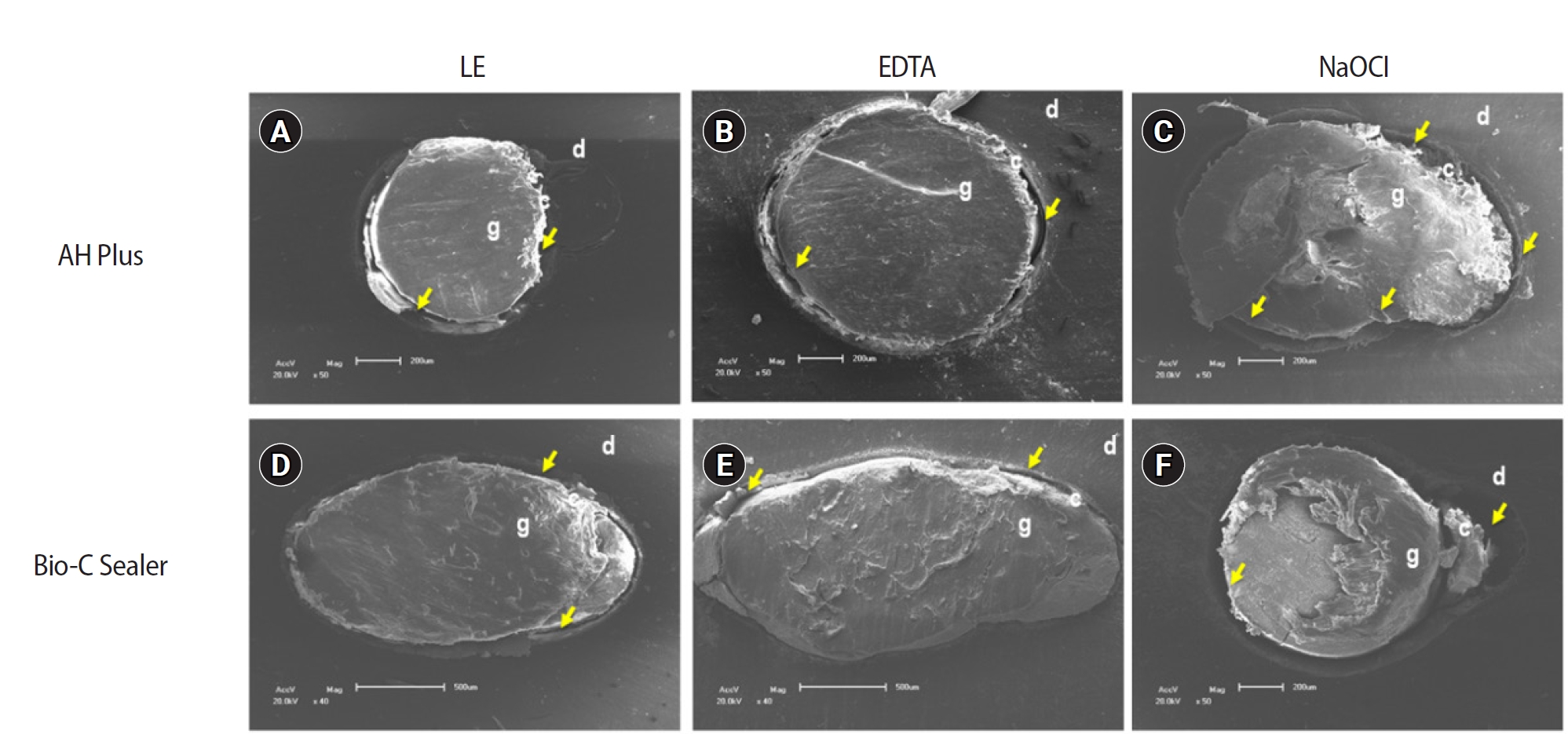
The study aimed to evaluate the effect of limonene extract (LE) on push-out bond strength (BS) to root dentin in endodontically treated teeth.
Methods
Single-rooted teeth were selected and instrumented using the reciprocating technique, then divided into three groups based on the final irrigating solution: 2.5% sodium hypochlorite (NaOCl), 17% ethylenediaminetetraacetic acid (EDTA), and 5% LE. The roots were further divided (n = 12) and obturated using the single-cone technique with epoxy resin-based (ERB) or bioceramic sealer (Bio-C). After 3 days, the roots were sectioned into 2-mm slices, obtaining two slices from each root third. Push-out BS testing was conducted at 0.5 mm/min, followed by failure pattern and adhesive interface analysis using scanning electron microscopy. Push-out BS data were analyzed by three-way analysis of variance and Tukey post-hoc test (p < 0.05).
Results
ERB showed higher BS when irrigated with EDTA (5.0 ± 2.3 MPa) compared to NaOCl (1.8 ± 1.1 MPa) (p = 0.0005), particularly in the cervical third. LE yielded intermediate values without significant differences from the other irrigants (3.5 ± 1.9 MPa) (p > 0.05). For Bio-C, the highest BS was observed in the apical third, especially with LE (9.4 ± 5.0 MPa), differing from other thirds and final irrigating solutions (p < 0.05). Mixed failure patterns were most prevalent, regardless of the irrigant solutions.
Conclusions
The combination of LE with Bio-C demonstrated superior BS in the apical third, suggesting its potential as a final irrigating solution in endodontic treatments.
- 2,286 View
- 206 Download

- Nanoleakage of apical sealing using a calcium silicate-based sealer according to canal drying methods
- Yoon-Joo Lee, Kyung-Mo Cho, Se-Hee Park, Yoon Lee, Jin-Woo Kim
- Restor Dent Endod 2024;49(2):e20. Published online April 19, 2024
- DOI: https://doi.org/10.5395/rde.2024.49.e20
-
 Abstract
 PDF
PDF PubReader
 ePub
ePub Objectives This study investigated the nanoleakage of root canal obturations using calcium silicate-based sealer according to different drying methods.
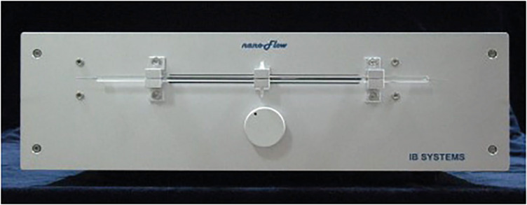
Materials and Methods Fifty-two extracted mandibular premolars with a single root canal and straight root were selected for this study. After canal preparation with a nickel-titanium rotary file system, the specimens were randomly divided into 4 groups according to canal drying methods (1: complete drying, 2: blot drying/distilled water, 3: blot drying/NaOCl, 4: aspiration only). The root canals were obturated using a single-cone filling technique with a calcium silicate–based sealer. Nanoleakage was evaluated using a nanoflow device after 24 hours, 1 week, and 1 month. Data were collected twice per second at the nanoscale and measured in nanoliters per second. Data were statistically analyzed using the Kruskal-Wallis and Mann–Whitney
U -tests (p < 0.05).Results The mean flow rate measured after 24 hours showed the highest value among the time periods in all groups. However, the difference in the flow rate between 1 week and 1 month was not significant. The mean flow rate of the complete drying group was the highest at all time points. After 1 month, the mean flow rate in the blot drying group and the aspiration group was not significantly different.
Conclusions Within the limitations of this study, the canal drying method had a significant effect on leakage and sealing ability in root canal obturations using a calcium silicate-based sealer. Thus, a proper drying procedure is critical in endodontic treatment.
- 2,295 View
- 104 Download

- The status of clinical trials regarding root canal sealers
- Ahmad AL Malak, Yasmina EL Masri, Mira Al Ziab, Nancy Zrara, Tarek Baroud, Pascale Salameh
- Restor Dent Endod 2024;49(1):e5. Published online January 15, 2024
- DOI: https://doi.org/10.5395/rde.2024.49.e5
-
 Abstract
 PDF
PDF PubReader
 ePub
ePub Objectives This study aimed to present the results and analyses of clinical trials, including updates on the different functions of root canal sealers.
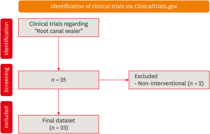
Materials and Methods In June 2023, we performed a comprehensive search of ClinicalTrials.gov to identify interventional clinical trials pertaining to root canal sealers. In total, 23 clinical trials conducted up to June 2023 were included in this study.
Results Approximately half of the trials (11 out of 23) were completed, while none were terminated or withdrawn. Each included trial had a minimum of 10 participants, with 11 trials having more than 100 participants. None of the assessed trials provided outcomes, and the majority (17 out of 23) lacked associated publications. In terms of geographic distribution, the USA and Canada did not contribute to any root canal sealer trials.
Conclusions This study highlights the lack of diversity in trial locations, the absence of reported results, and a scarcity of clinical trials examining the physicochemical properties of different sealers. Most published trials primarily focused on assessing the post-operative pain effect of these sealers, but no significant difference was found regarding post-operative pain control.
- 3,718 View
- 51 Download

- A scientometric, bibliometric, and thematic map analysis of hydraulic calcium silicate root canal sealers
- Anastasios Katakidis, Konstantinos Kodonas, Anastasia Fardi, Christos Gogos
- Restor Dent Endod 2023;48(4):e41. Published online November 13, 2023
- DOI: https://doi.org/10.5395/rde.2023.48.e41
-
 Abstract
 PDF
PDF PubReader
 ePub
ePub Objectives This scientometric and bibliometric analysis explored scientific publications related to hydraulic calcium silicate-based (HCSB) sealers used in endodontology, aiming to describe basic bibliometric indicators and analyze current research trends.
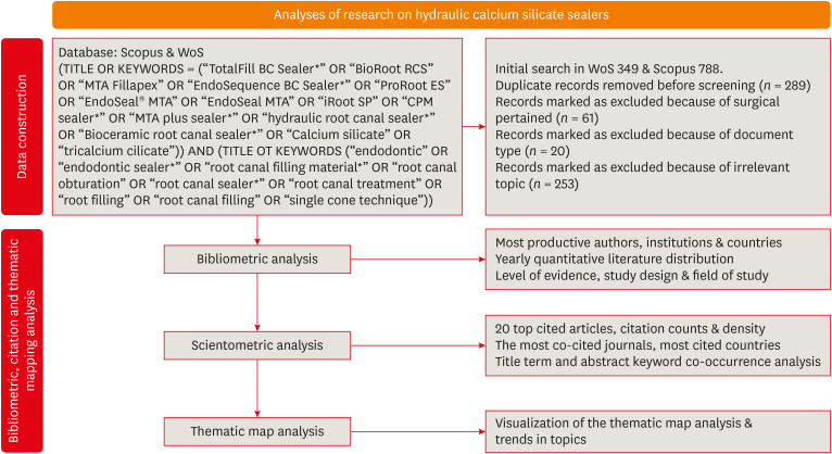
Materials and Methods A comprehensive search was conducted in Web of Science and Scopus using specific HCSB sealer and general endodontic-related terms. Basic research parameters were collected, including publication year, authorship, countries, institutions, journals, level of evidence, study design and topic of interest, title terms, author keywords, citation counts, and density.
Results In total, 498 articles published in 136 journals were retrieved for the period 2008–2023. Brazil was the leading country, and the universities of Bologna in Italy and Sao Paolo in Brazil were represented equally as leading institutions. The most frequently occurring keywords were “calcium silicate,” “root canal sealer MTA-Fillapex,” and “biocompatibility,” while title terms such as “calcium,” “sealers,” “root,” “canal,” “silicate based,” and “endodontic” occurred most often. According to the thematic map analysis, “solubility” appeared as a basic theme of concentrated research interest, and “single-cone technique” was identified as an emerging, inadequately developed theme. The co-occurrence analysis revealed 4 major clusters centered on sealers’ biological and physicochemical properties, obturation techniques, retreatability, and adhesion.
Conclusions This analysis presents bibliographic features and outlines changing trends in HCSB sealer research. The research output is dominated by basic science articles scrutinizing the biological and specific physicochemical properties of commonly used HCSB sealers. Future research needs to be guided by studies with a high level of evidence that utilize innovative, sophisticated technologies.
-
Citations
Citations to this article as recorded by- Agri-Food Sector: Contemporary Trends, Possible Gaps, and Prospective Directions
José Roberto Herrera Cantorani, Meire Ramalho de Oliveira, Luiz Alberto Pilatti, Thales Botelho de Sousa
Metrics.2025; 2(1): 3. CrossRef - Scientific mapping of experimental research on solar cookers: Global trends, evolution, and future directions
Flavio Odoi-Yorke, Bismark Baah, Richard Opoku
Solar Energy Advances.2025; 5: 100093. CrossRef - Bibliometric analysis of the GentleWave system: trends, collaborations, and research gaps
Raimundo Sales de Oliveira Neto, Thais de Moraes Souza, João Vitor Oliveira de Amorim, Thaine Oliveira Lima, Guilherme Ferreira da Silva, Rodrigo Ricci Vivan, Murilo Priori Alcalde, Marco Antonio Hungaro Duarte
Restorative Dentistry & Endodontics.2025; 50(2): e17. CrossRef - Top 100 Most Cited Articles on Antibiotics in Endodontics: A Bibliometric Analysis
Hajar Albanyan, Mohammed Asseery, Haitham Alahmari, Ikram Ul Haq, Ali Alaqla
Journal of Endodontics.2025;[Epub] CrossRef - A Scientometric Review of Practical Applications in Quantum Natural Language Processing (QNLP): Trends, Gaps, and Research Opportunities
Victor R. Silva, Fábio R. Barbosa, Jasson C. Silva, Francisco J. Santos, Ricardo A. L. Rabelo, Joel J. P. C. Rodrigues
IEEE Access.2025; 13: 210169. CrossRef - A bibliometric analysis of global research trend and progress on Dy doped materials
Sangeeta Kadyan, Manju Nain, Ashima Makhija, Poonam Punia, Anil Ohlan, Sajjan Dahiya, R. Punia, A.S. Maan
Journal of Alloys and Compounds Communications.2024; 3: 100006. CrossRef - Comparative bioactivity and immunomodulatory potential of the new Bioroot Flow and AH Plus Bioceramic sealer: An in vitro study on hPDLSCs
José Luis Sanz, Sergio López-García, David García-Bernal, Francisco Javier Rodríguez-Lozano, Leopoldo Forner, Adrián Lozano, Laura Murcia
Clinical Oral Investigations.2024;[Epub] CrossRef - Analyzing collaboration and impact: A bibliometric review of four highly published authors’ research profiles on collaborative maps
Willy Chou, Julie Chi Chow
Medicine.2024; 103(28): e38686. CrossRef
- Agri-Food Sector: Contemporary Trends, Possible Gaps, and Prospective Directions
- 2,639 View
- 44 Download
- 4 Web of Science
- 8 Crossref

- Bone repair in defects filled with AH Plus sealer and different concentrations of MTA: a study in rat tibiae
- Jessica Emanuella Rocha Paz, Priscila Oliveira Costa, Albert Alexandre Costa Souza, Ingrid Macedo de Oliveira, Lucas Fernandes Falcão, Carlos Alberto Monteiro Falcão, Maria Ângela Area Leão Ferraz, Lucielma Salmito Soares Pinto
- Restor Dent Endod 2021;46(4):e48. Published online September 2, 2021
- DOI: https://doi.org/10.5395/rde.2021.46.e48
-
 Abstract
 PDF
PDF PubReader
 ePub
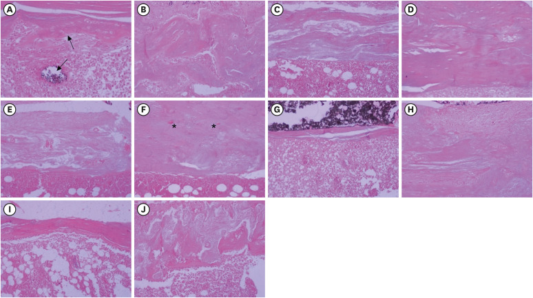
ePub Objectives This study aimed to evaluate the effects on bone repair of different concentrations of mineral trioxide aggregate (MTA) added to AH Plus.
Materials and Methods Bone tissue reactions were evaluated in 30 rats (
Rattus norvegicus ) after 7 and 30 days. In the AH + MTA10, AH + MTA20, and AH + MTA30 groups, defects in the tibiae were filled with AH Plus with MTA in proportions of 10%, 20% and 30%, respectively; in the MTA-FILL group, MTA Fillapex was used; and in the control group, no sealer was used. The samples were histologically analyzed to assess bone union and maturation. The Kruskal-Wallis and Mann-Whitney tests were performed for multiple pairwise comparisons (p ≤ 0.05).Results At the 7-day time point, AH + MTA10 was superior to MTA-FILL with respect to bone union, and AH + MTA20 was superior to MTA-FILL with respect to bone maturity (
p < 0.05). At the 30-day time point, both the AH + MTA10 and AH + MTA20 experimental sealers were superior not only to MTA-FILL, but also to AH + MTA30 with respect to both parameters (p < 0.05). The results of the AH + MTA10 and AH + MTA20 groups were superior to those of the control group for both parameters and experimental time points (p < 0.05).Conclusions The results suggest the potential benefit of using a combination of these materials in situations requiring bone repair.
-
Citations
Citations to this article as recorded by- Analysis of the cytotoxicity and bioactivity of CeraSeal, BioRoot™ and AH Plus® sealers in pre-osteoblast lineage cells
Luciano Aparecido de Almeida-Junior, Giuliana de Campos Chaves Lamarque, Henry Herrera, Maya Fernanda Manfrin Arnez, Francine Lorencetti-Silva, Raquel Assed Bezerra Silva, Léa Assed Bezerra Silva, Francisco Wanderley Garcia Paula-Silva
BMC Oral Health.2024;[Epub] CrossRef - A Review of the research methods and progress of biocompatibility evaluation of root canal sealers
Xiliang Yang, Tianxia Zheng, Nuoya Yang, Zihan Yin, Wuliang Wang, Yuhong Bai
Australian Endodontic Journal.2023; 49(S1): 508. CrossRef - Effect of Vitapex Combined with AH-Plus Paste on Inflammation in Middle-Aged and Elderly Patients with Periodontal-Endodontic Disease
Rong Hu, Fulan Zhang, Xiangyu Guo, Youren Jing, Xiaowan Lin, Liping Tian, Min Tang
Computational and Mathematical Methods in Medicine.2022; 2022: 1. CrossRef
- Analysis of the cytotoxicity and bioactivity of CeraSeal, BioRoot™ and AH Plus® sealers in pre-osteoblast lineage cells
- 1,810 View
- 15 Download
- 4 Web of Science
- 3 Crossref

- Micro-computed tomographic evaluation of single-cone obturation with three sealers
- Sahar Zare, Ivy Shen, Qiang Zhu, Chul Ahn, Carolyn Primus, Takashi Komabayashi
- Restor Dent Endod 2021;46(2):e25. Published online April 16, 2021
- DOI: https://doi.org/10.5395/rde.2021.46.e25
-
 Abstract
 PDF
PDF PubReader
 ePub
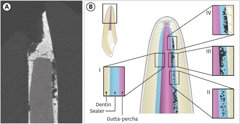
ePub Objectives This study used micro-computed tomography (µCT) to compare voids and interfaces in single-cone obturation among AH Plus, EndoSequence BC, and prototype surface pre-reacted glass ionomer (S-PRG) sealers and to determine the percentage of sealer contact at the dentin and gutta-percha (GP) interfaces.
Materials and Methods Fifteen single-rooted human teeth were shaped using ProTaper NEXT size X5 rotary files using 2.5% NaOCl irrigation. Roots were obturated with a single-cone ProTaper NEXT GP point X5 with AH Plus, EndoSequence BC, or prototype S-PRG sealer (
n = 5/group).Results The volumes of GP, sealer, and voids were measured in the region of 0–2, 2–4, 4–6, and 6–8 mm from the apex, using image analysis of sagittal µCT scans. GP volume percentages were: AH Plus (75.5%), EndoSequence BC (87.3%), and prototype S-PRG (94.4%). Sealer volume percentages were less: AH Plus (14.3%), EndoSequence BC (6.8%), and prototype S-PRG (4.6%). Void percentages were AH Plus (10.1%), EndoSequence BC (5.9%), and prototype S-PRG (1.0%). Dentin-sealer contact ratios of AH Plus, EndoSequence BC, and prototype S-PRG groups were 82.4% ± 6.8%, 71.6% ± 25.3%, and 70.2% ± 9.4%, respectively. GP-sealer contact ratios of AH Plus, EndoSequence BC, and prototype S-PRG groups were 65.6% ± 29.1%, 80.7% ± 25.8%, and 87.0% ± 8.6%, respectively.
Conclusions Prototype S-PRG sealer created a low-void obturation, similar to EndoSequence BC sealer with similar dentin-sealer contact (> 70%) and GP-sealer contact (> 80%). Prototype S-PRG sealer presented comparable filling quality to EndoSequence BC sealer.
-
Citations
Citations to this article as recorded by- Assessment of gap areas of root filling techniques in teeth with 3D-printed different configurations of C-shaped root canals: a micro-computed tomography study
Tuba Gok, Adem Gok, Haydar Onur Aciksoz
BMC Oral Health.2025;[Epub] CrossRef - Assessment of the quality of root canal filling using three different sealers: Micro-computed tomography and scanning electron microscope study
Loai Alsofi, Mohammed Yagmoor, Tariq AbuHaimed, Hassan Abed, Ehab Alshouibi, Rafif Mandura, Turki Bakhsh, Hanaa Ashkar, Mey Al-Habib
Saudi Endodontic Journal.2025; 15(2): 152. CrossRef - Comparative evaluation of bioactive calcium silicate coating on functionalized gutta-percha and its effect on bioceramic sealer wettability – An in vitro study
Bollineni Swetha, B. Devi Priya, K. Hanisha Reddy, G. Prasanthi, T. Murali Mohan, Dumpa Tejaswi
Journal of Conservative Dentistry and Endodontics.2025; 28(7): 613. CrossRef - Assessment of isthmus filling using two obturation techniques performed by students with different levels of clinical experience
Yang Yu, Chong-Yang Yuan, Xing-Zhe Yin, Xiao-Yan Wang
Journal of Dental Sciences.2024; 19(1): 169. CrossRef - Micro-CT determination of the porosity of two tricalcium silicate sealers applied using three obturation techniques
Jinah Kim, Kali Vo, Gurmukh S. Dhaliwal, Aya Takase, Carolyn Primus, Takashi Komabayashi
Journal of Oral Science.2024; 66(3): 163. CrossRef - Ex-vivo evaluation of clinically-set hydraulic sealers used with different canal dryness protocols and obturation techniques: a randomized clinical trial
Nawar Naguib Nawar, Mohamed Mohamed Elashiry, Ahmed El Banna, Shehabeldin Mohamed Saber, Edgar Schäfer
Clinical Oral Investigations.2024;[Epub] CrossRef - Hydraulic (Single Cone) Versus Thermogenic (Warm Vertical Compaction) Obturation Techniques: A Systematic Review
Haytham S Jaha
Cureus.2024;[Epub] CrossRef - Sealing ability of various endodontic sealers with or without ethylenediaminetetraacetic acid (EDTA) treatment on bovine root canal
Yusuke AIGAMI, Tomofumi SAWADA, Shunsuke SHIMIZU, Akiko ASANO, Mamoru NODA, Shinji TAKEMOTO
Dental Materials Journal.2024; 43(3): 420. CrossRef - A Literature Review of the Effect of Heat on the Physical-Chemical Properties of Calcium Silicate–Based Sealers
Israa Ashkar, José Luis Sanz, Leopoldo Forner, James Ghilotti, María Melo
Journal of Endodontics.2024; 50(8): 1044. CrossRef - Assessment of the Prevalence of Head Lice Infestation and Parents’ Attitudes Towards Its Management: A School-based Epidemiological Study in İstanbul, Türkiye
Özben Özden, İnci Timur, Hale Ezgi Açma, Duygu Şimşekli, Barış Gülerman, Özgür Kurt
Turkish Journal of Parasitology.2023; 47(2): 112. CrossRef - Calcium-doped zinc oxide nanocrystals as an innovative intracanal medicament: a pilot study
Gabriela Leite de Souza, Thamara Eduarda Alves Magalhães, Gabrielle Alves Nunes Freitas, Nelly Xiomara Alvarado Lemus, Gabriella Lopes de Rezende Barbosa, Anielle Christine Almeida Silva, Camilla Christian Gomes Moura
Restorative Dentistry & Endodontics.2022;[Epub] CrossRef - Micro‐CT assessment of gap‐containing areas along the gutta‐percha‐sealer interface in oval‐shaped canals
Gustavo De‐Deus, Gustavo O. Santos, Iara Zamboni Monteiro, Daniele M. Cavalcante, Marco Simões‐Carvalho, Felipe G. Belladonna, Emmanuel J. N. L. Silva, Erick M. Souza, Raphael Licha, Carla Zogheib, Marco A. Versiani
International Endodontic Journal.2022; 55(7): 795. CrossRef - A critical analysis of research methods and experimental models to study root canal fillings
Gustavo De‐Deus, Erick Miranda Souza, Emmanuel João Nogueira Leal Silva, Felipe Gonçalves Belladonna, Marco Simões‐Carvalho, Daniele Moreira Cavalcante, Marco Aurélio Versiani
International Endodontic Journal.2022; 55(S2): 384. CrossRef - Use of micro-CT to examine effects of heat on coronal obturation
Ivy Shen, Joan Daniel, Kali Vo, Chul Ahn, Carolyn Primus, Takashi Komabayashi
Journal of Oral Science.2022; 64(3): 224. CrossRef - Obturation of Root Canals By Vertical Condensation of Gutta-Percha – Benefits and Pitfalls
Calkovsky Bruno, Slobodnikova Ladislava, Bacinsky Martin, Janickova Maria
Acta Medica Martiniana.2021; 21(3): 103. CrossRef
- Assessment of gap areas of root filling techniques in teeth with 3D-printed different configurations of C-shaped root canals: a micro-computed tomography study
- 1,976 View
- 30 Download
- 11 Web of Science
- 15 Crossref

- Effects of radiation therapy on the dislocation resistance of root canal sealers applied to dentin and the sealer-dentin interface: a pilot study
- Pallavi Yaduka, Rubi Kataki, Debosmita Roy, Lima Das, Shachindra Goswami
- Restor Dent Endod 2021;46(2):e22. Published online March 29, 2021
- DOI: https://doi.org/10.5395/rde.2021.46.e22
-
 Abstract
 PDF
PDF PubReader
 ePub
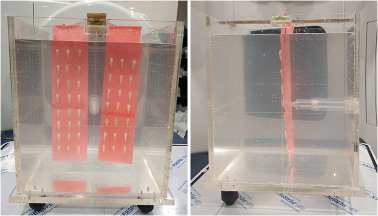
ePub Objectives This study evaluated and compared the effects of radiation therapy on the dislocation resistance of AH Plus and BioRoot RCS applied to dentin and the sealer-dentin interface.
Materials and Methods Thirty single-rooted teeth were randomly assigned to 2 groups (
n = 15 each): AH Plus (Dentsply DeTrey) and BioRoot RCS (Septodont). Each group was subdivided into control and experimental groups. The experimental group was subjected to a total radiation dose of 60 Gy. The root canals of all samples were cleaned, shaped, and obturated using the single-cone technique. Dentin slices (1 mm) were sectioned from each root third for the push-out test and scanning electron microscopy (SEM) was done to examine the sealer-dentin interface. The failure mode was determined using stereomicroscopy. Bond strength data were analyzed by the independentt -test, 1-way analysis of variance, and the Tukeypost hoc test (α = 0.05).Results Significantly lower bond strength was observed in irradiated teeth than non-irradiated teeth in the AH Plus group (
p < 0.05). The BioRoot RCS group showed no significant reduction in bond strength after irradiation (p > 0.05) and showed a higher post-irradiation bond strength (209.92 ± 172.26 MPa) than the AH Plus group. SEM revealed slightly larger gap-containing regions in irradiated specimens from both groups.Conclusions The dislocation resistance of BioRoot RCS was not significantly changed by irradiation and was higher than that of AH Plus. BioRoot RCS may be the sealer of choice for root canal treatment in patients undergoing radiation therapy.
-
Citations
Citations to this article as recorded by- Effects of radiotherapy dose and endodontic irrigants on universal resin cement bonding to root dentin: mechanical and interfacial analyses
Lívia Ribeiro, Luíz Carlos de Lima Dias-Júnior, Paulo Henrique dos Santos, Mariana Comparotto Minamisako, Paulo Marcelo Rodrigues, Vicente Ribeiro Netto, Bruno Alexandre Pacheco de Castro Henriques, Renata Gondo Machado, Cleonice da Silveira Teixeira, Luc
International Journal of Adhesion and Adhesives.2026; 146: 104252. CrossRef - Impact of radiation therapy regimen on the dislodgement resistance of endodontic sealers: A micro push-out test
Marcos Testa Magoga, Rafaela Lourdes de Sousa, Luiz Carlos Lima Dias-Junior, Rayssa Sabino-Silva, Mariana Comparotto Minamisako, Paulo Marcelo Rodrigues, Vicente Ribeiro Netto, Ricardo Machado, Cleonice da Silveira Teixeira, Lucas da Fonseca Roberti Garci
International Journal of Adhesion and Adhesives.2025; 136: 103894. CrossRef - The impact of radiotherapy on endodontic treatment: a scoping review
Guilherme Pauletto, Giovanna Isabel Mittmann Voigt, Sidnei Flores de Pellegrin, Yasmin Padoin, Carlos Alexandre Souza Bier
Odontology.2025;[Epub] CrossRef - Evaluation of the root dentin bond strength and intratubular biomineralization of a premixed calcium aluminate-based hydraulic bioceramic endodontic sealer
Yu-Na Lee, Min-Kyeong Kim, Hee-Jin Kim, Mi-Kyung Yu, Kwang-Won Lee, Kyung-San Min
Journal of Oral Science.2024; 66(2): 96. CrossRef - Effects of radiotherapy dose and application time on the load-to-failure values of teeth filled with different sealers
Ozgun Gulderen, Esma Saricam, Sedef Gökhan Açikgöz, Yılmaz Tezcan
BMC Oral Health.2024;[Epub] CrossRef - Ultrasonic activation of the endodontic sealer enhances its intratubular penetration and bond strength to irradiated root dentin
Luana Duart Jordani, Amanda Freitas da Rosa, Luiz Carlos de Lima Dias-Junior, Julia Menezes Savaris, Mariana Comparotto Minamisako, Luciano Roberto da Silva, Marcio Toshio Umeda Takashima, Eduardo Antunes Bortoluzzi, Cleonice da Silveira Teixeira, Lucas d
Odontology.2024; 112(3): 917. CrossRef - Effect of the timing of primary endodontic treatment and dosage of radiation therapy on the filling material removal
Bruna Venzke Fischer, Luiz Carlos de Lima Dias‐Junior, Mariana Comparotto Minamisako, Cristiane Maria Almeida, Luciano Roberto da Silva, Eduardo Antunes Bortoluzzi, Cleonice da Silveira Teixeira, Lucas da Fonseca Roberti Garcia
Australian Endodontic Journal.2024; 50(2): 321. CrossRef - Does radiation therapy affect adhesion of tricalcium silicate cements to root dentin?
Lochan KHULLAR, Nidambur Vasudev BALLAL, Tan Fırat EYÜBOĞLU, Mutlu ÖZCAN
Journal of Applied Oral Science.2023;[Epub] CrossRef - Effect of the timing of radiation therapy on the push‐out strength of resin cement to root dentine
Patrícia da Agostim Cancelier, Renata Gondo Machado, Júlia Menezes Savaris, Eduardo Antunes Bortoluzzi, Cleonice da Silveira Teixeira, Mariana Comparotto Minamisako, Paulo Marcelo Rodrigues, Vicente Ribeiro Netto, Kamile Leonardi Dutra‐Horstmann, Lucas da
Australian Endodontic Journal.2023; 49(S1): 122. CrossRef - Influence of irrigation and laser assisted root canal disinfection protocols on dislocation resistance of a bioceramic sealer
Ivona Bago, Ana Sandrić, Katarina Beljic-Ivanovic, Boris Pažin
Photodiagnosis and Photodynamic Therapy.2022; 40: 103067. CrossRef - Influence of 2% chlorhexidine on the dislodgement resistance of AH plus, bioroot RCS, and GuttaFlow 2 sealer to dentin and sealer-dentin interface
Debosmita Roy, Rubi Kataki, Lima Das, Khushboo Jain
Journal of Conservative Dentistry.2022; 25(6): 642. CrossRef
- Effects of radiotherapy dose and endodontic irrigants on universal resin cement bonding to root dentin: mechanical and interfacial analyses
- 2,131 View
- 23 Download
- 11 Web of Science
- 11 Crossref

- Biological assessment of a new ready-to-use hydraulic sealer
- Francine Benetti, João Eduardo Gomes-Filho, India Olinta de Azevedo-Queiroz, Marina Carminatti, Letícia Citelli Conti, Alexandre Henrique dos Reis-Prado, Sandra Helena Penha de Oliveira, Edilson Ervolino, Elói Dezan-Júnior, Luciano Tavares Angelo Cintra
- Restor Dent Endod 2021;46(2):e21. Published online March 24, 2021
- DOI: https://doi.org/10.5395/rde.2021.46.e21
-
 Abstract
 PDF
PDF PubReader
 ePub
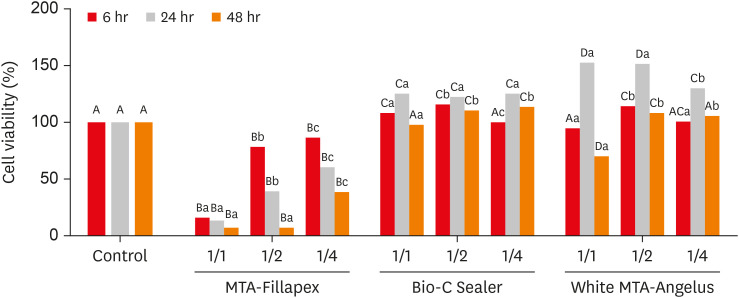
ePub Objectives This study compared the cytotoxicity, biocompatibility, and tenascin immunolabeling of a new ready-to-use hydraulic sealer (Bio-C Sealer) with MTA-Fillapex and white MTA-Angelus.
Materials and Methods L929 fibroblasts were cultivated and exposed to undiluted and diluted material extracts. Polyethylene tubes with or without (the control) the materials were implanted into the dorsa of rats. At 7 days and 30 days, the rats were euthanized, and the specimens were prepared for analysis; inflammation and immunolabeling were measured, and statistical analysis was performed (
p < 0.05).Results MTA-Fillapex exhibited greater cytotoxicity than the other materials at all time points (
p < 0.05). The undiluted Bio-C Sealer exhibited greater cytocompatibility at 6 and 48 hours than white MTA-Angelus, with higher cell viability than in the control (p < 0.05). White MTA-Angelus displayed higher cell viability than the control at 24 hours, and the one-half dilution displayed similar results at both 6 and 48 hours (p < 0.05). At 7 days and 30 days, the groups exhibited moderate inflammation with thick fibrous capsules and mild inflammation with thin fibrous capsules, respectively (p > 0.05). At 7 days, moderate to strong immunolabeling was observed (p > 0.05). After 30 days, the control and MTA-Fillapex groups exhibited strong immunolabeling, the white MTA-Angelus group exhibited moderate immunolabeling (p > 0.05), and the Bio-C Sealer group exhibited low-to-moderate immunolabeling, differing significantly from the control (p < 0.05).Conclusions Bio-C Sealer and white MTA-Angelus exhibited greater cytocompatibility than MTA-Fillapex; all materials displayed adequate biocompatibility and induced tenascin immunolabeling.
-
Citations
Citations to this article as recorded by- Clinical and radiographic assessment of mineral trioxide aggregate with platelet rich fibrin as pulp capping biomaterials: a 12-month randomized trial
Rahma Ahmed Ibrahem Hafiz Abuhashema, Mona El Saied Essa, Shereen Hafez Ibrahim, Omaima Mohamed Safwat
Scientific Reports.2025;[Epub] CrossRef - Biocompatibility and bioactivity of bioceramic endodontic sealer: NeoSealer Flo
Evelin Carine Alves SILVA, Jéssica Arielli PRADELLI, Guilherme Ferreira da SILVA, Paulo Sérgio CERRI, Mario TANOMARU-FILHO, Juliane Maria GUERREIRO-TANOMARU
Journal of Applied Oral Science.2025;[Epub] CrossRef - Influence of photoactivation on tissue response to different dyes used in photodynamic therapy and laser ablation therapy
Luciano Tavares Angelo Cintra, Cristiane Cantiga-Silva, Henrique Augusto Banci, Flávio Duarte Faria, Nathália Evelyn da Silva Machado, Carolina de Barros Morais Cardoso, Pedro Henrique Chaves de Oliveira, Lucas Rodrigues de Araújo Estrela, Gustavo Sivieri
Journal of Photochemistry and Photobiology B: Biology.2024; 251: 112843. CrossRef - Bleaching effectiveness and cytotoxicity of new experimental formulation of niobium-based bleaching gel
Camila de Sousa Caneschi, Francine Benetti, Luiz Carlos Alves de Oliveira, Jadson Cláudio Belchior, Raquel Conceição Ferreira, Allyson Nogueira Moreira, Luís Fernando dos Santos Alves Morgan
Clinical Oral Investigations.2023; 27(4): 1613. CrossRef - Biological investigation of resinous endodontic sealers containing calcium hydroxide
Carlos Roberto Emerenciano Bueno, Francine Benetti, Marina Tolomei Sandoval Cury, Ana Maria Veiga Vasques, Leopoldo Cosme-Silva, Índia Olinta de Azevedo Queiroz, Ana Cláudia Rodrigues da Silva, Rogério de Castilho Jacinto, Luciano Tavares Angelo Cintra, E
PLOS ONE.2023; 18(7): e0287890. CrossRef - Tricalcium silicate cement sealers
Anita Aminoshariae, Carolyn Primus, James C. Kulild
The Journal of the American Dental Association.2022; 153(8): 750. CrossRef - Comparative evaluation of push-out bond strength of bioceramic and epoxy sealers after using various final irrigants: An in vitro study
Chandrasekhar Veeramachaneni, Swathi Aravelli, Sreeja Dundigalla
Journal of Conservative Dentistry.2022; 25(2): 145. CrossRef
- Clinical and radiographic assessment of mineral trioxide aggregate with platelet rich fibrin as pulp capping biomaterials: a 12-month randomized trial
- 2,146 View
- 17 Download
- 7 Web of Science
- 7 Crossref

- Flow characteristics and alkalinity of novel bioceramic root canal sealers
- Anastasios Katakidis, Konstantinos Sidiropoulos, Elisabeth Koulaouzidou, Christos Gogos, Nikolaos Economides
- Restor Dent Endod 2020;45(4):e42. Published online August 18, 2020
- DOI: https://doi.org/10.5395/rde.2020.45.e42
-
 Abstract
 PDF
PDF PubReader
 ePub
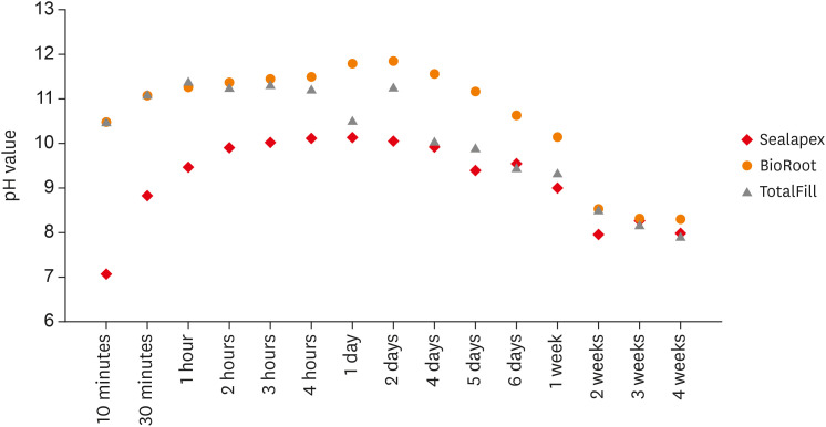
ePub Objective This study aimed to examine the physical properties (pH and flow) of 2 novel bioceramic sealers.
Materials and Methods The tested sealers were a calcium hydroxide sealer (Sealapex) and 2 bioceramic sealers (BioRoot RCS and TotalFill BC Sealer). Flow measurements were conducted according to ISO 6876/2012, with a press method of 0.05 mL of sealer. The pH of fresh samples was tested immediately after manipulation, while set samples were stored for 3 times the recommended setting time. The predetermined time intervals ranged from 3 minutes to 24 hours for fresh samples and from 10 minutes to 7 days and 4 weeks for the set samples. Analysis of variance was performed, with
p = 0.05 considered indicating significance.Results The mean flow values were 26.99 mm for BioRoot, 28.19 for Sealapex, and 30.8 mm for TotalFill BC Sealer, satisfying the ISO standard. In the set samples, BioRoot RCS had higher pH values at 24 hours to 1 week after immersion in distilled water. At 2 weeks, both bioceramic sealers had similar pH values, greater than that of Sealapex. In the fresh samples, the bioceramic sealers had significantly higher initial pH values than Sealapex (
p < 0.05). At 24 hours post-immersion, all sealers showed an alkaline pH, with the highest pH observed for TotalFill.Conclusions The TotalFill BC Sealer demonstrated the highest flow. The bioceramic sealers initially presented higher alkaline activity than the polymeric calcium hydroxide sealer. However, at 3 and 4 weeks post-immersion, all sealers had similar pH values.
-
Citations
Citations to this article as recorded by- In vitro comparative evaluation of physicochemical and mechanical properties, cytocompatibility, and antimicrobial efficacy of various bioceramic root canal sealers
Fushi Wang, Jiaxing Li, Jingjing Wan, Siyuan Li, Shijia Tang, Li Wang, Liuyan Meng
Ceramics International.2026;[Epub] CrossRef - Comparative analysis between resin-based root canal sealer and recent bioceramic-based root canal sealers using MicroCT, film thickness, and solubility
Amira Galal Ismail, Manar M. Galal, Tamer M. Hamdy
Journal of Oral Biology and Craniofacial Research.2026; 16(2): 101400. CrossRef - Functional and Bioactive Performance of Premixed Bioceramic Sealers with Warm Obturation: A Scoping Review
Patryk Wiśniewski, Stanisław Krokosz, Małgorzata Pietruska, Anna Zalewska
Gels.2025; 11(11): 932. CrossRef - Physicochemical properties of AH plus bioceramic sealer, Bio-C Sealer, and ADseal root canal sealer
Tamer M. Hamdy, Manar M. Galal, Amira Galal Ismail, Shehabeldin Saber
Head & Face Medicine.2024;[Epub] CrossRef - Characterization and Assessment of Physical Properties of 3 Single Syringe Hydraulic Cement–based Sealers
Veksina Raman, Josette Camilleri
Journal of Endodontics.2024; 50(3): 381. CrossRef - The Impact of Silver Nanoparticles on Dentinal Tubule Penetration of Endodontic Bioceramic Sealer
Sundus Bukhary, Sarah Alkahtany, Amal Almohaimede, Nourah Alkhayatt, Shahad Alsulaiman, Salma Alohali
Applied Sciences.2024; 14(24): 11639. CrossRef - Influence of root canal moisture on the penetration of TotalFill bioceramic sealer into the dentinal tubules: A confocal laser scanning microscopy study
Archika M Singh, Tarek M Elsewify, Walid S El-Sayed, Husam H Nuawafleh, Ranya F Elemam, Bassem M Eid
Saudi Endodontic Journal.2024; 14(2): 187. CrossRef - Unusual Canal Morphology in Mandibular Premolars With Two Distal and One Mesial Canal: A Case Series
Jinesh A, Sanjana Jayakumar Nair, Saurabh Gupta, Harsh Chansoria, Gaurav Rawat
Cureus.2024;[Epub] CrossRef - A scientometric, bibliometric, and thematic map analysis of hydraulic calcium silicate root canal sealers
Anastasios Katakidis, Konstantinos Kodonas, Anastasia Fardi, Christos Gogos
Restorative Dentistry & Endodontics.2023;[Epub] CrossRef - Thermal, chemical and physical analysis of VDW.1Seal, Fill Root ST, and ADseal root canal sealers
Shehabeldin Saber, Manar M. Galal, Amira Galal Ismail, Tamer M. Hamdy
Scientific Reports.2023;[Epub] CrossRef - α-tricalcium phosphate/fluorapatite-based cement - promising dental root canal filling material
Abdul Kazuz, Zeljko Radovanovic, Djordje Veljovic, Vesna Kojic, Dimitar Jakimov, Tamara Vlajic-Tovilovic, Vesna Miletic, Rada Petrovic, Djordje Janackovic
Processing and Application of Ceramics.2022; 16(1): 22. CrossRef
- In vitro comparative evaluation of physicochemical and mechanical properties, cytocompatibility, and antimicrobial efficacy of various bioceramic root canal sealers
- 2,778 View
- 26 Download
- 11 Crossref

- Calcium silicate-based root canal sealers: a literature review
- Miyoung Lim, Chanyong Jung, Dong-Hoon Shin, Yong-bum Cho, Minju Song
- Restor Dent Endod 2020;45(3):e35. Published online June 9, 2020
- DOI: https://doi.org/10.5395/rde.2020.45.e35
-
 Abstract
 PDF
PDF PubReader
 ePub
ePub Epoxy resin-based sealers are currently widely used, and several studies have considered AH Plus to be the gold-standard sealer. However, it still has limitations, including possible mutagenicity, cytotoxicity, inflammatory response, and hydrophobicity. Drawing upon the advantages of mineral trioxide aggregate, calcium silicate-based sealers were introduced with high levels of biocompatibility and hydrophilicity. Because of the hydrophilic environment in root canals, water resorption and solubility of root canal sealers are important factors contributing to their stability. Sealers displaying lower microleakage and stronger push-out bond strength are also needed to endure the dynamic tooth environment. Although the physical properties of calcium silicate-based sealers meet International Organization for Standardization recommendations, and they have consistently reported to be biocompatible, they have not overcome conventional resin-based sealers in actual practice. Therefore, further studies aiming to improve the physical properties of calcium silicate-based sealers are needed.
-
Citations
Citations to this article as recorded by- Evidence synthesis of postoperative pain with bioceramic vs. epoxy resin sealers: umbrella review of randomized trials within existing systematic reviews
Mrunali Dahikar, Ashish Mandwe, Kulvinder Singh Banga, Alexander Maniangat Luke, Suraj Arora, Unmesh Khanvilkar, Ajinkya M. Pawar
Frontiers in Dental Medicine.2026;[Epub] CrossRef - Effect of Different Tapered Gutta-Percha Points on Push-Out Bond Strength of Two Root Canal Sealers
Warattama Suksaphar, Pakit Tungsawat, Ninnita Wongwatanasanti, Siripat Lertnantapanya, Prattana Yodmanothum
European Journal of General Dentistry.2025; 14(03): 285. CrossRef - Effect of Electrical Heat Carrier Temperature on Bacterial Leakage of Endodontically Treated Teeth Using a Bioceramic Sealer
Mir Ahmad Nabavi, Mahmood Reza Kalantar Motamedi, Pedram Fattahi, Saber Khazaei
Clinical and Experimental Dental Research.2025;[Epub] CrossRef - Nanoparticles modified bioceramic sealers on solubility, antimicrobial efficacy, pushout bond strength and marginal adaptation at apical-third of canal dentin
Basil Almutairi, Fahad Alkhudhairy
PeerJ.2025; 13: e18840. CrossRef - Assessing the antimicrobial properties of bioceramic sealers enhanced with herbal extracts against E. faecalis
KS Sachin, K Shibani Shetty, KB Jeyalakshmi, S Harishma, S Harshini
Folia Medica.2025;[Epub] CrossRef - Estudio comparativo de la solubilidad de dos selladores endodónticos biocerámicos y un sellador a base de resinas
//Comparative study of the solubility of two bioceramic endodontic sealers and one epoxi-resin based sealer
Alejandro Leonhardt, Nicolás Paduli, Osvaldo Zmener, Miguel Chantiri
Revista de la Asociación Odontológica Argentina.2025; : 1. CrossRef - Enhancing root canal sealing: Exploring the sealing potential of epoxy and calcium silicate-based sealers with chitosan nanoparticle enhancement
S. Harishma, Srilekha Jayakumar, K Shibani Shetty, Barkavi Panchatcharam, Jwaalaa Rajkumar, S. Harshini
Endodontology.2025; 37(3): 306. CrossRef - Evaluation of the Genotoxicity and Cytotoxicity of Bioceramic Endodontic Sealers in HepG2 and V79 Cell Lines: An In Vitro Study Using the Comet and Micronucleus Assays
Antonija Tadin, Marija Badrov, Danijela Juric Kacunic, Nada Galic, Matea Macan, Ivan Kovacic, Davor Zeljezic
Journal of Functional Biomaterials.2025; 16(5): 169. CrossRef - In Vitro Apatite-Forming Ability of Different Root Canal Sealers (A Comparative Study)
Raghad A Al-Askary, Wiaam M. O. Al-Ashou, Sawsan H. Al-Jubori
Journal of International Society of Preventive and Community Dentistry.2025; 15(2): 173. CrossRef - Microstructural and elemental characterization of novel bioactive glass bioceramic sealer using Fourier transform infrared and X-ray diffraction analysis
Poulomi Guha, Pradeep Solete, Delphine Antony, Nishitha Arun, Mohmed Isaqali Karobari, Surendar Ramamoorthi
Journal of Conservative Dentistry and Endodontics.2025; 28(5): 412. CrossRef - Microstructural and Elemental Characterization of Calcium Silicate-Based Sealers
Mateusz Radwanski, Ireneusz Piwonski, Tomasz Szmechtyk, Salvatore Sauro, Monika Lukomska-Szymanska
Nanomaterials.2025; 15(10): 756. CrossRef - Apical negative pressure-enhanced sealer infiltration for obturating long oval-shaped root canals with the single-cone technique
Yaxu Feng, Brian E. Bergeron, Shijin Zhang, Danyang Sun, Kole Fisher, Franklin R. Tay, Bing Fan
Journal of Dentistry.2025; 160: 105909. CrossRef - Effects of different apical preparation sizes and root canal sealers on the fracture resistance of roots aged for 12 months in endodontically retreated mandibular premolars
Dilek Hancerliogullari, Sevda Durust Baris, Ali Turkyilmaz, Ali Erdemir
British Dental Journal.2025;[Epub] CrossRef - Influence of different endodontic treatment protocols on tooth survival: A retrospective cohort study with multistate analysis and group balancing
Ahmed Elmaasarawi, Mohamed Mekhemar, Andreas Bartols
International Endodontic Journal.2025; 58(10): 1529. CrossRef - Evaluation of 2,6-xylidine precipitate on sealer penetration of calcium silicate-based sealer and resin-based sealer: An in vitro study
M. B. Kalpana, Divya Shetty, Rajaram Naik
Endodontology.2025; 37(2): 183. CrossRef - Translational Advances in Regenerative Dentistry: Functional Biomaterials and Emerging Technologies
Seher Yaylacı, Hacer Eberliköse, Hakan Ceylan
Current Oral Health Reports.2025;[Epub] CrossRef - Marginal adaptation of heat and non-heat compatible bioceramic sealers in warm obturation: an in vitro SEM study
Thanomsuk Jearanaiphaisarn, Thanida Leelayuttakarn, Panisara Amatamahuthana, Pinmanus Chenpairojsakul, Keskanya Subbalekha, Pavena Chivatxaranukul
Scientific Reports.2025;[Epub] CrossRef - Influence of irrigating solutions on the hydration of calcium silicate-based dental biomaterials: An in vitro study
Pradeep M. Divya, Amit Jena, Saumyakanta Mohanty, Govind Shashirekha, Rashmi Rekha Mallick, Priyanka Sarangi
Journal of Conservative Dentistry and Endodontics.2025; 28(8): 758. CrossRef - Multispecies Biofilms Treated With Endodontic Sealers or Calcium Hydroxide: Antimicrobial Activity and Changes in Community Composition
Steven K. Uttech, Ronald Ordinola‐Zapata, W. Craig Noblett, Maria Martell, Bruno Lima, Christopher Staley
International Endodontic Journal.2025; 58(11): 1764. CrossRef - A comparative analysis of adhesion abilities between AH Plus® Bioceramic, Ceraseal® and AH Plus® on root canal dentine surfaces
Ike Dwi Maharti, Indira Larasputri, Nendar Herdianto, Anggraini Margono, Riesma Tasomara, Romilda Rosseti
Journal of Conservative Dentistry and Endodontics.2025; 28(9): 881. CrossRef - Clinical and radiographic success of single-cone bioceramic obturation versus traditional techniques: a systematic review and meta-analysis of randomized controlled trials
Firas Elmsmari, Yousef Elsayed, Abdelrahman Aboubakr, Mahdi Kaafarani, Osama Nour, Ajinkya M. Pawar
Journal of Oral Biology and Craniofacial Research.2025; 15(6): 1422. CrossRef - The Effect of Irrigation Solutions on the Setting Time, Solubility, and pH of Three Types of Premixed Bioceramic‐Based Root Canal Sealers
Kitichai Singharat, Ninnita Wongwatanasanti, Warattama Suksaphar, Pakit Tungsawat, Zhengrui Li
International Journal of Dentistry.2025;[Epub] CrossRef - Endodontie – State of the Art von A bis Z
Will Qian, Andreas Bartols
Zahnmedizin up2date.2025; 19(04): 281. CrossRef - Assessing Volume of Two Sealers’ Remnants after Reinstrumentation Using 3D Imaging Technology: An In Vitro Comparative Study
Khalel Mutaz Dawod, Raghad Abdulrazzaq Al-Hashimi
The Journal of Contemporary Dental Practice.2025; 26(8): 743. CrossRef - Functional and Bioactive Performance of Premixed Bioceramic Sealers with Warm Obturation: A Scoping Review
Patryk Wiśniewski, Stanisław Krokosz, Małgorzata Pietruska, Anna Zalewska
Gels.2025; 11(11): 932. CrossRef - Correlation of Bond Strength and Dentinal Tubule Penetration Evaluation of Four Different Endodontic Sealers: AH Plus, MTA Fillapex, Endoseal MTA, and Endoseal TCS (Maruchi): An In Vitro Study
Arezoo Mirzaei Sadeghloo, Seyedali Seyedmajidi, Akam Saeidi, Elham Mahmoudi, Murilo Baena Lopes
International Journal of Dentistry.2025;[Epub] CrossRef - Osteogenic Potential of Various Premixed Hydraulic Calcium Silicate-Based Sealers on Human Bone Marrow Stem Cells
Na-Hyun You, Donghee Lee, Yemi Kim, Sieun Nam, Sin-Young Kim
Materials.2025; 18(23): 5326. CrossRef - Polydopamine‐Functionalized Zinc Oxide Nanoparticles as a Root Canal Sealer: Characterization, Biological, and Physicochemical Properties
Arul Nayagi Raj, Aditya Shetty, Lakshmi Nidhi Rao, Giuseppe Ciccarella
Bioinorganic Chemistry and Applications.2025;[Epub] CrossRef - Does the Use of a Bioceramic Sealer Reduce Postoperative Pain Compared With an Epoxy Resin‐Based Sealer After Primary Root Canal Treatment and Retreatment?—An Umbrella Review
Lokhasudhan Govindaraju, Rajeswari Kalaiselvam, Mathan Rajan Rajendran, Aleksandar Jakovljevic, Jelena Jacomovic, Henry F. Duncan, Venkateshbabu Nagendrababu
International Endodontic Journal.2025;[Epub] CrossRef - Management of rarely seen internal tunnelling root resorption associated with a maxillary permanent incisor
Kirsty A. Carney, Thibault N. E. Colloc, Julie K. Kilgariff
British Dental Journal.2024; 236(12): 955. CrossRef - Top tips for treatment planning: tooth-by-tooth prognosis - Part 3: endodontic prognosis
Prashanti Eachempati, Andrew Harris, Guy Lambourn, Tony Francis, Ewen McColl
British Dental Journal.2024; 237(9): 686. CrossRef - Retreatability of calcium silicate-based sealers based on micro-computed tomographic evaluation − A systematic review
Sundus Mohammed Bukhary
The Saudi Dental Journal.2024; 36(10): 1278. CrossRef - Evaluation of Setting Time, Flowability, Film Thickness, and Radiopacity of Experimental Monocalcium Silicate‐Based Root Canal Sealers
Sukanya Juntha, Pakit Tungsawat, Ninnita Wongwatanasanti, Warattama Suksaphar, Siripat Lertnantapanya, Carlos M. Ardila
International Journal of Dentistry.2024;[Epub] CrossRef - Root Canal Treatment and Demand for Continuing Education among Thai Dental Practitioners
Ninnita Wongwatanasanti, Pakit Tungsawat, Warattama Suksaphar, Siripat Lertnantapanya, Prattana Yodmanotham
The Open Dentistry Journal.2024;[Epub] CrossRef - Clinical outcome of non-surgical root canal treatment using different sealers and techniques of obturation in 237 patients: A retrospective study
Mateusz Radwanski, Krystyna Pietrzycka, Tan Fırat Eyüboğlu, Mutlu Özcan, Monika Lukomska-Szymanska
Clinical Oral Investigations.2024;[Epub] CrossRef - Endodontic sealers after exposure to chlorhexidine digluconate: An assessment of physicochemical properties
Vasileios Kapralos, Josette Camilleri, Andreas Koutroulis, Håkon Valen, Dag Ørstavik, Pia Titterud Sunde
Dental Materials.2024; 40(3): 420. CrossRef - Assessment the bioactivity of zinc oxid eugenol sealer after the addition of different concentrations of nano hydroxyapatite-tyrosine amino acid
Rasha M. Al-Shamaa, Raghad A. Al-Askary
Brazilian Journal of Oral Sciences.2024; 23: e243733. CrossRef - Interfacial adaptation of newly prepared nano-tricalcium silicate-58s bioactive glass-based endodontic sealer
Nawal A. Al-Sabawi, Sawsan Hameed Al-Jubori
Journal of Dental Research, Dental Clinics, Dental Prospects.2024; 18(2): 115. CrossRef - Marginal adaptation of customized gutta percha cone with calcium silicate based sealer versus MTA and biodentine apical plugs in simulated immature permanent teeth (an in vitro study)
Mary M. Mina, Sybel M. Moussa, Mahmoud R. Aboelseoud
BMC Oral Health.2024;[Epub] CrossRef - Solubility of Endoseal and AH26 Root Canal Sealers
Nooshin Fakhari, Ali Reza Mirjani, Abbas Bagheri, Jalil Modaresi
Journal of Research in Dental and Maxillofacial Sciences.2024; 9(1): 1. CrossRef - Novel bioactive nanospheres show effective antibacterial effect against multiple endodontic pathogens
Jin Liu, Haoze Wu, Jun Qiu, Sirui Yang, Doudou Xiang, Xinhua Zhang, Jinxin Kuang, Min Xiao, Qing Yu, Xiaogang Cheng
Heliyon.2024; 10(7): e28266. CrossRef - Evaluation of canal patency and cleanliness following retreatment of bioceramic sealer‐obturated root canals using three different irrigant activation protocols
Daiasharailang Lyngdoh, Sharique Alam, Huma Iftekhar, Surendra Kumar Mishra
Australian Endodontic Journal.2024; 50(3): 475. CrossRef - Antibiofilm Efficacy of Calcium Silicate-Based Endodontic Sealers
Matilde Ruiz-Linares, Vsevolod Fedoseev, Carmen Solana, Cecilia Muñoz-Sandoval, Carmen María Ferrer-Luque
Materials.2024; 17(16): 3937. CrossRef - Enhancing the Biological Properties of Organic–Inorganic Hybrid Calcium Silicate Cements: An In Vitro Study
Minji Choi, Jiyoung Kwon, Ji-Hyun Jang, Duck-Su Kim, Hyun-Jung Kim
Journal of Functional Biomaterials.2024; 15(11): 337. CrossRef - Cytotoxicity and cell migration evaluation of a strontium silicate-based root canal sealer on stem cells from rat apical papilla: an in vitro study
Guanglei Zhou, Yu Zhao, Liangjing Cai, Liwei Liu, Xu Li, Lu Sun, Jiayin Deng
BMC Oral Health.2024;[Epub] CrossRef - An In Vitro Comparative Analysis of Physico–Mechanical Properties of Commercial and Experimental Bioactive Endodontic Sealers
Abdulmajeed Kashaf, Faisal Alonaizan, Khalid S. Almulhim, Dana Almohazey, Deemah Abdullah Alotaibi, Sultan Akhtar, Ashwin C. Shetty, Abdul Samad Khan
Bioengineering.2024; 11(11): 1079. CrossRef - Chemical, Antibacterial, and Cytotoxic Properties of Four Different Endodontic Sealer Leachates Over Time
Jo-Hsun Chen, Veksina Raman, Sarah A. Kuehne, Josette Camilleri, Josefine Hirschfeld
Journal of Endodontics.2024; 50(11): 1612. CrossRef - Comparative Analysis of Fracture Resistance of Endodontic Sealer Types and Filling Methods
Yun Song, Kee-Deog Kim, Bock-Young Jung, Wonse Park, Nan-Sim Pang
Materials.2024; 18(1): 40. CrossRef - Comparative Evaluation of Removal of Bioceramic Sealers Using Rotary Retreatment Files Supplemented with Passive Ultrasonic Activation: An In Vitro Study
Anuradha B Patil, Amrut Bambawale, Pooja R Barghare, Sumanthini V Margasahayam, Divya Naik, Jayeeta S Verma
World Journal of Dentistry.2024; 15(4): 292. CrossRef - Nonsurgical Endodontic Management of Nonperforating Internal Root Resorption in a Maxillary Central Incisor: A Case Report with a 4-Year Follow-Up
Paras M. Gehlot, Divya S. Rajkumar, Annapoorna B. Mariswamy, Upendra Natha N. Reddy, Chaitanya Chappidi
Journal of Pharmacy and Bioallied Sciences.2024; 16(Suppl 3): S3005. CrossRef - Evaluating the Sealing Performance of Endodontic Sealers: Insights Into Achieving Complete Sealing
Ajay Chhabra, Ramya K P., Saravana Prathap, Priyanka Yadav, Himani Mehra, Sona J Parvathy
Cureus.2024;[Epub] CrossRef - Effects of vehicles on the physical properties and biocompatibility of premixed calcium silicate cements
Gitae SON, Gyeung Mi SEON, Sang Hoon CHOI, Hyeong-Cheol YANG
Dental Materials Journal.2024; 43(2): 276. CrossRef - Comparative cytotoxicity study of putty- and powder-type calcium silicate cements
Sora Park, Dohyun Cho, Ji Hyeon Yoon, Yeonjoo Kang, Quang Canh Vo, Gitae Son, Hongjoo Park, Hyeong-Cheol Yang
Korean Journal of Dental Materials.2024; 51(4): 259. CrossRef - Physical-chemical properties and acellular bioactivity of newly prepared nano-tricalcium silicate-58s bioactive glass-based endodontic sealer
Nawal A. Al-Sabawi, Sawsan Hameed Al-Jubori
Journal of Oral Biosciences.2023; 65(4): 305. CrossRef - Dentinal Tubule Penetrability and Bond Strength of Two Novel Calcium Silicate-Based Root Canal Sealers
Karissa Shieh, Jack Yang, Elsa Heng Zhu, Ove Andreas Peters, Sepanta Hosseinpour
Materials.2023; 16(9): 3309. CrossRef - Cytotoxicity and Mineralization Activity of Calcium Silicate-Based Root Canal Sealers Compared to Conventional Resin-Based Sealer in Human Gingival Fibroblast Cells
Mohammad Shokrzadeh, Farzaneh Sadat Motafeghi, Anahita Lotfizadeh, Mohammad Ghorbani, Azam Haddadi Kohsar, Cesar Rogério Pucci
International Journal of Dentistry.2023; 2023: 1. CrossRef - Effect of three different photosensitizers in photodynamic therapy on bond strength of a calcium silicate‐based sealer to radicular dentin
Cihan Küden, Seda Nur Karakaş
Australian Endodontic Journal.2023; 49(S1): 265. CrossRef - Effect of endodontic sealer on postoperative pain: a network meta-analysis
Cynthia Maria Chaves Monteiro, Ana Cristina Rodrigues Martins, Alessandra Reis, Juliana Larocca de Geus
Restorative Dentistry & Endodontics.2023;[Epub] CrossRef - Antimicrobial Activity of Five Calcium Silicate Based Root Canal Sealers against a Multispecies Engineered Biofilm: An In Vitro Study
Carla Zogheib, Issam Khalil, Wajih Hage, Dolla Karam Sarkis, Mireille Kallasy, Germain Sfeir, May Mallah, Roula El Hachem
The Journal of Contemporary Dental Practice.2023; 24(9): 707. CrossRef - Calcium silicate sealers in endodontics
Archana Chavan, Nidambur Ballal
Acta stomatologica Naissi.2023; 39(87): 2624. CrossRef - Assessing the Sealing Performance and Clinical Outcomes of Endodontic Treatment in Patients with Chronic Apical Periodontitis Using Epoxy Resin and Calcium Salicylate Seals
Razvan Mihai Horhat, Bogdan Andrei Bumbu, Laura Orel, Oana Velea-Barta, Laura Cirligeriu, Gratiana Nicoleta Chicin, Marius Pricop, Mircea Rivis, Stefania Dinu, Delia Ioana Horhat, Felix Bratosin, Roxana Manuela Fericean, Rodica Anamaria Negrean, Luminita
Medicina.2023; 59(6): 1137. CrossRef -
In Vitro Cytotoxicity and Mineralization Potential of an Endodontic Bioceramic Material
Soumya Sheela, Mohannad Nassar, Fatma M. AlGhalban, Mehmet O. Gorduysus
European Journal of Dentistry.2023; 17(02): 548. CrossRef - Dislodgment Resistance, Adhesive Pattern, and Dentinal Tubule Penetration of a Novel Experimental Algin Biopolymer-Incorporated Bioceramic-Based Root Canal Sealer
Galvin Sim Siang Lin, Norhayati Luddin, Huwaina Abd Ghani, Josephine Chang Hui Lai, Tahir Yusuf Noorani
Polymers.2023; 15(5): 1317. CrossRef - Impact of Final Irrigation Protocol on the Push-Out Bond Strength of Two Types of Endodontic Sealers
Germain Sfeir, Frédéric Bukiet, Wajih Hage, Roula El Hachem, Carla Zogheib
Materials.2023; 16(5): 1761. CrossRef - Clinical Approaches to the Three-Dimensional Endodontic Obturation Protocol for Teeth with Periapical Bone Lesions
Angela Gusiyska, Elena Dyulgerova
Applied Sciences.2023; 13(17): 9755. CrossRef - Evaluating the bioactivity of endodontic sealers with respect to their thermo-nanomechanical properties
Andreea Marica, Luminita Fritea, Florin Banica, Iosif Hulka, Gerlinde Rusu, Cosmin Sinescu, Traian Octavian Costea, Simona Cavalu
Materials Science-Poland.2023; 41(3): 126. CrossRef - Advances and challenges in regenerative dentistry: A systematic review of calcium phosphate and silicate-based materials on human dental pulp stem cells
B. Christie, N. Musri, N. Djustiana, V. Takarini, N. Tuygunov, M.N. Zakaria, A. Cahyanto
Materials Today Bio.2023; 23: 100815. CrossRef - Radiographic Evaluation of Periapical Healing Rates Between Bio-Ceramic Sealer and AH+ Sealer: A Retrospective Study
Dalia Nayil Alharith, Iman T. Mansi, YoumnaElsaid Abdulmotalib, HebaFuad Amous, TagreedSuliman Aljulban, Haifa Mohammed Al Aiban, Sali Mohamad Haffar
Annals of Dental Specialty.2023; 11(2): 124. CrossRef - Obturation canalaire
N. Linas, M.-L. Munoz-Sanchez, N. Decerle, P.-Y. Cousson
EMC - Médecine buccale.2023; 16(5): 1. CrossRef - Biodentine Inhibits the Initial Microbial Adhesion of Oral Microbiota In Vivo
Ali Al-Ahmad, Michael Haendel, Markus Altenburger, Lamprini Karygianni, Elmar Hellwig, Karl Wrbas, Kirstin Vach, Christian Tennert
Antibiotics.2022; 12(1): 4. CrossRef - Pilot Evaluation of Sealer-Based Root Canal Obturation Using Epoxy-Resin-Based and Calcium-Silicate-Based Sealers: A Randomized Clinical Trial
Minju Song, Min-Gyu Park, Sang-Won Kwak, Ruben H. Kim, Jung-Hong Ha, Hyeon-Cheol Kim
Materials.2022; 15(15): 5146. CrossRef - The antibacterial activity of mineral trioxide aggregate containing calcium fluoride
Miyoung Lim, Seunghoon Yoo
Journal of Dental Sciences.2022; 17(2): 836. CrossRef - Physicochemical and Mechanical Properties of Premixed Calcium Silicate and Resin Sealers
Naji Kharouf, Salvatore Sauro, Ammar Eid, Jihed Zghal, Hamdi Jmal, Anta Seck, Valentina Macaluso, Frédéric Addiego, Francesco Inchingolo, Christine Affolter-Zbaraszczuk, Florent Meyer, Youssef Haikel, Davide Mancino
Journal of Functional Biomaterials.2022; 14(1): 9. CrossRef - Comparison of Fracture Resistance between Single-cone and Warm Vertical Compaction Technique Using Bio-C Sealer® in Mandibular Incisors: An In Vitro Study
Raphael Lichaa, George Deeb, Rami Mhanna, Carla Zogheib
The Journal of Contemporary Dental Practice.2022; 23(2): 143. CrossRef - In vitro physicochemical characterization of five root canal sealers and their influence on an ex vivo oral multi‐species biofilm community
Flavia M. Saavedra, Lauter E. Pelepenko, William S. Boyle, Anqi Zhang, Christopher Staley, Mark C. Herzberg, Marina A. Marciano, Bruno P. Lima
International Endodontic Journal.2022; 55(7): 772. CrossRef - Premixed Calcium Silicate-Based Root Canal Sealer Reinforced with Bioactive Glass Nanoparticles to Improve Biological Properties
Min-Kyung Jung, So-Chung Park, Yu-Jin Kim, Jong-Tae Park, Jonathan C. Knowles, Jeong-Hui Park, Khandmaa Dashnyam, Soo-Kyung Jun, Hae-Hyoung Lee, Jung-Hwan Lee
Pharmaceutics.2022; 14(9): 1903. CrossRef - A critical analysis of research methods and experimental models to study root canal fillings
Gustavo De‐Deus, Erick Miranda Souza, Emmanuel João Nogueira Leal Silva, Felipe Gonçalves Belladonna, Marco Simões‐Carvalho, Daniele Moreira Cavalcante, Marco Aurélio Versiani
International Endodontic Journal.2022; 55(S2): 384. CrossRef - Bioactivity Potential of Bioceramic-Based Root Canal Sealers: A Scoping Review
Mauro Schmitz Estivalet, Lucas Peixoto de Araújo, Felipe Immich, Adriana Fernandes da Silva, Nadia de Souza Ferreira, Wellington Luiz de Oliveira da Rosa, Evandro Piva
Life.2022; 12(11): 1853. CrossRef - The influence of humidity on bond strength of AH Plus, BioRoot RCS, and Nanoseal-S sealers
Sunanda Laxman Gaddalay, Damini Vilas Patil, Ramchandra Kabir
Endodontology.2022; 34(3): 202. CrossRef - The Effect of Bioceramic HiFlow and EndoSequence Bioceramic Sealers on Increasing the Fracture Resistance of Endodontically Treated Teeth: An In Vitro Study
Mohamad Khir Abdulsamad Alskaf, Hassan Achour, Hasan Alzoubi
Cureus.2022;[Epub] CrossRef - Unravelling the effects of ibuprofen-acetaminophen infused copper-bioglass towards the creation of root canal sealant
Chitra S, Riju Chandran, Ramya R, Durgalakshmi D, Balakumar S
Biomedical Materials.2022; 17(3): 035001. CrossRef - A Micro-CT Analysis of Initial and Long-Term Pores Volume and Porosity of Bioactive Endodontic Sealers
Mateusz Radwanski, Michal Leski, Adam K. Puszkarz, Jerzy Sokolowski, Louis Hardan, Rim Bourgi, Salvatore Sauro, Monika Lukomska-Szymanska
Biomedicines.2022; 10(10): 2403. CrossRef - A comprehensive in vitro comparison of the biological and physicochemical properties of bioactive root canal sealers
Sabina Noreen Wuersching, Christian Diegritz, Reinhard Hickel, Karin Christine Huth, Maximilian Kollmuss
Clinical Oral Investigations.2022; 26(10): 6209. CrossRef - Stability and solubility test of endodontic materials
Ivan Matovic, Jelena Vucetic
Stomatoloski glasnik Srbije.2022; 69(4): 169. CrossRef - Antimicrobial effectiveness of root canal sealers againstEnterococcus faecalis
Paola Castillo-Villagomez, Elizabeth Madla-Cruz, Fanny Lopez-Martinez, Idalia Rodriguez-Delgado, Jorge Jaime Flores-Treviño, Guadalupe Ismael Malagon-Santiago, Myriam Angelica de La Garza-Ramos
Biomaterial Investigations in Dentistry.2022; 9(1): 47. CrossRef - Tricalcium silicate cement sealers
Anita Aminoshariae, Carolyn Primus, James C. Kulild
The Journal of the American Dental Association.2022; 153(8): 750. CrossRef - Influence of variations in the environmental pH on the solubility and water sorption of a calcium silicate‐based root canal sealer
E. J. N. L. Silva, C. M. Ferreira, K. P. Pinto, A. F. A. Barbosa, M. V. Colaço, L. M. Sassone
International Endodontic Journal.2021; 54(8): 1394. CrossRef - Calcium Silicate-Based Root Canal Sealers: A Narrative Review and Clinical Perspectives
Germain Sfeir, Carla Zogheib, Shanon Patel, Thomas Giraud, Venkateshbabu Nagendrababu, Frédéric Bukiet
Materials.2021; 14(14): 3965. CrossRef - Development of A Nano-Apatite Based Composite Sealer for Endodontic Root Canal Filling
Angelica Bertacci, Daniele Moro, Gianfranco Ulian, Giovanni Valdrè
Journal of Composites Science.2021; 5(1): 30. CrossRef - Bone repair in defects filled with AH Plus sealer and different concentrations of MTA: a study in rat tibiae
Jessica Emanuella Rocha Paz, Priscila Oliveira Costa, Albert Alexandre Costa Souza, Ingrid Macedo de Oliveira, Lucas Fernandes Falcão, Carlos Alberto Monteiro Falcão, Maria Ângela Area Leão Ferraz, Lucielma Salmito Soares Pinto
Restorative Dentistry & Endodontics.2021;[Epub] CrossRef - Characterization, Antimicrobial Effects, and Cytocompatibility of a Root Canal Sealer Produced by Pozzolan Reaction between Calcium Hydroxide and Silica
Mi-Ah Kim, Vinicius Rosa, Prasanna Neelakantan, Yun-Chan Hwang, Kyung-San Min
Materials.2021; 14(11): 2863. CrossRef - Synthesis and Characterization of Novel Calcium-Silicate Nanobioceramics with Magnesium: Effect of Heat Treatment on Biological, Physical and Chemical Properties
Konstantina Kazeli, Ioannis Tsamesidis, Anna Theocharidou, Lamprini Malletzidou, Jonathan Rhoades, Georgia K. Pouroutzidou, Eleni Likotrafiti, Konstantinos Chrissafis, Theodoros Lialiaris, Lambrini Papadopoulou, Eleana Kontonasaki, Evgenia Lymperaki
Ceramics.2021; 4(4): 628. CrossRef - Calcium Silicate Cements vs. Epoxy Resin Based Cements: Narrative Review
Mario Dioguardi, Cristian Quarta, Diego Sovereto, Giuseppe Troiano, Khrystyna Zhurakivska, Maria Bizzoca, Lorenzo Lo Muzio, Lucio Lo Russo
Oral.2021; 1(1): 23. CrossRef - In Vitro Microleakage Evaluation of Bioceramic and Zinc-Eugenol Sealers with Two Obturation Techniques
Francesco De Angelis, Camillo D’Arcangelo, Matteo Buonvivere, Rachele Argentino, Mirco Vadini
Coatings.2021; 11(6): 727. CrossRef - Efficacy Of Calcium Silicate-Based Sealers In Root Canal Treatment: A Systematic Review
Hattan Mohammed Omar Baismail, Mohammed Ghazi Moiser Albalawi, Alaa Mofareh Thoilek Alanazi, Muhannad Atallah Saleem Alatawi, Badr Soliman Alhussain
Annals of Dental Specialty.2021; 9(1): 87. CrossRef - Apical Sealing Ability of Two Calcium Silicate-Based Sealers Using a Radioactive Isotope Method: An In Vitro Apexification Model
Inês Raquel Pereira, Catarina Carvalho, Siri Paulo, José Pedro Martinho, Ana Sofia Coelho, Anabela Baptista Paula, Carlos Miguel Marto, Eunice Carrilho, Maria Filomena Botelho, Ana Margarida Abrantes, Manuel Marques Ferreira
Materials.2021; 14(21): 6456. CrossRef
- Evidence synthesis of postoperative pain with bioceramic vs. epoxy resin sealers: umbrella review of randomized trials within existing systematic reviews
- 12,923 View
- 223 Download
- 96 Crossref

- Effect of ultrasonic cleaning on the bond strength of fiber posts in oval canals filled with a premixed bioceramic root canal sealer
- Fernando Peña Bengoa, Maria Consuelo Magasich Arze, Cristobal Macchiavello Noguera, Luiz Felipe Nunes Moreira, Augusto Shoji Kato, Carlos Eduardo Da Silveira Bueno
- Restor Dent Endod 2020;45(2):e19. Published online February 20, 2020
- DOI: https://doi.org/10.5395/rde.2020.45.e19
-
 Abstract
 PDF
PDF PubReader
 ePub
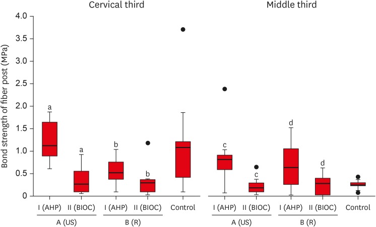
ePub Objective This study aimed to evaluate the effect of ultrasonic cleaning of the intracanal post space on the bond strength of fiber posts in oval canals filled with a premixed bioceramic (Bio-C Sealer [BIOC]) root canal sealer.
Materials and Methods Fifty premolars were endodontically prepared and divided into 5 groups (
n = 10), based on the type of root canal filling material used and the post space cleaning protocol. A1: gutta-percha + AH Plus (AHP) and post space preparation with ultrasonic cleaning, A2: gutta-percha + BIOC and post space preparation with ultrasonic cleaning, B1: gutta-percha + AHP and post space preparation, B2: gutta-percha + BIOC and post space preparation, C: control group. Fiber posts were cemented with a self-adhesive luting material, and 1 mm thick slices were sectioned from the middle and cervical third to evaluate the remaining filling material microscopically. The samples were subjected to a push-out test to analyze the bond strength of the fiber post, and the results were analyzed with the Shapiro-Wilk, Bonferroni, Kruskal-Wallis, and Mann-Whitney tests (p < 0.05). Failure modes were evaluated using optical microscopy.Results The results showed that the fiber posts cemented in canals sealed with BIOC had lower bond strength than those sealed with AHP. The ultrasonic cleaning of the post space improved the bond strength of fiber posts in canals sealed with AHP, but not with BIOC.
Conclusions BIOC decreased the bond strength of fiber posts in oval canals, regardless of ultrasonic cleaning.
-
Citations
Citations to this article as recorded by- Cleaning protocols to enhance bond strength of fiberglass posts on root canals filled with bioceramic sealer: an in vitro comparative study
Thiago Bessa Marconato Antunes, Juliana Delatorre Bronzato, Joice Graciani, Ana Cristina Padilha Janini, Rocharles Cavalcante Fontenele, Francisco Haiter Neto, Brenda Paula Figueiredo de Almeida Gomes, Marina Angélica Marciano da Silva
Restorative Dentistry & Endodontics.2025; 50(2): e20. CrossRef - In Vitro Effect of Using Endo‐Activator on Pushout Bond Strength of Radicular Dentin to Prefabricated Fiber Post in Using Natural Matrix Metalloproteinase Inhibitors
Nadia Elyassi Gorji, Homayoun Alaghemand, Faraneh Mokhtarpour, Elham Mahmodnia
Clinical and Experimental Dental Research.2025;[Epub] CrossRef - Evaluation of different mechanical cleaning protocols associated with 2.5% sodium hypochlorite in the removal of residues from the post space
Matheus Sousa Vitória, Eran Nair Mesquita de Almeida, Antonia Patricia Oliveira Barros, Eliane Cristina Gulin de Oliveira, Joatan Lucas de Sousa Gomes Costa, Andrea Abi Rached Dantas, Milton Carlos Kuga
Journal of Conservative Dentistry and Endodontics.2024; 27(3): 274. CrossRef - Fiber post cemented using different adhesive strategies to root canal dentin obturated with calcium silicate-based sealer
Lalita Patthanawijit, Kallaya Yanpiset, Pipop Saikaew, Jeeraphat Jantarat
BMC Oral Health.2024;[Epub] CrossRef - Effect of endodontic sealers on push-out bond strength of CAD-CAM or prefabricated fiber glass posts
Andréa Pereira de Souza PINTO, Fabiana Mantovani Gomes FRANÇA, Roberta Tarkany BASTING, Cecilia Pedroso TURSSI, José Joatan RODRIGUES JÚNIOR, Flávia Lucisano Botelho AMARAL
Brazilian Oral Research.2023;[Epub] CrossRef - Effect of mechanical cleaning protocols in the fiber post space on the adhesive interface between universal adhesive and root dentin
Gabriela Mariana Castro‐Núnez, José Rodolfo Estruc Verbicário dos Santos, Joissi Ferrari Zaniboni, Wilfredo Gustavo Escalante‐Otárola, Thiago Soares Porto, Milton Carlos Kuga
Microscopy Research and Technique.2022; 85(6): 2131. CrossRef - Effect of bioceramic root canal sealers on the bond strength of fiber posts cemented with resin cements
Rafael Nesello, Isadora Ames Silva, Igor Abreu De Bem, Karolina Bischoff, Matheus Albino Souza, Marcus Vinícius Reis Só, Ricardo Abreu Da Rosa
Brazilian Dental Journal.2022; 33(2): 91. CrossRef - Effect of irrigation protocols on root canal wall after post preparation: a micro-CT and microhardness study
Camila Maria Peres de Rosatto, Danilo Cassiano Ferraz, Lilian Vieira Oliveira, Priscilla Barbosa Ferreira Soares, Carlos José Soares, Mario Tanomaru Filho, Camilla Christian Gomes Moura
Brazilian Oral Research.2021;[Epub] CrossRef
- Cleaning protocols to enhance bond strength of fiberglass posts on root canals filled with bioceramic sealer: an in vitro comparative study
- 1,997 View
- 24 Download
- 8 Crossref

- A micro-computed tomographic evaluation of root canal filling with a single gutta-percha cone and calcium silicate sealer
- Jong Cheon Kim, Maung Maung Kyaw Moe, Sung Kyo Kim
- Restor Dent Endod 2020;45(2):e18. Published online February 12, 2020
- DOI: https://doi.org/10.5395/rde.2020.45.e18
-
 Abstract
 PDF
PDF PubReader
 ePub
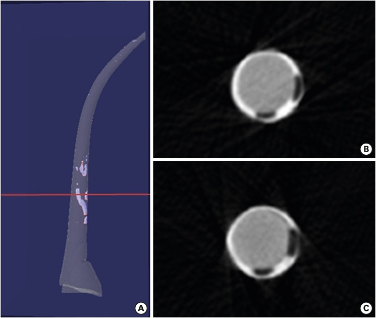
ePub Objectives The purpose of this study was to evaluate the void of root canal filling over time when a calcium silicate sealer was used in the single gutta-percha cone technique.
Materials and Methods Twenty-four J-shaped simulated root canals and twenty-four palatal root canals from extracted human maxillary molars were instrumented with ProFile Ni-Ti rotary instruments up to size 35/0.06 or size 40/0.06, respectively. Half of the canals were filled with Endoseal MTA and the other half were with AH Plus Jet using the single gutta-percha cone technique. Immediately after and 4 weeks after the root canal filling, the samples were scanned using micro-computed tomography at a resolution of 12.8 μm. The scanned images were reconstructed using the NRecon software and the void percentages were calculated using the CTan software, and statistically analyzed by 1-way analysis of variance, paired
t- test and Tukeypost hoc test.Results After 4 weeks, there were no significant changes in the void percentages at all levels in both material groups (
p > 0.05), except at the apical level of the AH Plus Jet group (p < 0.05) in the simulated root canal showing more void percentage compared to other groups. Immediately after filling the extracted human root canals, the Endoseal MTA group showed significantly less void percentage compared to the AH Plus Jet group (p < 0.05).Conclusions Under the limitations of this study, the Endoseal MTA does not seem to reduce the voids over time.
-
Citations
Citations to this article as recorded by- Comparative analysis between resin-based root canal sealer and recent bioceramic-based root canal sealers using MicroCT, film thickness, and solubility
Amira Galal Ismail, Manar M. Galal, Tamer M. Hamdy
Journal of Oral Biology and Craniofacial Research.2026; 16(2): 101400. CrossRef - Evaluation of various obturation techniques with bioceramic sealers in 3D-printed C-shaped canals
Maryam Gharechahi, Melika Hoseinzadeh, Saeed Moradi, Mina Mehrjouei
BMC Oral Health.2024;[Epub] CrossRef - Comparison of obturation quality in natural and replica teeth root-filled using different sealers and techniques
Chuta Kooanantkul, Richard M Shelton, Josette Camilleri
Clinical Oral Investigations.2023; 27(5): 2407. CrossRef - Obturation canalaire
N. Linas, M.-L. Munoz-Sanchez, N. Decerle, P.-Y. Cousson
EMC - Médecine buccale.2023; 16(5): 1. CrossRef - The Effect of Sealer Application Methods on Voids Volume after Aging of Three Calcium Silicate-Based Sealers: A Micro-Computed Tomography Study
Amre R. Atmeh, Rakan Alharbi, Ibrahim Aljamaan, Abdulrahman Alahmari, Ashwin C. Shetty, Ahmed Jamleh, Imran Farooq
Tomography.2022; 8(2): 778. CrossRef - Clinical Efficacy of Sealer-based Obturation Using Calcium Silicate Sealers: A Randomized Clinical Trial
Ji-hyung Kim, Sin-Yeon Cho, Yoonwoo Choi, Do-hyun Kim, Su-Jung Shin, Il-Young Jung
Journal of Endodontics.2022; 48(2): 144. CrossRef - A critical analysis of research methods and experimental models to study root canal fillings
Gustavo De‐Deus, Erick Miranda Souza, Emmanuel João Nogueira Leal Silva, Felipe Gonçalves Belladonna, Marco Simões‐Carvalho, Daniele Moreira Cavalcante, Marco Aurélio Versiani
International Endodontic Journal.2022; 55(S2): 384. CrossRef - Calcium Silicate Cements vs. Epoxy Resin Based Cements: Narrative Review
Mario Dioguardi, Cristian Quarta, Diego Sovereto, Giuseppe Troiano, Khrystyna Zhurakivska, Maria Bizzoca, Lorenzo Lo Muzio, Lucio Lo Russo
Oral.2021; 1(1): 23. CrossRef - Physico-Chemical Properties of Calcium-Silicate vs. Resin Based Sealers—A Systematic Review and Meta-Analysis of Laboratory-Based Studies
Viresh Chopra, Graham Davis, Aylin Baysan
Materials.2021; 15(1): 229. CrossRef - Micro-computed tomography in preventive and restorative dental research: A review
Mehrsima Ghavami-Lahiji, Reza Tayefeh Davalloo, Gelareh Tajziehchi, Paria Shams
Imaging Science in Dentistry.2021; 51(4): 341. CrossRef - Main and Accessory Canal Filling Quality of a Premixed Calcium Silicate Endodontic Sealer According to Different Obturation Techniques
Su-Yeon Ko, Hae Won Choi, E-Deun Jeong, Vinicius Rosa, Yun-Chan Hwang, Mi-Kyung Yu, Kyung-San Min
Materials.2020; 13(19): 4389. CrossRef
- Comparative analysis between resin-based root canal sealer and recent bioceramic-based root canal sealers using MicroCT, film thickness, and solubility
- 2,096 View
- 17 Download
- 11 Crossref

- A micro-computed tomographic study of remaining filling materials of two bioceramic sealers and epoxy resin sealer after retreatment
- KyungJae Kim, Da Vin Kim, Sin-Young Kim, SungEun Yang
- Restor Dent Endod 2019;44(2):e18. Published online April 26, 2019
- DOI: https://doi.org/10.5395/rde.2019.44.e18
-
 Abstract
 PDF
PDF PubReader
 ePub
ePub Objective This study evaluated the presence of residual root canal filling material after retreatment using micro-computed tomography (micro-CT).
Materials and Methods Extracted human teeth (single- and double-rooted,
n = 21/each; C-shaped,n = 15) were prepared with ProFile and randomly assigned to three subgroups for obturation with gutta-percha and three different sealers (EndoSeal MTA, EndoSequence BC sealer, and AH Plus). After 10 days, the filling material was removed and the root canals were instrumented one size up from the previous master apical file size. The teeth were scanned using micro-CT before and after retreatment. The percentage of remaining filling material after retreatment was calculated at the coronal, middle, and apical thirds. Data were analyzed using the Kruskal-Wallis test and Mann-WhitneyU test with Bonferronipost hoc correction.Results The tested sealers showed no significant differences in the percentage of remaining filling material in single- and double-rooted teeth, although EndoSeal MTA showed the highest value in C-shaped roots (
p < 0.05). The percentage of remaining filling material of AH Plus and EndoSeal MTA was significantly higher in C-shaped roots than in single- or double-roots (p < 0.05), while that of BC sealer was similar across all root types. EndoSeal MTA showed the highest values at the apical thirds of single- and double-roots (p < 0.05); otherwise, no significant differences were observed among the coronal, middle, and apical thirds.Conclusions Within the limitations of this study, a large amount of EndoSeal MTA remained after retreatment, especially in C-shaped root canals.
-
Citations
Citations to this article as recorded by- Development of a deep neural network and empirical model for predicting local gas holdup profiles in bubble columns
Sebastián Uribe, Ahmed Alalou, Mario E. Cordero, Muthanna Al‐Dahhan
The Canadian Journal of Chemical Engineering.2025; 103(6): 2918. CrossRef - An In Vitro Comparison of Epoxy Resin Sealer Removal During Endodontic Retreatment
Prashant A Bondarde, Aditi S Patkar, Aishwarya R Pawar, Rukmini Pande, Akshata Deshpande, Rachana S Agrawal, Seema Gupta
Cureus.2025;[Epub] CrossRef - Calcium silicate-based sealers remnants in isthmuses of mesial roots of mandibular molars: an in vitro evaluation
David Saldanha de Brito Alencar, Ana Cristina Padilha Janini, Lauter Eston Pelepenko, Brenda Fornazaro Moraes, Francisco Haiter Neto, Marco Antonio Hungaro Duarte, Marina Angélica Marciano
Restorative Dentistry & Endodontics.2025; 50(3): e25. CrossRef - Push-out bond strength of two endodontic sealers in retreated canals using different solvents
Sara Gamal Ghanem, Walaa M. Ghoneim, Ahmed H. Labib
Tanta Dental Journal.2025; 22(3): 504. CrossRef - Assessing Volume of Two Sealers’ Remnants after Reinstrumentation Using 3D Imaging Technology: An In Vitro Comparative Study
Khalel Mutaz Dawod, Raghad Abdulrazzaq Al-Hashimi
The Journal of Contemporary Dental Practice.2025; 26(8): 743. CrossRef - Removal efficacy of two different root canal sealers in retrograde cavities: a micro-CT study
Özge Başar, Ahter Şanal Çıkman, Cangül Keskin
BMC Oral Health.2025;[Epub] CrossRef - Evaluation of the retreatability of bioceramic root canal sealers with various formulations in simulated grooves
Meltem Sümbüllü, Afzal Ali, Abdulaziz Bakhsh, Hakan Arslan
PeerJ.2025; 13: e20398. CrossRef - Root canal cleanliness and debris extrusion following retreatment of thermoplastic injection technique and bioceramic-based root canal sealer
Deniz Bender, Mert Ocak, Emel Uzunoğlu Özyürek
Clinical Oral Investigations.2024;[Epub] CrossRef - The Effect of Different Obturation Techniques Using Different Root Canal Sealers on the Residual Filling Material After Retreatment Procedures
M Sarı, K Yılmaz
Nigerian Journal of Clinical Practice.2024; 27(2): 174. CrossRef - Effect of Different Obturation Techniques on the Amount of Debris Extrusion During Endodontic Retreatment Using XP Endo Retreatment Set Files (In vitro Study)
Pawan Mohamad Amin, Hawzhen Mohammed Saeed
Sulaimani Dental Journal.2023; 10: 49. CrossRef - The efficiency of different irrigation activation techniques in the removal of calcium silicate‐based endodontic sealer from artificially created groove
Meltem Sümbüllü, Afzal Ali, Mine Büker, Hakan Arslan
Australian Endodontic Journal.2023; 49(S1): 238. CrossRef - Efficiency of diode laser and ultrasonic‐activated irrigation in retreatment of gutta percha and bioceramic sealer: An in vitro study
Rahaf A. Almohareb, Reem M. Barakat, Noor Aljarallah, Halah Mudhish, Amjaad Almutairi, Fahda N. Algahtani
Australian Endodontic Journal.2023; 49(2): 318. CrossRef - Efficiency of the new reciprocating and rotary systems with or without ultrasonics in removing root-canals filling with calcium silicate-based sealer (MTA)
Ahmad A. Madarati, Aya M. N. Sammani, Ahmad A. Alnazzawi, Ali Alrahlah
BMC Oral Health.2023;[Epub] CrossRef - Retreatability of calcium silicate‐based root canal sealer using reciprocating instrumentation with different irrigation activation techniques in single‐rooted canals
Daniele Angerame, Matteo De Biasi, Davide Porrelli, Lorenzo Bevilacqua, Riccardo Zanin, Matteo Olivi, Vassilios Kaitsas, Giovanni Olivi
Australian Endodontic Journal.2022; 48(3): 415. CrossRef - Critical analysis of research methods and experimental models to study removal of root filling materials
Mahdi A. Ajina, Pratik K. Shah, Bun San Chong
International Endodontic Journal.2022; 55(S1): 119. CrossRef - An Updated Review on Properties and Indications of Calcium Silicate‐Based Cements in Endodontic Therapy
Fateme Eskandari, Alireza Razavian, Rozhina Hamidi, Khadije Yousefi, Susan Borzou, Zohaib Khurshid
International Journal of Dentistry.2022;[Epub] CrossRef - How do imaging protocols affect the assessment of root-end fillings?
Fernanda Ferrari Esteves Torres, Reinhilde Jacobs, Mostafa EzEldeen, Karla de Faria-Vasconcelos, Juliane Maria Guerreiro-Tanomaru, Bernardo Camargo dos Santos, Mário Tanomaru-Filho
Restorative Dentistry & Endodontics.2022;[Epub] CrossRef - The Efficacy of Er:YAG Laser-Activated Shock Wave-Enhanced Emission Photoacoustic Streaming Compared to Ultrasonically Activated Irrigation and Needle Irrigation in the Removal of Bioceramic Filling Remnants from Oval Root Canals—An Ex Vivo Study
Gabrijela Kapetanović Petričević, Marko Katić, Valentina Brzović Rajić, Ivica Anić, Ivona Bago
Bioengineering.2022; 9(12): 820. CrossRef - An in vitro comparative evaluation of retreatability of a bioceramic and resin sealer using cone-beam computed tomography analysis
Sumit Sharma, Ramya Raghu, Ashish Shetty, Subhashini Rajasekhara, Harika Lakshmisetty, G. Bharath
Endodontology.2022; 34(3): 173. CrossRef - Positive and negative properties of four endodontic sealant groups: a systematic review
E. V. Chestnyh, I. O. Larichkin, M. V. Iusufova, D. I. Oreshkina, E. I. Oreshkina, V. S. Minakova, S. V. Plekhanova
Kuban Scientific Medical Bulletin.2021; 28(3): 130. CrossRef - Retrievability of bioceramic-based sealers in comparison with epoxy resin-based sealer assessed using microcomputed tomography: A systematic review of laboratory-based studies
Buvaneshwari Arul, Aswathi Varghese, Anisha Mishra, Subashini Elango, Sairathna Padmanaban, Velmurugan Natanasabapathy
Journal of Conservative Dentistry.2021; 24(5): 421. CrossRef - Micro CT pilot evaluation of removability of two endodontic sealers
David Colmenar, Tenzin Tamula, Qiang Zhu, Chul Ahn, Carolyn Primus, Takashi Komabayashi
Journal of Oral Science.2021; 63(4): 306. CrossRef - Comparison of Obturation Quality between Calcium Silicate-Based Sealers and Resin-Based Sealers for Endodontic Re-treatment
Hye-Ryeon Jin, Young-Eun Jang, Yemi Kim
Materials.2021; 15(1): 72. CrossRef - Micro-computed tomographic evaluation of a new system for root canal filling using calcium silicate-based root canal sealers
Mario Tanomaru-Filho, Fernanda Ferrari Esteves Torres, Jader Camilo Pinto, Airton Oliveira Santos-Junior, Karina Ines Medina Carita Tavares, Juliane Maria Guerreiro-Tanomaru
Restorative Dentistry & Endodontics.2020;[Epub] CrossRef - Micro-computed tomographic evaluation of the flow and filling ability of endodontic materials using different test models
Fernanda Ferrari Esteves Torres, Juliane Maria Guerreiro-Tanomaru, Gisselle Moraima Chavez-Andrade, Jader Camilo Pinto, Fábio Luiz Camargo Villela Berbert, Mario Tanomaru-Filho
Restorative Dentistry & Endodontics.2020;[Epub] CrossRef - Retreatment efficacy of hydraulic calcium silicate sealers used in single cone obturation
M. Garrib, J. Camilleri
Journal of Dentistry.2020; 98: 103370. CrossRef
- Development of a deep neural network and empirical model for predicting local gas holdup profiles in bubble columns
- 2,206 View
- 23 Download
- 26 Crossref

- Bacterial leakage and micro-computed tomography evaluation in round-shaped canals obturated with bioceramic cone and sealer using matched single cone technique
- Kallaya Yanpiset, Danuchit Banomyong, Kanet Chotvorrarak, Ratchapin Laovanitch Srisatjaluk
- Restor Dent Endod 2018;43(3):e30. Published online July 5, 2018
- DOI: https://doi.org/10.5395/rde.2018.43.e30
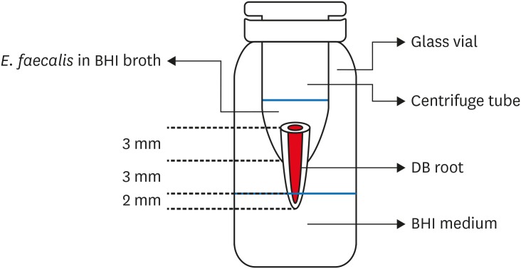
-
 Abstract
 PDF
PDF PubReader
 ePub
ePub Objectives To evaluate sealing ability of root canals obturated with bioceramic-impregnated gutta percha cone (BCC) or gutta percha (GP), with bioceramic sealer (BCS) or AH Plus (AH; Dentsply-Maillefer), in roundly-prepared canals using matched single-cone technique, based on bacterial leakage test, and to analyze obturation quality using micro-computed tomography (CT) analysis.
Materials and Methods Ninety-two distobuccal roots of maxillary molars were prepared using nickel-titanium files to apical size 40/0.06. The roots were divided into 4 groups (
n = 20) that were obturated with a master cone and sealer: GP/AH, BCC/AH, GP/BCS, and BCC/BCS. Bacterial leakage model usingEnterococcus faecalis was used to evaluate sealing ability for 60-day period. Obturated samples from each group (n = 4) were analyzed using micro-CT.Results All groups showed bacterial leakage at 20%–45% of samples with mean leakage times of 42–52 days. There were no significant differences in bacterial leakage among the groups. Micro-CT showed minimal gaps and voids in all groups at less than 1%.
Conclusions In roundly-prepared canals, the single cone obturation with BCC/BCS was comparable to GP/AH for bacterial leakage at 60 days.
-
Citations
Citations to this article as recorded by- Effect of Root Dentin Moisture on the Apical Sealing Ability of Root Canal Sealers: In vitro Study
Zahraa Khalil Alani, Manal Hussain Abd-alla
Al-Rafidain Journal of Medical Sciences ( ISSN 2789-3219 ).2025; 8(2): 122. CrossRef - Synthesis, physical properties, and root canal sealing of experimental MTA- and salicylate-based root canal sealers
Rafael Pino Vitti, Kusai Baroudi, Tarun Walia, Raghavandra M. Shetty, Flávia Goulart da Rosa Cardoso, Flávia de Moura Pereira, Evandro Piva, Cesar Henrique Zanchi, Gabriel Flores Abuna, Carolina Oliveira de Lima, Emmanuel João Nogueira Leal Silva, Flávio
PLOS One.2025; 20(7): e0329476. CrossRef - Impact of cone system compatibility on single cone bioceramic obturation in canals prepared with variable taper NiTi rotary files
Reem M. Barakat, Rahaf A. Almohareb, Njoom Aleid, Hoor Almowais, Aljawhara Alharbi, Meshal Al-Sharafa, Ali Alrahlah
Scientific Reports.2025;[Epub] CrossRef - Estudio de la obturación con selladores biocerámicos de conductos radiculares de premolares inferiores
Alicia Beatriz Bonafé, Cecilia Inés Rourera, Carla Pedraza, Yamila Victoria Zanoni, Soledad Salduna, Cecilia Noemi De Caso, Gabriela Martín
Methodo Investigación Aplicada a las Ciencias Biológicas.2025; 10(3): 31. CrossRef - Sealing ability of mineral trioxide aggregate: A scoping review of laboratory assessment methods
Kenta Tsuchiya, Salvatore Sauro, Jukka P. Matinlinna, Hidehiko Sano, Monica Yamauti, Deepak Mehta, Kyung‐San Min, Atsushi Tomokiyo
European Journal of Oral Sciences.2025;[Epub] CrossRef - Bacterial Leakage Testing in Dentistry: A Comprehensive Review on Methods, Models, and Clinical Relevance
Niher Tabassum Snigdha, Mohmed Isaqali Karobari, Sukhamoy Gorai
Scientifica.2025;[Epub] CrossRef - In vitro comparative evaluation of apical leakage using a bioceramic sealer with three different obturating techniques: A glucose leakage model
Tanvi S Agrawal, Shishir Singh, Rajesh S Podar, Gaurav Kulkarni, Anuprita Gadkari, Navin Agarwal
Journal of Conservative Dentistry and Endodontics.2024; 27(1): 76. CrossRef - In Vitro Microscopical and Microbiological Assessment of the Sealing Ability of Calcium Silicate-Based Root Canal Sealers
Karin Christine Huth, Sabina Noreen Wuersching, Leander Benz, Stefan Kist, Maximilian Kollmuss
Journal of Functional Biomaterials.2024; 15(11): 341. CrossRef - Comparison between AH plus sealer and total fill bioceramic sealer performance in previously untreated and retreatment cases of maxillary incisors with large-sized periapical lesion: a randomized controlled trial
Eisa Wahbi, Hassan Achour, Yasser Alsayed Tolibah
BDJ Open.2024;[Epub] CrossRef - Bacterial sealing ability of calcium silicate-based sealer for endodontic surgery: an in-vitro study
Mai M. Mansour, Sybel M. Moussa, Marwa A. Meheissen, Mahmoud R. Aboelseoud
BMC Oral Health.2024;[Epub] CrossRef - Assessment the bioactivity of zinc oxid eugenol sealer after the addition of different concentrations of nano hydroxyapatite-tyrosine amino acid
Rasha M. Al-Shamaa, Raghad A. Al-Askary
Brazilian Journal of Oral Sciences.2024; 23: e243733. CrossRef - Assessment of Bacterial Sealing Ability of Two Different Bio-Ceramic Sealers in Single-Rooted Teeth Using Single Cone Obturation Technique: An In Vitro Study
Doaa M. AlEraky, Ahmed M. Rahoma, Hatem M. Abuohashish, Abdullh AlQasser, Abbas AlHamali, Hussain M. AlHussain, Hussain M. AlShoalah, Zakrya AlSaghah, Abdulrahman Khattar, Shimaa Rifaat
Applied Sciences.2023; 13(5): 2906. CrossRef - How do imaging protocols affect the assessment of root-end fillings?
Fernanda Ferrari Esteves Torres, Reinhilde Jacobs, Mostafa EzEldeen, Karla de Faria-Vasconcelos, Juliane Maria Guerreiro-Tanomaru, Bernardo Camargo dos Santos, Mário Tanomaru-Filho
Restorative Dentistry & Endodontics.2022;[Epub] CrossRef - The impact of Morse taper implant design on microleakage at implant-healing abutment interface
Soyeon KIM, Joo Won LEE, Jae-Heon KIM, Van Mai TRUONG, Young-Seok PARK
Dental Materials Journal.2022; 41(5): 767. CrossRef - A critical analysis of research methods and experimental models to study root canal fillings
Gustavo De‐Deus, Erick Miranda Souza, Emmanuel João Nogueira Leal Silva, Felipe Gonçalves Belladonna, Marco Simões‐Carvalho, Daniele Moreira Cavalcante, Marco Aurélio Versiani
International Endodontic Journal.2022; 55(S2): 384. CrossRef - Micro‐CT assessment of gap‐containing areas along the gutta‐percha‐sealer interface in oval‐shaped canals
Gustavo De‐Deus, Gustavo O. Santos, Iara Zamboni Monteiro, Daniele M. Cavalcante, Marco Simões‐Carvalho, Felipe G. Belladonna, Emmanuel J. N. L. Silva, Erick M. Souza, Raphael Licha, Carla Zogheib, Marco A. Versiani
International Endodontic Journal.2022; 55(7): 795. CrossRef - Comparison of Sealing Ability of Bioceramic Sealer, AH Plus, and GuttaFlow in Conservatively Prepared Curved Root Canals Obturated with Single-Cone Technique: An In vitro Study
Shalan Kaul, Ajay Kumar, Bhumika Kamal Badiyani, Laxmi Sukhtankar, M. Madhumitha, Amit Kumar
Journal of Pharmacy and Bioallied Sciences.2021; 13(Suppl 1): S857. CrossRef - Micro-CT Evaluation of Four Root Canal Obturation Techniques
Mahmood Reza Kalantar Motamedi, Amin Mortaheb, Maryam Zare Jahromi, Brett E. Gilbert, Marilena Vivona
Scanning.2021; 2021: 1. CrossRef - Effects of Both Fiber Post/Core Resin Construction System and Root Canal Sealer on the Material Interface in Deep Areas of Root Canal
Hiroki Miura, Shinji Yoshii, Masataka Fujimoto, Ayako Washio, Takahiko Morotomi, Hiroshi Ikeda, Chiaki Kitamura
Materials.2021; 14(4): 982. CrossRef - Sealing ability and microbial leakage of root-end filling materials: MTA versus epoxy resin: A systematic review and meta-analysis
Mario Dioguardi, Mario Alovisi, Diego Sovereto, Giuseppe Troiano, Giancarlo Malagnino, Michele Di Cosola, Angela Pia Cazzolla, Luigi Laino, Lorenzo Lo Muzio
Heliyon.2021; 7(7): e07494. CrossRef - Development of A Nano-Apatite Based Composite Sealer for Endodontic Root Canal Filling
Angelica Bertacci, Daniele Moro, Gianfranco Ulian, Giovanni Valdrè
Journal of Composites Science.2021; 5(1): 30. CrossRef - BIOCERAMIC-BASED ROOT CANAL SEALERS
L Somolová, Z Zapletalová, M Rosa, B Novotná, I Voborná, Y Morozova
Česká stomatologie a praktické zubní lékařství.2021; 121(4): 116. CrossRef - Calcium Silicate-Based Root Canal Sealers: A Narrative Review and Clinical Perspectives
Germain Sfeir, Carla Zogheib, Shanon Patel, Thomas Giraud, Venkateshbabu Nagendrababu, Frédéric Bukiet
Materials.2021; 14(14): 3965. CrossRef - Physico-Chemical Properties of Calcium-Silicate vs. Resin Based Sealers—A Systematic Review and Meta-Analysis of Laboratory-Based Studies
Viresh Chopra, Graham Davis, Aylin Baysan
Materials.2021; 15(1): 229. CrossRef - Comparison of apical sealing ability of bioceramic sealer and epoxy resin-based sealer using the fluid filtration technique and scanning electron microscopy
Widcha Asawaworarit, Thitapa Pinyosopon, Kanittha Kijsamanmith
Journal of Dental Sciences.2020; 15(2): 186. CrossRef - Micro-computed tomographic evaluation of a new system for root canal filling using calcium silicate-based root canal sealers
Mario Tanomaru-Filho, Fernanda Ferrari Esteves Torres, Jader Camilo Pinto, Airton Oliveira Santos-Junior, Karina Ines Medina Carita Tavares, Juliane Maria Guerreiro-Tanomaru
Restorative Dentistry & Endodontics.2020;[Epub] CrossRef - A micro-computed tomographic evaluation of root canal filling with a single gutta-percha cone and calcium silicate sealer
Jong Cheon Kim, Maung Maung Kyaw Moe, Sung Kyo Kim
Restorative Dentistry & Endodontics.2020;[Epub] CrossRef - Comparative evaluation of sealing ability of gutta percha and resilon as root canal filling materials- a systematic review
Pragya Pandey, Himanshi Aggarwal, A.P. Tikku, Arpit Singh, Rhythm Bains, Shambhavi Mishra
Journal of Oral Biology and Craniofacial Research.2020; 10(2): 220. CrossRef - Micro-computed tomographic evaluation of the flow and filling ability of endodontic materials using different test models
Fernanda Ferrari Esteves Torres, Juliane Maria Guerreiro-Tanomaru, Gisselle Moraima Chavez-Andrade, Jader Camilo Pinto, Fábio Luiz Camargo Villela Berbert, Mario Tanomaru-Filho
Restorative Dentistry & Endodontics.2020;[Epub] CrossRef - Root fillings with a matched-taper single cone and two calcium silicate–based sealers: an analysis of voids using micro-computed tomography
Eugenio Pedullà, Roula S. Abiad, Gianluca Conte, Giusy R. M. La Rosa, Ernesto Rapisarda, Prasanna Neelakantan
Clinical Oral Investigations.2020; 24(12): 4487. CrossRef - Influence of different disinfection protocols on gutta-percha cones surface roughness assessed by two different methods
A.M. Nunes, J.P. Gouvea, L. da Silva
Journal of Materials Research and Technology.2019; 8(6): 5464. CrossRef - Endodontic sealers based on calcium silicates: a systematic review
David Donnermeyer, Sebastian Bürklein, Till Dammaschke, Edgar Schäfer
Odontology.2019; 107(4): 421. CrossRef
- Effect of Root Dentin Moisture on the Apical Sealing Ability of Root Canal Sealers: In vitro Study
- 2,320 View
- 35 Download
- 32 Crossref

- Retreatability of two endodontic sealers, EndoSequence BC Sealer and AH Plus: a micro-computed tomographic comparison
- Enrique Oltra, Timothy C. Cox, Matthew R. LaCourse, James D. Johnson, Avina Paranjpe
- Restor Dent Endod 2017;42(1):19-26. Published online December 8, 2016
- DOI: https://doi.org/10.5395/rde.2017.42.1.19

-
 Abstract
 PDF
PDF PubReader
 ePub
ePub Objectives Recently, bioceramic sealers like EndoSequence BC Sealer (BC Sealer) have been introduced and are being used in endodontic practice. However, this sealer has limited research related to its retreatability. Hence, the aim of this study was to evaluate the retreatability of two sealers, BC Sealer as compared with AH Plus using micro-computed tomographic (micro-CT) analysis.
Materials and Methods Fifty-six extracted human maxillary incisors were instrumented and randomly divided into 4 groups of 14 teeth: 1A, gutta-percha, AH Plus retreated with chloroform; 1B, gutta-percha, AH Plus retreated without chloroform; 2A, gutta-percha, EndoSequence BC Sealer retreated with chloroform; 2B, gutta-percha, EndoSequence BC Sealer retreated without chloroform. Micro-CT scans were taken before and after obturation and retreatment and analyzed for the volume of residual material. The specimens were longitudinally sectioned and digitized images were taken with the dental operating microscope. Data was analyzed using an ANOVA and a
post-hoc Tukey test. Fisher exact tests were performed to analyze the ability to regain patency.Results There was significantly less residual root canal filling material in the AH Plus groups retreated with chloroform as compared to the others. The BC Sealer samples retreated with chloroform had better results than those retreated without chloroform. Furthermore, patency could be re-established in only 14% of teeth in the BC Sealer without chloroform group.
Conclusion The results of this study demonstrate that the BC Sealer group had significantly more residual filling material than the AH Plus group regardless of whether or not both sealers were retreated with chloroform.
-
Citations
Citations to this article as recorded by- Retreatment of Bioceramic Sealers Using a Novel Solvent: Dissolution, Patency, and Dentinal Effects
Jemma Hanson, Michelle Wu, Ajay Shakya, Ryan Walsh, Poorya Jalali
Journal of Endodontics.2026; 52(2): 279. CrossRef - Laser-Activated versus Sonic-Activated Irrigation for Removing Residual Filling Materials in Mandibular Molars: A Micro-CT Study
Yuhong Lin, Fabricio Teixeira, Jianing He, Megan Yamaguchi, Alex Huynh, Poorya Jalali
Journal of Endodontics.2026;[Epub] CrossRef - The effect of different bioceramic root canal sealer removal protocols on the bond strength of composite to coronal dentin in diabetic and non-diabetic patients: an ex vivo study
Arzu Şahin Mantı, Cemile Kedici Alp
BMC Oral Health.2025;[Epub] CrossRef - Influence of the Filling Technique on Endodontic Retreatment in Curved Mesial Canals of Mandibular Molars: An In Vitro Study
Felipe Andretta Copelli, Lisa Yurie Oda, Renata Maira de Souza Leal, Clarissa Teles Rodrigues, Marco Antonio Hungaro Duarte, Bruno Cavalini Cavenago
Journal of Endodontics.2025; 51(6): 755. CrossRef - Efficacy of irrigation activation methods in removal of bioceramic-based sealer in retreatment
Büşra Nur Cıkrık, Selen İnce Yusufoğlu
Odontology.2025; 113(3): 1119. CrossRef - Patterns and Practices in the Use of Endodontic Materials: Insights from Romanian Dental Practices
Diana Marian, Ramona Amina Popovici, Iustin Olariu, Dana Emanuela Pitic (Cot), Maria-Monica Marta, Ioana Veja (Ilyes)
Applied Sciences.2025; 15(3): 1272. CrossRef - Microcracks induced by XP-endo retreatment system in root canals filled with bioceramic sealer: A micro-computed tomographic analysis
Sarah M. Alkahtany
The Saudi Dental Journal.2025;[Epub] CrossRef - Retrieval of AH Plus Bioceramic and Ceraseal Versus AH Plus in Endodontic Retreatment
Eurok Shim, Jee Woo Son, Jiyoung Kwon, Hyun-Jung Kim, Ji-Hyun Jang, Seok Woo Chang, Soram Oh
Journal of Clinical Medicine.2025; 14(6): 1826. CrossRef - Comparative Evaluation of Retreatability of Two Bioceramic Sealers and Epoxy Resin Sealer with Two Different File Systems: An In Vitro Cone Beam Computed Tomography Volumetric Analysis
Rashmi Nair, Riya Jain, Neetu Maurya, Neha D Singh, Labdhi Maloo, Shanu Khan
World Journal of Dentistry.2025; 16(1): 56. CrossRef - Comparative evaluation of the efficacy of three different retreatment files in removing root canal filling material: An In vitro confocal microscopy study
Meghna Sarah Abraham, Aravind R. Kudva, Prathap M. S. Nair, Shravan Kini, Samreena Kalander, Faseeh Muhammed Bin Farookh
Endodontology.2025; 37(2): 136. CrossRef - Comparative Evaluation of Removal of Gutta Percha and Bioceramic Sealer Using EdgeFile‐XR and ProTaper Universal Retreatment Files: Micro‐Computed Tomography Study
Sindhuja Srirama, Sooriaprakas Chandrasekaran, Buvaneshwari Arul, Velmurugan Natanasabapathy
Australian Endodontic Journal.2025; 51(2): 358. CrossRef - Achieving Patency in Straight Canals Obturated with AH Plus Bioceramic Sealer: An Ex Vivo Study
Inês Ferreira, Beatriz Fernandes, Ana Cristina Braga, Maria Ascensão Lopes, Irene Pina-Vaz
Applied Sciences.2025; 15(11): 5855. CrossRef - Cleaning protocols to enhance bond strength of fiberglass posts on root canals filled with bioceramic sealer: an in vitro comparative study
Thiago Bessa Marconato Antunes, Juliana Delatorre Bronzato, Joice Graciani, Ana Cristina Padilha Janini, Rocharles Cavalcante Fontenele, Francisco Haiter Neto, Brenda Paula Figueiredo de Almeida Gomes, Marina Angélica Marciano da Silva
Restorative Dentistry & Endodontics.2025; 50(2): e20. CrossRef - Surface Alterations of Ni‐Ti Files After Retreatment of Root Canals Filled With Different Sealers: AFM and SEM Study
Duygu Aksoy, Sibel Koçak, Mustafa Murat Koçak, Baran Can Sağlam
Microscopy Research and Technique.2025; 88(10): 2704. CrossRef - Resistance of gutta-percha and calcium silicate-based sealer to dislocation after non-surgical root canal retreatment
Reza Mahjourianqomi, Noushin Shokouhinejad, Fatemeh Hamidzadeh, Pegah Sarraf
BMC Oral Health.2025;[Epub] CrossRef - The Impact of Different Access Cavity Designs on the Retreatment Procedure of Single Oval Canals Using Reciprocating File Systems: An Ex-Vivo Study
Samia Elsherief, Sahar M Elmarsafy, Nora Abdelgawad, Haytham Sabri Abdul Hameed Jaha
The Open Dentistry Journal.2025;[Epub] CrossRef - Retrievability of NeoMTA 2 vs AH Plus Sealer from Retreated Mesial Canals of Mandibular First Molars: A Microcomputed Tomography Ex Vivo Study
Mey A Al-Habib, Mona Alsulaiman
The Journal of Contemporary Dental Practice.2025; 26(5): 493. CrossRef - Efficacy of self-adjusting file and E18 isthmus ultrasonic tip as supplementary instruments in removal of gutta-percha and bioceramic sealer from oval canals in endodontic retreatment: A cone-beam computed tomography study
Joel George Thomas Madolil, I. Sujatha, K. B. Jayalakshmi, Deena Elizabeth, B. Sowmya, Bhargavi Krishnaraj
Endodontology.2025; 37(3): 276. CrossRef - Calcium silicate-based sealers remnants in isthmuses of mesial roots of mandibular molars: an in vitro evaluation
David Saldanha de Brito Alencar, Ana Cristina Padilha Janini, Lauter Eston Pelepenko, Brenda Fornazaro Moraes, Francisco Haiter Neto, Marco Antonio Hungaro Duarte, Marina Angélica Marciano
Restorative Dentistry & Endodontics.2025; 50(3): e25. CrossRef - Thermal Behaviour of Teeth With Internal Root Resorption During Obturation and Enhancing Thermal Simulations: A Finite-Element Analysis
Alper Kabakci, Ayca Yilmaz, Dilek Helvacioglu-Yigit, Nawar Naguib Nawar, Hyeon-Cheol Kim
International Dental Journal.2025; 75(6): 103903. CrossRef - Assessing Volume of Two Sealers’ Remnants after Reinstrumentation Using 3D Imaging Technology: An In Vitro Comparative Study
Khalel Mutaz Dawod, Raghad Abdulrazzaq Al-Hashimi
The Journal of Contemporary Dental Practice.2025; 26(8): 743. CrossRef - Comparative analysis of the push-out bond strength of fiber posts: Immediate vs. delayed post-space preparation with two obturation techniques
Weilin Long, Xiongjun Xu, Li Tang, Hongwei Jiang, Yihua Huang, Miriam Fatima Zaccaro Scelza
PLOS One.2025; 20(10): e0333880. CrossRef - Removal efficacy of two different root canal sealers in retrograde cavities: a micro-CT study
Özge Başar, Ahter Şanal Çıkman, Cangül Keskin
BMC Oral Health.2025;[Epub] CrossRef - In Vitro and Ex Vivo Evaluation of a Novel Solvent for Tricalcium Silicate-based Sealers
David Colmenar, Saaya Sakoda, Tanguy Terlier, Takashi Komabayashi, Timothy Kirkpatrick, Ji Wook Jeong
Journal of Endodontics.2025;[Epub] CrossRef - Marginal adaptation of customized gutta percha cone with calcium silicate based sealer versus MTA and biodentine apical plugs in simulated immature permanent teeth (an in vitro study)
Mary M. Mina, Sybel M. Moussa, Mahmoud R. Aboelseoud
BMC Oral Health.2024;[Epub] CrossRef - Retreatability of Bioceramic-Filled Teeth: Comparative Analysis of Single-Cone and Carrier-Based Obturation Using a Reciprocating Technique
Andrea Spinelli, Fausto Zamparini, Jacopo Lenzi, Davide Carboni, Maria Giovanna Gandolfi, Carlo Prati
Applied Sciences.2024; 14(15): 6444. CrossRef - Retreatability of calcium silicate-based sealers based on micro-computed tomographic evaluation − A systematic review
Sundus Mohammed Bukhary
The Saudi Dental Journal.2024; 36(10): 1278. CrossRef - Bioceramic and Resin-Based Sealers Removal Using XP-Endo Finisher: A Scanning Electron Microscopy Study
Meriem Fejjeri, Kawther Bel Haj Salah, Sabra Jaafoura, Saida Sahtout
European Journal of General Dentistry.2024; 13(02): 110. CrossRef - Comparative Evaluation of the Efficacy of Different Solvents on the Removal of Endodontic Bioceramic Sealers: An In vitro Study
Ruaa A. Alamoudi
Journal of the International Clinical Dental Research Organization.2024; 16(2): 126. CrossRef - The Efficiency of Different Supplementary Irrigation Techniques After Nickel-Titanium Rotary System in Endodontic Retreatment
Selin Goker Kamali, Dilek Turkaydin
European Journal of Therapeutics.2024; 30(6): 859. CrossRef - Evaluation of canal patency and cleanliness following retreatment of bioceramic sealer‐obturated root canals using three different irrigant activation protocols
Daiasharailang Lyngdoh, Sharique Alam, Huma Iftekhar, Surendra Kumar Mishra
Australian Endodontic Journal.2024; 50(3): 475. CrossRef - Optimizing Non-surgical Endodontic Retreatment: A 3D CBCT Quantification of Root Canal Bioceramic Filling Material Removal
Kostadin Zhekov, Vesela Stefanova
The Open Dentistry Journal.2024;[Epub] CrossRef - The Effect of Different Obturation Techniques Using Different Root Canal Sealers on the Residual Filling Material After Retreatment Procedures
M Sarı, K Yılmaz
Nigerian Journal of Clinical Practice.2024; 27(2): 174. CrossRef - Dentinal Tubule Penetrability and Bond Strength of Two Novel Calcium Silicate-Based Root Canal Sealers
Karissa Shieh, Jack Yang, Elsa Heng Zhu, Ove Andreas Peters, Sepanta Hosseinpour
Materials.2023; 16(9): 3309. CrossRef - Efficiency of diode laser and ultrasonic‐activated irrigation in retreatment of gutta percha and bioceramic sealer: An in vitro study
Rahaf A. Almohareb, Reem M. Barakat, Noor Aljarallah, Halah Mudhish, Amjaad Almutairi, Fahda N. Algahtani
Australian Endodontic Journal.2023; 49(2): 318. CrossRef - The efficiency of different irrigation activation techniques in the removal of calcium silicate‐based endodontic sealer from artificially created groove
Meltem Sümbüllü, Afzal Ali, Mine Büker, Hakan Arslan
Australian Endodontic Journal.2023; 49(S1): 238. CrossRef - Global survey of endodontic practice and adoption of newer technologies
Monique Charlene Cheung, Ove Andreas Peters, Peter Parashos
International Endodontic Journal.2023; 56(12): 1517. CrossRef - Efficiency of the new reciprocating and rotary systems with or without ultrasonics in removing root-canals filling with calcium silicate-based sealer (MTA)
Ahmad A. Madarati, Aya M. N. Sammani, Ahmad A. Alnazzawi, Ali Alrahlah
BMC Oral Health.2023;[Epub] CrossRef - Effect of endodontic sealers on push-out bond strength of CAD-CAM or prefabricated fiber glass posts
Andréa Pereira de Souza PINTO, Fabiana Mantovani Gomes FRANÇA, Roberta Tarkany BASTING, Cecilia Pedroso TURSSI, José Joatan RODRIGUES JÚNIOR, Flávia Lucisano Botelho AMARAL
Brazilian Oral Research.2023;[Epub] CrossRef - Microcomputed tomographic analysis of the efficiency of two retreatment techniques in removing root canal filling materials from mandibular incisors
Xueqin Yang, Ye Wang, Mengzhen Ji, Yanyao Li, Hao Wang, Tao Luo, Yuan Gao, Ling Zou
Scientific Reports.2023;[Epub] CrossRef - Effectiveness of Two Endodontic Instruments in Calcium Silicate-Based Sealer Retreatment
Antoun Farrayeh, Samar Akil, Ammar Eid, Valentina Macaluso, Davide Mancino, Youssef Haïkel, Naji Kharouf
Bioengineering.2023; 10(3): 362. CrossRef - Clinical applications of calcium silicate‐based materials: a narrative review
S Küçükkaya Eren
Australian Dental Journal.2023;[Epub] CrossRef - Retreatability of Bioceramic Sealer Using One Curve Rotary File Assessed by Microcomputed Tomography
Dina G Mufti, Saad A Al-Nazhan
The Journal of Contemporary Dental Practice.2022; 22(10): 1175. CrossRef - Medium- and Long-Term Re-Treatment of Root Canals Filled with a Calcium Silicate-Based Sealer: An Experimental Ex Vivo Study
Giulia Bardini, Elisabetta Cotti, Terenzio Congiu, Claudia Caria, Davide Aru, Montse Mercadè
Materials.2022; 15(10): 3501. CrossRef - An in vitro comparative evaluation of retreatability of a bioceramic and resin sealer using cone-beam computed tomography analysis
Sumit Sharma, Ramya Raghu, Ashish Shetty, Subhashini Rajasekhara, Harika Lakshmisetty, G. Bharath
Endodontology.2022; 34(3): 173. CrossRef - Removal of the Previous Root Canal Filling Material for Retreatment: Implications and Techniques
Flávio R. F. Alves, Isabela N. Rôças, José C. Provenzano, José F. Siqueira
Applied Sciences.2022; 12(20): 10217. CrossRef - Efficacy of retreatment NiTi files for root canals filled with calcium silicate-based sealer
Jae-Yun Hyun, Kyung-Mo Cho, Se-Hee Park, Yoon Lee, Yoon-Joo Lee, Jin-Woo Kim
Journal of Dental Rehabilitation and Applied Science.2022; 38(4): 213. CrossRef - Dissolving efficacy of xylene on epoxy resin-based and bioceramic-based root canal sealers
Cindy Willie
Scientific Dental Journal.2022; 6(1): 32. CrossRef - Comparison of the efficacy of three different supplementary cleaning protocols in root-filled teeth with a bioceramic sealer after retreatment—a micro-computed tomographic study
Chanakarn Sinsareekul, Sirawut Hiran-us
Clinical Oral Investigations.2022; 26(4): 3515. CrossRef - The efficacy of different sealer removal protocols on the microtensile bond strength of adhesives to a bioceramic sealer-contaminated dentin
ZG Bek Kurklu, HO Yoldas
Nigerian Journal of Clinical Practice.2022; 25(3): 336. CrossRef - A micro-computed tomographic analysis of obturation quality and retreatability of an epoxy resin-based sealer
Roula S. ABIAD, Prasanna NEELAKANTAN, Ivan BUSCEMA, Islam A. ALI, Gianluca CONTE, Giusy R. M. LA ROSA, Luigi GENERALI, Marco CICCIÙ, Eugenio PEDULLÀ
Minerva Dental and Oral Science.2022;[Epub] CrossRef - Tricalcium silicate cement sealers
Anita Aminoshariae, Carolyn Primus, James C. Kulild
The Journal of the American Dental Association.2022; 153(8): 750. CrossRef - Antimicrobial effectiveness of root canal sealers againstEnterococcus faecalis
Paola Castillo-Villagomez, Elizabeth Madla-Cruz, Fanny Lopez-Martinez, Idalia Rodriguez-Delgado, Jorge Jaime Flores-Treviño, Guadalupe Ismael Malagon-Santiago, Myriam Angelica de La Garza-Ramos
Biomaterial Investigations in Dentistry.2022; 9(1): 47. CrossRef - Perspective of endodontic sealers based on calcium silicate
Yong-Bum Cho
Korean Journal of Dental Materials.2022; 49(4): 243. CrossRef - Efficiency of reciprocating systems reciprocated at different angles in removing root-canals fillings with an MTA-type sealer: an Ex-vivo study
Ahmad A. Madarati, Aya M. N. Sammani, Ahmad A. Alnazzawi, Ali Alrahlah, Eugenio Pedullà
BMC Oral Health.2022;[Epub] CrossRef - Comparative Evaluation of the Depth of Penetration and Persistence of Sealer Residues in Retreated Dentinal Tubules
Jeslee A Jose, Karthik V Thomas, Prathap MS Nair, Vivian F D'Costa, Nithin Suvarna
World Journal of Dentistry.2022; 13(6): 647. CrossRef - Calcium Silicate-Based Root Canal Sealers: A Narrative Review and Clinical Perspectives
Germain Sfeir, Carla Zogheib, Shanon Patel, Thomas Giraud, Venkateshbabu Nagendrababu, Frédéric Bukiet
Materials.2021; 14(14): 3965. CrossRef - The effect of two endodontic sealers and interval before post-preparation and cementation on the bond strength of fiber posts
He Yuanli, Wu Juan, Ji Mengzhen, Chen Xuan, Xiong Kaixin, Yang Xueqin, Qiao Xin, Hu Hantao, Gao Yuan, Zou Ling
Clinical Oral Investigations.2021; 25(11): 6211. CrossRef - Comparison of the efficacy of laser-activated and ultrasonic-activated techniques for the removal of tricalcium silicate-based sealers and gutta-percha in root canal retreatment: a microtomography and scanning electron microscopy study
Ruiqi Yang, Yuqing Han, Zhaohui Liu, Zhezhen Xu, Hongyan Liu, Xi Wei
BMC Oral Health.2021;[Epub] CrossRef - Improvement of the efficacy of endodontic solvents by ultrasonic agitation
Inês Ferreira, Ana Cristina Braga, Maria Ascensão Lopes, Irene Pina-Vaz
The Saudi Dental Journal.2021; 33(1): 39. CrossRef - Retreatability of BC Sealer and AH Plus root canal sealers using new supplementary instrumentation protocol during non-surgical endodontic retreatment
Bruno Monguilhott Crozeta, Fabiane Carneiro Lopes, Renato Menezes Silva, Yara Teresinha Correa Silva-Sousa, Letícia Freiria Moretti, Manoel Damião Sousa-Neto
Clinical Oral Investigations.2021; 25(3): 891. CrossRef - Retrievability of bioceramic-based sealers in comparison with epoxy resin-based sealer assessed using microcomputed tomography: A systematic review of laboratory-based studies
Buvaneshwari Arul, Aswathi Varghese, Anisha Mishra, Subashini Elango, Sairathna Padmanaban, Velmurugan Natanasabapathy
Journal of Conservative Dentistry.2021; 24(5): 421. CrossRef - Positive and negative properties of four endodontic sealant groups: a systematic review
E. V. Chestnyh, I. O. Larichkin, M. V. Iusufova, D. I. Oreshkina, E. I. Oreshkina, V. S. Minakova, S. V. Plekhanova
Kuban Scientific Medical Bulletin.2021; 28(3): 130. CrossRef - Comparative Evaluation of Retreatability of Calcium Silicate-Based Root Canal Sealers and Epoxy Resin-Based Root Canal Sealers in Curved Canals-An In-Vitro Micro-Ct Analysis
MV Mavishna, Kondas Vijay Venkatesh
Indian Journal of Dental Research.2021; 32(1): 79. CrossRef - Micro CT pilot evaluation of removability of two endodontic sealers
David Colmenar, Tenzin Tamula, Qiang Zhu, Chul Ahn, Carolyn Primus, Takashi Komabayashi
Journal of Oral Science.2021; 63(4): 306. CrossRef - Efficacy of Reciprocating Instruments in the Removal of Bioceramic and Epoxy Resin-Based Sealers: Micro-CT Analysis
Marko Rajda, Ivana Miletić, Gorana Baršić, Silvana Jukić Krmek, Damir Šnjarić, Anja Baraba
Materials.2021; 14(21): 6670. CrossRef - Comparison of Obturation Quality between Calcium Silicate-Based Sealers and Resin-Based Sealers for Endodontic Re-treatment
Hye-Ryeon Jin, Young-Eun Jang, Yemi Kim
Materials.2021; 15(1): 72. CrossRef - Micro-computed tomographic evaluation of the flow and filling ability of endodontic materials using different test models
Fernanda Ferrari Esteves Torres, Juliane Maria Guerreiro-Tanomaru, Gisselle Moraima Chavez-Andrade, Jader Camilo Pinto, Fábio Luiz Camargo Villela Berbert, Mario Tanomaru-Filho
Restorative Dentistry & Endodontics.2020;[Epub] CrossRef - Evaluation of Passive Ultrasonic Irrigation and GentleWave System as Adjuvants in Endodontic Retreatment
Bruno Monguilhott Crozeta, Letícia Chaves de Souza, Yara Teresinha Correa Silva-Sousa, Manoel D. Sousa-Neto, David Enrique Jaramillo, Renato Menezes Silva
Journal of Endodontics.2020; 46(9): 1279. CrossRef - Micro-computed tomographic evaluation of a new system for root canal filling using calcium silicate-based root canal sealers
Mario Tanomaru-Filho, Fernanda Ferrari Esteves Torres, Jader Camilo Pinto, Airton Oliveira Santos-Junior, Karina Ines Medina Carita Tavares, Juliane Maria Guerreiro-Tanomaru
Restorative Dentistry & Endodontics.2020;[Epub] CrossRef - Micro–computed Tomographic Assessment of Supplementary Cleaning Techniques for Removing Bioceramic Sealer and Gutta-percha in Oval Canals
Andrea Volponi, Rina Andréa Pelegrine, Augusto Shoji Kato, Carolina Pessoa Stringheta, Ricardo Tadeu Lopes, Aline Saddock de Sá Silva, Carlos Eduardo da Silveira Bueno
Journal of Endodontics.2020; 46(12): 1901. CrossRef - Reciproc and Reciproc Blue in the removal of bioceramic and resin-based sealers in retreatment procedures
Kaline Romeiro, Andressa de Almeida, Marcely Cassimiro, Luciana Gominho, Eugênia Dantas, Nayane Chagas, Christianne Velozo, Laila Freire, Diana Albuquerque
Clinical Oral Investigations.2020; 24(1): 405. CrossRef - Micro-CT analysis of dentinal microcracks on root canals filled with a bioceramic sealer and retreated with reciprocating instruments
Andressa Almeida, Kaline Romeiro, Marcely Cassimiro, Luciana Gominho, Eugênia Dantas, Silmara Silva, Diana Albuquerque
Scientific Reports.2020;[Epub] CrossRef - An international survey on the use of calcium silicate-based sealers in non-surgical endodontic treatment
M. Guivarc’h, C. Jeanneau, T. Giraud, L. Pommel, I. About, A.A. Azim, Frédéric Bukiet
Clinical Oral Investigations.2020; 24(1): 417. CrossRef - Efficacy and Cytotoxicity of Binary Mixtures as Root Canal Filling Solvents
Inês Ferreira, Liliana Grenho, Pedro Gomes, Ana Cristina Braga, Maria Helena Fernandes, Maria Ascensão Lopes, Irene Pina-Vaz
Materials.2020; 13(14): 3237. CrossRef - Comprehensive review of current endodontic sealers
Takashi KOMABAYASHI, David COLMENAR, Nicholas CVACH, Aparna BHAT, Carolyn PRIMUS, Yohji IMAI
Dental Materials Journal.2020; 39(5): 703. CrossRef - Effect of ultrasonic cleaning on the bond strength of fiber posts in oval canals filled with a premixed bioceramic root canal sealer
Fernando Peña Bengoa, Maria Consuelo Magasich Arze, Cristobal Macchiavello Noguera, Luiz Felipe Nunes Moreira, Augusto Shoji Kato, Carlos Eduardo Da Silveira Bueno
Restorative Dentistry & Endodontics.2020;[Epub] CrossRef - Retreatment efficacy of hydraulic calcium silicate sealers used in single cone obturation
M. Garrib, J. Camilleri
Journal of Dentistry.2020; 98: 103370. CrossRef - Influence of endodontic sealers with different chemical compositions on bond strength of the resin cement/glass fiber post junction to root dentin
Isadora Mello Vilarinho Soares, Bruno Monguilhott Crozeta, Rodrigo Dantas Pereira, Ricardo Gariba Silva, Antonio Miranda da Cruz-Filho
Clinical Oral Investigations.2020; 24(10): 3417. CrossRef - Micro-computed tomography evaluation of root canal filling quality with apical negative pressure
Jinxia Li, Brian E. Bergeron, Jing Chao, Ting Xu, Franklin R. Tay, Bing Fan
Journal of Dentistry.2020; 100: 103431. CrossRef - Comparative Evaluation of Retreatability of Endodontically Treated Teeth using AH 26, Fluoride Varnish and Mineral Trioxide Aggregate-based Endodontic Sealers
Zakiyeh Donyavi, Abbas Shokri, Zahra Pakseresht, Leili Tapak, Alireza Falahi, Hadiseh Abbaspourrokni
The Open Dentistry Journal.2019; 13(1): 183. CrossRef - Bioactive Glass-Based Endodontic Sealer as a Promising Root Canal Filling Material without Semisolid Core Materials
Ayako Washio, Takahiko Morotomi, Shinji Yoshii, Chiaki Kitamura
Materials.2019; 12(23): 3967. CrossRef - Micro‐CT evaluation of the removal of root fillings using the ProTaper Universal Retreatment system supplemented by the XP‐Endo Finisher file
H. Aksel, S. Küçükkaya Eren, S. Askerbeyli Örs, A. Serper, M. Ocak, H. H. Çelik
International Endodontic Journal.2019; 52(7): 1070. CrossRef - Retreatability of two hydraulic calcium silicate‐based root canal sealers using rotary instrumentation with supplementary irrigant agitation protocols: a laboratory‐based micro‐computed tomographic analysis
E. Pedullà, R. S. Abiad, G. Conte, K. Khan, K. Lazaridis, E. Rapisarda, P. Neelakantan
International Endodontic Journal.2019; 52(9): 1377. CrossRef - A micro-computed tomographic study of remaining filling materials of two bioceramic sealers and epoxy resin sealer after retreatment
KyungJae Kim, Da Vin Kim, Sin-Young Kim, SungEun Yang
Restorative Dentistry & Endodontics.2019;[Epub] CrossRef - Endodontic sealers based on calcium silicates: a systematic review
David Donnermeyer, Sebastian Bürklein, Till Dammaschke, Edgar Schäfer
Odontology.2019; 107(4): 421. CrossRef - Ex Vivo Evaluation of Endodontic Retreatment Using Four Rotary File Systems and Hand Hedstrom Files in the Removal of Gutta-percha and MTA-based Salicylate Resin Sealer
Vasundhara Shivanna, Nerulgundi M Dhanyakumar, Sushant Sharma
CODS - Journal of Dentistry.2018; 10(2): 29. CrossRef - Mineral trioxide aggregate and other bioactive endodontic cements: an updated overview – part II: other clinical applications and complications
M. Torabinejad, M. Parirokh, P. M. H. Dummer
International Endodontic Journal.2018; 51(3): 284. CrossRef - Effect of different endodontic sealers and time of cementation on push-out bond strength of fiber posts
Danielle Araújo Vilas-Boas, Renata Grazziotin-Soares, Diego Machado Ardenghi, José Bauer, Patrícia Oliveira de Souza, George Táccio de Miranda Candeiro, Etevaldo Matos Maia-Filho, Ceci Nunes Carvalho
Clinical Oral Investigations.2018; 22(3): 1403. CrossRef
- Retreatment of Bioceramic Sealers Using a Novel Solvent: Dissolution, Patency, and Dentinal Effects
- 3,492 View
- 52 Download
- 89 Crossref

-
In vitro evaluation of a newly produced resin-based endodontic sealer - Yoo-Seok Song, Yoorina Choi, Myung-Jin Lim, Mi-Kyung Yu, Chan-Ui Hong, Kwang-Won Lee, Kyung-San Min
- Restor Dent Endod 2016;41(3):189-195. Published online July 26, 2016
- DOI: https://doi.org/10.5395/rde.2016.41.3.189
-
 Abstract
 PDF
PDF PubReader
 ePub
ePub Objectives A variety of root canal sealers were recently launched to the market. This study evaluated physicochemical properties, biocompatibility, and sealing ability of a newly launched resin-based sealer (Dia-Proseal, Diadent) compared to the existing root canal sealers (AHplus, Dentsply DeTrey and ADseal, Metabiomed).
Materials and Methods The physicochemical properties of the tested sealers including pH, solubility, dimensional change, and radiopacity were evaluated. Biocompatibility was measured using the 3-(4,5-dimethylthiazol-2-yl)-2,5-diphenyltetrazolium bromide (MTT) assay. For microleakage test, single-rooted teeth were instrumented, and obturated with gutta-percha and one of the sealers (
n = 10). After immersion in 1% methylene blue solution for 2 weeks, the specimens were split longitudinally. Then, the maximum length of staining was measured. Statistical analysis was performed by one-way analysis of variance followed by Tukey test (p = 0.05).Results Dia-Proseal showed the highest pH value among the tested sealers (
p < 0.05). ADseal showed higher dimensional change compared to AHplus and Dia-Proseal (p < 0.05). The solubility values of AHplus and Dia-Proseal were similar, whereas ADseal had the lowest solubility value (p < 0.05). The flow values of sealer in increasing order were AHplus, DiaProseal, and ADseal (p < 0.05). The radiopacity of AHplus was higher than those of ADseal and Dia-Proseal (p < 0.05). The cell viability of the tested materials was statistically similar throughout the experimental period. There were no significant differences in microleakage values among the tested samples.Conclusions The present study indicates that Dia-Proseal has acceptable physicochemical properties, biocompatibility, and sealing ability.
-
Citations
Citations to this article as recorded by- Comparison of Apical Sealing Ability of Different Endodontic Sealers – An In Vitro Study
Supriya Patil, Rahul Singh, B Jyothi Lekshmi, Sameer Ahmed Khan, H Shalini, Prashanth Kumar Katta
Journal of Pharmacy and Bioallied Sciences.2025; 17(Suppl 1): S513. CrossRef - Comparative evaluation of ICON resin infiltration and bioactive glass adhesive for managing initial caries lesions using quantitative light-induced fluorescence: a randomized clinical trial
Zakereyya S.M. Albashaireh, Susan N. Al-Khateeb, Malak K. Altallaq
Journal of Dentistry.2025; 159: 105853. CrossRef - An In Vitro Comparison of Epoxy Resin Sealer Removal During Endodontic Retreatment
Prashant A Bondarde, Aditi S Patkar, Aishwarya R Pawar, Rukmini Pande, Akshata Deshpande, Rachana S Agrawal, Seema Gupta
Cureus.2025;[Epub] CrossRef - Stereomicroscopic evaluation of sealing ability of four different root canal sealers: an in-vitro study
Sonam Sah, Panna Mangat, Ajay Kumar, Neha Sah, Ganiga Channaiah Shivakumar, Marco Di Blasio, Gabriele Cervino, Giuseppe Minervini
BMC Oral Health.2024;[Epub] CrossRef - Physicochemical properties of AH plus bioceramic sealer, Bio-C Sealer, and ADseal root canal sealer
Tamer M. Hamdy, Manar M. Galal, Amira Galal Ismail, Shehabeldin Saber
Head & Face Medicine.2024;[Epub] CrossRef - Biological investigation of resinous endodontic sealers containing calcium hydroxide
Carlos Roberto Emerenciano Bueno, Francine Benetti, Marina Tolomei Sandoval Cury, Ana Maria Veiga Vasques, Leopoldo Cosme-Silva, Índia Olinta de Azevedo Queiroz, Ana Cláudia Rodrigues da Silva, Rogério de Castilho Jacinto, Luciano Tavares Angelo Cintra, E
PLOS ONE.2023; 18(7): e0287890. CrossRef - Comparison of the apical seal obtained by Adseal, Proseal, and AH26 sealers in root canal obturation with lateral compaction technique
Akam Saeidi, Romina Hajipour, Elham Mahmoudi, Farideh Feizi, Soraya Khafri
Dental Research Journal.2023;[Epub] CrossRef - Evaluation of Cytotoxicity of Calcium Silicate-based Mineral Trioxide Aggregate Sealers: A Systematic Review of In Vitro Studies
Nezar Boreak, Mazen Ahmed Qadi, Faisal Hadi Khormi, Luay Mutaen Faqiri, Sadeem Omar Zaylai, Yaser Ali Jad, Bassam Ali Hamdi, Asayil Juraybi
The Journal of Contemporary Dental Practice.2023; 24(8): 610. CrossRef - Comparative evaluation of push-out bond strength of bioceramic and epoxy sealers after using various final irrigants: An in vitro study
Chandrasekhar Veeramachaneni, Swathi Aravelli, Sreeja Dundigalla
Journal of Conservative Dentistry.2022; 25(2): 145. CrossRef - Comparative Evaluation of Root Reinforcement Using MTA-based, Epoxy Resin-based, and Silicone-based Endodontic Sealers in Canals Instrumented with Single-file Rotary System: An In Vitro Study
Reshma Rajasekhar, Varsha Maria Sebastian, Farhat Nasreen, Pramod Junjanna, Azeem Hassan, Venkidesh Hari Maratt
The Journal of Contemporary Dental Practice.2022; 22(10): 1098. CrossRef - The Short-Term Antibacterial Activity of Three Selected Endodontic Sealers against Enterococcus faecalis Bacterial Culture
Matej Rosa, Yuliya Morozova, Roman Moštěk, Pavel Holík, Lucia Somolová, Barbora Novotná, Soňa Zábojníková, Kateřina Bogdanová, Kateřina Langová, Iva Voborná, Lenka Pospíšilová, Josef Paul Kovařík
Life.2022; 12(2): 158. CrossRef - Antimicrobial potential of AH Plus supplemented with bismuth lipophilic nanoparticles on E. faecalis isolated from clinical isolates
Jesús Alejandro Torres-Betancourt, Rene Hernandez-Delgadillo, Jorge Jaime Flores-Treviño, Juan Manuel Solís-Soto, Nayely Pineda-Aguilar, Maria Argelia Akemi Nakagoshi-Cepeda, Rosa Isela Sánchez-Nájera, Shankararaman Chellam, Claudio Cabral-Romero
Journal of Applied Biomaterials & Functional Materials.2022;[Epub] CrossRef - A micro-computed tomographic study using a novel test model to assess the filling ability and volumetric changes of bioceramic root repair materials
Fernanda Ferrari Esteves Torres, Jader Camilo Pinto, Gabriella Oliveira Figueira, Juliane Maria Guerreiro-Tanomaru, Mario Tanomaru-Filho
Restorative Dentistry & Endodontics.2021;[Epub] CrossRef - Energy-Dispersive X-Ray Spectrometry Analysis and Radiopacity of Five Different Root Canal Sealers
Gözde Kandemir Demirci, Mehmet Emin Kaval, Seniha Miçooğulları Kurt, Burcu Serefoglu, Pelin Güneri, Michael Hülsmann, Mehmet Kemal Caliskan
Brazilian Dental Journal.2021; 32(5): 1. CrossRef - Ultrasonic vibration and thermo‐hydrodynamic technique for filling root canals: Technical overview and a case series
Yong‐Sik Cho
International Endodontic Journal.2021; 54(9): 1668. CrossRef - Physicochemical Properties of Two Generations of MTA-Based Root Canal Sealers
Sawsan Abu Zeid, Hadeel Yaseen Edrees, Abeer Abdulaziz Mokeem Saleh, Osama S. Alothmani
Materials.2021; 14(20): 5911. CrossRef - Micro-computed tomographic evaluation of a new system for root canal filling using calcium silicate-based root canal sealers
Mario Tanomaru-Filho, Fernanda Ferrari Esteves Torres, Jader Camilo Pinto, Airton Oliveira Santos-Junior, Karina Ines Medina Carita Tavares, Juliane Maria Guerreiro-Tanomaru
Restorative Dentistry & Endodontics.2020;[Epub] CrossRef - Radiopacity of endodontic materials using two models for conversion to millimeters of aluminum
Victor Manuel OCHOA-RODRÍGUEZ, Jorge Homero WILCHES-VISBAL, Barbara ROMA, Hernán COAGUILA-LLERENA, Mário TANOMARU-FILHO, Andréa GONÇALVES, Rubens SPIN-NETO, Gisele FARIA
Brazilian Oral Research.2020;[Epub] CrossRef - Flow characteristics and alkalinity of novel bioceramic root canal sealers
Anastasios Katakidis, Konstantinos Sidiropoulos, Elisabeth Koulaouzidou, Christos Gogos, Nikolaos Economides
Restorative Dentistry & Endodontics.2020;[Epub] CrossRef - Micro-computed tomographic evaluation of the flow and filling ability of endodontic materials using different test models
Fernanda Ferrari Esteves Torres, Juliane Maria Guerreiro-Tanomaru, Gisselle Moraima Chavez-Andrade, Jader Camilo Pinto, Fábio Luiz Camargo Villela Berbert, Mario Tanomaru-Filho
Restorative Dentistry & Endodontics.2020;[Epub] CrossRef - SELECTED PROPERTIES OF CONTEMPORARY ENDODONTIC SEALERS: PART 1
M Rosa, Y Morozova, R Moštěk, A Jusku, V Kováčová, L Somolová, I Voborná, T Kovalský
Česká stomatologie a praktické zubní lékařství.2020; 120(4): 107. CrossRef - Calcium phosphates as fillers for methacrylate-based sealer
Flávia Veronezi Rostirolla, Vicente Castelo Branco Leitune, Fabio Rocha Bohns, Fernando Freitas Portella, Susana Maria Werner Samuel, Fabrício Mezzomo Collares
Clinical Oral Investigations.2019; 23(12): 4417. CrossRef - Do in vitro solubility studies on endodontic sealers demonstrate a high level of evidence? A systematic review
Ankur Razdan, Ana Raquel Benetti, Lars Bjørndal
Acta Odontologica Scandinavica.2019; 77(4): 253. CrossRef - Physicochemical properties of two epoxy resin-based sealants: Topseal® and Adseal™. a comparative study
Julio César Cardona-Hidalgo, José Manuel González-Carreño, Julio César Avendaño-Rueda
Revista Facultad de Odontología.2019;[Epub] CrossRef - In Vitro Comparison of Biocompatibility of Calcium Silicate-Based Root Canal Sealers
Ju Kyung Lee, Sunil Kim, Sukjoon Lee, Hyeon-Cheol Kim, Euiseong Kim
Materials.2019; 12(15): 2411. CrossRef - Physicochemical Properties of Epoxy Resin-Based and Bioceramic-Based Root Canal Sealers
Ju Kyung Lee, Sang Won Kwak, Jung-Hong Ha, WooCheol Lee, Hyeon-Cheol Kim
Bioinorganic Chemistry and Applications.2017; 2017: 1. CrossRef
- Comparison of Apical Sealing Ability of Different Endodontic Sealers – An In Vitro Study
- 1,868 View
- 19 Download
- 26 Crossref

- Push-out bond strength and dentinal tubule penetration of different root canal sealers used with coated core materials
- Derya Deniz Sungur, Nuhan Purali, Erdal Coşgun, Semra Calt
- Restor Dent Endod 2016;41(2):114-120. Published online May 4, 2016
- DOI: https://doi.org/10.5395/rde.2016.41.2.114
-
 Abstract
 PDF
PDF PubReader
 ePub
ePub Objectives The aim of this study was to compare the push-out bond strength and dentinal tubule penetration of root canal sealers used with coated core materials and conventional gutta-percha.
Materials and Methods A total of 72 single-rooted human mandibular incisors were instrumented with NiTi rotary files with irrigation of 2.5% NaOCl. The smear layer was removed with 17% ethylenediaminetetraacetic acid (EDTA). Specimens were assigned into four groups according to the obturation system: Group 1, EndoRez (Ultradent Product Inc.); Group 2, Activ GP (Brasseler); Group 3, SmartSeal (DFRP Ltd. Villa Farm); Group 4, AH 26 (Dentsply de Trey)/gutta-percha (GP). For push-out bond strength measurement, two horizontal slices were obtained from each specimen (
n = 20). To compare dentinal tubule penetration, remaining 32 roots assigned to 4 groups as above were obturated with 0.1% Rhodamine B labeled sealers. One horizontal slice was obtained from the middle third of each specimen (n = 8) and scanned under confocal laser scanning electron microscope. Tubule penetration area, depth, and percentage were measured. Kruskall-Wallis test was used for statistical analysis.Results EndoRez showed significantly lower push-out bond strength than the others (
p < 0.05). No significant difference was found amongst the groups in terms of percentage of sealer penetration. SmartSeal showed the least penetration than the others (p < 0.05).Conclusions The bond strength and sealer penetration of resin-and glass ionomer-based sealers used with coated core was not superior to resin-based sealer used with conventional GP. Dentinal tubule penetration has limited effect on bond strength. The use of conventional GP with sealer seems to be sufficient in terms of push-out bond strength.
-
Citations
Citations to this article as recorded by- Comparative evaluation of pushout bond strength of three different sealers: An in vitro study
J. S. Beautlin, M. S. Ravisankar, Arvind Kumar Alexander, R. C. Neil Ananth, V. Jevina Christy, K. T. Manoj
Journal of Conservative Dentistry and Endodontics.2025; 28(7): 637. CrossRef - Comparative analysis of dentinal tubule penetration: effects of irrigation activation methods and root canal sealers: in-vitro study
Yagmur Kilic, Mustafa Mert Tulgar, Emrah Karataslioglu, Tugba Turk
BMC Oral Health.2025;[Epub] CrossRef - Comparative evaluation of bioactive calcium silicate coating on functionalized gutta-percha and its effect on bioceramic sealer wettability – An in vitro study
Bollineni Swetha, B. Devi Priya, K. Hanisha Reddy, G. Prasanthi, T. Murali Mohan, Dumpa Tejaswi
Journal of Conservative Dentistry and Endodontics.2025; 28(7): 613. CrossRef - In Vitro and In Vivo Evaluation of a New Experimental Polydimethylsiloxane-Based Endodontic Sealer
Fabiola Cardoso Maldonado, Cesar Gaitan Fonseca, Carlos Bermudez Jimenez, Luis Alejandro Aguilera Galaviz, Margarita L. Martinez-Fierro, Lorena Troncoso Vazquez, Martha Eugenia Reyes Ortiz
Journal of Functional Biomaterials.2025; 16(11): 402. CrossRef - A Comparative Evaluation of Push-Out Bond Strength of Fiber-Reinforced Composite Posts Luted with Two Different Cement Systems: An Ex vivo Study
Shahnaz Shahnawaz, R. P. Shanoj, K. Nandakumar, M. C. Juraise, Smrithi Sudhakaran, R. Shaheena
Journal of Pharmacy and Bioallied Sciences.2025; 17(Suppl 4): S3084. CrossRef - Efficacy of Different Irrigation Activation Techniques on Dentinal Tubule Penetration of the Novel AH-Plus Bioceramic Sealer
Alhasan Almasri, Mohamad Abduljalil, Umut Aksoy
Applied Sciences.2024; 14(2): 701. CrossRef - In Vitro Microscopical and Microbiological Assessment of the Sealing Ability of Calcium Silicate-Based Root Canal Sealers
Karin Christine Huth, Sabina Noreen Wuersching, Leander Benz, Stefan Kist, Maximilian Kollmuss
Journal of Functional Biomaterials.2024; 15(11): 341. CrossRef - Comparative evaluation of dentinal tubule penetration and push-out bond strength of new injectable hydraulic calcium disilicate based root canal sealer: A single blinded in vitro study
Aman Verma, Anshul Arora, Sonali Taneja
Journal of Oral Biology and Craniofacial Research.2024; 14(2): 143. CrossRef - Scanning Electron Microscopy Analysis of the Intratubular Radicular Dentin Penetration of Calcium Hydroxide, Triple Antibiotic Paste, and Nitrofurantoin
Unmesh Khanvilkar, Sanika Pawar, Siddhesh Bandekar, Vaishnavi Dhok, Suraj Arora, Ajinkya M. Pawar, Francesco Pagnoni, Rodolfo Reda, Luca Testarelli
Journal of Personalized Medicine.2023; 13(11): 1554. CrossRef - Influence of access cavity design on calcium hydroxide removal using different cleaning protocols: a confocal laser scanning microscopy study
Seda Falakaloğlu, Merve Yeniçeri Özata, Betül Güneş, Emmanuel João Nogueira Leal Silva, Mustafa Gündoğar, Burcu Güçyetmez Topal
Restorative Dentistry & Endodontics.2023;[Epub] CrossRef - Efficacy of antimicrobial photodynamic therapy and Er,Cr:YSGG laser-activated irrigation on dentinal tubule penetration of MTA-based root canal sealer: a confocal microscopy study
Gül Keskin, Mehmet Çiloğlu
Photodiagnosis and Photodynamic Therapy.2021; 36: 102584. CrossRef - Smear layer removal and sealer penetration with different tapers after using photon-initiated photoacoustic streaming technique
Islam Mohamed Eldeeb, Nawar Naguib Nawar, Shehabeldin Mohamed Saber, Ehab El-Sayed Hassanein, Edgar Schäfer
Clinical Oral Investigations.2021; 25(8): 5025. CrossRef - The Effect of Different Final Irrigation Regimens on the Dentinal Tubule Penetration of Three Different Root Canal Sealers: A Confocal Laser Scanning Microscopy Study In Vitro
Tufan Ozasir, Birgul Eren, Kamran Gulsahi, Mete Ungor, Stefan G. Stanciu
Scanning.2021; 2021: 1. CrossRef - Effect of Vehicle and Agitation Methods on the Penetration of Calcium Hydroxide Paste in the Dentinal Tubules
Mariana de Almeida Barbosa, Kauhanna Vianna de Oliveira, Vinícius Rodrigues dos Santos, Wander José da Silva, Flávia Sens Fagundes Tomazinho, Flares Baratto-Filho, Marilisa Carneiro Leão Gabardo
Journal of Endodontics.2020; 46(7): 980. CrossRef - Micro-computed tomographic evaluation of a new system for root canal filling using calcium silicate-based root canal sealers
Mario Tanomaru-Filho, Fernanda Ferrari Esteves Torres, Jader Camilo Pinto, Airton Oliveira Santos-Junior, Karina Ines Medina Carita Tavares, Juliane Maria Guerreiro-Tanomaru
Restorative Dentistry & Endodontics.2020;[Epub] CrossRef - Dentinal tubule penetration of endodontic sealers after nonthermal plasma treatment: A confocal laser scanning microscopy study
Betul Gunes, Kubra Y. Yeter, Arslan Terlemez, Basak Seker, Yasin Altay
Microscopy Research and Technique.2019; 82(6): 903. CrossRef - Tooth crown discoloration induced by endodontic sealers: a 3-year ex vivo evaluation
Mügem Aslı Ekici, Adil Ekici, Tolga Kaskatı, Bağdagül Helvacıoğlu Kıvanç
Clinical Oral Investigations.2019; 23(5): 2097. CrossRef - Effect of Calcium Hydroxide Dressing on the Dentinal Tubule Penetration of 2 Different Root Canal Sealers: A Confocal Laser Scanning Microscopic Study
Emel Uzunoglu-Özyürek, Özge Erdoğan, Sevinç Aktemur Türker
Journal of Endodontics.2018; 44(6): 1018. CrossRef - Design Variability of the Push-out Bond Test in Endodontic Research: A Systematic Review
James Brichko, Michael F. Burrow, Peter Parashos
Journal of Endodontics.2018; 44(8): 1237. CrossRef - Evaluation of dentinal tubule penetration depth and push-out bond strength of AH 26, BioRoot RCS, and MTA Plus root canal sealers in presence or absence of smear layer
Sevinç Aktemur Türker, Emel Uzunoğlu, Nuhan Purali
Journal of Dental Research, Dental Clinics, Dental Prospects.2018; 12(4): 294. CrossRef - Comparison of Tubular Penetration of AH26, EasySeal, and SureSeal Root Canal Sealers in Single-Rooted Teeth Using Scanning Electron Microscopy
S Toursavadkohi, F Zameni, M Afkar
Journal of Research in Dental and Maxillofacial Sciences.2018; 3(3): 27. CrossRef - Effect of a Low Surface Tension Vehicle on the Dentinal Tubule Penetration of Calcium Hydroxide and Triple Antibiotic Paste
Derya Deniz Sungur, Hacer Aksel, Nuhan Purali
Journal of Endodontics.2017; 43(3): 452. CrossRef
- Comparative evaluation of pushout bond strength of three different sealers: An in vitro study
- 2,155 View
- 13 Download
- 22 Crossref

-
In vitro cytotoxicity of four calcium silicate-based endodontic cements on human monocytes, a colorimetric MTT assay - Sedigheh Khedmat, Somayyeh Dehghan, Jamshid Hadjati, Farimah Masoumi, Mohammad Hossein Nekoofar, Paul Michael Howell Dummer
- Restor Dent Endod 2014;39(3):149-154. Published online April 30, 2014
- DOI: https://doi.org/10.5395/rde.2014.39.3.149
-
 Abstract
 PDF
PDF PubReader
 ePub
ePub Objectives This study was performed to evaluate the cytotoxicity of four calcium silicate-based endodontic cements at different storage times after mixing.
Materials and Methods Capillary tubes were filled with Biodentine (Septodont), Calcium Enriched Mixture (CEM cement, BioniqueDent), Tech Biosealer Endo (Tech Biosealer) and ProRoot MTA (Dentsply Tulsa Dental). Empty tubes and tubes containing Dycal were used as negative and positive control groups respectively. Filled capillary tubes were kept in 0.2 mL microtubes and incubated at 37℃. Each material was divided into 3 groups for testing at intervals of 24 hr, 7 day and 28 day after mixing. Human monocytes were isolated from peripheral blood mononuclear cells and cocultered with 24 hr, 7 day and 28 day samples of different materials for 24 and 48 hr. Cell viability was evaluated using an MTT assay.
Results In all groups, the viability of monocytes significantly improved with increasing storage time regardless of the incubation time (
p < 0.001). After 24 hr of incubation, there was no significant difference between the materials regarding monocyte viability. However, at 48 hr of incubation, ProRoot MTA and Biodentine were less cytotoxic than CEM cement and Biosealer (p < 0.01).Conclusions Biodentine and ProRoot MTA had similar biocompatibility. Mixing ProRoot MTA with PBS in place of distilled water had no effect on its biocompatibility. Biosealer and CEM cement after 48 hr of incubation were significantly more cytotoxic to on monocyte cells compared to ProRoot MTA and Biodentine.
-
Citations
Citations to this article as recorded by- Comprehensive review of composition, properties, clinical applications, and future perspectives of calcium-enriched mixture (CEM) cement: a systematic analysis
Saeed Asgary, Mahtab Aram, Mahta Fazlyab
BioMedical Engineering OnLine.2024;[Epub] CrossRef - Biocompatibility of mineral trioxide aggregate and biodentine as root-end filling materials: an in vivo study
Mohamed Nabeel, Ashraf M. Abu-Seida, Abeer A. Elgendy, Hossam M. Tawfik
Scientific Reports.2024;[Epub] CrossRef - Apoptotic effects of biodentine, calcium-enriched mixture (CEM) cement, ferric sulfate, and mineral trioxide aggregate (MTA) on human mesenchymal stem cells isolated from the human pulp of exfoliated deciduous teeth
Bahareh NAZEMI SALMAN, Mahshid MOHEBBI RAD, Ehsan SABURI
Minerva Dental and Oral Science.2024;[Epub] CrossRef - Evaluation of the Effects of Retro-Cavity Preconditioning with or Without Ethylenediaminetetraacetic Acid on Root Surface pH and Dislodgement Resistance of NeoMTA2 and Mineral Trioxide Aggregate Flow Retro-Fills: An Ex Vivo Investigation
Sedigheh Khedmat, Seyyed Ali Abaee, Hadi Assadian, Antonio Signore, Stefano Benedicenti
Journal of Functional Biomaterials.2024; 16(1): 3. CrossRef - Bone morphogenetic proteins in biomineralization of two endodontic restorative cements
Tamara A. Souza, Mirna M. Bezerra, Paulo G. B. Silva, José J. N. Costa, Rayssa F. L. A. Carneiro, Janice O. F. Barcelos, Bruno C. Vasconcelos, Hellíada V. Chaves
Journal of Biomedical Materials Research Part B: Applied Biomaterials.2021; 109(3): 348. CrossRef - Cytotoxicity and Bioactivity of Mineral Trioxide Aggregate and Bioactive Endodontic Type Cements: A Systematic Review
Uma Dixit, Rucha Shivajirao Bhise Patil, Rupanshi Parekh
International Journal of Clinical Pediatric Dentistry.2021; 14(1): 30. CrossRef - Physicochemical and Biological Properties of Mg-Doped Calcium Silicate Endodontic Cement
Kyung-Hyeon Yoo, Yong-Il Kim, Seog-Young Yoon
Materials.2021; 14(8): 1843. CrossRef - Comparative evaluation of the effect of cold ceramic and MTA-Angelus on cell viability, attachment and differentiation of dental pulp stem cells and periodontal ligament fibroblasts: an in vitro study
Sedigheh Khedmat, Pegah Sarraf, Ehsan Seyedjafari, Parisa Sanaei-rad, Faranak Noori
BMC Oral Health.2021;[Epub] CrossRef - Cytotoxicity of universal dental adhesive systems: Assessment in vitro assays on human gingival fibroblasts
Stefano Pagano, Guido Lombardo, Stefania Balloni, Maria Bodo, Stefano Cianetti, Antonella Barbati, Azadeh Montaseri, Lorella Marinucci
Toxicology in Vitro.2019; 60: 252. CrossRef - The effect of desiccation on water sorption, solubility and hygroscopic volumetric expansion of dentine replacement materials
Ruba Mustafa, Ruwaida Z. Alshali, Nick Silikas
Dental Materials.2018; 34(8): e205. CrossRef - Evaluation of the Viability of Rat Periodontal Ligament Cells after Storing at 0℃/2 MPa Condition up to One Week: In Vivo MTT Method
Sun Mi Jang, Sin-Yeon Cho, Eui-Seong Kim, Il-Young Jung, Seung Jong Lee
Journal of Korean Dental Science.2016; 9(1): 1. CrossRef - Cytotoxic effects of one‐step self‐etching adhesives on an odontoblast cell line
Yoon Lee, So‐Youn An, Yoon‐Jung Park, Frank H. Yu, Joo‐Cheol Park, Deog‐Gyu Seo
Scanning.2016; 38(1): 36. CrossRef - Influence of Biodentine® - A Dentine Substitute - On Collagen Type I Synthesis in Pulp Fibroblasts In Vitro
Frangis Nikfarjam, Kim Beyer, Anke König, Matthias Hofmann, Manuel Butting, Eva Valesky, Stefan Kippenberger, Roland Kaufmann, Detlef Heidemann, August Bernd, Nadja Nicole Zöller, Dimitrios Karamichos
PLOS ONE.2016; 11(12): e0167633. CrossRef - Evaluation of cytotoxicity and gelatinases activity in 3T3 fibroblast cell by root repair materials
Varol Basak, Tuna Elif Bahar, Karsli Emine, Kasimoglu Yelda, Koruyucu Mine, Seymen Figen, Nurten Rustem
Biotechnology & Biotechnological Equipment.2016; 30(5): 984. CrossRef - In VitroCytotoxicity of Calcium Silicate-Based Endodontic Cement as Root-End Filling Materials
Selen Küçükkaya, Mehmet Ömer Görduysus, Naciye Dilara Zeybek, Sevda Fatma Müftüoğlu
Scientifica.2016; 2016: 1. CrossRef - Cytotoxic effects of mineral trioxide aggregate, calcium enrichedmixture cement, Biodentine and octacalcium pohosphate onhuman gingival fibroblasts
Eshagh A. Saberi, Narges Farhadmollashahi, Faroogh Ghotbi, Hamed Karkeabadi, Roholla Havaei
Journal of Dental Research, Dental Clinics, Dental Prospects.2016; 10(2): 75. CrossRef - Cytotoxicity and Initial Biocompatibility of Endodontic Biomaterials (MTA and Biodentine™) Used as Root-End Filling Materials
Diana María Escobar-García, Eva Aguirre-López, Verónica Méndez-González, Amaury Pozos-Guillén
BioMed Research International.2016; 2016: 1. CrossRef - Cytotoxicity and osteogenic potential of silicate calcium cements as potential protective materials for pulpal revascularization
Eduardo A. Bortoluzzi, Li-na Niu, Chithra D. Palani, Ahmed R. El-Awady, Barry D. Hammond, Dan-dan Pei, Fu-cong Tian, Christopher W. Cutler, David H. Pashley, Franklin R. Tay
Dental Materials.2015; 31(12): 1510. CrossRef
- Comprehensive review of composition, properties, clinical applications, and future perspectives of calcium-enriched mixture (CEM) cement: a systematic analysis
- 1,787 View
- 6 Download
- 18 Crossref

-
A comparative evaluation of cytotoxicity of root canal sealers: an
in vitro study - Gautam Pyarelal Badole, Manjusha Madhukar Warhadpande, Ganesh Kothiramji Meshram, Rakesh Namdeoraoji Bahadure, Shubha Gopal Tawani, Gopal Tawani, Shital Gautam Badole
- Restor Dent Endod 2013;38(4):204-209. Published online November 12, 2013
- DOI: https://doi.org/10.5395/rde.2013.38.4.204
-
 Abstract
 PDF
PDF PubReader
 ePub
ePub Objectives The objective of this
in vitro study was to evaluate and compare the cytotoxicity of four different root canal sealersi.e. Apexit Plus (Ivoclar Vivadent), Endomethasone N (Septodont), AH-26 (Dentsply) and Pulpdent Root Canal Sealer (Pulpdent), on a mouse fibroblast cell line (L929).Materials and Methods Thirty two discs for each sealer (5 mm in diameter and 2 mm in height) were fabricated in Teflon mould. The sealer extraction was made in cell culture medium (Dulbecco's Modified Eagle's Medium, DMEM) using the ratio 1.25 cm2/mL between the surface of the sealer samples and the volume of medium in a shaker incubator. Extraction of each sealer was obtained at 24 hr, 7th day, 14th day, and one month of interval. These extracts were incubated with L929 cell line and 3-(4,5-dimethylthiazol-2yl)-2,5-diphenyltetrazolium bromide (MTT) assay was done. Two-way ANOVA for interaction effects between sealer and time and Post-hoc multiple comparison using Tukey's test across all the 16 different groups were used for statistical analysis.
Results Apexit Plus root canal sealer was significantly less toxic than other sealers (
p < 0.05) and showed higher cellular growth than control. Endomethasone N showed mild cytotoxicity. AH-26 showed severe toxicity which became mild after one month while Pulpdent Root Canal Sealer showed severe to moderate toxicity.Conclusions Apexit Plus was relatively biocompatible sealer as compared to other three sealers which were cytotoxic at their initial stages, however, they became biocompatible with time.
-
Citations
Citations to this article as recorded by- The Evolution of In Vitro Toxicity Assessment Methods for Oral Cavity Tissues—From 2D Cell Cultures to Organ-on-a-Chip
Alexandra Jităreanu, Luminița Agoroaei, Ioana-Cezara Caba, Florina-Daniela Cojocaru, Liliana Vereștiuc, Mădălina Vieriu, Ioana Mârțu
Toxics.2025; 13(3): 195. CrossRef - Comparative analysis of the inflammatory response of human gingival fibroblasts to NeoSEALER Flo and CeraSeal bioceramic sealers: an in vitro study
Sarah Salah Gaafar, Abdel Rahman O. El Mekkawi, Rehab Ali Farag, Mohamed H. A. Gadelmawla, Ahmad Mostafa Hussein Mohamad Hussein, Mohamed Sayed, Mohammad Rayyan, Doaa Gamal AbdelMouez Basta
BMC Oral Health.2025;[Epub] CrossRef - Surface characteristics of nanostructured titanium surface modified via UV irradiation during SLA
S. Khorasani, G. Faraji
Applied Physics A.2025;[Epub] CrossRef - Cytotoxicity and Cell Viability of Two Bioactive Root Canal Sealers, Mineral Trioxide Aggregate, and BioRoot Root Canal Sealer: An In Vitro Study
Emmanuel Samson, Lata B Gangurde, Jaiprakash R Rathod, Pradnya S Jadhav, Sangeeta Ambhore, Pranav S Jadhav
CODS - Journal of Dentistry.2023; 14(2): 57. CrossRef - Human Gingival Fibroblasts Response to Different Endodontic Sealers: An In Vitro Study
Rita Noites, Inês Tavares, Miguel Cardoso, Isabel M. Carreira, Maria Bartolomeu, Ana S. Duarte, Ilda P. Ribeiro
Applied Sciences.2023; 13(19): 10976. CrossRef - Antibacterial and Cytotoxicity of Root Canal Sealer with the Addition of Chitosan Nanoparticle at Various Concentrations
Diatri Nari Ratih, Ema Mulyawati, Rika Kurnia Santi, Yulita Kristanti
European Journal of Dentistry.2023; 17(02): 398. CrossRef - Transforaminal and systemic diffusion of an active agent from a zinc oxide eugenol-based endodontic sealer containing hydrocortisone—in an in vivo model
Davy Aubeux, Anne Valot-Salengro, Gaelle Gautier, Arnaud Malet, Fabienne Pérez
Clinical Oral Investigations.2020; 24(12): 4395. CrossRef - PEGylated curcumin-loaded nanofibrous mats with controlled burst release through bead knot-on-spring design
Mahdi Saeed, Hamid Mirzadeh, Mojgan Zandi, Jalal Barzin
Progress in Biomaterials.2020; 9(4): 175. CrossRef - A New Calcium Silicate-Based Root Canal Dressing: Physical and Chemical Properties, Cytotoxicity and Dentinal Tubule Penetration
Natália Villa, Vanessa Valgas Dos Santos, Ubirajara Maciel da Costa, Aline Teixeira Mendes, Pedro Henrique Marks Duarte, Ricardo Abreu da Rosa, Jefferson Ricardo Pereira, Marcus Vinícius Reis Só
Brazilian Dental Journal.2020; 31(6): 598. CrossRef - Evaluation of the Antimicrobial Efficacy of Herbal Extracts Added to Root Canal Sealers of Different Bases: An In Vitro Study
Abhay M Tripathi, Minarani T Devi, Sonali K Kalra, Ujjala Ghoshal
International Journal of Clinical Pediatric Dentistry.2019; 12(5): 398. CrossRef - Endodontic-related inferior alveolar nerve injuries: A review and a therapeutic flow chart
R. Castro, M. Guivarc'h, J.M. Foletti, J.H. Catherine, C. Chossegros, L. Guyot
Journal of Stomatology, Oral and Maxillofacial Surgery.2018; 119(5): 412. CrossRef - Comparison between direct contact and extract exposure methods for PFO cytotoxicity evaluation
Girish K. Srivastava, Maria L. Alonso-Alonso, Ivan Fernandez-Bueno, Maria T. Garcia-Gutierrez, Fernando Rull, Jesús Medina, Rosa M. Coco, J. Carlos Pastor
Scientific Reports.2018;[Epub] CrossRef - Novel endodontic sealers induce cell cytotoxicity and apoptosis in a dose-dependent behavior and favorable response in mice subcutaneous tissue
L. A. B. Silva, L. U. Azevedo, A. Consolaro, F. Barnett, Y. Xu, R. A. Battaglino, P. S. Cañadas, Katharina Morant Holanda de Oliveira, R. A. B. Silva
Clinical Oral Investigations.2017; 21(9): 2851. CrossRef - Designing and fabrication of curcumin loaded PCL/PVA multi-layer nanofibrous electrospun structures as active wound dressing
Seyed Mahdi Saeed, Hamid Mirzadeh, Mojgan Zandi, Jalal Barzin
Progress in Biomaterials.2017; 6(1-2): 39. CrossRef
- The Evolution of In Vitro Toxicity Assessment Methods for Oral Cavity Tissues—From 2D Cell Cultures to Organ-on-a-Chip
- 1,713 View
- 5 Download
- 14 Crossref

- A comparative study on radiopacity of root canal sealers
- Tae-Min Kim, Seo-Kyoung Kim, In-Nam Hwang, Yun-Chan Hwang, Byung-Cheol Kang, Suk-Ja Yoon, Jae-Seo Lee, Won-Mann Oh
- J Korean Acad Conserv Dent 2009;34(1):61-68. Published online January 31, 2009
- DOI: https://doi.org/10.5395/JKACD.2009.34.1.061
-
 Abstract
 PDF
PDF PubReader
 ePub
ePub This study was performed to assess the radiopacity of a variety of root canal sealers according to the specification concerning root canal sealers.
Ten materials including Tubli-Seal™, Kerr Pulp Canal Sealer™, AH 26®, AH plus®, AH plus jet™, Ad sea l™, Sealapex™, NOGENOL™, ZOB seal™, Epiphany™ and dentin were evaluated in this study. In the first part, densitometric reading of an each step of aluminum step wedge on occlusal film was performed at different voltage and exposure time. In the second part, ten specimens were radiographed simultaneously with an aluminum step wedges on the occlusal films under decided condition. The mean radiographic den sity values of the materials were transformed into radiopacity expressed equivalent thickness of aluminum (mm Al).
The following results were obtained.
1. Among the various conditions, the appropriate voltage and exposure time that meet the requirement density was 60 kVp at 0.2 s
2. All of the materials had greater radiopacity than 3 mm Al requirement of ANSI/ADA specification No. 57 (2000) and ISO No. 6876 (2001) standards.
3. The radiopacity of materials increased as thickness of materials increased.
4. The mm Al value of each specimen at 1mm in thickness has a significant difference in the statistics.
It suggests that root canal sealers have a sufficient radiopacity that meet the requirement.
-
Citations
Citations to this article as recorded by- Evaluation of radiopacity and discriminability of various fiber reinforced composite posts
Eun-Hye Lee, Hang-Moon Choi, Se-Hee Park, Jin-Woo Kim, Kyung-Mo Cho
Journal of Korean Academy of Conservative Dentistry.2010; 35(3): 188. CrossRef
- Evaluation of radiopacity and discriminability of various fiber reinforced composite posts
- 1,289 View
- 4 Download
- 1 Crossref

- Microleakage of resilon by methacrylate-based sealer and self-adhesive resin cement
- Sun-Young Ham, Jin-Woo Kim, Hye-Jin Shin, Kyung-Mo Cho, Se-Hee Park
- J Korean Acad Conserv Dent 2008;33(3):204-212. Published online May 31, 2008
- DOI: https://doi.org/10.5395/JKACD.2008.33.3.204
-
 Abstract
 PDF
PDF PubReader
 ePub
ePub The purpose of this study was to compare the apical microleakage in root canal filled with Resilon by methacrylate-based root canal sealer or 2 different self-adhesive resin cements. Seventy single-rooted extracted human teeth were sectioned at the CEJ perpendicular to the long axis of the roots with diamond disk. Canal preparation was performed with crown-down technique using Profile NiTi rotary instruments and GG drill. Each canal was prepared to ISO size 40, .04 taper and 1 mm short from the apex. The prepared roots were randomly divided into 4 experimental groups of 15 roots each and 5 roots each for positive and negative control group. The root canals were filled by lateral condensation as follows. Group 1: Guttapercha with AH-26, Group 2: Resilon with RealSeal primer & sealer, Group 3: Resilon with Rely-X Unicem, Group 4: Resilon with BisCem. After stored in 37℃, 100% humidity chamber for 7 days, the roots were coated with 2 layers of nail varnish except apical 3 mm. The roots were then immersed in 1% methylene blue dye for 7 days. Apical microleakage was measured by a maximum length of linear dye penetration after roots were separated longitudinally. One way ANOVA and Scheffe's post-hoc test were performed for statistical analysis. Group 1 showed the least apical leakage and there was no statistical significance between Group 2, 3, 4. According to the results, the self adhesive resin cement is possible to use as sealer instead of primer & sealant when root canal filled by Resilon.
- 736 View
- 1 Download

- Microleakage of resilon: Effects of several self-etching primer
- Jong-Hyeon O, Se-Hee Park, Hye-Jin Shin, Kyung-Mo Cho, Jin-Woo Kim
- J Korean Acad Conserv Dent 2008;33(2):133-140. Published online March 31, 2008
- DOI: https://doi.org/10.5395/JKACD.2008.33.2.133
-
 Abstract
 PDF
PDF PubReader
 ePub
ePub The purpose of this study was to compare the apical microleakage in root canal filled with Resilon by several self-etching primers and methacrylate-based root canal sealer. Seventy single-rooted human teeth were used in this study. The canals were instrumented by a crown-down manner with Gate-Glidden drills and .04 Taper Profile to ISO #40. The teeth were randomly divided into four experimental groups of 15 teeth each according to root canal filling material and self-etching primers and two control groups (positive and negative) of 5 teeth each as follows: group 1 - gutta percha and AH26® sealer; group 2 - Resilon, RealSeal™ primer and RealSeal™ sealer; group 3 - Resilon, Clearfil SE Bond® primer and RealSeal™ sealer group 4 - Resilon, AdheSe® primer and RealSeal™ sealer. Apical leakage was measured by a maximum length of linear dye penetration of roots sectioned longitudinally by diamond disk. Statistical analysis was performed using the One-way ANOVA followed by Scheffe's test. There were no statistical differences in the mean apical dye penetration among the groups 2, 3 and 4 of self-etching primers. And group 1, 2 and 3 had also no statistical difference in apical dye penetration. But, there was statistical difference between group 1 and 4 (p < 0.05). The group 1 showed the least dye penetration. According to the results of this study, Resilon with self-etching primer was not sealed root canal better than gutta precha with AH26® at sealing root canals. And there was no significant difference in apical leakage among the three self-etching primers.
- 674 View
- 1 Download

- Effect of irrigation methods on the adhesion of Resilon/Epiphany sealer and gutta-percha/AH 26 sealer to intracanal dentin
- Seo-Kyong Kim, Yun-Chan Hwang, In-Nam Hwang, Won-Mann Oh
- J Korean Acad Conserv Dent 2008;33(2):98-106. Published online March 31, 2008
- DOI: https://doi.org/10.5395/JKACD.2008.33.2.098
-
 Abstract
 PDF
PDF PubReader
 ePub
ePub The purpose of this study was to evaluate whether intracanal irrigation method could affect the adhesion between intracanal dentin and root canal filling materials (Gutta-percha/AH 26 sealer and Resilon/Epiphany sealer).
Thirty extracted human incisor teeth were prepared. Canals were irrigated with three different irrigation methods as a final rinse and obturated with two different canal filling materials (G groups : Gutta-percha/AH 26 sealer, R groups : Resilon/Epiphany sealer) respectively.
Group G1, R1 - irrigated with 5.25% NaOCl
Group G2, R2 - irrigated with 5.25% NaOCl, sterile saline
Group G3, R3 - irrigated with 5.25% NaOCl, 17% EDTA, sterile saline
Thirty obturated roots were horizontally sliced and push-out bond strength test was performed in the universal testing machine. After test, the failure patterns of the specimens were observed using Image-analyzing microscope.
The results were as follows.
Gutta-percha/AH 26 sealer groups had significantly higher push-out bond strength compared with the Resilon/Epiphany sealer groups (p < 0.05).
Push-out bond strength was higher when using 17% EDTA followed by sterile saline than using NaOCl as a final irrigation solution in the Resilon/Epiphany sealer groups (p < 0.05).
In the failure pattern analysis, there was no cohesive failure in Group G1, G2, and R1. Gutta-percha/AH 26 sealer groups appeared to exhibit predominantly adhesive and mixed failure patterns, whereas Resilon/Epiphany sealer groups exhibited mixed failures with the cohesive failure occurred within the Resilon substrate.
- 760 View
- 4 Download

- Evaluation of the radiopacity and cytotoxicity of resinous root canal sealers
- Chang-Kyu Kim, Hyun-Wook Ryu, Hoon-Sang Chang, Byung-Do Lee, Kyung-San Min, Chan-Ui Hong
- J Korean Acad Conserv Dent 2007;32(5):419-425. Published online September 30, 2007
- DOI: https://doi.org/10.5395/JKACD.2007.32.5.419
-
 Abstract
 PDF
PDF PubReader
 ePub
ePub The aim of this study was to evaluate the radiopacity and cytotoxicity of three resin-based (AH 26, EZ fill and AD Seal), a zinc oxide-eugenol-based (ZOB Seal), and a calcium hydroxide-based (Sealapex) root canal sealers. Specimens, 10 mm in diameter and 1 mm in thickness, were radiographed simultaneously with an aluminum step wedge using occlusal films, according to ISO 6876/2001 standards. Radiographs were digitized, and the radiopacity of sealers was compared to the different thicknesses of the aluminum step wedge, using the Scion image software. Using the 3-(4,5-dimethylthiazol-2-yl)-2,5-diphenyltetrazolium bromide (MTT) assay, the cytotoxicity of each material was determined in immortalized human periodontal ligament (IPDL) cells.
The results demonstrated that EZ fill was the most radiopaque sealer, while Sealapex was the least radiopaque (p < 0.05). AH 26, AD Seal and ZOB Seal presented intermediate radiopacity values. All the materials evaluated, except for Sealapex, presented the minimum radiopacity required by ISO standards. The cell viabilities of resin-based root canal sealers were statistically higher than that of other type of root canal sealers through the all experimental time. Further, EZ fill showed statistically lower cell viability in 24 and 48 hours compared to AD Seal and in 72 hours compared to all other resin-based root canal sealers. However, there was no correlation between the radiopacity and cytotoxicity of three resin-based root canals sealers (p > 0.05).
These results indicate that resin-based root canal sealer is more biocompatible and has advantage in terms of radiopacity.
-
Citations
Citations to this article as recorded by- A Comparative Evaluation of Two Commonly Used GP Solvents on Different Epoxy Resin-based Sealers: An In Vitro Study
Sakshi Tyagi, Ekta Choudhary, Rajat Chauhan, Ashish Choudhary
International Journal of Clinical Pediatric Dentistry.2020; 13(1): 35. CrossRef - Evaluation of softening ability of Xylene & Endosolv-R on three different epoxy resin based sealers within 1 to 2 minutes - anin vitrostudy
Pratima Ramakrishna Shenoi, Gautam Pyarelal Badole, Rajiv Tarachand Khode
Restorative Dentistry & Endodontics.2014; 39(1): 17. CrossRef - A comparative evaluation of cytotoxicity of root canal sealers: anin vitrostudy
Gautam Pyarelal Badole, Manjusha Madhukar Warhadpande, Ganesh Kothiramji Meshram, Rakesh Namdeoraoji Bahadure, Shubha Gopal Tawani, Gopal Tawani, Shital Gautam Badole
Restorative Dentistry & Endodontics.2013; 38(4): 204. CrossRef - Evaluation of radiopacity and discriminability of various fiber reinforced composite posts
Eun-Hye Lee, Hang-Moon Choi, Se-Hee Park, Jin-Woo Kim, Kyung-Mo Cho
Journal of Korean Academy of Conservative Dentistry.2010; 35(3): 188. CrossRef - A comparative study on radiopacity of root canal sealers
Tae-Min Kim, Seo-Kyoung Kim, In-Nam Hwang, Yun-Chan Hwang, Byung-Cheol Kang, Suk-Ja Yoon, Jae-Seo Lee, Won-Mann Oh
Journal of Korean Academy of Conservative Dentistry.2009; 34(1): 61. CrossRef - A comparative study on radiopacity of canal filling and retrograde root-end filling materials
Yong-Sang Kim, Seo-Kyong Kim, Yun-Chan Hwang, In-Nam Hwang, Won-Mann Oh
Journal of Korean Academy of Conservative Dentistry.2008; 33(2): 107. CrossRef
- A Comparative Evaluation of Two Commonly Used GP Solvents on Different Epoxy Resin-based Sealers: An In Vitro Study
- 1,697 View
- 3 Download
- 6 Crossref

- The influence of AH-26 and zinc oxide-eugenol root canal sealer on the shear bond strength of composite resin to dentin
- Ju-Yeon Cho, Myoung-Uk Jin, Young-Kyung Kim, Sung Kyo Kim
- J Korean Acad Conserv Dent 2006;31(3):147-152. Published online May 31, 2006
- DOI: https://doi.org/10.5395/JKACD.2006.31.3.147
-
 Abstract
 PDF
PDF PubReader
 ePub
ePub The purpose of this study was to evaluate the influence of the AH-26 root canal sealer on the shear bond strength of composite resin to dentin.
One hundred and forty four (144) extracted, sound human molars were used. After embedding in a cylindrical mold, the occlusal part of the anatomical crown was cut away and trimmed in order to create a flat dentin surface. The teeth were randomly divided into three groups; the AH-26 sealer was applied to the AH-26 group, and zinc-oxide eugenol (ZOE) paste was applied to the ZOE group. The dentin surface of the control group did not receive any sealer.
A mount jig was placed against the surface of the teeth and the One-step dentin bonding agent was applied after acid etching. Charisma composite resin was packed into the mold and light cured. After polymerization, the alignment tube and mold were removed and the specimens were placed in distilled water at 37℃ for twenty four hours. The shear bond strength was measured by an Instron testing machine. The data for each group were subjected to one-way ANOVA and Tukey's studentized rank test so as to make comparisons between the groups.
The AH-26 group and the control group showed significantly higher shear bond strength than the ZOE group (
p < 0.05).There were no significant differences between the AH-26 group and the control one (
p > 0.05).Under the conditions of this study, the AH-26 root canal sealer did not seem to affect the shear bond strength of the composite resin to dentin while the ZOE sealer did. Therefore, there may be no decrease in bond strength when the composite resin core is built up immediately after a canal filling with AH-26 as a root canal sealer.
-
Citations
Citations to this article as recorded by- Is Zinc Oxide Eugenol Cement Still Impeding the Use of Resin-based Restoration? A Systematic Review
Fawaz Pullishery, Hajer Ayed Alhejoury, Mohammed Turkistani, Yasser Refay Souror
Dentistry and Medical Research.2021; 9(2): 59. CrossRef - A comparative evaluation of removal of gutta percha using two retreatment file system: An in vitro study
Shruthi Mary Sunil, Balakrishnan Rajkumar, Vishesh Gupta, Akanksha Bhatt, Pragyan Paliwal
IP Indian Journal of Conservative and Endodontics.2020; 5(2): 53. CrossRef - Evaluation of softening ability of Xylene & Endosolv-R on three different epoxy resin based sealers within 1 to 2 minutes - anin vitrostudy
Pratima Ramakrishna Shenoi, Gautam Pyarelal Badole, Rajiv Tarachand Khode
Restorative Dentistry & Endodontics.2014; 39(1): 17. CrossRef - Influence of Sodium Ascorbate on Microtensile Bond Strengths to Pulp Chamber Dentin treated with NaOCl
Soo-Yeon Jeon, Kwang-Won Lee, Mi-Kyung Yu
Journal of Korean Academy of Conservative Dentistry.2008; 33(6): 545. CrossRef
- Is Zinc Oxide Eugenol Cement Still Impeding the Use of Resin-based Restoration? A Systematic Review
- 2,075 View
- 2 Download
- 4 Crossref

- Cytotoxicity and genotoxicity of newly developed calcium phosphate-based root canal sealers
- Hee-Jung Kim, Seung-Ho Baek, Kwang-Shik Bae
- J Korean Acad Conserv Dent 2006;31(1):36-49. Published online January 31, 2006
- DOI: https://doi.org/10.5395/JKACD.2006.31.1.036
-
 Abstract
 PDF
PDF PubReader
 ePub
ePub The purpose of this study was to compare the cytotoxicity by MTT test and genotoxicity by Ames test of new calcium phosphate-based root canal sealers (CAPSEAL I, CAPSEAL II) with commercially available resin-based sealers (AH 26, AH Plus), zinc oxide eugenol-based sealers (Tubliseal EWT, Pulp Canal Sealer EWT), calcium hydroxide-based sealer (Sealapex), and tricalcium phosphate based sealers (Sankin Apatite Root Canal Sealer I, II, III).
According to this study, the results were as follows:
The extracts of freshly mixed group showed higher toxicity than those of 24 h set group in MTT assay (p < 0.001).
CAPSEAL I and CAPSEAL II were less cytotoxic than AH 26, AH Plus, Tubliseal EWT, Pulp Canal Sealer EWT, Sealapex and SARCS II in freshly mixed group (p < 0.01).
AH 26 in freshly mixed group showed mutagenicity to TA98 and TA100 with and without S9 mix and AH Plus extracts also were mutagenic to TA100 with and without S9 mix.
Tubliseal EWT, Pulp Canal Sealer EWT and Sealapex in freshly mixed group were mutagenic to TA100 with S9 mix.
Among those of 24 h set groups, the extracts of SARCS II were mutagenic to TA98 with and without S9 mix and AH 26 showed mutagenic effects to TA98 with S9 mix.
No mutagenic effect of CAPSEAL I and CAPSEAL II was detected.
There is no statistically significant difference between CAPSEAL I and CAPSEAL II at MTT assay and Ames test in both freshly mixed group and 24 h set group.
-
Citations
Citations to this article as recorded by- In vitrocytotoxicity of four calcium silicate-based endodontic cements on human monocytes, a colorimetric MTT assay
Sedigheh Khedmat, Somayyeh Dehghan, Jamshid Hadjati, Farimah Masoumi, Mohammad Hossein Nekoofar, Paul Michael Howell Dummer
Restorative Dentistry & Endodontics.2014; 39(3): 149. CrossRef - A comparative evaluation of cytotoxicity of root canal sealers: anin vitrostudy
Gautam Pyarelal Badole, Manjusha Madhukar Warhadpande, Ganesh Kothiramji Meshram, Rakesh Namdeoraoji Bahadure, Shubha Gopal Tawani, Gopal Tawani, Shital Gautam Badole
Restorative Dentistry & Endodontics.2013; 38(4): 204. CrossRef - Comparison of biocompatibility of four root perforation repair materials
Min-Kyung Kang, In-Ho Bae, Jeong-Tae Koh, Yun-Chan Hwang, In-Nam Hwang, Won-Mann Oh
Journal of Korean Academy of Conservative Dentistry.2009; 34(3): 192. CrossRef
- In vitrocytotoxicity of four calcium silicate-based endodontic cements on human monocytes, a colorimetric MTT assay
- 1,415 View
- 5 Download
- 3 Crossref

- Cytotoxicity of resin-based root canal sealer, adseal
- Hee-Jung Kim, Seung-Ho Baek, Woo-Cheol Lee, Han-Soo Park, Kwang-Shik Bae
- J Korean Acad Conserv Dent 2004;29(6):498-503. Published online November 30, 2004
- DOI: https://doi.org/10.5395/JKACD.2004.29.6.498
-
 Abstract
 PDF
PDF PubReader
 ePub
ePub The properties of ideal root canal sealers include the ability of sealing the total root canal system and no toxic effects to periradicular tissues. Cytotoxicity test using cell culture is a common screening method for evaluation of the biocompatibility of root canal sealers. The purpose of this study was to investigate the cytotoxic effect of newly developed resin-based sealer (Adseal 1, 2, and 3) comparing with those commercial resin-based sealers (AH26 and AH Plus), ZOE-based sealers (Tubliseal EWT, Pulp Canal Sealer EWT) and calcium hydroxide based sealer (Sealapex). An indirect contact test of cytotoxicity by agar diffusion was performed according to the international standard ISO 10993-5. L929 fibroblast cells were incubated at 37℃ in humidified 5% CO2-containing air atmosphere. The freshly mixed test materials were inserted into glass rings of internal diameter 5 mm and height 5 mm placed on the agar. After the 24 hrs incubation period, the decolorization zones around the test materials were assessed using an inverted microscope with a calibrated screen. A Decolorization Index was determined for each specimen. Adseal 1, 2, and 3 did not exert any cytotoxic effects, whereas AH26, AH Plus, Tubliseal EWT, Pulp Canal Sealer EWT, and Sealapex produced mild cytotoxicity.
-
Citations
Citations to this article as recorded by- Cytotoxicity and subcutaneous tissue response of beta RCS in comparison with ADSeal and AH plus endodontic sealers: in vitro/in vivo study
Mehdi Dastorani, Mohammad Reza Babaie, Babak Farzaneh, Atousa Haghdoost
The Saudi Dental Journal.2025;[Epub] CrossRef - Cytotoxicity Comparison of Sure-seal root and Adseal Sealers on mouse fibroblast Cells:Invitro study
Azam haddadikohsar, Mohammad shokrzade, Marjan Fallah, Fatemeh Shakeri
journal of research in dental sciences.2024; 21(1): 46. CrossRef - Antimicrobial efficacy of Kerr pulp canal sealer (EWT) in combination with 10% amoxicillin on Enterococcus faecalis: A confocal laser scanning microscopic study
Madhureema De Sarkar, Kundabala Mala, Suchitra Shenoy Mala, Shama Prasada Kabekkodu, Srikant Natarajan, Neeta Shetty, Priyanka Madhav Kamath, Manuel Thomas
F1000Research.2023; 12: 725. CrossRef - Antimicrobial efficacy of Kerr pulp canal sealer (EWT) in combination with 10% amoxicillin on Enterococcus faecalis: A confocal laser scanning microscopic study
Madhureema De Sarkar, Kundabala Mala, Suchitra Shenoy Mala, Shama Prasada Kabekkodu, Srikant Natarajan, Neeta Shetty, Priyanka Madhav Kamath, Manuel Thomas
F1000Research.2023; 12: 725. CrossRef - Cytotoxicity and Genotoxicity of Epoxy Resin-Based Root Canal Sealers before and after Setting Procedures
Mijoo Kim, Marc Hayashi, Bo Yu, Thomas K. Lee, Reuben H. Kim, Deuk-won Jo
Life.2022; 12(6): 847. CrossRef - Biocompatibility and Bioactivity of Four Different Root Canal Sealers in Osteoblastic Cell Line MC3T3-El
Nu-Ri Jun, Sun-Kyung Lee, Sang-Im Lee
Journal of Dental Hygiene Science.2021; 21(4): 243. CrossRef
- Cytotoxicity and subcutaneous tissue response of beta RCS in comparison with ADSeal and AH plus endodontic sealers: in vitro/in vivo study
- 1,168 View
- 7 Download
- 6 Crossref

- Influence of plugger penetration depth on the apical extrusion of root canal sealer in Continuous Wave of Condensation Technique
- Ho-Young So, Young-Mi Lee, Kwang-Keun Kim, Ki-Ok Kim, Young-Kyung Kim, Sung-Kyo Kim
- J Korean Acad Conserv Dent 2004;29(5):439-445. Published online January 14, 2004
- DOI: https://doi.org/10.5395/JKACD.2004.29.5.439
-
 Abstract
 PDF
PDF PubReader
 ePub
ePub ABSTRACT The purpose of this study was to evaluate the influence of plugger penetration depth on the apical extrusion of root canal sealer during root canal obturation with Continuous Wave of Condensation Technique.
Root canals of forty extracted human teeth were divided into four groups and were prepared up to size 40 of 0.06 taper with ProFile. After drying, canals of three groups were filled with Continuous Wave of Condensation Technique with System B™ and different plugger penetration depths of 3, 5, and 7 mm from the apex. Canals of one group were filled with cold lateral compaction technique as a control. Canals were filled with non-standardized master gutta-percha cones and 0.02 mL of Sealapex. Apical extruded sealer was collected in a container and weighed. Data was analyzed with one-way ANOVA and Duncan’s Multiple Range Test. 3 and 5 mm penetration depth groups in Continuous Wave of Condensation Technique showed significantly more extrusion of root canal sealer than 7 mm penetration depth group (
p < 0.05). However, there was no significant difference between 7 mm depth group in Continuous Wave of Condensation Technique and cold lateral compaction group (p < 0.05).The result of this study demonstrates that deeper plugger penetration depth causes more extrusion of root canal sealer in root canal obturation by Continuous Wave of Condensation Technique. Therefore, special caution is needed when plugger penetration is deeper in the canal in Continuous Wave of Condensation Technique to minimize the amount of sealer extrusion beyond apex.
-
Citations
Citations to this article as recorded by- Influence of plugger penetration depth on the area of the canal space occupied by gutta-percha
Young Mi Lee, Ho-young So, Young Kyung Kim, Sung Kyo Kim
Journal of Korean Academy of Conservative Dentistry.2006; 31(1): 66. CrossRef
- Influence of plugger penetration depth on the area of the canal space occupied by gutta-percha
- 1,268 View
- 7 Download
- 1 Crossref


 KACD
KACD

 First
First Prev
Prev


