Search
- Page Path
- HOME > Search
- Analysis of temperature change during polymerization according to resin thickness: an in vitro experimental study
- Kkot-Byeol Bae, Eun-Young Noh, Young-Tae Cho, Bin-Na Lee, Hoon-Sang Chang, Yun-Chan Hwang, Won-Mann Oh, In-Nam Hwang
- Restor Dent Endod 2025;50(4):e34. Published online November 12, 2025
- DOI: https://doi.org/10.5395/rde.2025.50.e34
-
 Abstract
 PDF
PDF PubReader
 ePub
ePub - Objectives
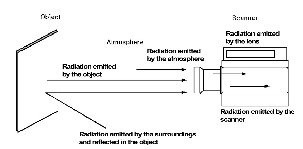
This study aimed to analyze the temperature changes during the light curing of conventional flowable composite resin and bulk-fill composite resin of various thicknesses using an infrared thermographic camera.
Methods
Flowable composite resin (G-aenial Flo, GC Co.) and bulk-fill composite resin (SDR, Dentsply Caulk) were used. Specimens with thicknesses from 0.5 mm to 5.0 mm were prepared. The infrared thermographic camera measured the temperature changes at the maximum temperature rise point during light curing. The data were analyzed for maximum temperature, time to peak temperature, and temperature rise patterns.
Results
For G-aenial Flo, the maximum temperature tended to decrease with increasing thickness, whereas for SDR, the maximum temperature decreased up to 2.0 mm and then remained relatively consistent from 2.0 mm to 5.0 mm. At thicknesses of 1.5 mm or less, both resins showed a rapid temperature increase within the first 5 seconds, followed by a reduced rate of increase up to 80 seconds. At thicknesses of 2.0 mm or greater, the temperature peaked and then gradually decreased. Across all thicknesses, SDR was observed to reach peak temperature more rapidly than G-aenial Flo.
Conclusions
Observable differences in polymerization dynamics were identified between the two resin types, particularly at greater thicknesses. Although no statistical analysis was performed, these descriptive findings suggest that infrared thermographic cameras may be useful for indirectly assessing polymerization dynamics during resin polymerization.
- 771 View
- 57 Download

- Effects of different curing methods on the color stability of composite resins
- Massimo Pisano, Alfredo Iandolo, Dina Abdellatif, Andrea Chiacchio, Marzio Galdi, Stefano Martina
- Restor Dent Endod 2024;49(4):e33. Published online September 5, 2024
- DOI: https://doi.org/10.5395/rde.2024.49.e33
-
 Abstract
 PDF
PDF PubReader
 ePub
ePub Objectives The aim of this study was to compare the effects of different polymerization strategies and the effectiveness of finishing and polishing procedures of composite resins on color stability.
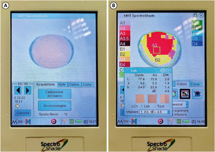
Materials and Methods The samples were divided into 4 main groups according to the polymerization strategy, and all groups except the control group received surface treatment. Each group was subsequently divided into 3 subgroups respectively: Kuraray Clearfil Majesty ES-2 Classic, Premium and Universal. Approximately 24 hours after preparation of the samples, they were immersed for 7 days in a coffee solution. A first color measurement was performed after the preparation of the samples, the second measurement was performed after 7 days in the coffee solution. All measurements were carried out using a dental spectrophotometer to assess the CIE
L *a *b * color parameters.Results There was a statistically significant difference between ΔE values for different procedures (
p = 0.003); in particular, the differences were found only between the groups that received surface treatment and the control group. In addition, a statistically significant difference was observed between the values of ΔE for different composites in the different procedure groups.Conclusions Spectrophotometric analysis showed that the additional photopolymerization and oxygen inhibition procedures did not yield better results in relation to color stability. In addition, finishing and polishing provided better color stability compared to not performing these procedures.
-
Citations
Citations to this article as recorded by- Abrasiveness and Bleaching Level of Toothpastes on Composite Resins: A Quantitative Analysis Using a Novel Brushing Simulator
Simge Meseli, Elif Alkan, Bora Korkut, Ozlem Kanar, Dilek Tagtekin
Applied Sciences.2025; 15(5): 2314. CrossRef - Comparative Evaluation of Direct and Indirect Composite Restorations in Class II Tooth Preparations - An In vivo Study
Akshun Gupta, Garima Arora, Aprajita Mehta, Satish Sane, Siddhi Nevrekar, Apurva Nagrale
Advances in Human Biology.2025; 15(4): 550. CrossRef - Micro- and Nanoplastics and the Oral Cavity: Implications for Oral and Systemic Health, Dental Practice, and the Environment—A Narrative Review
Federica Di Spirito, Veronica Folliero, Maria Pia Di Palo, Giuseppina De Benedetto, Leonardo Aulisio, Stefano Martina, Luca Rinaldi, Gianluigi Franci
Journal of Functional Biomaterials.2025; 16(9): 332. CrossRef
- Abrasiveness and Bleaching Level of Toothpastes on Composite Resins: A Quantitative Analysis Using a Novel Brushing Simulator
- 5,900 View
- 319 Download
- 3 Web of Science
- 3 Crossref

- Relationship between battery level and irradiance of light-curing units and their effects on the hardness of a bulk-fill composite resin
- Fernanda Harumi Oku Prochnow, Patricia Valéria Manozzo Kunz, Gisele Maria Correr, Marina da Rosa Kaizer, Carla Castiglia Gonzaga
- Restor Dent Endod 2022;47(4):e45. Published online November 3, 2022
- DOI: https://doi.org/10.5395/rde.2022.47.e45
-
 Abstract
 PDF
PDF PubReader
 ePub
ePub Objectives This study evaluated the relationship between the battery charge level and irradiance of light-emitting diode (LED) light-curing units (LCUs) and how these variables influence the Vickers hardness number (VHN) of a bulk-fill resin.
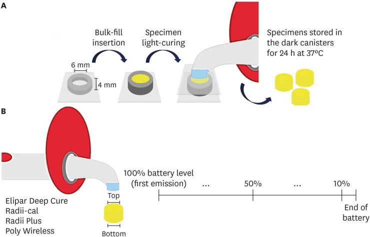
Materials and Methods Four LCUs were evaluated: Radii Plus (SDI), Radii-cal (SDI), Elipar Deep Cure (Filtek Bulk Fill, 3M Oral Care), and Poly Wireless (Kavo Kerr). Irradiance was measured using a radiometer every ten 20-second activations until the battery was discharged. Disks (4 mm thick) of a bulk-fill resin (Filtek Bulk Fill, 3M Oral Care) were prepared, and the VHN was determined on the top and bottom surfaces when light-cured with the LCUs with battery levels at 100%, 50% and 10%. Data were analyzed by 2-way analysis of variance, the Tukey’s test, and Pearson correlations (α = 5%).
Results Elipar Deep Cure and Poly Wireless showed significant differences between the irradiance when the battery was fully charged versus discharged (10% battery level). Significant differences in irradiance were detected among all LCUs, within each battery condition tested. Hardness ratios below 80% were obtained for Radii-cal (10% battery level) and for Poly Wireless (50% and 10% battery levels). The battery level showed moderate and strong, but non-significant, positive correlations with the VHN and irradiance.
Conclusions Although the irradiance was different among LCUs, it decreased in half of the devices along with a reduction in battery level. In addition, the composite resin effectiveness of curing, measured by the hardness ratio, was reduced when the LCUs’ battery was discharged.
-
Citations
Citations to this article as recorded by- Effect of erosive solutions and thermal cycling on the surface properties of universal injectable and regular consistency resin composites
Ahmed Abbas Rhaif, Hoda Saleh Ismail, Tawakol Ahmed Ahmed Enab, Nadia Mohamed Zaghloul
BMC Oral Health.2025;[Epub] CrossRef - Effect of Battery Level During Successive Charging Cycles on the Performance of Certified and Low-cost Uncertified Light-curing Units Available on E-commerce
TS Peres, G Oliveira, SP da Silva Sakamoto, M da Silva Faria, HL Carlo, CJ Soares
Operative Dentistry.2024; 49(6): 673. CrossRef - Influence of Exposure Distance on Light Irradiance of Dental Curing Lamps in Various Operating Modes
Anna Lehmann, Kacper Nijakowski, Marta Mroczyk, Filip Podgórski, Beata Czarnecka, Anna Surdacka
Applied Sciences.2024; 14(21): 9999. CrossRef - ESTADO DA INTENSIDADE LUMINOSA DAS LÂMPADAS DE FOTOPOLIMERIZAÇÃO DAS CLÍNICAS ODONTOLÓGICAS DOS CENTROS DE SAÚDE DA CIDADE DE CUENCA
Milton Alexis Quinchiguano Caraguay, David Ismael Bravo Achundia , Esteban Eduardo Amoroso Calle, Manuel Estuardo Bravo Calderon
RECISATEC - REVISTA CIENTÍFICA SAÚDE E TECNOLOGIA - ISSN 2763-8405.2023; 3(6): e36296. CrossRef
- Effect of erosive solutions and thermal cycling on the surface properties of universal injectable and regular consistency resin composites
- 1,780 View
- 27 Download
- 5 Web of Science
- 4 Crossref

- Errors in light-emitting diodes positioning when curing bulk fill and incremental composites: impact on properties after aging
- Abdulrahman A. Balhaddad, Isadora M. Garcia, Haifa Maktabi, Maria Salem Ibrahim, Qoot Alkhubaizi, Howard Strassler, Fabrício M. Collares, Mary Anne S. Melo
- Restor Dent Endod 2021;46(4):e51. Published online September 24, 2021
- DOI: https://doi.org/10.5395/rde.2021.46.e51

-
 Abstract
 PDF
PDF PubReader
 ePub
ePub Objectives This study aimed to evaluate the effect of improper positioning single-peak and multi-peak lights on color change, microhardness of bottom and top, and surface topography of bulk fill and incremental composites after artificial aging for 1 year.
Materials and Methods Bulk fill and incremental composites were cured using multi-peak and single-peak light-emitting diode (LED) following 4 clinical conditions: (1) optimal condition (no angulation or tip displacement), (2) tip-displacement (2 mm), (3) slight tip angulation (α = 20°) and (4) moderate tip angulation (α = 35°). After 1-year of water aging, the specimens were analyzed for color changes (ΔE), Vickers hardness, surface topography (Ra, Rt, and Rv), and scanning electron microscopy.
Results For samples cured by single-peak LED, the improper positioning significantly increases the color change compared to the optimal position regardless of the type of composite (
p < 0.001). For multi-peak LED, the type of resin composite and the curing condition displayed a significant effect on ΔE (p < 0.001). For both LEDs, the Vickers hardness and bottom/top ratio of Vickers hardness were affected by the type of composite and the curing condition (p < 0.01).Conclusions The bulk fill composite presented greater resistance to wear, higher color stability, and better microhardness than the incremental composite when subjected to improper curing. The multi-peak LED improves curing under improper conditions compared to single-peak LED. Prevention of errors when curing composites requires the attention of all personnel involved in the patient's care once the clinical relevance of the appropriate polymerization reflects on reliable long-term outcomes.
-
Citations
Citations to this article as recorded by- A clinical survey of the output intensity of 50 light-curing units in dental clinics across Davangere and Mangalore region using a spectrometer system
Elizbeth Christy Jose, Sakshi Jha, Prema Shantagouda Biradar, J Arun, TN Nandini, Thushara Mohanan
International Journal of Oral Health Sciences.2025; 15(1): 41. CrossRef - The demineralization resistance and mechanical assessments of different bioactive restorative materials for primary and permanent teeth: an in vitro study
Maria Salem Ibrahim, Fahad Rakad Aldhafeeri, Abdullah Sami Banaemah, Mana S. Alhaider, Yousif A. Al-Dulaijan, Abdulrahman A. Balhaddad
BDJ Open.2024;[Epub] CrossRef - Inorganic Compounds as Remineralizing Fillers in Dental Restorative Materials: Narrative Review
Leena Ibraheem Bin-Jardan, Dalal Ibrahim Almadani, Leen Saleh Almutairi, Hadi A. Almoabid, Mohammed A. Alessa, Khalid S. Almulhim, Rasha N. AlSheikh, Yousif A. Al-Dulaijan, Maria S. Ibrahim, Afnan O. Al-Zain, Abdulrahman A. Balhaddad
International Journal of Molecular Sciences.2023; 24(9): 8295. CrossRef
- A clinical survey of the output intensity of 50 light-curing units in dental clinics across Davangere and Mangalore region using a spectrometer system
- 1,532 View
- 17 Download
- 2 Web of Science
- 3 Crossref

- Physicochemical characterization of two bulk fill composites at different depths
- Guillermo Grazioli, Carlos Enrique Cuevas-Suárez, Leina Nakanishi, Alejandro Francia, Rafael Ratto de Moraes
- Restor Dent Endod 2021;46(3):e39. Published online July 5, 2021
- DOI: https://doi.org/10.5395/rde.2021.46.e39
-
 Abstract
 PDF
PDF PubReader
 ePub
ePub Objectives This study analyzed the physical-chemical behavior of 2 bulk fill resin composites (BFCs; Filtek Bulk Fill [FBF], and Tetric-N-Ceram Bulk Fill [TBF]) used in 2- and 4-mm increments and compared them with a conventional resin composite (Filtek Z250).
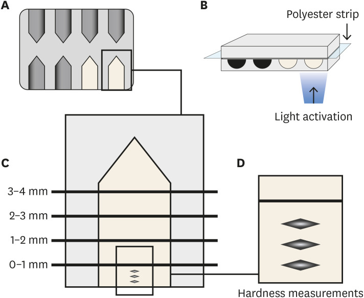
Materials and Methods Flexural strength and elastic modulus were evaluated by using a 3-point bending test. Knoop hardness was measured at depth areas 0–1, 1–2, 2–3, and 3–4 mm. The translucency parameter was measured using an optical spectrophotometer. Real-time polymerization kinetics was analyzed using Fourier transform infrared spectroscopy.
Results Flexural strength was similar among the materials, while TBF showed lower elastic modulus (Z250: 6.6 ± 1.3, FBF: 6.4 ± 0.9, TBF: 4.3 ± 1.3). The hardness of Z250 was similar only between 0–1 mm and 1–2 mm. Both BFCs had similar hardness until 2–3 mm, and showed significant decreases at 3–4 mm (FBF: 33.45 ± 1.95 at 0–1 mm to 23.19 ± 4.32 at 3–4 mm, TBF: 23.17 ± 2.51 at 0–1 mm to 15.11 ± 1.94 at 3–4 mm). The BFCs showed higher translucency than Z250. The polymerization kinetics of all the materials were similar at 2-mm increments. At 4-mm, only TBF had a similar degree of conversion compared with 2 mm.
Conclusions The BFCs tested had similar performance compared to the conventional composite when used in up to 2-mm increments. When the increment was thicker, the BFCs were properly polymerized only up to 3 mm.
-
Citations
Citations to this article as recorded by- Microhardness According to Surface, Distance and Time of Photopolymerization of a Bulk-Fill Resin: In Vitro Study
María José Loayza-Gallegos, Gino Hernan Vidalón-Romo, Julissa Amparo Dulanto-Vargas
Odovtos - International Journal of Dental Sciences.2026; 1(1): 384. CrossRef - Comparative In Vitro Analysis of Mechanical Properties in Three High-Viscosity Bulk-Fill Composite Resins
Carlos I. Santacruz, Jorge I. Fajardo, César A. Paltán, Ana del Carmen Armas-Vega, Eleonor Vélez León
Journal of Composites Science.2025; 9(11): 623. CrossRef - Translucency of bulk‐fill composite materials: A systematic review
Gaetano Paolone, Sofia Baldani, Niccolò De Masi, Mauro Mandurino, Giacomo Collivasone, Nicola Scotti, Enrico Gherlone, Giuseppe Cantatore
Journal of Esthetic and Restorative Dentistry.2024; 36(7): 995. CrossRef - Can composite packaging and selective enamel etching affect the clinical behavior of bulk-fill composite resin in posterior restorations? 24-month results of a randomized clinical trial
Marcos de Oliveira BARCELEIRO, Chane TARDEM, Elisa Gomes ALBUQUERQUE, Leticia de Souza LOPES, Stella Soares MARINS, Luiz Augusto POUBEL, Roberta BARCELOS, Romina ÑAUPARI-VILLASANTE, Alessandro Dourado LOGUERCIO, Fernanda Signorelli CALAZANS
Journal of Applied Oral Science.2023;[Epub] CrossRef - No-Cap Flowable Bulk-Fill Composite: Physico-Mechanical Assessment
Abdullah Alshehri, Feras Alhalabi, Ali Robaian, Mohammed A. S. Abuelqomsan, Abdulrahman Alshabib, Eman Ismail, Faisal Alzamil, Nawaf Alotaibi, Hamad Algamaiah
Polymers.2023; 15(8): 1847. CrossRef - The Microhardness and Surface Roughness Assessment of Bulk-Fill Resin Composites Treated with and without the Application of an Oxygen-Inhibited Layer and a Polishing System: An In Vitro Study
Ann Carrillo-Marcos, Giuliany Salazar-Correa, Leonor Castro-Ramirez, Marysela Ladera-Castañeda, Carlos López-Gurreonero, Hernán Cachay-Criado, Ana Aliaga-Mariñas, Alberto Cornejo-Pinto, Luis Cervantes-Ganoza, César Félix Cayo-Rojas
Polymers.2022; 14(15): 3053. CrossRef
- Microhardness According to Surface, Distance and Time of Photopolymerization of a Bulk-Fill Resin: In Vitro Study
- 1,979 View
- 19 Download
- 6 Web of Science
- 6 Crossref

- Assessment of the radiant emittance of damaged/contaminated dental light-curing tips by spectrophotometric methods
- Abdulrahman A. Balhaddad, Isadora Garcia, Fabrício Collares, Cristopher M. Felix, Nisha Ganesh, Qoot Alkabashi, Ward Massei, Howard Strassler, Mary Anne Melo
- Restor Dent Endod 2020;45(4):e55. Published online November 3, 2020
- DOI: https://doi.org/10.5395/rde.2020.45.e55
-
 Abstract
 PDF
PDF PubReader
 ePub
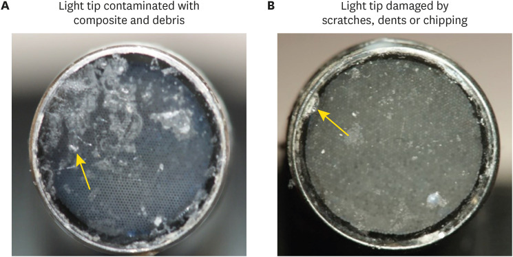
ePub Objectives This study investigated the effects of physically damaged and resin-contaminated tips on radiant emittance, comparing them with new undamaged, non-contaminated tips using 3 pieces of spectrophotometric laboratory equipment.
Materials and Methods Nine tips with damage and/or resin contaminants from actual clinical situations were compared with a new tip without damage or contamination (control group). The radiant emittance was recorded using 3 spectrophotometric methods: a laboratory-grade thermopile, a laboratory-grade integrating sphere, and a portable light collector (checkMARC).
Results A significant difference between the laboratory-grade thermopile and the laboratory-grade integrating sphere was found when the radiant emittance values of the control or damaged/contaminated tips were investigated (
p < 0.05), but both methods were comparable to checkMARC (p > 0.05). Regardless of the method used to quantify the light output, the mean radiant emittance values of the damaged/contaminated tips were significantly lower than those of the control (p < 0.05). The beam profile of the damaged/contaminated tips was less homogeneous than that of the control.Conclusions Damaged/contaminated tips can reduce the radiant emittance output and the homogeneity of the beam, which may affect the energy delivered to composite restorations. The checkMARC spectrophotometer device can be used in dental offices, as it provided values close to those produced by a laboratory-grade integrated sphere spectrophotometer. Dentists should assess the radiant emittance of their light-curing units to ensure optimal curing in photoactivated, resin-based materials.
-
Citations
Citations to this article as recorded by- Effect of damage or contamination to the tips of 200 light-curing units
Abdulrahman A. Balhaddad, Afnan O. Al-Zain, Hassan A. Alyami, Husain A. Almakrami, Osama A. Alsulaiman, Eman H. Ismail, Richard B. Price, Ahmed A. Alsulaiman
BMC Oral Health.2025;[Epub] CrossRef - The Performance of Light-curing Units Used in Different Clinics at Aseer Region, Saudi Arabia: A Cross-sectional Study
Mohammed M Al Moaleem, Ghadeer S Alwadai, Nada A Alamoudi, Naif N Abogazalah, Saleh A Alqahtani, Faisal H Alshehri, Wafa H Alaajam, Mohammad A Alamri, Amjad Y Alhaydan
The Journal of Contemporary Dental Practice.2025; 26(8): 784. CrossRef - Evaluation of Radiant Power of the Light Curing Units Used in Clinics at Governmental and Privates Dental Faculties
Sami Ali Hasan, Ibrahim Al-Shami, Mohsen Al-Hamzi, Ghadeer Alwadai, Nada Alamoudi, Saleh Alqahtani, Arwa Daghrery, Wafa Alaajam, Mansoor Shariff, Hussain Kinani, Mohammed Al Moaleem
Medical Devices: Evidence and Research.2024; Volume 17: 301. CrossRef - Evaluation of the information provided in the instruction manuals of dental light‐curing units
Afnan O. Al‐Zain, Eman H. Ismail, Abdulrahman A. Balhaddad, Osamah Toras, Yousif Alharthy, Rafa Alsultan, Abeer Alrossais, Richard B. Price
Journal of Esthetic and Restorative Dentistry.2024; 36(10): 1466. CrossRef - Utilizing Light Cure Units: A Concise Narrative Review
Fatin A. Hasanain, Hani M. Nassar
Polymers.2021; 13(10): 1596. CrossRef - Improper Light Curing of Bulkfill Composite Drives Surface Changes and Increases S. mutans Biofilm Growth as a Pathway for Higher Risk of Recurrent Caries around Restorations
Haifa Maktabi, Maria Salem Ibrahim, Abdulrahman A. Balhaddad, Qoot Alkhubaizi, Isadora Martini Garcia, Fabrício Mezzomo Collares, Howard Strassler, Ana Paula P. Fugolin, Carmem S. Pfeifer, Mary Anne S. Melo
Dentistry Journal.2021; 9(8): 83. CrossRef
- Effect of damage or contamination to the tips of 200 light-curing units
- 1,552 View
- 8 Download
- 6 Crossref

- The influence of nanofillers on the properties of ethanol-solvated and non-solvated dental adhesives
- Leonardo Bairrada Tavares da Cruz, Marcelo Tavares Oliveira, Cintia Helena Coury Saraceni, Adriano Fonseca Lima
- Restor Dent Endod 2019;44(3):e28. Published online July 24, 2019
- DOI: https://doi.org/10.5395/rde.2019.44.e28
-
 Abstract
 PDF
PDF PubReader
 ePub
ePub Objectives The aim of this study was to evaluate the influence of different concentrations of nanofillers on the chemical and physical properties of ethanol-solvated and non-solvated dental adhesives.
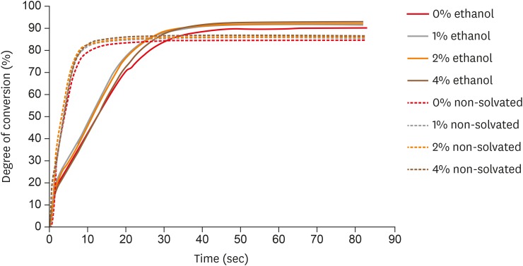
Materials and Methods Eight experimental adhesives were prepared with different nanofiller concentrations (0, 1, 2, and 4 wt%) and 2 solvent concentrations (0% and 10% ethanol). Several properties of the experimental adhesives were evaluated, such as water sorption and solubility (
n = 5, 20 seconds light activation), real-time degree of conversion (DC;n = 3, 20 and 40 seconds light activation), and stability of cohesive strength at 6 months (CS;n = 20, 20 seconds light activation) using the microtensile test. A light-emitting diode (Bluephase 20i, Ivoclar Vivadent) with an average light emittance of 1,200 mW/cm2 was used.Results The presence of solvent reduced the DC after 20 seconds of curing, but increased the final DC, water sorption, and solubility of the adhesives. Storage in water reduced the strength of the adhesives. The addition of 1 wt% and 2 wt% nanofillers increased the polymerization rate of the adhesives.
Conclusions The presence of nanofillers and ethanol improved the final DC, although the DC of the solvated adhesives at 20 seconds was lower than that of the non-solvated adhesives. The presence of ethanol reduced the strength of the adhesives and increased their water sorption and solubility. However, nanofillers did not affect the water sorption and strength of the tested adhesives.
-
Citations
Citations to this article as recorded by- Effect of boric acid on the color stability and mechanical properties of 3D-printed permanent resins
Dalndushe Abdulai, Rafat Sasany, Raghib Suradi, Mehran Moghbel, Seyed Ali Mosaddad
Scientific Reports.2025;[Epub] CrossRef - Development of a Boron Nitride-Filled Dental Adhesive System
Senthilguru Kulanthaivel, Jeremiah Poppen, Sandra Ribeiro Cunha, Benjamin Furman, Kyumin Whang, Erica C. Teixeira
Polymers.2023; 15(17): 3512. CrossRef - Analyses of Experimental Dental Adhesives Based on Zirconia/Silver Phosphate Nanoparticles
Abdul Khan, Yasmin Alhamdan, Hala Alibrahim, Khalid Almulhim, Muhammad Nawaz, Syed Ahmed, Khalid Aljuaid, Ijlal Ateeq, Sultan Akhtar, Mohammad Ansari, Intisar Siddiqui
Polymers.2023; 15(12): 2614. CrossRef - Mechanical characterization and adhesive properties of a dental adhesive modified with a polymer antibiotic conjugate
Camila Sabatini, Russell J. Aguilar, Ziwen Zhang, Steven Makowka, Abhishek Kumar, Megan M. Jones, Michelle B. Visser, Mark Swihart, Chong Cheng
Journal of the Mechanical Behavior of Biomedical Materials.2022; 129: 105153. CrossRef
- Effect of boric acid on the color stability and mechanical properties of 3D-printed permanent resins
- 1,315 View
- 8 Download
- 4 Crossref

- The effect of thermocycling on the degree of conversion and mechanical properties of a microhybrid dental resin composite
- Mehrsima Ghavami-Lahiji, Melika Firouzmanesh, Hossein Bagheri, Tahereh S. Jafarzadeh Kashi, Fateme Razazpour, Marjan Behroozibakhsh
- Restor Dent Endod 2018;43(2):e26. Published online April 26, 2018
- DOI: https://doi.org/10.5395/rde.2018.43.e26
-
 Abstract
 PDF
PDF PubReader
 ePub
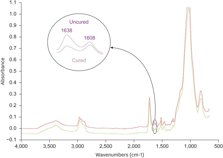
ePub Objective The purpose of this study was to investigate the degree of conversion (DC) and mechanical properties of a microhybrid Filtek Z250 (3M ESPE) resin composite after aging.
Method The specimens were fabricated using circular molds to investigate Vickers microhardness (Vickers hardness number [VHN]) and DC, and were prepared according to ISO 4049 for flexural strength testing. The initial DC (%) of discs was recorded using attenuated total reflectance-Fourier transforming infrared spectroscopy. The initial VHN of the specimens was measured using a microhardness tester under a load of 300 g for 15 seconds and the flexural strength test was carried out with a universal testing machine (crosshead speed, 0.5 mm/min). The specimens were then subjected to thermocycling in 5°C and 55°C water baths. Properties were assessed after 1,000–10,000 cycles of thermocycling. The surfaces were evaluated using scanning electron microscopy (SEM). Data were analyzed using 1-way analysis of variance followed by the Tukey honest significant difference
post hoc test.Results Statistical analysis showed that DC tended to increase up to 4,000 cycles, with no significant changes. VHN and flexural strength values significantly decreased upon thermal cycling when compared to baseline (
p < 0.05). However, there was no significant difference between initial and post-thermocycling VHN results at 1,000 cycles. SEM images after aging showed deteriorative changes in the resin composite surfaces.Conclusions The Z250 microhybrid resin composite showed reduced surface microhardness and flexural strength and increased DC after thermocycling.
-
Citations
Citations to this article as recorded by- Clinical Decision‐Making of Repair vs. Replacement of Defective Direct Dental Restorations: A Multinational Cross‐Sectional Study With Meta‐Analysis
Ömer Hatipoğlu, João Filipe Brochado Martins, Mohmed Isaqali Karobari, Nessrin Taha, Thiyezen Abdullah Aldhelai, Daoud M. Ayyad, Ahmed A. Madfa, Benjamin Martin‐Biedma, Rafael Fernández‐Grisales, Bakhyt A. Omarova, Wen Yi Lim, Suha Alfirjani, Kacper Nijak
Journal of Esthetic and Restorative Dentistry.2025; 37(4): 977. CrossRef - An In Vitro Evaluation of Novel Bioactive Liner's Effect on Marginal Adaptation of Class II Composite Restorations: A Scanning Electron Microscope Analysis
Girija S Sajjan, Naveena Ponnada, Praveen Dalavai, Madhu Varma Kanumuri, Venkata Karteek Varma Penmatsa, B V Sindhuja
World Journal of Dentistry.2025; 15(9): 749. CrossRef - Different contemporary resin cements for intracanal luting of glass fiber posts - Bonding and polymerization assessments
Anna Caroliny Detogni, Vitaliano Gomes de Araújo Neto, Caio Felipe de Almeida Nobre, Victor Pinheiro Feitosa, Mário Alexandre Coelho Sinhoreti
International Journal of Adhesion and Adhesives.2025; 138: 103951. CrossRef - Effect of food-simulating liquids and polishing times on the color stability of microhybrid and nanohybrid resin composites
Muhammet Fidan, Nevin Çankaya
Discover Nano.2025;[Epub] CrossRef - Effect of irrigation protocols for post space preparation on the bond of the resin luting agent and post to a hydraulic calcium silicate filled root: An in vitro study
Nuttanun Poeaim, Sirawut Hiran-us, Yanee Tantilertanant
The Journal of Prosthetic Dentistry.2025; 133(4): 1039.e1. CrossRef - Influence of Different Adhesives and Surface Treatments on Shear and Tensile Bond Strength and Microleakage with Micro-CT of Repaired Bulk-Fill Composites
Handan Yıldırım-Işık, Mediha Büyükgöze-Dindar
Polymers.2025; 17(12): 1680. CrossRef - Effect of thermal ageing on physico-mechanical properties and self-healing potential of experimental 3D-printed denture base resin composites
Mariam Raza Syed, Amr Fawzy
Journal of the Mechanical Behavior of Biomedical Materials.2025; 170: 107123. CrossRef - Effects of aging on the physicomechanical, antimicrobial, and cytotoxicity properties of flowable composite resin with strontium-modified phosphate-based glass
Seo-Hyun Kim, Hye-Bin Go, Myung-Jin Lee, Jae-Sung Kwon
Scientific Reports.2025;[Epub] CrossRef - Colour Stability and Optical Properties of Provisional Crowns Fabricated With Milling, 3D Printing, and Direct Technique
Tommaso Rinaldi, Carlos Serrano Granger, Andrea Santamaría Laorden, Jaime Orejas-Perez, Pablo Gómez Cogolludo
International Dental Journal.2025; 75(6): 103932. CrossRef - EVALUATE DEGREE OF CONVERSION OF NEW BIOACTIVE ORTHODONTIC ADHESIVE WITH COLOR CHANGE & FLUORESCENCE PROPERTY
Mohammed Younis, Neam Fakhri Neam Fakhri
BULLETIN OF STOMATOLOGY AND MAXILLOFACIAL SURGERY.2025; : 39. CrossRef - Antibacterial activity and physicochemical properties of light-curable fluoride varnishes containing strontium phosphate-based glass
Na-Yeon Kim, Mi-Sol Ryu, Ji-Min Lee, Soo-Yeon Jeong, Hye-Been Choi, Myung-Jin Lee, Song-Yi Yang
Clinical Oral Investigations.2025;[Epub] CrossRef - Systematic Review of Studies Comparing Microleakage After Restoration With Cention and Conventional Glass Ionomer Cement in Human Extracted Teeth
Rashmi Misra, Mansi Vandekar, Gayatri Pendse, Omkar Bhosale, Pauravi Hegde, Aashaka Vaishnav
Cureus.2025;[Epub] CrossRef - Evaluation of the radiopacity of different universal composite resins aged by thermocycling
Dilber Çölkesen, Alican Kuran, Neslihan Tekçe
Odontology.2025;[Epub] CrossRef - Exploring the sources and routes of micro- and nanoplastics from dental products and materials: their impact on human health - a systematic review
Vidhya Selvaraj, R. Saravanan, N. Raj Vikram, Uma revathi Gopalakrishnan, Ramsamy M
Next Research.2025; 2(4): 100925. CrossRef - Investigation of the mechanical response of MWCNTs infused carbon/glass fiber-based hybrid composites using digital image correlation
Somaiah Chowdary Mallampati, Ujendra Kumar Komal, Paladugu Rakesh, Parthapratim Barman
Construction and Building Materials.2025; 492: 143068. CrossRef - Mechanical, Surface and Physicochemical Properties of Nanozeolite‐Modified 3D Printed Hybrid Ceramics at Varying Concentrations: An In Vitro Study
Ahmed A. Holiel, Yomna M. Ibrahim, Noha Morsy
Journal of Esthetic and Restorative Dentistry.2025;[Epub] CrossRef - Impact of Graphitic Carbon Nitride on Dental Composite’s Mechanical and Antibacterial Properties
Zainab Rafaqat, Saad Liaqat, Ahmed Bari, Warda Khan Yousafzai, Umar Nishan, Sandleen Feroz, Nawshad Muhammad
Journal of Materials Engineering and Performance.2025;[Epub] CrossRef - Exploring the Biological and Chemical Properties of Emerging 3D-Printed Dental Resin Composites Compared to Conventional Light-Cured Materials
Nikola Živković, Stefan Vulović, Miloš Lazarević, Anja Baraba, Aleksandar Jakovljević, Mina Perić, Jelena Mitrić, Aleksandra Milić Lemić
Materials.2025; 18(22): 5170. CrossRef - Awareness of possible complications associated with direct composite restorations: A multinational survey among dentists from 13 countries with meta-analysis
Anna Lehmann, Kacper Nijakowski, Jakub Jankowski, David Donnermeyer, Paulo J. Palma, Milan Drobac, João Filipe Brochado Martins, Fatma Pertek Hatipoğlu, Indira Tulegenova, Muhammad Qasim Javed, Hamad Mohammad Alharkan, Olga Bekjanova, Sylvia Wyzga, Moataz
Journal of Dentistry.2024; 145: 105009. CrossRef - Comparative evaluation of bond strength and color stability of polyetheretherketone and zirconia layered with indirect composite before and after thermocycling: An in vitro study
Pooja Singh, Subhabrata Maiti, Amrutha Shenoy
The Journal of Indian Prosthodontic Society.2024; 24(3): 252. CrossRef - Biaxial flexural strength of hydrothermally aged resin-based materials
Rodrigo Ricci Vivan, Mariana Miranda de Toledo Piza, Bruna de Mello Silva, Thalya Fernanda Horsth Maltarollo, Gustavo Sivieri-Araujo, Murilo Priori Alcalde, Marco Antonio Hungaro Duarte, Estevam Augusto Bonfante, Henrico Badaoui Strazzi-Sahyon
Journal of the Mechanical Behavior of Biomedical Materials.2024; 155: 106568. CrossRef - Comparative Strength Study of Indirect Permanent Restorations: 3D-Printed, Milled, and Conventional Dental Composites
João Paulo Mendes Tribst, Adelheid Veerman, Gabriel Kalil Rocha Pereira, Cornelis Johannes Kleverlaan, Amanda Maria de Oliveira Dal Piva
Clinics and Practice.2024; 14(5): 1940. CrossRef - Influencia del termociclado sobre la estabilidad del color de dos resinas compuestas
//Influence of thermocycling on the color stability of two composite resins
Verónica Lucía Ventrera, María Eugenia Alejandra Barrionuevo
Revista de la Asociación Odontológica Argentina.2024; : 1. CrossRef - Efeito do protocolo de polimento e do armazenamento em meio úmido na variação de cor, massa e rugosidade superficial de resinas compostas
Leonardo Cruz Morais, Mateus Victória Gontijo, Gabriela Rodrigues Pires, Victor de Morais Gomes, Milton Carlos Kuga, Francisco Fernando Massola Filho, Amanda Gonçalves Franco, Alberto Nogueira da Gama Antunes
Cuadernos de Educación y Desarrollo.2024; 16(6): e4556. CrossRef - A comparison of the mechanical properties of 3D-printed, milled, and conventional denture base resin materials
Hyeong-Ju YU, You-Jung KANG, Yeseul PARK, Hoon KIM, Jee-Hwan KIM
Dental Materials Journal.2024; 43(6): 813. CrossRef - Effect of aging and fiber‐reinforcement on color stability, translucency, and microhardness of single‐shade resin composites versus multi‐shade resin composite
Muhammet Fidan, Özhan Yağci
Journal of Esthetic and Restorative Dentistry.2024; 36(4): 632. CrossRef - Impact of Artificial Aging on the Physical and Mechanical Characteristics of Denture Base Materials Fabricated via 3D Printing
Ahmed Altarazi, Julfikar Haider, Abdulaziz Alhotan, Nick Silikas, Hugh Devlin, Weihao Yuan
International Journal of Biomaterials.2024;[Epub] CrossRef - Synthesis, monomer conversion, and mechanical properties of polylysine based dental composites
Saadia Bano Lone, Rabia Zeeshan, Hina Khadim, Muhammad Adnan Khan, Abdul Samad Khan, Anila Asif
Journal of the Mechanical Behavior of Biomedical Materials.2024; 151: 106398. CrossRef - Bond strength and surface roughness assessment of novel antimicrobial polymeric coated dental cement
Ghada Naguib, Hisham Mously, Jumana Mazhar, Ibrahim Alkanfari, Abdulelah Binmahfooz, Mohammed Zahran, Mohamed T. Hamed
Discover Nano.2024;[Epub] CrossRef - Evaluation of microhardness, degree of conversion, and abrasion resistance of nanoglass and multiwalled carbon nanotubes reinforced three‐dimensionally printed denture base resins
Pansai Ashraf Mohamed, Yomna Mohamed Ibrahim, Kenda Ibrahim Hisham Hanno, Mohamed Mahmoud Abdul‐Monem
Journal of Prosthodontics.2024;[Epub] CrossRef - Effect of CAD-CAM block thickness and translucency on the polymerization of luting materials
Bengü Doğu Kaya, Selinsu Öztürk, Ayşe Aslı Şenol, Erkut Kahramanoğlu, Pınar Yılmaz Atalı, Bilge Tarçın
BMC Oral Health.2024;[Epub] CrossRef - Simulation of oral environmental conditions through artificial aging of teeth for the assessment of enamel discoloration in orthodontics
Celal Irgın
BMC Oral Health.2024;[Epub] CrossRef - Do universal adhesive systems affect color coordinates and color change of single-shade resin composites compared with a multi-shade composite?
Muhammet FİDAN, Özhan YAĞCI
Dental Materials Journal.2023; 42(6): 886. CrossRef - Fabrication, Evaluation, and Performance Ranking of Tri-calcium Phosphate and Silica Reinforced Dental Resin Composite Materials
Sonu Saini, Anoj Meena, Ramkumar Yadav, Amar Patnaik
Silicon.2023; 15(18): 8045. CrossRef - Can Modification with Urethane Derivatives or the Addition of an Anti-Hydrolysis Agent Influence the Hydrolytic Stability of Resin Dental Composite?
Agata Szczesio-Wlodarczyk, Izabela M. Barszczewska-Rybarek, Marta W. Chrószcz-Porębska, Karolina Kopacz, Jerzy Sokolowski, Kinga Bociong
International Journal of Molecular Sciences.2023; 24(5): 4336. CrossRef - Effect of veneering material type and thickness ratio on flexural strength of bi-layered PEEK restorations before and after thermal cycling
Ahmed Gouda, Ashraf Sherif, Mennatallah Wahba, Tarek Morsi
Clinical Oral Investigations.2023; 27(6): 2629. CrossRef - 3D printed denture base material: The effect of incorporating TiO2 nanoparticles and artificial ageing on the physical and mechanical properties
Ahmed Altarazi, Julfikar Haider, Abdulaziz Alhotan, Nick Silikas, Hugh Devlin
Dental Materials.2023; 39(12): 1122. CrossRef - Influence of silane coupling agent and aging on the repair bond strength of dental composites
Gustavo Jusué-Esparza, José Alejandro Rivera-Gonzaga, Guillermo Grazioli, Ana Josefina Monjarás-Ávila, J. Eliezer Zamarripa-Calderón, Carlos Enrique Cuevas-Suárez
Journal of Adhesion Science and Technology.2023; 37(5): 913. CrossRef - Degree of conversion of light‐polymerized composite resin in implant prosthesis screw access opening
Se‐Hyun Park, Yoon‐Hyuk Huh, Chan‐Jin Park, Lee‐Ra Cho, Kyung‐Ho Ko
Journal of Prosthodontics.2023; 32(9): 829. CrossRef - Investigation of the effect of matrix-interface formed with silane-based coupling agents on physico-chemical behavior and flow distance of dental composites
Zerin Yeşil Acar, Merve Tunç Koçyiğit, Meltem Asiltürk
Journal of Molecular Liquids.2023; 378: 121600. CrossRef - Evaluation of Water Sorption and Solubility of 3D-Printed, CAD/CAM Milled, and PMMA Denture Base Materials Subjected to Artificial Aging
Mariya Dimitrova, Angelina Vlahova, Ilian Hristov, Rada Kazakova, Bozhana Chuchulska, Stoyan Kazakov, Marta Forte, Vanja Granberg, Giuseppe Barile, Saverio Capodiferro, Massimo Corsalini
Journal of Composites Science.2023; 7(8): 339. CrossRef - Effect of thermocycling on internal microhardness of high and low viscosity bulk fill composite resins in class I cavities
Sâmara Luciana de Andrade LIMA, Lais Lemos CABRAL, Natália Russo CARLOS, Saulo André de Andrade LIMA, Kamila Rosamilia KANTOVITZ, Flávia Lucisano Botelho do AMARAL
RGO - Revista Gaúcha de Odontologia.2023;[Epub] CrossRef - Effects of an Acidic Environment on the Strength and Chemical Changes of Resin-based Composites
S Kang, B-H Cho
Operative Dentistry.2023; 48(4): E81. CrossRef - Influence of compressive forces and aging through thermocycling on the strength of mono incremental dental composite resins
Cristian Roberto Sigcho Romero, Henry Fabricio Mejía Mosquera, Sandra Marcela Quisiguiña Guevara, Yudy Jacqueline Alvarado Aguayo
Bionatura.2023; 8(4): 1. CrossRef - Push-out Bond Strength of Two Fiber Posts in Composite Resin Using Different Types of Silanization
RM Novis, BLT Leon, FMG França, CP Turssi, RT Basting, FLB Amaral
Operative Dentistry.2022; 47(2): 173. CrossRef - Penetration and Adaptation of the Highly Viscous Zinc-Reinforced Glass Ionomer Cement on Contaminated Fissures: An In Vitro Study with SEM Analysis
Galiah AlJefri, Sunil Kotha, Muhannad Murad, Reham Aljudaibi, Fatmah Almotawah, Sreekanth Mallineni
International Journal of Environmental Research and Public Health.2022; 19(10): 6291. CrossRef - Surface Characteristics and Color Stability of Dental PEEK Related to Water Saturation and Thermal Cycling
Liliana Porojan, Flavia Roxana Toma, Mihaela Ionela Bîrdeanu, Roxana Diana Vasiliu, Ion-Dragoș Uțu, Anamaria Matichescu
Polymers.2022; 14(11): 2144. CrossRef - Effects of aging and light-curing unit type on the volume and internal porosity of bulk-fill resin composite restoration
Afnan O. Al-Zain, Elaf A. Alboloshi, Walaa A. Amir, Maryam A. Alghilan, Eliseu A. Münchow
The Saudi Dental Journal.2022; 34(3): 243. CrossRef - An Evaluation of the Hydrolytic Stability of Selected Experimental Dental Matrices and Composites
Agata Szczesio-Wlodarczyk, Karolina Kopacz, Malgorzata Iwona Szynkowska-Jozwik, Jerzy Sokolowski, Kinga Bociong
Materials.2022; 15(14): 5055. CrossRef - Comparison of the Mechanical Properties and Push-out Bond Strength of Self-adhesive and Conventional Resin Cements on Fiber Post Cementation
MR Santi, RBE Lins, BO Sahadi, JR Soto-Montero, LRM Martins
Operative Dentistry.2022; 47(3): 346. CrossRef - Effect of Different Polymerization Times on Color Change, Translucency Parameter, and Surface Hardness of Bulk-Fill Resin Composites
HY Gonder, M Fidan
Nigerian Journal of Clinical Practice.2022; 25(10): 1751. CrossRef - Surface degradation and biofilm formation on hybrid and nanohybrid composites after immersion in different liquids
Gabriela Escamilla-Gómez, Octavio Sánchez-Vargas, Diana M. Escobar-García, Amaury Pozos-Guillén, Norma V. Zavala-Alonso, Mariana Gutiérrez-Sánchez, José E. Pérez-López, Gregorio Sánchez-Balderas, Gabriel F. Romo-Ramírez, Marine Ortiz-Magdaleno
Journal of Oral Science.2022; 64(4): 263. CrossRef - Effects of Different Adhesive Systems and Orthodontic Bracket Material on Enamel Surface Discoloration: An In Vitro Study
Ali Alqerban, Doaa R. M. Ahmed, Ali S. Aljhani, Dalal Almadhi, Amjad AlShahrani, Hussah AlAdwene, Abdulaziz Samran
Applied Sciences.2022; 12(24): 12885. CrossRef - Effects of Immediate Coating on Unset Composite with Different Bonding Agents to Surface Hardness
Nantawan Krajangta, Supissara Ninbanjong, Sunisa Khosook, Kanjana Chaitontuak, Awiruth Klaisiri
European Journal of Dentistry.2022; 16(04): 828. CrossRef - Rational durability of optical properties of chameleon effect of Omnichroma and Essentia composite thermocycled in black dark drinks (in vitro study)
Bassma Abdelhamed, Asmaa Abdel-Hakeem Metwally, Heba A. Shalaby
Bulletin of the National Research Centre.2022;[Epub] CrossRef - Comparative Evaluation of Shear Bond Strength of Nanohybrid Composite Restoration After the Placement of Flowable Compomer and Composite Using the Snowplow Technique
Meghna Dugar, Anuja Ikhar, Pradnya Nikhade, Manoj Chandak, Nidhi Motwani
Cureus.2022;[Epub] CrossRef - The First Step in Standardizing an Artificial Aging Protocol for Dental Composites—Evaluation of Basic Protocols
Agata Szczesio-Wlodarczyk, Magdalena Fronczek, Katarzyna Ranoszek-Soliwoda, Jarosław Grobelny, Jerzy Sokolowski, Kinga Bociong
Molecules.2022; 27(11): 3511. CrossRef - Effect of Different Surface Treatments on the Long‐Term Repair Bond Strength of Aged Methacrylate‐Based Resin Composite Restorations: A Systematic Review and Network Meta‐analysis
Mahdi Hadilou, Amirmohammad Dolatabadi, Morteza Ghojazadeh, Hossein Hosseinifard, Parnian Alizadeh Oskuee, Fatemeh Pournaghi Azar, Victor Feitosa
BioMed Research International.2022;[Epub] CrossRef - Edge chipping resistance of veneering composite resins
Parissa Nassary Zadeh, Bogna Stawarczyk, Rüdiger Hampe, Anja Liebermann, Felicitas Mayinger
Journal of the Mechanical Behavior of Biomedical Materials.2021; 116: 104349. CrossRef - The effect of radiation exposure and storage time on the degree of conversion and flexural strength of different resin composites
Ragia M. Taher, Lamiaa M. Moharam, Amin E. Amin, Mohamed H. Zaazou, Farid S. El-Askary, Mokhtar N. Ibrahim
Bulletin of the National Research Centre.2021;[Epub] CrossRef - Fracture Load of CAD/CAM Fabricated Cantilever Implant-Supported Zirconia Framework: An In Vitro Study
Ibraheem F. Alshiddi, Syed Rashid Habib, Muhammad Sohail Zafar, Salwa Bajunaid, Nawaf Labban, Mohammed Alsarhan
Molecules.2021; 26(8): 2259. CrossRef - A numerical, theoretical and experimental study of the effect of thermocycling on the matrix-filler interface of dental restorative materials
Yoan Boussès, Nathalie Brulat-Bouchard, Pierre-Olivier Bouchard, Yannick Tillier
Dental Materials.2021; 37(5): 772. CrossRef - Impact of polymerization and storage on the degree of conversion and mechanical properties of veneering resin composites
Felicitas MAYINGER, Marcel REYMUS, Anja LIEBERMANN, Marc RICHTER, Patrick KUBRYK, Henning GROẞEKAPPENBERG, Bogna STAWARCZYK
Dental Materials Journal.2021; 40(2): 487. CrossRef - Intrapulpal Concentration of Hydrogen Peroxide of Teeth Restored With Bulk Fill and Conventional Bioactive Composites
DP Silva, BA Resende, M Kury, CB André, CPM Tabchoury, M Giannini, V Cavalli
Operative Dentistry.2021; 46(3): E158. CrossRef - Silane content influences physicochemical properties in nanostructured model composites
Larissa Maria Cavalcante, Lucielle Guimarães Ferraz, Karinne Bueno Antunes, Isadora Martini Garcia, Luis Felipe Jochims Schneider, Fabrício Mezzomo Collares
Dental Materials.2021; 37(2): e85. CrossRef - AĞIZ GARGARALARI VE ANTİSEPTİKLERİNİN FARKLI KOMPOZİT REZİNLERİN RENK STABİLİTESİNE ETKİSİ
Turan Emre KUZU, Özcan KARATAŞ
Atatürk Üniversitesi Diş Hekimliği Fakültesi Dergisi.2021; : 1. CrossRef - Evaluation of Immediate and Delayed Microleakage of Class V Cavities Restored with Chitosan-incorporated Composite Resins: An In Vitro Study
Roopa R Nadig, Veena Pai, Arpita Deb
International Journal of Clinical Pediatric Dentistry.2021; 14(5): 621. CrossRef - Influence of Diode Laser for the Treatment of Dentin Hypersensitivity on Microleakage of Cervical Restorations
Doaa R. M. Ahmed, Diana G. Shaath, Jomana B. Alakeel, Abdulaziz A. Samran, Mona Abbassy
BioMed Research International.2021;[Epub] CrossRef - Ageing of Dental Composites Based on Methacrylate Resins—A Critical Review of the Causes and Method of Assessment
Agata Szczesio-Wlodarczyk, Jerzy Sokolowski, Joanna Kleczewska, Kinga Bociong
Polymers.2020; 12(4): 882. CrossRef - Flexural strength and surface microhardness of materials used for temporary dental disocclusion submitted to thermal cycling: An in vitro study
Tamires Borges de Lima, José Guilherme Neves, Ana Paula Terossi de Godoi, Ana Rosa Costa, Viviane Veroni Degan, Américo Bortolazzo Correr, Heloisa Cristina Valdrighi
International Orthodontics.2020; 18(3): 519. CrossRef - Evaluation of the repair capacities and color stabilities of a resin nanoceramic and hybrid CAD/CAM blocks
Hasibe Sevilay Bahadır, Yusuf Bayraktar
The Journal of Advanced Prosthodontics.2020; 12(3): 140. CrossRef - Effect of Different Surface Treatments of Resin Relined Fiber Posts Cemented With Self-adhesive Resin Cement on Push-out and Microtensile Bond Strength Tests
RV Machry, PE Fontana, TC Bohrer, LF Valandro, OB Kaizer
Operative Dentistry.2020; 45(4): E185. CrossRef - Influences of Successive Exposure to Bleaching and Fluoride Preparations on the Surface Hardness and Roughness of the Aged Resin Composite Restoratives
Khalid M. Abdelaziz, Shugufta Mir, Shafait Ullah Khateeb, Suheel M. Baba, Saud S. Alshahrani, Eman A. Alshahrani, Zahra A. Alsafi
Medicina.2020; 56(9): 476. CrossRef - Fracture Resistance of Lithıum Disilicate, Indirect Resin Composite and Zirconıa by Using Dual Cure Resin Cements
Mohammed BADWAN, Erkut KAHRAMANOĞLU
Clinical and Experimental Health Sciences.2020; 10(4): 435. CrossRef - Effect of Stress-decreasing Resin Thickness as an Intermediate Layer on Fracture Resistance of Class II Composite Restoration: An In Vitro Study
Dennis Dennis, Arwin Leonardy, Trimurni Abidin
World Journal of Dentistry.2020; 11(2): 91. CrossRef - Effect of Thermocycling on Biaxial Flexural Strength of CAD/CAM, Bulk Fill, and Conventional Resin Composite Materials
EB Benalcázar Jalkh, CM Machado, M Gianinni, I Beltramini, MMT Piza, PG Coelho, R Hirata, EA Bonfante
Operative Dentistry.2019; 44(5): E254. CrossRef - Mechanical properties of hybrid computer-aided design/computer-aided manufacturing (CAD/CAM) materials after aging treatments
Hae-Yong Jeong, Hae-Hyoung Lee, Yu-Sung Choi
Ceramics International.2018; 44(16): 19217. CrossRef
- Clinical Decision‐Making of Repair vs. Replacement of Defective Direct Dental Restorations: A Multinational Cross‐Sectional Study With Meta‐Analysis
- 6,416 View
- 47 Download
- 77 Crossref

- Post space preparation timing of root canals sealed with AH Plus sealer
- Hae-Ri Kim, Young Kyung Kim, Tae-Yub Kwon
- Restor Dent Endod 2017;42(1):27-33. Published online December 19, 2016
- DOI: https://doi.org/10.5395/rde.2017.42.1.27

-
 Abstract
 PDF
PDF PubReader
 ePub
ePub Objectives To determine the optimal timing for post space preparation of root canals sealed with epoxy resin-based AH Plus sealer in terms of its polymerization and influence on apical leakage.
Materials and Methods The epoxy polymerization of AH Plus (Dentsply DeTrey) as a function of time after mixing (8, 24, and 72 hours, and 1 week) was evaluated using Fourier transform infrared (FTIR) spectroscopy and microhardness measurements. The change in the glass transition temperature (
Tg ) of the material with time was also investigated using differential scanning calorimetry (DSC). Fifty extracted human single-rooted premolars were filled with gutta-percha and AH Plus, and randomly separated into five groups (n = 10) based on post space preparation timing (immediately after root canal obturation and 8, 24, and 72 hours, and 1 week after root canal obturation). The extent of apical leakage (mm) of the five groups was compared using a dye leakage test. Each dataset was statistically analyzed by one-way analysis of variance and Tukey'spost hoc test (α = 0.05).Results Continuous epoxy polymerization of the material with time was observed. Although the
Tg values of the material gradually increased with time, the specimens presented no clearTg value at 1 week after mixing. When the post space was prepared 1 week after root canal obturation, the leakage was significantly higher than in the other groups (p < 0.05), among which there was no significant difference in leakage.Conclusions Poor apical seal was detected when post space preparation was delayed until 1 week after root canal obturation.
-
Citations
Citations to this article as recorded by- Bacterial microleakage in endodontically treated teeth following two methods of postspace preparation at two-time intervals: An in vitro study
AzamS Mostafavi, Mahsa Rasoulzadehsheikh, Naghmeh Meraji, Maryam Pourhajibagher
The Journal of Indian Prosthodontic Society.2022; 22(3): 233. CrossRef - Comparison of the effect of post space preparation time on the apical seal of two different sealers
Neda Hajihassani, Navid Mohammadi, Ahmad Karimi Kelayeh, Shima Aalaei
BMC Oral Health.2022;[Epub] CrossRef - Immediate and Delayed Post Space Preparations in Endodontically Treated Teeth: A Scoping Review
Sadaf Mahmoudi, Pedram Iranmanesh, Saber Khazaei, Maryam Zare Jahromi
BMC Oral Health.2022;[Epub] CrossRef - Physicochemical properties of a novel bioceramic silicone-based root canal sealer
Wei-Jia Lyu, Wei Bai, Xiao-Yan Wang, Yu-Hong Liang
Journal of Dental Sciences.2022; 17(2): 831. CrossRef - Impact of Immersion Media on Physical Properties and Bioactivity of Epoxy Resin-Based and Bioceramic Endodontic Sealers
Thais Gomes de Moraes, Alan Silva de Menezes, Renata Grazziotin-Soares, Rafael Ubaldo Moreira e Moraes, Paulo Vitor Campos Ferreira, Ceci Nunes Carvalho, Jose Bauer, Edilausson Moreno Carvalho
Polymers.2022; 14(4): 729. CrossRef - The effect of two endodontic sealers and interval before post-preparation and cementation on the bond strength of fiber posts
He Yuanli, Wu Juan, Ji Mengzhen, Chen Xuan, Xiong Kaixin, Yang Xueqin, Qiao Xin, Hu Hantao, Gao Yuan, Zou Ling
Clinical Oral Investigations.2021; 25(11): 6211. CrossRef - Sealing Ability of Various Types of Root Canal Sealers at Different Levels of Remaining Gutta Percha After Post Space Preparation at Two Time Intervals
Wiaam M O Al-Ashou, Rasha M Al-Shamaa, Shaymaa S Hassan
Journal of International Society of Preventive and Community Dentistry.2021; 11(6): 721. CrossRef - Comparison between immediate and delayed post space preparations: a systematic review and meta-analysis
Alexandre Henrique dos Reis-Prado, Lucas Guimarães Abreu, Warley Luciano Fonseca Tavares, Isabella Faria da Cunha Peixoto, Ana Cecília Diniz Viana, Elen Marise Castro de Oliveira, Juliana Vilela Bastos, Antônio Paulino Ribeiro-Sobrinho, Francine Benetti
Clinical Oral Investigations.2021; 25(2): 417. CrossRef - Apical Displacement and Residual Root Canal Filling with Single-Cone After Post Space Preparation: A Micro-CT Analysis
Camila Maria Peres de Rosatto, Lilian Vieira Oliveira, Danilo Cassiano Ferraz, Priscilla Barbosa Ferreira Soares, Carlos José Soares, Camilla Christian Gomes Moura
Brazilian Dental Journal.2020; 31(1): 25. CrossRef - Do Contaminating Substances Influence the Rheological Properties of Root Canal Sealers?
Jéssica Vavassori de Freitas, Johannes Ebert, Jardel Francisco Mazzi-Chaves, Manoel Damião de Sousa-Neto, Ulrich Lohbauer, Flares Baratto-Filho
Journal of Endodontics.2020; 46(2): 258. CrossRef
- Bacterial microleakage in endodontically treated teeth following two methods of postspace preparation at two-time intervals: An in vitro study
- 1,654 View
- 10 Download
- 10 Crossref

- The effect of resin thickness on polymerization characteristics of silorane-based composite resin
- Sung-Ae Son, Hyoung-Mee Roh, Bock Hur, Yong-Hoon Kwon, Jeong-Kil Park
- Restor Dent Endod 2014;39(4):310-318. Published online September 5, 2014
- DOI: https://doi.org/10.5395/rde.2014.39.4.310
-
 Abstract
 PDF
PDF PubReader
 ePub
ePub Objectives This study examined the influence of the resin thickness on the polymerization of silorane- and methacrylate-based composites.
Materials and Methods One silorane-based (Filtek P90, 3M ESPE) and two methacrylate-based (Filtek Z250 and Z350, 3M ESPE) composite resins were used. The number of photons were detected using a photodiode detector at the different thicknesses (thickness, 1, 2 and 3 mm) specimens. The microhardness of the top and bottom surfaces was measured (
n = 15) using a Vickers hardness with 200 gf load and 15 sec dwell time conditions. The degree of conversion (DC) of the specimens was determined using Fourier transform infrared spectroscopy (FTIR). Scratched powder of each top and bottom surface of the specimen dissolved in ethanol for transmission FTIR spectroscopy. The refractive index was measured using a Abbe-type refractometer. To measure the polymerization shrinkage, a linometer was used. The results were analyzed using two-way ANOVA and Tukey's test atp < 0.05 level.Results The silorane-based resin composite showed the lowest filler content and light attenuation among the specimens. P90 showed the highest values in the DC and the lowest microhardness at all depth. In the polymerization shrinkage, P90 showed a significantly lower shrinkage than the rest two resin products (
p < 0.05). P90 showed a significantly lower refractive index than the remaining two resin products (p < 0.05).Conclusions DC, microhardness, polymerization rate and refractive index linearly decreased as specimen thickness linearly increased. P90 showed much less polymerization shrinkage compared to other specimens. P90, even though achieved the highest DC, showed the lowest microhardness and refractive index.
-
Citations
Citations to this article as recorded by- A Year-Long Comparison of Dentin Bond Strength Using the Co-Curing Technique and Conventional Adhesive Application
Josipa Vukelja Bosnić, Eva Klarić, Ivan Sever, Zrinka Tarle
Journal of Composites Science.2025; 9(3): 131. CrossRef - Chameleon Effect of Universal Shade Composite Polymers in Repairing CAD/CAM Lithium Disilicate
Gaetano Paolone, Giacomo Collivasone, Niccolò De Masi, Alicia Heinichen, Katia Greco, Enrico Gherlone, Giuseppe Cantatore
Materials.2025; 18(13): 3020. CrossRef - The influence of inorganic fillers on the light transmission through resin-matrix composites during the light-curing procedure: an integrative review
Rita Fidalgo-Pereira, Daniela Carpio, Orlanda Torres, Oscar Carvalho, Filipe Silva, Bruno Henriques, Mutlu Özcan, Júlio C. M. Souza
Clinical Oral Investigations.2022; 26(9): 5575. CrossRef - Conversion, Polymerization Shrinkage, Heat Generation, and Depth of Cure of Novel Dental Composites
Saad Liaqat, Humaira Jabeen
Pakistan BioMedical Journal.2022;[Epub] CrossRef - Effect of Polymerization on the Color of Resin Composites
B Korkut, G Dokumacigil, N Murat, PY Atali, B Tarcin, GB Gocmen
Operative Dentistry.2022; 47(5): 514. CrossRef - Shrinkage Stress and Temperature Variation in Resin Composites Cured via Different Photoactivation Methods: Insights for Standardisation of the Photopolymerisation
Guilherme dos Santos Sousa, Gabriel Felipe Guimarães, Edilmar Marcelino, José Eduardo Petit Rodokas, Arilson José de Oliveira Júnior, Ivana Cesarino, Alcides Lopes Leão, Carla dos Santos Riccardi, Mohammad Arjmand, Rafael Plana Simões
Polymers.2021; 13(13): 2065. CrossRef - Effect of the incorporation of silica blow spun nanofibers containing silver nanoparticles (SiO2/Ag) on the mechanical, physicochemical, and biological properties of a low-viscosity bulk-fill composite resin
Soraya Salmanzadeh Ardestani, Roberta Ferreti Bonan, Mariaugusta Ferreira Mota, Rosiane Maria da Costa Farias, Romualdo Rodrigues Menezes, Paulo Rogério Ferreti Bonan, Panmella Pereira Maciel, Flávia Maria de Moraes Ramos-Perez, André Ulisses Dantas Batis
Dental Materials.2021; 37(10): 1615. CrossRef - Light-Curing Units, Photoinitiators System, and Monomers on Physico-Mechanical Properties of Experimental Composite Resins
Gustavo Furlan da Silva Prezotto, Weverteon Soares de Lima, Rafael Pino Vitti, Ariel Farias da Silva, Mário Alexandre Coelho Sinhoreti, William Cunha Brandt
Matéria (Rio de Janeiro).2020;[Epub] CrossRef - Influence of Different Cordless Light-emitting-diode Units and Battery Levels on Chemical, Mechanical, and Physical Properties of Composite Resin
IO Cardoso, AC Machado, DNR Teixeira, FC Basílio, A Marletta, PV Soares
Operative Dentistry.2020; 45(4): 377. CrossRef - Shrinkage in composites: An enigma
Dhakshinamoorthy Malarvizhi, Arumugam Karthick, NewBegin Selvakumar Gold Pearlin Mary, Alagarsamy Venkatesh
Journal of International Oral Health.2019; 11(5): 244. CrossRef - Development and status of resin composite as dental restorative materials
Xinxuan Zhou, Xiaoyu Huang, Mingyun Li, Xian Peng, Suping Wang, Xuedong Zhou, Lei Cheng
Journal of Applied Polymer Science.2019;[Epub] CrossRef - Effect of the Time of Salivary Contamination during Light Curing on Degree of Conversion and Microhardness of a Restorative Composite Resin
Rasoul Sahebalam, Alireza Boruziniat, Fahimeh Mohammadzadeh, Abdolrasoul Rangrazi
Biomimetics.2018; 3(3): 23. CrossRef - LIGHT POLYMERIZATION OF PHOTO-CURED COMPOSITE MATERIALS: MODERN APPROACHES AND APPLICATION PECULIARITIES
O. A. Udod, V. H. Tsentilo, O. M. Adamenko
Bulletin of Problems Biology and Medicine.2018; 2(4): 72. CrossRef - Resistencia a la compresión del ionómero de vidrio y de la resina compuesta. Estudio in vitro
Sara Blanco Lerech, Sebastián Frías Tarón, Arnulfo Tarón Dunoyer, José María Bustillo Arrieta, Antonio Díaz Caballero
Revista Odontológica Mexicana.2017; 21(2): 109. CrossRef - Compressive strength of glass ionomer and composite resin. In vitro study
Sara Blanco Lerech, Sebastián Frías Tarón, Arnulfo Tarón Dunoyer, José María Bustillo Arrieta, Antonio Díaz Caballero
Revista Odontológica Mexicana.2017; 21(2): e107. CrossRef - Influência de três modos de fotopolimerização sobre a microdureza de três resinas compostas
Andréa Cristina Schneider, Márcio José Mendonça, Roberta Bento Rodrigues, Priscilla do Monte Ribeiro Busato, Veridiana Camilotti
Polímeros.2016; 26(spe): 37. CrossRef - Vickers microhardness comparison of 4 composite resins with different types of filler.
Rene García-Contreras, Rogelio Scougall-Vilchis, Laura Acosta-Torres, Concepción Arenas-Arrocena, Rigoberto García-Garduño, Javier de la Fuente-Hernández
Journal Oral Of Research.2015; 4(5): 313. CrossRef
- A Year-Long Comparison of Dentin Bond Strength Using the Co-Curing Technique and Conventional Adhesive Application
- 1,721 View
- 8 Download
- 17 Crossref

- Temperature changes under demineralized dentin during polymerization of three resin-based restorative materials using QTH and LED units
- Sayed-Mostafa Mousavinasab, Maryam Khoroushi, Mohammadreza Moharreri, Mohammad Atai
- Restor Dent Endod 2014;39(3):155-163. Published online May 13, 2014
- DOI: https://doi.org/10.5395/rde.2014.39.3.155
-
 Abstract
 PDF
PDF PubReader
 ePub
ePub Objectives Light-curing of resin-based materials (RBMs) increases the pulp chamber temperature, with detrimental effects on the vital pulp. This
in vitro study compared the temperature rise under demineralized human tooth dentin during light-curing and the degrees of conversion (DCs) of three different RBMs using quartz tungsten halogen (QTH) and light-emitting diode (LED) units (LCUs).Materials and Methods Demineralized and non-demineralized dentin disks were prepared from 120 extracted human mandibular molars. The temperature rise under the dentin disks (
n = 12) during the light-curing of three RBMs, i.e. an Ormocer-based composite resin (Ceram. X, Dentsply DeTrey), a low-shrinkage silorane-based composite (Filtek P90, 3M ESPE), and a giomer (Beautifil II, Shofu GmbH), was measured with a K-type thermocouple wire. The DCs of the materials were investigated using Fourier transform infrared spectroscopy.Results The temperature rise under the demineralized dentin disks was higher than that under the non-demineralized dentin disks during the polymerization of all restorative materials (
p < 0.05). Filtek P90 induced higher temperature rise during polymerization than Ceram.X and Beautifil II under demineralized dentin (p < 0.05). The temperature rise under demineralized dentin during Filtek P90 polymerization exceeded the threshold value (5.5℃), with no significant differences between the DCs of the test materials (p > 0.05).Conclusions Although there were no significant differences in the DCs, the temperature rise under demineralized dentin disks for the silorane-based composite was higher than that for dimethacrylate-based restorative materials, particularly with QTH LCU.
-
Citations
Citations to this article as recorded by- Effect of Shade and Light Curing Mode on the Degree of Conversion of Silorane-Based and Methacrylate-Based Resin Composites
Sayed-Mostafa Mousavinasab, Mohammad Atai, Negar Salehi, Arman Salehi
SSRN Electronic Journal.2024;[Epub] CrossRef - Pulp chamber temperature rise in light-cure bonding of brackets with and without primer, in intact versus restored teeth
Gabriela Cenci SCHMITZ, Fernanda de Souza HENKIN, Mauricio MEZOMO, Mariana MARQUEZAN, Gabriela BONACINA, Maximiliano Schünke GOMES, Eduardo Martinelli Santayana de LIMA
Dental Press Journal of Orthodontics.2023;[Epub] CrossRef -
In Vivo Pulp Temperature Changes During Class V Cavity Preparation and Resin Composite Restoration in Premolars
DC Zarpellon, P Runnacles, C Maucoski, DJ Gross, U Coelho, FA Rueggeberg, CAG Arrais
Operative Dentistry.2021; 46(4): 374. CrossRef
- Effect of Shade and Light Curing Mode on the Degree of Conversion of Silorane-Based and Methacrylate-Based Resin Composites
- 1,427 View
- 2 Download
- 3 Crossref

- Evaluation of polymerization shrinkage stress in silorane-based composites
- Seung-Ji Ryu, Ji-Hoon Cheon, Jeong-Bum Min
- J Korean Acad Conserv Dent 2011;36(3):188-195. Published online May 31, 2011
- DOI: https://doi.org/10.5395/JKACD.2011.36.3.188
-
 Abstract
 PDF
PDF PubReader
 ePub
ePub Objectives The purpose of this study was to evaluate the polymerization shrinkage stress among conventional methacrylate-based composite resins and a silorane-based composite resin.
Materials and Methods The strain gauge method was used for the determination of polymerization shrinkage strain. Specimens were divided by 3 groups according to various composite materials. Filtek Z-250 (3M ESPE) and Filtek P-60 (3M ESPE) were used as a conventional methacrylate-based composites and Filtek P-90 (3M ESPE) was used as a silorane-based composites. Measurements were recorded at each 1 second for the total of 800 seconds including the periods of light application. The results of polymerization shrinkage stress were statistically analyzed using One way ANOVA and Tukey test (
p = 0.05).Results The polymerization shrinkage stress of a silorane-based composite resin was lower than those of conventional methacrylate-based composite resins (
p < 0.05). The shrinkage stress between methacrylate-based composite resin groups did not show significant difference (p > 0.05).Conclusions Within the limitation of this study, silorane-based composites showed lower polymerization shrinkage stress than methacrylate-based composites. We need to investigate more into polymerization shrinkage stress with regard to elastic modulus of silorane-based composites for the precise result.
-
Citations
Citations to this article as recorded by- Polymerization shrinkage and stress analysis during dental restoration observed by digital image correlation method
Jung-Hoon Park, Nak-Sam Choi
Journal of Mechanical Science and Technology.2021; 35(12): 5435. CrossRef - Evaluation of the color stability of light cured composite resins according to the resin matrices
Da-Hye Yu, Hyun-Jin Jung, Sung-Hyeon Choi, In-Nam Hwang
Korean Journal of Dental Materials.2019; 46(2): 109. CrossRef - Behavior of Polymerization Shrinkage Stress of Methacrylate-based Composite and Silorane-based Composite during Dental Restoration
Jung-Hoon Park, Nak-Sam Choi
Composites Research.2015; 28(1): 6. CrossRef - Microtensile bond strength of silorane-based composite specific adhesive system using different bonding strategies
Laura Alves Bastos, Ana Beatriz Silva Sousa, Brahim Drubi-Filho, Fernanda de Carvalho Panzeri Pires-de-Souza, Lucas da Fonseca Roberti Garcia
Restorative Dentistry & Endodontics.2015; 40(1): 23. CrossRef
- Polymerization shrinkage and stress analysis during dental restoration observed by digital image correlation method
- 1,625 View
- 8 Download
- 4 Crossref

- A new method to measure the linear polymerization shrinkage of composites using a particle tracking method with computer vision
- In-Bog Lee, Sun-Hong Min, Deog-Gyu Seo, Sun-Young Kim, Youngchul Kwon
- J Korean Acad Conserv Dent 2010;35(3):180-187. Published online May 31, 2010
- DOI: https://doi.org/10.5395/JKACD.2010.35.3.180
-
 Abstract
 PDF
PDF PubReader
 ePub
ePub Since the introduction of restorative dental composites, their physical properties have been significantly improved. However, polymerization shrinkage is still a major drawback. Many efforts have been made to develop a low shrinking composite, and silorane-based composites have recently been introduced into the market. In addition, many different methods have been developed to measure the polymerization shrinkage.
In this study, we developed a new method to measure the linear polymerization shrinkage of composites without direct contact to a specimen using a particle tracking method with computer vision. The shrinkage kinetics of a commercial silorane-based composite (P90) and two conventional methacrylate-based composites (Z250 and Z350) were investigated and compared. The results were as follows:
The linear shrinkage of composites was 0.33-1.41%. Shrinkage was lowest for the silorane-based (P90) composite, and highest for the flowable Z350 composite.
The new instrument was able to measure the true linear shrinkage of composites in real time without sensitivity to the specimen preparation and geometry.
-
Citations
Citations to this article as recorded by- Effect of layering methods, composite type, and flowable liner on the polymerization shrinkage stress of light cured composites
Youngchul Kwon, Jack Ferracane, In-Bog Lee
Dental Materials.2012; 28(7): 801. CrossRef
- Effect of layering methods, composite type, and flowable liner on the polymerization shrinkage stress of light cured composites
- 1,128 View
- 4 Download
- 1 Crossref

- Effect of the exponential curing of composite resin on the microtensile dentin bond strength of adhesives
- So-Rae Seong, Duck-kyu Seo, In-Bog Lee, Ho-Hyun Son, Byeong-Hoon Cho
- J Korean Acad Conserv Dent 2010;35(2):125-133. Published online March 31, 2010
- DOI: https://doi.org/10.5395/JKACD.2010.35.2.125
-
 Abstract
 PDF
PDF PubReader
 ePub
ePub Objectives Rapid polymerization of overlying composite resin causes high polymerization shrinkage stress at the adhesive layer. In order to alleviate the shrinkage stress, increasing the light intensity over the first 5 seconds was suggested as an exponential curing mode by an LED light curing unit (Elipar FreeLight2, 3M ESPE). In this study, the effectiveness of the exponential curing mode on reducing stress was evaluated with measuring microtensile bond strength of three adhesives after the overlying composite resin was polymerized with either continuous or exponential curing mode.
Methods Scotchbond Multipurpose Plus (MP, 3M ESPE), Single Bond 2 (SB, 3M ESPE), and Adper Prompt (AP, 3M ESPE) were applied onto the flat occlusal dentin of extracted human molar. The overlying hybrid composite (Denfil, Vericom, Korea) was cured under one of two exposing modes of the curing unit. At 48h from bonding, microtensile bond strength was measured at a crosshead speed of 1.0 mm/min. The fractured surfaces were observed under FE-SEM.
Results There was no statistically significant difference in the microtensile bond strengths of each adhesive between curing methods (Two-way ANOVA, p > 0.05). The microtensile bond strengths of MP and SB were significantly higher than that of AP (p < 0.05). Mixed failures were observed in most of the fractured surfaces, and differences in the failure mode were not observed among groups.
Conclusion The exponential curing method had no beneficial effect on the microtensile dentin bond strengths of three adhesives compared to continuous curing method.
-
Citations
Citations to this article as recorded by- The effect of the strength and wetting characteristics of Bis-GMA/TEGDMA-based adhesives on the bond strength to dentin
Eun-Sook Park, Chang-Keun Kim, Ji-Hyun Bae, Byeong-Hoon Cho
Journal of Korean Academy of Conservative Dentistry.2011; 36(2): 139. CrossRef
- The effect of the strength and wetting characteristics of Bis-GMA/TEGDMA-based adhesives on the bond strength to dentin
- 1,015 View
- 1 Download
- 1 Crossref

- Polymerization shrinkage kinetics of silorane-based composites
- Youngchul Kwon, In-Bog Lee
- J Korean Acad Conserv Dent 2010;35(1):51-58. Published online January 31, 2010
- DOI: https://doi.org/10.5395/JKACD.2010.35.1.051
-
 Abstract
 PDF
PDF PubReader
 ePub
ePub Dental composites have improved significantly in physical properties over the past few decades. However, polymerization shrinkage and stress is still the major drawback of composites, limiting its use to selected cases. Much effort has been made to make low shrinking composites to overcome this issue and silorane-based composites have recently been introduced into the market.
The aim of this study was to measure the volumetric polymerization shrinkage kinetics of a silorane-based composite and compare it with conventional methacrylate-based composites in order to evaluate its effectiveness in reducing polymerization shrinkage.
Five commercial methacrylate-based (Beautifil, Z100, Z250, Z350 and Gradia X) and a silorane-based (P90) composites were investigated. The volumetric change of the composites during light polymerization was detected continuously as buoyancy change in distilled water by means of Archemedes'principle, using a newly made volume shrinkage measurement instrument. The null hypothesis was that there were no differences in polymerization shrinkage, peak polymerization shrinkage rate and peak shrinkage time between the silorane-based composite and methacrylate-based composites. The results were as follows:
The shrinkage of silorane-based (P90) composites was the lowest (1.48%), and that of Beautifil composite was the highest (2.80%). There were also significant differences between brands among the methacrylate-based composites.
Peak polymerization shrinkage rate was the lowest in P90 (0.13%/s) and the highest in Z100 (0.34%/s).
The time to reach peak shrinkage rate of the silorane-based composite (P90) was longer (6.7 s) than those of the methacrylate-based composites (2.4-3.1 s).
Peak shrinkage rate showed a strong positive correlation with the product of polymerization shrinkage and the inverse of peak shrinkage time (R = 0.95).
-
Citations
Citations to this article as recorded by- Comparison of polymerization shrinkage of dual-cure core build-up resin according to shade and curing mode
Yoorina Choi, Karl Lee, Hoon-Sang Chang
Oral Biology Research.2019; 43(4): 243. CrossRef - Evaluation of polymerization shrinkage stress in silorane-based composites
Seung-Ji Ryu, Ji-Hoon Cheon, Jeong-Bum Min
Journal of Korean Academy of Conservative Dentistry.2011; 36(3): 188. CrossRef - A new method to measure the linear polymerization shrinkage of composites using a particle tracking method with computer vision
In-Bog Lee, Sun-Hong Min, Deog-Gyu Seo, Sun-Young Kim, Youngchul Kwon
Journal of Korean Academy of Conservative Dentistry.2010; 35(3): 180. CrossRef
- Comparison of polymerization shrinkage of dual-cure core build-up resin according to shade and curing mode
- 1,290 View
- 8 Download
- 3 Crossref

- Effect of fiber direction on the polymerization shrinkage of fiber-reinforced composites
- Joongwon Yom, In-Bog Lee
- J Korean Acad Conserv Dent 2009;34(4):364-370. Published online July 31, 2009
- DOI: https://doi.org/10.5395/JKACD.2009.34.4.364
-
 Abstract
 PDF
PDF PubReader
 ePub
ePub The aim of this study was to evaluate the effect of fiber direction on the polymerization shrinkage of fiber-reinforced composite. The disc-shaped flowable composite specimens (d = 10 mm, h = 2 mm, Aeliteflo A2, Bisco, Inc., IL, USA) with or without glass fiber bundle (X-80821P Glass Fiber, Bisco, Inc., IL, USA) inside were prepared, and the longitudinal and transversal polymerization shrinkage of the specimens on radial plane were measured with strain gages (Linear S-series 350ω, CAS, Seoul, Korea). In order to measure the free polymerization shrinkage of the flowable composite itself, the disc-shaped specimens (d = 7 mm, h = 1 mm) without fiber were prepared, and the axial shrinkage was measured with an LVDT (linear variable differential transformer) displacement sensor. The cross-section of the polymerized specimens was observed with a scanning electron microscope to examine the arrangement of the fiber bundle in composite. The mean polymerization shrinkage value of each specimen group was analyzed with ANOVA and Scheffe post-hoc test (α=0.05).
The radial polymerization shrinkage of fiber-reinforced composite was decreased in the longitudinal direction of fiber, but increased in the transversal direction of fiber (p<0.05). We can conclude that the polymerization shrinkage of fiber-reinforced composite splint or restoratives is dependent on the direction of fiber.
- 758 View
- 2 Download

- Effect of instrument compliance on the polymerization shrinkage stress measurements of dental resin composites
- Deog-Gyu Seo, Sun-Hong Min, In-Bog Lee
- J Korean Acad Conserv Dent 2009;34(2):145-153. Published online March 31, 2009
- DOI: https://doi.org/10.5395/JKACD.2009.34.2.145
-
 Abstract
 PDF
PDF PubReader
 ePub
ePub The purpose of this study was to evaluate the effect of instrument compliance on the polymerization shrinkage stress measurements of dental composites. The contraction strain and stress of composites during light curing were measured by a custom made stress-strain analyzer, which consisted of a displacement sensor, a cantilever load cell and a negative feedback mechanism. The instrument can measure the polymerization stress by two modes: with compliance mode in which the instrument compliance is allowed, or without compliance mode in which the instrument compliance is not allowed.
A flowable (Filtek Flow: FF) and two universal hybrid (Z100: Z1 and Z250: Z2) composites were studied. A silane treated metal rod with a diameter of 3.0 mm was fixed at free end of the load cell, and other metal rod was fixed on the base plate. Composite of 1.0 mm thickness was placed between the two rods and light cured. The axial shrinkage strain and stress of the composite were recorded for 10 minutes during polymerization, and the tensile modulus of the materials was also determined with the instrument. The statistical analysis was conducted by ANOVA, paired t-test and Tukey's test (α<0.05).
There were significant differences between the two measurement modes and among materials. With compliance mode, the contraction stress of FF was the highest: 3.11 (0.13), followed by Z1: 2.91 (0.10) and Z2: 1.94 (0.09) MPa. When the instrument compliance is not allowed, the contraction stress of Z1 was the highest: 17.08 (0.89), followed by FF: 10.11 (0.29) and Z2: 9.46 (1.63) MPa. The tensile modulus for Z1, Z2 and FF was 2.31 (0.18), 2.05 (0.20), 1.41 (0.11) GPa, respectively. With compliance mode, the measured stress correlated with the axial shrinkage strain of composite; while without compliance the elastic modulus of materials played a significant role in the stress measurement.
-
Citations
Citations to this article as recorded by- Effects of cuspal compliance and radiant emittance of LED light on the cuspal deflection of replicated tooth cavity
Chang-Ha LEE, In-Bog LEE
Dental Materials Journal.2021; 40(3): 827. CrossRef - Polymerization Shrinkage and Stress of Silorane-based Dental Restorative Composite
In-Bog Lee, Sung-Hwan Park, Hyun-Jeong Kweon, Ja-Uk Gu, Nak-Sam Choi
Composites Research.2013; 26(3): 182. CrossRef - Evaluation of polymerization shrinkage stress in silorane-based composites
Seung-Ji Ryu, Ji-Hoon Cheon, Jeong-Bum Min
Journal of Korean Academy of Conservative Dentistry.2011; 36(3): 188. CrossRef - The change of the initial dynamic visco-elastic modulus of composite resins during light polymerization
Min-Ho Kim, In-Bog Lee
Journal of Korean Academy of Conservative Dentistry.2009; 34(5): 450. CrossRef
- Effects of cuspal compliance and radiant emittance of LED light on the cuspal deflection of replicated tooth cavity
- 1,189 View
- 1 Download
- 4 Crossref

- Effect of intermittent polymerization on the rate of polymerization shrinkage and cuspal deflection in composite resin
- Min Kyung Kim, Sung Ho Park, Deog Gyu Seo, Yun Jung Song, Yoon Lee, Chan Young Lee
- J Korean Acad Conserv Dent 2008;33(4):341-351. Published online July 31, 2008
- DOI: https://doi.org/10.5395/JKACD.2008.33.4.341
-
 Abstract
 PDF
PDF PubReader
 ePub
ePub This study investigated the effect of intermittent polymerization on the rate of polymerization shrinkage and cuspal deflection in composite resins.
The linear polymerization shrinkage of each composite was measured using the custom-made linometer along with the light shutter specially devised to block the light at the previously determined interval. Samples were divided into 4 groups by light curing method; Group 1) continuous light (60s with light on); Group 2) intermittent light (cycles of 3s with 2s light on & 1s with light off for 90s); Group 3) intermittent light (cycles of 2s with 1s light on & 1s with light off for 120s); Group 4) intermittent light (cycles of 3s with 1s light on & 2s with light off for 180s). The amount of linear polymerization shrinkage was measured and its maximum rate (Rmax) and peak time (PT) in the first 15 seconds were calculated. For the measurement of cuspal deflection of teeth, MOD cavities were prepared in 10 extracted maxillary premolars. Reduction in the intercuspal distance was measured by the custom-made cuspal deflection measuring machine. ANOVA analysis was used for the comparison of the light curing groups and t-test was used to determine significant difference between the composite resins.
Pyramid showed the greater amount of polymerization shrinkage than Heliomolar (p < 0.05). There was no significant difference in the linear polymerization shrinkage among the groups. The Rmax was group 4 < 3, 2 < 1 in Heliomolar and group 3 < 4 < 2, 1 in Pyramid (p < 0.05). Pyramid demonstrated greater cuspal deflection than Heliomolar. The cuspal deflection in Heliomolar was group 4 < 3 < 2, 1 and group 4, 3 < 2, 1 in Pyramid (p < 0.05).
It was concluded that the reduced rate of polymerization shrinkage by intermittent polymerization can help to decrease the cuspal deflection.
- 808 View
- 5 Download

- Polymerization of dual cured composites by different thickness
- Yun Ju Kim, Myoung Uk Jin, Sung Kyo Kim, Tae-Yub Kwon, Young Kyung Kim
- J Korean Acad Conserv Dent 2008;33(3):169-176. Published online May 31, 2008
- DOI: https://doi.org/10.5395/JKACD.2008.33.3.169
-
 Abstract
 PDF
PDF PubReader
 ePub
ePub The purpose of this study was to evaluate the effect of thickness, filling methods and curing methods on the polymerization of dual cured core materials by means of microhardness test.
Two dual cured core materials, MultiCore Flow (Ivoclar Vivadent AG, Schaan, Liechtenstein) and Bis-Core (Bisco Inc., Schaumburg, IL, USA) were used in this study. 2 mm (bulky filled), 4 mm (bulky filled), 6 mm (bulky and incrementally filled) and 8 mm (bulky and incrementally filled)-thickness specimens were prepared with light cure or self cure mode. After storage at 37℃ for 24 hours, the Knoop hardness values (KHN) of top and bottom surfaces were measured and the microhardness ratio of top and bottom surfaces was calculated. The data were analyzed using one-way ANOVA and Scheffe multiple comparison test, with α = 0.05.
The effect of thickness on the polymerization of dual cured composites showed material specific results. In 2, 4 and 6 mm groups, the KHN of two materials were not affected by thickness. However, in 8 mm group of MultiCore Flow, the KHN of the bottom surface was lower than those of other groups (
p < 0.05). The effect of filling methods on the polymerization of dual cured composites was different by their thickness or materials. In 6 mm thickness, there was no significant difference between bulk and incremental filling groups. In 8 mm thickness, Bis-Core showed no significant difference between groups. However, in MultiCore Flow, the microhardness ratio of bulk filling group was lower than that of incremental filling group (p < 0.05). The effect of curing methods on the polymerization of dual cured composites showed material specific results. In Bis-Core, the KHN of dual cured group were higher than those of self cured group at both surfaces (p < 0.05). However, in MultiCore Flow, the results were not similar at both surfaces. At the top surface, dual cured group showed higher KHN than that of self cured group (p < 0.05). However, in the bottom surface, dual cured group showed lower value than that of self cured group (p < 0.05).-
Citations
Citations to this article as recorded by- Comparison of polymerization behaviors, microhardness and compressive strength between bulk-fill resin and dual-cured core resin
Hye Jeong Kim, Jiyoung Kwon, Hyun-Jung Kim, Reuben H. Kim, Duck-Su Kim, Ji-Hyun Jang
BMC Oral Health.2025;[Epub] CrossRef - Effect of curing modes on micro-hardness of dual-cure resin cements
Ki-Deok Lee, Se-Hee Park, Jin-Woo Kim, Kyung-Mo Cho
Journal of Korean Academy of Conservative Dentistry.2011; 36(2): 132. CrossRef
- Comparison of polymerization behaviors, microhardness and compressive strength between bulk-fill resin and dual-cured core resin
- 1,823 View
- 5 Download
- 2 Crossref

- Polymerization shrinkage, hygroscopic expansion and microleakage of resin-based temporary filling materials
- Nak Yeon Cho, In-Bog Lee
- J Korean Acad Conserv Dent 2008;33(2):115-124. Published online March 31, 2008
- DOI: https://doi.org/10.5395/JKACD.2008.33.2.115
-
 Abstract
 PDF
PDF PubReader
 ePub
ePub The purpose of this study was to measure the polymerization shrinkage and hygroscopic expansion of resin-based temporary filling materials and to evaluate microleakage at the interface between the materials and cavity wall.
Five resin-based temporary filing materials were investigated: Fermit (Vivadent), Quicks (Dentkist), Provifil (Promedica), Spacer (Vericom), Clip (Voco). Caviton (GC) was also included for comparison. Polymerization shrinkage of five resin-based temporary filling materials was measured using the bonded disc method. For the measurement of hygroscopic expansion, the discs of six cured temporary filling materials were immersed in saline and a LVDT displacement sensor was used to measure the expansion for 7 days. For estimating of microleakage, Class I cavities were prepared on 120 extracted human molars and randomly assigned to 6 groups of 20 each. The cavities in each group were filled with six temporary filling materials. All specimens were submitted to 1000 thermo-cycles, with temperature varying from 5℃/55℃. Microleakage was determined using a dye penetration test.
The results were as follows:
Fermit had significantly less polymerization shrinkage than the other resin-based temporary filling materials. Fermit (0.22 %) < Spacer (0.38 %) < Quicks (0.64 %), Provifil (0.67 %), Clip (0.67 %)
Resin-based temporary filling materials showed 0.43 - 1.1 % expansion in 7 days.
Fermit showed the greatest leakage, while Quicks exhibited the least leakage.
There are no correlation between polymerization shrinkage or hygroscopic expansion and microleakage of resin-based temporary filling materials.
-
Citations
Citations to this article as recorded by- Comparison of color stability, gloss, mechanical and physical properties according to dental temporary filling materials type
Ji-Won Choi, You-Young Shin, Song-Yi Yang
Korean Journal of Dental Materials.2022; 49(3): 97. CrossRef - Comparative analysis of strain according to two wavelengths of light source and constant temperature bath deposition in ultraviolet-curing resin for dental three-dimensional printing
Dong-Yeon Kim, Gwang-Young Lee, Hoo-Won Kang, Cheon-Seung Yang
Journal of Korean Acedemy of Dental Technology.2020; 42(3): 208. CrossRef - Effect of cavity disinfectants on antibacterial activity and microtensile bond strength in class I cavity
Bo-Ram KIM, Man-Hwan OH, Dong-Hoon SHIN
Dental Materials Journal.2017; 36(3): 368. CrossRef - Shear bond strength of a self-adhesive resin cement to resin-coated dentin
Jee-Youn Hong, Cheol-Woo Park, Jeong-Uk Heo, Min-Ki Bang, Jae-Jun Ryu
The Journal of Korean Academy of Prosthodontics.2013; 51(1): 27. CrossRef - Coronal microleakage of four temporary restorative materials in Class II-type endodontic access preparations
Sang-Mi Yun, Lorena Karanxha, Hee-Jin Kim, Sung-Ho Jung, Su-Jung Park, Kyung-San Min
Restorative Dentistry & Endodontics.2012; 37(1): 29. CrossRef - Microtensile bond strength of resin inlay bonded to dentin treated with various temporary filling materials
Tae-Woo Kim, Bin-Na Lee, Young-Jung Choi, So-Young Yang, Hoon-Sang Chang, Yun-Chan Hwang, In-Nam Hwang, Won-Mann Oh
Journal of Korean Academy of Conservative Dentistry.2011; 36(5): 419. CrossRef - The Effect of Temporary Filling Materials on The Adhesion between Dentin Adhesive-coated Surface and Resin Inlay
Tae-Gun Kim, Kwang-Won Lee, Mi-Kyung Yu
Journal of Korean Academy of Conservative Dentistry.2008; 33(6): 553. CrossRef
- Comparison of color stability, gloss, mechanical and physical properties according to dental temporary filling materials type
- 1,161 View
- 8 Download
- 7 Crossref

- Cuspal deflection in class V cavities restored with composite resins
- Jun-Gyu Park, Bum-Soon Lim, In-Bog Lee
- J Korean Acad Conserv Dent 2008;33(2):83-89. Published online March 31, 2008
- DOI: https://doi.org/10.5395/JKACD.2008.33.2.083
-
 Abstract
 PDF
PDF PubReader
 ePub
ePub The purpose of this study was to evaluate the effect of the polymerization shrinkage and modulus of elasticity of composites on the cusp deflection of class V restoration in premolars. The sixteen extracted upper premolars were divided into 2 groups with similar size. The amounts of cuspal deflection were measured in Class V cavities restored with a flowable composite (Filtek flow) or a universal hybrid composite (Z-250). The bonded interfaces of the sectioned specimens were observed using a scanning electron microscopy (SEM). The polymerization shrinkage and modulus of elasticity of the composites were measured to find out the effect of physical properties of composite resins on the cuspal deflection. The results were as follows.
The amounts of cuspal deflection restored with Filtek flow or Z-250 were 2.18 ± 0.92 µm and 2.95 ± 1.13 µm, respectively. Filtek flow showed less cuspal deflection but there was no statistically significant difference (p > 0.05).
The two specimens in each group showed gap at the inner portion of the cavity.
The polymerization shrinkages of Filtek flow and Z-250 were 4.41% and 2.23% respectively, and the flexural modulus of elasticity of cured Filtek flow (7.77 GPa) was much lower than that of Z-250 (17.43 GPa).
The cuspal deflection depends not only on the polymerization shrinkage but also on the modulus of elasticity of composites.
-
Citations
Citations to this article as recorded by- Comparison of marginal microleakage between low and high flowable resins in class V cavity
Sang-Bae Bae, Young-Gon Cho, Myeong-Seon Lee
Journal of Korean Academy of Conservative Dentistry.2009; 34(6): 477. CrossRef - A survey on the use of composite resin in Class II restoration in Korea
Dong-Ho Shin, Se-Eun Park, In-Seok Yang, Juhea Chang, In-Bog Lee, Byeong-Hoon Cho, Ho-Hyun Son
Journal of Korean Academy of Conservative Dentistry.2009; 34(2): 87. CrossRef - Effect of instrument compliance on the polymerization shrinkage stress measurements of dental resin composites
Deog-Gyu Seo, Sun-Hong Min, In-Bog Lee
Journal of Korean Academy of Conservative Dentistry.2009; 34(2): 145. CrossRef - The change of the initial dynamic visco-elastic modulus of composite resins during light polymerization
Min-Ho Kim, In-Bog Lee
Journal of Korean Academy of Conservative Dentistry.2009; 34(5): 450. CrossRef
- Comparison of marginal microleakage between low and high flowable resins in class V cavity
- 1,164 View
- 7 Download
- 4 Crossref

- Effect of a new resin monomer on the microleakage of composite resin restorations
- JH Bae, YK Kim, PY Yoon, MA Lee, BH Cho
- J Korean Acad Conserv Dent 2007;32(5):469-475. Published online September 30, 2007
- DOI: https://doi.org/10.5395/JKACD.2007.32.5.469
-
 Abstract
 PDF
PDF PubReader
 ePub
ePub The purpose of this study was to evaluate the effect of a new resin monomer on the microleakage of composite resin restorations. By adding new methoxylated Bis-GMA (Bis-M-GMA, 2,2-bis[4-(2-methoxy-3-methacryloyloxy propoxy) phenyl] propane) having low viscosity, the content of TEGDMA which has adverse effects on polymerization shrinkage might be decreased. As a result, microleakage might be improved.
2 mm × 2 mm × 2 mm cavities with occlusal margins in enamel and gingival margins in dentin were prepared on buccal and lingual surfaces of 40 extracted human premolars. Prepared teeth were randomly divided into four groups and restored with Clearfil SE bond (Kuraray, Japan) and one of experimental composite resins; EX1, Experimental composite resin1 (Bis-M-GMA/TEGDMA = 95/5 wt%, 40 nm nanofillers); EX2, Experimental composite resin2 (Bis-M-GMA/TEGDMA = 95/5 wt%, 20 nm nanofillers); EX3, Experimental composite resin3 (Bis-GMA/TEGDMA = 70/30 wt%, 40 nm nanofillers); and Filtek Z250 (3M ESPE, USA) was filled as a control group. The restored teeth were thermocycled, and immersed in 2% methylene blue solution for 24 hours. The teeth were sectioned buccolingually with a low speed diamond saw and evaluated for microleakage under stereomicroscope. The data were statistically analyzed by Pearson Chi-Square test and Fisher Exact test (p = 0.05).
The microleakage scores seen at the enamel margin were significantly lower than those of dentin margin (p = 0.007). There were no significant differences among the composite resins in the microleakage scores within each margin (p > 0.05). Bis-M-GMA, a new resin monomer having low viscosity, might in part replace high viscous Bis-GMA and might improve the quality of composite resin.
-
Citations
Citations to this article as recorded by- Sealing Ability of Three Different Materials Used as Retrograde Filling
Ji-Hoon Park, Seung-Bok Kang, Yong-Hoon Choi, Ji-Hyun Bae
Journal of Korean Dental Science.2012; 5(2): 60. CrossRef - Surface roughness of experimental composite resins using confocal laser scanning microscope
JH Bae, MA Lee, BH Cho
Journal of Korean Academy of Conservative Dentistry.2008; 33(1): 1. CrossRef
- Sealing Ability of Three Different Materials Used as Retrograde Filling
- 1,094 View
- 4 Download
- 2 Crossref

- The effect of intermittent composite curing on marginal adaptation
- Yong-Hwan Yun, Sung-Ho Park
- J Korean Acad Conserv Dent 2007;32(3):248-259. Published online May 31, 2007
- DOI: https://doi.org/10.5395/JKACD.2007.32.3.248
-
 Abstract
 PDF
PDF PubReader
 ePub
ePub The aim of this research was to study the effect of intermittent polymerization on marginal adaptation by comparing the marginal adaptation of intermittently polymerized composite to that of continuously polymerized composite.
The materials used for this study were Pyramid (Bisco Inc., Schaumburg, U.S.A.) and Heliomolar (Ivoclar Vivadent, Liechtenstein). The experiment was carried out in class II MOD cavities prepared in 48 extracted human maxillary premolars. The samples were divided into 4 groups by light curing method; group 1- continuous curing (60s light on with no light off); group 2- intermittent curing (cycles of 3s with 2s light on & 1s light off for 90s); group 3- intermittent curing (cycles of 2s with 1s light on & 1s light off for 120s); group 4- intermittent curing (cycles of 3s with 1s light on & 2s light off for 180s). Consequently the total amount of light energy radiated was same in all the groups. Each specimen went through thermo-mechanical loading (TML) which consisted of mechanical loading (720,000 cycles, 5.0 kg) with a speed of 120 rpm for 100 hours and thermocycling (6000 thermocycles of alternating water of 50℃ and 55℃). The continuous margin (CM) (%) of the total margin and regional margins, occlusal enamel (OE), vertical enamel (VE), and cervical enamel (CE)) was measured before and after TML under a × 200 digital light microscope.
Three-way ANOVA and Duncan's Multiple Range Test was performed at 95% level of confidence to test the effect of 3 variables on CM (%) of the total margin: light curing conditions, composite materials and effect of TML. In each group, One-way ANOVA and Duncan's Multiple Range Test was additionally performed to compare CM (%) of regions (OE, VE, CE).
The results indicated that all the three variables were statistically significant (p < 0.05). Before TML, in groups using Pyramid, groups 3 and 4 showed higher CM (%) than groups 1 and 2, and in groups using Heliomolar, groups 3 and 4 showed higher CM (%) than group 1 (p < 0.05). After TML, in both Pyramid and Heliomolar groups, group 3 showed higher CM (%) than group 1 (p < 0.05). CM (%) of the regions are significantly different in each group (p < 0.05). Before TML, no statistical difference was found between groups within the VE and CE region. In the OE region, group 4 of Pyramid showed higher CM (%) than group 2, and groups 2 and 4 of Heliomolar showed higher CM (%) than group 1 (p < 0.05). After TML, no statistical difference was found among groups within the VE and CE region. In the OE region, group 3 of Pyramid showed higher CM (%) than groups 1 and 2, and groups 2,3 and 4 of Heliomolar showed higher CM (%) than group 1 (p < 0.05).
It was concluded that intermittent polymerization may be effective in reducing marginal gap formation.
-
Citations
Citations to this article as recorded by- Effect of the exponential curing of composite resin on the microtensile dentin bond strength of adhesives
So-Rae Seong, Duck-kyu Seo, In-Bog Lee, Ho-Hyun Son, Byeong-Hoon Cho
Journal of Korean Academy of Conservative Dentistry.2010; 35(2): 125. CrossRef
- Effect of the exponential curing of composite resin on the microtensile dentin bond strength of adhesives
- 914 View
- 6 Download
- 1 Crossref

- Polymerization shrinkage of composite resins cured by variable light intensities
- Mi-Young Lim, Kyung-Mo Cho, Chan-Ui Hong
- J Korean Acad Conserv Dent 2007;32(1):28-36. Published online January 31, 2007
- DOI: https://doi.org/10.5395/JKACD.2007.32.1.028
-
 Abstract
 PDF
PDF PubReader
 ePub
ePub The purpose of this study was to compare the effect of exponential curing method with conventional curing and soft start curing method on polymerization shrinkage of composite resins.
Three brands of composite resins (Synergy Duo Shade, Z250, Filtek Supreme) and three brands of light curing units (Spectrum 800, Elipar Highlight, Elipar Trilight) were used. 40 seconds curing time was given. The shrinkage was measured using linometer for 90 seconds.
The effect of time on polymerization shrinkage was analysed by one-way ANOVA and the effect of curing modes and materials on polymerization shrinkage at the time of 90s were analysed by two-way ANOVA. The shrinkage ratios at the time of 20s to 90s were taken and analysed the same way. The results were as follows:
1. All the groups except Supreme shrank almost within 20s. Supreme cured by soft start and exponential curing had no further shrinkage after 30s (p < 0.05).
2. Statistical analysis revealed that polymerization shrinkage varied among materials (p = 0.000) and curing modes (p = 0.003). There was no significant interaction between material and curing mode.
3. The groups cured by exponential curing showed the statistically lower polymerization shrinkage at 90s than the groups cured by conventional curing and soft start curing (p < 0.05).
4. The initial shrinkage ratios of soft start and exponential curing were statistically lower than conventional curing (p < 0.05).
From this study, the use of low initial light intensities may reduce the polymerization rate and, as a result, reduce the stress of polymerization shrinkage.
-
Citations
Citations to this article as recorded by- Degree of Conversion and Polymerization Shrinkage of Low and High Viscosity Bulk-Fill Giomer-based and Resin-based composites
Heera Kim, Jaesik Lee, Hyunjung Kim, Taeyub Kwon, Soonhyeun Nam
THE JOURNAL OF THE KOREAN ACADEMY OF PEDTATRIC DENTISTRY.2019; 46(1): 1. CrossRef - Effect of the exponential curing of composite resin on the microtensile dentin bond strength of adhesives
So-Rae Seong, Duck-kyu Seo, In-Bog Lee, Ho-Hyun Son, Byeong-Hoon Cho
Journal of Korean Academy of Conservative Dentistry.2010; 35(2): 125. CrossRef
- Degree of Conversion and Polymerization Shrinkage of Low and High Viscosity Bulk-Fill Giomer-based and Resin-based composites
- 1,357 View
- 7 Download
- 2 Crossref

- The effect of C-factor and volume on microleakage of composite resin restorations with enamel margins
- Bong-Joo Koo, Dong-Hoon Shin
- J Korean Acad Conserv Dent 2006;31(6):452-459. Published online November 30, 2006
- DOI: https://doi.org/10.5395/JKACD.2006.31.6.452
-
 Abstract
 PDF
PDF PubReader
 ePub
ePub Competition will usually develop between the opposing walls as the restorative resin shrinks during polymerization. Magnitude of this phenomenon may be depended upon cavity configuration and volume.
The purpose of this sturdy was to evaluate the effect of cavity configuration and volume on microleakage of composite resin restoration that has margins on the enamel site only.
The labial enamel of forty bovine teeth was ground using a model trimmer to expose a flat enamel surface. Four groups with cylindrical cavities were defined, according to volume and configuration factor (Depth × Diameter / C-factor) - Group I: 1.5 mm × 2.0 mm / 4.0, Group II: 1.5 mm × 6.0 mm / 2.0, Group III: 2.0 mm × 1.72 mm / 5.62, Group IV: 2.0 mm × 5.23 mm / 2.54.
After treating with fifth-generation one-bottle adhesive - BC Plus™ (Vericom, AnYang, Korea), cavities were bulk filled with microhybrid composite resin - Denfill™ (Vericom). Teeth were stored in distilled water for one day at room temperature and were finished and polished with Sof-Lex system. Specimens were thermocycled 500 times between 5℃ and 55℃ for 30 second at each temperature.
Teeth were isolated with two layers of nail varnish except the restoration surface and 1 mm surrounding margins. Electrical conductivity (µA) was recorded in distilled water by electrochemical method. Microleakage scores were compared and analyzed using two-way ANOVA at 95% level.
The results were as follows:
1. Small cavity volume showed lower microleakage score than large one, however, there was no statistically significant difference.
2. There was no relationship between cavity configuration and microleakage.
Factors of cavity configuration and volume did not affect on microleakage of resin restorations with enamel margins only.
-
Citations
Citations to this article as recorded by- Influence of rebonding procedures on microleakage of composite resin restorations
Mi-Ae Lee, Duck-Kyu Seo, Ho-Hyun Son, Byeong-Hoon Cho
Journal of Korean Academy of Conservative Dentistry.2010; 35(3): 164. CrossRef - Microleakage of the experimental composite resin with three component photoinitiator systems
Ji-Hoon Kim, Dong-Hoon Shin
Journal of Korean Academy of Conservative Dentistry.2009; 34(4): 333. CrossRef - A survey on the use of composite resin in Class II restoration in Korea
Dong-Ho Shin, Se-Eun Park, In-Seok Yang, Juhea Chang, In-Bog Lee, Byeong-Hoon Cho, Ho-Hyun Son
Journal of Korean Academy of Conservative Dentistry.2009; 34(2): 87. CrossRef - Difference in bond strength according to filling techniques and cavity walls in box-type occlusal composite resin restoration
Eun-Joo Ko, Dong-Hoon Shin
Journal of Korean Academy of Conservative Dentistry.2009; 34(4): 350. CrossRef
- Influence of rebonding procedures on microleakage of composite resin restorations
- 1,200 View
- 4 Download
- 4 Crossref

- Influence of thickness on the degree of cure of composite resin core material
- Pyoung-Cheol Kwon, Jeong-Won Park
- J Korean Acad Conserv Dent 2006;31(5):352-358. Published online September 30, 2006
- DOI: https://doi.org/10.5395/JKACD.2006.31.5.352
-
 Abstract
 PDF
PDF PubReader
 ePub
ePub The purpose of this study was to investigate the influence of thickness on the degree of cure of dual-cured composite core.
2, 4, 6, 8 mm thickness Luxacore Dual and Luxacore Self (DMG Inc, Hamburg, Germany) core composites were cured by bulk or incremental filling with halogen curing unit or self-cure mode. The specimens were stored at 37℃ for 24 hours and the Knoop's hardness of top and bottom surfaces were measured.
The statistical analysis was performed using ANOVA and Tukey's test at p = 0.05 significance level.
In self cure mode, polymerization is not affected by the thickness. In Luxacore dual, polymerization of the bottom surface was effective in 2, 4 and 6 (incremental) mm specimens. However the 6 (bulk) and 8 (bulk, incremental) mm filling groups showed lower bottom/top hardness ratio (p < 0.05). Within the limitation of this experiment, incremental filling is better than bulk filling in case of over 4 mm depth, and bulk filling should be avoided.
-
Citations
Citations to this article as recorded by- Strategies for enhancing the shear bond strength using highly acidic universal adhesive and dual-cured composite resin
Sumin Choi, Jin-Woo Kim, Kyung-Mo Cho, Se-Hee Park, Yoon Lee
Journal of Dental Rehabilitation and Applied Science.2025; 41(1): 11. CrossRef - A Study of Composite Surface Hardness When Cured Using Special Accessories with a Curing Light
Samir Koheil
Dental Research and Management.2020; : 11. CrossRef - Effect of curing modes on micro-hardness of dual-cure resin cements
Ki-Deok Lee, Se-Hee Park, Jin-Woo Kim, Kyung-Mo Cho
Journal of Korean Academy of Conservative Dentistry.2011; 36(2): 132. CrossRef
- Strategies for enhancing the shear bond strength using highly acidic universal adhesive and dual-cured composite resin
- 1,184 View
- 3 Download
- 3 Crossref

- Effect of cavity shape, bond quality and volume on dentin bond strength
- Hyo-Jin Lee, Jong-Soon Kim, Shin-Jae Lee, Bum-Soon Lim, Seung-Ho Baek, Byeong-Hoon Cho
- J Korean Acad Conserv Dent 2005;30(6):450-460. Published online November 30, 2005
- DOI: https://doi.org/10.5395/JKACD.2005.30.6.450
-
 Abstract
 PDF
PDF PubReader
 ePub
ePub The aim of this study was to evaluate the effect of cavity shape, bond quality of bonding agent and volume of resin composite on shrinkage stress developed at the cavity floor. This was done by measuring the shear bond strength with respect to iris materials (cavity shape; adhesive-coated dentin as a high C-factor and Teflon-coated metal as a low C-factor), bonding agents (bond quality; Scotchbond™ Multi-purpose and Xeno®III) and iris hole diameters (volume; 1 mm or 3 mm in diameter × 1.5 mm in thickness). Ninety-six molars were randomly divided into 8 groups (2 × 2 × 2 experimental setup). In order to simulate a Class I cavity, shear bond strength was measured on the flat occlusal dentin surface with irises. The iris hole was filled with Z250 restorative resin composite in a bulk-filling manner. The data was analyzed using three-way ANOVA and the Tukey test. Fracture mode analysis was also done. When the cavity had high C-factor, good bond quality and large volume, the bond strength decreased significantly. The volume of resin composite restricted within the well-bonded cavity walls is also be suggested to be included in the concept of C-factor, as well as the cavity shape and bond quality. Since the bond quality and volume can exaggerate the effect of cavity shape on the shrinkage stress developed at the resin-dentin bond, resin composites must be filled in a method, which minimizes the volume that can increase the C-factor.
- 1,051 View
- 0 Download

- Correlation between Linear polymerization shrinkage & tooth cuspal deflection
- Soon-Young Lee, Sung-Ho Park
- J Korean Acad Conserv Dent 2005;30(6):442-449. Published online November 30, 2005
- DOI: https://doi.org/10.5395/JKACD.2005.30.6.442
-
 Abstract
 PDF
PDF PubReader
 ePub
ePub The purpose of the present study was to evaluate the relationship between the amount of cuspal deflection and linear polymerization shrinkage in resin composite and polyacid modified resin composite. For cuspal defelction and shrinkage measurement, Dyract AP, Compoglass F, Z100, Surefil, Pyramid, Synergy Compact, Heliomolar and Heliomolar HB were used.
For measuring polymerization shrinkage, a custom made linometer (R&B, Daejon, Korea) was used. The amount of shrinkage among materials was compared using One-way ANOVA analysis and Tukey's test at the 95% of confidence level.
For measuring cuspal deflection of teeth, standardized MOD cavities were prepared in extracted maxillary premolars. After a self-etching adhesive was applied, cavities were bulk filled with one of the filling materials.Fifteen teeth were used for each material. Cuspal deflection was measured by a custom made cuspal-deflection measuring device. One-way ANOVA analysis and Tukey's test were used to determine differences between the materials at the 95% of confidence level.
Correlation of polymerization shrinkage and cuspal deflection were analyzed by regression analysis.
The amount of polymerization shrinkage from least to greatest was Heliomolar, Surefil < Heliomolar HB < Z100, Synergy Compact < Dyract AP < Pyramid, Compoglass F (p < 0.05).
The amount of cuspal deflection from least to greatest was Z100, Heliomolar, Heliomolar HB, Synergy Compact Surefil < Compoglass F < Pyramid, Dyract AP (p < 0.05).
The amount of polymerization shrinkage and cuspal deflection showed a correlation (p < 0.001).
-
Citations
Citations to this article as recorded by- Comparison of Premolar Cuspal Deflection in Bulk or in Incremental Composite Restoration Methods
ME Kim, SH Park
Operative Dentistry.2011; 36(3): 326. CrossRef
- Comparison of Premolar Cuspal Deflection in Bulk or in Incremental Composite Restoration Methods
- 1,437 View
- 5 Download
- 1 Crossref

- Surface hardness of the dental composite cured by light that penetrate tooth structure according to thickness of tooth structure, light intensity and curing time
- Soo-Kyung Cho, Dong-Jun Kim, Yun-Chan Hwang, Won-Mann Oh, In-Nam Hwang
- J Korean Acad Conserv Dent 2005;30(2):128-137. Published online March 31, 2005
- DOI: https://doi.org/10.5395/JKACD.2005.30.2.128
-
 Abstract
 PDF
PDF PubReader
 ePub
ePub In this study we measured the amount of light energy that was projected through the tooth material and analyzed the degree of polymerization by measuring the surface hardness of composites. For polymerization, Optilux 501 (Demetron, USA) with two types of light guide was used: a 12 mm diameter light guide with 840 mW/cm2 light intensity and a 7 mm diameter turbo light guide with 1100 mW/cm2.
Specimens were divided into three groups according to thickness of penetrating tooth (1 mm, 2 mm, 0 mm). Each group was further divided into four subgroups according to type of light guide and curing time (20 seconds, 40 seconds). Vickers'hardness was measured by using a microhardness tester. In 0 mm and 1 mm penetrating tooth group, which were polymerized by a turbo light guide for 40 seconds, showed the highest hardness values. The specimens from 2 mm penetrating tooth group, which were polymerized for 20 seconds, demonstrated the lowest hardness regardless of the types of light guides (p < 0.05).
The results of this study suggest that, when projecting tooth material over a specified thickness, the increase of polymerization will be limited even if light intensity or curing time is increased.
-
Citations
Citations to this article as recorded by- Comparison of Flexural Strength According to Post-Curing Treatment Time of Cast Resin Printed by 3D Printing Method
Jihyun Kim
International Journal of Clinical Preventive Dentistry.2024; 20(4): 170. CrossRef - Comparison of Surface Microhardness of the Flowable Bulk-Fill Resin and the Packable Bulk-Fill Resin according to Light Curing Time and Distance
Hyung-Min Kim, Moon-Jin Jeong, Hee-Jung Lim, Do-Seon Lim
Journal of Dental Hygiene Science.2023; 23(2): 123. CrossRef - Comparison of polymerization by time of light curing for dental 3D printing
Dong-Yeon Kim, Gwang-Young Lee
Journal of Korean Acedemy of Dental Technology.2022; 44(3): 76. CrossRef - Comparative analysis of the flexural strength of provisional restorative resins using a digital light processing printer according to the post-curing method
Young-Dae Park, Wol Kang
Journal of Korean Acedemy of Dental Technology.2020; 42(4): 341. CrossRef
- Comparison of Flexural Strength According to Post-Curing Treatment Time of Cast Resin Printed by 3D Printing Method
- 1,246 View
- 3 Download
- 4 Crossref

- Measurements of shrinkage stress and reduction of inter-cuspal distance in maxillary premolars resulting from polymerization of composites and compomers
- Soon-Young Lee, Sung-Ho Park
- J Korean Acad Conserv Dent 2004;29(4):346-352. Published online July 31, 2004
- DOI: https://doi.org/10.5395/JKACD.2004.29.4.346
-
 Abstract
 PDF
PDF PubReader
 ePub
ePub The purpose of present study was to evaluate the polymerization shrinkage stress and cuspal deflection in maxillary premolars resulting from polymerization shrinkage of composites and compomers.
Composites and compomers which were used in this study were as follows: Dyract AP, Z100, Surefil, Pyramid, Synergy Compact, Heliomolar, Heliomolar HB, and Compoglass F. For measuring of polymerization shrinkage stress, Stress measuring machine (R&B, Daejon, Korea) was used. One-way ANOVA analysis with Duncan's multiple comparison test were used to determine significant differences between the materials.
For measuring of cuspal deflection of tooth, MOD cavities were prepared in 10 extracted maxillary premolars. And reduction of intercuspal distance was measured by strain measuring machine (R&B, Daejon, Korea) One-way ANOVA analysis with Turkey test were used to determine significant differences between the materials.
Polymerization shrinkage stress is 『Heliomolar, Z100, Pyramid < Synergy Compact Compoglass F < Dyract AP < Heliomolr HB, surefil』 (P < 0.05). And cuspal delfelction is 『Z100, Heliomolar, Heliomolar HB, Synergy Compact Surefil, < Compoglass F < Pyramid, Dyract AP』 (P < 0.05).
Measurements of ploymerization shrinkage stress and those of cuspal deflection of the teeth was different. There is no correlation between polymerization shrinkage stress and cuspal deflection of the teeth (p > 0.05).
-
Citations
Citations to this article as recorded by- Influence of thermal and thermomechanical stimuli on a molar tooth treated with resin-based restorative dental composites
Jerrin Thadathil Varghese, Behzad Babaei, Paul Farrar, Leon Prentice, B. Gangadhara Prusty
Dental Materials.2022; 38(5): 811. CrossRef - Effect of intermittent polymerization on the rate of polymerization shrinkage and cuspal deflection in composite resin
Min Kyung Kim, Sung Ho Park, Deog Gyu Seo, Yun Jung Song, Yoon Lee, Chan Young Lee
Journal of Korean Academy of Conservative Dentistry.2008; 33(4): 341. CrossRef - The effect of intermittent composite curing on marginal adaptation
Yong-Hwan Yun, Sung-Ho Park
Journal of Korean Academy of Conservative Dentistry.2007; 32(3): 248. CrossRef - Correlation Between the Amount of Linear Polymerization Shrinkage and Cuspal Deflection
S-Y. Lee, S-H. Park
Operative Dentistry.2006; 31(3): 364. CrossRef - Correlation between Linear polymerization shrinkage & tooth cuspal deflection
Soon-Young Lee, Sung-Ho Park
Journal of Korean Academy of Conservative Dentistry.2005; 30(6): 442. CrossRef
- Influence of thermal and thermomechanical stimuli on a molar tooth treated with resin-based restorative dental composites
- 896 View
- 0 Download
- 5 Crossref

- A study on the material properties of various composite resins for core build-up
- Soo-Il Shin, Dong-Hoon Shin
- J Korean Acad Conserv Dent 2004;29(2):191-199. Published online March 31, 2004
- DOI: https://doi.org/10.5395/JKACD.2004.29.2.191
-
 Abstract
 PDF
PDF PubReader
 ePub
ePub The purposes of this study were to estimate the material properties of the recently developed domestic composite resins for core filling material (Chemical, Dual A, Dual B; Vericom, Korea) and to compare them with other marketed foreign products (CorePaste, Den-Mat, USA; Ti-Core, Essential Dental Systems, USA; Support, SCI-Pharm, USA). Six assessments were made; working time, setting time, depth of polymerization, flexural strength, bonding strength, and marginal leakage. All items were compared to ISO standards.
All domestic products satisfied the minimum requirements from ISO standards (working time: above 90 seconds, setting time: within 5 minutes), and showed significantly higher flexural strength than Core Paste. Dual A and B could, especially, reduce the setting time to 60 seconds when cured with 600 mW/cm2 light intensity. All experimental materials showed 6 mm depth of polymerization.
Bond strengths of Ti-Core and Dual B materials were significantly higher than the other materials. Furthermore, three domestic products and Ti-Core could reduce the microleakage effectively.
-
Citations
Citations to this article as recorded by- Effect of surface treatments of fiber posts on bond strength to composite resin cores
Hye-Jo Keum, Hyun-Mi Yoo
Journal of Korean Academy of Conservative Dentistry.2010; 35(3): 173. CrossRef
- Effect of surface treatments of fiber posts on bond strength to composite resin cores
- 1,102 View
- 1 Download
- 1 Crossref

- The effect of viscosity, specimen geometry and adhesion on the linear polymerization shrinkage measurement of light cured composites
- In-Bog Lee, Ho-Hyun Son, Hyuk-Chun Kwon, Chung-Moon Um, Byeong-Hoon Cho
- J Korean Acad Conserv Dent 2003;28(6):457-466. Published online November 30, 2003
- DOI: https://doi.org/10.5395/JKACD.2003.28.6.457
-
 Abstract
 PDF
PDF PubReader
 ePub
ePub Objectives The aim of study was to investigate the effect of flow, specimen geometry and adhesion on the measurement of linear polymerization shrinkage of light cured composite resins using linear shrinkage measuring device.
Methods Four commercially available composites - an anterior posterior hybrid composite Z100, a posterior packable composite P60 and two flowable composites, Filtek flow and Tetric flow - were studied. The linear polymerization shrinkage of composites was determined using 'bonded disc method' and 'non-bonded' free shrinkage method at varying C-factor in the range of 1~8 by changing specimen geometry. These measured linear shrinkage values were compared with free volumetric shrinkage values.
The viscosity and flow of composites were determined and compared by measuring the dropping speed of metal rod under constant load.
Results In non-bonded method, the linear shrinkage approximated one third of true volumetric shrinkage by isotropic contraction. However, in bonded disc method, as the bonded surface increased the linear shrinkage increased up to volumetric shrinkage value by anisotropic contraction. The linear shrinkage value increased with increasing C-factor and approximated true volumetric shrinkage and reached plateau at about C-factor 5~6. The more flow the composite was, reduced linear shrinkage was measured by compensation radial flow.
-
Citations
Citations to this article as recorded by- Influence of cavity size and restoration methods on the cusp deflection in composite restoration
Mi-Ra Lee, In-Bog Lee, Chang-In Seok, Sang-Tag Lee, Chung-Moon Um
Journal of Korean Academy of Conservative Dentistry.2004; 29(6): 532. CrossRef
- Influence of cavity size and restoration methods on the cusp deflection in composite restoration
- 1,117 View
- 0 Download
- 1 Crossref

- The amounts and speed of polymerization shrinkage and microhardness in LED cured composites
- Sung-Ho Park, Su-Sun Kim, Yong-Sik Cho, Soon-Young Lee, Do-Hyun Kim, Yong-Joo Jang, Hyun-Sung Mun, Jung-Won Seo, Byung-Duk Noh
- J Korean Acad Conserv Dent 2003;28(4):354-359. Published online July 31, 2003
- DOI: https://doi.org/10.5395/JKACD.2003.28.4.354
-
 Abstract
 PDF
PDF PubReader
 ePub
ePub This study evaluated the effectiveness of the light emitting diode(LED) units for composite curing. To compare its effectiveness with conventional quartz tungsten halogen (QTH) light curing unit, the microhardness of 2mm composite, Z250, which had been light cured by the LEDs (Ultralume LED2, FreeLight, Developing product D1) or QTH (XL 3000) were compared on the upper and lower surface. One way ANOVA with Tukey and Paired t-test was used at 95% levels of confidence. In addition, the amount of linear polymerization shrinkage was compared between composites which were light cured by QTH or LEDs using a custom-made linometer in 10s and 60s of light curing, and the amount of linear polymerization shrinkage was compared by one way ANOVA with Tukey.
The amount of polymerization shrinkage at 10s was
XL3000 > Ultralume 2, 40, 60> FreeLight, D1 (P<0.05)
The amount of polymerization shrinkage at 60s was
XL3000 > Ultralume 2, 60> Ultralume 2,40> FreeLight, D1 (P<0.05)
The microhardness on the upper and lower surface was as follows;
It was concluded that the LEDs produced lower polymerization shrinkage in 10s and 60s compared with QTH unit. In addition, the microhardness of samples which had been cured with LEDs was lower on the lower surfaces than the upper surfaces whereas there was no difference in QTH cured samples.
-
Citations
Citations to this article as recorded by- The polymerization rate and the degree of conversion of composite resins by different light sources
Joo-Hee Ryoo, In-Bog Lee, Hyun-Mee Yoo, Mi-Ja Kim, Chang-In Seok, Hyuk-Choon Kwon
Journal of Korean Academy of Conservative Dentistry.2004; 29(4): 386. CrossRef - Measurements of shrinkage stress and reduction of inter-cuspal distance in maxillary premolars resulting from polymerization of composites and compomers
Soon-Young Lee, Sung-Ho Park
Journal of Korean Academy of Conservative Dentistry.2004; 29(4): 346. CrossRef
- The polymerization rate and the degree of conversion of composite resins by different light sources
- 1,036 View
- 0 Download
- 2 Crossref

- Amount of polymerization shrinkage and shrinkage stress in composites and compomers for posterior restoration
- Sung-Ho Park, Soon-Young Lee, Yong-Sik Cho, Su-Sun Kim, Chang-Jae Lee, Young-Joo Kim, Bong-Hee Lee, Kouang-Sung Lee, Byung-Duk Noh
- J Korean Acad Conserv Dent 2003;28(4):348-353. Published online July 31, 2003
- DOI: https://doi.org/10.5395/JKACD.2003.28.4.348
-
 Abstract
 PDF
PDF PubReader
 ePub
ePub The purpose of present study was to evaluate the polymerization shrinkage stress and amount of linear shrinkage of composites and compomers for posterior restoration.
For this purpose, linear polymerization shrinkage and polymerization stress were measured.
For linear polymerization shrinklage and polymerization stress measurement, custom made Linometer (R&B, Daejon, Korea) and Stress measuring machine was used (R&B, Daejon, Korea). Compositers and compomers were evaluated; Dyract AP (Dentsply Detrey, Gumbh. German) Z100 (3M Dental Products, St. Paul, USA) Surefil (Dentsply Caulk, Milford, USA) Pyramid(Bisco, Schaumburg, USA) Synergy Compact (Coltene, Altstatten, Switzerland), Heliomolar (Vivadent/Ivoclar, Liechtenstein), and Compoglass (Vivadent Ivoclar/Liechtenstein) were used. 15 measurements were made for each material. Linear polymerization shrinkage or polymerization stress for each material was compared with one way ANOVA with Tukey at 95% levels of confidence.
For linear shrinkage; Heliomolar, Surefil<Synergy Compact, Z100<Dyract AP<Pyramid, Compoglass F (p<0.05)
For Shrinkage stress; Heliomolar<Z100, Pyramid<Synergy Compact, Compoglass F<Dyract AP<Heliomolar HB, Surefil (p<0.05)
-
Citations
Citations to this article as recorded by- A comparative study on color and dimensional stability of esthetic indirect dental materials
Hye-Yun Heo, Hyo-Jin Son, Yu-Ri Heo, Mee-Kyoung Son
Oral Biology Research.2019; 43(4): 306. CrossRef - Measurement of the Internal Adaptation of Resin Composites Using Micro-CT and Its Correlation With Polymerization Shrinkage
HJ Kim, SH Park
Operative Dentistry.2014; 39(2): e57. CrossRef - Comparison of Premolar Cuspal Deflection in Bulk or in Incremental Composite Restoration Methods
ME Kim, SH Park
Operative Dentistry.2011; 36(3): 326. CrossRef - Effect of intermittent polymerization on the rate of polymerization shrinkage and cuspal deflection in composite resin
Min Kyung Kim, Sung Ho Park, Deog Gyu Seo, Yun Jung Song, Yoon Lee, Chan Young Lee
Journal of Korean Academy of Conservative Dentistry.2008; 33(4): 341. CrossRef - Correlation Between the Amount of Linear Polymerization Shrinkage and Cuspal Deflection
S-Y. Lee, S-H. Park
Operative Dentistry.2006; 31(3): 364. CrossRef - Correlation between Linear polymerization shrinkage & tooth cuspal deflection
Soon-Young Lee, Sung-Ho Park
Journal of Korean Academy of Conservative Dentistry.2005; 30(6): 442. CrossRef
- A comparative study on color and dimensional stability of esthetic indirect dental materials
- 1,034 View
- 1 Download
- 6 Crossref

- Effect of light intensity on the polymerization rate of composite resin using real-time measurement of volumetric change
- Sung-Ho La, In-Bog Lee, Chang-Keun Kim, Byeong-Hoon Cho, Kwang-Won Lee, Ho-Hyun Son
- J Korean Acad Conserv Dent 2002;27(2):135-141. Published online March 31, 2002
- DOI: https://doi.org/10.5395/JKACD.2002.27.2.135
-
 Abstract
 PDF
PDF PubReader
 ePub
ePub Objectives The aim of this study is to evaluate the effect of light intensity variation on the polymerization rate of composite resin using IB system (the experimental equipment designed by Dr. IB Lee) by which real-time volumetric change of composite can be measured.
Methods Three commercial composite resins [Z100(Z1), AeliteFil(AF), SureFil(SF)] were photopolymerized with Variable Intensity Polymerizer unit (Bisco, U.S.A.) under the variable light intensity (75/150/225/300/375/450mW2) during 20 sec. Polymerization shrinkage of samples was detected continuously by IB system during 110 sec and the rate of polymerization shrinkage was obtained by its shrinkage data. Peak time(P.T.) showing the maximum rate of polymerization shrinkage was used to compare the polymerization rate.
Results Peak time decreased with increasing light intensity(p<0.05). Maximum rate of polymerization shrinkage increased with increasing light intensity(p<0.05). Statistical analysis revealed a significant positive correlation between peak time and inverse square root of the light intensity (AF:R=0.965, Z1:R=0.974, SF:R=0.927). Statistical analysis revealed a significant negative correlation between the maximum rate of polymerization shrinkage and peak time(AF:R=-0.933, Z1:R=-0.892, SF:R=-0.883), and a significant positive correlation between the maximum rate of polymerization shrinkage and square root of the light intensity (AF:R=0.988, Z1:R=0.974, SF:R=0.946).
Discussion and Conclusions The polymerization rate of composite resins used in this study was proportional to the square root of light intensity. Maximum rate of polymerization shrinkage as well as peak time can be used to compare the polymerization rate. Real-time volume method using IB system can be a simple, alternative method to obtain the polymerization rate of composite resins.
-
Citations
Citations to this article as recorded by- Effect of instrument compliance on the polymerization shrinkage stress measurements of dental resin composites
Deog-Gyu Seo, Sun-Hong Min, In-Bog Lee
Journal of Korean Academy of Conservative Dentistry.2009; 34(2): 145. CrossRef
- Effect of instrument compliance on the polymerization shrinkage stress measurements of dental resin composites
- 1,011 View
- 3 Download
- 1 Crossref


 KACD
KACD

 First
First Prev
Prev


