Articles
- Page Path
- HOME > Restor Dent Endod > Volume 32(3); 2007 > Article
- Original Article The effect of intermittent composite curing on marginal adaptation
- Yong-Hwan Yun, Sung-Ho Park
-
2007;32(3):-259.
DOI: https://doi.org/10.5395/JKACD.2007.32.3.248
Published online: May 31, 2007
Department of Conservative Dentistry, The Gaduate School, Yonsei University, Korea.
- Corresponding Author: Sung-Ho Park. Department of Conservative Dentistry, College of Dentistry, Yonsei University, 134 Shinchon-Dong, Seodaemun-Gu, Seoul, Korea, 120-752. Tel: 82-2-2228-3147, Fax: 82-2-361-7575, sunghopark@yumc.yonsei.ac.kr
Copyright © 2007 Korean Academy of Conservative Dentistry
- 927 Views
- 6 Download
- 1 Crossref
Abstract
-
The aim of this research was to study the effect of intermittent polymerization on marginal adaptation by comparing the marginal adaptation of intermittently polymerized composite to that of continuously polymerized composite.The materials used for this study were Pyramid (Bisco Inc., Schaumburg, U.S.A.) and Heliomolar (Ivoclar Vivadent, Liechtenstein). The experiment was carried out in class II MOD cavities prepared in 48 extracted human maxillary premolars. The samples were divided into 4 groups by light curing method; group 1- continuous curing (60s light on with no light off); group 2- intermittent curing (cycles of 3s with 2s light on & 1s light off for 90s); group 3- intermittent curing (cycles of 2s with 1s light on & 1s light off for 120s); group 4- intermittent curing (cycles of 3s with 1s light on & 2s light off for 180s). Consequently the total amount of light energy radiated was same in all the groups. Each specimen went through thermo-mechanical loading (TML) which consisted of mechanical loading (720,000 cycles, 5.0 kg) with a speed of 120 rpm for 100 hours and thermocycling (6000 thermocycles of alternating water of 50℃ and 55℃). The continuous margin (CM) (%) of the total margin and regional margins, occlusal enamel (OE), vertical enamel (VE), and cervical enamel (CE)) was measured before and after TML under a × 200 digital light microscope.Three-way ANOVA and Duncan's Multiple Range Test was performed at 95% level of confidence to test the effect of 3 variables on CM (%) of the total margin: light curing conditions, composite materials and effect of TML. In each group, One-way ANOVA and Duncan's Multiple Range Test was additionally performed to compare CM (%) of regions (OE, VE, CE).The results indicated that all the three variables were statistically significant (p < 0.05). Before TML, in groups using Pyramid, groups 3 and 4 showed higher CM (%) than groups 1 and 2, and in groups using Heliomolar, groups 3 and 4 showed higher CM (%) than group 1 (p < 0.05). After TML, in both Pyramid and Heliomolar groups, group 3 showed higher CM (%) than group 1 (p < 0.05). CM (%) of the regions are significantly different in each group (p < 0.05). Before TML, no statistical difference was found between groups within the VE and CE region. In the OE region, group 4 of Pyramid showed higher CM (%) than group 2, and groups 2 and 4 of Heliomolar showed higher CM (%) than group 1 (p < 0.05). After TML, no statistical difference was found among groups within the VE and CE region. In the OE region, group 3 of Pyramid showed higher CM (%) than groups 1 and 2, and groups 2,3 and 4 of Heliomolar showed higher CM (%) than group 1 (p < 0.05).It was concluded that intermittent polymerization may be effective in reducing marginal gap formation.
- 1. Burke FJ. Light-activated composites. the current status. Dent Update. 1985;12: 182. 184-188.PubMed
- 2. Feilzer AJ, De Gee AJ, Davidson CL. Curing contraction of composites and glass ionomer cements. J Prosthet Dent. 1988;59: 297-300.ArticlePubMed
- 3. Carvalho RM, Pereira JC, Yoshiyama M, Pashley DH. A review of polymerization contraction: The influence of stress development versus stress relief. Oper Dent. 1996;21: 17-24.PubMed
- 4. Hansen EK. Visible light-cured composite resins: polymerization contraction, contraction pattern and hygroscopic expansion. Scand J Dent Res. 1982;90(4):329-335.ArticlePubMed
- 5. Suliman AA, Boyer DB, Lakes RS. Cusp movement in premolars resulting from composite polymerization shrinkage. Dent Mater. 1993 01;9: 6-10.ArticlePubMed
- 6. Unterbrink GL, Muessner R. Influence of light intensity on two restorative system. J Dent. 1995;23: 183-189.PubMed
- 7. Goracci G, Mori G, De Martinis LC. Curing light intensity and marginal leakage of resin composite restorations. Quintessence Int. 1996;27: 355-362.
- 8. Mehl A, Hickel R, Kunzelmann KH. Physical properties and gap formation of a light-cured composites with and without 'softstart-polymerization'. J Dent. 1997;25: 321-330.PubMed
- 9. Kanca J 3rd, Suh BI. Pulse activation. Reducing resin-based composite contraction stresses at the enamel cavosurface margins. Am J Dent. 1999;12: 107-112.PubMed
- 10. Alonso RC, Cunha LG, Correr GM. Association of photoactivation methods and low modulus liners on marginal adaptation of composite restoration. Acta Odontol Scand. 2004;62: 298-304.PubMed
- 11. Rueggeberg F. Contemporary issues in photocuring. Compend Contin Educ Dent Suppl. 1999;(25):S4-S15.PubMed
- 12. Obici AC, Sinhoreti MAG, Goes MF, Consani S, Sobrinho LC. Effect of photo-activation method on polymerization shrinkage of restorative composites. Oper Dent. 2002;27: 192-198.PubMed
- 13. Kim MK, Lee CY. Effect of intermittent polymerization on the rate of polymerization shrinkage, microhardness and cuspal deflection in composite resin. 2005;Yonsei University; PhD thesis.
- 14. Lee SY, Park SH. Measurements of shrinkage stress and reduction of inter-cuspal distance in maxillary premolars resulting from polymerization of composites and compomers. J Korean Acad Conserv Dent. 2004;29: 346-352.Article
- 15. Krejci I, Lutz F, Reimer M. Marginal adaptation and fit of adhesive ceramic inlays. J Dent. 1993;21: 39-46.ArticlePubMed
- 16. Park SH, Jung IY, Lee KY. Development of chewing simulator. J Korean Acad Conserv Dent. 2003;28: 34-40.Article
- 17. Luescher B, Lutz F. Obschenbein H and Muehlemann HR: Microleakage and marginal adaptation in conventional and adhesive class II restorations. J Prosthet Dent. 1977;37: 300-309.PubMed
- 18. Dietschi D, Bindi G. Marginal and internal adaptation of stratified compomer-compostie class II restorations. Oper Dent. 2002;27: 500-509.PubMed
- 19. Choi IB, Park SH. Marginal adaptation of indirect composite resin systems in three different base materials. 2003;Yonsei University; MD thesis.
- 20. Feilzer AJ, De Gee AJ, Davidson CL. Quantitative determination of stress reduction by flow in composite restorations. Dent Mater. 1990;6: 167-171.ArticlePubMed
- 21. Davidson CL, de Gee AJ. Relaxation of polymerization contraction stress by flow in dental composites. J Dent Res. 1984;63: 146-148.ArticlePubMedPDF
- 22. Uno S, Asmussen E. Marginal adaptation of restorative resin polymerized at reduced rate. Scand J Dent Res. 1991;99: 440-444.PubMed
- 23. Davidson CL, Feilzer AJ. Polymerization shrinkage and polymerization shrinkage stress in polymer-based restoratives. J Dent. 1997;25: 435-440.ArticlePubMed
- 24. Alvarez-Gayosso C, Barcelo-Santana F, Guerrero-Ibarra J, Saes-Espinola G, Canseco-Martinez MA. Calculation of contraction rates due to shrinkage in light-cured composites. Dent Mater. 2004;20(3):228-235.ArticlePubMed
- 25. Shimada Y, Tagami J. Effects of regional enamel and prism orientation on resin bonding. Oper Dent. 2003;28: 20-27.PubMed
- 26. Jung W, Park SH. The effect of additional etching on the marginal adaptation of self etching adhesives; evaluation through thermo-mechanical loading. 2005;Yonsei University; MD thesis.
REFERENCES
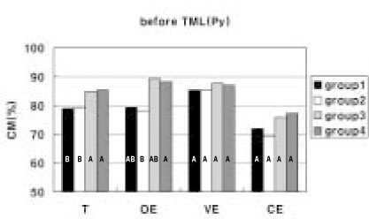
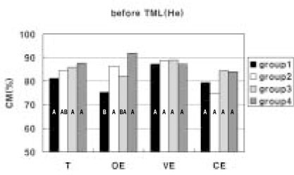
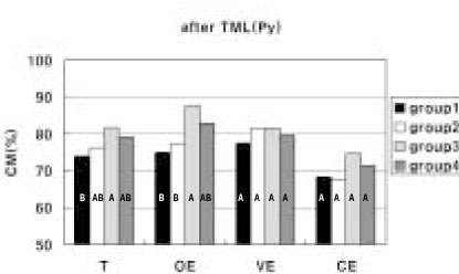
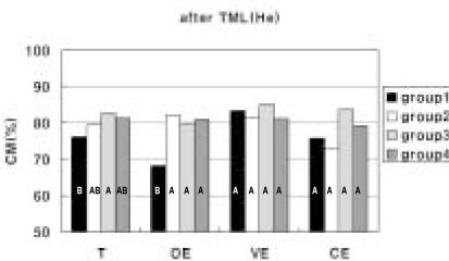
CM: continuous margin, DMR: Duncan's Multiple Range Test, TML: thermo-mechanical loading
*: statistically significant difference between Pyramid and Heliomolar (p < 0.05).

Tables & Figures
REFERENCES
Citations

- Effect of the exponential curing of composite resin on the microtensile dentin bond strength of adhesives
So-Rae Seong, Duck-kyu Seo, In-Bog Lee, Ho-Hyun Son, Byeong-Hoon Cho
Journal of Korean Academy of Conservative Dentistry.2010; 35(2): 125. CrossRef


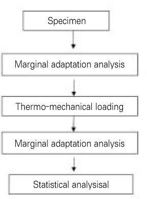


Figure 1
Figure 2
Figure 3
Figure 4
Figure 5
Figure 6
Figure 7
Figure 8
Figure 9
Figure 10
Figure 11
Restorative materials used in this study
List of investigated photoactivation methods with their curing cycles
Three-way ANOVA for 3 variables
a): before or after TML (thermo-mechanical loading)
b): Pyramid or Heliomolar
Mean total CM (%) of each group and DMR Grouping
CM: continuous margin, DMR: Duncan's Multiple Range Test, TML: thermo-mechanical loading
*: statistically significant difference between Pyramid and Heliomolar (p < 0.05).
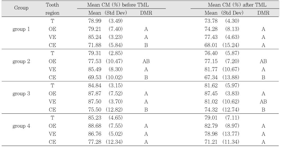
Mean CM (%) of tooth regions and DMR Grouping (Pyramid)
T: total margin, OE: occlusal enamel, VE: vertical enamel, CE: cervical enamel
DMR: Duncan's Multiple Range Test
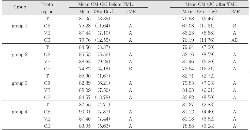
Mean CM (%) of tooth regions and DMR Grouping (Heliomolar)
T: total margin, OE: occlusal enamel, VE: vertical enamel, CE: cervical enamel
DMR: Duncan's Multiple Range Test
a): before or after TML (thermo-mechanical loading) b): Pyramid or Heliomolar
CM: continuous margin, DMR: Duncan's Multiple Range Test, TML: thermo-mechanical loading *: statistically significant difference between Pyramid and Heliomolar (p < 0.05).
T: total margin, OE: occlusal enamel, VE: vertical enamel, CE: cervical enamel DMR: Duncan's Multiple Range Test
T: total margin, OE: occlusal enamel, VE: vertical enamel, CE: cervical enamel DMR: Duncan's Multiple Range Test

 KACD
KACD


 ePub Link
 Cite
Cite

