Articles
- Page Path
- HOME > Restor Dent Endod > Volume 29(4); 2004 > Article
- Original Article Measurements of shrinkage stress and reduction of inter-cuspal distance in maxillary premolars resulting from polymerization of composites and compomers
- Soon-Young Lee, Sung-Ho Park
-
2004;29(4):-352.
DOI: https://doi.org/10.5395/JKACD.2004.29.4.346
Published online: July 31, 2004
Department of Conservative Dentistry, College of Dentistry, Yonsei University, Korea.
- Corresponding author: Sung-Ho Park. Department of Conservative Dentistry, College of Dentistry, Yonsei University, 134 Shinchon-Dong, Seodaemoon Gu, Seoul, Korea, 120-752. Tel: 82-2-361-8709, Fax: 82-2-313-7575, sunghopark@yumc.yonsei.ac.kr
Copyright © 2004 Korean Academy of Conservative Dentistry
- 895 Views
- 0 Download
- 5 Crossref
Abstract
-
The purpose of present study was to evaluate the polymerization shrinkage stress and cuspal deflection in maxillary premolars resulting from polymerization shrinkage of composites and compomers.Composites and compomers which were used in this study were as follows: Dyract AP, Z100, Surefil, Pyramid, Synergy Compact, Heliomolar, Heliomolar HB, and Compoglass F. For measuring of polymerization shrinkage stress, Stress measuring machine (R&B, Daejon, Korea) was used. One-way ANOVA analysis with Duncan's multiple comparison test were used to determine significant differences between the materials.For measuring of cuspal deflection of tooth, MOD cavities were prepared in 10 extracted maxillary premolars. And reduction of intercuspal distance was measured by strain measuring machine (R&B, Daejon, Korea) One-way ANOVA analysis with Turkey test were used to determine significant differences between the materials.Polymerization shrinkage stress is 『Heliomolar, Z100, Pyramid < Synergy Compact Compoglass F < Dyract AP < Heliomolr HB, surefil』 (P < 0.05). And cuspal delfelction is 『Z100, Heliomolar, Heliomolar HB, Synergy Compact Surefil, < Compoglass F < Pyramid, Dyract AP』 (P < 0.05).Measurements of ploymerization shrinkage stress and those of cuspal deflection of the teeth was different. There is no correlation between polymerization shrinkage stress and cuspal deflection of the teeth (p > 0.05).
- 1. Baush JR, de Lange K, Davidson CR, Peters A, De Gee AJ. Clinical significance of polymerization shrinkage of composite resins. J Prosthet Dent. 1982;48: 59-67.ArticlePubMed
- 2. Eick JD, Welch FH. Polymerization shrinkage of posterior composites resins and its possible influence on postoperative sensitivity. Quintessence Int. 1986;17: 103-111.PubMed
- 3. Kemp-Scholte CM, Davidson CL. Marginal sealing of curing contractions gaps in class V composite resin restorations. J Dent Res. 1988;67: 841-845.ArticlePubMedPDF
- 4. Robert JC, Powers JM, Craig RG. Fracture toughness of composite and unfilled restorative resins. J Dent Res. 1977;56: 748.ArticlePubMedPDF
- 5. Feilzer AJ, De Gee AJ, Davidson CL. Setting stress in composite resin in relation to configuration of the restoration. J Dent Res. 1987;66: 1636-1639.ArticlePubMedPDF
- 6. Feilzer AJ, De Gee AJ, Davidson CL. Increased wall to wall curing contraction in thin bonded resin layers. J Dent Res. 1989;68: 48-50.ArticlePubMedPDF
- 7. Hansen EK. Effect of cavity depth and application technique on marginal adaptation of resins in dentin cavities. J Dent Res. 1986;65(11):1319-1321.ArticlePubMedPDF
- 8. Krejci I, Sperr D, Lutz F. A three-sited light curing technique for conventional Class II composite restoraions. Quintessence Int. 1987;38: 125-131.
- 9. Lutz F, Krejci I, Barbakow F. The importance of proximal curing in posterior composite resin restorations. Quintessence Int. 1992;23: 605-607.PubMed
- 10. Krejci I, Lutz F. Marginal adaptation of class V restorations using different restorative technique. J Dent. 1991;19: 24-32.PubMed
- 11. McCullock AJ, Smith B. In vitro studies of cuspal movement produced by adhesive materials. Br Dent J. 1986;161: 405-409.ArticlePubMedPDF
- 12. Park SH, Krejci I, Lutz F. Consistency in the amount of linear polymerization shrinkage in syringe type composite. Dent Mater. 1999;442-446.PubMed
- 13. Park SH, Krejci I, Lutz F. A comparison of microhardness of resin composites polymerized by plasma arc or conventional visible light curing. Oper Dent. 2002;27: 30-37.PubMed
- 14. Suliman AA, Boyer DB, Lakes RS. Cusp movement in premolars resulting from composite polymerization shrinkage. Dent Mater. 1993;9: 6-10.ArticlePubMed
- 15. Ericson D, Paulsson L, Sowaik H, Derand T. Reduction of cusp deflection resulting from composite polymerization shrinkage, using a light-transmitting cone. Scand J Dent Res. 1994;102: 244-248.PubMed
- 16. Park SH, Kim SS, Cho YS, Lee SY, Kim DH, Jang YJ, Mun HS, Seo JW, Noh BD. Amount of polymerization shrinkage stress in composites and compomers for posterior restoration. J Korean Acad Conserv Dent. 2003;28: 354-359.
- 17. Lim BS, Ferracane JL, Sakaguchi RL, Condon JR. Reduction of polymerization contraction stress for dental composites by two-step light activation. Dent Mater. 2002;18: 436-444.ArticlePubMed
- 18. Jacobsen PH. The polymerization shrinkage of composite resins. Dent Mater. 1989;5: 41-44.ArticlePubMed
REFERENCES
Tables & Figures
REFERENCES
Citations
Citations to this article as recorded by 

- Influence of thermal and thermomechanical stimuli on a molar tooth treated with resin-based restorative dental composites
Jerrin Thadathil Varghese, Behzad Babaei, Paul Farrar, Leon Prentice, B. Gangadhara Prusty
Dental Materials.2022; 38(5): 811. CrossRef - Effect of intermittent polymerization on the rate of polymerization shrinkage and cuspal deflection in composite resin
Min Kyung Kim, Sung Ho Park, Deog Gyu Seo, Yun Jung Song, Yoon Lee, Chan Young Lee
Journal of Korean Academy of Conservative Dentistry.2008; 33(4): 341. CrossRef - The effect of intermittent composite curing on marginal adaptation
Yong-Hwan Yun, Sung-Ho Park
Journal of Korean Academy of Conservative Dentistry.2007; 32(3): 248. CrossRef - Correlation Between the Amount of Linear Polymerization Shrinkage and Cuspal Deflection
S-Y. Lee, S-H. Park
Operative Dentistry.2006; 31(3): 364. CrossRef - Correlation between Linear polymerization shrinkage & tooth cuspal deflection
Soon-Young Lee, Sung-Ho Park
Journal of Korean Academy of Conservative Dentistry.2005; 30(6): 442. CrossRef
Measurements of shrinkage stress and reduction of inter-cuspal distance in maxillary premolars resulting from polymerization of composites and compomers
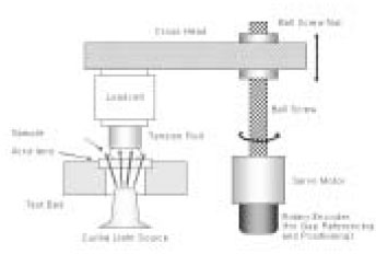
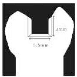
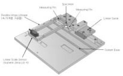
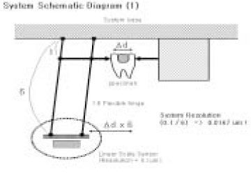
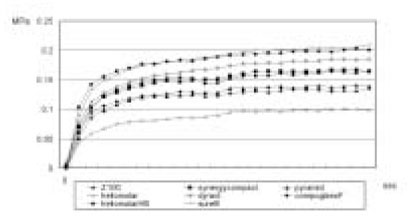
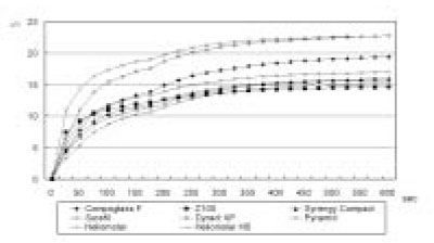
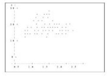
Figure 1
Schematic drawing of the custom made polymerization shrinkage stress measuring machine with a sample in place.
Figure 2
Schematic drawing of cavity preparation.
Figure 3
Cuspal deflection measuring machine.
Figure 4
Schematic drawing of the cuspal deflection measuring machine.
Figure 5
Change in the polymerization stress versus time.
Figure 6
Change of cuspal deflection of premolars versus time.
Figure 7
Regression analysis.
Figure 1
Figure 2
Figure 3
Figure 4
Figure 5
Figure 6
Figure 7
Measurements of shrinkage stress and reduction of inter-cuspal distance in maxillary premolars resulting from polymerization of composites and compomers
Restorative materials used in this study.
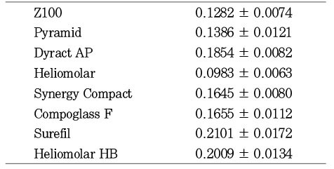
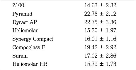
Mean value of shrinkage stress at 120s (MPa)
Mean value of cuspal deflection at 10 min (µm)
Elastic modulus and filler contents of posterior composites
a: Obtained from manufactures' technical manual and home page
b: Data taken from Y. Abe (2001)
Table 1
Restorative materials used in this study.
Table 2
Mean value of shrinkage stress at 120s (MPa)
Table 3
Mean value of cuspal deflection at 10 min (µm)
Table 4
Elastic modulus and filler contents of posterior composites
a: Obtained from manufactures' technical manual and home page b: Data taken from Y. Abe (2001)

 KACD
KACD



 ePub Link
 Cite
Cite

