Articles
- Page Path
- HOME > Restor Dent Endod > Volume 49(4); 2024 > Article
- Research Article Effects of different curing methods on the color stability of composite resins
-
Massimo Pisano
 , Alfredo Iandolo
 , Dina Abdellatif
 , Andrea Chiacchio
 , Marzio Galdi
 , Stefano Martina
-
Restor Dent Endod 2024;49(4):e33.
DOI: https://doi.org/10.5395/rde.2024.49.e33
Published online: September 5, 2024
Department of Medicine, Surgery and Dentistry ‘Scuola Medica Salernitana’, University of Salerno, Baronissi, Italy.
- Correspondence to Massimo Pisano, DDS. Department of Medicine, Surgery and Dentistry ‘Scuola Medica Salernitana’, University of Salerno, Via S. Allende, 84081 Baronissi, Italy. pisano.studio@virgilio.it
Copyright © 2024. The Korean Academy of Conservative Dentistry
This is an Open Access article distributed under the terms of the Creative Commons Attribution Non-Commercial License (https://creativecommons.org/licenses/by-nc/4.0/) which permits unrestricted non-commercial use, distribution, and reproduction in any medium, provided the original work is properly cited.
Abstract
-
Objectives The aim of this study was to compare the effects of different polymerization strategies and the effectiveness of finishing and polishing procedures of composite resins on color stability.
-
Materials and Methods The samples were divided into 4 main groups according to the polymerization strategy, and all groups except the control group received surface treatment. Each group was subsequently divided into 3 subgroups respectively: Kuraray Clearfil Majesty ES-2 Classic, Premium and Universal. Approximately 24 hours after preparation of the samples, they were immersed for 7 days in a coffee solution. A first color measurement was performed after the preparation of the samples, the second measurement was performed after 7 days in the coffee solution. All measurements were carried out using a dental spectrophotometer to assess the CIE L* a* b* color parameters.
-
Results There was a statistically significant difference between ΔE values for different procedures (p = 0.003); in particular, the differences were found only between the groups that received surface treatment and the control group. In addition, a statistically significant difference was observed between the values of ΔE for different composites in the different procedure groups.
-
Conclusions Spectrophotometric analysis showed that the additional photopolymerization and oxygen inhibition procedures did not yield better results in relation to color stability. In addition, finishing and polishing provided better color stability compared to not performing these procedures.
INTRODUCTION
MATERIALS AND METHODS
• Clearfil Majesty ES-2 Universal (Kuraray Co. Ltd., Tokyo, Japan)
• Clearfil Majesty ES-2 Classic (Kuraray Co. Ltd.)
• Clearfil Majesty ES-2 Premium Dentin (Kuraray Co. Ltd.)
• Control group (Group C): A single light-cure was performed, and the composite resins were light-cured for 20 seconds.
• Single application of curing light + Polishing (Group 1): A single 20-second light curing and subsequent polishing was performed.
• Application of curing light + Polishing + Additional application of curing light (Group 2): After the first 20 seconds of photoactivation and subsequent polishing maneuvers, an additional 20 seconds of photo polishing was carried out.
• Glycerin + Single application of curing light + Polishing (Group 3): Before light-curing the samples for 20 seconds, the surface was sprinkled with glycerin (Liquid Strip - Glycerin Gel; Ivoclar Vivadent, Amherst, NY, USA) followed by polishing.
Division of the sample into groups and sub-groups (n = 120)
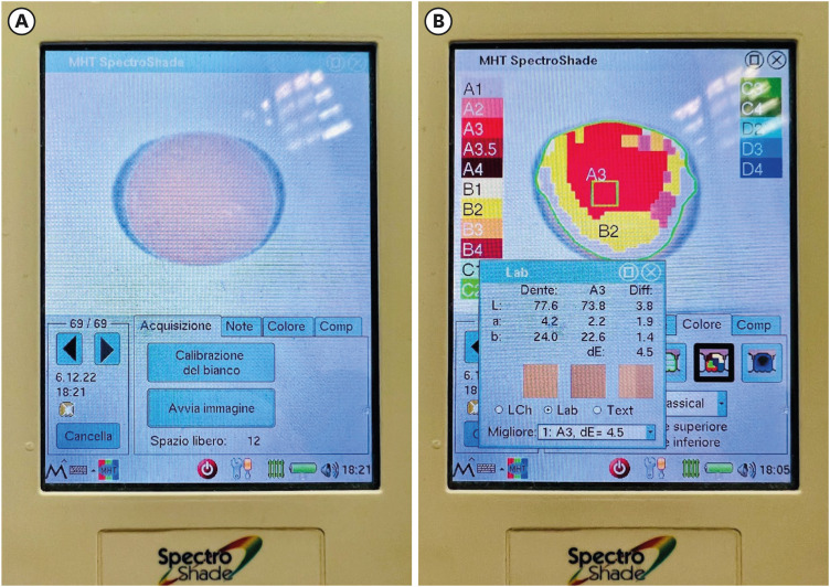
Procedure for using spectrophotometer on composite samples. A sample before (A) and during (B) the spectrophotometer measurement, the spectrophotometer was used to measure the CIE L* a* b* color parameters.

• ΔE is a parameter used to evaluate the discoloration; therefore, it follows that the higher its value, the greater the degree of difference between the final and initial color of the sample examined.
• Δa is the difference between the red-green shades of the sample after and before coffee immersion.
• Δb is the difference between the blue-yellow shades of the sample after and before coffee immersion.
• ΔL is the difference between the brightness of the sample after and before coffee immersion.
RESULTS
ΔE according to different procedure groups, and composite sub-groups
| Groups | ΔE | |
|---|---|---|
| Composites groups | ||
| Kuraray Universal | 7.5 ± 2.5 | |
| Kuraray Classic | 3.3 ± 1.0 | |
| Kuraray Premium | 5.6 ± 2.3 | |
| Procedures groups* | ||
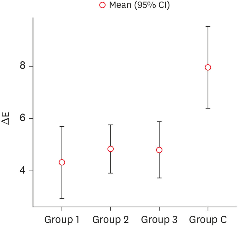
| Group C | 8.0 ± 2.8 | |
| Group 1 | 4.3 ± 2.5 | |
| Group 2 | 4.8 ± 1.7 | |
| Group 3 | 4.8 ± 1.9 | |
ΔE according to different groups and sub-groups
Comparison between the different procedures and the control group as a result of post-hoc analysis with Tukey test
| Comparison between groups* | p value | |
|---|---|---|
| Group 1 | Group C | 0.004 |
| Group 1 | Group 2 | 0.909 |
| Group 1 | Group 3 | 0.933 |
| Group 2 | Group C | 0.006 |
| Group 2 | Group 3 | 1.000 |
| Group 3 | Group C | 0.008 |
Comparison between the distribution of ΔE values in different procedure groups (Group 1: 20 seconds Photopolymerization + Polishing; Group 2: 20 seconds Photopolymerization + Polishing + 20 seconds Photopolymerization; Group 3: Glycerin + 20 seconds Photopolymerization + Polishing) and control group (Group C).

DISCUSSION
CONCLUSION
-
Conflict of Interest: No potential conflict of interest relevant to this article was reported.
-
Author Contributions:
Conceptualization: Pisano M.
Data curation: Chiacchio A, Pisano M.
Formal analysis: Galdi M.
Investigation: Iandolo A, Martina S.
Methodology: Adbellatif D, Galdi M.
Project administration: Chiacchio A, Pisano M.
Software: Galdi M, Chiacchio A.
Supervision: Martina S.
Validation: Pisano M.
Visualization: Iandolo A.
Writing - original draft: Iandolo A, Pisano M,.
Writing - review & editing: Abdellatif D, Martina S.
- 1. Mante FK, Ozer F, Walter R, Atlas AM, Saleh N, Dietschi D, et al. The current state of adhesive dentistry: a guide for clinical practice. Compend Contin Educ Dent 2013;34 Spec 9:2-8.PubMed
- 2. Bagheri R, Burrow MF, Tyas M. Influence of food-simulating solutions and surface finish on susceptibility to staining of aesthetic restorative materials. J Dent 2005;33:389-398.ArticlePubMed
- 3. Bollen CM, Lambrechts P, Quirynen M. Comparison of surface roughness of oral hard materials to the threshold surface roughness for bacterial plaque retention: a review of the literature. Dent Mater 1997;13:258-269.ArticlePubMed
- 4. Sampath S. Survival times of restorative parameters vis-à-vis material combinations. J Res Dent 2015;3:14-20.Article
- 5. Hervás-García A, Martínez-Lozano MA, Cabanes-Vila J, Barjau-Escribano A, Fos-Galve P. Composite resins. A review of the materials and clinical indications. Med Oral Patol Oral Cir Bucal 2006;11:E215-E220.PubMed
- 6. Manhart J, Kunzelmann KH, Chen HY, Hickel R. Mechanical properties of new composite restorative materials. J Biomed Mater Res 2000;53:353-361.ArticlePubMed
- 7. dos Santos Bertoldo CE, Miranda DA, Souza-Junior EJ, Aguiar FHB, Lima DANL, Lovadino JR. Evaluation of surface roughness and color stability of direct resin composites after different polishing protocols. Int J Dent Clin 2011;3:4-7.
- 8. Zhu S, Platt JA. Curing efficiency of three different curing lights at different distances for a hybrid composite. Am J Dent 2009;22:381-386.PubMed
- 9. Price RBT. Light curing in dentistry. Dent Clin North Am 2017;61:751-778.ArticlePubMed
- 10. Guler AU, Yilmaz F, Kulunk T, Guler E, Kurt S. Effects of different drinks on stainability of resin composite provisional restorative materials. J Prosthet Dent 2005;94:118-124.ArticlePubMed
- 11. Sarembe S, Kiesow A, Pratten J, Webster C. The impact on dental staining caused by beverages in combination with chlorhexidine digluconate. Eur J Dent 2022;16:911-918.ArticlePubMedPMC
- 12. Kumari RV, Nagaraj H, Siddaraju K, Poluri RK. evaluation of the effect of surface polishing, oral beverages and food colorants on color stability and surface roughness of nanocomposite resins. J Int Oral Health 2015;7:63-70.
- 13. Tekçe N, Tuncer S, Demirci M, Serim ME, Baydemir C. The effect of different drinks on the color stability of different restorative materials after one month. Restor Dent Endod 2015;40:255-261.ArticlePubMedPMC
- 14. Villalta P, Lu H, Okte Z, Garcia-Godoy F, Powers JM. Effects of staining and bleaching on color change of dental composite resins. J Prosthet Dent 2006;95:137-142.ArticlePubMed
- 15. Kocaagaoglu H, Aslan T, Gürbulak A, Albayrak H, Taşdemir Z, Gumus H. Efficacy of polishing kits on the surface roughness and color stability of different composite resins. Niger J Clin Pract 2017;20:557-565.ArticlePubMed
- 16. Attar N. The effect of finishing and polishing procedures on the surface roughness of composite resin materials. J Contemp Dent Pract 2007;8:27-35.Article
- 17. Reis AF, Giannini M, Lovadino JR, Ambrosano GM. Effects of various finishing systems on the surface roughness and staining susceptibility of packable composite resins. Dent Mater 2003;19:12-18.ArticlePubMed
- 18. Başeren M. Surface roughness of nanofill and nanohybrid composite resin and ormocer-based tooth-colored restorative materials after several finishing and polishing procedures. J Biomater Appl 2004;19:121-134.ArticlePubMedPDF
- 19. Gönülol N, Yilmaz F. The effects of finishing and polishing techniques on surface roughness and color stability of nanocomposites. J Dent 2012;40(Supplement 2):e64-e70.Article
- 20. Barakah HM, Taher NM. Effect of polishing systems on stain susceptibility and surface roughness of nanocomposite resin material. J Prosthet Dent 2014;112:625-631.ArticlePubMed
- 21. International Commission on Illumination (CIE). CIE 142-2001. Improvement to industrial colour-difference evaluation. Vienna: CIE; 2001.
- 22. Asmussen E, Hansen EK. Surface discoloration of restorative resins in relation to surface softening and oral hygiene. Scand J Dent Res 1986;94:174-177.ArticlePubMed
- 23. Ferracane JL, Moser JB, Greener EH. Ultraviolet light-induced yellowing of dental restorative resins. J Prosthet Dent 1985;54:483-487.ArticlePubMed
- 24. Satou N, Khan AM, Matsumae I, Satou J, Shintani H. In vitro color change of composite-based resins. Dent Mater 1989;5:384-387.PubMed
- 25. Um CM, Ruyter IE. Staining of resin-based veneering materials with coffee and tea. Quintessence Int 1991;22:377-386.PubMed
- 26. D’Ambrosio F, Pisano M, Amato A, Iandolo A, Caggiano M, Martina S. Periodontal and peri-implant health status in traditional vs. heat-not-burn tobacco and electronic cigarettes smokers: a systematic review. Dent J 2022;10:103.ArticlePubMedPMC
- 27. Liberato WF, Barreto IC, Costa PP, de Almeida CC, Pimentel W, Tiossi R. A comparison between visual, intraoral scanner, and spectrophotometer shade matching: a clinical study. J Prosthet Dent 2019;121:271-275.ArticlePubMed
- 28. Saegusa M, Kurokawa H, Takahashi N, Takamizawa T, Ishii R, Shiratsuchi K, et al. Evaluation of color-matching ability of a structural colored resin composite. Oper Dent 2021;46:306-315.ArticlePubMedPDF
- 29. Mundim FM, Garcia LF, Pires-de-Souza FC. Effect of staining solutions and repolishing on color stability of direct composites. J Appl Oral Sci 2010;18:249-254.ArticlePubMedPMC
- 30. Alharbi A, Ardu S, Bortolotto T, Krejci I. Stain susceptibility of composite and ceramic CAD/CAM blocks versus direct resin composites with different resinous matrices. Odontology 2017;105:162-169.ArticlePubMedPDF
- 31. Ardu S, Duc O, Di Bella E, Krejci I, Daher R. Color stability of different composite resins after polishing. Odontology 2018;106:328-333.ArticlePubMedPDF
- 32. Alshehri A, Alhalabi F, Mustafa M, Awad MM, Alqhtani M, Almutairi M, et al. effects of accelerated aging on color stability and surface roughness of a biomimetic composite: an in vitro study. Biomimetics (Basel) 2022;7:158.PubMedPMC
- 33. Patel SB, Gordan VV, Barrett AA, Shen C. The effect of surface finishing and storage solutions on the color stability of resin-based composites. J Am Dent Assoc 2004;135:587-594.ArticlePubMed
- 34. Gaviria-Martinez A, Castro-Ramirez L, Ladera-Castañeda M, Cervantes-Ganoza L, Cachay-Criado H, Alvino-Vales M, et al. Surface roughness and oxygen inhibited layer control in bulk-fill and conventional nanohybrid resin composites with and without polishing: in vitro study. BMC Oral Health 2022;22:258.PubMedPMC
- 35. Seyidaliyeva A, Rues S, Evagorou Z, Hassel AJ, Rammelsberg P, Zenthöfer A. Color stability of polymer-infiltrated-ceramics compared with lithium disilicate ceramics and composite. J Esthet Restor Dent 2020;32:43-50.ArticlePubMedPDF
- 36. Marghalani HY. Effect of filler particles on surface roughness of experimental composite series. J Appl Oral Sci 2010;18:59-67.ArticlePubMedPMC
- 37. Mailart MC, Rocha RS, Contreras SCM, Torres CRG, Borges AB, Caneppele TMF. Effects of artificial staining on bulk-filled resin composites. Am J Dent 2018;31:144-148.PubMed
- 38. Ugurlu M. Effect of repolishing on the discoloration of indirect composite block, nanohybrid, and microhybrid resin composites. Eur Oral Res 2022;56:158-163.ArticlePubMedPMC
- 39. de Abreu JLB, Sampaio CS, Benalcázar Jalkh EB, Hirata R. Analysis of the color matching of universal resin composites in anterior restorations. J Esthet Restor Dent 2021;33:269-276.ArticlePubMedPDF
- 40. Silva MVMD, Batista JMN, Fraga MAA, Correr AB, Campos EA, Geraldeli S, et al. Surface analysis of a universal resin composite and effect of preheating on its physicochemical properties. Braz Dent J 2023;34:115-126.ArticlePubMedPMC
- 41. Hardan L, Bourgi R, Cuevas-Suárez CE, Lukomska-Szymanska M, Monjarás-Ávila AJ, Zarow M, et al. Novel trends in dental color match using different shade selection methods: a systematic review and meta-analysis. Materials (Basel) 2022;15:468.ArticlePubMedPMC
- 42. Sideridou I, Tserki V, Papanastasiou G. Effect of chemical structure on degree of conversion in light-cured dimethacrylate-based dental resins. Biomaterials 2002;23:1819-1829.ArticlePubMed
- 43. Rueggeberg FA, Caughman WF, Curtis JW Jr. Effect of light intensity and exposure duration on cure of resin composite. Oper Dent 1994;19:26-32.PubMed
- 44. Unsal KA, Karaman E. Effect of additional light curing on colour stability of composite resins. Int Dent J 2022;72:346-352.ArticlePubMed
- 45. Alkhudhairy F. Wear resistance of bulk-fill composite resin restorative materials polymerized under different curing intensities. J Contemp Dent Pract 2017;18:39-43.ArticlePubMed
- 46. Panchal AC, Asthana G. Oxygen inhibition layer: a dilemma to be solved. J Conserv Dent 2020;23:254-258.ArticlePubMedPMC
- 47. Rodrigues-Junior SA, Chemin P, Piaia PP, Ferracane JL. surface roughness and gloss of actual composites as polished with different polishing systems. Oper Dent 2015;40:418-429.ArticlePubMedPDF
- 48. Park HH, Lee IB. Effect of glycerin on the surface hardness of composites after curing. J Korean Acad Conserv Dent 2011;36:483-439.Article
- 49. Borges MG, Silva GR, Neves FT, Soares CJ, Faria-E-Silva AL, Carvalho RF, et al. Oxygen inhibition of surface composites and its correlation with degree of conversion and color stability. Braz Dent J 2021;32:91-97.ArticlePubMed
REFERENCES
Tables & Figures
REFERENCES
Citations

- Color Stability Under Challenge: Effects of Thermo-Aging and Mouthrinse Exposure on Anterior Teeth and Esthetic Composites
Gökçe Keçeci, Zehra Güner, Süleyman Ziya Şenyurt, Kamile Erciyas
European Journal of Therapeutics.2026;[Epub] CrossRef - Abrasiveness and Bleaching Level of Toothpastes on Composite Resins: A Quantitative Analysis Using a Novel Brushing Simulator
Simge Meseli, Elif Alkan, Bora Korkut, Ozlem Kanar, Dilek Tagtekin
Applied Sciences.2025; 15(5): 2314. CrossRef - Comparative Evaluation of Direct and Indirect Composite Restorations in Class II Tooth Preparations - An In vivo Study
Akshun Gupta, Garima Arora, Aprajita Mehta, Satish Sane, Siddhi Nevrekar, Apurva Nagrale
Advances in Human Biology.2025; 15(4): 550. CrossRef - Micro- and Nanoplastics and the Oral Cavity: Implications for Oral and Systemic Health, Dental Practice, and the Environment—A Narrative Review
Federica Di Spirito, Veronica Folliero, Maria Pia Di Palo, Giuseppina De Benedetto, Leonardo Aulisio, Stefano Martina, Luca Rinaldi, Gianluigi Franci
Journal of Functional Biomaterials.2025; 16(9): 332. CrossRef


Figure 1
Figure 2
Division of the sample into groups and sub-groups (n = 120)
| Composites groups | Procedures groups* | |||
|---|---|---|---|---|
| Group C | Group 1 | Group 2 | Group 3 | |
| Kuraray Universal | ||||
| Kuraray Classic | ||||
| Kuraray Premium | ||||
*The samples were divided into the following groups: Group C, A single light-cure was performed, and the composite resins were light-cured for 20 seconds; Group 1, 20 seconds Photopolymerization + Polishing; Group 2, 20 seconds Photopolymerization + Polishing + 20 seconds Photopolymerization; and Group 3, Glycerin + 20 seconds Photopolymerization + Polishing.
ΔE according to different procedure groups, and composite sub-groups
| Groups | ΔE | |
|---|---|---|
| Composites groups | ||
| Kuraray Universal | 7.5 ± 2.5 | |
| Kuraray Classic | 3.3 ± 1.0 | |
| Kuraray Premium | 5.6 ± 2.3 | |
| Procedures groups* | ||
| Group C | 8.0 ± 2.8 | |
| Group 1 | 4.3 ± 2.5 | |
| Group 2 | 4.8 ± 1.7 | |
| Group 3 | 4.8 ± 1.9 | |
Values are presented as mean ± standard deviation.
*The samples were divided into the following groups: Group C, A single light-cure was performed, and the composite resins were light-cured for 20 seconds; Group 1, 20 seconds Photopolymerization + Polishing; Group 2, 20 seconds Photopolymerization + Polishing + 20 seconds Photopolymerization; and Group 3, Glycerin + 20 seconds Photopolymerization + Polishing.
ΔE according to different groups and sub-groups
| Procedures* | Composite | ΔE | |
|---|---|---|---|
| Group C | Kuraray Universal | 10.4 ± 1.5 | < 0.001 |
| Kuraray Classic | 9.0 ± 0.6 | ||
| Kuraray Premium | 4.4 ± 0.8 | ||
| Group 1 | Kuraray Universal | 6.5 ± 2.7 | 0.011 |
| Kuraray Classic | 4.2 ± 1.4 | ||
| Kuraray Premium | 2.2 ± 0.8 | ||
| Group 2 | Kuraray Universal | 6.2 ± 1.3 | < 0.001 |
| Kuraray Classic | 5.3 ± 1.0 | ||
| Kuraray Premium | 3.0 ± 0.5 | ||
| Group 3 | Kuraray Universal | 7.0 ± 1.8 | < 0.001 |
| Kuraray Classic | 3.7 ± 0.8 | ||
| Kuraray Premium | 3.7 ± 0.5 |
The
*The samples were divided into the following groups: Group C, A single light-cure was performed, and the composite resins were light-cured for 20 seconds; Group 1, 20 seconds Photopolymerization + Polishing; Group 2, 20 seconds Photopolymerization + Polishing + 20 seconds Photopolymerization; and Group 3, Glycerin + 20 seconds Photopolymerization + Polishing.
Comparison between the different procedures and the control group as a result of post-hoc analysis with Tukey test
| Comparison between groups* | ||
|---|---|---|
| Group 1 | Group C | 0.004 |
| Group 1 | Group 2 | 0.909 |
| Group 1 | Group 3 | 0.933 |
| Group 2 | Group C | 0.006 |
| Group 2 | Group 3 | 1.000 |
| Group 3 | Group C | 0.008 |
*The samples were divided into the following groups: Group C, A single light-cure was performed, and the composite resins were light-cured for 20 seconds; Group 1, 20 seconds Photopolymerization + Polishing; Group 2, 20 seconds Photopolymerization + Polishing + 20 seconds Photopolymerization; and Group 3, Glycerin + 20 seconds Photopolymerization + Polishing.
*The samples were divided into the following groups: Group C, A single light-cure was performed, and the composite resins were light-cured for 20 seconds; Group 1, 20 seconds Photopolymerization + Polishing; Group 2, 20 seconds Photopolymerization + Polishing + 20 seconds Photopolymerization; and Group 3, Glycerin + 20 seconds Photopolymerization + Polishing.
Values are presented as mean ± standard deviation.
*The samples were divided into the following groups: Group C, A single light-cure was performed, and the composite resins were light-cured for 20 seconds; Group 1, 20 seconds Photopolymerization + Polishing; Group 2, 20 seconds Photopolymerization + Polishing + 20 seconds Photopolymerization; and Group 3, Glycerin + 20 seconds Photopolymerization + Polishing.
The
*The samples were divided into the following groups: Group C, A single light-cure was performed, and the composite resins were light-cured for 20 seconds; Group 1, 20 seconds Photopolymerization + Polishing; Group 2, 20 seconds Photopolymerization + Polishing + 20 seconds Photopolymerization; and Group 3, Glycerin + 20 seconds Photopolymerization + Polishing.
*The samples were divided into the following groups: Group C, A single light-cure was performed, and the composite resins were light-cured for 20 seconds; Group 1, 20 seconds Photopolymerization + Polishing; Group 2, 20 seconds Photopolymerization + Polishing + 20 seconds Photopolymerization; and Group 3, Glycerin + 20 seconds Photopolymerization + Polishing.

 KACD
KACD
 ePub Link
 Cite
Cite

