
Search
- Page Path
- HOME > Search
Research Articles
- Fracture resistance of regenerated immature teeth in different simulated stages of root development: an in vitro cyclic loading study
- Kyveli-Artemis Polydora, Konstantinos Kodonas, Anastasia Fardi, Christos Gogos
- Restor Dent Endod 2026;51(2):e21. Published online April 28, 2026
- DOI: https://doi.org/10.5395/rde.2026.51.e21
-
 Abstract
 PDF
PDF Supplementary Material
 PubReader
 ePub
ePub - Objectives
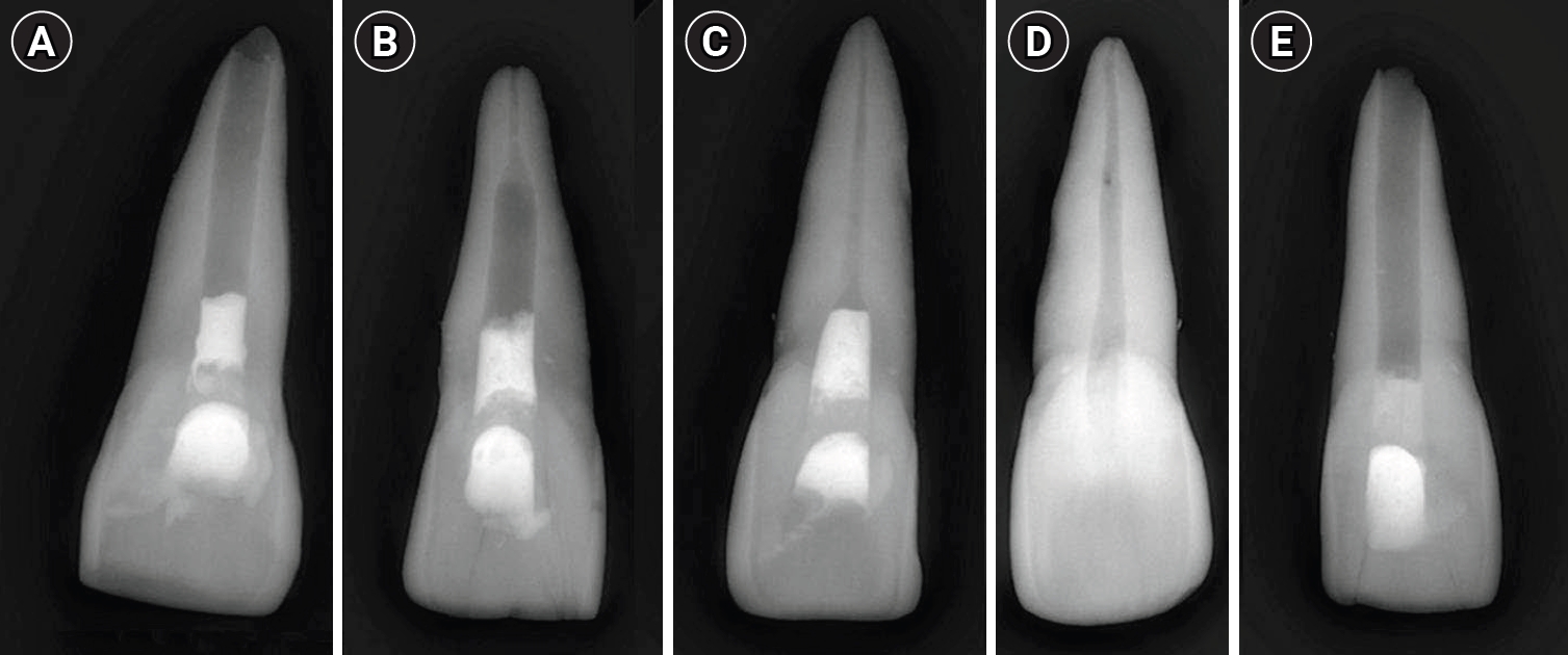
This in vitro study aimed to assess the fracture resistance of simulated stages of root maturation following regenerative endodontic treatment using a cyclic loading method.
Methods
Ninety extracted maxillary central incisors were randomly allocated into three experimental groups representing different stages of root development, following revitalization: Group A for completely immature teeth immediately after treatment; Group B for teeth with apical closure, and Group C for teeth with apical closure and wall thickening. Two control groups were also included: Group D for intact teeth and Group E for simulated immature teeth without the bioceramic material. Following simulation of immature apices and treatment with a bioceramic material, all specimens were subjected to cyclic loading using a step-stress fatigue protocol until failure. The number of cycles to fracture and the peak load were recorded and statistically analyzed.
Results
Statistically significant differences in loading forces were observed between the negative control group (Group D) and Groups A, B, and E (p < 0.05). However, no statistically significant differences were detected among the experimental groups. These results indicate that apical closure and dentinal wall thickening alone did not substantially improve mechanical reinforcement under cyclic loading conditions.
Conclusions
Although intact teeth exhibited superior mechanical performance, apical closure and wall thickening alone were insufficient to enhance reinforcement under cyclic loading.
- 540 View
- 51 Download

- Determination of optimal horizontal beam angulations for canal separation in mandibular molars using cone-beam computed tomography: a retrospective image-based analysis
- Benedikt Schneider, Tamina Tepe, Daniel Rapp, Wilhelm Frank, Maria Lessani, Constantin von See, Sebastian Fitzek, Jörg Philipp Tchorz
- Restor Dent Endod 2026;51(1):e9. Published online February 26, 2026
- DOI: https://doi.org/10.5395/rde.2026.51.e9
-
Abstract
PDFPubReaderePub
- Objectives
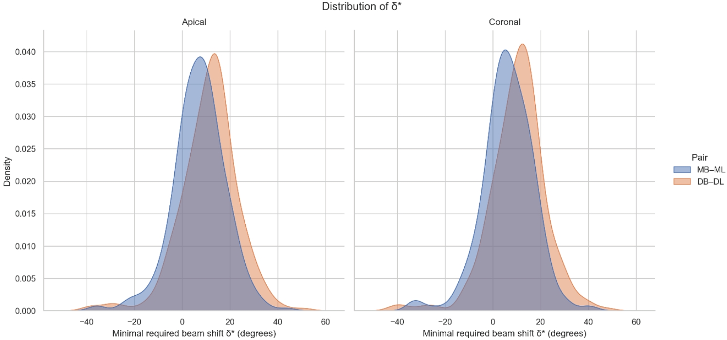
Two-dimensional intraoral radiographs often obscure canals due to superimposition, especially in mandibular molars with complex anatomy. This cone-beam computed tomography (CBCT) study identified the horizontal beam angles at which first and second molar canals overlap and derived clinically applicable angulations for enhanced canal separation.
Methods
Eighty-five CBCT datasets from 100 patients met the inclusion criteria, yielding 318 mandibular molars (160 first, 158 second). Using ImageJ, absolute horizontal overlap angles (α) were measured to determine the corresponding theoretical separation angles defined as δ* = 90° – α. Separability was modeled across horizontal beam angulation increments from −45° to +45° in five steps, and Wilson’s 95% confidence intervals were computed. Group comparisons used the Mann-Whitney U and independent t-tests (p ≤ 0.05)
Results
Minimal mesial beam angulations for effective canal separability (δ* = 90° − α) ranged from approximately 7° to 15° for mesial roots and approximately 10° to 13° for distal roots. No significant mesial differences were observed between first and second molars (p > 0.30). Distal roots of second molars exhibited significantly higher angulations (p = 0.003 coronal, p < 0.001 apical). Mesial canals achieved ≥95% separability at approximately 25° and ≥99% at approximately 35°; distal canals required approximately 30° and approximately 40°.
Conclusions
A mesial beam angulation of 30° to 35° provides probable canal differentiation in mandibular molars, separating mesial canals in ≥99% and distal canals in ≥95% of cases. This range refines previous recommendations and supports the as low as reasonably achievable (ALARA) principle.
- 1,130 View
- 32 Download
- Analysis of the reciprocating kinematics of the VDW Silver Reciproc, E-Connect Pro, Ecom, and Endopen endodontic motors: an in vitro experimental study
- Cristielly França, Juliana D. Bronzato, Dieimes Braambati, Adriana de-Jesus-Soares, Carla C. R. B. Félix, Michelle A. N. S. Ferreira, Marcos Frozoni
- Restor Dent Endod 2026;51(1):e5. Published online January 20, 2026
- DOI: https://doi.org/10.5395/rde.2026.51.e5
-
Abstract
PDFPubReaderePub
- Objectives
This study aimed to evaluate the actual parameters of four endodontic motors, each adjusted for reciprocating motion, and compare them to the manufacturers’ declared values.
Methods
The motors used were the VDW Silver Reciproc (VDW GmbH), E-Connect Pro (MK Life), Ecom (Woodpecker), and Endopen (Schuster Woodpecker). A custom optical target was attached to the motor contra-angle, the movements were recorded with a high-resolution camera, and the images were analyzed. Engagement, disengagement, net angles, and speed for each operation cycle, duration of clockwise (CW) and counter-clockwise (CCW) movement, duration of standstill after CW and CCW movement, and the number of cycles to complete a full rotation were analyzed. The data were statistically analyzed at a significance level of 5%. The replicability of all reciprocal parameters analyzed was statistically different from that reported by the manufacturers.
Results
There was no statistically significant difference between the VDW Silver Reciproc, Ecom, and Endopen for the engagement angle. The E-Connect Pro was the least reliable at the 150°/30° settings for both angle parameters. There was no significant difference between the set and actual cycle net angles for the VDW Silver Reciproc (p = 0.493). While the actual values for the Ecom and E-Connect Pro were significantly higher than the set (p < 0.001), the actual values for the Endopen were significantly lower than the set (p < 0.001).
Conclusions
Experiments on four commercially available reciprocating endodontic motors revealed that the actual motor values differed significantly from the set values.
- 1,614 View
- 79 Download
Case Report
- Fifty-year follow-up of dens invaginatus treated by nonsurgical and surgical endodontic treatments: a case report
- Qais Arow, Eyal Rosen, Galit Sela, Shlomo Elbahary, Igor Tsesis
- Restor Dent Endod 2026;51(1):e1. Published online December 18, 2025
- DOI: https://doi.org/10.5395/rde.2026.51.e1
-
Abstract
PDFPubReaderePub
- This case report presents a lateral maxillary incisor with dens invaginatus (DI) type IIIb that was treated both nonsurgically and surgically over 50 years. Treatment of teeth with DI can be challenging. Suggested options may include nonsurgical root canal treatment, endodontic surgery, or extraction. In this case report, a 13-year-old patient with a lateral maxillary incisor with DI type IIIb was treated by nonsurgical root canal treatment, modern endodontic surgery, and reoperation over the course of 50 years. There was complete healing at the last follow-up, 11 years after the reoperation. Correct diagnosis and proper treatment using modern endodontic techniques can enable teeth with DI to survive throughout the life span of the patient.
- 1,686 View
- 143 Download
- 1 Web of Science
Research Articles
- Evaluation of platelet concentrates in regenerative endodontics: a systematic review and meta-analysis
- Anna Tsiolaki, Dimitrios Theocharis, Nikolaos Tsitsipas, Anastasia Fardi, Konstantinos Kodonas
- Restor Dent Endod 2025;50(4):e38. Published online November 28, 2025
- DOI: https://doi.org/10.5395/rde.2025.50.e38
-
Abstract
PDFSupplementary MaterialPubReaderePub
- Objectives
The aim of this systematic review is to compare the effectiveness of advanced platelet concentrates as regenerative endodontic therapeutic alternatives to blood clot (BC) revascularization in immature permanent necrotic teeth.
Methods
Randomized controlled trials (RCTs) comparing regenerative endodontic therapies using platelet-rich plasma (PRP), platelet-rich fibrin (PRF), or platelet pellet (PP) with the BC revascularization approach in immature permanent necrotic teeth were systematically searched in PubMed, Scopus, Cochrane Library, and Web of Science until May 2025. Data was extracted and analyzed both qualitatively and quantitatively. Study quality was assessed using the Cochrane Risk of Bias tool. A meta-analysis was conducted using IBM SPSS software (version 29.0), with success rates expressed as risk ratios and 95% confidence intervals (CIs).
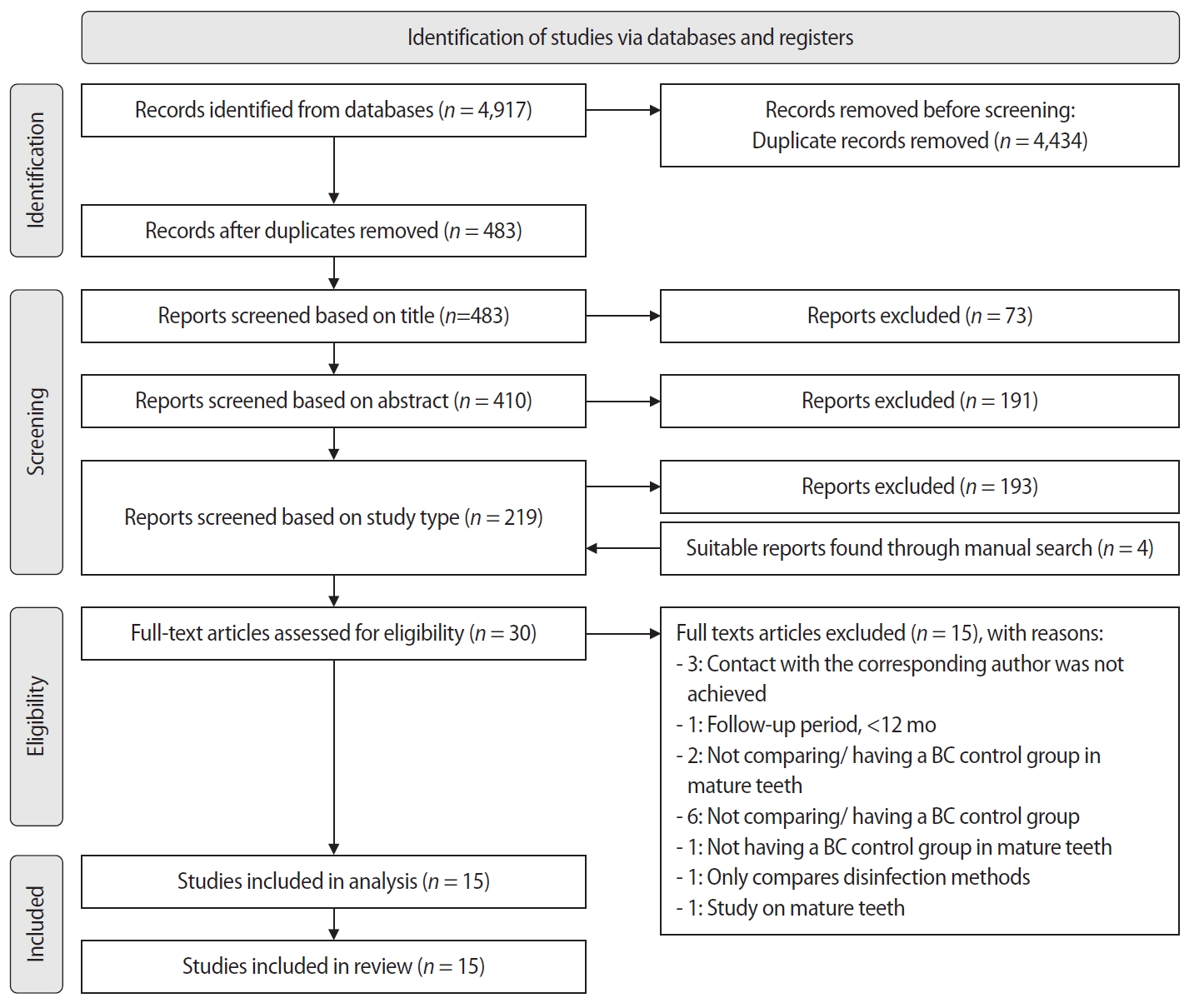
Results
The initial search yielded 4,917 studies. After removing duplicates and applying eligibility criteria, 15 RCTs were included. Meta-analysis indicated no significant difference in the risk ratio (RR), as the BC method has similar success rates with PRP (10 studies; RR = 1.01; 95% CI, 0.94–1.09; p = 0.76) and PRF (8 studies; RR = 0.98; 95% CI, 0.89–1.08; p = 0.65) at 12 months. The primary outcomes evaluated were based on clinical and radiographic success.
Conclusions
Current evidence suggests PRP, PRF, and BC are all effective in treating immature permanent necrotic teeth with similar success rates. However, further research is needed to assess long-term outcomes. -
Citations
Citations to this article as recorded by
- Longitudinal periapical radiographic evaluation of apexification, vital pulpotomy, and revascularization in immature permanent teeth: a retrospective comparative study
Xiaona Sun, Kailing Zhu
Frontiers in Bioengineering and Biotechnology.2026;[Epub] CrossRef
- Longitudinal periapical radiographic evaluation of apexification, vital pulpotomy, and revascularization in immature permanent teeth: a retrospective comparative study
- 2,186 View
- 111 Download
- 1 Web of Science
- 1 Crossref
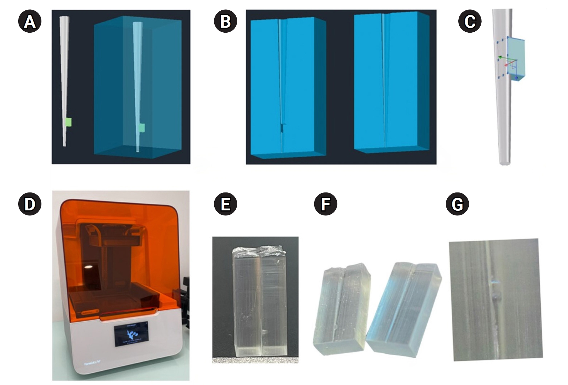
- In vitro experimental study comparing continuous and intermittent irrigation protocols: influence of sodium hypochlorite volume and contact time on tissue dissolution
- Alfredo Iandolo, Dina Abdellatif, Davide Mancino, Gwenael Rolin, Camille Coussens, Aurelian Louvrier, Felipe G Belladonna, Edouard Euvrard, Emmanuel João Nogueira Leal da Silva
- Restor Dent Endod 2025;50(4):e36. Published online October 15, 2025
- DOI: https://doi.org/10.5395/rde.2025.50.e36
-
Abstract
PDFPubReaderePub
- Objectives
This study aimed to evaluate whether continuous irrigation with larger volumes or allowing sodium hypochlorite (NaOCl) resting time is more critical for pulp tissue dissolution using a controlled artificial root canal system.
Methods
A three-dimensional printed artificial root canal with a lateral canal in the apical third was fabricated. Standardized bovine pulp tissue specimens were inserted, and three irrigation protocols were tested: group A (continuous NaOCl irrigation at 1 mL/min via syringe pump), group B (intermittent NaOCl irrigation with 0.1 mL and a 3-minute resting period), and group C (control, saline irrigation). The time for complete dissolution and the total NaOCl volume were recorded.
Results
Complete dissolution occurred in groups A and B, with significant differences in NaOCl volume and time (p < 0.05). In group A, complete dissolution was consistently observed after the 6th irrigation cycle, corresponding to a total NaOCl volume of 6.0 ± 0.66 mL per test. The average time required for complete dissolution in this group was 6 ± 0.66 minutes. In group B, complete dissolution occurred after the 4th cycle, with a total NaOCl volume of 0.4 ± 0.06 mL per test and a mean dissolution time of 12.6 ± 1.8 minutes.
Conclusions
NaOCl volume and exposure time significantly influence pulp tissue dissolution.
- 2,122 View
- 182 Download
Case Report
- Multidisciplinary management of an endo-perio lesion complicated by a cemental tear: a case report
- Nishanth D. Sadhak, Akshaya Pallod, Shreyas Oza
- Restor Dent Endod 2025;50(3):e31. Published online August 22, 2025
- DOI: https://doi.org/10.5395/rde.2025.50.e31

-
Abstract
PDFPubReaderePub
- Endodontic-periodontal lesions (EPLs) complicated by cemental tears present a diagnostic and therapeutic challenge. This case report describes the successful management of a 66-year-old male patient with a mandibular second molar (#18) exhibiting an EPL complicated by a cemental tear. Clinical examination revealed a draining sinus tract, deep periodontal pockets, and radiographic evidence of a “J-shaped” lesion and a radiopaque cemental fragment. The tooth had previously initiated endodontic treatment. A multidisciplinary approach involving endodontic treatment and surgical removal of the cemental tear was implemented. At 24-month follow-up, clinical and radiographic examination revealed significant improvement in periodontal health, bone regeneration, and resolution of the lesion. This case highlights the importance of considering cemental tears in the differential diagnosis of EPLs and demonstrates the efficacy of a combined endodontic-periodontal approach for achieving predictable outcomes.
- 4,266 View
- 325 Download
Research Articles
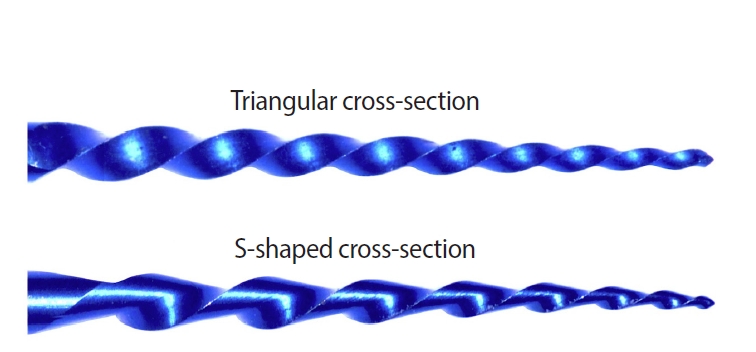
- Isolating design variables by assessing the impact of cross-section geometry on the mechanical performance of nickel-titanium rotary instruments: a comparative in vitro study
- Anne Rafaella Tenório Vieira, Guilherme Ferreira da Silva, Emmanuel João Nogueira Leal da Silva, Rodrigo Ricci Vivan, João Vitor Oliveira de Amorim, Thaine Oliveira Lima, Raimundo Sales de Oliveira Neto, Marco Antonio Hungaro Duarte, Murilo Priori Alcalde
- Restor Dent Endod 2025;50(3):e28. Published online July 24, 2025
- DOI: https://doi.org/10.5395/rde.2025.50.e28
-
Abstract
PDFPubReaderePub
- Objectives
This study aimed to assess the effect of cross-section geometry on the mechanical properties of nickel-titanium (NiTi) instruments by comparing two instruments with identical tip size, taper, and thermal treatment but differing in cross-section design.
Methods
One hundred four NiTi rotary instruments, being S-shaped and triangular cross-section, manufactured with Blueish thermal treatment, were tested (n = 52 per group). Differential scanning calorimetry was employed, and the metal mass volume and cross-section area were assessed. The cyclic fatigue, torsional, and bending resistance tests were assessed. Data were analyzed using the Kolmogorov-Smirnov and Student t tests, and the level of significance was set at 5%.
Results
The instruments exhibited similar start and finish temperatures of phase transformation. The S-shaped instruments had significantly lower metal mass volume and cross-sectional area (p < 0.05). S-shaped instruments demonstrated superior cyclic fatigue resistance, greater angular deflection, and lower bending stiffness (p < 0.05).
Conclusions
Cross-section geometry significantly influences the mechanical properties of NiTi rotary instruments. -
Citations
Citations to this article as recorded by- Mechanical properties and micro-CT-based biomechanical performance of NiTi instrumentation systems in mandibular molars
Jeneffer Vieira Rodrigues, Julia Godoi-Lopes, Heitor Silva Prado, Anne Rafaella Tenório Vieira, Rafael Verardino Camargo, Graziela Bianchi Leoni, Marco Antonio Hungaro Duarte, Igor Bassi Ferreira Petean, Manoel Damião Sousa-Neto, Murilo Priori Alcalde, Fa
Odontology.2026;[Epub] CrossRef
- Mechanical properties and micro-CT-based biomechanical performance of NiTi instrumentation systems in mandibular molars
- 3,116 View
- 106 Download
- 2 Web of Science
- 1 Crossref
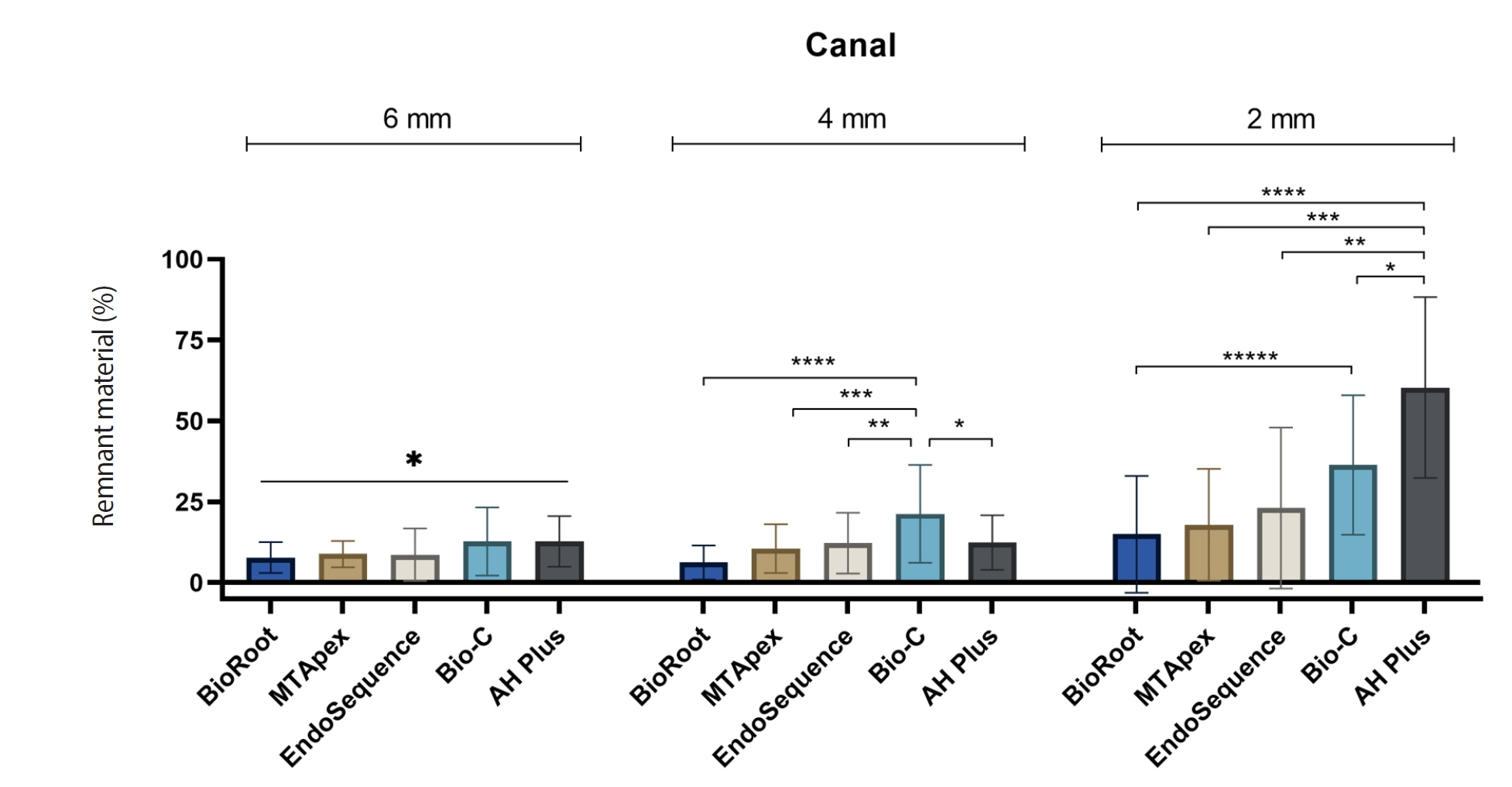
- Calcium silicate-based sealers remnants in isthmuses of mesial roots of mandibular molars: an in vitro evaluation
- David Saldanha de Brito Alencar, Ana Cristina Padilha Janini, Lauter Eston Pelepenko, Brenda Fornazaro Moraes, Francisco Haiter Neto, Marco Antonio Hungaro Duarte, Marina Angélica Marciano
- Restor Dent Endod 2025;50(3):e25. Published online July 15, 2025
- DOI: https://doi.org/10.5395/rde.2025.50.e25
-
Abstract
PDFPubReaderePub
- Objectives
Endodontic retreatment aims to address treatment failure through the removal of root canal filling materials. This in vitro study evaluated the presence of filling material remnants in the mesial root canals, specifically focusing on the isthmuses, of mandibular molars after retreatment.
Methods
One hundred extracted mandibular molar mesial roots with isthmuses were prepared with an R25 file, obturated with one of five calcium silicate-based sealers (BioRoot RCS [Septodont], MTApex [Ultradent Products Inc.], EndoSequence BC Sealer HiFlow [Brasseler USA], Bio-C Sealer [Angelus]) or an epoxy resin-based sealer (AH Plus Jet [Dentsply Maillefer]), all stained with rhodamine B, and stored at 37ºC for 30 days to allow for setting. Retreatment was subsequently performed using R40 and XP-endo Finisher R instruments (FKG Dentaire) with 2.5% sodium hypochlorite irrigation. The presence of remaining filling material was then assessed using confocal microscopy, and setting times were tested per ISO 6876:2012.
Results
AH Plus Jet showed the most remnants at 2 mm and the longest retreatment time. Calcium silicate-based sealers exhibited prolonged setting times under dry conditions, with EndoSequence BC Sealer HiFlow showing a particularly extended setting period.
Conclusions
Despite retreatment, residues remained in all canals and isthmus regions, particularly Bio-C Sealer and AH Plus Jet in apical areas, emphasizing the difficulty of complete removal and the persistence of filling material. -
Citations
Citations to this article as recorded by- Bonding effects of mechanical removal of bioceramic sealer residues using glycine or glass microparticles abrasion
Jesus Aranda, Julia de Freitas Ceccato, Eduardo Fernández Godoy, João Felipe Besegato, Joissi Ferrari Zaniboni, Regina Guenka Palma-Dibb, Milton Carlos Kuga
International Journal of Adhesion and Adhesives.2026; 148: 104289. CrossRef
- Bonding effects of mechanical removal of bioceramic sealer residues using glycine or glass microparticles abrasion
- 2,621 View
- 123 Download
- 1 Web of Science
- 1 Crossref
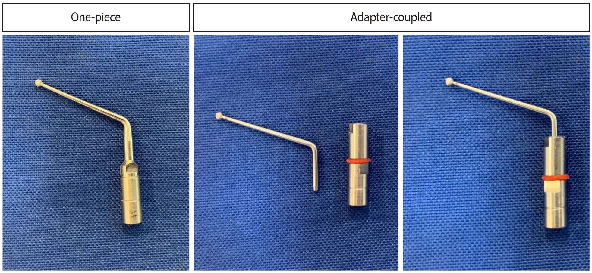
- Analysis of thermal profiles on tooth structure and insert during one-piece or adapter-coupled ultrasonic insert use: an in vitro experimental study
- Gabriela Loewen Brotto, Bruno Monguilhott Crozeta, Bruno Marques-da-Silva, Alysson Nunes Diógenes, Emmanuel João Nogueira Leal da Silva, Flávia Sens Fagundes Tomazinho
- Restor Dent Endod 2025;50(3):e24. Published online July 11, 2025
- DOI: https://doi.org/10.5395/rde.2025.50.e24
-
Abstract
PDFPubReaderePub
- Objectives
This in vitro study aimed to evaluate temperature variation on the external surface of mandibular molars and within ultrasonic inserts when using adapter-coupled versus one-piece inserts.
Methods
Twenty-four extracted human mandibular molars were divided into two groups based on the type of ultrasonic insert used: adapter-coupled and one-piece inserts. Temperature on the external surface of each tooth was measured with a thermocouple probe positioned in the furcation area, capturing data continuously. The temperature of the ultrasonic inserts was monitored in real-time using a thermal imaging camera. Measurements were taken in a controlled environment without cooling for over 120 seconds. Statistical analysis was conducted using analysis of variance (ANOVA) and two-way ANOVA with repeated measures to evaluate temperature variations between groups and over time, with significance set at 5%.
Results
In the external tooth surface temperature measurements, no significant differences were observed between the groups during the initial 15 seconds (p = 0.185) and 30 seconds (p = 0.067). However, significant differences emerged at 60 seconds (p = 0.025), 90 seconds (p = 0.024), and 120 seconds (p = 0.020), with the one-piece insert group demonstrating higher temperatures in the furcation region. Thermal imaging of the inserts revealed a significant difference at all time points (p < 0.001), with adapter-coupled inserts showing greater heating.
Conclusions
The use of ultrasonic inserts leads to a gradual rise in temperature on the external tooth surface. One-piece inserts generated higher temperatures on the tooth, while adapter-coupled inserts exhibited greater heating within the insert.
- 2,370 View
- 104 Download
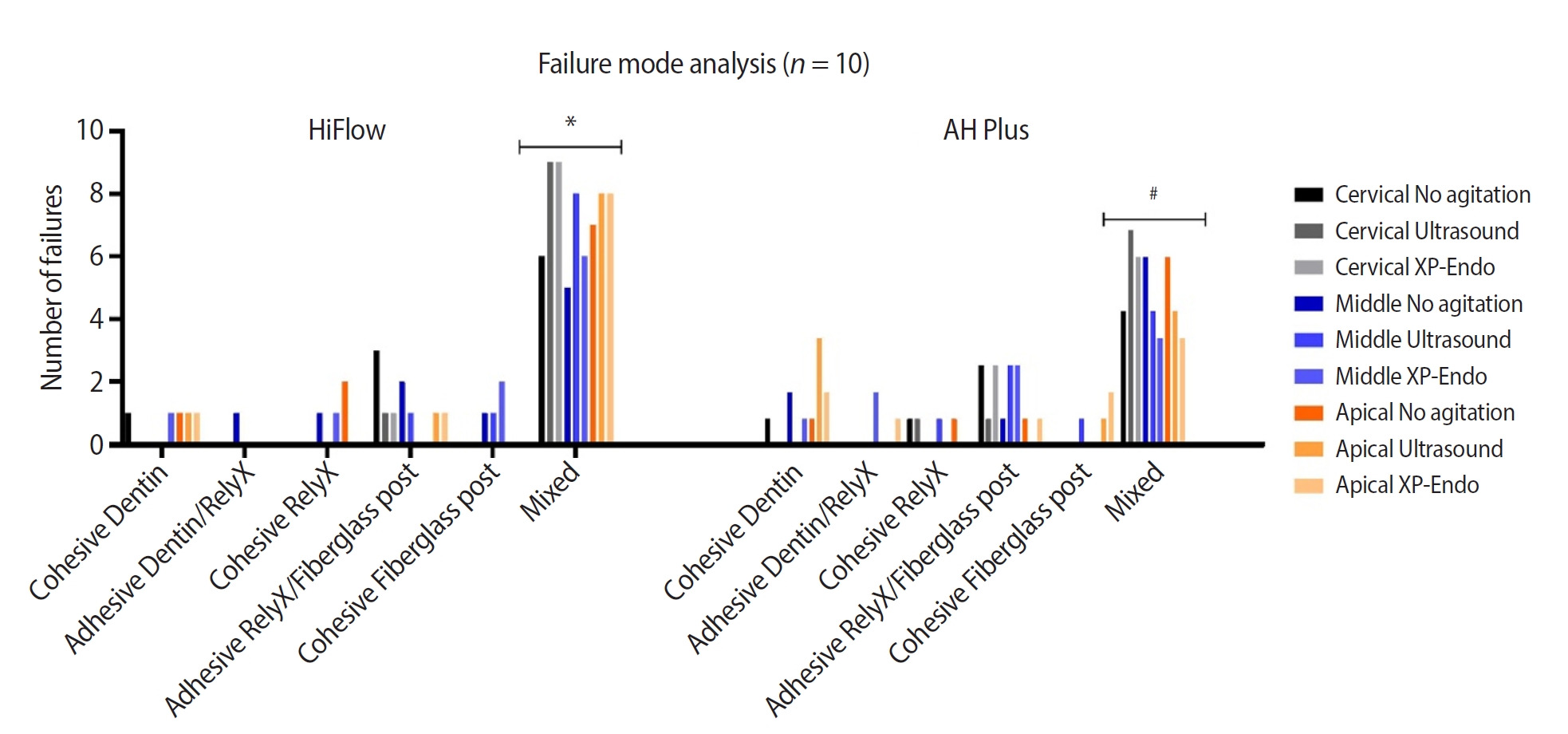
- Cleaning protocols to enhance bond strength of fiberglass posts on root canals filled with bioceramic sealer: an in vitro comparative study
- Thiago Bessa Marconato Antunes, Juliana Delatorre Bronzato, Joice Graciani, Ana Cristina Padilha Janini, Rocharles Cavalcante Fontenele, Francisco Haiter Neto, Brenda Paula Figueiredo de Almeida Gomes, Marina Angélica Marciano da Silva
- Restor Dent Endod 2025;50(2):e20. Published online May 21, 2025
- DOI: https://doi.org/10.5395/rde.2025.50.e20
-
Abstract
PDFPubReaderePub
- Objectives
This study aimed to evaluate whether the agitation protocols using ultrasonic inserts or the XP-endo Finisher R file improved the removal of two different endodontic sealer remnants and the bond strength of fiberglass posts to dentin.
Methods
Seventy-two human teeth were selected. The canals were prepared with Reciproc 50 and Easy ProDesign 30/.10 and root filled according to the endodontic sealer groups: AH Plus or EndoSequence BC Sealer HiFlow. The samples were kept at 37ºC and 95% humidity for 28 days. During the post space preparation, the obturation was removed with Largo burs, and the groups were divided according to the irrigant agitation protocols (n = 12): no agitation, agitation with R1-Clearsonic associated with E1-Irrisonic ultrasonic inserts, or agitation with XP-endo Finisher R file. The fiberglass posts were cemented with RelyX ARC. The roots were sectioned into slices and submitted to the push-out test. Micro-computed tomography analysis was used to check the effectiveness of irrigating solution agitation in the elimination of remnants.
Results
The cleaning protocols with agitation were more effective in increasing the bond strength of posts to dentin for both sealer groups compared to non-agitation (p < 0.05). There was no difference between the same cleaning protocols for the different sealers. Among the different thirds, there was no statistical difference for the same sealer in the different cleaning protocols (p > 0.05).
Conclusions
Both agitation protocols effectively clean root-filled canals sealed with resin-based and calcium silicate-based sealers during fiberglass post space preparation. These protocols result in improved bond strength compared to non-agitation methods. -
Citations
Citations to this article as recorded by- Cleaning efficacy and bond interaction of glycine-based air polishing and glass microparticles abrasion on dentin impregnated with premixed bioceramic sealer
Ândresson Aurélio Fernandes Martins, Maria Carolina Sidonio Alves, Bruno Martins Maciel, José Rodolfo Estruc Verbicário, João Felipe Besegato, Wilfredo Gustavo Escalante-Otárola, Milton Carlos Kuga
International Journal of Adhesion and Adhesives.2026; 147: 104277. CrossRef - Effect of Endodontic Sealers on the Bond Strength of Glass Fibre Posts: A Systematic Review
Thiago Bessa Marconato Antunes, Juliana D. Bronzato, Vanessa Gallego Arias Pecorari, Jennifer Santos Pereira, Talita Tartari, Adriana de Jesus Soares, Brenda P. F. A. Gomes, Marina Angélica Marciano
Australian Endodontic Journal.2026;[Epub] CrossRef
- Cleaning efficacy and bond interaction of glycine-based air polishing and glass microparticles abrasion on dentin impregnated with premixed bioceramic sealer
- 4,921 View
- 240 Download
- 2 Web of Science
- 2 Crossref
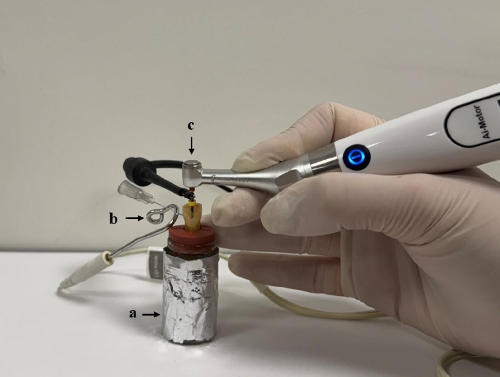
- Impact of the use of high-power 810-nm diode laser as monotherapy on the clinical and tomographic success of the treatment of teeth with periapical lesions: an observational clinical study
- Fabricio Hinojosa Pedraza, Abel Victor Isidro Teves-Cordova, Murilo Priori Alcalde, Marco Antonio Hungaro Duarte
- Restor Dent Endod 2025;50(2):e15. Published online May 15, 2025
- DOI: https://doi.org/10.5395/rde.2025.50.e15
-
Abstract
PDFPubReaderePub
- Objectives
The aim of this study was to demonstrate the impact of a high-power 810-nm diode laser as monotherapy on the clinical and tomographic success of treating teeth with periapical lesions, through a series of 31 cases.
Methods
Teeth with apical lesions underwent endodontic treatment in which a high-power 810-nm diode laser with saline solution was used as monotherapy for disinfection. This type of therapy aimed to replace the traditional irrigation protocol with sodium hypochlorite. This research is the first to assess the clinical success of this alternative treatment, along with tomographic evaluations conducted over periods ranging from 2 to 7 years, analyzed using the periapical index based on cone-beam computed tomography (CBCTPAI). All cases were performed by a single clinician following the same laser protocol, which involved using 1 W of continuous power and four cycles of 20 seconds of laser activation.
Results
All teeth showed no clinical symptoms upon follow-up examination. However, the tomographic evaluation revealed that the success rates for teeth receiving primary treatment were 60% and 80% according to strict and loose criteria, respectively. For teeth requiring retreatment, the success rates were 12.5% and 37.5% using strict and loose criteria, respectively.
Conclusions
The teeth with apical lesions that underwent primary treatment did not present clinical symptoms, but they showed a moderate success rate on tomographic evaluation. However, despite lacking clinical symptoms, teeth with apical lesions that required retreatment had a very low success rate on tomographic evaluation. -
Citations
Citations to this article as recorded by- Adherence to core outcome set for endodontic treatments (COSET) international consensus: Two-year before/after bibliometric systematic review
Carolina Bender Hoppe, Pauline Mastella Lang, Lara Dotto, Mateus Silveira Martins Hartmann, Fabiana Soares Grecca
Journal of Dentistry.2026; : 106834. CrossRef - Diode Laser-Guided Protocol for Endo-Perio Lesions: Toward a Multi-Stage Therapeutic Strategy—A Case Series and Brief Literature Review
Ioana-Roxana Munteanu, George-Dumitru Constantin, Ruxandra-Elena Luca, Ioana Veja, Mariana-Ioana Miron
Medicina.2025; 61(12): 2157. CrossRef
- Adherence to core outcome set for endodontic treatments (COSET) international consensus: Two-year before/after bibliometric systematic review
- 4,863 View
- 223 Download
- 1 Web of Science
- 2 Crossref
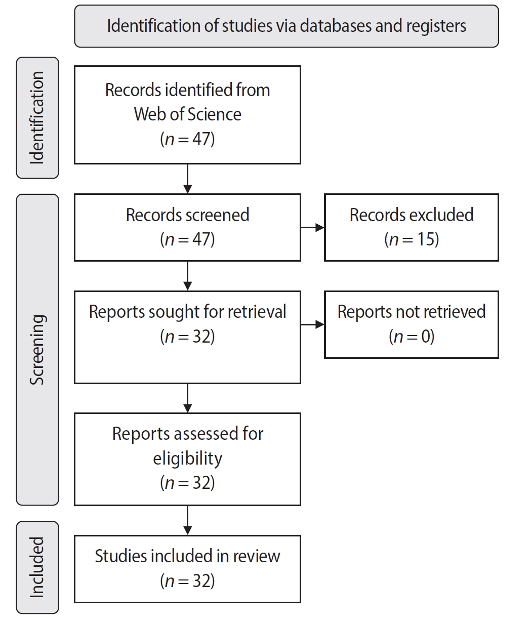
- Bibliometric analysis of the GentleWave system: trends, collaborations, and research gaps
- Raimundo Sales de Oliveira Neto, Thais de Moraes Souza, João Vitor Oliveira de Amorim, Thaine Oliveira Lima, Guilherme Ferreira da Silva, Rodrigo Ricci Vivan, Murilo Priori Alcalde, Marco Antonio Hungaro Duarte
- Restor Dent Endod 2025;50(2):e17. Published online May 12, 2025
- DOI: https://doi.org/10.5395/rde.2025.50.e17
-
Abstract
PDFSupplementary MaterialPubReaderePub
- Objectives
The study aimed to conduct a bibliometric analysis of the GentleWave system (Sonendo, Inc.).
Methods
An electronic search was conducted in June 2024 using the Web of Science Collection database. Two reviewers independently screened publications, extracting data on authorship, publication details, study design, and citation metrics. Statistical analyses were performed in R to assess variable correlations, while the VOSviewer (Visualization of Similarities Viewer) software was used to map author and keyword networks.
Results
The search yielded 47 records, with 32 studies included. Publications spanned 2014 to 2024. The Journal of Endodontics published the highest number of studies (n = 15), and the International Endodontic Journal had the highest impact factor (5.4). The University of British Columbia and Sonendo, Inc. were the most frequent affiliations. Among the 32 articles, 28 were in vitro studies, primarily focusing on microbiology (n = 9). A total of 95 authors were identified, with Haapasalo and Shen being the most cited (n = 229). The articles accumulated 495 citations, demonstrating a strong positive correlation between the number of studies and citation counts (r = 0.98).
Conclusions
The analysis highlights a predominance of in vitro studies. Geographic concentration in the United States and Canada limits diversity, while the strong correlation between study numbers and citations suggests that increased publication volume enhances visibility. -
Citations
Citations to this article as recorded by- Three-year Outcomes of Conventional Versus Minimally Invasive Endodontic Treatment Protocols: A Retrospective Study
Kiavash Hossini, He Liu, Ya Shen, Jolanta Aleksejuniene, Fahda Algahtani, Ahmed Hieawy
Journal of Endodontics.2026; 52(4): 558. CrossRef
- Three-year Outcomes of Conventional Versus Minimally Invasive Endodontic Treatment Protocols: A Retrospective Study
- 4,243 View
- 99 Download
- 1 Web of Science
- 1 Crossref
- The effect of limonene extract on the adhesion of different endodontic cements to root dentin: an in vitro experimental study
- Nayara Lima Ferraz Aguiar, Eduardo José Soares, Guilherme Nilson Alves dos Santos, Anna Luísa Araújo Pimenta, Laryssa Karla Romano, Ricardo Gariba Silva, Fernanda de Carvalho Panzeri
- Restor Dent Endod 2025;50(2):e16. Published online May 12, 2025
- DOI: https://doi.org/10.5395/rde.2025.50.e16
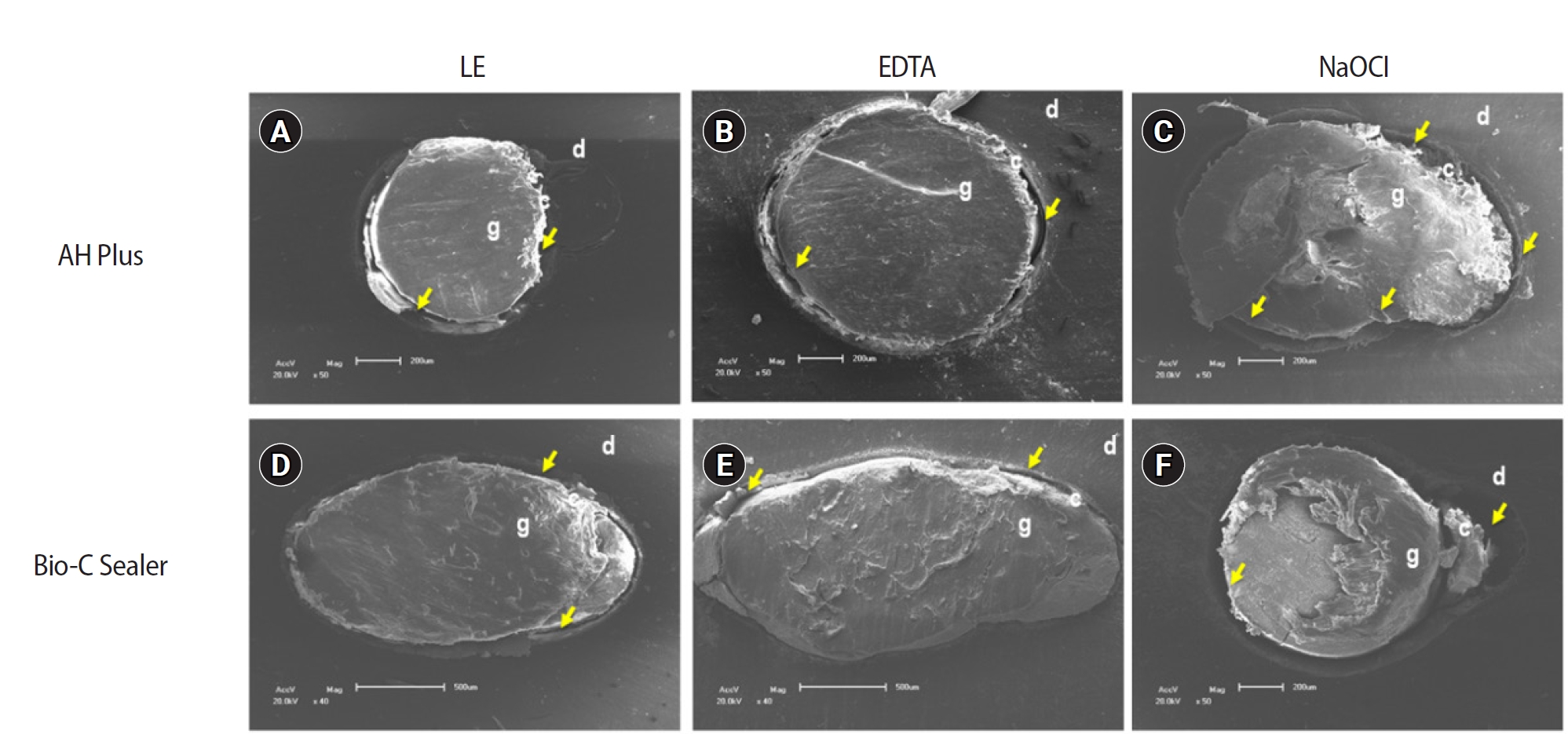
-
Abstract
PDFPubReaderePub
- Objectives
The study aimed to evaluate the effect of limonene extract (LE) on push-out bond strength (BS) to root dentin in endodontically treated teeth.
Methods
Single-rooted teeth were selected and instrumented using the reciprocating technique, then divided into three groups based on the final irrigating solution: 2.5% sodium hypochlorite (NaOCl), 17% ethylenediaminetetraacetic acid (EDTA), and 5% LE. The roots were further divided (n = 12) and obturated using the single-cone technique with epoxy resin-based (ERB) or bioceramic sealer (Bio-C). After 3 days, the roots were sectioned into 2-mm slices, obtaining two slices from each root third. Push-out BS testing was conducted at 0.5 mm/min, followed by failure pattern and adhesive interface analysis using scanning electron microscopy. Push-out BS data were analyzed by three-way analysis of variance and Tukey post-hoc test (p < 0.05).
Results
ERB showed higher BS when irrigated with EDTA (5.0 ± 2.3 MPa) compared to NaOCl (1.8 ± 1.1 MPa) (p = 0.0005), particularly in the cervical third. LE yielded intermediate values without significant differences from the other irrigants (3.5 ± 1.9 MPa) (p > 0.05). For Bio-C, the highest BS was observed in the apical third, especially with LE (9.4 ± 5.0 MPa), differing from other thirds and final irrigating solutions (p < 0.05). Mixed failure patterns were most prevalent, regardless of the irrigant solutions.
Conclusions
The combination of LE with Bio-C demonstrated superior BS in the apical third, suggesting its potential as a final irrigating solution in endodontic treatments.
- 3,101 View
- 237 Download
- Evaluation of the effects of different file systems and apical functions of integrated endodontic motors on debris extrusion: an ex vivo experimental study
- Sıla Nur Usta, Antonio Magan-Fernandez, Cumhur Aydın
- Restor Dent Endod 2025;50(2):e14. Published online April 14, 2025
- DOI: https://doi.org/10.5395/rde.2025.50.e14
-
Abstract
PDFPubReaderePub
- Objectives
This study aimed to evaluate the effects of two different file systems operated with three apical functions of an endodontic motor integrated with an electronic apex locator on debris extrusion.
Methods
Sixty single-rooted teeth were prepared and divided into two main groups and three subgroups based on the file system (OneShape [Micro-Mega SA] and WaveOne [Dentsply Maillefer]) and apical function of the endodontic motor used (auto apical stop [AAS], auto apical reverse [AAR], and auto apical slowdown [ASD]). The teeth were mounted in pre-weighed glass tubes filled with 0.9% sodium chloride to complete the circuit with the apex locator. Files were advanced until the respective apical function (stop, reverse, or slowdown) was activated. The extruded debris was collected, dried, and weighed by subtracting pre-weighed values from post-weighed values. Preparation time was also recorded. Statistical analyses were performed to compare the groups.
Results
OneShape was associated with significantly less debris extrusion compared to WaveOne, regardless of the apical function (p < 0.05). The ASD function resulted in the least debris extrusion compared to AAS and AAR (p < 0.05). Preparation time was significantly longer in the ASD function (p < 0.05), while no differences were observed between the file systems (p > 0.05).
Conclusions
The OneShape file system and the ASD function produced the least amount of apical debris. While the ASD function requires more preparation time, its potential to minimize debris extrusion suggests it may reduce postoperative symptoms. -
Citations
Citations to this article as recorded by- Inflammatory Mediator Levels and Postoperative Pain Following Root Canal Shaping with Different Apical Actions: A Randomized Controlled Trial
Mustafa Mert Tulgar, Yağmur Kılıç, Oğuz Karalar, Huriye Erbak Yılmaz, Emrah Karataşlıoğlu
Journal of Endodontics.2026; 52(4): 574. CrossRef
- Inflammatory Mediator Levels and Postoperative Pain Following Root Canal Shaping with Different Apical Actions: A Randomized Controlled Trial
- 5,150 View
- 249 Download
- 1 Web of Science
- 1 Crossref
Case Report
- Surgical management of maxillary sinusitis of endodontic origin after reestablishing maxillary sinus floor healing through a nonsurgical approach: a case report
- Eun-Sook Kang, Min-Kyeong Kim, Mi-Kyung Yu, Kyung-San Min
- Restor Dent Endod 2025;50(2):e12. Published online April 8, 2025
- DOI: https://doi.org/10.5395/rde.2025.50.e12
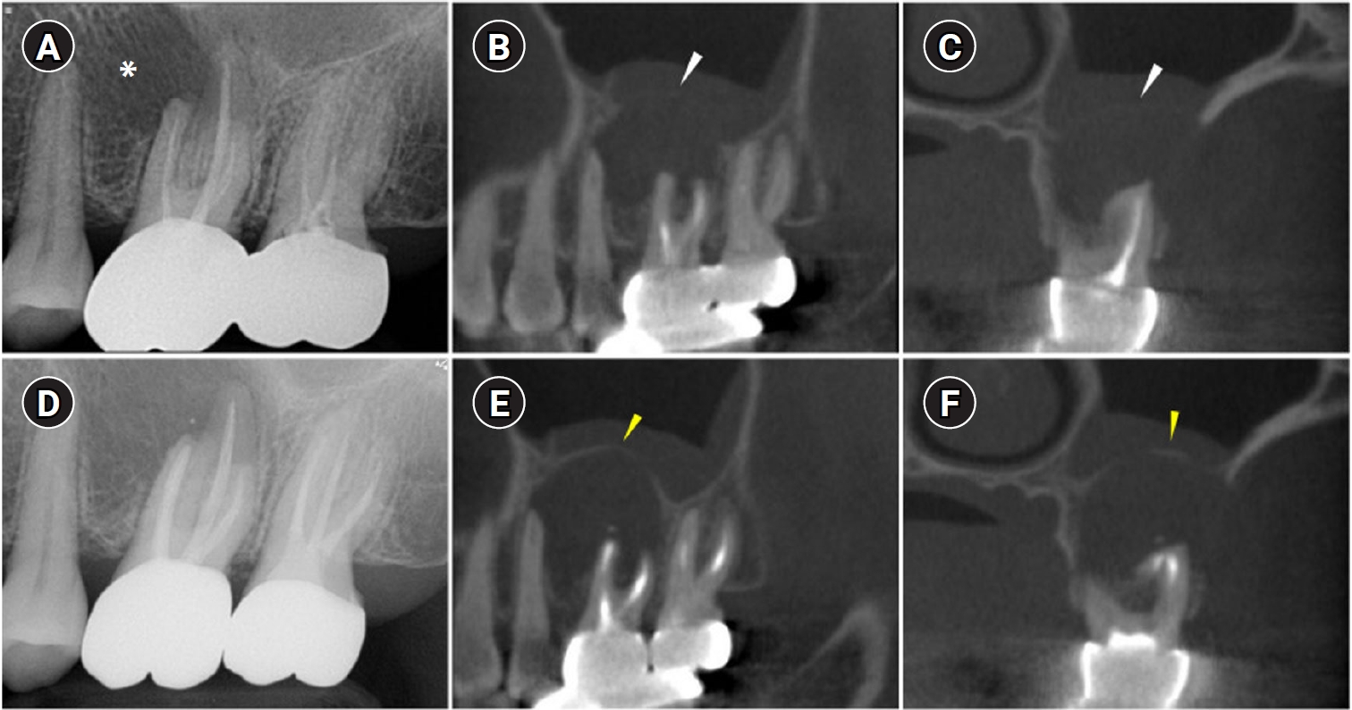
-
Abstract
PDFPubReaderePub
- When root canal infections breach the maxillary sinus floor (MSF), maxillary sinusitis of endodontic origin (MSEO) can result. This case illustrates the surgical management of MSEO following the nonsurgical reestablishment of the MSF. A 55-year-old woman presented with left facial pain and was diagnosed with MSEO originating from the left upper first molar. Despite undergoing nonsurgical root canal treatment, there was no evidence of bony healing after 6 months. However, cone-beam computed tomographic (CBCT) scans revealed the reestablishment of MSF. Subsequently, surgical intervention was carried out using a dental operating microscope. Two years after surgery, CBCT images indicated that the mucosal edema had resolved, and the MSF was well reestablished. Preserving the MSF is crucial for the success of endodontic surgery. When MSEO is present, the integrity of the MSF must be assessed to determine appropriate treatment options.
- 5,156 View
- 233 Download
Research Articles
- Pattern of endodontic instrument separation and factors affecting its retrieval: a 10-year retrospective observational study in a postgraduate institute
- Velmurugan Natanasabapathy, Aswathi Varghese, Paul Kevin Abishek Karthikeyan, Srinivasan Narasimhan
- Restor Dent Endod 2025;50(1):e7. Published online February 19, 2025
- DOI: https://doi.org/10.5395/rde.2025.50.e7
-
Abstract
PDFPubReaderePub
- Objectives
This study aimed to assess the pattern of endodontic instrument separation, their retrievability, and factors affecting its retrieval, in a postgraduate institute.
Methods
Cases referred for the management of separated endodontic instruments (SEI) from 2013 to 2023 were considered for this study. Data related to demographics, tooth type, file type, and retrieval were documented in an Excel sheet. Eight prognostic factors assumed to influence the retrieval were analyzed in this study. The secondary aim was to compare the pattern of SEI and retrievability between conventional nickel-titanium files and newer generation heat-treated nickel-titanium files. Retrieval was attempted by a senior endodontist under the dental operating microscope. Various ultrasonic tips and a Broken Tool Removal loop system were used during retrieval. Simple descriptive statistics were performed. Binomial logistic regression was done to identify the effect of the eight prognostic factors on the retrieval outcome.
Results
A total of 190 SEI was reported. SEI occurred more often in posterior teeth than anterior teeth, mandibular arch than maxillary arch, and in larger files than smaller files. Separation occurred more often in the apical third compared to the other levels. Retrieval was attempted in 88 cases and successful in 70 cases (79.5%). The larger taper and apical position of the SEI negatively influenced the retrieval by 1.4 and 8.7 times, respectively.
Conclusions
Retrieval of SEI was successful in the majority of the cases. An increase in taper and apically placed SEI negatively impacted the retrieval. There was no difference in the pattern of separation nor retrievability between conventional nickel-titanium files and newer generation heat-treated nickel-titanium files. -
Citations
Citations to this article as recorded by- Risk Factors for Failure of Separated Instrument Removal: A Systematic Review and Meta‐Analysis
Le Zhao, WangYu Luo, Yue Shen, WanNing Yu, Liu Yang, Xiaolei Zhang
Australian Endodontic Journal.2026; 52(1): 331. CrossRef - Efficiency of ultrasonic retrieval for separated instruments within the middle third of root canals using modified staging platform: a comparative in-vitro study
Basim Samir Mohamed, Nihal Ezzat Sabet, Dina Ahmed Morsy
BMC Oral Health.2026;[Epub] CrossRef - Influence of the Cause of File Fracture on the Successful Removal of Fragments from Root Canals: An In Vivo Study
Ricardo Portigliatti, Eugenia Pilar Consoli Lizzi, Pablo Alejandro Rodríguez
Applied Sciences.2026; 16(8): 3832. CrossRef - Prevalence and management techniques for separated endodontic files among endodontic postgraduate students in the Qassim region: A retrospective cross-sectional study
Hanin Abdulaziz Alsalhi, Rana Rabeh Alharbi, Amnah Ameen Hawsa
Saudi Endodontic Journal.2026; 16(2): 160. CrossRef - Effectiveness of microscope-assisted root canal treatment in permanent posterior teeth: A retrospective cohort study
Ya-Ching Chang, Ting-Ya Wang
Journal of Dentistry.2025; 157: 105771. CrossRef - Deep Learning-Based Detection of Separated Root Canal Instruments in Panoramic Radiographs Using a U2-Net Architecture
Nildem İnönü, Umut Aksoy, Dilan Kırmızı, Seçil Aksoy, Nurullah Akkaya, Kaan Orhan
Diagnostics.2025; 15(14): 1744. CrossRef - MANAGEMENT OF INTRACANAL SEPARATED INSTRUMENTS: FACTORS CONTRIBUTING TO ENDODONTIC FILE SEPARATION — A NARRATIVE REVIEW
Tareq Hajaj, Paul Freiman , Serban Talpos Niculescu , Mihai Rominu , Tiberiu Hosszu , Ioana Veja
Romanian Journal of Oral Rehabilitation.2025; 17(2): 993. CrossRef
- Risk Factors for Failure of Separated Instrument Removal: A Systematic Review and Meta‐Analysis
- 8,969 View
- 420 Download
- 5 Web of Science
- 7 Crossref
- Effect of quality of radiographs taken during root canal treatment on technical quality of root canal fillings and endodontic outcome
- Jia Min Ng, Yan Yee Lee, Prashanti Chippagiri, Elaheh Ahanin, Abhishek Parolia
- Restor Dent Endod 2025;50(1):e3. Published online January 7, 2025
- DOI: https://doi.org/10.5395/rde.2025.50.e3
-
Abstract
PDFPubReaderePub
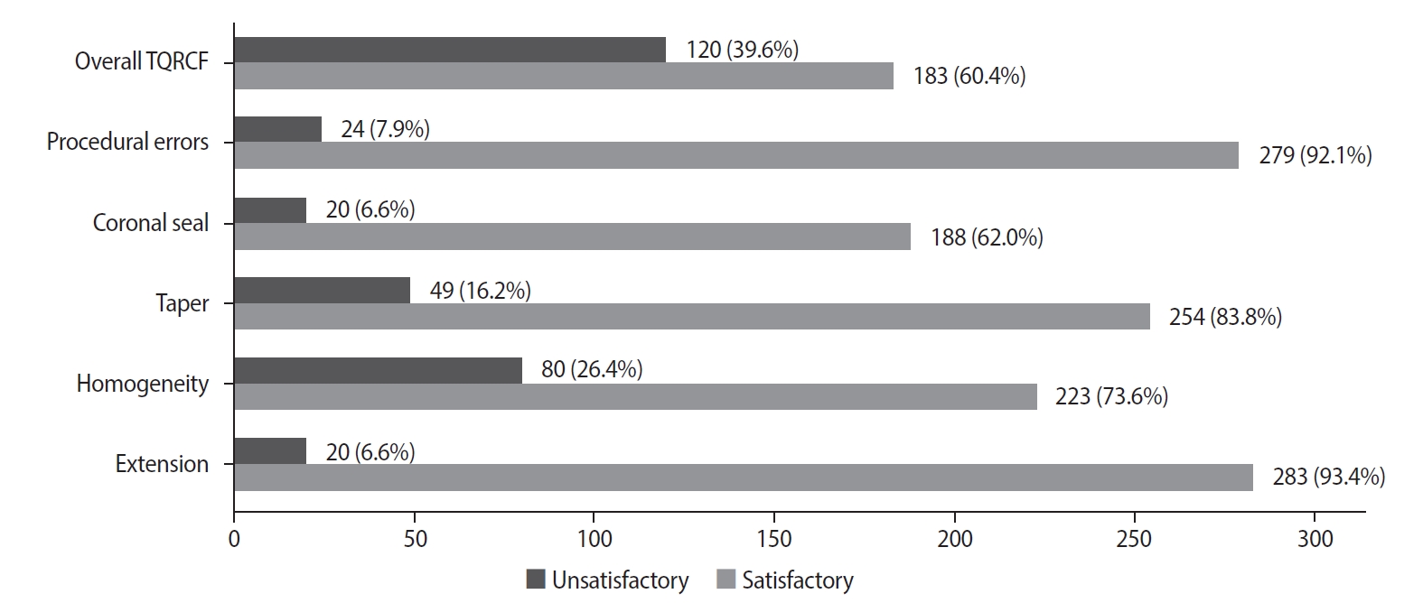
- Objectives
This study evaluated the number and quality of working length (WL) and master cone (MC) radiographs taken during root canal treatment by dental undergraduates, and their associations with the technical quality of root canal fillings (TQRCF) and endodontic outcomes (EO).
Methods
A retrospective evaluation of radiographs from 303 root canal-treated teeth in 231 patients was conducted, with 72 patients attending recall visits to assess EO. The chi-square and one-way analysis of variance tests were performed.
Results
A total of 505 WL and 557 MC radiographs were reviewed, with 72.9% and 75% deemed satisfactory, respectively. Satisfactory TQRCF was achieved in 60.4% of cases. Significant associations were found between the extension of the file in WL and gutta-percha in MC radiographs and TQRCF (p = 0.000). Misinterpretation of these radiographs resulted in poor TQRCF. Furthermore, 64.2% of teeth had satisfactory EO. A significant relationship was noted between the quality of MC radiographs and both TQRCF (p = 0.043) and EO (p = 0.003).
Conclusions
Unsatisfactory MC radiographs were linked to poor TQRCF and unfavorable EO. Regular radiographic training is recommended to enhance EO. -
Citations
Citations to this article as recorded by- Radiographic evaluation of root canal fillings: can undergraduate dental students perform it?
Emine Odabaşı Tezer, Fadi Nahas, Alhabab Shbitah, İrem Dilara Kılıç, Ahmet Bölük, Meltem Öztan
BMC Medical Education.2026;[Epub] CrossRef - Assessment of radiographic errors and repetition rates in undergraduate endodontic education: a retrospective clinical study
Marwa Ameen, Abdul Rahman Saleh, Dunia Alhadi, Manal Almaslamani
The Saudi Dental Journal.2025;[Epub] CrossRef - Application of Periapical Radiography in Root Canal Treatment: A Literature Review
Jennifer Lois Violita Malau, Keizha Allysia Nabila, Widiani Harrista, Regina Amara Ginting, Tassa Kusuma Arya Putri, Jatu Rachel Keshena
Acta Odontologica Indonesia.2025; 1(2): 49. CrossRef
- Radiographic evaluation of root canal fillings: can undergraduate dental students perform it?
- 12,993 View
- 296 Download
- 2 Web of Science
- 3 Crossref
Case Report
- Guided endodontics, precision and predictability: a case series of mineralized anterior teeth with follow-up cone-beam computed tomography
- Rafael Fernández-Grisales, Wilder Javier Rojas-Gutierrez, Pamela Mejía, Carolina Berruecos-Orozco, Néstor Ríos-Osorio
- Restor Dent Endod 2025;50(1):e4. Published online January 6, 2025
- DOI: https://doi.org/10.5395/rde.2025.50.e4
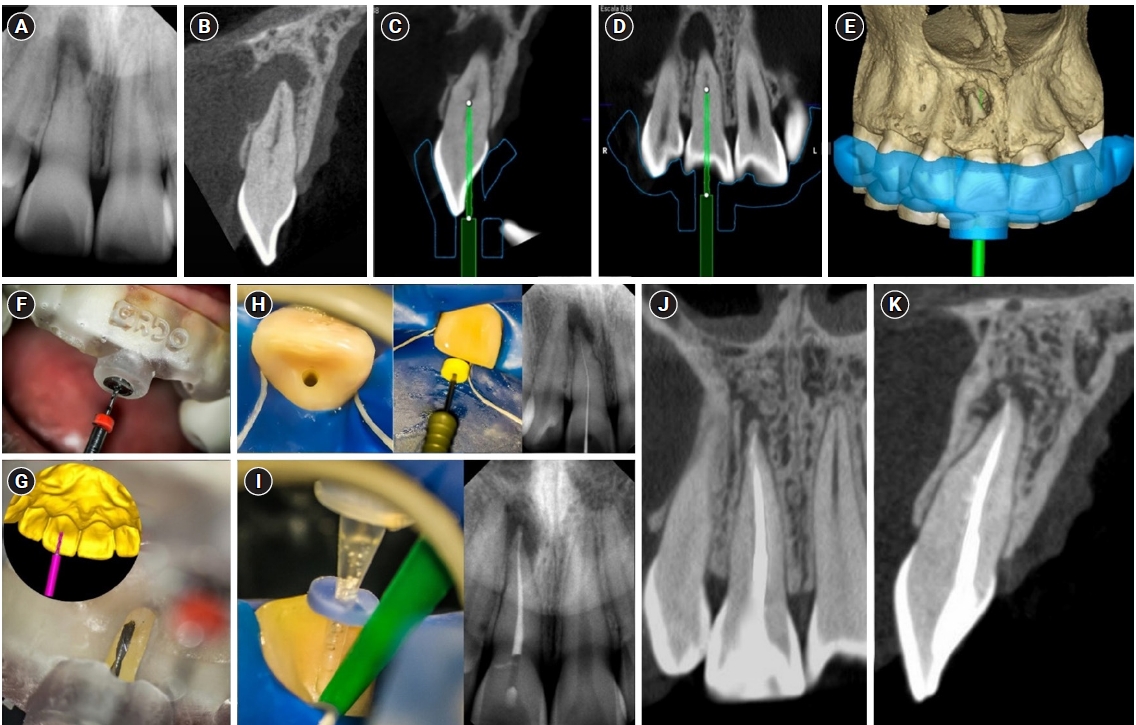
-
Abstract
PDFPubReaderePub
- Pulp chamber and root canal obliteration (PCO/RCO) presents a challenge for clinicians when nonsurgical endodontic treatment is indicated. Guided endodontics (GE) aims to precisely locate the root canal (RC) system while preserving as much pericervical dentin as possible. GE involves integrating cone-beam computed tomography (CBCT) of the affected tooth with a digital impression of the maxillary/mandibular arch, allowing for careful planning of the drilling path to the RC system through a three-dimensional (3D) static guide. This article reports four cases of teeth with PCO/RCO, accompanied by additional diagnoses of internal and external root resorption and horizontal tooth fracture, all successfully treated with GE. These cases highlight the clinical and radiographic success of GE treatments using CBCT, establishing this technique as a predictable approach for managing mineralized teeth.
-
Citations
Citations to this article as recorded by- Static Guided Endodontics in Primary Endodontic Treatment of Anterior Teeth: A Narrative Review
Monika Kuczmaja, Wiesława Puchalska, Agata Żółtowska
Dentistry Journal.2026; 14(4): 195. CrossRef - Effect of different rotary instrument designs (protaper ultimate and protaper gold) on postoperative pain and bacterial reduction: a randomized clinical trial
Khaled Hassan Abed, Ahmed Abdel Rahman Hashem, Reem Ahmed Lutfy, Somaia Abdellatif Eissa, Dina Ahmed Morsy
BMC Oral Health.2026;[Epub] CrossRef
- Static Guided Endodontics in Primary Endodontic Treatment of Anterior Teeth: A Narrative Review
- 5,038 View
- 372 Download
- 2 Web of Science
- 2 Crossref
Review Article
- Success rate of direct pulp capping on permanent teeth using bioactive materials: a systematic review and meta-analysis of randomized clinical trials
- Karem Paula Pinto, Gabriela Ribeiro da Silva, Cláudio Malizia Alves Ferreira, Luciana Moura Sassone, Emmanuel João Nogueira Leal da Silva
- Restor Dent Endod 2024;49(4):e34. Published online September 6, 2024
- DOI: https://doi.org/10.5395/rde.2024.49.e34
-
Abstract
PDFSupplementary MaterialPubReaderePub
This systematic review and meta-analysis aimed to evaluate the success rate of direct pulp capping (DPC) on permanent teeth, comparing the use of MTA with calcium hydroxide and calcium silicate-based cements. A systematic search was carried out in 4 databases until July 2023. The selection was based on PICOS criteria and only randomized clinical trials were included. The risk of bias was assessed using RoB-2 tool, and meta-analyses were performed using RevMan 5.3 software. The overall quality of evidence was determined using the GRADE tool. Thirteen studies were included. Meta-analyses indicated significantly higher success rate for DPC using MTA compared to calcium hydroxide, while no significant difference was observed between MTA and Biodentine, showing a success rate from 80% to 100% even after 3 years of follow-up. Five studies were classified as having high risk of bias and the GRADE assessment revealed low certainty of evidence. DPC is highly effective for permanent teeth when using MTA or Biodentine. There is a need for future well-designed randomized clinical trials to evaluate the efficacy of DPC using newer bioceramic materials.
-
Citations
Citations to this article as recorded by- Physicochemical effects of nano type-B bone substitute on pulp protective cement formulations
Njwan Fadhel SHEHAB
Dental Materials Journal.2026; 45(1): 92. CrossRef - Photobiomodulation-assisted pulp capping using nano-hydroxyapatite and mineral trioxide aggregate: Report of two cases
Priya Pal, Rhythm Bains, Promila Verma, Vivek Kumar Bains
Journal of Healthcare Research and Education.2026; 2: 2. CrossRef - Histological Tissue Response to Calcium Silicate-Based Cements Assessed in Human Tooth Culture Models: A Systematic Review
Alberto Cabrera-Fernández, Hebertt Gonzaga dos Santos Chaves, Aránzazu Díaz-Cuenca, Juan J. Segura-Egea, Jenifer Martín-González, João Peça, Diana B. Sequeira, João Miguel Marques dos Santos
Journal of Functional Biomaterials.2026; 17(2): 78. CrossRef - Translational Pathways for Smart and Bioactive Dental Biomaterials: Biocompatibility Standards, Sterilisation, Sustainability and Regulation
Katarzyna Chojnacka, Marcin Mikulewicz
MedComm – Biomaterials and Applications.2026;[Epub] CrossRef - Decision-ready evidence for vital pulp therapy: a network meta-analysis of bioactive materials in mature permanent teeth
Firas Elmsmari, Reem B. Abdelsayed, Qamar Albasoumi, Tareq Aljafarawi, Swadheena Patro, Ajinkya M. Pawar
Frontiers in Dental Medicine.2026;[Epub] CrossRef - Comparative Evaluation of Platelet-Rich Fibrin and Mineral Trioxide Aggregate in the Direct Pulp Capping of Carious Exposures: A Randomized Clinical Trial
Geeta Asthana, Rajashree Tamuli, Sadhna Manglani, Saloni Mandhare, Ragini Kulkarni, Anooja Mathirat, Parneet Kaur, P Jyothirmayee, Surabhi Landge
Cureus.2026;[Epub] CrossRef - Indian Association of Conservative Dentistry and Endodontics consensus statement on deep caries management
Deepak Kumar Sharma, R. S. Mohan Kumar, Shishir Singh, Suparna Ganguly Saha, Meenal Nithin Gulve, Dipali Y. Shah, Sathish Abraham, Shruthi Nagaraja, Raksha Bhat
Journal of Conservative Dentistry and Endodontics.2025; 28(8): 714. CrossRef
- Physicochemical effects of nano type-B bone substitute on pulp protective cement formulations
- 22,504 View
- 693 Download
- 4 Web of Science
- 7 Crossref
Research Articles
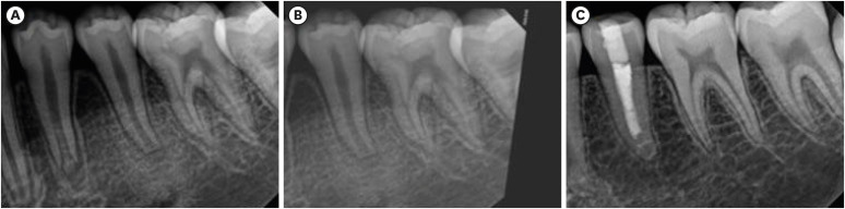
- Endodontic characteristics of mandibular premolar with dens evaginatus: a retrospective study
- Minjin Kim, Sujin Jeon, Min-Seock Seo
- Restor Dent Endod 2024;49(3):e28. Published online July 11, 2024
- DOI: https://doi.org/10.5395/rde.2024.49.e28
-
Abstract
PDFPubReaderePub
Objectives This study aimed to investigate the endodontic characteristics of mandibular premolars with dens evaginatus (DE) that require endodontic treatment.
Materials and Methods Patients who underwent endodontic treatment were enrolled. The inclusion criteria were patients who underwent root canal treatment in the lower permanent teeth with DE and were followed up for at least 1 year. Preoperative clinical and radiographic variables were obtained. The frequency distribution of the preoperative variables was compared using the χ2 or Fisher’s exact tests. The significance of the change in periapical health index (PAI) and root development stages before and after treatment was examined using the Wilcoxon signed-rank test.
Results A total of 150 teeth of 134 patients with an average age of 15.3 years were included. The percentage distribution comparison of the preoperative variables and obturation techniques revealed significant differences in pulpal and periapical diagnosis, and percussion, and especially regarding age, root development stage, and PAI. Age was the only statistically significant preoperative variable associated with root growth (
p < 0.05).Conclusions Approximately, 60% of DEs requiring endodontic treatment had immature roots. Age being the most significant predisposing factor, early treatment provides the greatest opportunity for full root development.
-
Citations
Citations to this article as recorded by- A tooth with multiple supernumerary cusps and taurodontism concurrently accompanied with other taurodont teeth: a rare case report
Zihui Tang, Hongchen Zhang, Rongrong Dang, Qiushi Zhang, Yan Huang, Yanwei Yang
Surgical and Radiologic Anatomy.2025;[Epub] CrossRef
- A tooth with multiple supernumerary cusps and taurodontism concurrently accompanied with other taurodont teeth: a rare case report
- 4,174 View
- 119 Download
- 1 Web of Science
- 1 Crossref
-
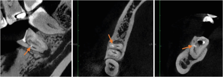
Procedural errors detected by cone beam tomography in cases with indication for retreatment:
in vivo cross-sectional study - Henry Paul Valverde Haro, Carmen Rosa Garcia Rupaya, Flávio R. F. Alves
- Restor Dent Endod 2024;49(3):e26. Published online June 24, 2024
- DOI: https://doi.org/10.5395/rde.2024.49.e26
-
Abstract
PDFPubReaderePub
Objectives This study aimed to investigate the frequency and type of endodontic procedural errors in cases indicated for retreatment through cone-beam computed tomography (CBCT) analysis.
Materials and Methods The sample consisted of 96 CBCT scans, encompassing 122 permanent teeth with fully formed roots. Errors included perforation, instrument fracture, canal transportation, missed canals, and inadequate apical limit of filling. Additionally, potential risk factors were analyzed and subjected to statistical modeling.
Results The most frequent procedural error observed was the inadequate apical limit of filling, followed by canal transportation, perforation, missed canal, and instrument fracture. Statistically significant associations were identified between various procedural errors and specific factors. These include canal transportation and root canal wall, with the buccal wall being the most commonly affected; missed canal and tooth type, particularly the palatine and second mesiobuccal canal canals; inadequate apical limit of filling and root curvature, showing a higher deviation to the mesial direction in severely curved canals; inadequate apical limit of filling and the presence of calcifications, with underfilling being the most frequent; canal transportation and periapical lesion, notably with deviation to the buccal direction; and the direction of perforation and periapical lesion, most frequently occurring to buccal direction.
Conclusions CBCT emerges as a valuable tool in identifying procedural errors and associated factors, crucial for their prevention and management.
-
Citations
Citations to this article as recorded by- Regenerative endodontic treatment of a necrotic immature taurodont mandibular second molar with endodontic infection
Ali Mohammed Addokhi, Ahmed Abuhaimed
Saudi Endodontic Journal.2026; 16(2): 271. CrossRef - Repair of furcal perforations using different calcium silicate cements: An in vitro study
Ariana Esperanza Apolo Aguilar, Maria Soledad Peñaherrera Manosalvas, Henry Paul Valverde Haro
Journal of Conservative Dentistry and Endodontics.2025; 28(10): 1007. CrossRef - Impact of Downward Load and Rotational Kinematics on Root Canal Instrumentation with a Heat-Treated Nickel–Titanium Rotary Instrument
Risako Yamamoto, Keiichiro Maki, Shunsuke Kimura, Satoshi Omori, Keiko Hirano, Arata Ebihara, Yoshio Yahata, Takashi Okiji
Materials.2025; 19(1): 108. CrossRef - ANALYSIS OF THE QUALITY OF ROOT CANAL OBTURATION AND PREVALENCE OF APICAL PERIODONTITIS IN ENDODONTICALLY TREATED TEETH
Cristina Coralia Nistor, Ioana Suciu , Elena Zabrac , Ruxandra Ioana Bartok , Bogdan Dimitriu , Andreea Baluta
Romanian Journal of Oral Rehabilitation.2024; 16(4): 311. CrossRef
- Regenerative endodontic treatment of a necrotic immature taurodont mandibular second molar with endodontic infection
- 3,848 View
- 136 Download
- 2 Web of Science
- 4 Crossref
- Color stability and solubility of Biodentine and NeoPutty in contact with different irrigation solutions
- Sıla Nur Usta, Cangül Keskin
- Restor Dent Endod 2024;49(3):e25. Published online June 19, 2024
- DOI: https://doi.org/10.5395/rde.2024.49.e25
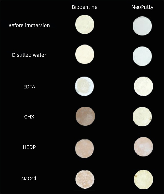
-
Abstract
PDFPubReaderePub
Objectives This study aimed to evaluate the color stability and solubility of Biodentine and NeoPutty in contact with different irrigation solutions.
Materials and Methods Biodentine and NeoPutty were set in cylindrical molds with 7 mm diameter and 1.5 mm high and immersed in distilled water, 17% ethylenediaminetetraacetic acid (EDTA), 2% chlorhexidine (CHX), 9% 1-hydroxyethylidene 1,1-diphosphonate (HEDP), and 5% sodium hypochlorite (NaOCl) solutions for 24 hours. The color change was measured with a spectrophotometer. The solubility values were calculated as the mass loss was expressed as a percentage of the original mass using an analytical balance with 10−4 g accuracy. Data were analyzed with Kruskal-Wallis followed by Mann-Whitney
U tests, and 2-way analysis of variance test followed by Bonferroni corrections for pairwise comparisons for solubility and color stability with a 5% significance threshold, respectively.Results Biodentine exhibited higher color changes compared to the NeoPutty contact with all solutions except distilled water (
p < 0.05). Both hydraulic cements (HCs) showed higher discoloration values immersion in CHX followed by NaOCl. No statistically significant difference was found between Biodentine and NeoPutty regardless of irrigation solution in terms of solubility (p > 0.05). Solubility values were lower in the distilled water group compared to EDTA and CHX (p < 0.05).Conclusions Tested HCs showed solubility and color changes at various rates. NeoPutty could be an appropriate material in aesthetic areas. The usage of HEDP as an irrigant solution can be considered suitable for various endodontic treatments due to its relatively lower solubility and discoloration values.
-
Citations
Citations to this article as recorded by- Sealing ability of Biodentine, zirconia reinforced glass ionomer cement and Mineral Trioxide Aggregate as furcation perforation repair materials: an in vitro analysis
Sumita Panwar, Yajuvender Singh Hada
Biomaterial Investigations in Dentistry.2026; 13: 21. CrossRef - Effect of calcium silicate-based materials on tooth discolouration in repairing root perforations of lower molars: an in-vitro study
Sevil Zırhlı, Davut Celık, Tugba Kosar
Journal of the Australian Ceramic Society.2026;[Epub] CrossRef - Influence of endodontic irrigants on hydraulic cements: solubility, color alteration and surface changes
Sıla Usta, Cangül Keskin, Ayşe Oktay, Emmanuel João Nogueira Leal Silva
European Oral Research.2026; 60(1): 230. CrossRef
- Sealing ability of Biodentine, zirconia reinforced glass ionomer cement and Mineral Trioxide Aggregate as furcation perforation repair materials: an in vitro analysis
- 3,546 View
- 172 Download
- 3 Web of Science
- 3 Crossref
Review Article
- The prevalence of apical periodontitis in patients prior to hematopoietic cell transplantation: a systematic review
- Letícia Tainá de Oliveira Lemes, Carolina Horn Troian-Michel, Theodoro Weissheimer, Marcus Vinicius Reis Só
- Restor Dent Endod 2024;49(2):e22. Published online May 9, 2024
- DOI: https://doi.org/10.5395/rde.2024.49.e22
-
Abstract
PDFSupplementary MaterialPubReaderePub
Objectives This systematic review addressed the question: “What is the prevalence of apical periodontitis in patients prior to hematopoietic cell transplantation?”
Materials and Methods A systematic search was conducted in MEDLINE/PubMed, Cochrane Library, Scopus, Web of Science, Embase, and Grey Literature Report. Eligibility criteria were based on the condition, content, and population strategy: the condition was the radiographic prevalence of apical periodontitis, the content comprised patients scheduled for hematopoietic stem cell transplantation, and the population consisted of adult and pediatric patients. The revised Risk of Bias in Nonrandomized Studies of Exposure tool was used to assess the quality of studies. The Grading Recommendations Assessments, Development, and Evaluation (GRADE) tool was used to assess the quality of evidence.
Results Eight studies were included in this review. The average number of patients with apical periodontitis was 15.65% (range, 2.1%–43.34%). One study was classified as having a very high risk of bias, 1 with a high risk of bias, and 6 with some concern for bias. GRADE analysis showed a very low certainty of evidence. Significant limitations concerning the absence of control over confounding variables were identified.
Conclusions With the caveat of the very low quality of evidence in the studies reviewed, there was a low to moderate prevalence of apical periodontitis in patients prior to undergoing hematopoietic cell transplantation.
- 2,720 View
- 59 Download
Research Article
- Prevalence of apical periodontitis and quality of root canal treatment in an adult Kuwaiti sub-population: a cross-sectional study
- Abdulrahman A. Alhailaa, Saad A Al-Nazhan, Mazen A Aldosimani
- Restor Dent Endod 2024;49(2):e16. Published online March 22, 2024
- DOI: https://doi.org/10.5395/rde.2024.49.e16
-
Abstract
PDFPubReaderePub
Objectives This cross-sectional study evaluated the prevalence of apical periodontitis (AP) and the technical quality of root canal fillings in an adult Kuwaiti subpopulation using cone-beam computed tomography (CBCT) images.
Materials and Methods Two experienced examiners analyzed 250 CBCT images obtained from Kuwaiti patients aged 15–65 years who attended government dental specialist clinics between January 2019 and September 2020. The assessment followed the radiographic scoring criteria proposed by De Moor for periapical status and the technical quality of root canal filling. Chi-square and Fisher’s exact tests were used for statistical analysis, with significance level set at
p < 0.05.Results Among the 2,762 examined teeth, 191 (6.91%) exhibited radiographic signs of AP, and 176 (6.37%) had undergone root canal filling. AP prevalence in root canal-treated teeth was 32.38%, with a significant difference between males and females. Most of the endodontically treated teeth exhibited adequate root canal filling (71.5%).
Conclusions The study demonstrated a comparable prevalence of AP and satisfactory execution of root canal treatment compared to similar studies in different countries.
-
Citations
Citations to this article as recorded by- RISK FACTORS FOR CHRONIC APICAL PERIODONTITIS ACCORDING TO THE CASE-CONTROL STUDY
N. Bagryantseva
Vrach.2026; : 43. CrossRef - A Retrospective Study of CBCT-Based Detection of Endodontic Failures and Periapical Lesions in a Romanian Cohort
Oana Andreea Diaconu, Lelia Mihaela Gheorghiță, Anca Gabriela Gheorghe, Mihaela Jana Țuculină, Maria Cristina Munteanu, Cătălina Alexandra Iacov, Virginia Maria Rădulescu, Mihaela Ionescu, Adina Andreea Mirea, Carina Alexandra Bănică
Journal of Clinical Medicine.2025; 14(18): 6364. CrossRef
- RISK FACTORS FOR CHRONIC APICAL PERIODONTITIS ACCORDING TO THE CASE-CONTROL STUDY
- 6,399 View
- 100 Download
- 1 Web of Science
- 2 Crossref
Review Article
- Cone-beam computed tomography in endodontics: from the specific technical considerations of acquisition parameters and interpretation to advanced clinical applications
- Néstor Ríos-Osorio, Sara Quijano-Guauque, Sandra Briñez-Rodríguez, Gustavo Velasco-Flechas, Antonieta Muñoz-Solís, Carlos Chávez, Rafael Fernandez-Grisales
- Restor Dent Endod 2024;49(1):e1. Published online December 11, 2023
- DOI: https://doi.org/10.5395/rde.2024.49.e1
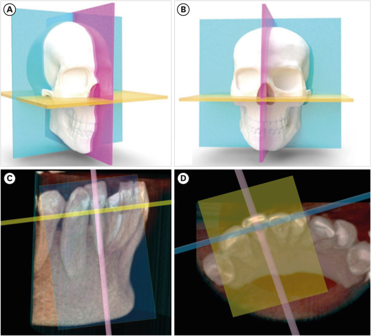
-
Abstract
PDFPubReaderePub
The implementation of imaging methods that enable sensitive and specific observation of anatomical structures has been a constant in the evolution of endodontic therapy. Cone-beam computed tomography (CBCT) enables 3-dimensional (3D) spatial anatomical navigation in the 3 volumetric planes (sagittal, coronal and axial) which translates into great accuracy for the identification of endodontic pathologies/conditions. CBCT interpretation consists of 2 main components: (i) the generation of specific tasks of the image and (ii) the subsequent interpretation report. A systematic and reproducible method to review CBCT scans can improve the accuracy of the interpretation process, translating into greater precision in terms of diagnosis and planning of endodontic clinical procedures. MEDLINE (PubMed), Web of Science, Google Scholar, Embase and Scopus were searched from inception to March 2023. This narrative review addresses the theoretical concepts, elements of interpretation and applications of the CBCT scan in endodontics. In addition, the contents and rationale for reporting 3D endodontic imaging are discussed.
-
Citations
Citations to this article as recorded by- Evaluation of Maxillary Sinus Pathologies in Children and Adolescents with Cleft Lip and Palate Using Cone Beam Computed Tomography: A Retrospective Study
Ayşe Çelik, Nilüfer Ersan, Senem Selvi-Kuvvetli
The Cleft Palate Craniofacial Journal.2026; 63(4): 554. CrossRef - Comparative Evaluation of the Accuracy of Two-Dimensional and Three-Dimensional Radiographic Assessment of Bony Defects Before and After Endodontic Surgery
Aishwarya Talakeri, Pravin Kumar, Soundharrajan P, Vinay Kumar Chugh , Rajat Sharma, Arun Patnana
Cureus.2026;[Epub] CrossRef - Prevalence and morphometric assessment of the middle mesial canal in mandibular first molars in a turkish population: A CBCT study
Elif Solakoğlu, Özge Kurt
Odontology.2026;[Epub] CrossRef - Three-Dimensional CBCT Analysis of Second Mesiobuccal Canal Anatomy in Maxillary Molars
Hanadi Sabban, Maysoon Albahiti, Suha S. Maddah
Diagnostics.2026; 16(9): 1299. CrossRef - Comparative Evaluation of Platelet-Rich Fibrin and Mineral Trioxide Aggregate in the Direct Pulp Capping of Carious Exposures: A Randomized Clinical Trial
Geeta Asthana, Rajashree Tamuli, Sadhna Manglani, Saloni Mandhare, Ragini Kulkarni, Anooja Mathirat, Parneet Kaur, P Jyothirmayee, Surabhi Landge
Cureus.2026;[Epub] CrossRef - Machine Learning Models in the Detection of MB2 Canal Orifice in CBCT Images
Shishir Shetty, Meliz Yuvali, Ilker Ozsahin, Saad Al-Bayatti, Sangeetha Narasimhan, Mohammed Alsaegh, Hiba Al-Daghestani, Raghavendra Shetty, Renita Castelino, Leena R David, Dilber Uzun Ozsahin
International Dental Journal.2025; 75(3): 1640. CrossRef - Early diagnosis of acute lymphoblastic leukemia utilizing clinical, radiographic, and dental age indicators
Rehab F Ghouraba, Shaimaa S. EL-Desouky, Mohamed R. El-Shanshory, Ibrahim A. Kabbash, Nancy M. Metwally
Scientific Reports.2025;[Epub] CrossRef - Tomographic evaluation of apexogenesis with human treated dentin matrix in young permanent molars: a split-mouth randomized controlled clinical trial
Nora M. Abo Shanady, Nahed A. Abo Hamila, Gamal M. El Maghraby, Rehab F. Ghouraba
BMC Oral Health.2025;[Epub] CrossRef - The Integration of Cone Beam Computed Tomography, Artificial Intelligence, Augmented Reality, and Virtual Reality in Dental Diagnostics, Surgical Planning, and Education: A Narrative Review
Aida Meto, Gerta Halilaj
Applied Sciences.2025; 15(11): 6308. CrossRef - Healing Outcomes of Through‐And‐Through Bone Defects in Periapical Surgery: A Systematic Review and Meta‐Analysis
Bibi Fatima, Farhan Raza Khan, Syeda Abeerah Tanveer
Australian Endodontic Journal.2025; 51(2): 518. CrossRef - Genotoxic and cytotoxic effects of cone beam computed tomography on exfoliated epithelial cells in different age groups
Maged Bakr, Fatma Ata, Asmaa Saleh Elmahdy, Bassant Mowafey
BMC Oral Health.2025;[Epub] CrossRef - Bridging the gap in aberrant root canal systems: Case series
Seethalakshmi Tamizhselvan, Diana Davidson, Srinivasan Manali Ramakrishnan
Journal of Conservative Dentistry and Endodontics.2025; 28(8): 833. CrossRef - IMAGING TECHNIQUES IN ENDODONTIC DIAGNOSIS: A REVIEW OF LITERATURE
Mihaela Salceanu, Anca Melian , Tudor Hamburda , Cristina Antohi , Corina Concita , Claudiu Topoliceanu , Cristian Levente Giuroiu
Romanian Journal of Oral Rehabilitation.2025; 17(1): 705. CrossRef - A Three-rooted Deciduous Second Molar in a 13-year-old Caucasian Female
Daniel Traub, Robert Walsh, Colleen Ahern
International Journal of Medical Case Reports.2025; 4(3): 51. CrossRef - Critical success factors for digital transformation in government organizations using a structural model approach
Abdalla Al Maazmi, Zehra Canan Araci, Sujan Piya
Discover Applied Sciences.2025;[Epub] CrossRef - AGE ESTIMATION BASED ON PULP / TOOTH VOLUME BY CONE BEAM COMPUTERIZED TOMOGRAPHY IMAGE
Ramadhan Rasheed, Salah Faraj
BULLETIN OF STOMATOLOGY AND MAXILLOFACIAL SURGERY.2025; : 288. CrossRef - Clinical Benefits and Limitations of Cone-Beam Computed Tomography in Endodontic Practice: A Contemporary Evidence-Based Review
Jasmine Wong, Chengfei Zhang, Angeline Hui Cheng Lee
Diagnostics.2025; 15(24): 3117. CrossRef - On the Causes of Persistent Apical Periodontitis. Findings From Endodontic Microsurgery: A Case Report
Mateo José Pesántez-Ibarra, Carolina Berruecos-Orozco, Jeimmy Katherine Molina-Barrera, Néstor Ríos-Osorio, Rafael Fernández-Grisales
Journal of Endodontic Microsurgery.2025;[Epub] CrossRef - Bildgebung im ZMK-Bereich – aber in welcher Reihenfolge?
Rainer Lutz
Zahnmedizin up2date.2024; 18(04): 297. CrossRef - Cone-beam computed tomography evaluation of shaping ability of kedo-S square and fanta AF™ baby rotary files compared to manual K-files in root canal preparation of primary anterior teeth
Shaimaa S. El-Desouky, Bassem N. El Fahl, Ibrahim A. Kabbash, Shimaa M. Hadwa
Clinical Oral Investigations.2024;[Epub] CrossRef - Analysis of Endodontic Successes and Failures in the Removal of Fractured Endodontic Instruments during Retreatment: A Systematic Review, Meta-Analysis, and Trial Sequential Analysis
Mario Dioguardi, Corrado Dello Russo, Filippo Scarano, Fariba Esperouz, Andrea Ballini, Diego Sovereto, Mario Alovisi, Angelo Martella, Lorenzo Lo Muzio
Healthcare.2024; 12(14): 1390. CrossRef
- Evaluation of Maxillary Sinus Pathologies in Children and Adolescents with Cleft Lip and Palate Using Cone Beam Computed Tomography: A Retrospective Study
- 20,255 View
- 875 Download
- 22 Web of Science
- 21 Crossref
Research Articles
- A scientometric, bibliometric, and thematic map analysis of hydraulic calcium silicate root canal sealers
- Anastasios Katakidis, Konstantinos Kodonas, Anastasia Fardi, Christos Gogos
- Restor Dent Endod 2023;48(4):e41. Published online November 13, 2023
- DOI: https://doi.org/10.5395/rde.2023.48.e41
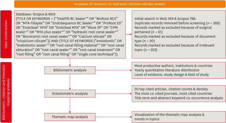
-
Abstract
PDFPubReaderePub
Objectives This scientometric and bibliometric analysis explored scientific publications related to hydraulic calcium silicate-based (HCSB) sealers used in endodontology, aiming to describe basic bibliometric indicators and analyze current research trends.
Materials and Methods A comprehensive search was conducted in Web of Science and Scopus using specific HCSB sealer and general endodontic-related terms. Basic research parameters were collected, including publication year, authorship, countries, institutions, journals, level of evidence, study design and topic of interest, title terms, author keywords, citation counts, and density.
Results In total, 498 articles published in 136 journals were retrieved for the period 2008–2023. Brazil was the leading country, and the universities of Bologna in Italy and Sao Paolo in Brazil were represented equally as leading institutions. The most frequently occurring keywords were “calcium silicate,” “root canal sealer MTA-Fillapex,” and “biocompatibility,” while title terms such as “calcium,” “sealers,” “root,” “canal,” “silicate based,” and “endodontic” occurred most often. According to the thematic map analysis, “solubility” appeared as a basic theme of concentrated research interest, and “single-cone technique” was identified as an emerging, inadequately developed theme. The co-occurrence analysis revealed 4 major clusters centered on sealers’ biological and physicochemical properties, obturation techniques, retreatability, and adhesion.
Conclusions This analysis presents bibliographic features and outlines changing trends in HCSB sealer research. The research output is dominated by basic science articles scrutinizing the biological and specific physicochemical properties of commonly used HCSB sealers. Future research needs to be guided by studies with a high level of evidence that utilize innovative, sophisticated technologies.
-
Citations
Citations to this article as recorded by- Top 100 Most Cited Articles on Antibiotics in Endodontics: A Bibliometric Analysis
Hajar Albanyan, Mohammed Asseery, Haitham Alahmari, Ikram Ul Haq, Ali Alaqla
Journal of Endodontics.2026; 52(3): 345. CrossRef - Top-cited articles on vital pulp therapy for irreversible pulpitis in permanent teeth: a bibliometric analysis
İkbal Sena Çelebi Keskin, Ferda Karabay
Odontology.2026;[Epub] CrossRef - Agri-Food Sector: Contemporary Trends, Possible Gaps, and Prospective Directions
José Roberto Herrera Cantorani, Meire Ramalho de Oliveira, Luiz Alberto Pilatti, Thales Botelho de Sousa
Metrics.2025; 2(1): 3. CrossRef - Scientific mapping of experimental research on solar cookers: Global trends, evolution, and future directions
Flavio Odoi-Yorke, Bismark Baah, Richard Opoku
Solar Energy Advances.2025; 5: 100093. CrossRef - Bibliometric analysis of the GentleWave system: trends, collaborations, and research gaps
Raimundo Sales de Oliveira Neto, Thais de Moraes Souza, João Vitor Oliveira de Amorim, Thaine Oliveira Lima, Guilherme Ferreira da Silva, Rodrigo Ricci Vivan, Murilo Priori Alcalde, Marco Antonio Hungaro Duarte
Restorative Dentistry & Endodontics.2025; 50(2): e17. CrossRef - A Scientometric Review of Practical Applications in Quantum Natural Language Processing (QNLP): Trends, Gaps, and Research Opportunities
Victor R. Silva, Fábio R. Barbosa, Jasson C. Silva, Francisco J. Santos, Ricardo A. L. Rabelo, Joel J. P. C. Rodrigues
IEEE Access.2025; 13: 210169. CrossRef - A bibliometric analysis of global research trend and progress on Dy doped materials
Sangeeta Kadyan, Manju Nain, Ashima Makhija, Poonam Punia, Anil Ohlan, Sajjan Dahiya, R. Punia, A.S. Maan
Journal of Alloys and Compounds Communications.2024; 3: 100006. CrossRef - Comparative bioactivity and immunomodulatory potential of the new Bioroot Flow and AH Plus Bioceramic sealer: An in vitro study on hPDLSCs
José Luis Sanz, Sergio López-García, David García-Bernal, Francisco Javier Rodríguez-Lozano, Leopoldo Forner, Adrián Lozano, Laura Murcia
Clinical Oral Investigations.2024;[Epub] CrossRef - Analyzing collaboration and impact: A bibliometric review of four highly published authors’ research profiles on collaborative maps
Willy Chou, Julie Chi Chow
Medicine.2024; 103(28): e38686. CrossRef
- Top 100 Most Cited Articles on Antibiotics in Endodontics: A Bibliometric Analysis
- 3,663 View
- 52 Download
- 7 Web of Science
- 9 Crossref
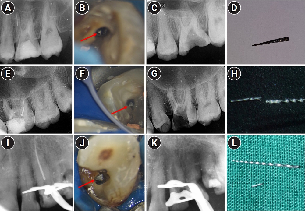
- Investigation of fracture prevalence of instruments used in root canal treatments at a faculty of dentistry: a prospective study
- Mehmet Eskibağlar, Merve Yeniçeri Özata, Mevlüt Sinan Ocak, Faruk Öztekin
- Restor Dent Endod 2023;48(4):e38. Published online November 1, 2023
- DOI: https://doi.org/10.5395/rde.2023.48.e38
-
Abstract
PDFPubReaderePub
Objectives The aim of this study was to examine the use of hand or rotary files by pre-graduation (fourth- and fifth-year) and postgraduate students in endodontic treatments and to determine the incidence of file fracture and the management of cases with broken instruments.
Materials and Methods A total of 2,168 teeth undergoing primary endodontic treatment were included in this study. It was determined that 79 of these teeth resulted in broken tools. In the case of broken tools, the education level of the treating clinician, the tooth that was being treated, the canal and fracture level, the curvature of the tooth and the management of the broken instrument were recorded. Periapical radiographs of the patients were used to calculate curvature following the Schneider method.
Results There was no significant difference in the incidence of broken tools according to education level (
p > 0.05). The incidence of file fracture in molar teeth (73.4%) was higher than in other teeth (p < 0.05). More files were broken in the mandibular molar MB canal (20.25%) and in the apical third of the canals (72.1%). The risk of instrument fracture was high in teeth with moderate (44.3%) and severe (38%) curvature canals. The management of apically broken (80%) files mostly involved lefting (p < 0.05).Conclusions There was no statistically significant difference between fourth-year students, fifth-year students and postgraduate students in terms of instrument fracture.
-
Citations
Citations to this article as recorded by- Assessment of Techniques of Surpassing Ledges in Curved Canals and Its Effect on File: An In Vitro Study
Nezar Boreak, Abdulrahman Ali Sawadi, Yahya Yaqoup Hazazi, Sameer J Oqayshi, Wed AY Ayash, Essa Ibrahim Al Essa, Abdulaziz Ismail Shafei, Amjad Hassan Khawaji
World Journal of Dentistry.2026; 16(11): 1032. CrossRef - Secondary retrieval and complication management for failed separated instrument removal in the apical third in curved root canals: a case series
Mei Fu, Jia Guo, Fengting Wang, Benxiang Hou, Chen Zhang
BMC Oral Health.2026;[Epub] CrossRef - Prevalence and management techniques for separated endodontic files among endodontic postgraduate students in the Qassim region: A retrospective cross-sectional study
Hanin Abdulaziz Alsalhi, Rana Rabeh Alharbi, Amnah Ameen Hawsa
Saudi Endodontic Journal.2026; 16(2): 160. CrossRef - Case Study of a Broken Instrument in a Primary Tooth and Literature Review
Masashi Nakano, Tatsuya Akitomo, Masashi Ogawa, Mariko Kametani, Momoko Usuda, Satoru Kusaka, Chieko Mitsuhata, Ryota Nomura
Children.2025; 12(2): 149. CrossRef - Neodymium-Doped Yttrium Aluminum Perovskite (Nd:YAP) Laser in the Elimination of Endodontic Nickel-Titanium Files Fractured in Rooted Canals (Part 2: Teeth With Significant Root Curvature)
Amaury Namour, Marwan El Mobadder, Clément Cerfontaine, Patrick Matamba, Lucia Misoaga, Delphine Magnin , Praveen Arany, Samir Nammour
Cureus.2025;[Epub] CrossRef - Pattern of endodontic instrument separation and factors affecting its retrieval: a 10-year retrospective observational study in a postgraduate institute
Velmurugan Natanasabapathy, Aswathi Varghese, Paul Kevin Abishek Karthikeyan, Srinivasan Narasimhan
Restorative Dentistry & Endodontics.2025; 50(1): e7. CrossRef - Remoção de instrumentos fraturados nos canais radiculares: Desafios, estratégias e perspectivas clínicas
João Victor da Fonseca Barbosa, Eduardo Kitto Miranda Teixeira , Laura Rodrigues Barbosa, Martinelle Ferreira da Rocha Taranto, Jáder Camilo Pinto
Research, Society and Development.2025; 14(10): e98141049749. CrossRef - Comparative evaluation of curvature severity of mesiobuccal canals in permanent maxillary 1st molar using multiple complexity risk criteria: A cone-beam computed tomography-based cross-sectional study of central Indian subpopulation
Mahima Mathur, Suparna Ganguly Saha, Rolly S. Agarwal, Shakti Singh, Manasi Kewlani, Shaurya Sahu
Endodontology.2025; 37(4): 393. CrossRef - Methods for preventing fragmentation of endodontic instruments: a systematic review
A. V. Mitronin, D. A. Ostanina, K. A. Archakov, Yu. A. Mitronin
Endodontics Today.2025; 23(4): 672. CrossRef - Perception of Dental Interns About Intracanal Fracture of Endodontic Instruments in the Central Region of Saudi Arabia: A Cross-Sectional Study
Abdullah Ahmad A. Aloyouni, Muhammad Atif Saleem Agwan, Saleh Suliman S. Almuzaini, Faris Saleh A. Alqazlan, Abdulaziz Abdulrhman A. Alshumaym, Khalid Abdullah G. Alfuryah
Journal of Pharmacy and Bioallied Sciences.2024; 16(Suppl 4): S3890. CrossRef - Predictive factors in the retrieval of endodontic instruments: the relationship between the fragment length and location
Ricardo Portigliatti, Eugenia Pilar Consoli Lizzi, Pablo Alejandro Rodríguez
Restorative Dentistry & Endodontics.2024;[Epub] CrossRef - Causes and prevention of endodontic file fractures: a review of the literature
Erkal Damla, Er Kürşat
Acta Stomatologica Marisiensis Journal.2024; 7(2): 33. CrossRef - PREVALENCE AND ENDODONTIC MANAGEMENT OF SEPARATED INSTRUMENTS INSIDE THE ROOT CANAL
Cristina Coralia Nistor, Ana Maria Țâncu , Elena Claudia Coculescu , Albu Cristina Crenguta , Stefan Milicescu , Bogdan Dimitriu
Romanian Journal of Oral Rehabilitation.2024; 16(1): 96. CrossRef
- Assessment of Techniques of Surpassing Ledges in Curved Canals and Its Effect on File: An In Vitro Study
- 4,120 View
- 109 Download
- 5 Web of Science
- 13 Crossref
- Micro-CT evaluation of the removal of root fillings using rotary and reciprocating systems supplemented by XP-Endo Finisher, the Self-Adjusting File, or Er,Cr:YSGG laser
- Gülsen Kiraz, Bulem Üreyen Kaya, Mert Ocak, Muhammet Bora Uzuner, Hakan Hamdi Çelik
- Restor Dent Endod 2023;48(4):e36. Published online October 23, 2023
- DOI: https://doi.org/10.5395/rde.2023.48.e36
-
Abstract
PDFPubReaderePub
Objectives This study aimed to compare the effectiveness of a single-file reciprocating system (WaveOne Gold, WOG) and a multi-file rotary system (ProTaper Universal Retreatment, PTUR) in removing canal filling from severely curved canals and to evaluate the possible adjunctive effects of XP-Endo Finisher (XPF), the Self-Adjusting File (SAF), and an erbium, chromium: yttrium, scandium, gallium garnet (Er,Cr:YSGG) laser using micro-computed tomography (μCT).
Materials and Methods Sixty-six curved mandibular molars were divided into 2 groups based on the retreatment technique and then into 3 based on the supplementary method. The residual filling volumes and root canals were evaluated with μCT before and after retreatment, and after the supplementary steps. The data were statistically analyzed with the
t -test, Mann-WhitneyU test, analysis of covariance, and factorial analysis of variance (p < 0.05).Results PTUR and WOG showed no significant difference in removing filling materials (
p > 0.05). The supplementary techniques were significantly more effective than reciprocating or rotary systems only (p < 0.01). The supplementary steps showed no significant differences in canal filling removal effectiveness (p > 0.05), but XPF showed less dentin reduction than the SAF and Er,Cr:YSGG laser (p < 0.01).Conclusions The supplementary methods significantly decreased the volume of residual filling materials. XPF caused minimal changes in root canal volume and might be preferred for retreatment in curved root canals. Supplementary approaches after retreatment procedures may improve root canal cleanliness.
-
Citations
Citations to this article as recorded by- A laboratory study comparing two methods for removing plastic carrier obturators from severely curved root canals
Tania Gancedo-Gancedo, Patricia Pereira-Lores, Venkateshbabu Nagendrababu, Paul MH Dummer, Jenifer Martín-González, Alba Bello-Castro, Inmaculada Tomás, Benjamín Martín-Biedma, Pablo Castelo-Baz
BMC Oral Health.2026;[Epub] CrossRef - Reciproc and XP-endo Shaper Outperform WaveOne Gold in Apical Debris Removal: A Micro-CT Study in 3D-Printed Molars
Yu-juan Zhu, Xue Zhong, Yu-xuan Wu, Shao-lan Li, Qiu-hui Li
Current Medical Science.2026;[Epub] CrossRef - Trends in dentomaxillofacial radiology
Kıvanç Kamburoğlu
World Journal of Radiology.2025;[Epub] CrossRef - Retrieval of AH Plus Bioceramic and Ceraseal Versus AH Plus in Endodontic Retreatment
Eurok Shim, Jee Woo Son, Jiyoung Kwon, Hyun-Jung Kim, Ji-Hyun Jang, Seok Woo Chang, Soram Oh
Journal of Clinical Medicine.2025; 14(6): 1826. CrossRef - Characteristics and Effectiveness of XP‐Endo Files and Systems: A Narrative Review
Sarah M. Alkahtany, Rana Alfadhel, Aseel AlOmair, Sarah Bin Durayhim, Kee Y. Kum
International Journal of Dentistry.2024;[Epub] CrossRef - Effect of the filling technique on the filling removal from oval-shaped canals
Lislaine Valerio, Lisa Yurie Oda, Felipe Andretta Copelli, Clarissa Teles Rodrigues, Everdan Carneiro, Marco Antonio Hungaro Duarte, Bruno Cavalini Cavenago
Clinical Oral Investigations.2024;[Epub] CrossRef
- A laboratory study comparing two methods for removing plastic carrier obturators from severely curved root canals
- 4,972 View
- 115 Download
- 10 Web of Science
- 6 Crossref
- Incidence of interappointment emergencies in multiple-visit root canal treatments performed with or without intracanal medicament by undergraduate students
- Annemarie Baaij, Corine Mirjam Visscher, Manon Jansen, Ahmet Rifat Özok
- Restor Dent Endod 2023;48(3):e31. Published online August 14, 2023
- DOI: https://doi.org/10.5395/rde.2023.48.e31
-
Abstract
PDFPubReaderePub
Objectives This retrospective cohort study examined the incidence of interappointment emergencies during multiple-visit molar root canal treatments conducted by undergraduate students. Treatments performed without the use of intracanal medicament were compared to treatments that incorporated calcium hydroxide as an intracanal medicament.
Materials and Methods Interappointment emergencies, defined as instances of pain or swelling that required the patient to make an unscheduled follow-up visit, were recorded for up to 2 months after the intervention. To avoid the influence of obturation on the observed incidence of emergency visits, only unscheduled visits occurring between the start and end of the root canal treatment were included.
Results Of the 719 patients included in this study, 77 (10.7%) were recorded as experiencing interappointment emergencies. Of these emergencies, 62% occurred within 2 weeks following the most recent intervention. In the group of patients who did not receive intracanal medicament, the incidence of interappointment emergencies was 11.9% (46 of 385 patients). In comparison, this rate was 9.3% (31 of 334 patients) among those who received calcium hydroxide as an intracanal medicament (odds ratio, 1.33; 95% confidence interval, 0.82–2.15;
p = 0.249).Conclusions Interappointment emergencies may arise at any point during root canal treatment, but they most commonly occur within the first 2 weeks following intervention. The omission of intracanal medicament in multiple-visit molar root canal treatments, performed by undergraduate students, did not significantly increase the incidence of these emergencies.
-
Citations
Citations to this article as recorded by- An Observational Study on Pain Occurrence After Root Canal Treatment: Role of Operator Experience When Using a Bioceramic Sealer
Mihai Merfea, Ioana Sofia Pop-Ciutrila, Mindra Eugenia Badea, Ada Gabriela Delean, Oana Cimponeriu, Razvan Corneliu Pop, Maria Peter, Iulia Clara Badea, Sanda Ileana Cimpean
Journal of Clinical Medicine.2025; 14(13): 4558. CrossRef
- An Observational Study on Pain Occurrence After Root Canal Treatment: Role of Operator Experience When Using a Bioceramic Sealer
- 5,164 View
- 50 Download
- 1 Web of Science
- 1 Crossref
-
Effect of cryotherapy duration on experimentally induced connective tissue inflammation
in vivo - Jorge Vera, Mayra Alejandra Castro-Nuñez, María Fernanda Troncoso-Cibrian, Ana Gabriela Carrillo-Varguez, Edgar Ramiro Méndez Sánchez, Viviana Sarmiento, Lourdes Lanzagorta-Rebollo, Prasanna Neelakantan, Monica Romero, Ana Arias
- Restor Dent Endod 2023;48(3):e29. Published online August 2, 2023
- DOI: https://doi.org/10.5395/rde.2023.48.e29
-
Abstract
PDFPubReaderePub
Objectives This study tested the hypothesis that cryotherapy duration influences lipopolysaccharide (LPS)-induced inflammation in a rat model.
Materials and Methods Six Wistar rats (
Rattus norvegicus albinus ) were used. Five sites were selected per animal and divided into 5 groups: a negative control group (NC), 2 positive control groups (PC1 and PC2), and 2 experimental groups (E1 and E2). Cryotherapy was applied for 1 minute (E1) or 5 minutes (E2). An acute inflammatory response was induced in the PC and E groups via subcutaneous administration of 0.5 mL/kg. In the PC2 group, a catheter was inserted without additional treatment. For the E1 and E2 groups, 2.5°C saline solution was administered through the implanted catheters for 1 and 5 minutes, respectively. The rats were sacrificed, and samples were obtained and processed for histological analysis, specifically examining the presence of polymorphonuclear neutrophils and hemorrhage. The χ2 test was used to compare the presence of acute inflammation across groups. Dependent variables were compared using the linear-by-linear association test.Results Inflammation and hemorrhage varied significantly among the groups (
p = 0.001). A significantly higher degree of acute inflammation was detected (p = 0.0002) in the PC and E1 samples than in the E2 group, in which cryotherapy was administered for 5 minutes. The PC and E1 groups also exhibited significantly greater numbers of neutrophils (p = 0.007), which were essentially absent in both the NC and E2 groups.Conclusions Cryotherapy administration for 5 minutes reduced the acute inflammation associated with LPS and catheter implantation.
-
Citations
Citations to this article as recorded by- The impact of using cold irrigation on postoperative endodontic pain and substance P level: a randomized clinical trial
Reem Mohammed Amr Sharaf, Tariq Yehia Abdelrahman, Maram Farouk Obeid
Odontology.2026; 114(2): 527. CrossRef - Cryotherapy as a supplementary aid to inferior alveolar nerve block in patients with symptomatic irreversible pulpitis: A randomized controlled trial
Setu Katyal, Poonam Bogra, Rajinder Bansal, Vishakha Grover, Saurabh Gupta, Saru Gupta
Medicine International.2025; 5(5): 1. CrossRef - Determining Efficacy of Intracanal Cryotherapy on Post Endodontic Pain in Irreversible Pulpitis
Anam Fayyaz Bashir, Ussamah Waheed Jatala, Moeen ud din Ahmad, Muhammad Talha Khan, Saima Razzaq Khan, Aisha Arshad Butt
Pakistan Journal of Health Sciences.2024; : 68. CrossRef
- The impact of using cold irrigation on postoperative endodontic pain and substance P level: a randomized clinical trial
- 4,440 View
- 62 Download
- 2 Web of Science
- 3 Crossref
- Effect of irrigation protocols on smear layer removal, bond strength and nanoleakage of fiber posts using a self-adhesive resin cement
- Rodrigo Stadler Alessi, Renata Terumi Jitumori, Bruna Fortes Bittencourt, Giovana Mongruel Gomes, João Carlos Gomes
- Restor Dent Endod 2023;48(3):e28. Published online July 27, 2023
- DOI: https://doi.org/10.5395/rde.2023.48.e28
-
Abstract
PDFPubReaderePub
Objectives This study aimed to investigate the effect of the application method of 2% chlorhexidine (CHX) and its influence on the adhesion of fiberglass posts cemented with a self-adhesive resin cement.
Materials and Methods Sixty human mandibular premolars were endodontically treated and divided into 5 groups (
n = 12), according to the canal irrigant and its application method: 2 groups with conventional syringe irrigation (CSI)—2.5% sodium hypochlorite (NaOCl) (control) and 2% CHX— and 3 groups with 2% CHX irrigation/activation—by passive ultrasonic irrigation (PUI), Easy Clean file, and XP-Endo Finisher file. Two roots per group were evaluated for smear layer (SL) removal by scanning electron microscopy. For other roots, fiber posts were luted using a self-adhesive resin cement. The roots were sectioned into 6 slices for push-out bond strength (BS) (7/group) and nanoleakage (NL) (3/group). Data from SL removal were submitted to Kruskal-Wallis and Student-Newman-Keuls tests (α = 0.05). Data from BS and NL were evaluated by 2-way analysis of variance and Tukey’s test (α = 0.05).Results For SL removal and BS, the CHX irrigation/activation promoted better values than CSI with CHX (
p < 0.05), but it was not significantly different from CSI with NaOCl (p > 0.05). For NL, the lowest values were obtained by the chlorhexidine irrigation/activation groups (p < 0.05).Conclusions Active 2% CHX irrigation can be used to improve the post space cleaning and adhesion before fiber post cementation with self-adhesive resin cements.
-
Citations
Citations to this article as recorded by- Effects of radiotherapy dose and endodontic irrigants on universal resin cement bonding to root dentin: mechanical and interfacial analyses
Lívia Ribeiro, Luíz Carlos de Lima Dias-Júnior, Paulo Henrique dos Santos, Mariana Comparotto Minamisako, Paulo Marcelo Rodrigues, Vicente Ribeiro Netto, Bruno Alexandre Pacheco de Castro Henriques, Renata Gondo Machado, Cleonice da Silveira Teixeira, Luc
International Journal of Adhesion and Adhesives.2026; 146: 104252. CrossRef - Effect of Erbium: Yttrium–Aluminum Garnet Laser, XP-Endo Finisher, and Passive Ultrasonic Irrigation in the Cleaning of Post Spaces on Push-Out Bond Strength
Dilek Hançerlioğulları, Gökhan Karadağ
Current Research in Dental Sciences.2026; 36(2): 121. CrossRef - Laser‐Activated Irrigation via Photon‐Induced Photoacoustic Streaming and Shock Wave Enhanced Emission on Smear Layer Removal Efficacy, Pushout Bond Strength, and Sealer Adaptation: A SEM Assessment
Basil Almutairi, Fahad Alkhudhairy
Microscopy Research and Technique.2025; 88(6): 1806. CrossRef - The impact of passive ultrasonic irrigation on the bond strength of two different self-etch adhesives to human pulp chamber dentine: a laboratory investigation
Mohammed Turky, Jukka Matinlinna, Monika Lukomska-Szymanska, Venkateshbabu Nagendrababu, Paul M. H. Dummer, Ahmad Abdel Hamid Elheeny, Nermin Alsayed Mahmoud
BMC Oral Health.2025;[Epub] CrossRef - The effect of nanoparticles incorporation titanium dioxide and zirconium oxide within self-adhesive resin cement on the push-out bond strength of the fiber post to the radicular dentin: An in vitro study
Sawsan Hameed Al-Jubori, Maha Anwer AL-Murad
Saudi Endodontic Journal.2025; 15(2): 162. CrossRef - The Effects of Different Post Space Conditioning Procedures and Different Endodontic Sealers on the Push-Out Bond Strengths of Fiber Posts
Leyla Ayranci, Ahmet Serkan Küçükekenci, Fatih Sarı, Ahmet Çetinkaya
Clinical and Experimental Health Sciences.2025; 15(3): 620. CrossRef - Evaluation of Microleakage Using Different Luting Cements in Kedo Zirconia Crowns: An In Vitro Assessment
Guru Vishnu, Ganesh Jeevanandan
Cureus.2024;[Epub] CrossRef
- Effects of radiotherapy dose and endodontic irrigants on universal resin cement bonding to root dentin: mechanical and interfacial analyses
- 4,354 View
- 82 Download
- 7 Web of Science
- 7 Crossref
- Contemporary research trends on nanoparticles in endodontics: a bibliometric and scientometric analysis of the top 100 most-cited articles
- Sıla Nur Usta, Zeliha Uğur-Aydın, Kadriye Demirkaya, Cumhur Aydın
- Restor Dent Endod 2023;48(3):e27. Published online July 26, 2023
- DOI: https://doi.org/10.5395/rde.2023.48.e27
-
Abstract
PDFSupplementary MaterialPubReaderePub
Objectives Advancements in nanotechnology have led to the widespread usage of nanoparticles in the endodontic field. This bibliometric study aimed to determine and analyze the top 100 most-cited articles about nanoparticles in endodontics from 2000 to 2022.
Materials and Methods A detailed electronic search was conducted on the “Clarivate Analytics Web of Science, All Databases” to receive the most-cited articles related to the topic. Articles were ranked in descending order based on their citation counts, and the first 100 were selected for bibliometric analysis. Parameters such as citation density, publication year, journal, country, institution, author, study design, study field, evidence level, and keywords were analyzed.
Results The top 100 most-cited articles received 4,698 citations (16–271) with 970.21 (1.91–181) citation density in total. Among decades, citations were significantly higher in 2011–2022 (
p < 0.001).Journal of Endodontics had the largest number of publications. Canada and the University of Toronto made the highest contribution as country and institution, respectively. Anil Kishen was the 1 who participated in the largest number of articles. The majority of the articles were designedin vitro . The main study field was “antibacterial effect.” Among keywords, “nanoparticles” followed by “Enterococcus faecalis ” were used more frequently.Conclusions Developments in nanotechnology had an impact on the increasing number of studies in recent years. This bibliometric study provides a comprehensive view of nanoparticle advances and trends using citation analysis.
-
Citations
Citations to this article as recorded by- Research trends and mapping knowledge for maxillary sinus augmentation in oral and maxillofacial surgery
Özlem Saraç Atagün, Şeyma Çardakcı Bahar, Seval Ceylan Şen, Gülbahar Ustaoğlu
Journal of Stomatology Oral and Maxillofacial Surgery.2025; 126(4): 102116. CrossRef - Visualized bibliometric panorama of ureteral stents (1975–present): trends and hotspots revealed
Renjie Wei, Fudong Liu, Xinjie Ji, Xu Luo, Chunyu Gong, Ruitu Ran
World Journal of Urology.2025;[Epub] CrossRef - Bibliometric analysis of the publications that list the most-cited articles in endodontics
Oscar Alejandro Gutiérrez-Alvarez, Luis Alberto Pantoja-Villa, Benigno Miguel Calderón-Rojas
Endodontology.2025; 37(2): 128. CrossRef - Cyclic fatigue in NiTi files: a bibliometric and science mapping analysis
Huda Melike Bayram, Emre Bayram
Odontology.2025;[Epub] CrossRef - Synergistic antibiofilm activity of methylene blue and silver nanoparticle-mediated photothermal therapy against Enterococcus faecalis biofilm
Eman M. Fouad, Hossam Tawfiq, Soha Abdelrahman Elhady, Ali M. Saafan
Scientific Reports.2025;[Epub] CrossRef - Exploring vital pulp Therapies: A bibliometric analysis of the most cited articles
Gustavo Henrique Sousa, Rodolfo Lima Gonçalves, Barbara Figueiredo, Vilton Cardozo Moreira Dias, Ana Carolina Soares Mendes, Valéria de Cássia Bueno Melo, Adriana Guimarães Rodrigues, Hebertt Gonzaga dos Santos Chaves
The Saudi Dental Journal.2024; 36(5): 778. CrossRef - The cutting-edge roles of lasers in endodontics: A bibliometric and scientometric analysis of the 100 most-cited articles
Sıla Nur Usta, Pablo Betancourt, Alper Ceylan, Cangül Keskin
Lasers in Medical Science.2024;[Epub] CrossRef
- Research trends and mapping knowledge for maxillary sinus augmentation in oral and maxillofacial surgery
- 2,946 View
- 38 Download
- 6 Web of Science
- 7 Crossref
- Tip and taper compatibility of accessory gutta-percha points with rotary and reciprocating instruments
- Júlia Niero Zanatta Streck, Sabrina Arcaro, Renan Antônio Ceretta, Eduardo Antunes Bortoluzzi, Lucas da Fonseca Roberti Garcia, Josiane de Almeida, Patrícia Maria Poli Kopper, Anarela Vassen Bernardi
- Restor Dent Endod 2023;48(3):e22. Published online June 8, 2023
- DOI: https://doi.org/10.5395/rde.2023.48.e22
-
Abstract
PDFPubReaderePub
Objectives This study was conducted to evaluate and compare the tip and taper compatibility of accessory gutta-percha points (AGPs) with various rotary and reciprocating instruments.
Materials and Methods Using a profile analyzer, tip and taper measurements were taken of 10 AGPs of each of the 14 models available from Odous de Deus and the 4 models available from Dentsply-Maillefer. Diameter measurements were taken at 1-mm intervals, from 3 mm from the tip (D3) to 16 mm.
Results Based on the mean values obtained, 3-dimensional (3D) models of the AGPs were drawn in Autodesk Fusion 360 and superimposed on 3D models of each instrument selected (Mtwo, Reciproc, RaCe, K3, and ProDesign Logic) to determine the compatibility between the instrument and the AGP. Data corresponding to the tips and tapers of the various AGPs, as well as the tip and taper differences between the AGPs and the instruments, were analyzed using descriptive statistics. The tapers of the AGPs were subject to the American National Standards Institute/American Dental Association No. 57 standard. The Odous de Deus extra-long medium and extra-long extra-medium AGPs were shown to be compatible with Mtwo, K3, and ProDesign Logic instruments with taper 0.06 and tip sizes 25 and 30, while the Dentsply fine and fine medium cones were compatible with Mtwo, RaCe, and K3 instruments with conicity of 0.04 and tip sizes 35 and 40.
Conclusions Both the Odous de Deus and Dentsply commercial brands included 2 AGP models with tip (D3) and taper compatibility with Mtwo, RaCe, K3, and/or Prodesign Logic instruments.
- 3,013 View
- 50 Download
Review Article
- Stem cell-derived exosomes for dentin-pulp complex regeneration: a mini-review
- Dina A. Hammouda, Alaa M Mansour, Mahmoud A. Saeed, Ahmed R. Zaher, Mohammed E. Grawish
- Restor Dent Endod 2023;48(2):e20. Published online May 3, 2023
- DOI: https://doi.org/10.5395/rde.2023.48.e20
-
Abstract
PDFPubReaderePub
This mini-review was conducted to present an overview of the use of exosomes in regenerating the dentin-pulp complex (DPC). The PubMed and Scopus databases were searched for relevant articles published between January 1, 2013 and January 1, 2023. The findings of basic
in vitro studies indicated that exosomes enhance the proliferation and migration of mesenchymal cells, as human dental pulp stem cells, via mitogen-activated protein kinases and Wingless-Int signaling pathways. In addition, they possess proangiogenic potential and contribute to neovascularization and capillary tube formation by promoting endothelial cell proliferation and migration of human umbilical vein endothelial cells. Likewise, they regulate the migration and differentiation of Schwann cells, facilitate the conversion of M1 pro-inflammatory macrophages to M2 anti-inflammatory phenotypes, and mediate immune suppression as they promote regulatory T cell conversion. Basicin vivo studies have indicated that exosomes triggered the regeneration of dentin-pulp–like tissue, and exosomes isolated under odontogenic circumstances are particularly strong inducers of tissue regeneration and stem cell differentiation. Exosomes are a promising regenerative tool for DPC in cases of small pulp exposure or for whole-pulp tissue regeneration.-
Citations
Citations to this article as recorded by- Extracellular vesicles derived from dental mesenchymal stem cells for regenerative medicine: a scoping review
Maria Emília Mota, Márcia Martins Marques, Thaís Gimenez, Suely Kunimi Kubo Ariga, Tiago Góss dos Santos, Fábio Abreu Alves, Maria Stella Moreira
Molecular Biology Reports.2026;[Epub] CrossRef - Stem cell-derived exosomes in tissue regeneration of oral and maxillofacial region: A systematic review
Qianlei Dai, Qianfen Dai
Medicine.2026; 105(1): e46948. CrossRef - Stem Cell-Derived Exosomes in Wound Healing and Skin Regeneration: Emerging Therapeutic Strategies and Mechanisms
Nithin Vidiyala, Pavani Sunkishala, Prashanth Reddy Parupathi, Dinesh Nyavanandi
Cells.2026; 15(10): 872. CrossRef - Cell Homing Strategies in Regenerative Endodontic Therapy
David Kim, Sahng G. Kim
Cells.2025; 14(3): 201. CrossRef - Impact of dental pulp cells-derived small extracellular vesicles on the properties and behavior of dental pulp cells: an in-vitro study
Dina A. Hammouda, Alaa M. Mansour, Ahmed R. Zaher, Mohammed E. Grawish
BMC Oral Health.2025;[Epub] CrossRef - Methodological Approaches for Economic Comparison of Mesenchymal Stem Cell and Exosome-based Therapies with Conventional Endodontic Treatments in Regenerative Endodontics
Madina A. Kurmanalina Kurmanalina, Nadiar M. Mussin, Aigul M. Sumanova, Violetta R. Detochkina, Maryam Mardani, Nader Tanideh, Amin Tamadon
West Kazakhstan Medical Journal.2025; 67(2): 188. CrossRef - Exosomal circ_0003057 promotes osteo/odontogenic differentiation of hDPSCs by binding with EIF4A3 through upregulated parental gene ANKH
Bingtao Wang, Yuanyuan Kong, Huixian Dong, Feng Lai, Zixin Guo, Liecong Lin, Jingyi Xu, Jingkun Zhang, Yiguo Jiang, Qianzhou Jiang
International Endodontic Journal.2025; 58(9): 1433. CrossRef - Mechanistic insights into dental stem cells‐derived exosomes in regenerative endodontics
Paras Ahmad, Nathan Estrin, Nima Farshidfar, Yufeng Zhang, Richard J. Miron
International Endodontic Journal.2025; 58(9): 1384. CrossRef - Development and characterization of an exosome-loaded biomimetic hydroxyapatite/gelatin scaffold for enhanced dental pulp regeneration
Yuen-Shan Tsai, Shih-Jung Cheng, Tsao-Li Chuang, Shu-Fang Chang, Feng-Huei Lin, Chun-Pin Lin
Journal of Dental Sciences.2025;[Epub] CrossRef - Exosomes as Promising Therapeutic Tools for Regenerative Endodontic Therapy
Qingyue Kong, Yujie Wang, Nan Jiang, Yifan Wang, Rui Wang, Xiaohan Hu, Jing Mao, Xin Shi
Biomolecules.2024; 14(3): 330. CrossRef - Role and Molecular Mechanism of miR-586 in the Differentiation of Dental Pulp Stem Cells into Odontoblast-like Cells
Gang Pan, Qianwen Zhou, Chenhua Pan, Yingxue Zhang
Cell Biochemistry and Biophysics.2024; 83(1): 507. CrossRef
- Extracellular vesicles derived from dental mesenchymal stem cells for regenerative medicine: a scoping review
- 6,598 View
- 137 Download
- 9 Web of Science
- 11 Crossref
Research Articles
- Effectiveness of endodontic retreatment using WaveOne Primary files in reciprocating and rotary motions
- Patricia Marton Costa, Renata Maíra de Souza Leal, Guilherme Hiroshi Yamanari, Bruno Cavalini Cavenago, Marco Antônio Húngaro Duarte
- Restor Dent Endod 2023;48(2):e15. Published online April 25, 2023
- DOI: https://doi.org/10.5395/rde.2023.48.e15
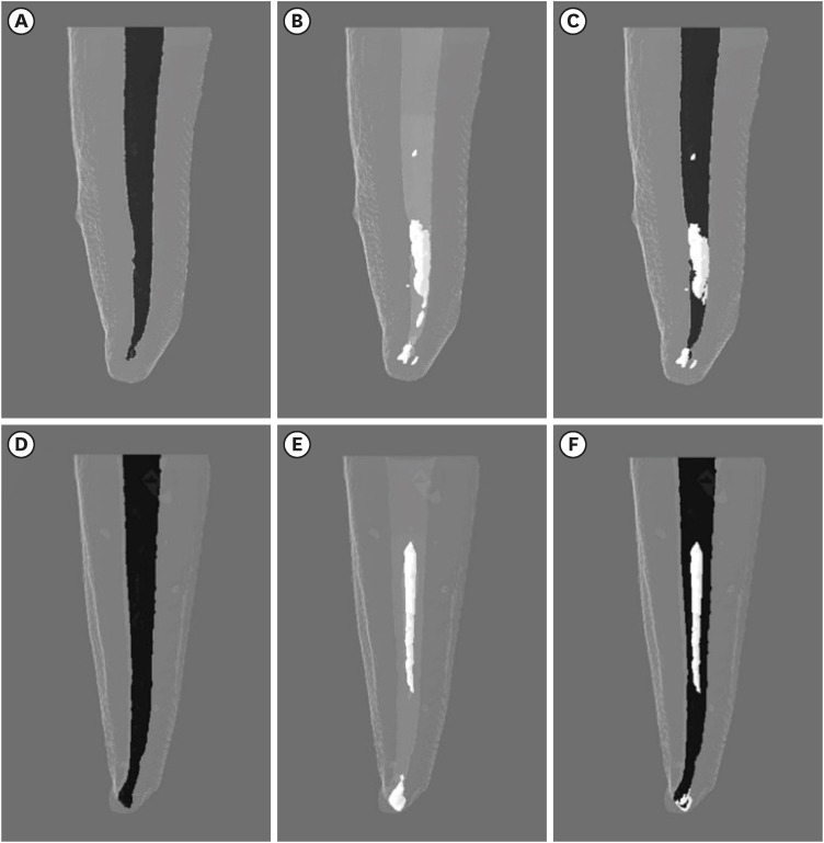
-
Abstract
PDFPubReaderePub
Objectives This study evaluated the efficiency of WaveOne Primary files (Dentsply Sirona) for removing root canal fillings with 2 types of movement: reciprocating (RCP) and continuous counterclockwise rotation (CCR).
Materials and Methods Twenty mandibular incisors were prepared with a RCP instrument (25.08) and filled using the Tagger hybrid obturation technique. The teeth were retreated with a WaveOne Primary file and randomly allocated to 2 experimental retreatment groups (
n = 10) according to movement type: RCP and CCR. The root canals were emptied of filling material in the first 3 steps of insertion, until reaching the working length. The timing of retreatment and procedure errors were recorded for all samples. The specimens were scanned before and after the retreatment procedure with micro-computed tomography to calculate the percentage and volume (mm3) of the residual filling material. The results were statistically evaluated using paired and independentt -tests, with a significance level set at 5%.Results No significant difference was found in the timing of filling removal between the groups, with a mean of 322 seconds (RCP) and 327 seconds (CCR) (
p < 0.05). There were 6 instrument fractures: 1 in a RCP motion file and 5 in continuous rotation files. The volumes of residual filling material were similar (9.94% for RCP and 15.94% for CCR;p > 0.05).Conclusions The WaveOne Primary files used in retreatment performed similarly in both RCP and CCR movements. Neither movement type completely removed the obturation material, but the RCP movement provided greater safety.
-
Citations
Citations to this article as recorded by- Micro-CT evaluation of the removal of root fillings using rotary and reciprocating systems supplemented by XP-Endo Finisher, the Self-Adjusting File, or Er,Cr:YSGG laser
Gülsen Kiraz, Bulem Üreyen Kaya, Mert Ocak, Muhammet Bora Uzuner, Hakan Hamdi Çelik
Restorative Dentistry & Endodontics.2023;[Epub] CrossRef
- Micro-CT evaluation of the removal of root fillings using rotary and reciprocating systems supplemented by XP-Endo Finisher, the Self-Adjusting File, or Er,Cr:YSGG laser
- 3,273 View
- 73 Download
- 1 Crossref
- Comparative analysis of torsional and cyclic fatigue resistance of ProGlider, WaveOne Gold Glider, and TruNatomy Glider in simulated curved canal
- Pedro de Souza Dias, Augusto Shoji Kato, Carlos Eduardo da Silveira Bueno, Rodrigo Ricci Vivan, Marco Antonio Hungaro Duarte, Pedro Henrique Souza Calefi, Rina Andréa Pelegrine
- Restor Dent Endod 2023;48(1):e4. Published online December 8, 2022
- DOI: https://doi.org/10.5395/rde.2023.48.e4
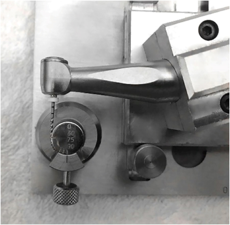
-
Abstract
PDFPubReaderePub
Objectives This study aimed to compare the torsional and cyclic fatigue resistance of ProGlider (PG), WaveOne Gold Glider (WGG), and TruNatomy Glider (TNG).
Materials and Methods A total of 15 instruments of each glide path system (
n = 15) were used for each test. A custom-made device simulating an angle of 90° and a radius of 5 millimeters was used to assess cyclic fatigue resistance, with calculation of number of cycles to failure. Torsional fatigue resistance was assessed by maximum torque and angle of rotation. Fractured instruments were examined by scanning electron microscopy (SEM). Data were analyzed with Shapiro-Wilk and Kruskal-Wallis tests, and the significance level was set at 5%.Results The WGG group showed greater cyclic fatigue resistance than the PG and TNG groups (
p < 0.05). In the torsional fatigue test, the TNG group showed a higher angle of rotation, followed by the PG and WGG groups (p < 0.05). The TNG group was superior to the PG group in torsional resistance (p < 0.05). SEM analysis revealed ductile morphology, typical of the 2 fracture modes: cyclic fatigue and torsional fatigue.Conclusions Reciprocating WGG instruments showed greater cyclic fatigue resistance, while TNG instruments were better in torsional fatigue resistance. The significance of these findings lies in the identification of the instruments’ clinical applicability to guide the choice of the most appropriate instrument and enable the clinician to provide a more predictable glide path preparation.
-
Citations
Citations to this article as recorded by- Mechanical Behaviour of Three Heat‐Treated NiTi Glide Path Systems at Simulated Intracanal Temperature
Ahmad Jamleh, Taher Omari, Abdulmohsen Alfadley, Abdullah Alqedairi, Hussam Alfawaz
Australian Endodontic Journal.2026;[Epub] CrossRef - Buckling resistance of various pathfinding endodontic instruments: An in vitro study
Ujjwal Das, Rajesh Kumar Das, Kallol Kumar Saha, Lugu Buru Murmu, Srimanta Banerjee, Rishila Nag
Journal of Conservative Dentistry and Endodontics.2025; 28(4): 384. CrossRef - Comparative evaluation of the remaining dentin volume following instrumentation with rotary, reciprocating, and hand files during root canal treatment in primary molars: An ex vivo study
İrem Eren, Berkant Sezer
Journal of Dental Sciences.2024; 19(4): 2126. CrossRef - Screw-in force, torque generation, and performance of glide-path files with three rotation kinetics
Jee-Yeon Woo, Ji-Hyun Jang, Seok Woo Chang, Soram Oh
Odontology.2024; 112(3): 761. CrossRef - Evaluation of shaping ability of different glide path instruments: a micro-computed tomography study
Merve Yeniçeri Özata, Seda Falakaloğlu, Ali Keleş, Özkan Adıgüzel, Mustafa Gündoğar
BMC Oral Health.2023;[Epub] CrossRef
- Mechanical Behaviour of Three Heat‐Treated NiTi Glide Path Systems at Simulated Intracanal Temperature
- 3,711 View
- 79 Download
- 4 Web of Science
- 5 Crossref
- Dentinal tubule penetration of sodium hypochlorite in root canals with and without mechanical preparation and different irrigant activation methods
- Renata Aqel de Oliveira, Theodoro Weissheimer, Gabriel Barcelos Só, Ricardo Abreu da Rosa, Matheus Albino Souza, Rodrigo Gonçalves Ribeiro, Marcus Vinicius Reis Só
- Restor Dent Endod 2023;48(1):e1. Published online December 1, 2022
- DOI: https://doi.org/10.5395/rde.2023.48.e1
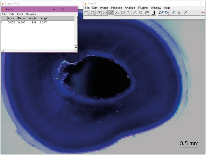
-
Abstract
PDFPubReaderePub
Objectives This study evaluated the dentinal penetration depth of 2.5% sodium hypochlorite (NaOCl) in root canals with and without preparation and different irrigant activation protocols.
Materials and Methods Sixty-three bovine mandibular incisors were randomly allocated to 6 groups (
n = 10): G1, preparation + conventional needle irrigation (CNI); G2, preparation + passive ultrasonic irrigation (PUI); G3, preparation + Odous Clean (OC); G4, no preparation + CNI; G5, no preparation + PUI; G6, no preparation + OC; and CG (negative control;n = 3). Samples were filled with crystal violet for 72 hours. Irrigant activation was performed. Samples were sectioned perpendicularly along the long axis, 3 mm and 7 mm from the apex. Images of the root thirds of each block were captured with a stereomicroscope and analyzed with an image analysis software. One-way analysis of variance, followed by the Tukeypost hoc test, and the Student’st -test were used for data analysis, with a significance level of 5%.Results The NaOCl penetration depth was similar when preparation was performed, regardless of the method of irrigation activation (
p > 0.05). In the groups without preparation, G6 showed greater NaOCl penetration depth (p < 0.05). The groups without preparation had a greater NaOCl penetration depth than those with preparation (p = 0.0019).Conclusions The NaOCl penetration depth was similar in groups with root canal preparation. Without root canal preparation, OC allowed deeper NaOCl penetration. The groups without preparation had greater NaOCl penetration than those undergoing root canal preparation.
-
Citations
Citations to this article as recorded by- Novel approaches involving curcumin in endodontic and periodontal diseases: a scoping review
Yuxi Xing, Yanbing Zhu, Yukai Shen, Yuou Xu, Ziman Xu, Mengxue Wang, Xudong Ma, Lehua Liu, Shu Chen
BMC Oral Health.2026;[Epub] CrossRef - Influence of passive ultrasonic irrigation cycles on the penetration depth of sodium hypochlorite into root dentin
Hüseyin Gündüz, Esin Özlek, Züleyha Baş
Scientific Reports.2025;[Epub] CrossRef - Evaluating the Effects of Various Antioxidants on Dentinal Tubule Penetrability of a Resin-Based Sealer: A Confocal Laser Microscopic Study
Sanjeev Srivastava, Shijita Sinha, Abhishek Singh, Aditya Singh, Pragyan Paliwal, Syed H Mehdii
Cureus.2025;[Epub] CrossRef - Impact of different activation procedures on sodium hypochlorite penetration into dentinal tubules after endodontic retreatment via confocal laser scanning microscopy
Betul Gunes, Kübra Yeşildal Yeter, Yasin Altay
BMC Oral Health.2024;[Epub] CrossRef - Debridement ability of the WaveOne Gold and TruNatomy systems in the apical third of root canals: ex vivo assessment
Sara Carvalho Avelar de Oliveira, Carlos Eduardo da Silveira Bueno, Rina Andréa Pelegrine, Carlos Eduardo Fontana, Alexandre Sigrist de Martin, Carolina Pessoa Stringheta
Brazilian Dental Journal.2024;[Epub] CrossRef - Combined effect of electrical energy and graphene oxide on Enterococcus faecalis biofilms
Myung-Jin LEE, Mi-Ah KIM, Kyung-San MIN
Dental Materials Journal.2023; 42(6): 844. CrossRef
- Novel approaches involving curcumin in endodontic and periodontal diseases: a scoping review
- 3,312 View
- 79 Download
- 5 Web of Science
- 6 Crossref
- Evaluation of blood clot, platelet-rich plasma, and platelet-rich fibrin–mediated regenerative endodontic procedures in teeth with periapical pathology: a CBCT study
- Swati Markandey, Haridas Das Adhikari
- Restor Dent Endod 2022;47(4):e41. Published online October 21, 2022
- DOI: https://doi.org/10.5395/rde.2022.47.e41
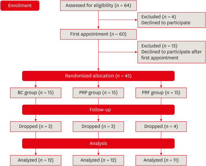
-
Abstract
PDFSupplementary MaterialPubReaderePub
Objectives This study compared the clinical and radiological outcomes of regenerative endodontic procedures (REPs) using blood clots (BCs), platelet-rich plasma (PRP), and platelet-rich fibrin (PRF) through intraoral periapical radiography (IOPAR) and cone-beam computed tomography (CBCT).
Materials and Methods Forty-five single-rooted necrotic teeth with periapical pathology were randomly allocated to receive BC, PRP, or PRF as an individual scaffold. Outcomes were evaluated in 35 teeth in 23 patients with a follow-up period of 12–24 months through qualitative IOPAR scoring and quantitative CBCT measurements. Healing of periapical lesions and in immature teeth, changes in the apical foramen diameter (AFD), root wall thickness (RWT), and root length (RL) were assessed. A
p value less than 0.05 was considered to indicate statistical significance.Results All teeth were asymptomatic except 1 in the PRP group. Periapical lesion healing was seen in all except 2 teeth in the BC group and 3 in the PRP group. Both IOPAR and CBCT revealed no significant differences in bone healing or changes in AFD, RWT, and RL among the 3 groups. A positive pulp sensibility response to the cold test was seen in 2 teeth in the BC group, but none to the electric pulp test. Intracanal calcification (ICC) was evident in more teeth in the BC group than in the PRP and PRF groups, and was also significantly higher in immature teeth.
Conclusions Our results revealed that BC, PRP, and PRF have similar potential as scaffolds in REPs, and ICC may be a concern for long-term outcomes.
-
Citations
Citations to this article as recorded by- Regenerative Endodontics and Stem Cell-Based Therapies – A Systematic Review
Wjoud Ahmed Alshamrani, Sarah Sulaiman Alzarea, Joud Khalid Alabbas, Ayah Khalid Alabbas, Mawiyah Ibrahim Aljaddua, Osama Khattak, Rakhi Issrani
Journal of Pharmacy and Bioallied Sciences.2026; 18(Suppl 1): S29. CrossRef - Collagen Scaffolds in Regenerative Endodontic Procedures: Current Evidence, Limitations, and Future Perspectives
Qiong-Ling Shi, Xiao Zhu, Chen Chen, Jing-Yi Chen, Dan-Yang Lu, Ying Shi, Yan-Qi Chen, Zhi-Fang Wu
Polymers.2026; 18(7): 894. CrossRef - Regenerative potential of concentrated growth factor compared to platelet-rich fibrin in treatment of necrotic mature teeth: a randomized clinical trial
Taghreed Salah, Wael Hussein, Heba Abdelkafy
BDJ Open.2025;[Epub] CrossRef - Clinical and radiographic outcomes of non-surgical retreatment of mature maxillary incisors using two regenerative endodontic techniques in adolescents: a 24-month randomized clinical trial
Ahmad Abdel Hamid Elheeny, Sherif Shafik EL Bahnasy, Yassmin Mohamed ElMakawi, Mohammed Turky, Eman Farouk Ahmed, Norhan Khaled Omar Wahba
BDJ Open.2025;[Epub] CrossRef - Clinical and Radiographic Outcomes of Autologous Platelet-Rich Products in Regenerative Endodontics: A Systematic Review and Meta-Analysis
Raewyn Huang, Wei Chen, Matthew Fang, Ove A. Peters, Sepanta Hosseinpour
Dentistry Journal.2025; 13(6): 236. CrossRef - Regenerative Endodontics in the Treatment of Periapical Lesions of Endodontic Origin: A Systematic Review of Randomized Controlled Trials
BelHaj Salah Kawthar, Berrima Fatma, Boukhris Hanen, Gnaba Imen, Ben Youssef Souha
The Journal of Contemporary Dental Practice.2025; 26(6): 623. CrossRef - Advanced Platelet-Rich Fibrin Plus Sealed Exclusively with Glass Ionomer Cement: Setting a New Standard for Healing, Aesthetics and Predictive Modelling in Regenerative Endodontics
Dubravka Turjanski, Dragutin Lisjak, Petra Bučević Sojčić, Jelena Valpotić, Tea Borojević Renić, Kristina Goršeta, Domagoj Glavina
Materials.2025; 18(18): 4421. CrossRef - Evaluation of different scaffolds and intracanal medications in revascularization of nonvital immature permanent teeth
Mona M. A. Sameia, Abeer M. Darrag, Neveen A. Shaheen, Dina A. Attia
Tanta Dental Journal.2025; 22(4): 693. CrossRef - Biomimetic pulp scaffolds prepared from extracellular matrix derived from stem cells from human exfoliated deciduous teeth promote pulp–dentine complex regeneration
Ning Yang, Rou Shen, Wenxiao Yang, Shengcai Zhang, Tianxing Gong, Yao Liu
International Endodontic Journal.2024; 57(9): 1279. CrossRef - Arrest and Repair of Inflammatory Root Resorption After an Endodontic Regeneration Procedure – A Hypothesis and Case Report
Arieh Y. Kaufman, Bill Kahler
Journal of Endodontics.2024; 50(12): 1743. CrossRef - Effect of Platelet Rich Plasma in Regenerative Endodontic Treatment: A Review of Clinical Trials
Hojat Rezazadeh, Mehrnaz Okhovatfard
Research Journal of Pharmacy and Technology.2023; : 5562. CrossRef
- Regenerative Endodontics and Stem Cell-Based Therapies – A Systematic Review
- 5,985 View
- 156 Download
- 7 Web of Science
- 11 Crossref
Review Article
- Influence of the root canal filling technique on the success rate of primary endodontic treatments: a systematic review
- Daniel Feijolo Marconi, Giovana Siocheta da Silva, Theodoro Weissheimer, Isadora Ames Silva, Gabriel Barcelos Só, Leonardo Thomasi Jahnke, Jovito Adiel Skupien, Marcus Vinicius Reis Só, Ricardo Abreu da Rosa
- Restor Dent Endod 2022;47(4):e40. Published online October 11, 2022
- DOI: https://doi.org/10.5395/rde.2022.47.e40
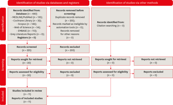
-
Abstract
PDFSupplementary MaterialPubReaderePub
Objectives This study aimed to investigate the influence of different obturation techniques compared to cold lateral compaction on the success rate of primary non-surgical endodontic treatments.
Materials and Methods Systematic searches were performed for studies published up to May 17th, 2022 in MEDLINE/PubMed, Cochrane Library, Web of Science, Scopus, EMBASE, and Grey Literature Reports. Randomized clinical trials and nonrandomized (nonrandomized clinical trials, prospective or retrospective) studies that evaluated the success rate of primary non-surgical endodontic treatments obturated with the cold lateral compaction (control) and other obturation techniques were included. The revised Cochrane risk of bias tools for randomized trials (RoB 2) and nonrandomized studies of interventions (ROBINS-I) were used to evaluate the risk of bias. The Grading of Recommendations Assessment, Development, and Evaluation (GRADE) tool was used to evaluate the certainty of evidence.
Results Eleven studies (4 randomized clinical trials (RCTs), 4 prospective, and 3 retrospectives) were included. Two RCTs were classified as having some concerns risk of bias and 2 as a low risk of bias. Two nonrandomized studies were classified as having a critical risk of bias and 5 as having a moderate risk of bias. The GRADE analysis demonstrated a very low to moderate certainty of evidence.
Conclusions This systematic review generally evidenced no differences in the success rate of primary non-surgical endodontic treatments when the cold lateral compaction technique and other obturation techniques are performed. Further well-designed studies are still necessary.
-
Citations
Citations to this article as recorded by- Assessing Sealing Ability of C-Root SP Strontium Silicate Sealer With Different Obturation Techniques: An in vitro Study
Suixin Hu, Jianshe Li, Meng Xu, Laiqing Xu, Yangming Yin, Peng Xue, Liping Dong, Lin Wang, Huixia He, Ying Liu, Qiang Luo, Fei Chen
International Dental Journal.2026; 76(1): 109283. CrossRef - Effect of root canal filling techniques and materials on endodontic treatment outcomes: a systematic review and meta-analysis
Ahmed Mushtaq, Sura Alsanafi, Firas Elmsmari, José A. González, Marc Garcia-Font, Francesc Abella Sans, Kelvin I. Afrashtehfar, Paul V. Abbott
Scientific Reports.2026;[Epub] CrossRef - Dimensional and cross-sectional compatibility of contemporary nickel–titanium files and their corresponding gutta-percha cones: A micro-computed tomography study
Cem Sarkan, Faruk Haznedaroglu
The Saudi Dental Journal.2026;[Epub] CrossRef - Comparative Analysis Of Obturation Techniques In Endodontics: Lateral Vs. Thermoplasticized. Thermoplasticized
Juan Esteban Díaz Pacheco , Rómulo Guillermo López Torres , Verónica Alejandra Salame Ortíz
Salud, Ciencia y Tecnología.2025; 5: 1626. CrossRef - Effect of ultrasonic activation of endodontic sealers on root canal filling quality during the single-cone obturation procedure: a systematic review and meta-analysis of laboratory-based studies
Shuting Feng, Weiqing Zhou, Xiaojun Chu, Shuaimei Xu, Xiongqun Zeng
Odontology.2025; 113(4): 1380. CrossRef - In Vitro and In Vivo Evaluation of a New Experimental Polydimethylsiloxane-Based Endodontic Sealer
Fabiola Cardoso Maldonado, Cesar Gaitan Fonseca, Carlos Bermudez Jimenez, Luis Alejandro Aguilera Galaviz, Margarita L. Martinez-Fierro, Lorena Troncoso Vazquez, Martha Eugenia Reyes Ortiz
Journal of Functional Biomaterials.2025; 16(11): 402. CrossRef - Evaluation of three obturation techniques in 3D-printed models of oval canals with standardized prepared morphology: a micro-CT study
Wenjun Xia, Qisheng Gu, Yingshuang Song, Yunjia Liu, Xuetao Deng, Wenhao Qian
BMC Oral Health.2025;[Epub] CrossRef - Clinical and Radiographic Failure of Nonsurgical Endodontic Treatment and Retreatment Using Single-cone Technique With Calcium Silicate-based Sealers: A Systematic Review and Meta-analysis
Mohammad A. Sabeti, Negah Karimpourtalebi, Arash Shahravan, Omid Dianat
Journal of Endodontics.2024; 50(6): 735. CrossRef - Method of microbial decontamination of endodontic absorbent paper points: a randomised experimental study
O. A. Pavlovskaya, O. A. Kachanova, V. V. Volobuev, M. N. Mitropanova, A. R. Gazarova, V. Y. Zobenko, A. G. Uvarova
Pediatric dentistry and dental prophylaxis.2024; 24(2): 157. CrossRef - The Push-Out Bond Strength, Surface Roughness, and Antimicrobial Properties of Endodontic Bioceramic Sealers Supplemented with Silver Nanoparticles
Karla Navarrete-Olvera, Nereyda Niño-Martínez, Idania De Alba-Montero, Nuria Patiño-Marín, Facundo Ruiz, Horacio Bach, Gabriel-Alejandro Martínez-Castañón
Molecules.2024; 29(18): 4422. CrossRef - Clinical outcome of non-surgical root canal treatment using different sealers and techniques of obturation in 237 patients: A retrospective study
Mateusz Radwanski, Krystyna Pietrzycka, Tan Fırat Eyüboğlu, Mutlu Özcan, Monika Lukomska-Szymanska
Clinical Oral Investigations.2024;[Epub] CrossRef
- Assessing Sealing Ability of C-Root SP Strontium Silicate Sealer With Different Obturation Techniques: An in vitro Study
- 8,368 View
- 132 Download
- 10 Web of Science
- 11 Crossref
Research Articles
- Outcome of endodontic treatments performed by Brazilian undergraduate students: 3- to 8-year follow up
- Jéssica Gabriele da Rocha, Isabella Marian Lena, Jéssica Lopes Trindade, Gabriela Salatino Liedke, Renata Dornelles Morgental, Carlos Alexandre Souza Bier
- Restor Dent Endod 2022;47(3):e34. Published online August 18, 2022
- DOI: https://doi.org/10.5395/rde.2022.47.e34
-
Abstract
PDFPubReaderePub
Objectives This study aimed to evaluate the success rate of endodontic treatments performed by undergraduate students and the factors associated with the outcome.
Materials and Methods A follow-up of 3 to 8 years after root canal filling was carried out in 91 patients. At the follow-up visits, medical and dental history questionnaires were applied along with clinical and radiographic examinations. Data collected in the clinical exam included: the presence of pain, swelling, sinus tract, mobility, tenderness to palpation and percussion, periodontal probing profile, and type/quality of coronal restoration. Postoperative and follow-up radiographs were digitalized and analyzed by 2 trained and calibrated examiners to assess periapical healing. The treatment outcome was based on strict clinical and radiographic criteria and classified as success (absence of any clinical and radiographic sign of apical periodontitis) or failure (other combination). Logistic regression was used to investigate the impact of clinical and radiographic variables on endodontic treatment outcomes at a 5% significance level.
Results The success rate of endodontic treatments was 60.7%. The only risk factor significantly associated with failure was the presence of a periapical lesion on the postoperative radiograph (odds ratio, 3.35; 95% confidence interval, 1.17–9.54).
Conclusions The success rate of endodontic treatments performed by undergraduate students was low and was jeopardized by the presence of a periapical lesion on the postoperative radiograph.
-
Citations
Citations to this article as recorded by- Outcomes of root canal treatment performed by undergraduate students: A systematic review and meta‐analysis
Philip Y.‐H. Chien, Sepanta Hosseinpour, Ove A. Peters, Christine I. Peters
International Endodontic Journal.2026; 59(6): 929. CrossRef - Effect of quality of radiographs taken during root canal treatment on technical quality of root canal fillings and endodontic outcome
Jia Min Ng, Yan Yee Lee, Prashanti Chippagiri, Elaheh Ahanin, Abhishek Parolia
Restorative Dentistry & Endodontics.2025; 50(1): e3. CrossRef - Factors Influencing the Long-Term Survival and Success of Endodontically Treated and Retreated Teeth: An Ambispective Study at an Educational Hospital
Reem Barakat, Rahaf Almohareb, Ghaliah Alsawah, Hadeel Busuhail, Shahad A. Alshihri, Ghadah T. Alrashid, Ghadeer Y. Alotaibi, Mamata Hebbal
Journal of Clinical Medicine.2025; 14(21): 7826. CrossRef - A bibliometric comparison of undergraduate and postgraduate endodontic education publications: The topics, trends, and challenges
Jinglan Zhang, Xiaowei Liu, Lei Yang, Yiran Wang, Dingming Huang, Xuelian Tan
Journal of Dental Education.2023; 87(12): 1661. CrossRef
- Outcomes of root canal treatment performed by undergraduate students: A systematic review and meta‐analysis
- 5,592 View
- 48 Download
- 5 Web of Science
- 4 Crossref
- Proximity of maxillary molar apexes to the cortical bone surface and the maxillary sinus
- Han Shin Lee, Dokyung Kim, Sung Kyo Kim
- Restor Dent Endod 2022;47(3):e33. Published online August 8, 2022
- DOI: https://doi.org/10.5395/rde.2022.47.e33
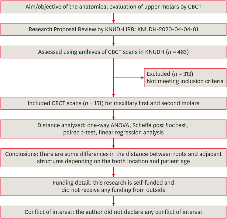
-
Abstract
PDFPubReaderePub
Objectives This study aimed to analyze the proximity of maxillary molar roots to their overlying cortical bone surfaces and the maxillary sinus.
Materials and Methods Cone-beam computed tomographic images of 151 patients with completely erupted upper molars that had 3 separate roots were studied. The following distances were measured: from the root apex to the cortical plate and maxillary sinus floor, and from the apical 3-mm level of the root to the cortical plate. Differences between groups were analyzed with 1-way analysis of variance and the Scheffé
post hoc test, the significance of differences between cone-beam computed tomography views with the pairedt -test, and the significance of differences among age groups with linear regression analysis. The significance level was set atp < 0.05.Results The mesiobuccal and distobuccal root apexes of maxillary second molars were more distant from the buccal cortical plate than the maxillary first molars (
p < 0.05). The apical 3-mm level of the mesiobuccal root of the first molar was closer to the buccal cortical bone than the second molar (p < 0.05). In the maxillary first molars, the thickness of the buccal cortical bone decreased in all roots with age (p < 0.05). In all root apexes of both molars, the difference in the vertical level between the maxillary sinus floor and the root apex increased with age (p < 0.05).Conclusions Awareness of the anatomical profile of maxillary molar apices in relation to the cortical bones and maxillary sinus will be beneficial for apical surgery.
-
Citations
Citations to this article as recorded by- Proximity of maxillary molar palatal roots to adjacent structures for endodontic microsurgery: a cone-beam computed tomography study
Xiaoxiang Huang, Jun Xu, Benxiang Hou, Ying Wang
BMC Oral Health.2025;[Epub] CrossRef - Periapical bone loss configuration in sub-Saudi patients afflicted with periapical abscesses: A 3D cone-beam computed tomography analysis
Swati A. Srivastava, Rahaf A. Alawajy, Rehab Abdelaziz, Elzahraa A. Eldwakhly, Selma A. Saadaldin, Rahaf A. Almohareb, Fahda Nabeel Algahtani, Mai Salah Soliman, Manal M. Abdelhafeez
Saudi Endodontic Journal.2025; 15(2): 144. CrossRef
- Proximity of maxillary molar palatal roots to adjacent structures for endodontic microsurgery: a cone-beam computed tomography study
- 3,947 View
- 32 Download
- 1 Web of Science
- 2 Crossref
- Clinical and radiographic outcomes of regenerative endodontic treatment performed by endodontic postgraduate students: a retrospective study
- Hadi Rajeh Alfahadi, Saad Al-Nazhan, Fawaz Hamad Alkazman, Nassr Al-Maflehi, Nada Al-Nazhan
- Restor Dent Endod 2022;47(2):e24. Published online May 9, 2022
- DOI: https://doi.org/10.5395/rde.2022.47.e24
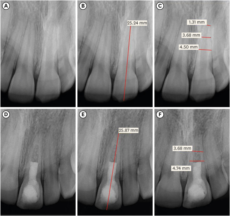
-
Abstract
PDFPubReaderePub
Objectives Regenerative endodontic treatment is a clinical procedure aimed at biologically regenerating damaged root canal tissue of immature permanent teeth. This study aimed to report the outcomes of regenerative endodontic treatment performed by endodontic postgraduate students.
Materials and Methods Clinical and radiographic data of 27 patients, aged 10–22 years, who underwent regenerative treatment of immature permanent teeth from 2015 to 2019 were followed up, wherein clinical and radiographic examinations were performed for each patient. Postoperative success rate and tooth survival were analyzed, and the postoperative radiographic root area changes were quantified.
Results A total of 23 patients attended the dental appointments, showing that all teeth survived and were asymptomatic. Specifically, 7 periapical pathosis cases were completely healed, 12 were incompletely healed, and 4 cases failed. Moreover, significant differences were found between discolored and non-discolored teeth, and between the presence or absence of periapical radiolucency. Additionally, 3 anterior teeth showed complete closure of the apical foramen, while the apical foramen width was reduced in 17 teeth and failed in 3 teeth. Root length was also found to have been increased in 7 anterior and 4 posterior teeth, and the average length ranged from 4.00–0.63 mm in the anterior teeth, 2.85–1.48 mm of the mesial root, and 2.73–2.16 mm of the molar teeth distal root. Furthermore, calcified tissue deposition was observed in 7 teeth.
Conclusions A favorable outcome of regenerative endodontic treatment of immature permanent teeth with necrotic pulp was achieved with a high survival rate.
-
Citations
Citations to this article as recorded by- Regenerative Endodontics and Stem Cell-Based Therapies – A Systematic Review
Wjoud Ahmed Alshamrani, Sarah Sulaiman Alzarea, Joud Khalid Alabbas, Ayah Khalid Alabbas, Mawiyah Ibrahim Aljaddua, Osama Khattak, Rakhi Issrani
Journal of Pharmacy and Bioallied Sciences.2026; 18(Suppl 1): S29. CrossRef - Pre‐Operative Factors on Prognosis of Regenerative Endodontic Procedures: A Systematic Review and Meta‐Analysis
Filipe Colombo Vitali, Alexandre Henrique dos Reis‐Prado, Pablo Silveira Santos, Ana Paula Portes Zeno, Patrícia de Andrade de Risso, Lucianne Cople Maia, Francine Benetti, Cleonice da Silveira da Teixeira
International Endodontic Journal.2025; 58(12): 1814. CrossRef - Clinical, radiographic, and biomarker perspectives of low-level laser therapy during regenerative endodontic procedures in necrotic immature young teeth: a randomized clinical study
Pragya Pandey, Neha Jasrasaria, Ramesh Bharti, Rakesh Kumar Yadav, Monika Kumari, Abinia Vaishnavi, Rahul Pandey
Lasers in Medical Science.2025;[Epub] CrossRef - Allogeneic Bone Marrow Mesenchymal Stromal Cell Transplantation Induces Dentin Pulp Complex-like Formation in Immature Teeth with Pulp Necrosis and Apical Periodontitis
Jose Francisco Gomez-Sosa, José E. Cardier, Olga Wittig, Dylana Díaz-Solano, Eloisa Lara, Kharelys Duque, Giselle Ramos-González
Journal of Endodontics.2024; 50(4): 483. CrossRef - Radiographic assessment of dental post and core placement at different educational levels in an undergraduate student clinic: a 4-year retrospective study
Turki Alshehri, Nourhan M. Aly, Raand Altayyar, Deena Alghamdi, Shahad Alotaibi, Passent Ellakany
F1000Research.2024; 12: 976. CrossRef - Evaluation of the efficacy of injectable platelet‐rich fibrin versus platelet‐rich plasma in the regeneration of traumatized necrotic immature maxillary anterior teeth: A randomized clinical trial
Maha Mohamed Abo‐Heikal, Jealan M. El‐Shafei, Samia A. Shouman, Nehal N. Roshdy
Dental Traumatology.2024; 40(1): 61. CrossRef - Radiographical assessment of post and core placement errors encountered by Saudi dental students at different educational levels
Turki Alshehri, Nourhan M. Aly, Raand Altayyar, Deena Alghamdi, Shahad Alotaibi, Passent Ellakany
F1000Research.2023; 12: 976. CrossRef
- Regenerative Endodontics and Stem Cell-Based Therapies – A Systematic Review
- 5,286 View
- 103 Download
- 7 Web of Science
- 7 Crossref
- Difficulties experienced by endodontics researchers in conducting studies and writing papers
- Betul Aycan Alim-Uysal, Selin Goker-Kamali, Ricardo Machado
- Restor Dent Endod 2022;47(2):e20. Published online March 15, 2022
- DOI: https://doi.org/10.5395/rde.2022.47.e20
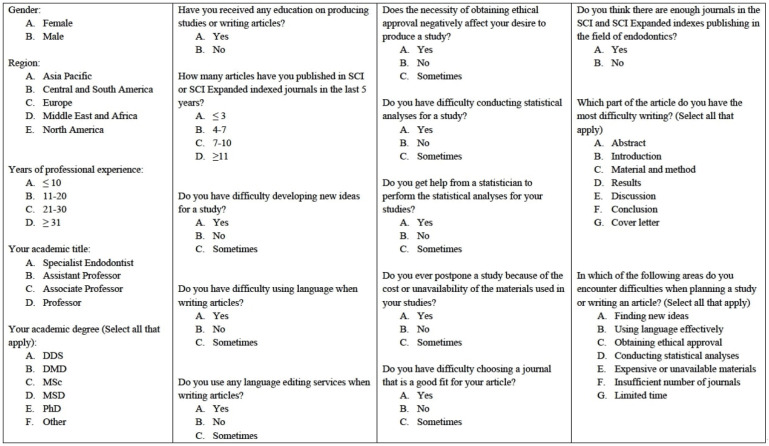
-
Abstract
PDFPubReaderePub
Objectives The study investigated the difficulties experienced by endodontics researchers around the world in conducting studies and writing papers.
Materials and Methods A survey consisting of 18 questions on the difficulties experienced by endodontics researchers in performing studies and writing papers was e-mailed to academics in the field of endodontics working at 202 universities. The independent risk factors were analyzed using binary logistic regression at a significance level of 0.05.
Results A total of 581 individuals (10.7%) agreed to participate in the study. Almost half the participants (48.2%) reported that they had received some type of training in conducting studies and writing papers. In response to the question, “Do you get help from a statistician to perform the statistical analyses of your studies?,” 77.1% answered “yes.” Around 40% of the participants stated that the need to obtain ethical approval negatively affected their desire to conduct studies. The participants’ regions had no effect on the reported difficulties associated with writing papers in English or conducting statistical analyses (
p > 0.05). Most participants (81.8%) reported difficulties in writing the Discussion section, regardless of their region, academic degrees, or years of experience.Conclusions The participants stated they experienced difficulties in many areas, such as conducting statistical analyses, finding new ideas, and writing in English. Engaging in a detailed examination of ethics committee rules, expanding biostatistics education, increasing the number of institutions providing research funding, and increasing the number of endodontics journals can increase the enthusiasm of endodontics researchers to publish papers.
-
Citations
Citations to this article as recorded by- Prevalence of radix molaris in mandibular molars of a subpopulation of Brazil’s Northeast region: a cross-sectional CBCT study
Yasmym Martins Araújo de Oliveira, Maria Clara Mendes Gomes, Maria Fernanda da Silva Nascimento, Ricardo Machado, Danna Mota Moreira, Hermano Camelo Paiva, George Táccio de Miranda Candeiro
Scientific Reports.2025;[Epub] CrossRef - Statistical pitfalls in endodontic research
Nandini Suresh
Endodontology.2023; 35(1): 1. CrossRef
- Prevalence of radix molaris in mandibular molars of a subpopulation of Brazil’s Northeast region: a cross-sectional CBCT study
- 2,745 View
- 31 Download
- 1 Web of Science
- 2 Crossref
Case Report
- Leukocyte platelet-rich fibrin in endodontic microsurgery: a report of 2 cases
- Mariana Domingos Pires, Jorge N. R. Martins, Abayomi Omokeji Baruwa, Beatriz Pereira, António Ginjeira
- Restor Dent Endod 2022;47(2):e17. Published online March 4, 2022
- DOI: https://doi.org/10.5395/rde.2022.47.e17
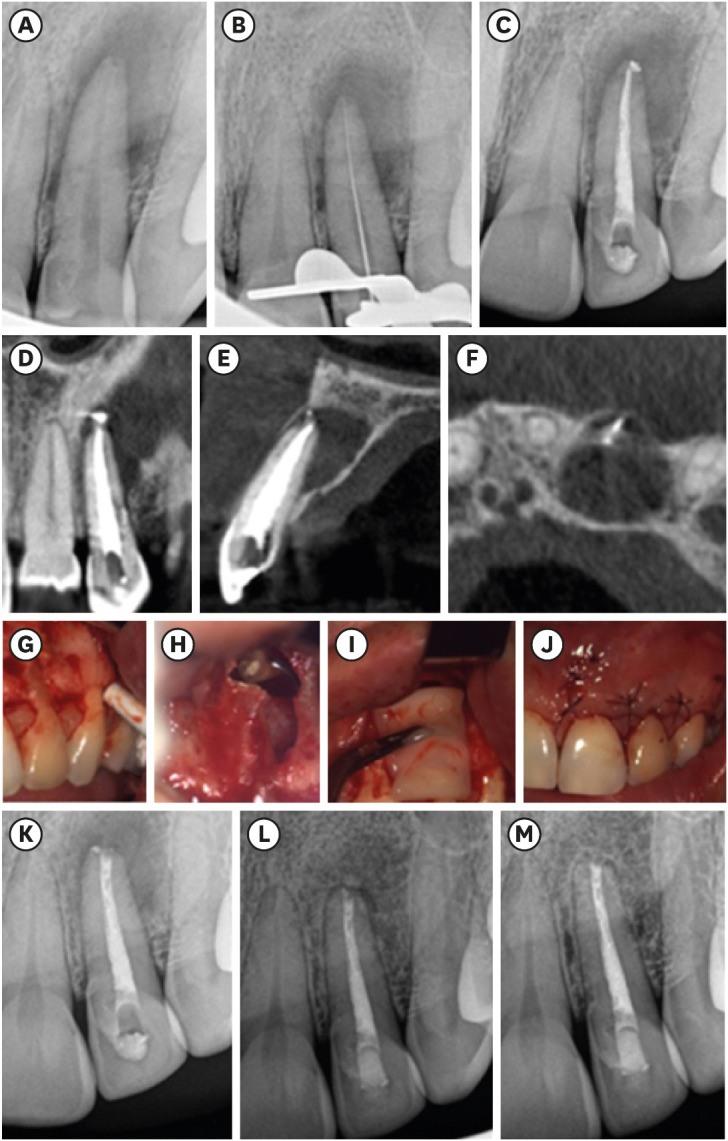
-
Abstract
PDFPubReaderePub
Endodontic microsurgery is a predictable treatment option when orthograde treatment or retreatment is unsuccessful or unfeasible. However, when there is a gross compromise of periapical bone, achievement of bone regeneration after the surgical procedure may be hampered. In such cases, the application of guided tissue regeneration principles, with adjunctive use of leukocyte platelet-rich fibrin to fill the bone defect as a bone substitute and as a membrane to cover the site, provides a cost-effective solution with the benefits of accelerated physiological healing and reduced post-surgical pain and discomfort. This case report presents 2 cases of endodontic microsurgery of the upper lateral incisors with loss of buccal cortical plate, where platelet-rich fibrin was successfully applied.
-
Citations
Citations to this article as recorded by- Focuses and Trends of Research on Platelet-Rich Fibrin: A Bibliometric and Visual Analysis
Ying Zhao, Chen Dong, Liumeizi Fan, Ting Lei, Xin Ge, Zhou Yu, Sheng Hu
Indian Journal of Plastic Surgery.2024; 57(05): 356. CrossRef
- Focuses and Trends of Research on Platelet-Rich Fibrin: A Bibliometric and Visual Analysis
- 2,235 View
- 36 Download
- 1 Web of Science
- 1 Crossref
Research Articles
- Comparison of the cyclic fatigue resistance of One Curve, F6 Skytaper, Protaper Next, and Hyflex CM endodontic files
- Charlotte Gouédard, Laurent Pino, Reza Arbab-Chirani, Shabnam Arbab-Chirani, Valérie Chevalier
- Restor Dent Endod 2022;47(2):e16. Published online March 4, 2022
- DOI: https://doi.org/10.5395/rde.2022.47.e16
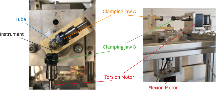
-
Abstract
PDFPubReaderePub
Objectives This study compared the cyclic fatigue resistance of One Curve (C wire) and F6 Skytaper (conventional austenite nickel-titanium [NiTi]), and 2 instruments with thermo-mechanically treated NiTi: Protaper Next X2 (M wire) and Hyflex CM (CM wire).
Materials and Methods Ten new instruments of each group (size: 0.25 mm, 6% taper in the 3 mm tip region) were tested using a rotary bending machine with a 60° curvature angle and a 5 mm curvature radius, at room temperature. The number of cycles until fracture was recorded. The length of the fractured instruments was measured. The fracture surface of each fragment was examined with a scanning electron microscope (SEM). The data were analyzed using one-way analysis of variance and the
post hoc Tukey test. The significance level was set at 0.05.Results At 60°, One Curve, F6 Skytaper and Hyflex CM had significantly longer fatigue lives than Protaper Next X2 (
p < 0.05). No statistically significant differences were found in the cyclic fatigue lives of One Curve, F6 Skytaper, and Hyflex CM (p > 0.05). SEM images of the fracture surfaces of the different instruments showed typical features of fatigue failure.Conclusions Within the conditions of this study, at 60° and with a 5 mm curvature radius, the cyclic fatigue life of One Curve was not significantly different from those of F6 Skytaper and Hyflex CM. The cyclic fatigue lives of these 3 instruments were statistically significantly longer than that of Protaper Next.
-
Citations
Citations to this article as recorded by- Comparison of Canal Shaping and Transportation Between the Protaper Next and Protaper Ultimate in Simulated Double-curved Canals
Seher Pelda Biçer, Betül Aycan Uysal
ADO Klinik Bilimler Dergisi.2026; 15(1): 47. CrossRef - Influence of the Cause of File Fracture on the Successful Removal of Fragments from Root Canals: An In Vivo Study
Ricardo Portigliatti, Eugenia Pilar Consoli Lizzi, Pablo Alejandro Rodríguez
Applied Sciences.2026; 16(8): 3832. CrossRef - Evaluation of cyclic fatigue in three pediatric endodontic rotary file systems in root canals of primary molars: A finite element analysis (FEA)
Monika sri S.S., K.C. Vignesh, K. Vivek, Kavitha Swaminathan, Selvakumar Haridoss
Journal of Oral Biology and Craniofacial Research.2025; 15(2): 310. CrossRef - Stress analysis of different experimental finite element models of rotary endodontic instruments
Manar M. Galal, Amira Galal Ismail, Nada Omar
Bulletin of the National Research Centre.2025;[Epub] CrossRef - Understanding Cyclic Fatigue in Three Nickel–Titanium Pediatric Files: An In Vitro Study for Enhanced Patient Care
Alwaleed Abushanan, Rajashekhara Bhari Sharanesha, Fahd Aljarbou, Hadi Alamri, Mohammed Hamad Almasud, Abdulfatah AlAzmah, Sara Alghamdi, Mubashir Baig Mirza
Medicina.2025; 61(5): 830. CrossRef - Analyzing Surface Morphology Changes Induced by Cyclic Fatigue in Three Different Nickel–Titanium Rotary Files Using Scanning Electron Microscopy Analysis
Chintan Joshi, Mahima P Jain, Sweety M Thumar, Jay H Dave, Applu R Bhatt, Juhi I Dholani
World Journal of Dentistry.2024; 15(7): 579. CrossRef - Nickel ion release and surface analyses on instrument fragments fractured beyond the apex: a laboratory investigation
Sıdıka Mine Toker, Ekim Onur Orhan, Arzu Beklen
BMC Oral Health.2023;[Epub] CrossRef
- Comparison of Canal Shaping and Transportation Between the Protaper Next and Protaper Ultimate in Simulated Double-curved Canals
- 4,103 View
- 61 Download
- 3 Web of Science
- 7 Crossref
- Efficacy of reciprocating instruments and final irrigant activation protocols on retreatment of mesiobuccal roots of maxillary molars: a micro-CT analysis
- Lilian Tietz, Renan Diego Furlan, Ricardo Abreu da Rosa, Marco Antonio Hungaro Duarte, Murilo Priori Alcalde, Rodrigo Ricci Vivan, Theodoro Weissheimer, Marcus Vinicius Reis Só
- Restor Dent Endod 2022;47(1):e13. Published online February 15, 2022
- DOI: https://doi.org/10.5395/rde.2022.47.e13
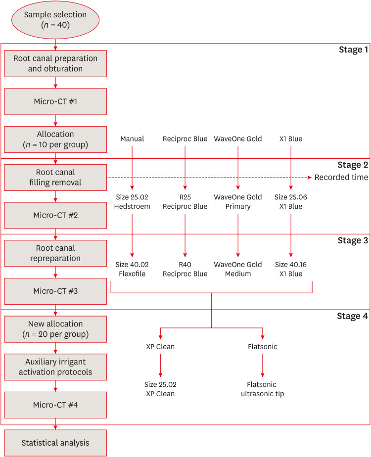
-
Abstract
PDFPubReaderePub
Objectives This study evaluated the efficacy of 3 reciprocating systems and the effects of 2 instruments for irrigant activation on filling material removal.
Materials and Methods Forty mesiobuccal roots of maxillary molars were prepared up to size 25.06 and obturated. Micro-computed tomography (micro-CT) examination #1 was performed. Teeth were then divided into 4 groups (
n = 10), according to the retreatment protocol: (1) manual, (2) Reciproc Blue, (3) WaveOne Gold, and (4) X1 Blue. Micro-CT examinations #2 and #3 were performed after filling removal and repreparation, respectively. Next, all teeth were divided into 2 new groups (n = 20) according to the irrigant activation protocol: XP Clean (XP Clean size 25.02) and Flatsonic (Flatsonic ultrasonic tip). Micro-CT examination #4 was performed after irrigant activation. Statistical analysis was performed with a significance level set at 5%.Results WaveOne Gold removed a significantly greater amount of filling material than the manual group (
p < 0.05). The time to reach the WL was similar for all reciprocating systems (p > 0.05). X1 Blue was faster than the manual group (p < 0.05). Only manual group improved the filling material removal after the repreparation stage (p < 0.05). Both activation protocols significantly improved the filling material removal (p < 0.05), without differences between them (p > 0.05).Conclusions None of the tested instruments completely removed the filling material. X1 Blue size 25.06 reached the working length in the shortest time. XP Clean and Flatsonic improved the filling material removal.
-
Citations
Citations to this article as recorded by- Impact of vehicle and insertion method on intratubular penetration of calcium hydroxide pastes: an in vitro confocal laser microscopy analysis
Mirela Barros, Daiara Da Silva Franco, Lidiane De Castro Pinto, Flaviana Bombarda De Andrade, Luciana Betti
European Oral Research.2026; 60(1): 202. CrossRef - Removal of Obturation Material from Mesial Roots and Isthmus of Mandibular Molars by Activation of Irrigating Solution During Endodontic Retreatment: Micro-CT Analysis
Eulália Mendes de Oliveira, Nathalie Murielly Rolim de Abreu, Alinne Patiery Pacífico Oliveira Feitosa, Ana Grasiela Limoeiro, Rodrigo Ricci Vivan, Frederico Barbosa de Sousa, Marco Antônio Húngaro Duarte, Bruno Carvalho de Vasconcelos
Pesquisa Brasileira em Odontopediatria e Clínica Integrada.2026;[Epub] CrossRef - Supplementary instrumentation did not enhance the removal of residual gutta-percha: a micro-computed tomography study
Selin Nur Ayaz, Meltem Kucuk, Deniz Yanık Nalbantoğlu, Ali Keles, Amine Yigit, Fugen Dagli Comert Tasman, Bekir Karabucak
Odontology.2025;[Epub] CrossRef - Supplementary methods for filling material removal: A systematic review and meta-analysis of micro-CT imaging studies
Bruna Venzke Fischer, Taynara Santos Goulart, Filipe Colombo Vitali, Diego Leonardo de Souza, Cleonice da Silveira Teixeira, Lucas da Fonseca Roberti Garcia
Journal of Dentistry.2024; 151: 105445. CrossRef - Evaluation of the Ability of 3 Reciprocating Instruments to Remove Obturation Material: A Micro–Computed Tomography Study
Fábio Luiz Cecagno, Alexandre Sigrist De Martin, Carlos Eduardo Fontana, Bruno Cavalini Cavenago, Wayne Martins Nascimento, Ana Grasiela da Silva Limoeiro, Carlos Eduardo da Silveira Bueno
Journal of Endodontics.2024; 50(3): 376. CrossRef - Comparative evaluation of cleaning efficiency of single file NiTi rotary system during root canal treatment procedure - A scanning electron microscope study
Ruchi Vashisht, Umesh Kumar, Swaty Jhamb, Ruchi Singla
Journal of Conservative Dentistry.2023; 26(3): 316. CrossRef - Influence of rotary and reciprocating kinematics on the accuracy of an integrated apex locator
Verônica de Almeida Gardelin, Júlia Itzel Acosta Moreno Vinholes, Renata Grazziotin‐Soares, Fernanda Geraldo Pappen, Fernando Branco Barletta
Australian Endodontic Journal.2023; 49(S1): 202. CrossRef
- Impact of vehicle and insertion method on intratubular penetration of calcium hydroxide pastes: an in vitro confocal laser microscopy analysis
- 3,452 View
- 48 Download
- 6 Web of Science
- 7 Crossref
Review Article
- Outcomes of the GentleWave system on root canal treatment: a narrative review
- Hernán Coaguila-Llerena, Eduarda Gaeta, Gisele Faria
- Restor Dent Endod 2022;47(1):e11. Published online February 14, 2022
- DOI: https://doi.org/10.5395/rde.2022.47.e11
-
Abstract
PDFPubReaderePub
This study aimed to describe the outcomes of the GentleWave system (GW) (Sonendo) on root canal treatment. Published articles were collected from scientific databases (MEDLINE/PubMed platform, Web of Science, Scopus, Science Direct and Embase). A total of 24 studies were collected from August/2014 to July/2021, 20
in vitro and 4 clinical. GW System was not associated with extrusion of the irrigant, promoted faster organic dissolution than conventional syringe irrigation (CSI), passive ultrasonic irrigation (PUI) continuous ultrasonic irrigation (CUI) and EndoVac, reduced more bacterial DNA and biofilm than PUI and CUI, promoted higher penetration of sodium hypochlorite into dentinal tubules than PUI and CUIin vitro , and removed more intracanal medication than CSI and PUI. GW was able to remove pulp tissue and calcifications. Moreover, its ability to remove hard-tissue debris and smear layer was better than that of CSI, and its ability to remove root canal obturation residues was lower or similar to that of PUI, and similar to that of CSI and EndoVac. Regarding root canal obturation of minimally instrumented molar canals, GW was associated with high-quality obturation. Clinically, the success rate of endodontic treatment using GW was 97.3%, and the short-term postoperative pain in the GW group was not different from CSI. Further research, mainly clinical, is needed to establish whether GW has any advantages over other available irrigation methods.-
Citations
Citations to this article as recorded by- Clinical and Laboratory Insights Into the GentleWave System: A Scoping Review
Lucas Peixoto de Araújo, Bruna Cavalcante Chaves de Araújo, Felipe Immich, Bruno das Neves Cavalcanti, Neville J. McDonald
Journal of Endodontics.2026; 52(2): 212. CrossRef - Efficacy of Supplementary Irrigation Methods Against Bacterial Biofilm‐Infected Root Canals Prepared With Minimally Invasive and Conventional Techniques
Giuliana Soimu, Abhishek Parolia, Anelise V. Masiero, Fang Qian, Thomas Moninger, Jeffrey A. Banas, Fabricio B. Teixeira
Australian Endodontic Journal.2026; 52(1): 46. CrossRef - Effectiveness of the GentleWave system in root canal disinfection: a systematic review
Sıla Nur Usta, Eda Doğuş, Mustafa Gündoğar
BMC Oral Health.2026;[Epub] CrossRef - GentleWave versus Established Irrigation Techniques: Current Evidence from a Scoping Review
Mohmed Isaqali Karobari, Abdul Habeeb Adil, Niher Tabassum Snigdha, Emmanuel João Nogueira Leal da Silva
Journal of Endodontics.2026; 52(5): 713. CrossRef - Endodontic bioceramics: current and futurity aspects
Roma M, Karthik Shetty, Laxmish Mallya, Krishna Prasad Shetty
Frontiers in Oral Health.2026;[Epub] CrossRef - Modern Endodontic Irrigation and Activation: From Disinfection to Dentin Substrate Conditioning—A Narrative Review
Angelo Aliberti, Mirko Piscopo, Roberta Gasparro, Gilberto Sammartino, Oreste Trosino, Francesco Riccitiello, Pietro Ausiello
Applied Sciences.2026; 16(10): 4593. CrossRef - Comparative Evaluation of Herbal Irrigants on Fracture Resistance of Endodontically Treated Teeth: An In Vitro Study
Sandhya K Punia, Shanya Goyal, Yogender Kumar, Deepak Sharma, Azhar Zeya, Vikas Pandey
Dental Journal of Advance Studies.2026; 14(1): 49. CrossRef - A retrospective pragmatic clinical study of healing and predictors of success following gentlewave and conventional endodontic irrigation
Ather Amber, Dybdal-Hargreaves Nicholas, Thakkar Shilpa, Slykhous Rene, Chrepa Vanessa, Carrico Caroline, Nikita B. Ruparel
Clinical Oral Investigations.2026;[Epub] CrossRef - Use of the gentlewave system in endodonticsUse of the gentlewave system in endodontics
Daiana Jacobi Lazzarotto, Mayara Colpo Prado, Lara Dotto, Rafael Sarkis-Onofre
Brazilian Journal of Oral Sciences.2025; 24: e254250. CrossRef - A Comparison Between Multisonic and Ultrasonic Irrigant Activation Techniques for Multispecies Biofilm Removal During Root Canal Disinfection: A Systematic Review
Preethi Varadan, Sangavi Ra, Mathan R Rajendran
Cureus.2025;[Epub] CrossRef - Improving fluid dynamics during root canal irrigation
Geeta Asthana, Sadhna Manglani, Rajashree Tamuli
Journal of Conservative Dentistry and Endodontics.2025; 28(6): 595. CrossRef - Bibliometric analysis of the GentleWave system: trends, collaborations, and research gaps
Raimundo Sales de Oliveira Neto, Thais de Moraes Souza, João Vitor Oliveira de Amorim, Thaine Oliveira Lima, Guilherme Ferreira da Silva, Rodrigo Ricci Vivan, Murilo Priori Alcalde, Marco Antonio Hungaro Duarte
Restorative Dentistry & Endodontics.2025; 50(2): e17. CrossRef - Effectiveness of the iVac System Compared to Conventional Irrigation and Ultrasonic Activation in Reducing Microbial Biofilm, Lipopolysaccharides and Apical Extrusion
Brenda P. F. A. Gomes, Ana B. S. Lopes, Emelly Aveiro, Lidiane M. Louzada, Ederaldo P. Godoi‐Junior, Pedro I. G. Fagundes, Esdras G. Alves‐Silva, Antônio A. L. Moura‐Filho, Rodrigo Arruda‐Vasconcelos, Juliana D. Bronzato
Australian Endodontic Journal.2025; 51(3): 598. CrossRef - Influence of Ultrasonic Activation of Endodontic Irrigants on Microbial Reduction and Postoperative Pain: A Scoping Review of In Vivo Studies
Jacob Marx, Corban Ward, Bayler Gunnell, Zachary Marx, Alicia Parry, Samuel Dyal, Amir Mohajeri, Man Hung
Dentistry Journal.2025; 13(10): 459. CrossRef - The effect of ultrasonic and multisonic irrigation on root canal microbial communities: An ex vivo study
Ki Hong Park, Ronald Ordinola‐Zapata, W. Craig Noblett, Bruno P. Lima, Christopher Staley
International Endodontic Journal.2024; 57(7): 895. CrossRef - An Experimental Anatomic CBCT Study on the Correlations Between MB1 and MB2 of the Mesio-Vestibular Root of the Upper First Molars
Luca Fiorillo, Cesare D’Amico, Giusy Rita Maria La Rosa, Francesco Calanna, Alfio Pappalardo, Eugenio Pedullà
Journal of Craniofacial Surgery.2024; 35(2): 672. CrossRef - Bioceramics in Endodontics: Updates and Future Perspectives
Xu Dong, Xin Xu
Bioengineering.2023; 10(3): 354. CrossRef - Comparative analysis of the effectiveness of modern irrigants activation techniques in the process of mechanical root canal system treatment (Literature review)
Anatoliy Potapchuk, Vasyl Almashi, Arsenii Horzov, Victor Buleza
InterConf.2023; (34(159)): 200. CrossRef - Evaluation of machine-assisted irrigation on removal of intracanal biofilm and extrusion of sodium hypochlorite using a three-dimensionally printed root canal model
Ji-Yoon Shin, Mi-Ah Kim, Hee-Jin Kim, Prasanna Neelakantan, Mi-Kyung Yu, Kyung-San Min
Journal of Oral Science.2023; 65(3): 158. CrossRef - Analysis of the efficiency of sound impact on the system of canals of the tooth root: A laboratory study
Anatolii A. Adamchik, Valerii V. Tairov, Irina O. Kamyshnikova, Ekaterina S. Zaporozhskaya-Abramova, Zhanna V. Solovyeva, Viktoria A. Ivashchenko, Natalia V. Lapina, Armenak V. Arutyunov, Olga N. Risovannaya, Ksenia D. Kirsch, Valeria D. Golubina
Russian Journal of Dentistry.2023; 27(4): 261. CrossRef - Comparative analysis of the effectiveness of modern irrigants activation techniques in the protocol of chemomechanical root canal system treatment (literature review)
A. Potapchuk, V. Almashi, Y. Rak, Y. Melnyk, V. Buleza, A. Horzov
SUCHASNA STOMATOLOHIYA.2023; 114(3): 4. CrossRef - Multispecies biofilm removal by a multisonic irrigation system in mandibular molars
Hernán Coaguila‐Llerena, Ronald Ordinola‐Zapata, Christopher Staley, Matthew Dietz, Ruoqiong Chen, Gisele Faria
International Endodontic Journal.2022; 55(11): 1252. CrossRef
- Clinical and Laboratory Insights Into the GentleWave System: A Scoping Review
- 5,389 View
- 94 Download
- 15 Web of Science
- 22 Crossref
Research Articles
- How do imaging protocols affect the assessment of root-end fillings?
- Fernanda Ferrari Esteves Torres, Reinhilde Jacobs, Mostafa EzEldeen, Karla de Faria-Vasconcelos, Juliane Maria Guerreiro-Tanomaru, Bernardo Camargo dos Santos, Mário Tanomaru-Filho
- Restor Dent Endod 2022;47(1):e2. Published online December 15, 2021
- DOI: https://doi.org/10.5395/rde.2022.47.e2
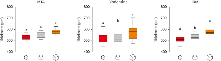
-
Abstract
PDFPubReaderePub
Objectives This study investigated the impact of micro-computed tomography (micro-CT)-based voxel size on the analysis of material/dentin interface voids and thickness of different endodontic cements.
Materials and Methods Following root-end resection and apical preparation, maxillary premolars were filled with mineral trioxide aggregate (MTA), Biodentine, and intermediate restorative material (IRM) (
n = 24). The samples were scanned using micro-CT (SkyScan 1272; Bruker) and the cement/dentin interface and thickness of materials were evaluated at voxel sizes of 5, 10, and 20 µm. Analysis of variance and the Tukey test were conducted, and the degree of agreement between different voxel sizes was evaluated using the Bland and Altman method (p < 0.05).Results All materials showed an increase in thickness from 5 to 10 and 20 µm (
p < 0.05). When evaluating the interface voids, materials were similar at 5 µm (p > 0.05), while at 10 and 20 µm Biodentine showed the lowest percentage of voids (p < 0.05). A decrease in the interface voids was observed for MTA and IRM at 20 µm, while Biodentine showed differences among all voxel sizes (p < 0.05). The Bland-Altman plots for comparisons among voxel sizes showed the largest deviations when comparing images between 5 and 20 µm.Conclusions Voxel size had an impact on the micro-CT evaluation of thickness and interface voids of endodontic materials. All cements exhibited an increase in thickness and a decrease in the void percentage as the voxel size increased, especially when evaluating images at 20 µm.
-
Citations
Citations to this article as recorded by- Effect of ultrasonic activation of endodontic sealers on root canal filling quality during the single-cone obturation procedure: a systematic review and meta-analysis of laboratory-based studies
Shuting Feng, Weiqing Zhou, Xiaojun Chu, Shuaimei Xu, Xiongqun Zeng
Odontology.2025; 113(4): 1380. CrossRef - Marginal Adaptation and Porosity of a Novel MTA Brand Applied as Root-End Filling Material: A Micro-CT Study
Yaneta Kouzmanova, Ivanka Dimitrova
Applied Sciences.2024; 14(7): 2758. CrossRef - Supplementary methods for filling material removal: A systematic review and meta-analysis of micro-CT imaging studies
Bruna Venzke Fischer, Taynara Santos Goulart, Filipe Colombo Vitali, Diego Leonardo de Souza, Cleonice da Silveira Teixeira, Lucas da Fonseca Roberti Garcia
Journal of Dentistry.2024; 151: 105445. CrossRef
- Effect of ultrasonic activation of endodontic sealers on root canal filling quality during the single-cone obturation procedure: a systematic review and meta-analysis of laboratory-based studies
- 2,702 View
- 27 Download
- 3 Web of Science
- 3 Crossref
- Shape and anatomical relationship of the mental foramen to the mandibular premolars in an Indian sub-population: a retrospective CBCT analysis
- Komal Sheth, Kulvinder Singh Banga, Ajinkya M. Pawar, James L. Gutmann, Hyeon-Cheol Kim
- Restor Dent Endod 2022;47(1):e1. Published online December 13, 2021
- DOI: https://doi.org/10.5395/rde.2022.47.e1
-
Abstract
PDFPubReaderePub
Objectives This study assessed the shape and anatomical relationship of the mental foramen (MF) to mandibular posterior teeth in an Indian sub-population.
Materials and Methods In total, 475 existing cone-beam computed tomography records exhibiting 950 MFs and including the bilateral presence of mandibular premolars and first molars were assessed. Images were evaluated 3-dimensionally to ascertain the position, shape, and anatomical proximity of MFs to mandibular teeth. The position and shape of MFs were measured and calculated. The Pythagorean theorem was used to calculate the distance between the root apex of the mandibular teeth and the MF.
Results MFs exhibited a predominantly round shape (left: 67% and right: 65%) followed by oval (left: 30% and right: 31%) in both males and females and in different age groups. The root apices of mandibular second premolars (left: 71% and right: 62%) were closest to the MF, followed by distal to the first premolars and mesial to the second premolars. The mean vertical distance between the MF and the nearest tooth apex calculated on sagittal sections was 2.20 mm on the right side and 2.32 mm on the left side; no significant difference was found according to sex or age. The distance between the apices of the teeth and the MF was ≥ 4 mm (left; 4.09 ± 1.27 mm and right; 4.01 ± 1.15 mm).
Conclusions These findings highlight the need for clinicians to be aware of the location of the MF in treatment planning and while performing non-surgical and surgical endodontic procedures.
-
Citations
Citations to this article as recorded by- Morphometric analysis of mental foramen in retained cadaveric specimens of mandibles of Sri Lankan population
Dadallage Tharanga De Silva, Usliyanage Clifford Priyantha Perera
Anatomical Science International.2026; 101(3): 340. CrossRef - Anatomical and radiographic assessment of variations of the mental foramen and their impact on success of local anaesthesia administration
Isratul Jannat, M. Ummay Salma, Nipu Rani Chowdhury, Kulsum Nahar, Dilruba Binte Mostafa, Khandokar Emanuzzaman Emon, Shahela Sarmin
International Journal of Research in Medical Sciences.2026; 14(3): 823. CrossRef - Clinical Implications of the Localization and Morphological Variability of the Mental Foramen—A Systematic Review
Mariola Krzykawska-Krupska, Janusz Pach, Piotr Regulski, Jacek Tomczyk, Izabela Strużycka, Kazimierz Szopiński, Katarzyna Osipowicz, Anna Pogorzelska
Diagnostics.2026; 16(5): 779. CrossRef - Optimising Treatment Strategies: Labial versus Labio-inferior Plating Using Three-dimensional Miniplates for Mandibular Symphysis and Parasymphysis Fractures
Akash P Muralidharan, Kalyani Bhate, K Mithun Nilgiri, Sumithra S Nair, Lakshmi Shetty, Rose Johnson
Advances in Human Biology.2025; 15(2): 242. CrossRef - A Cross-Sectional CBCT Study of Anterior Loop, Accessory Mental Foramen, and Lingual Foramina in Patients’ Mandibles: Implications for Safer Implant Planning
Abbas Shokri, Mohammad Mahdi Maleki, Leili Tapak
Journal of Maxillofacial and Oral Surgery.2025;[Epub] CrossRef - Radiographic Recognition of Mental Nerve for Secured Dental Implant Placement by Cone-Beam Computed Tomography in Mosul City Population
Asmaa B. Al-Saffar, Mekdad H. Alrigbo, Rawaa Y. Al-Rawee
Journal of Craniofacial Surgery.2024; 35(7): 2049. CrossRef - Accuracy of Implant Size Prediction Based on Edentulous Ridge Dimension on Cone-beam Computed Tomography - A Retrospective Study
Hunter R. Jolicoeur, Gerard A. Camargo, Tamara G. Stephenson, Wenjian Zhang
Annals of Maxillofacial Surgery.2024; 14(2): 187. CrossRef - Mental Foramenin Panoramik Radyografi ve Konik Işınlı Bilgisayarlı Tomografi Görüntüleri Üzerinde Morfolojik Analizi
Ezgi UZUN, Burak Kerem APAYDIN, Ayşen TİL
Selcuk Dental Journal.2023; 10(3): 540. CrossRef - Evaluation of the Possible Relationship between the Curvature and
Horizontal Course of the Inferior Alveolar Canal
Cansu G. Koca, M. Fatih Çiçek, Sanaz Sadry, Ozan Yenidünya, Fatma Akkoca Kaplan, Aras Erdil
Current Medical Imaging Formerly Current Medical Imaging Reviews.2023;[Epub] CrossRef
- Morphometric analysis of mental foramen in retained cadaveric specimens of mandibles of Sri Lankan population
- 3,743 View
- 63 Download
- 9 Web of Science
- 9 Crossref
 First
First Prev
Prev

