Abstract
-
Objectives
This study evaluated the efficacy of 3 reciprocating systems and the effects of 2 instruments for irrigant activation on filling material removal.
-
Materials and Methods
Forty mesiobuccal roots of maxillary molars were prepared up to size 25.06 and obturated. Micro-computed tomography (micro-CT) examination #1 was performed. Teeth were then divided into 4 groups (n = 10), according to the retreatment protocol: (1) manual, (2) Reciproc Blue, (3) WaveOne Gold, and (4) X1 Blue. Micro-CT examinations #2 and #3 were performed after filling removal and repreparation, respectively. Next, all teeth were divided into 2 new groups (n = 20) according to the irrigant activation protocol: XP Clean (XP Clean size 25.02) and Flatsonic (Flatsonic ultrasonic tip). Micro-CT examination #4 was performed after irrigant activation. Statistical analysis was performed with a significance level set at 5%.
-
Results
WaveOne Gold removed a significantly greater amount of filling material than the manual group (p < 0.05). The time to reach the WL was similar for all reciprocating systems (p > 0.05). X1 Blue was faster than the manual group (p < 0.05). Only manual group improved the filling material removal after the repreparation stage (p < 0.05). Both activation protocols significantly improved the filling material removal (p < 0.05), without differences between them (p > 0.05).
-
Conclusions
None of the tested instruments completely removed the filling material. X1 Blue size 25.06 reached the working length in the shortest time. XP Clean and Flatsonic improved the filling material removal.
-
Keywords: Endodontic; Retreatment; Micro computed tomography; Reciprocating
INTRODUCTION
Nonsurgical endodontic retreatment can be challenging, and it is traditionally considered as a conservative approach for tooth maintenance. Many of the difficulties in this process involve obtaining access to the pulp chamber through extensive coronary restorations, and it is typically recommended to completely remove the restorative materials and as much filling material as possible from all roots regardless of the presence or absence of periapical disease [
1,
2,
3].
Nickel-titanium (NiTi) reciprocating instruments were developed to improve and simplify the preparation of the root canal system, allowing better canal centering, requiring a shorter learning curve, and presenting an increase in cyclic fatigue resistance compared to continuous rotary instruments [
4,
5,
6]. In addition, thermal treatments aimed to improve the mechanical properties of reciprocating files, such as cyclic fatigue, flexibility, torsional resistance, and controlled memory [
7].
X1 Blue is a reciprocating system exhibiting a heat treatment similar to that of Blue technology with an inactive tip, presented in 3 tip diameters and taper sizes (size 20.06, size 25.06, and size 40.06), with a convex triangular cross-section, according to the manufacturer [
6]. WaveOne Gold, a reciprocating system manufactured with Gold technology and having an inactive tip, is presented with 4 tip diameters and taper sizes (size 20.07, size 25.07, size 35.06, and size 45.05), with a parallelogram cross-section [
8]. Reciproc Blue, a reciprocating system manufactured with Blue technology and also having an inactive tip, is presented in 3 tip diameters and taper sizes (size 25.08, size 40.06, and size 50.05), with an S-shaped cross-section [
8]. Few studies have evaluated and compared the mechanical properties of these instruments [
6,
9], and none have investigated their performance during filling material removal and root canal repreparation.
Some anatomical configurations can harbor gutta-percha, endodontic sealer, microorganisms, and other debris that can compromise the success rate of endodontic retreatment. According to Nair
et al. [
10], the organization of the microbiota in biofilms also occurs in regions of complex anatomy that are inaccessible to endodontic instruments. Recent studies have shown that 30%–45% of the root canal walls remain untouched after reciprocation preparation [
11,
12]. Thus, methods that complement the action of endodontic instruments must be used.
Irrigant activation can serve as an interesting auxiliary method to improve the cleaning and disinfection of the main canal and regions of the isthmus that are inaccessible to mechanized instruments [
13,
14]. XP Clean (MK Life Medical and Dental Products, Porto Alegre, RS, Brazil), consists of a snake-like file, presenting a size 25 tip diameter, and a 0.02 taper, and it can be used after root canal preparation in a rotary motion set at a speed of 800 rpm and 1 N·cm of torque. According to the manufacturer, its action promotes the agitation of the irrigant, potentializing its antibacterial effects, and due to its expanded action, the debridement of areas that were not touched by the preparation files. Flatsonic (R2, Helse Ultrasonic, Santa Rosa de Viterbo, SP, Brazil) is an ultrasonic insert made of stainless steel, with a size 25 tip diameter, a flat arrow design, and a smooth surface, developed to actively remove the root filling remnants adhered to the canal walls. Previous reports have demonstrated the efficacy of this protocol in reducing residual endodontic sealer and gutta-percha from oval-shaped and flattened canals [
15,
16].
Although XP Clean and Flatsonic were developed to improve root canal disinfection, there is limited evidence in the literature regarding the efficacy of these instruments in retreatment situations, especially in canals from molar teeth. Therefore, the aims of this study were to evaluate (i) the efficacy of 3 reciprocating systems in removing root canal filling material from mesiobuccal roots of maxillary molars; (ii) the time required to reach the working length (WL); (iii) the efficacy of these reciprocating systems on root canal repreparation; and (iv) the effects of 2 instruments designed for irrigant activation on filling removal from mesiobuccal roots of maxillary molars.
MATERIALS AND METHODS
This experimental, randomized,
in vitro study was approved by the local Research Ethics Committee (CAAE: 81863318.8.0000.5347).
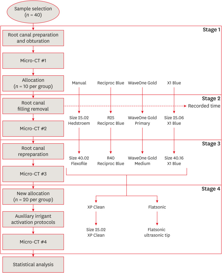
Figure 1 presents a flowchart depicting the key stages of the experimental protocols.
Figure 1
Flowchart representing all stages of the experimental procedures.
Micro-CT, micro-computed tomography.

For the sample calculation, the statistical package BioEstat 5.0 (Mamirauá Foundation, Belém, PA, Brazil) was used, and 10 sample units per group were suggested. In this calculation, the parameters described by Colombo
et al. [
17] were considered, as follows: statistical test: F test (analysis of variance [ANOVA]); minimum difference between treatments (mean values) = 3.3; standard error = 2.2; number of treatments = 4; test power = 0.80; significance level = 0.05.
Forty human maxillary molars extracted for reasons not related to this study were used. All teeth used in this study were donated from patients after signing an informed consent form. The inclusion criteria were fully formed mesiobuccal root canals presenting 1 or 2 (second mesiobuccal canal) canals, with a curvature angle ranging from 20° to 30° [
18] and a curvature radius ranging from 4 to 8 mm [
19]. The exclusion criteria were teeth presenting root resorption, calcifications, previous endodontic treatment, fractures, or cracks. Digital periapical radiographs (Dürr Dental, Porto Alegre, RS, Brazil) were performed to confirm the complete root development; absence of root resorption, calcifications, and previous endodontic treatment; and the curvature angle and radius. A visual inspection was performed to verify fractures or cracks under a stereomicroscope (Stemi 2000C, Carl Zeiss, Jena, Germany). After sample selection, the teeth were then stored in a 0.9% saline solution (Eurofarma, São Paulo, SP, Brazil) until the experimental procedures.
The teeth were attached to a metal stub (IBT Usinagem, Piracicaba, SP, Brazil) for better stabilization during endodontic procedures.
Coronary access was performed with size 1014 diamond burs (Fava Metalúrgica, Franco da Rocha, SP, Brazil) under water cooling. The canals were negotiated with size 15.02 K-files (Dentsply Maillefer, Ballaigues, Switzerland) until their tips were visualized at the apical foramen. These measures were recorded, and the WL was established 1 mm shorter than this measure. The root apex of each root was sealed with utility wax (Lysanda Produtos Odontológicos, São Paulo, SP, Brazil) to simulate a closed system.
Root canal preparation was performed with the first (size 15–40/0.02) and second series (size 45–80/0.02) of FlexoFile hand files (Dentsply Maillefer) using the crown-down/stepback technique. The apical segment of the roots was enlarged up to a size 25.02 FlexoFile. The canals were irrigated during the preparation with 1 mL of 2.5% sodium hypochlorite (NaOCl) (Rioquímica, São José do Rio Preto, SP, Brazil) after the use of each instrument. NaOCl was delivered using a 5 mL disposable syringe (Injex, Ourinhos, SP, Brazil) with a 29-gauge needle (Navitip, Ultradent, South Jordan, UT, USA) placed 3 mm from the WL [
20]. At the end of the preparation, irrigation was performed with 5 mL of 0.9% saline solution (Eurofarma), followed by 3 mL of 17% ethylenediaminetetraacetic acid (EDTA) (Biodinâmica Química e Farmacêutica LTDA, Ibiporã, PR, Brazil) and hand activation with a size 20 FlexoFile for 3 minutes. The final irrigation was performed with 5 mL of 0.9% saline solution (Eurofarma) in order to inactivate the chemical solutions [
21,
22,
23], and the canals were dried with a size 25 absorbent paper points (AllPrime Dental Products, Brasília, DF, Brazil). The root canals were then filled with an epoxy resin-based sealer (Sealer Plus, MK Life Medical and Dental Products) and standard gutta-percha cones (AllPrime Dental Products) and obturated with Tagger’s hybrid technique. The excess of gutta-percha was removed and vertically condensed with a heated plugger. The coronal access was sealed with a temporary filling material (Villevie, Joinville, SC, Brazil) and the teeth were stored at 37ºC and 100% humidity for 7 days until complete setting of the sealer.
Stratified randomization was performed to divide the roots according to the anatomical characteristics of their mesiobuccal roots. The curvature angle and radius were measured by means of micro-computed tomography (micro-CT) based on previously described parameters [
18,
19]. For this purpose, a micro-CT scan was performed using a desktop X-ray microfocus CT scanner (SkyScan 1174 v2, Bruker-microCT, Kontich, Belgium). The scanning procedures were performed using the following parameters: 50 kV X-ray tube voltage, 800 μA anode current, and a voxel size of 14.4 μm. Only 1 specimen was scanned at a time. Scans with 1,304 × 1,304 pixels were obtained with acquisition intervals of 1° over 360° rotation. Thus, data were recorded, and the scan sequences were reconstructed using the NRecon v1.6.4.8 (NRecon v.1.6.3, Bruker-microCT) software. All micro-CT scans were reconstructed with the NRecon software (Bruker-microCT) after adjusting the following parameters: post-alignment, beam-hardening, ring artifacts, and smoothing. The volume of filling material, in cubic millimeters, was measured using CT Analyzer (CTAn) software version 1.5.4.0 (Bruker-microCT). Using the micro-CT examination #1 scans, the initial volume of the root canal fillings was statistically compared (ANOVA) to confirm that there was a similar amount of material in each sample.
In this stage, teeth were allocated to 1 of the following groups, and filling material removal (stage 2) and repreparation (stage 3) were performed as follow:
• Manual group (n = 10): the filling material was removed using a size 25.02 Hedstroem file (Dentsply Maillefer) in a back-and-forth motion until reaching the WL. The canals were then reprepared with size 30.02, size 35.02, and size 40.02 FlexoFile instruments (Dentsply/Sirona, York, PA, USA) until the WL.
• Reciproc Blue group (n = 10): filling material removal was performed with a R25 Reciproc Blue file (size 25.08, VDW GmbH, Munich, Germany) powered by an endodontic electric motor (VDW Silver, VDW GmbH) set in the Reciproc All program as recommended by the manufacturer in a pecking motion until reaching the WL. The canals were reprepared up to an R40 Reciproc Blue file (VDW GmbH);
• WaveOne Gold group (n = 10): the filling material was removed by using the WaveOne Gold Primary file (size 25.07, Dentsply Maillefer) powered by an endodontic electric motor (VDW Silver, VDW GmbH) set in the WaveOne All program as stipulated by the manufacturer, in a pecking motion until reaching the WL. The canals were reprepared up to a WaveOne Gold Medium file (size 35.06, Dentsply/Sirona);
• X1 Blue (n = 10): filling removal was performed with a size 25.06 X1 Blue file (MK Life Medical and Dental Products) operated by an endodontic electric motor (VDW Silver, VDW GmbH) set in the WaveOne All program, as recommended by the manufacturer [6], in a pecking motion until reaching the WL. The canals were reprepared up to a 40.06 X1 Blue file (MK Life Medical and Dental Products).
In the Reciproc Blue, WaveOne Gold, and X1 Blue groups, after 3 in-and-out movements, the files were removed and cleaned with wet gauze (0.9% saline solution, Eurofarma), and the canals were irrigated with 3 mL of 2.5% NaOCl (Rioquímica) using a disposable syringe (Injex) with a 29-gauge needle (Navitip, Ultradent) placed 2 mm from the WL. A total volume of 15 mL of 2.5% NaOCl was delivered in all groups.
In the manual group, after each advance of 4 mm toward the apex, the size 25.02 file (Dentsply Maillefer) was removed and cleaned, and the canals were irrigated. Irrigation was performed after each file change (size 30.02, size 35.02, and size 40.02), as described in the previous groups.
For all groups, final irrigation was performed with 5 mL of 0.9% saline solution, 3 mL of 17% EDTA (Biodinâmica Química e Farmacêutica LTDA), and 5 mL of 0.9% saline solution as described in the root canal preparation stage.
The time required to perform all retreatment procedures was measured using a digital timer (SportLine, Ontario, Canada). The time was recorded during filling removal stage until the instrument reached the WL. Then, the digital timer was stopped.
After root canal filling removal, a second micro-CT (micro-CT #2) scan was performed following the same parameters described before, to measure the volume of filling material remnants. After root canal repreparation, a third micro-CT examination (#3) was performed to determine the volume of residual filling material.
Irrigant activation protocols
After micro-CT examination #3, 5 samples of each group were randomly selected and allocated to 1 of the following irrigant activation protocols (n = 20; stage 4):
• XP Clean group: canals were irrigated with 2 mL of 2.5% NaOCl (Rioquímica), and XP Clean (MK Life Medical and Dental Products) was activated in a continuous rotation (800 rpm, torque of 1 N·cm) 2 mm shorter than the WL, according to the protocol established by the manufacturer. This protocol was repeated 3 times during 20 seconds and using 2 mL of 2.5% NaOCl each time, for a total of 1 minute of activation and 6 mL of the irrigant.
• Flatsonic group: canals were filled with 2.5% NaOCl (Rioquímica) and Flatsonic was positioned 2 mm from the WL, directed toward the buccal-palatal direction, and powered by an ultrasonic device (NAC Plus, Adiel Ltda, Ribeirão Preto, SP, Brazil) set at 40 kHz. Three ultrasonic activation cycles of the irrigant were performed for 20 seconds each, using the intermittent flush technique [15].
Finally, all specimens were subjected to micro-CT (examination #4) to analyze the volume of the remaining filling material.
Statistical analysis
All data were entered into a spreadsheet (Microsoft Office Excel 2016, Microsoft Corporation, Redmond, WA, USA) and analyzed statistically using SPSS software version 15.2 for Windows (SPSS Inc., Chicago, IL, USA). The Kruskal-Wallis and Dunn post hoc tests were performed to compare the volume of residual filling material among the experimental groups in each retreatment stage. Friedman analysis was used to analyze the retreatment stages in each group. For the final irrigation protocols, the Wilcoxon test was used to evaluate if they were useful and the Mann-Whitney test was used to compare both protocols. The significance level was set at 5%.
RESULTS
Filling removal, repreparation, and time required to reach the WL
Table 1 presents the volume of filling material after each stage of retreatment (
i.e., baseline, after filling removal, and after repreparation) and the time required to reach the WL. In the intergroup assessment, the volume of filling material at baseline was similar in all groups (
p > 0.05). After filling removal, WaveOne Gold group removed a significantly greater amount of filling material than the manual group (
p < 0.05), but without significant differences when compared to the other reciprocating groups (
p > 0.05). The Reciproc Blue, X1 Blue, and manual groups did not show statistically significant differences (
p > 0.05). An intragroup analysis revealed that a significant reduction of the filling material occurred after removal (stage 2) in the reciprocating groups (
p < 0.05), but not in the manual group (
p > 0.05).
Table 1Comparative analysis of the volume of filling material (mm3) after each retreatment stage and the time required to reach the working length
|
Variables |
Manual |
Reciproc Blue |
WaveOne Gold |
X1 Blue |
|
After root canal obturation |
3.35Aa (1.58–9.21) |
4.03Aa (2.17–6.69) |
3.61Aa (2.41–5.47) |
3.82Aa (1.34–7.26) |
|
After filling removal |
2.47Aa (0.79–5.16) |
1.33ABb (0.35–2.97) |
0.95Bb (0.01–1.72) |
1.14ABb (0.67–3.04) |
|
After repreparation |
1.15Ab (0.50–4.79) |
1.04Ab (0.29–2.58) |
0.77Ab (0.01–1.09) |
0.96Ab (0.59–2.90) |
|
Time required to reach the working length (in seconds) |
325B (178–773) |
239AB (93–477) |
147AB (66–195) |
100A (66–332) |
The time required to reach the WL during filling removal was similar for all reciprocating systems (p > 0.05), but it was shorter in the X1 Blue group than in the manual group (p < 0.05).
When the canals were reprepared, the amount of residual filling material was similar among the groups (
p > 0.05). After the repreparation stage (stage 3), the manual group presented significantly less filling material than in the removal stage (stage 2) (
p < 0.05). Meanwhile, repreparation with larger files in the reciprocating groups did not guarantee additional removal of filling material (
p > 0.05).
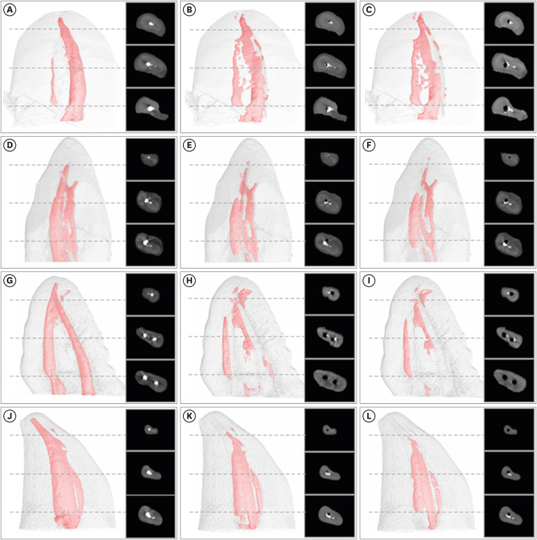
Figure 2 shows 3-dimensional reconstructions of representative micro-CT scans of each stage of the endodontic retreatment protocols.
Figure 2Two-dimensional and 3-dimensional micro-computed tomography scans of each stage of the endodontic retreatment protocols. Manual (A-C), Reciproc Blue (D-F), WaveOne Gold (G-I), and X1 Blue (J-L) groups after root canal filling (A, D, G, J), filling removal (B, E, H, K) and repreparation (C, F, I, L) stages.

Irrigant activation protocols
Because no significant differences were found among the groups after repreparation (micro-CT examination #3), all volume data were pooled and the isolated effect of the protocols was assessed using the Mann-Whitney test. Therefore, the XP Clean and Flatsonic protocols were compared.
XP Clean and Flatsonic significantly improved the removal of filling material (
Table 2) (
p < 0.05), without significant differences between them (
p > 0.05).
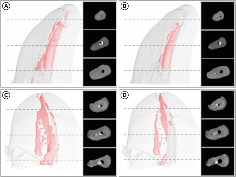
Figure 3 shows 3-dimensional reconstructions of representative micro-CT scans of the 2 final irrigant activation protocols.
Table 2Median, minimum, and maximum values of filling material volume (mm3) after using auxiliary protocols with XP Clean and Flatsonic regardless of the instrument used for repreparation
|
Variables |
XP Clean |
Flatsonic |
|
Before |
After |
Before |
After |
|
Median |
0.74A*
|
0.59B
|
0.96A
|
0.78B
|
|
Minimum |
0.31 |
0.27 |
0.01 |
0.00 |
|
Maximum |
2.90 |
2.23 |
4.79 |
3.58 |
Figure 3Two-dimensional and 3-dimensional micro-computed tomography scans of the 2 final irrigant activation protocols (before and after). XP Clean (A, B) and Flatsonic (C, D) groups after repreparation (A, C) and after final irrigant activation protocols (B, D).

DISCUSSION
The success rate of nonsurgical endodontic retreatment depends on the diagnosis of periapical disease, access to the apical foramen in all root canals, suitable disinfection after canal preparation, and adequate canal filling and coronal sealing [
10]. For this purpose, removal of the root canal filling material is mandatory, allowing better disinfection of the root canal system [
23].
Annually, several rotary and reciprocating systems are launched into the market. The X1 Blue file (MK Life Medical and Dental Products) consists of a Blue-like thermally treated NiTi alloy with controlled memory. Initially developed for use in primary root canal treatments, its efficacy for root canal filling removal has not been investigated. This study compared the performance of the X1 Blue file (MK Life Medical and Dental Products) with other well-established reciprocating systems (Reciproc Blue and WaveOne Gold).
Roots with isthmus, accessory, and additional canals, and moderate or severe curvatures are harder to disinfect than those with straight and single canals. For these reasons, the mesiobuccal roots of maxillary molars were used in this study. These roots were scanned using micro-CT to standardize the root canal volume for further stratified randomization according to the 4 experimental groups. Micro-CT assessment is a quite precise and a non-destructive method for the longitudinal evaluation of retreatment procedures [
24], and allows the standardization of samples, providing more reliable information on the investigated parameters.
The volume of filling material after the filling removal stage in the manual group (control) remained unchanged when compared to those presented after root canal obturation (p > 0.05). This can probably be explained by the fact that the filling removal stage, in this group, was completed with a size 25.02 Hedstrom (Dentsply Maillefer) file reaching the WL. The reduced taper of this hand file contributed to the poor reduction of the filling material.
In contrast, the tested reciprocating instruments promoted a significant decrease in the filling volume after the filling removal stage (p < 0.05). Despite the slight variation in the taper of these instruments (R25 Blue - size 25.08; WaveOne Gold Primary - size 25.07; X1 Blue - size 25.06), their performance was similar (p > 0.05).
Although WaveOne Gold Primary presents a smaller taper diameter (0.07 mm for 3 mm) than Reciproc R25 Blue (0.08 mm for 3 mm), only WaveOne Gold showed a significantly greater amount of filling material reduction than the manual group (
p < 0.05). Similar results were observed in a previous study [
25] that demonstrated no differences between these instruments. This result can probably be explained due to the WaveOne Gold parallelogram-shaped cross-section, which presents 2 cutting edges when in motion, and its offset design [
26], which may favor the highest percentage of touched canal surfaces, as presented in a previous study [
27]. Furthermore, the WaveOne Gold Primary was set in the WaveOne All program, reciprocating at a speed of 350 rpm, while the Reciproc All program reciprocates at a speed of 300 rpm. The highest speed of the WaveOne All program may favor a greater heat release during instrument motion [
28]. The heat generated can increase the temperature and plasticize the gutta-percha, facilitating its removal [
29]. Although the tested reciprocating instruments were not designed to remove filling material, it has been reported that individual systems specifically designed for use in retreatment did not differ in terms of filling removal efficacy when compared to those designed for primary root canal treatment [
30].
The time required to reach the WL was similar for all reciprocating instruments (
p > 0.05), but only the X1 Blue group showed a significantly shorter time than the manual group (
p < 0.05). A possible explanation for this result relates to the instrument’s geometrical features. It is known that triangular instruments can present a more even stress distribution, allowing the instrument to penetrate into the filling material without bending as easily [
30,
31], enabling the instrument to reach the WL in a shorter time. In addition, X1 Blue presents a heat treatment similar to that of the Blue technology. It is known that the Blue technology involves a higher amount of austenite in its composition, also favoring a higher bending stiffness [
7]. The repreparation procedures were performed using the subsequent file of each reciprocating system to promote further enlargement. Therefore, Reciproc Blue R40 (size 40.06), WaveOne Gold Medium (size 35.06), and X1 Blue (size 40.06) were used. It has been reported that additional enlargement of the apical third in retreatments promotes contact of the instruments with more canal walls, leading to better disinfection and removal of filling material [
32]. All reciprocating systems reduced the amount of filling material after repreparation when compared to the initial volume. Despite the slight differences in the taper and tip diameter of the files, their performance was similar, and no significant differences were observed among them (
p > 0.05). Once again, the absence of statistically significant differences among the reciprocating instruments was probably due to the offset motion and parallelogram cross-section design of the WaveOne Gold instruments [
25,
26], despite their smaller tip and taper diameter. Only repreparation with hand files reduced the volume of residual filling material in comparison with the filling removal stage (
p < 0.05), and this result may be explained by the increase in the instrument diameter (size 40.02) when compared to that used during filling material removal (size 25.02). In this study, none of the filling removal or repreparation strategies tested completely removed the filling material from the mesiobuccal roots of maxillary molars. These findings agree with previous studies [
33,
34]. For this reason, 2 final irrigation protocols were tested: an ultrasonic device (Flatsonic) with the passive ultrasonic irrigation protocol, and a snake-like (XP Clean) rotary file.
Both protocols significantly reduced the volume of filling material (
p < 0.05), but with no significant differences between them (
p > 0.05). Real effects of auxiliary protocols have been reported [
14,
17,
24,
32]. Some authors have found benefits to activating the irrigant [
14,
32,
34].
The results obtained with XP Clean in this study were comparable to those obtained for the Flatsonic with passive ultrasound irrigation, with a significant reduction of the amount of filling material. Its snake-like design seems to improve root canal cleaning, as described by the manufacturer, probably by touching the canal walls that were not touched by the repreparation files due to its expanded action. Regarding the results presented for the Flatsonic tip, recent reports also observed that Flatsonic improved the cleaning of flattened root canals after rotary and reciprocation preparation and reduced the percentage of non-instrumented areas [
17,
24]. These findings encourage the use of ultrasonic tips as an auxiliary method for filling removal after canal repreparation in endodontic retreatment.
It is important to emphasize some limitations of this study. First, this was an in vitro study; therefore, it was not possible to determine the influence of these protocols on clinical outcomes. Furthermore, only mesiobuccal roots of maxillary molars were evaluated. Thus, it is not possible to extrapolate these results to root canals of different teeth or even palatal canals. In addition, important factors such as canal transportation and debris extrusion were not evaluated. Therefore, more research evaluating such protocols is needed.
However, when considering the present findings, it is possible to infer that the evaluated protocols for filling removal and irrigant activation can provide satisfactory results when applied in mesiobuccal roots of maxillary molars.
CONCLUSIONS
In this study, more filling material was removed in the WaveOne Gold group than in the manual group during the filling material removal stage, but without significant differences in comparison to the other reciprocating systems. The X1 Blue group had a significantly shorter time to reach the WL than the manual group, but also without significant differences when compared to the other reciprocating instruments. Regarding the final irrigation protocols, both XP Clean and passive ultrasonic activation with Flatsonic promoted significant improvements in the removal of filling material residues. None of the protocols tested completely removed the filling material from the interior of the root canal system of mesiobuccal roots from maxillary molars.
-
Conflict of Interest: No potential conflict of interest relevant to this article was reported.
-
Author Contributions:
Conceptualization: Tietz L, Só MVR, Duarte MAH.
Data curation: Tietz L, Só MVR, da Rosa RA, Duarte MAH.
Formal analysis: Tietz L, Só MVR, da Rosa RA, Duarte MAH.
Investigation: Tietz L, Furlan RD, Alcalde MP, Vivan RR.
Methodology: Só MVR, Duarte MAH.
Project administration: Tietz L, Só MVR, Duarte MAH.
Supervision: Só MVR, da Rosa RA, Duarte MAH.
Writing - original draft: Tietz L, Furlan RD, Só MVR.
Writing - review & editing: da Rosa RA, Vivan RR, Alcalde MP, Weissheimer T.
REFERENCES
- 1. Nudera WJ. Selective root retreatment: a novel approach. J Endod 2015;41:1382-1388.ArticlePubMed
- 2. Soares C, Maia C, Vale F, Gadê-Neto C, Carvalho L, Oliveira H, Carvalho R. Comparison of endodontic retreatment in teeth obturated with resilon or gutta-percha: a review of literature. Iran Endod J 2015;10:221-225.PubMedPMC
- 3. Virdee SS, Thomas MB. A practitioner’s guide to gutta-percha removal during endodontic retreatment. Br Dent J 2017;222:251-257.ArticlePubMedPDF
- 4. Kim HC, Kwak SW, Cheung GS, Ko DH, Chung SM, Lee W. Cyclic fatigue and torsional resistance of two new nickel-titanium instruments used in reciprocation motion: Reciproc versus WaveOne. J Endod 2012;38:541-544.ArticlePubMed
- 5. Ferreira F, Adeodato C, Barbosa I, Aboud L, Scelza P, Zaccaro Scelza M. Movement kinematics and cyclic fatigue of NiTi rotary instruments: a systematic review. Int Endod J 2017;50:143-152.ArticlePubMedPDF
- 6. Klymus ME, Alcalde MP, Vivan RR, Só MV, de Vasconselos BC, Duarte MA. Effect of temperature on the cyclic fatigue resistance of thermally treated reciprocating instruments. Clin Oral Investig 2019;23:3047-3052.ArticlePubMedPDF
- 7. Zupanc J, Vahdat-Pajouh N, Schäfer E. New thermomechanically treated NiTi alloys - a review. Int Endod J 2018;51:1088-1103.ArticlePubMedPDF
- 8. Gündoğar M, Özyürek T. Cyclic fatigue resistance of OneShape, HyFlex EDM, WaveOne Gold, and Reciproc Blue nickel-titanium instruments. J Endod 2017;43:1192-1196.ArticlePubMed
- 9. Silva E, Oliveira de Lima C, Vieira V, Antunes H, Lima Moreira EJ, Versiani M. Cyclic fatigue and torsional resistance of four martensite-based nickel titanium reciprocating instruments. Eur Endod J 2020;5:231-235.PubMedPMC
- 10. Nair PN, Henry S, Cano V, Vera J. Microbial status of apical root canal system of human mandibular first molars with primary apical periodontitis after “one-visit” endodontic treatment. Oral Surg Oral Med Oral Pathol Oral Radiol Endod 2005;99:231-252.ArticlePubMed
- 11. Belladonna FG, Carvalho MS, Cavalcante DM, Fernandes JT, de Carvalho Maciel AC, Oliveira HE, Lopes RT, Silva EJ, De-Deus G. Micro-computed tomography shaping ability assessment of the new blue thermal treated Reciproc instrument. J Endod 2018;44:1146-1150.ArticlePubMed
- 12. De-Deus G, Belladonna FG, Silva EJ, Marins JR, Souza EM, Perez R, Lopes RT, Versiani MA, Paciornik S, Neves AA. Micro-CT evaluation of non-instrumented canal areas with different enlargements performed by NiTi systems. Braz Dent J 2015;26:624-629.ArticlePubMed
- 13. Cavenago BC, Ordinola-Zapata R, Duarte MA, del Carpio-Perochena AE, Villas-Bôas MH, Marciano MA, Bramante CM, Moraes IG. Efficacy of xylene and passive ultrasonic irrigation on remaining root filling material during retreatment of anatomically complex teeth. Int Endod J 2014;47:1078-1083.ArticlePubMed
- 14. Bernardes RA, Duarte MA, Vivan RR, Alcalde MP, Vasconcelos BC, Bramante CM. Comparison of three retreatment techniques with ultrasonic activation in flattened canals using micro-computed tomography and scanning electron microscopy. Int Endod J 2016;49:890-897.ArticlePubMedPDF
- 15. Rivera-Peña ME, Duarte MA, Alcalde MP, Furlan RD, Só MV, Vivan RR. Ultrasonic tips as an auxiliary method for the instrumentation of oval-shaped root canals. Braz Oral Res 2019;33:e011.PubMed
- 16. Santos-Júnior AO, Tanomaru-Filho M, Pinto JC, Tavares KI, Pivoto-João MM, Guerreiro-Tanomaru JM. New ultrasonic tip decreases uninstrumented surface and debris in flattened canals: a micro-computed tomographic study. J Endod 2020;46:1712-1718.ArticlePubMed
- 17. Colombo AP, Fontana CE, Godoy A, De Martin AS, Kato AS, Rocha DG, Pelegrine RA, Bueno CE. Efectiveness of the waveone and ProTaper D systems for removing gutta-percha with or without a solvent. Acta Odontol Latinoam 2016;29:262-267.PubMed
- 18. Schneider SW. A comparison of canal preparations in straight and curved root canals. Oral Surg Oral Med Oral Pathol 1971;32:271-275.ArticlePubMed
- 19. Estrela C, Bueno MR, Sousa-Neto MD, Pécora JD. Method for determination of root curvature radius using cone-beam computed tomography images. Braz Dent J 2008;19:114-118.ArticlePubMed
- 20. Boutsioukis C, Lambrianidis T, Verhaagen B, Versluis M, Kastrinakis E, Wesselink PR, van der Sluis LW. The effect of needle-insertion depth on the irrigant flow in the root canal: evaluation using an unsteady computational fluid dynamics model. J Endod 2010;36:1664-1668.ArticlePubMed
- 21. Prado M, Santos Júnior HM, Rezende CM, Pinto AC, Faria RB, Simão RA, Gomes BP. Interactions between irrigants commonly used in endodontic practice: a chemical analysis. J Endod 2013;39:505-510.ArticlePubMed
- 22. Neelakantan P, Subbarao C, Subbarao CV, De-Deus G, Zehnder M. The impact of root dentine conditioning on sealing ability and push-out bond strength of an epoxy resin root canal sealer. Int Endod J 2011;44:491-498.ArticlePubMed
- 23. Torabinejad M, Corr R, Handysides R, Shabahang S. Outcomes of nonsurgical retreatment and endodontic surgery: a systematic review. J Endod 2009;35:930-937.ArticlePubMed
- 24. Jung M, Lommel D, Klimek J. The imaging of root canal obturation using micro-CT. Int Endod J 2005;38:617-626.ArticlePubMed
- 25. Bago I, Plotino G, Katić M, Ročan M, Batinić M, Anić I. Evaluation of filling material remnants after basic preparation, apical enlargement and final irrigation in retreatment of severely curved root canals in extracted teeth. Int Endod J 2020;53:962-973.ArticlePubMedPDF
- 26. Thomas JP, Lynch M, Paurazas S, Askar M. Micro-computed tomographic evaluation of the shaping ability of Wave One Gold, TRUShape, EdgeCoil and XP-3D Shaper endodontic files in single, oval-shaped canals: an in vitro study. J Endod 2020;46:244-251.e1.PubMed
- 27. Pérez Morales ML, González Sánchez JA, Olivieri JG, Elmsmari F, Salmon P, Jaramillo DE, Terol FD. Micro-computed tomographic assessment and comparative study of the shaping ability of 6 nickel-titanium files: an in vitro study. J Endod 2021;47:812-819.PubMed
- 28. Bramante CM, Fidelis NS, Assumpção TS, Bernardineli N, Garcia RB, Bramante AS, de Moraes IG. Heat release, time required, and cleaning ability of MTwo R and ProTaper universal retreatment systems in the removal of filling material. J Endod 2010;36:1870-1873.ArticlePubMed
- 29. Zanettini PR, Barletta FB, de Mello Rahde N.
In vitro comparison of different reciprocating systems used during endodontic retreatment. Aust Endod J 2008;34:80-85.PubMed
- 30. Rios MA, Villela AM, Cunha RS, Velasco RC, De Martin AS, Kato AS, Bueno CE. Efficacy of 2 reciprocating systems compared with a rotary retreatment system for gutta-percha removal. J Endod 2014;40:543-546.ArticlePubMed
- 31. Kim HC, Kim HJ, Lee CJ, Kim BM, Park JK, Versluis A. Mechanical response of nickel-titanium instruments with different cross-sectional designs during shaping of simulated curved canals. Int Endod J 2009;42:593-602.ArticlePubMed
- 32. Alves FR, Marceliano-Alves MF, Sousa JC, Silveira SB, Provenzano JC, Siqueira JF Jr. Removal of root canal fillings in curved canals using either reciprocating single- or rotary multi-instrument systems and a supplementary step with the XP-Endo Finisher. J Endod 2016;42:1114-1119.ArticlePubMed
- 33. Martins MP, Duarte MA, Cavenago BC, Kato AS, da Silveira Bueno CE. Effectiveness of the ProTaper Next and Reciproc systems in removing root canal filling material with sonic or ultrasonic irrigation: a micro–computed tomographic study. J Endod 2017;43:467-471.ArticlePubMed
- 34. Fruchi LC, Ordinola-Zapata R, Cavenago BC, Hungaro Duarte MA, Bueno CE, De Martin AS. Efficacy of reciprocating instruments for removing filling material in curved canals obturated with a single-cone technique: a micro-computed tomographic analysis. J Endod 2014;40:1000-1004.ArticlePubMed
 , Renan Diego Furlan2
 , Ricardo Abreu da Rosa1
 , Marco Antonio Hungaro Duarte2
 , Murilo Priori Alcalde3
 , Rodrigo Ricci Vivan2
 , Theodoro Weissheimer1
 , Marcus Vinicius Reis Só1


 KACD
KACD
 ePub Link
 Cite
Cite

