Search
- Page Path
- HOME > Search
- Comparison of remineralization in caries-affected dentin using calcium silicate, glass ionomer cement, and resin-modified glass ionomer cement: an in vitro study
- Kwanchanok Youcharoen, Onwara Akkaratham, Papichaya Intajak, Pipop Saikaew, Sirichan Chiaraputt
- Restor Dent Endod 2025;50(4):e37. Published online November 14, 2025
- DOI: https://doi.org/10.5395/rde.2025.50.e37
-
 Abstract
 PDF
PDF PubReader
 ePub
ePub - Objectives
This study evaluated the ability of calcium silicate cement (CSC) as a remineralizing agent compared with conventional glass ionomer cement (GIC) and resin-modified GIC (RMGIC) to remineralize artificial caries-affected dentin.
Methods
Twenty-five class V cavities were prepared on extracted human third molars. Twenty teeth underwent artificial caries induction. The remaining five teeth with sound dentin serve as the positive control. The twenty demineralized teeth were subdivided into four groups (n = 5): carious dentin without restoration (negative control [NC]), carious dentin restored with CSC (Biodentine, Septodont), carious dentin restored with GI (Fuji IX, GC Corporation), and carious dentin restored with RMGIC (Fuji II LC, GC Corporation). Following restoration, the specimens were stored in artificial saliva for 7 days. The elastic modulus was evaluated by a nanoindentation test. The mineral composition was analyzed by scanning electron microscopy-energy-dispersive X-ray spectroscopy (SEM-EDX), and the mineral composition at the dentin-material interface.
Results
CSC had a higher modulus of elasticity compared to GI, RMGI, and NC groups (p < 0.05). Higher calcium and phosphorus content was observed under CSC restorations, as indicated by SEM-EDX examination, which may lead to better remineralization.
Conclusions
Compared to GI and RMGI, CSC showed the best remineralization and mechanical reinforcement in caries-affected dentin, indicating CSC for use in minimally invasive restorative dentistry.
- 1,225 View
- 163 Download

- Calcium silicate-based sealers remnants in isthmuses of mesial roots of mandibular molars: an in vitro evaluation
- David Saldanha de Brito Alencar, Ana Cristina Padilha Janini, Lauter Eston Pelepenko, Brenda Fornazaro Moraes, Francisco Haiter Neto, Marco Antonio Hungaro Duarte, Marina Angélica Marciano
- Restor Dent Endod 2025;50(3):e25. Published online July 15, 2025
- DOI: https://doi.org/10.5395/rde.2025.50.e25
-
 Abstract
 PDF
PDF PubReader
 ePub
ePub - Objectives
Endodontic retreatment aims to address treatment failure through the removal of root canal filling materials. This in vitro study evaluated the presence of filling material remnants in the mesial root canals, specifically focusing on the isthmuses, of mandibular molars after retreatment.
Methods
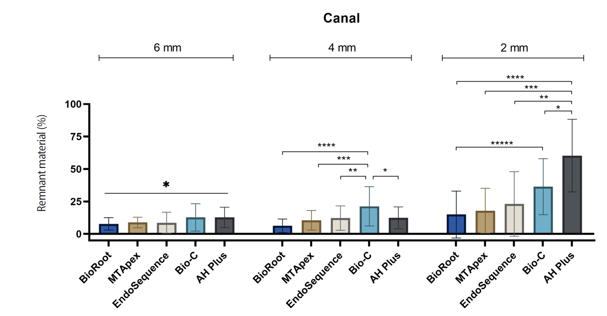
One hundred extracted mandibular molar mesial roots with isthmuses were prepared with an R25 file, obturated with one of five calcium silicate-based sealers (BioRoot RCS [Septodont], MTApex [Ultradent Products Inc.], EndoSequence BC Sealer HiFlow [Brasseler USA], Bio-C Sealer [Angelus]) or an epoxy resin-based sealer (AH Plus Jet [Dentsply Maillefer]), all stained with rhodamine B, and stored at 37ºC for 30 days to allow for setting. Retreatment was subsequently performed using R40 and XP-endo Finisher R instruments (FKG Dentaire) with 2.5% sodium hypochlorite irrigation. The presence of remaining filling material was then assessed using confocal microscopy, and setting times were tested per ISO 6876:2012.
Results
AH Plus Jet showed the most remnants at 2 mm and the longest retreatment time. Calcium silicate-based sealers exhibited prolonged setting times under dry conditions, with EndoSequence BC Sealer HiFlow showing a particularly extended setting period.
Conclusions
Despite retreatment, residues remained in all canals and isthmus regions, particularly Bio-C Sealer and AH Plus Jet in apical areas, emphasizing the difficulty of complete removal and the persistence of filling material.
- 1,836 View
- 106 Download

-
Influence of disinfecting solutions on the surface topography of gutta-percha cones: a systematic review of
in vitro studies - Lora Mishra, Gathani Dash, Naomi Ranjan Singh, Manoj Kumar, Saurav Panda, Franck Diemer, Monika Lukomska-Szymanska, Barbara Lapinska, Abdul Samad Khan
- Restor Dent Endod 2024;49(4):e42. Published online November 1, 2024
- DOI: https://doi.org/10.5395/rde.2024.49.e42
-
 Abstract
 PDF
PDF Supplementary Material
 PubReader
 ePub
ePub The surface integrity of gutta-percha cones is a crucial factor in the success of endodontic procedures. Disinfecting solutions play a pivotal role in sterilizing gutta-percha cones, but their influence on gutta-percha surface topography remains a subject of concern. This systematic review aimed to present a qualitative synthesis of available laboratory studies assessing the influence of disinfecting solutions on the surface topography of gutta-percha and offers insights into the implications for clinical practice. The present review followed PRISMA 2020 guidelines. An advanced database search was performed in PubMed, Google Scholar, Embase, Scopus, LILAC, non-indexed citations and reference lists of eligible studies in May 2024. Laboratory studies, in English language, were considered for inclusion. The quality (risk of bias) of the included studies was assessed using parameters for
in vitro studies. A total of 28 studies were included in the qualitative synthesis. Based on the included in vitro studies, surface deposits and alterations in the physical properties of gutta-percha cones were observed after the disinfection protocol. A comprehensive review of the available literature indicates that the choice of disinfecting solution, its concentration, and immersion time significantly affect the surface topography of gutta-percha cones.-
Citations
Citations to this article as recorded by- In Vitro Evaluation of Disinfectants on Gutta-Percha Cones: Antimicrobial Efficacy Against Enterococcus faecalis and Candida albicans
Tringa Kelmendi, Donika Bajrami Shabani, Aida Meto, Hani Ounsi
Journal of Clinical Medicine.2025; 14(19): 6846. CrossRef
- In Vitro Evaluation of Disinfectants on Gutta-Percha Cones: Antimicrobial Efficacy Against Enterococcus faecalis and Candida albicans
- 3,661 View
- 189 Download
- 1 Web of Science
- 1 Crossref

- Impact of different agitation methods on smear layer cleaning of mesial canals with accentuated curvature
- Abel Teves Cordova, Murilo Priori Alcalde, Michel Espinosa Klymus, Leonardo Rigoldi Bonjardim, Rodrigo Ricci Vivan, Marco Antonio Hungaro Duarte
- Restor Dent Endod 2024;49(2):e12. Published online March 4, 2024
- DOI: https://doi.org/10.5395/rde.2024.49.e12

-
 Abstract
 PDF
PDF PubReader
 ePub
ePub Objectives This study evaluated the impact of different methods of irrigant agitation on smear layer removal in the apical third of curved mesial canals of 3 dimensionally (D) printed mandibular molars.
Materials and Methods Sixty 3D-printed mandibular second molars were used, presenting a 70° curvature and a Vertucci type II configuration in the mesial root. A round cavity was cut 2 mm from the apex using a trephine of 2 mm in diameter, 60 bovine dentin disks were made, and a smear layer was formed. The dentin disks had the adaptation checked in the apical third of the teeth with wax. The dentin disks were evaluated in environmental scanning electron microscope before and after the following irrigant agitation methods: G1(PIK Ultrasonic Tip), G2 (Passive Ultrasonic Irrigation with Irrisonic– PUI), G3 (Easy Clean), G4 (HBW Ultrasonic Tip), G5 (Ultramint X Ultrasonic tip), and G6 (conventional irrigation-CI) (
n = 10). All groups were irrigated with 2.5% sodium hypochlorite and 17% ethylenediaminetetraacetic acid.Results All dentin disks were 100% covered by the smear layer before treatment, and all groups significantly reduced the percentage of the smear layer after treatment. After the irrigation protocols, the Ultra-X group showed the lowest coverage percentage, statistically differing from the conventional, PIK, and HBW groups (
p < 0.05). There was no significant difference among Ultramint X, PUI-Irrisonic, and Easy Clean (p > 0.05). None of the agitation methods could remove the smear layer altogether.Conclusions Ultramint X resulted in the most significant number of completely clean specimens.
-
Citations
Citations to this article as recorded by- A new cleaning protocol in minimally invasive endodontic surgery: RUA (“retro irrigant activation”)
Dina Abdellatif, Davide Mancino, Massimo Pisano, Sara De Fontaine, Alfredo Iandolo
Journal of Conservative Dentistry and Endodontics.2025; 28(3): 297. CrossRef - Impact of the use of high-power 810-nm diode laser as monotherapy on the clinical and tomographic success of the treatment of teeth with periapical lesions: an observational clinical study
Fabricio Hinojosa Pedraza, Abel Victor Isidro Teves-Cordova, Murilo Priori Alcalde, Marco Antonio Hungaro Duarte
Restorative Dentistry & Endodontics.2025; 50(2): e15. CrossRef - Smear layer removal comparing conventional irrigation, passive ultrasonic irrigation, EndoActivator System, and a new sonic device (Perfect Clean System) by scanning electron microscopy: An ex vivo study
Bruna Fernanda Alionço Gonçalves, Divya Reddy, Ricardo Machado, Paulo César Soares Júunior, Sérgio Aparecido Ignácio, Douglas Augusto Fernandes Couto, Karine Santos Frasquetti, Vânia Portela Ditzel Westphalen, Everdan Carneiro, Ulisses Xavier da Silva Net
PLOS ONE.2024; 19(12): e0314940. CrossRef
- A new cleaning protocol in minimally invasive endodontic surgery: RUA (“retro irrigant activation”)
- 2,286 View
- 126 Download
- 2 Web of Science
- 3 Crossref

- Influence of access cavity design on calcium hydroxide removal using different cleaning protocols: a confocal laser scanning microscopy study
- Seda Falakaloğlu, Merve Yeniçeri Özata, Betül Güneş, Emmanuel João Nogueira Leal Silva, Mustafa Gündoğar, Burcu Güçyetmez Topal
- Restor Dent Endod 2023;48(3):e25. Published online July 24, 2023
- DOI: https://doi.org/10.5395/rde.2023.48.e25
-
 Abstract
 PDF
PDF PubReader
 ePub
ePub Objectives The purpose of this study was to evaluate the influence of endodontic access cavities design on the removal of calcium hydroxide medication of the apical third of mandibular incisor root canal walls and dentinal tubules with different cleaning protocols: EDDY sonic activation, Er,Cr:YSGG laser-activated irrigation, or conventional irrigation with IrriFlex.
Materials and Methods Seventy-eight extracted human mandibular incisors were assigned to 6 experimental groups (
n = 13) according to the endodontic access cavity and cleaning protocol for calcium hydroxide removal: traditional access cavity (TradAC)/EDDY; ultraconservative access cavity performed in the incisal edge (UltraAC.Inc)/EDDY; TradAC/Er,Cr:YSGG; UltraAC.Inc/Er,Cr:YSGG; TradAC/IrriFlex; or UltraAC.Inc/IrriFlex. Confocal laser scanning microscopy images were used to measure the non-penetration percentage, maximum residual calcium hydroxide penetration depth, and penetration area at 2 and 4 mm from the apex. Data were statistically analyzed using Shapiro-Wilk and WRS2 package for 2-way comparison of non-normally distributed parameters (depth of penetration, area of penetration, and percentage of non-penetration) according to cavity and cleaning protocol with the significance level set at 5%.Results The effect of cavity and cleaning protocol interactions on penetration depth, penetration area and non-penetration percentage was not found statistically significant at 2 and 4 mm levels (
p > 0.05).Conclusions The present study demonstrated that TradAC or UltraAC.Inc preparations with different cleaning protocols in extracted mandibular incisors did not influence the remaining calcium hydroxide at 2 and 4 mm from the apex.
-
Citations
Citations to this article as recorded by- Effect of Apical Preparation Size and Preparation Taper on Smear Layer Removal Using Two Different Irrigation Needles: A Scanning Electron Microscopy Study
Rania Lebbos, Naji Kharouf, Deepak Mehta, Jamal Jabr, Cynthia Kamel, Roula El Hachem, Youssef Haikel, Marc Krikor Kaloustian
European Journal of Dentistry.2025; 19(03): 678. CrossRef - Combination of Chitosan Nanoparticles, EDTA, and Irrigation Activation Enhances TGF-β1 Release from Dentin: A Laboratory Study
Sıla Nur Usta, Emre Avcı, Ayşe Nur Oktay, Cangül Keskin
Journal of Endodontics.2025; 51(8): 1081. CrossRef
- Effect of Apical Preparation Size and Preparation Taper on Smear Layer Removal Using Two Different Irrigation Needles: A Scanning Electron Microscopy Study
- 2,756 View
- 68 Download
- 2 Web of Science
- 2 Crossref

- Comparison of the cyclic fatigue resistance of One Curve, F6 Skytaper, Protaper Next, and Hyflex CM endodontic files
- Charlotte Gouédard, Laurent Pino, Reza Arbab-Chirani, Shabnam Arbab-Chirani, Valérie Chevalier
- Restor Dent Endod 2022;47(2):e16. Published online March 4, 2022
- DOI: https://doi.org/10.5395/rde.2022.47.e16
-
 Abstract
 PDF
PDF PubReader
 ePub
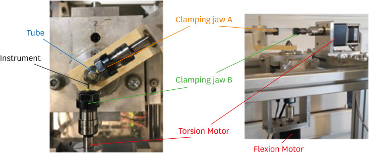
ePub Objectives This study compared the cyclic fatigue resistance of One Curve (C wire) and F6 Skytaper (conventional austenite nickel-titanium [NiTi]), and 2 instruments with thermo-mechanically treated NiTi: Protaper Next X2 (M wire) and Hyflex CM (CM wire).
Materials and Methods Ten new instruments of each group (size: 0.25 mm, 6% taper in the 3 mm tip region) were tested using a rotary bending machine with a 60° curvature angle and a 5 mm curvature radius, at room temperature. The number of cycles until fracture was recorded. The length of the fractured instruments was measured. The fracture surface of each fragment was examined with a scanning electron microscope (SEM). The data were analyzed using one-way analysis of variance and the
post hoc Tukey test. The significance level was set at 0.05.Results At 60°, One Curve, F6 Skytaper and Hyflex CM had significantly longer fatigue lives than Protaper Next X2 (
p < 0.05). No statistically significant differences were found in the cyclic fatigue lives of One Curve, F6 Skytaper, and Hyflex CM (p > 0.05). SEM images of the fracture surfaces of the different instruments showed typical features of fatigue failure.Conclusions Within the conditions of this study, at 60° and with a 5 mm curvature radius, the cyclic fatigue life of One Curve was not significantly different from those of F6 Skytaper and Hyflex CM. The cyclic fatigue lives of these 3 instruments were statistically significantly longer than that of Protaper Next.
-
Citations
Citations to this article as recorded by- Evaluation of cyclic fatigue in three pediatric endodontic rotary file systems in root canals of primary molars: A finite element analysis (FEA)
Monika sri S.S., K.C. Vignesh, K. Vivek, Kavitha Swaminathan, Selvakumar Haridoss
Journal of Oral Biology and Craniofacial Research.2025; 15(2): 310. CrossRef - Stress analysis of different experimental finite element models of rotary endodontic instruments
Manar M. Galal, Amira Galal Ismail, Nada Omar
Bulletin of the National Research Centre.2025;[Epub] CrossRef - Understanding Cyclic Fatigue in Three Nickel–Titanium Pediatric Files: An In Vitro Study for Enhanced Patient Care
Alwaleed Abushanan, Rajashekhara Bhari Sharanesha, Fahd Aljarbou, Hadi Alamri, Mohammed Hamad Almasud, Abdulfatah AlAzmah, Sara Alghamdi, Mubashir Baig Mirza
Medicina.2025; 61(5): 830. CrossRef - Analyzing Surface Morphology Changes Induced by Cyclic Fatigue in Three Different Nickel–Titanium Rotary Files Using Scanning Electron Microscopy Analysis
Chintan Joshi, Mahima P Jain, Sweety M Thumar, Jay H Dave, Applu R Bhatt, Juhi I Dholani
World Journal of Dentistry.2024; 15(7): 579. CrossRef - Nickel ion release and surface analyses on instrument fragments fractured beyond the apex: a laboratory investigation
Sıdıka Mine Toker, Ekim Onur Orhan, Arzu Beklen
BMC Oral Health.2023;[Epub] CrossRef
- Evaluation of cyclic fatigue in three pediatric endodontic rotary file systems in root canals of primary molars: A finite element analysis (FEA)
- 3,231 View
- 43 Download
- 2 Web of Science
- 5 Crossref

- Effects of dentin surface preparations on bonding of self-etching adhesives under simulated pulpal pressure
- Chantima Siriporananon, Pisol Senawongse, Vanthana Sattabanasuk, Natchalee Srimaneekarn, Hidehiko Sano, Pipop Saikaew
- Restor Dent Endod 2022;47(1):e4. Published online December 28, 2021
- DOI: https://doi.org/10.5395/rde.2022.47.e4
-
 Abstract
 PDF
PDF PubReader
 ePub
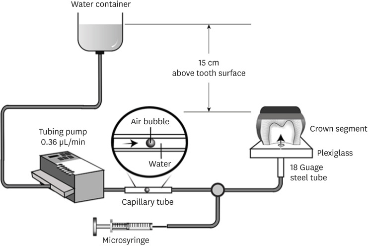
ePub Objectives This study evaluated the effects of different smear layer preparations on the dentin permeability and microtensile bond strength (µTBS) of 2 self-etching adhesives (Clearfil SE Bond [CSE] and Clearfil Tri-S Bond Universal [CTS]) under dynamic pulpal pressure.
Materials and Methods Human third molars were cut into crown segments. The dentin surfaces were prepared using 4 armamentaria: 600-grit SiC paper, coarse diamond burs, superfine diamond burs, and carbide burs. The pulp chamber of each crown segment was connected to a dynamic intra-pulpal pressure simulation apparatus, and the permeability test was done under a pressure of 15 cmH2O. The relative permeability (%P) was evaluated on the smear layer-covered and bonded dentin surfaces. The teeth were bonded to either of the adhesives under pulpal pressure simulation, and cut into sticks after 24 hours water storage for the µTBS test. The resin-dentin interface and nanoleakage observations were performed using a scanning electron microscope. Statistical comparisons were done using analysis of variance and
post hoc tests.Results Only the method of surface preparation had a significant effect on permeability (
p < 0.05). The smear layers created by the carbide and superfine diamond burs yielded the lowest permeability. CSE demonstrated a higher µTBS, with these values in the superfine diamond and carbide bur groups being the highest. Microscopic evaluation of the resin-dentin interface revealed nanoleakage in the coarse diamond bur and SiC paper groups for both adhesives.Conclusions Superfine diamond and carbide burs can be recommended for dentin preparation with the use of 2-step CSE.
-
Citations
Citations to this article as recorded by- The effect of different adhesive strategies and diamond burs on dentin bond strength of universal resin cements
Chavakorn Atsavathavornset, Pipop Saikaew, Choltacha Harnirattisai, Hidehiko Sano
Clinical Oral Investigations.2025;[Epub] CrossRef - Universal adhesive systems in dentistry: A narrative review
Svetlana N. Razumova, Anzhela S. Brago, Oxana R. Ruda, Zoya A. Guryeva, Elvira V. Adzhieva
Russian Journal of Dentistry.2024; 28(5): 512. CrossRef - Delayed light activation of resin composite affects the bond strength of adhesives under dynamic simulated pulpal pressure
Nattaporn Sukprasert, Choltacha Harnirattisai, Pisol Senawongse, Hidehiko Sano, Pipop Saikaew
Clinical Oral Investigations.2022; 26(11): 6743. CrossRef
- The effect of different adhesive strategies and diamond burs on dentin bond strength of universal resin cements
- 2,900 View
- 42 Download
- 2 Web of Science
- 3 Crossref

- The effect of using nanoparticles in bioactive glass on its antimicrobial properties
- Maram Farouk Obeid, Kareim Moustafa El-Batouty, Mohammed Aslam
- Restor Dent Endod 2021;46(4):e58. Published online October 29, 2021
- DOI: https://doi.org/10.5395/rde.2021.46.e58
-
 Abstract
 PDF
PDF PubReader
 ePub
ePub Objectives This study addresses the effect of using nanoparticles (np) on the antimicrobial properties of bioactive glass (BAG) when used in intracanal medicaments against
Enterococcus faecalis (E. faecalis ) biofilms.Materials and Methods E. faecalis biofilms, grown inside 90 root canals for 21 days, were randomly divided into 4 groups according to the antimicrobial regimen followed (n = 20; BAG-np, BAG, calcium hydroxide [CaOH], and saline). After 1 week, residual live bacteria were quantified in terms of colony-forming units (CFU), while dead bacteria were assessed with a confocal laser scanning microscope.Results Although there was a statistically significant decrease in the mean CFU value among all groups, the nano-group performed the best. The highest percentage of dead bacteria was detected in the BAG-np group, with a significant difference from the BAG group.
Conclusions The reduction of particle size and use of a nano-form of BAG improved the antimicrobial properties of the intracanal treatment of
E. faecalis biofilms-
Citations
Citations to this article as recorded by- Size matters: Radiation shielding superiority of borate glasses with nano vs. micro ZnO
Aljawhara H. Almuqrin, M.I. Sayyed, M. Elsafi
Nuclear Engineering and Technology.2025; 57(9): 103614. CrossRef - Effect of Chitosan and bioactive glass nanomaterials as intracanal medicaments on TGF-β1 release from intraradicular dentin
Sarah Salah Hashem, Mohammed M. Khalefa, Mahmoud Hassan Mohamed, Hemat M. ELSheikh, Fatma Abd El-Rahman Taher
BMC Oral Health.2025;[Epub] CrossRef - Effect of Er: YAG laser, phthalocyanine activated photodynamic therapy, and bioactive glass nanoparticles on smear layer removal and push out bond strength of quartz fiber posts to canal dentin: a SEM assessment
Okba Mahmoud, Erum Zain
Frontiers in Dental Medicine.2025;[Epub] CrossRef - Advancements in Root Canal Therapy: Translational Innovations and the Role of Nanoparticles in Endodontic Treatment
Noha M. Badawi, Mohamed M. Kataia, Hadeel A. Mousa, Mozhgan Afshari
Journal of Nanotechnology.2025;[Epub] CrossRef - Propolis in Endodontics—Unveiling Its Therapeutic Potential: A Narrative Review
Poorani Durai, Santha Devy A, Mithila Mohan, Harish Ramalingam, Shasidharan P, Rahul Chaurasia M
World Journal of Dentistry.2025; 16(10): 959. CrossRef - Application of Nanomaterials in Endodontics
Farzaneh Afkhami, Yuan Chen, Laurence J. Walsh, Ove A. Peters, Chun Xu
BME Frontiers.2024;[Epub] CrossRef - Antimicrobial efficacy of newly prepared nano-tricalcium silicate-58s bioactive glass-based endodontic sealer
Nawal Atiya Al-Sabawi, Sawsan Hameed Al-Jubori
Endodontology.2024;[Epub] CrossRef - Antimicrobial Effects of Formulations of Various Nanoparticles and Calcium Hydroxide as Intra-canal Medications Against Enterococcus faecalis: A Systematic Review
Seema H Bukhari, Dax Abraham, Shakila Mahesh
Cureus.2024;[Epub] CrossRef - Effect of nanoparticles on antibacterial efficacy of intracanal medicament: A scoping review
Alpa Gupta, Arundeep Singh, Vivek Aggarwal
Endodontology.2023; 35(4): 283. CrossRef - Physical properties, marginal adaptation and bioactivity of an experimental mineral trioxide aggregate-like cement modified with bioactive materials
Abigailt Flores-Ledesma, Adriana Tejeda-Cruz, María A. Moyaho-Bernal, Ana Wintergerst, Yoshamin A. Moreno-Vargas, Jacqueline A. Rodríguez-Chávez, Carlos E. Cuevas-Suárez, Kenya Gutiérrez-Estrada, Jesús A. Arenas-Alatorre
Journal of Oral Science.2023; 65(2): 141. CrossRef - Nanopartículas antimicrobianas en endodoncia: Revisión narrativa
Gustavo Adolfo Tovar Rangel , Fanny Mildred González Sáenz , Ingrid Ximena Zamora Córdoba , Lina María García Zapata
Revista Estomatología.2023;[Epub] CrossRef
- Size matters: Radiation shielding superiority of borate glasses with nano vs. micro ZnO
- 1,501 View
- 25 Download
- 6 Web of Science
- 11 Crossref

- Push-out bond strength and marginal adaptation of apical plugs with bioactive endodontic cements in simulated immature teeth
- Maria Aparecida Barbosa de Sá, Eduardo Nunes, Alberto Nogueira da Gama Antunes, Manoel Brito Júnior, Martinho Campolina Rebello Horta, Rodrigo Rodrigues Amaral, Stephen Cohen, Frank Ferreira Silveira
- Restor Dent Endod 2021;46(4):e53. Published online October 20, 2021
- DOI: https://doi.org/10.5395/rde.2021.46.e53
-
 Abstract
 PDF
PDF PubReader
 ePub
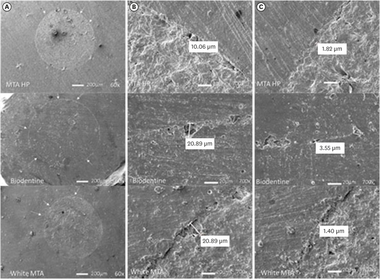
ePub Objectives This study evaluates the bond strength and marginal adaptation of mineral trioxide aggregate (MTA) Repair HP and Biodentine used as apical plugs; MTA was used as reference material for comparison.
Materials and Methods A total of 30 single-rooted teeth with standardized, artificially created open apices were randomly divided into 3 groups (
n = 10 per group), according to the material used to form 6-mm-thick apical plugs: group 1 (MTA Repair HP); group 2 (Biodentine); and group 3 (white MTA). Subsequently, the specimens were transversely sectioned to obtain 2 (cervical and apical) 2.5-mm-thick slices per root. Epoxy resin replicas were observed under a scanning electron microscope to measure the gap size at the material/dentin interface (the largest and smaller gaps were recorded for each replica). The bond strength of the investigated materials to dentin was determined using the push-out test. The variable bond strengths and gap sizes were evaluated independently at the apical and cervical root dentin slices. Data were analyzed using descriptive and analytic statistics.Results The comparison between the groups regarding the variables' bond strengths and gap sizes showed no statistical difference (
p > 0.05) except for a single difference in the smallest gap at the cervical root dentin slice, which was higher in group 3 than in group 1 (p < 0.05).Conclusions The bond strength and marginal adaptation to root canal walls of MTA HP and Biodentine cement were comparable to white MTA.
-
Citations
Citations to this article as recorded by- Application of Biodentine for Apexification of Immature Teeth of Children: A Scoping Review
Liz M Gerard, Sumit Gaur
International Journal of Clinical Pediatric Dentistry.2025; 18(5): 573. CrossRef - Evaluation of the root dentin bond strength and intratubular biomineralization of a premixed calcium aluminate-based hydraulic bioceramic endodontic sealer
Yu-Na Lee, Min-Kyeong Kim, Hee-Jin Kim, Mi-Kyung Yu, Kwang-Won Lee, Kyung-San Min
Journal of Oral Science.2024; 66(2): 96. CrossRef - Managing Cracked Teeth with Root Extension: A Prospective Preliminary Study Using Biodentine™ Material
Kênia Maria Soares de Toubes, Isabella Sousa Corrêa, Regina Célia Lopes Valadares, Stephanie Quadros Tonelli, Fábio Fernandes Borém Bruzinga, Frank Ferreira Silveira, Dr Karthikeyan Ramalingam
International Journal of Dentistry.2024;[Epub] CrossRef - Marginal adaptation of customized gutta percha cone with calcium silicate based sealer versus MTA and biodentine apical plugs in simulated immature permanent teeth (an in vitro study)
Mary M. Mina, Sybel M. Moussa, Mahmoud R. Aboelseoud
BMC Oral Health.2024;[Epub] CrossRef - Comparative Evaluation of Push-Out Bond Strength of Conventional Mineral Trioxide Aggregate, Biodentine, a Modified Mineral Trioxide Aggregate, and Two Novel Antibacterial-Enhanced Mineral Trioxide Aggregates
Arokia Rajkumar Shancy Merlin, Vignesh Ravindran, Ganesh Jeevanandan, Rajalakshmanan Eswaramoorthy, Abirami Arthanari
Cureus.2024;[Epub] CrossRef - Push out bond strength of hydraulic cements used at different thicknesses
C. Ruiz Durán, Dra L. Gancedo-Caravia, V. Vera González, C. González Losada
BMC Oral Health.2023;[Epub] CrossRef - Effects of different calcium-silicate based materials on fracture resistance of immature permanent teeth with replacement root resorption and osteoclastogenesis
Gabriela Leite de Souza, Gabrielle Alves Nunes Freitas, Maria Tereza Hordones Ribeiro, Nelly Xiomara Alvarado Lemus, Carlos José Soares, Camilla Christian Gomes Moura
Restorative Dentistry & Endodontics.2023;[Epub] CrossRef
- Application of Biodentine for Apexification of Immature Teeth of Children: A Scoping Review
- 2,315 View
- 24 Download
- 8 Web of Science
- 7 Crossref

-
Comparative evaluation of
Emblica officinalis as an etchant and an MMP inhibitor with orthophosphoric acid and chlorhexidine on the microshear bond strength of composite resin: anex vivo study - Divya Sangeetha Rajkumar, Annapoorna Ballagere Mariswamy
- Restor Dent Endod 2021;46(3):e36. Published online June 8, 2021
- DOI: https://doi.org/10.5395/rde.2021.46.e36

-
 Abstract
 PDF
PDF PubReader
 ePub
ePub Objectives This study aimed to evaluate
Emblica officinalis (Indian gooseberry or amla) as an acid etchant and matrix metalloproteinase (MMP) inhibitor, and to compare its effect on the microshear bond strength of composite resin with orthophosphoric acid (OPA) and 2% chlorhexidine (CHX) as an acid etchant and MMP inhibitor, respectively.Materials and Methods The etching effect and MMP-inhibiting action of amla on dentin samples were confirmed by scanning electron microscopy (SEM) and gelatin zymography, respectively. Dentinal slabs (3 mm thick) from 80 extracted human molars were divided into 10 and 20 samples to form 2 control groups and 3 experimental groups. Groups 1, 2, and 4 were etched with OPA and groups 3 and 5 with amla juice. An MMP inhibitor was then applied: CHX for group 2 and amla extract for groups 4 and 5. Groups 1 and 3 received no MMP inhibitor. All specimens received a standardized bonding protocol and composite resin build-up, and were subjected to microshear bond strength testing. The force at which the fracture occurred was recorded and statistically analyzed.
Results Amla juice had a similar etching effect as a self-etch adhesive in SEM and 100% amla extract was found to inhibit MMP-9 by gelatin zymography. The microshear bond strength values of amla were lower than those obtained for OPA and CHX, but the difference was not statistically significant.
Conclusions Amla has a promising role as an acid etchant and MMP inhibitor, but further studies are necessary to substantiate its efficacy.
-
Citations
Citations to this article as recorded by- In vitro assessment of anti-glioblastoma potential of Emblica officinalis methanolic fruit extract and green nanoparticles in U87-MG cells
Kokkonda Jackson Sugunakara Chary, Anuradha Sharma, Amrita Singh
Medical Oncology.2025;[Epub] CrossRef - Eco-conscious synthesis of novel 1,2,4-triazolo[1,5-a]pyrimidine derivatives as potent Anti-microbial agent and comparative study of cell viability and cytotoxicity in HEK-293 cell line utilizing Indian gooseberry (Phyllanthus emblica) fruit extract
Bhaktiben R. Bhatt, Kamalkishor Pandey, Tarosh Patel, Anupama Modi, Chandani Halpani, Vaibhav D. Bhatt, Bharat C. Dixit
Bioorganic Chemistry.2024; 153: 107936. CrossRef - Cell mediated ECM-degradation as an emerging tool for anti-fibrotic strategy
Peng Zhao, Tian Sun, Cheng Lyu, Kaini Liang, Yanan Du
Cell Regeneration.2023;[Epub] CrossRef - Insight into the development of versatile dentin bonding agents to increase the durability of the bonding interface
Isabel Cristina Celerino de Moraes Porto, Teresa de Lisieux Guedes Ferreira Lôbo, Raphaela Farias Rodrigues, Rodrigo Barros Esteves Lins, Marcos Aurélio Bomfim da Silva
Frontiers in Dental Medicine.2023;[Epub] CrossRef
- In vitro assessment of anti-glioblastoma potential of Emblica officinalis methanolic fruit extract and green nanoparticles in U87-MG cells
- 1,663 View
- 20 Download
- 4 Web of Science
- 4 Crossref

-
Cyclic fatigue resistance of M-Pro and RaCe Ni-Ti rotary endodontic instruments in artificial curved canals: a comparative
in vitro study - Hadeer Mostafa El Feky, Khalid Mohammed Ezzat, Marwa Mahmoud Ali Bedier
- Restor Dent Endod 2019;44(4):e44. Published online November 7, 2019
- DOI: https://doi.org/10.5395/rde.2019.44.e44
-
 Abstract
 PDF
PDF PubReader
 ePub
ePub Objectives To compare the flexural cyclic fatigue resistance and the length of the fractured segments (FLs) of recently introduced M-Pro rotary files with that of RaCe rotary files in curved canals and to evaluate the fracture surface by scanning electron microscopy (SEM).
Materials and Methods Thirty-six endodontic files with the same tip size and taper (size 25, 0.06 taper) were used. The samples were classified into 2 groups (n = 18): the M-Pro group (M-Pro IMD) and the RaCe group (FKG). A custom-made simulated canal model was fabricated to evaluate the total number of cycles to failure and the FL. SEM was used to examine the fracture surfaces of the fragmented segments. The data were statistically analyzed and comparisons between the 2 groups for normally distributed numerical variables were carried out using the independent Student's
t -test. Ap value less than 0.05 was considered to indicate statistical significance.Results The M-Pro group showed significantly higher resistance to flexural cyclic fatigue than the RaCe group (
p < 0.05), but there was no significant difference in the FLs between the 2 groups (p ≥ 0.05).Conclusions Thermal treatment of nickel-titanium instruments can improve the flexural cyclic fatigue resistance of rotary endodontic files, and the M-Pro rotary system seems to be a promising rotary endodontic file.
-
Citations
Citations to this article as recorded by- The Effect of Canal Curvature and Different Manufacturing Processes of Five Different NiTi Rotary Files on Cyclic Fatigue Resistance
Panupat Phumpatrakom, Awiruth Klaisiri, Sukitti Techapatiphandee, Thippawan Saekow, Panuroot Aguilar
European Journal of General Dentistry.2025; 14(03): 264. CrossRef - EndoMagic Gold M06 Eğelerinde Boyut ve Konikliğin Döngüsel Yorgunluğa Etkisi: Bir İn Vitro Çalışma
Bircan Kuloğlu, Ayşe Çoban, Hatice Büyüközer Özkan
Akdeniz Diş Hekimliği Dergisi.2025; 4(3): 212. CrossRef - Evaluatation of two nickle-titanium systems’ (Neolix and X Pro Gold) resistance to fracture after immersion in sodium hypochlorite.
Solmaz Araghi, Abbas Delvarani, Faeze dehghan, Parisa Kaghazloo
journal of research in dental sciences.2024; 21(1): 17. CrossRef - Endodontic Ni–Ti Rotary Instruments for Glide-path, Are They Still Necessary and How to Think about the Ideal Instrument?
Shilpa Bhandi, Rodolfo Reda, Luca Testarelli, Elisa Maccari
The Journal of Contemporary Dental Practice.2024; 25(6): 505. CrossRef - Comparative evaluation of cyclic fatigue resistance of thermomechanically treated NiTi rotary instruments in simulated curved canals with two different radii of curvature: An in vitro Study
Tahira Hamid, Azhar Malik, Ajay Kumar, Shamim Anjum
Journal of Conservative Dentistry and Endodontics.2024; 27(4): 393. CrossRef - New heat-treated vs electropolished nickel-titanium instruments used in root canal treatment: Influence of autoclave sterilization on surface roughness
Rahaf A. Almohareb, Reem Barakat, Fatimah Albohairy, Hannes C. Schniepp
PLOS ONE.2022; 17(3): e0265226. CrossRef - The Effect of Taper and Apical Diameter on the Cyclic Fatigue Resistance of Rotary Endodontic Files Using an Experimental Electronic Device
Vicente Faus-Llácer, Nirmine Hamoud Kharrat, Celia Ruiz-Sánchez, Ignacio Faus-Matoses, Álvaro Zubizarreta-Macho, Vicente Faus-Matoses
Applied Sciences.2021; 11(2): 863. CrossRef
- The Effect of Canal Curvature and Different Manufacturing Processes of Five Different NiTi Rotary Files on Cyclic Fatigue Resistance
- 2,134 View
- 10 Download
- 7 Crossref

- Cyclic fatigue, bending resistance, and surface roughness of ProTaper Gold and EdgeEvolve files in canals with single- and double-curvature
- Wafaa A. Khalil, Zuhair S. Natto
- Restor Dent Endod 2019;44(2):e19. Published online April 26, 2019
- DOI: https://doi.org/10.5395/rde.2019.44.e19
-
 Abstract
 PDF
PDF PubReader
 ePub
ePub Objectives The purpose of this study was to evaluate the cyclic fatigue, bending resistance, and surface roughness of EdgeEvolve (EdgeEndo) and ProTaper Gold (Dentsply Tulsa Dental Specialties) nickel-titanium (NiTi) rotary files.
Materials and Methods The instruments (
n = 15/each) were tested for cyclic fatigue in single- (60° curvature, 5-mm radius) and double-curved (coronal curvature 60°, 5-mm radius, and apical curvature of 30° and 2-mm radius) artificial canals. The number of cycles to fracture was calculated. The bending resistance of both files were tested using a universal testing machine where the files were bent until reach 45°. Scanning electron microscopy and x-ray energy-dispersive spectrometric analysis were used for imaging the fractured segments, while the atomic force microscope was used to quantify the surface roughness average (Ra).Results EdgeEvolve files exhibited higher cyclic fatigue resistance than ProTaper Gold files in single- and double-curved canals (
p < 0.05) and both files were more resistant to cyclic fatigue in single-curved canals than double-curved canals (p < 0.05). EdgeEvolve files exhibited significantly more flexibility than did ProTaper Gold files (p < 0.05). Both files had approximately similar Ni and Ti contents (p > 0.05). EdgeEvolve files showed significantly lower Ra values than ProTaper Gold files (p < 0.05).Conclusions Within the limitation of this study, EdgeEvolve files exhibited significantly higher cyclic fatigue resistance than ProTaper Gold files in both single- and double-curved canals.
-
Citations
Citations to this article as recorded by- Comparison of Design, Cyclic Fatigue Resistance, and Metallurgical Properties of Original, Replica‐Like, and Counterfeit Nickel‐Titanium Files
Mert Unal, Elif Bahar Cakici
Microscopy Research and Technique.2026; 89(1): 87. CrossRef - An in vitro comparison of alterations in surface topographies of three different rotary files after root canal preparation with different irrigating solutions: Atomic force microscopic study
PremSai Parepalli, TB. V G. Raju, PKrishna Prasad, GowtamDev Dondapati, VenkataSrija Kintada, Alekhya Mediboyina
Journal of Conservative Dentistry.2023; 26(3): 299. CrossRef - Assessment of surface topographic changes of nickel–titanium rotary endodontic file at repeated usage: An in vitro study
E. Viswas, VSS Krishna, E. Sridevi, A. J. Sai Sankar, K. Siva Sankar, B. Nagesh
Endodontology.2023; 35(2): 149. CrossRef - Cyclic Fatigue Resistance and Surface Roughness of Rotary NiTi Instruments after Simulated Clinical Use in Curved Root Canals – An Atomic Force Microscopy Study
Raksha Bhat, Arjun Kini, Preethesh Shetty, Payalben Kansara, Bapanaiah Penugonda
Pesquisa Brasileira em Odontopediatria e Clínica Integrada.2022;[Epub] CrossRef - Metallurgical Tests in Endodontics: A Narrative Review
Alessio Zanza, Marco Seracchiani, Rodolfo Reda, Gabriele Miccoli, Luca Testarelli, Dario Di Nardo
Bioengineering.2022; 9(1): 30. CrossRef - Influence of nickel-titanium rotary systems with varying cross-sectional, pitch, and rotational speed on deflection and cyclic fatigue: a finite element analysis study
Wignyo Hadriyanto, Lukita Wardani, Christina Nugrohowati, Ananto Alhasyimi, Rachmat Sriwijaya, Margareta Rinastiti, Widowati Siswomihardjo, Gunadi, T. Yamada, A.A.C. Pramana, Y. Ophinni, A. Gusnanto, W.A. Kusuma, J. Yunus, Afiahayati, R. Dharmastiti, T.
BIO Web of Conferences.2021; 41: 05005. CrossRef - Can the Separated Instrument be Removed From the Root Canal System out by Magnetism? A Hypothesis
Mohammad Daryaeian, Sanjay Miglani, AbdolMahmood Davarpanah, Hyeon-Cheol Kim, Mohsen Ramazani
Dental Hypotheses.2019; 10(4): 108. CrossRef - Resistance to cyclic fatigue of reciprocating instruments determined at body temperature and phase transformation analysis
Raymond Scott, Ana Arias, José C. Macorra, Sanjay Govindjee, Ove A. Peters
Australian Endodontic Journal.2019; 45(3): 400. CrossRef
- Comparison of Design, Cyclic Fatigue Resistance, and Metallurgical Properties of Original, Replica‐Like, and Counterfeit Nickel‐Titanium Files
- 1,401 View
- 8 Download
- 8 Crossref

- Improved dentin disinfection by combining different-geometry rotary nickel-titanium files in preparing root canals
- Marwa M. Bedier, Ahmed Abdel Rahman Hashem, Yosra M. Hassan
- Restor Dent Endod 2018;43(4):e46. Published online November 1, 2018
- DOI: https://doi.org/10.5395/rde.2018.43.e46
-
 Abstract
 PDF
PDF PubReader
 ePub
ePub Objectives This study was to evaluate the antibacterial effect of different instrumentation and irrigation techniques using confocal laser scanning microscopy (CLSM) after root canal inoculation with
Enterococcus faecalis (E. faecalis ).Materials and Methods Mesiobuccal and mesiolingual canals of extracted mandibular molars were apically enlarged up to a size 25 hand K-file, then autoclaved and inoculated with
E. faecalis . The samples were randomly divided into 4 main groups according to the system of instrumentation and irrigation: an XP-endo Shaper (XPS) combined with conventional irrigation (XPS/C) or an XP-endo Finisher (XPF) (XPS/XPF), and iRaCe combined with conventional irrigation (iRaCe/C) or combined with an XPF (iRaCe/XPF). A middle-third samplewas taken from each group, and then the bacterial reduction was evaluated using CLSM at a depth of 50 µm inside the dentinal tubules. The ratio of red fluorescence (dead cells) to green-and-red fluorescence (live and dead cells) represented the percentage of bacterial reduction. The data were then statistically analyzed using the Kruskal-Wallis test for comparisons across the groups and the Dunn test was used for pairwise comparisons.Results The instrumentation and irrigation techniques had a significant effect on bacterial reduction (
p < 0.05). The iRaCe/XPF group showed the strongest effect, followed by the XPS/XPF and XPS/C group, while the iRaCe/C group had the weakest effect.Conclusions Combining iRaCe with XPF improved its bacterial reduction effect, while combining XPS with XPF did not yield a significant improvement in its ability to reduce bacteria at a depth of 50 µm in the dentinal tubules.
-
Citations
Citations to this article as recorded by- Biofilm-forming activity of Enterococcus faecalis on basic materials of removable dental prosthetic bases
Oksana A. Shuliatnikova, Mikhail V. Yakovlev, Anatoliy P. Godovalov
HERALD of North-Western State Medical University named after I.I. Mechnikov.2025; 17(2): 89. CrossRef - A Short Report on the Effectiveness of Edge Taper Platinum and XP-3D Shaper for the Reduction of Enterococcus faecalis Count in the Root Canal System: An Ex Vivo Study
Hanie Moaveni, Parastou Ghahari, Samira Behrad, Majid Mirmohammadkhani, Sobhan Rashmee, Somayeh Teimoori
Avicenna Journal of Dental Research.2024; 16(2): 77. CrossRef - Shaping ability of non‐adaptive and adaptive core nickel–titanium single‐file systems with supplementary file in ribbon‐shaped canals analysed by micro‐computed tomography
Parichat Chinchiyanont, Kallaya Yanpiset, Danuchit Banomyong, Nathamon Thongbai‐On
Australian Endodontic Journal.2023; 49(1): 38. CrossRef - Impact XP-endo finisher on the 1-year follow-up success of posterior root canal treatments: a randomized clinical trial
Ludmila Smith de Jesus Oliveira, Fabricio Eneas Diniz de Figueiredo, Janaina Araújo Dantas, Maria Amália Gonzaga Ribeiro, Carlos Estrela, Manoel Damião Sousa-Neto, André Luis Faria-e-Silva
Clinical Oral Investigations.2023; 27(12): 7595. CrossRef - In vitro reduction in Enterococcus faecalis count following root canal preparation with Neolix and XP shaper rotary files
Mina Mehrjouei, Somayeh Teimoori, Majid Mirmohammadkhani, Seyed Majed Mortazavi, Maryam Khorasanchi
Saudi Endodontic Journal.2023; 13(3): 236. CrossRef - Antibacterial efficacy of sodium hypochlorite versus apple cider vinegar against Enterococcus faecalis in contracted endodontic cavity
Kaur Supreet, Karkala Venkappa Kishan, Nimisha Chinmay Shah
Endodontology.2022; 34(4): 254. CrossRef - Ex vivo evaluation of the effectiveness of XP-endo Finisher on the removal of smear layer from the root canal
Sângela Maria PEREIRA, Ceci Nunes CARVALHO, Rudys Rodolfo TAVAREZ, Paulo NELSON-FILHO, Léa Assed Bezerra DA SILVA, Etevaldo Matos MAIA FILHO
RGO - Revista Gaúcha de Odontologia.2022;[Epub] CrossRef - Biofilm elimination from infected root canals using four different single files
Sarah A. Hamed, Sarah Shabayek, Hayam Y. Hassan
BMC Oral Health.2022;[Epub] CrossRef - The effectiveness of the supplementary use of the XP-endo Finisher on bacteria content reduction: a systematic review and meta-analysis
Ludmila Smith de Jesus Oliveira, Rafaella Mariana Fontes de Bragança, Rafael Sarkis-Onofre, André Luis Faria-e-Silva
Restorative Dentistry & Endodontics.2021;[Epub] CrossRef - Combination of a new ultrasonic tip with rotary systems for the preparation of flattened root canals
Karina Ines Medina Carita Tavares, Jáder Camilo Pinto, Airton Oliveira Santos-Junior, Fernanda Ferrari Esteves Torres, Juliane Maria Guerreiro-Tanomaru, Mario Tanomaru-Filho
Restorative Dentistry & Endodontics.2021;[Epub] CrossRef - Effect of Adaptive, Rotary, and Manual Root Canal Instrumentation in Primary Molars: A Triple-Armed, Randomized Controlled Clinical Trial
Bhaggyashri A. Pawar, Ajinkya M. Pawar, Anuj Bhardwaj, Dian Agustin Wahjuningrum, Amelia Kristanti Rahardjo, Alexander Maniangat Luke, Zvi Metzger, Anda Kfir
Biology.2021; 10(1): 42. CrossRef - Complete Obturation—Cold Lateral Condensation vs. Thermoplastic Techniques: A Systematic Review of Micro-CT Studies
Shilpa Bhandi, Mohammed Mashyakhy, Abdulaziz S. Abumelha, Mazen F. Alkahtany, Mohamed Jamal, Hitesh Chohan, A. Thirumal Raj, Luca Testarelli, Rodolfo Reda, Shankargouda Patil
Materials.2021; 14(14): 4013. CrossRef - The Effects of Different Endodontic Access Cavity Design and Using XP-endo Finisher on the Reduction of Enterococcus faecalis in the Root Canal System
Pelin Tüfenkçi, Koray Yılmaz
Journal of Endodontics.2020; 46(3): 419. CrossRef - Irrigation in Endodontics: a Review
Sarah Bukhari, Alaa Babaeer
Current Oral Health Reports.2019; 6(4): 367. CrossRef
- Biofilm-forming activity of Enterococcus faecalis on basic materials of removable dental prosthetic bases
- 1,474 View
- 14 Download
- 14 Crossref

- Mineral content analysis of root canal dentin using laser-induced breakdown spectroscopy
- Selen Küçükkaya Eren, Emel Uzunoğlu, Banu Sezer, Zeliha Yılmaz, İsmail Hakkı Boyacı
- Restor Dent Endod 2018;43(1):e11. Published online February 4, 2018
- DOI: https://doi.org/10.5395/rde.2018.43.e11
-
 Abstract
 PDF
PDF PubReader
 ePub
ePub Objectives This study aimed to introduce the use of laser-induced breakdown spectroscopy (LIBS) for evaluation of the mineral content of root canal dentin, and to assess whether a correlation exists between LIBS and scanning electron microscopy/energy dispersive spectroscopy (SEM/EDS) methods by comparing the effects of irrigation solutions on the mineral content change of root canal dentin.
Materials and Methods Forty teeth with a single root canal were decoronated and longitudinally sectioned to expose the canals. The root halves were divided into 4 groups (
n = 10) according to the solution applied: group NaOCl, 5.25% sodium hypochlorite (NaOCl) for 1 hour; group EDTA, 17% ethylenediaminetetraacetic acid (EDTA) for 2 minutes; group NaOCl+EDTA, 5.25% NaOCl for 1 hour and 17% EDTA for 2 minutes; a control group. Each root half belonging to the same root was evaluated for mineral content with either LIBS or SEM/EDS methods. The data were analyzed statistically.Results In groups NaOCl and NaOCl+EDTA, the calcium (Ca)/phosphorus (P) ratio decreased while the sodium (Na) level increased compared with the other groups (
p < 0.05). The magnesium (Mg) level changes were not significant among the groups. A significant positive correlation was found between the results of LIBS and SEM/EDS analyses (r = 0.84,p < 0.001).Conclusions Treatment with NaOCl for 1 hour altered the mineral content of dentin, while EDTA application for 2 minutes had no effect on the elemental composition. The LIBS method proved to be reliable while providing data for the elemental composition of root canal dentin.
-
Citations
Citations to this article as recorded by- In vitro evaluation of antimicrobial photodynamic therapy with photosensitizers and calcium hydroxide on bond strength, chemical composition, and sealing of glass-fiber posts to root dentin
Thalya Fernanda Horsth Maltarollo, Paulo Henrique dos Santos, Henrique Augusto Banci, Mariana de Oliveira Bachega, Beatriz Melare de Oliveira, Marco Hungaro Antonio Duarte, Índia Olinta de Azevedo Queiroz, Rodrigo Rodrigues Amaral, Luciano Angelo Tavares
Lasers in Medical Science.2025;[Epub] CrossRef - Effect of Using 5% Apple Vinegar Irrigation Solution Adjunct to Diode Laser on Smear Layer Removal and Calcium/Phosphorus Ion Ratio during Root Canal Treatment
Tarek AA Salam, Haythem SA Kader, Elsayed E Abdallah
CODS - Journal of Dentistry.2024; 15(1): 3. CrossRef - Evaluation of chemical composition of root canal dentin between two age groups using different irrigating solutions: An in vitro sem-eds study
Naresh Kumar K, Abhijith Kallu, Surender L.R, Sravani Nirmala, Narender Reddy
International Dental Journal of Student's Research.2024; 12(1): 18. CrossRef - Minimally invasive management of vital teeth requiring root canal therapy
E. Karatas, M. Hadis, W. M. Palin, M. R. Milward, S. A. Kuehne, J. Camilleri
Scientific Reports.2023;[Epub] CrossRef - The Effects of a Novel Nanohydroxyapatite Gel and Er: YAG Laser Treatment on Dentin Hypersensitivity
Demet Sahin, Ceren Deger, Burcu Oglakci, Metehan Demirkol, Bedri Onur Kucukyildirim, Mehtikar Gursel, Evrim Eliguzeloglu Dalkilic
Materials.2023; 16(19): 6522. CrossRef - Chitosan Homogenizing Coffee Ring Effect for Soil Available Potassium Determination Using Laser-Induced Breakdown Spectroscopy
Xiaolong Li, Rongqin Chen, Zhengkai You, Tiantian Pan, Rui Yang, Jing Huang, Hui Fang, Wenwen Kong, Jiyu Peng, Fei Liu
Chemosensors.2022; 10(9): 374. CrossRef - Quantitative analysis of cadmium in rice roots based on LIBS and chemometrics methods
Wei Wang, Wenwen Kong, Tingting Shen, Zun Man, Wenjing Zhu, Yong He, Fei Liu
Environmental Sciences Europe.2021;[Epub] CrossRef
- In vitro evaluation of antimicrobial photodynamic therapy with photosensitizers and calcium hydroxide on bond strength, chemical composition, and sealing of glass-fiber posts to root dentin
- 1,626 View
- 10 Download
- 7 Crossref

- The effect of root canal preparation on the surface roughness of WaveOne and WaveOne Gold files: atomic force microscopy study
- Taha Özyürek, Koray Yılmaz, Gülşah Uslu, Gianluca Plotino
- Restor Dent Endod 2018;43(1):e10. Published online February 2, 2018
- DOI: https://doi.org/10.5395/rde.2018.43.e10
-
 Abstract
 PDF
PDF PubReader
 ePub
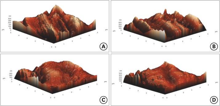
ePub Objectives To examine the surface topography of intact WaveOne (WO; Dentsply Sirona Endodontics) and WaveOne Gold (WOG; Dentsply Sirona Endodontics) nickel-titanium rotary files and to evaluate the presence of alterations to the surface topography after root canal preparations of severely curved root canals in molar teeth.
Materials and Methods Forty-eight severely curved canals of extracted molar teeth were divided into 2 groups (
n = 24/each group). In group 1, the canals were prepared using WO and in group 2, the canals were prepared using WOG files. After the preparation of 3 root canals, instruments were subjected to atomic force microscopy analysis. Average roughness and root mean square values were chosen to investigate the surface features of endodontic files. The data was analyzed using one-way analysis of variance andpost hoc Tamhane's tests at 5% significant level.Results The surface roughness values of WO and WOG files significantly changed after use in root canals (
p < 0.05). The used WOG files exhibited higher surface roughness change when compared with the used WO files (p < 0.05).Conclusions Using WO and WOG Primary files in 3 root canals affected the surface topography of the files. After being used in root canals, the WOG files showed a higher level of surface porosity value than the WO files.
-
Citations
Citations to this article as recorded by- Comparative Analysis of Surface Roughness and Plastic Deformation of Reciprocating Instruments after Clinical Use
Ángel Herrera, Magdalena Azabal, Jesús R. Jimenez-Octavio, Juan C. del Real-Romero, Sara López de Armentia, Juan M. Asensio-Gil, Ana Arias
Materials.2024; 17(16): 3978. CrossRef - Comparative Evaluation of Surface Roughness of Different Rotary Nickel-Titanium (NiTi) Files After Autoclaving: An Atomic Force Microscopic Study
Angela Alex, Ranjith Kumar Sivarajan, Vijay Venkatesh
Cureus.2024;[Epub] CrossRef - Impact of endodontic irrigants on surface roughness of various nickel-titanium rotary endodontic instruments
Tamer M. Hamdy, Yasmine Mohsen Alkabani, Amira Galal Ismail, Manar M. Galal
BMC Oral Health.2023;[Epub] CrossRef - Metallurgical Tests in Endodontics: A Narrative Review
Alessio Zanza, Marco Seracchiani, Rodolfo Reda, Gabriele Miccoli, Luca Testarelli, Dario Di Nardo
Bioengineering.2022; 9(1): 30. CrossRef - Cyclic Fatigue Resistance and Surface Roughness of Rotary NiTi Instruments after Simulated Clinical Use in Curved Root Canals – An Atomic Force Microscopy Study
Raksha Bhat, Arjun Kini, Preethesh Shetty, Payalben Kansara, Bapanaiah Penugonda
Pesquisa Brasileira em Odontopediatria e Clínica Integrada.2022;[Epub] CrossRef - In Vitro Study of Irrigation solution of Chitosan Nanoparticles to Inhibit the Adhesion and Biofilm Formation of Enterococcus faecalis in the Root Canal
Imelda Darmawi, Trimurni Abidin, Harry Agusnar, Basri A. Gani
Research Journal of Pharmacy and Technology.2022; : 2691. CrossRef - Alteration in surface roughness of reciprocating endodontic instruments
Khoa Van Pham
F1000Research.2021; 10: 875. CrossRef - Surface profile of different heat-treated nickel-titanium files before and after root canal preparation
Iandara de Lima Scardini, Denise Maria Zezell, Juliana Lisboa Couto Marques, Laila Gonzales Freire, Marcelo dos Santos
Brazilian Dental Journal.2021; 32(6): 8. CrossRef - Impact of Endodontic Instrumentation on Surface Roughness of Various Nickel-Titanium Rotary Files
Muhammad Sohail Zafar
European Journal of Dentistry.2021; 15(02): 273. CrossRef - A new method for assessment of nickel-titanium endodontic instrument surface roughness using field emission scanning electronic microscope
Khoa Van Pham, Canh Quang Vo
BMC Oral Health.2020;[Epub] CrossRef - Surface Alterations Induced on Endodontic Instruments by Sterilization Processes, Analyzed with Atomic Force Microscopy: A Systematic Review
Mario Dioguardi, Vito Crincoli, Luigi Laino, Mario Alovisi, Enrica Laneve, Diego Sovereto, Bruna Raddato, Khrystyna Zhurakivska, Filiberto Mastrangelo, Domenico Ciavarella, Lucio Lo Russo, Lorenzo Lo Muzio
Applied Sciences.2019; 9(22): 4948. CrossRef - Atomic force microscopy and energy dispersive X‐ray spectrophotometry analysis of reciprocating and continuous rotary nickel‐titanium instruments following root canal retreatment
Bulem Üreyen Kaya, Cevat Emre Erik, Gülsen Kiraz
Microscopy Research and Technique.2019; 82(7): 1157. CrossRef - Influence of heat treatment on torsional resistance and surface roughness of nickel‐titanium instruments
E. J. N. L. Silva, J. F. N. Giraldes, C. O. de Lima, V. T. L. Vieira, C. N. Elias, H. S. Antunes
International Endodontic Journal.2019; 52(11): 1645. CrossRef
- Comparative Analysis of Surface Roughness and Plastic Deformation of Reciprocating Instruments after Clinical Use
- 1,557 View
- 12 Download
- 13 Crossref

- Influence of size and insertion depth of irrigation needle on debris extrusion and sealer penetration
- Emel Uzunoglu-Özyürek, Hakan Karaaslan, Sevinç Aktemur Türker, Bahar Özçelik
- Restor Dent Endod 2018;43(1):e2. Published online December 22, 2017
- DOI: https://doi.org/10.5395/rde.2018.43.e2
-
 Abstract
 PDF
PDF PubReader
 ePub
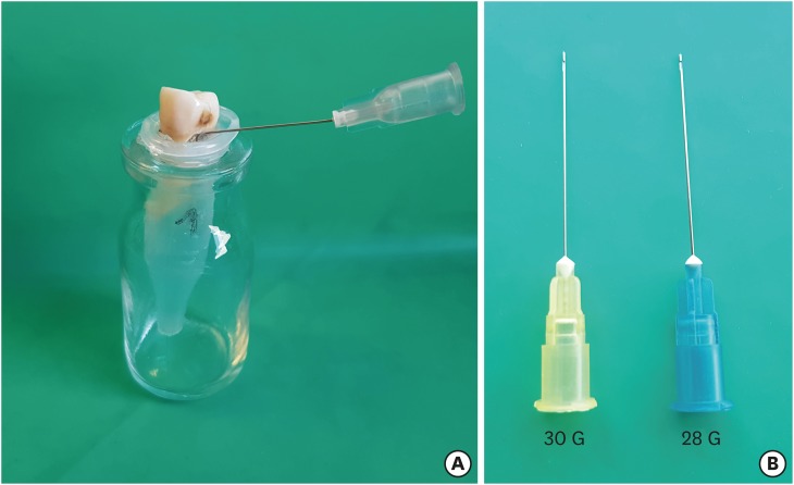
ePub Objectives To determine the effect of size and insertion depth of irrigation needle on the amount of apical extruded debris and the amount of penetration depth of sealer using a confocal laser scanning microscope (CLSM).
Materials and Methods Twenty maxillary premolars were assigned to 2 groups (
n = 10), according to the size of needle tip, 28 G or 30 G. Buccal roots of samples were irrigated with respective needle type inserted 1 mm short of the working length (WL), while palatal roots were irrigated with respective needle type inserted 3 mm short of the WL. Prepared teeth were removed from the pre-weighed Eppendorf tubes. Canals were filled with F3 gutta-percha cone and rhodamine B dye-labeled AH 26 sealer. Teeth were transversally sectioned at 1 and 3 mm levels from the apex and observed under a CLSM. Eppendorf tubes were incubated to evaporate the irrigant and were weighed again. The difference between pre- and post-weights was calculated, and statistical evaluation was performed.Results Inserting needles closer to the apex and using needles with wider diameters were associated with significantly more debris extrusion (
p < 0.05). The position of needles and level of sections had statistically significant effects on sealer penetration depth (p < 0.05 for both).Conclusions Following preparation, inserting narrower needles compatible with the final apical diameter of the prepared root canal at 3 mm short of WL during final irrigation might prevent debris extrusion and improve sealer penetration in the apical third.
-
Citations
Citations to this article as recorded by- Effect of laser-induced pulpal anesthesia of single-rooted teeth with irreversible pulpitis treated by single-visit root canal therapy - A randomized clinical trial
Geeta Asthana, Dhwani Morakhia, Ravina Parmar, Rajashree Tamuli
Endodontology.2025; 37(3): 244. CrossRef - Efficacy of different irrigation needles used in endodontics: an in silico and an in vitro investigation
Maulee Sheth, Ankit Arora, Sonali Kapoor, Balraj Shukla
Biomaterial Investigations in Dentistry.2025; 12: 264. CrossRef - Preliminary insights: exploring irrigation practices during endodontic treatment among general dental practitioners in Malaysia
Kai Qi Chiew, Xin Ni Lim, Shekhar Bhatia, Naveen Chhabra
British Dental Journal.2024;[Epub] CrossRef - Efficiency of diode laser in control of post-endodontic pain: a randomized controlled trial
Hend H. Ismail, Maram Obeid, Ehab Hassanien
Clinical Oral Investigations.2023; 27(6): 2797. CrossRef - Endodontic management of an aberrant germinated composite odontome: A case report
Ankit Arora, Kavina Desai, Sonali Kapoor, Seema Gajera
Australian Endodontic Journal.2023; 49(3): 684. CrossRef - Potentials of 3D-Modeling in the Preclinical Stage of Root Needle Research
Aleksandr V. Kuligin, Larisa N. Kazakova, Oksana S. Tereshchuk, Vadim V. Bokov
I.P. Pavlov Russian Medical Biological Herald.2022; 30(1): 95. CrossRef - Effect of root canal geometry and needle type on apical extrusion of irrigant: an ex vivo study
Büşra SERÇE FİKİRLİ, Bülent ALTUNKAYNAK, Güven KAYAOĞLU
Acta Odontologica Turcica.2022; 39(3): 58. CrossRef - An in vitro radiological evaluation of irrigant penetration in the root canals using three different irrigation systems: Waterpik WP-100 device, passive irrigation, and manual dynamic irrigation systems
Suragani Hemalatha, Archana Srinivasan, A Srirekha, Lekha Santhosh, C Champa, Ashwija Shetty
Journal of Conservative Dentistry.2022; 25(4): 403. CrossRef - Preparation Ability of ProTaper Next and XP-endo Shaper Instruments in Isthmus-containing Root Canal System
Mustafa Sarıkahya, Tayfun Alaçam
Conservative Dentistry and Endodontic Journal.2021; 5(2): 28. CrossRef - Penetration depth of irrigants into root dentine after sonic, ultrasonic and photoacoustic activation
K. M. Galler, V. Grubmüller, R. Schlichting, M. Widbiller, A. Eidt, C. Schuller, M. Wölflick, K.‐A. Hiller, W. Buchalla
International Endodontic Journal.2019; 52(8): 1210. CrossRef
- Effect of laser-induced pulpal anesthesia of single-rooted teeth with irreversible pulpitis treated by single-visit root canal therapy - A randomized clinical trial
- 1,593 View
- 18 Download
- 10 Crossref

- Smear layer removal by different chemical solutions used with or without ultrasonic activation after post preparation
- Daniel Poletto, Ana Claudia Poletto, Andressa Cavalaro, Ricardo Machado, Leopoldo Cosme-Silva, Cássia Cilene Dezan Garbelini, Márcio Grama Hoeppner
- Restor Dent Endod 2017;42(4):324-331. Published online November 1, 2017
- DOI: https://doi.org/10.5395/rde.2017.42.4.324
-
 Abstract
 PDF
PDF PubReader
 ePub
ePub Objectives This study evaluated smear layer removal by different chemical solutions used with or without ultrasonic activation after post preparation.
Materials and Methods Forty-five extracted uniradicular human mandibular premolars with single canals were treated endodontically. The cervical and middle thirds of the fillings were then removed, and the specimens were divided into 9 groups: G1, saline solution (NaCl); G2, 2.5% sodium hypochlorite (NaOCl); G3, 2% chlorhexidine (CHX); G4, 11.5% polyacrylic acid (PAA); G5, 17% ethylenediaminetetraacetic acid (EDTA). For the groups 6, 7, 8, and 9, the same solutions used in the groups 2, 3, 4, and 5 were used, respectively, but activated with ultrasonic activation. Afterwards, the roots were analyzed by a score considering the images obtained from a scanning electron microscope.
Results EDTA achieved the best performance compared with the other solutions evaluated regardless of the irrigation method (
p < 0.05).Conclusions Ultrasonic activation did not significantly influence smear layer removal.
-
Citations
Citations to this article as recorded by- O papel do ultrassom no tratamento e retratamento de canais radiculares: Revisão de literatura
Carlos Roberto Souza Hipp, Joaquim Carlos Fest da Silveira, Luiz Felipe Gilson de Oliveira Rangel, Tatiana Federici de Souza Fest da Silveira, Carla Minozzo Mello, Rodrigo Simões de Oliveira
Research, Society and Development.2025; 14(8): e1314849323. CrossRef - Effect of sodium hypochlorite, ethylenediaminetetraacetic acid, and dual-rinse irrigation on dentin adhesion using an etch-and-rinse or self-etch approach
Matej Par, Tobias Steffen, Selinay Dogan, Noah Walser, Tobias T. Tauböck
Scientific Reports.2024;[Epub] CrossRef - Evaluation of Effect of Poloxamer on Smear Layer Removal Using Apical Negative Pressure: An In Vitro Scanning Electron Microscopy Study
Chandra Prabha, Chitharanjan Shetty, Aditya Shetty
Journal of International Oral Health.2024; 16(6): 498. CrossRef - Laboratory Assessment of Antibacterial Efficacy of Five Different Herbal-based Potential Endodontic Irrigants
Anjali A Oak, Kailash Attur, Kamal Bagda, Nitish Mathur, Lubna Mohammad, Nikhat M Attar
Advances in Human Biology.2023; 13(4): 350. CrossRef - Dental Surface Conditioning Techniques to Increase the Micromechanical Retention to Fiberglass Posts: A Literature Review
Paulina Leticia Moreno-Sánchez, Maricela Ramírez-Álvarez, Alfredo del Rosario Ayala-Ham, Erika de Lourdes Silva-Benítez, Miguel Ángel Casillas-Santana, Diana Leyva del Rio, León Francisco Espinosa-Cristóbal, Erik Lizárraga-Verdugo, Mariana Melisa Avendaño
Applied Sciences.2023; 13(14): 8083. CrossRef - Effect of irrigation protocols on smear layer removal, bond strength and nanoleakage of fiber posts using a self-adhesive resin cement
Rodrigo Stadler Alessi, Renata Terumi Jitumori, Bruna Fortes Bittencourt, Giovana Mongruel Gomes, João Carlos Gomes
Restorative Dentistry & Endodontics.2023;[Epub] CrossRef - Effects of using different root canal sealers and protocols for cleaning intraradicular dentin on the bond strength of a composite resin used to reinforce weakened roots
Luiz Pascoal Vansan, Ricardo Machado, Celso Bernardes de Souza, Ricardo Gariba, Antônio Miranda da Cruz, Cinara Muniz, Jardel FranciscoX Jardel Francisco Mazzi-Chaves, Lucas da Fonseca Roberti Garcia
Journal of Oral Research.2022; 11(6): 1. CrossRef - Influence of the use of chelating agents as final irrigant on the push‐out bond strength of epoxy resin‐based root canal sealers: A systematic review
Carla M. Augusto, Miguel A. Cunha Neto, Karem P. Pinto, Ana Flavia A. Barbosa, Emmanuel J. N. L. Silva, Ana Paula P. dos Santos, Luciana M. Sassone
Australian Endodontic Journal.2022; 48(2): 347. CrossRef - Adhesion and whitening efficacy of P11-4 self-assembling peptide and HAP suspension after using NaOCl as a pre-treatment agent
Niloofar Hojabri, Karl-Heinz Kunzelmann
BMC Oral Health.2022;[Epub] CrossRef - Influence of resin cements and root canal disinfection techniques on the adhesive bond strength of fibre reinforced composite post to radicular dentin
Zaid A. Al Jeaidi
Photodiagnosis and Photodynamic Therapy.2021; 33: 102108. CrossRef - The Antibacterial Efficacy and In Vivo Toxicity of Sodium Hypochlorite and Electrolyzed Oxidizing (EO) Water-Based Endodontic Irrigating Solutions
Sung-Chih Hsieh, Nai-Chia Teng, Chia Chun Chu, You-Tai Chu, Chung-He Chen, Liang-Yu Chang, Chieh-Yun Hsu, Ching-Shuan Huang, Grace Ying-Wen Hsiao, Jen-Chang Yang
Materials.2020; 13(2): 260. CrossRef
- O papel do ultrassom no tratamento e retratamento de canais radiculares: Revisão de literatura
- 2,455 View
- 17 Download
- 11 Crossref

- The use of auxiliary devices during irrigation to increase the cleaning ability of a chelating agent
- Marina Carvalho Prado, Fernanda Leal, Renata Antoun Simão, Heloisa Gusman, Maíra do Prado
- Restor Dent Endod 2017;42(2):105-110. Published online February 3, 2017
- DOI: https://doi.org/10.5395/rde.2017.42.2.105
-
 Abstract
 PDF
PDF PubReader
 ePub
ePub Objectives This study investigated the cleaning ability of ultrasonically activated irrigation (UAI) and a novel activation system with reciprocating motion (EC, EasyClean, Easy Equipamentos Odontológicos) when used with a relatively new chelating agent (QMix, Dentsply). In addition, the effect of QMix solution when used for a shorter (1 minute) and a longer application time (3 minutes) was investigated.
Materials and Methods Fifty permanent human teeth were prepared with K3 rotary system and 6% sodium hypochlorite. Samples were randomly assigned to five groups (
n = 10) according to the final irrigation protocol: G1, negative control (distilled water); G2, positive control (QMix 1 minute); G3, QMix 1 minute/UAI; G4, QMix 1 minute/EC; G5, QMix 3 minutes. Subsequently the teeth were prepared and three photomicrographs were obtained in each root third of root walls, by scanning electron microscopy. Two blinded and pre-calibrated examiners evaluated the images using a four-category scoring system. Data were statistically analyzed using Kruskal-Wallis and Dunn tests (p < 0.05).Results There were differences among groups (
p < 0.05). UAI showed better cleaning ability than EC (p < 0.05). There were improvements when QMix was used with auxiliary devices in comparison with conventional irrigation (p < 0.05). Conventional irrigation for 3 minutes presented significantly better results than its use for 1 minute (p < 0.05).Conclusions QMix should be used for 1 minute when it is used with UAI, since this final irrigation protocol showed the best performance and also allowed clinical optimization of this procedure.
-
Citations
Citations to this article as recorded by- Comparative Evaluation of Different Methods of Activation of Chelating Solution for Smear Layer Removal in the Apical Portion of the Root Canal Using a Scanning Electron Microscopy: An In Vitro Study
Mrunal B Alhat, Sudha B Mattigatti, Rushikesh R Mahaparale, Kapil D Wahane, Apoorva Jadhav
Cureus.2024;[Epub] CrossRef - The Impact of Laser-Activated and Conventional Irrigation Techniques on Sealer Penetration into Dentinal Tubules
Dilara Koruk, Fatma Basmacı, Dilan Kırmızı, Umut Aksoy
Photobiomodulation, Photomedicine, and Laser Surgery.2022; 40(8): 565. CrossRef - Utilização dos atuais métodos de agitação de soluções endodônticas no canal radicular
Lívia Rodrigues Schneider, Larissa Giovanella
Revista Científica Multidisciplinar Núcleo do Conhecimento.2022; : 135. CrossRef - Smear layer removal by passive ultrasonic irrigation and 2 new mechanical methods for activation of the chelating solution
Ricardo Machado, Isadora da Silva, Daniel Comparin, Bianca Araujo Marques de Mattos, Luiz Rômulo Alberton, Ulisses Xavier da Silva Neto
Restorative Dentistry & Endodontics.2021;[Epub] CrossRef - Proteomic analysis of human dental pulp in different clinical diagnosis
Poliana Amanda Oliveira Silva, Stella Maris de Freitas Lima, Mirna de Souza Freire, André Melro Murad, Octávio Luiz Franco, Taia Maria Berto Rezende
Clinical Oral Investigations.2021; 25(5): 3285. CrossRef - Effect of QMix irrigant in removal of smear layer in root canal system: a systematic review of in vitro studies
Margaret Soo Yee Chia, Abhishek Parolia, Benjamin Syek Hur Lim, Jayakumar Jayaraman, Isabel Cristina Celerino de Moraes Porto
Restorative Dentistry & Endodontics.2020;[Epub] CrossRef - The effect of 17% EDTA and QMiX ultrasonic activation on smear layer removal and sealer penetration: ex vivo study
Felipe de Souza Matos, Fabrício Rutz da Silva, Luiz Renato Paranhos, Camilla Christian Gomes Moura, Eduardo Bresciani, Marcia Carneiro Valera
Scientific Reports.2020;[Epub] CrossRef - Micro-CT evaluation of different final irrigation protocols on the removal of hard-tissue debris from isthmus-containing mesial root of mandibular molars
Emmanuel João Nogueira Leal Silva, Carla Rodrigues Carvalho, Felipe Gonçalves Belladonna, Marina Carvalho Prado, Ricardo Tadeu Lopes, Gustavo De-Deus, Edson Jorge Lima Moreira
Clinical Oral Investigations.2019; 23(2): 681. CrossRef
- Comparative Evaluation of Different Methods of Activation of Chelating Solution for Smear Layer Removal in the Apical Portion of the Root Canal Using a Scanning Electron Microscopy: An In Vitro Study
- 1,259 View
- 6 Download
- 8 Crossref

- Antifungal effects of synthetic human β-defensin 3-C15 peptide
- Sang-Min Lim, Ki-Bum Ahn, Christine Kim, Jong-Won Kum, Hiran Perinpanayagam, Yu Gu, Yeon-Jee Yoo, Seok Woo Chang, Seung Hyun Han, Won-Jun Shon, Woocheol Lee, Seung-Ho Baek, Qiang Zhu, Kee-Yeon Kum
- Restor Dent Endod 2016;41(2):91-97. Published online March 17, 2016
- DOI: https://doi.org/10.5395/rde.2016.41.2.91
-
 Abstract
 PDF
PDF PubReader
 ePub
ePub Objectives The purpose of this
ex vivo study was to compare the antifungal activity of a synthetic peptide consisting of 15 amino acids at the C-terminus of human β-defensin 3 (HBD3-C15) with calcium hydroxide (CH) and Nystatin (Nys) againstCandida albicans (C. albicans ) biofilm.Materials and Methods C. albicans were grown on cover glass bottom dishes or human dentin disks for 48 hr, and then treated with HBD3-C15 (0, 12.5, 25, 50, 100, 150, 200, and 300 µg/mL), CH (100 µg/mL), and Nys (20 µg/mL) for 7 days at 37℃. On cover glass, live and dead cells in the biomass were measured by the FilmTracer Biofilm viability assay, and observed by confocal laser scanning microscopy (CLSM). On dentin, normal, diminished and ruptured cells were observed by field-emission scanning electron microscopy (FE-SEM). The results were subjected to a two-tailedt -test, a one way analysis variance and apost hoc test at a significance level ofp = 0.05.Results C. albicans survival on dentin was inhibited by HBD3-C15 in a dose-dependent manner. There were fewer aggregations ofC. albicans in the groups of Nys and HBD3-C15 (≥ 100 µg/mL). CLSM showedC. albicans survival was reduced by HBD3-C15 in a dose dependent manner. Nys and HBD3-C15 (≥ 100 µg/mL) showed significant fungicidal activity compared to CH group (p < 0.05).Conclusions Synthetic HBD3-C15 peptide (≥ 100 µg/mL) and Nys exhibited significantly higher antifungal activity than CH against
C. albicans by inhibiting cell survival and biofilm.-
Citations
Citations to this article as recorded by- Anti-fungal peptides: an emerging category with enthralling therapeutic prospects in the treatment of candidiasis
Jyoti Sankar Prusty, Ashwini Kumar, Awanish Kumar
Critical Reviews in Microbiology.2025; 51(5): 755. CrossRef - Current status of antimicrobial peptides databases and computational tools for optimization
Madhulika Jha, Akash Nautiyal, Kumud Pant, Navin Kumar
Environment Conservation Journal.2025; 26(1): 281. CrossRef - Harnessing antimicrobial peptides in endodontics
Xinzi Kong, Vijetha Vishwanath, Prasanna Neelakantan, Zhou Ye
International Endodontic Journal.2024; 57(7): 815. CrossRef - Human β-defensins and their synthetic analogs: Natural defenders and prospective new drugs of oral health
Mumian Chen, Zihe Hu, Jue Shi, Zhijian Xie
Life Sciences.2024; 346: 122591. CrossRef - Candida albicans Virulence Factors and Pathogenicity for Endodontic Infections
Yeon-Jee Yoo, A Reum Kim, Hiran Perinpanayagam, Seung Hyun Han, Kee-Yeon Kum
Microorganisms.2020; 8(9): 1300. CrossRef - Innate Inspiration: Antifungal Peptides and Other Immunotherapeutics From the Host Immune Response
Derry K. Mercer, Deborah A. O'Neil
Frontiers in Immunology.2020;[Epub] CrossRef - Human salivary proteins and their peptidomimetics: Values of function, early diagnosis, and therapeutic potential in combating dental caries
Kun Wang, Xuedong Zhou, Wei Li, Linglin Zhang
Archives of Oral Biology.2019; 99: 31. CrossRef - Endodontic biofilms: contemporary and future treatment options
Yeon-Jee Yoo, Hiran Perinpanayagam, Soram Oh, A-Reum Kim, Seung-Hyun Han, Kee-Yeon Kum
Restorative Dentistry & Endodontics.2019;[Epub] CrossRef - Bioactive Peptides Against Fungal Biofilms
Karen G. N. Oshiro, Gisele Rodrigues, Bruna Estéfani D. Monges, Marlon Henrique Cardoso, Octávio Luiz Franco
Frontiers in Microbiology.2019;[Epub] CrossRef - Anticandidal Potential of Stem Bark Extract from Schima superba and the Identification of Its Major Anticandidal Compound
Chun Wu, Hong-Tan Wu, Qing Wang, Guey-Horng Wang, Xue Yi, Yu-Pei Chen, Guang-Xiong Zhou
Molecules.2019; 24(8): 1587. CrossRef - Synthetic Human β Defensin-3-C15 Peptide in Endodontics: Potential Therapeutic Agent in Streptococcus gordonii Lipoprotein-Stimulated Human Dental Pulp-Derived Cells
Yeon-Jee Yoo, Hiran Perinpanayagam, Jue-Yeon Lee, Soram Oh, Yu Gu, A-Reum Kim, Seok-Woo Chang, Seung-Ho Baek, Kee-Yeon Kum
International Journal of Molecular Sciences.2019; 21(1): 71. CrossRef - Candida Infections and Therapeutic Strategies: Mechanisms of Action for Traditional and Alternative Agents
Giselle C. de Oliveira Santos, Cleydlenne C. Vasconcelos, Alberto J. O. Lopes, Maria do S. de Sousa Cartágenes, Allan K. D. B. Filho, Flávia R. F. do Nascimento, Ricardo M. Ramos, Emygdia R. R. B. Pires, Marcelo S. de Andrade, Flaviane M. G. Rocha, Cristi
Frontiers in Microbiology.2018;[Epub] CrossRef - Perspectives for clinical use of engineered human host defense antimicrobial peptides
María Eugenia Pachón-Ibáñez, Younes Smani, Jerónimo Pachón, Javier Sánchez-Céspedes
FEMS Microbiology Reviews.2017; 41(3): 323. CrossRef - The synthetic human beta-defensin-3 C15 peptide exhibits antimicrobial activity against Streptococcus mutans, both alone and in combination with dental disinfectants
Ki Bum Ahn, A. Reum Kim, Kee-Yeon Kum, Cheol-Heui Yun, Seung Hyun Han
Journal of Microbiology.2017; 55(10): 830. CrossRef - Antibiofilm peptides against oral biofilms
Zhejun Wang, Ya Shen, Markus Haapasalo
Journal of Oral Microbiology.2017; 9(1): 1327308. CrossRef - Humanβ-Defensin 3 Reduces TNF-α-Induced Inflammation and Monocyte Adhesion in Human Umbilical Vein Endothelial Cells
Tianying Bian, Houxuan Li, Qian Zhou, Can Ni, Yangheng Zhang, Fuhua Yan
Mediators of Inflammation.2017; 2017: 1. CrossRef - Antifungal Effects of Synthetic Human Beta-defensin-3-C15 Peptide on Candida albicans –infected Root Dentin
Yeon-Jee Yoo, Ikyung Kwon, So-Ram Oh, Hiran Perinpanayagam, Sang-Min Lim, Ki-Bum Ahn, Yoon Lee, Seung-Hyun Han, Seok-Woo Chang, Seung-Ho Baek, Qiang Zhu, Kee-Yeon Kum
Journal of Endodontics.2017; 43(11): 1857. CrossRef - A 15-amino acid C-terminal peptide of beta-defensin-3 inhibits bone resorption by inhibiting the osteoclast differentiation and disrupting podosome belt formation
Ok-Jin Park, Jiseon Kim, Ki Bum Ahn, Jue Yeon Lee, Yoon-Jeong Park, Kee-Yeon Kum, Cheol-Heui Yun, Seung Hyun Han
Journal of Molecular Medicine.2017; 95(12): 1315. CrossRef
- Anti-fungal peptides: an emerging category with enthralling therapeutic prospects in the treatment of candidiasis
- 1,569 View
- 5 Download
- 18 Crossref

-
Microorganism penetration in dentinal tubules of instrumented and retreated root canal walls.
In vitro SEM study - Saad Al-Nazhan, Alaa Al-Sulaiman, Fellwa Al-Rasheed, Fatimah Alnajjar, Bander Al-Abdulwahab, Abdulhakeem Al-Badah
- Restor Dent Endod 2014;39(4):258-264. Published online July 22, 2014
- DOI: https://doi.org/10.5395/rde.2014.39.4.258
-
 Abstract
 PDF
PDF PubReader
 ePub
ePub Objectives This
in vitro study aimed to investigate the ability ofCandida albicans (C. albicans ) andEnterococcus faecalis (E. faecalis ) to penetrate dentinal tubules of instrumented and retreated root canal surface of split human teeth.Materials and Methods Sixty intact extracted human single-rooted teeth were divided into 4 groups, negative control, positive control without canal instrumentation, instrumented, and retreated. Root canals in the instrumented group were enlarged with endodontic instruments, while root canals in the retreated group were enlarged, filled, and then removed the canal filling materials. The teeth were split longitudinally after canal preparation in 3 groups except the negative control group. The teeth were inoculated with both microorganisms separately and in combination. Teeth specimens were examined by scanning electron microscopy (SEM), and the depth of penetration into the dentinal tubules was assessed using the SMILE view software (JEOL Ltd).
Results Penetration of
C. albicans andE. faecalis into the dentinal tubules was observed in all 3 groups, although penetration was partially restricted by dentin debris of tubules in the instrumented group and remnants of canal filling materials in the retreated group. In all 3 groups,E. faecalis penetrated deeper into the dentinal tubules by way of cell division thanC. albicans which built colonies and penetrated by means of hyphae.Conclusions Microorganisms can easily penetrate dentinal tubules of root canals with different appearance based on the microorganism size and status of dentinal tubules.
-
Citations
Citations to this article as recorded by- Influence of photobiomodulation therapy on regenerative potential of non-vital mature permanent teeth in healthy canine dogs
S. F. Khattab, Y. F. Gomaa, E. A. E. Abdelaziz, N. M. A. Khattab
European Archives of Paediatric Dentistry.2025; 26(3): 493. CrossRef - Evaluation of the effectiveness of different supplemental cleaning techniques in the retreatment of roots with small simulated internal resorption cavities: an in vitro comparative study
Sine Güngör Us, Özgür Uzun, Nazlı Merve Güngör
BMC Oral Health.2025;[Epub] CrossRef - A 12-month randomized controlled trial to assess the efficacy of revitalization of retreated mature incisors with periapical radiolucency in adolescents
Ahmad Abdel Hamid Elheeny, Osama Seif-Elnasr Hussien, Mahmoud Ahmed Abdelmotelb, Yassmin Mohamed ElMakawi, Norhan Khaled Omar Wahba
Scientific Reports.2024;[Epub] CrossRef - Failed Regenerative Endodontic Case Treated by Modified Aspiration-irrigation Technique and Apexification
Loai Alsofi, Sara Almarzouki
The Journal of Contemporary Dental Practice.2024; 25(1): 92. CrossRef - Effectiveness of EndoActivator, PATS Vario system, and XP-endo Finisher files on smear layer removal under scanning electron microscope: A comparative study
Rishabh Patel, Gaurav Shinde, Prashant Bondarde, Aruna Vishwakarma, Madhuri Bhandare, Vaibhavi Pharne
Journal of Indian Society of Pedodontics and Preventive Dentistry.2024; 42(3): 195. CrossRef - Influence of root canal moisture on the penetration of TotalFill bioceramic sealer into the dentinal tubules: A confocal laser scanning microscopy study
Archika M Singh, Tarek M Elsewify, Walid S El-Sayed, Husam H Nuawafleh, Ranya F Elemam, Bassem M Eid
Saudi Endodontic Journal.2024; 14(2): 187. CrossRef - Fungi and bacteria occupy distinct spatial niches within carious dentin
Rosalyn M. Sulyanto, Clifford J. Beall, Kasey Ha, Joseph Montesano, Jason Juang, John R. Dickson, Shahr B. Hashmi, Seth Bradbury, Eugene J. Leys, Mira Edgerton, Sunita P. Ho, Ann L. Griffen, Alex Andrianopoulos
PLOS Pathogens.2024; 20(5): e1011865. CrossRef - The advancement in irrigation solution within the field of endodontics, A Review
Fatima Fahad , Raghad A Al-Hashimi , Munther J Hussain
Journal of Baghdad College of Dentistry.2024; 36(1): 54. CrossRef - Supplementary methods for filling material removal: A systematic review and meta-analysis of micro-CT imaging studies
Bruna Venzke Fischer, Taynara Santos Goulart, Filipe Colombo Vitali, Diego Leonardo de Souza, Cleonice da Silveira Teixeira, Lucas da Fonseca Roberti Garcia
Journal of Dentistry.2024; 151: 105445. CrossRef - Impact of calcium hydroxide and 2-hydroxyisocaproic acid on the microhardness of root dentine: an in vitro study
Nandini T. Niranjan, Protim Ghosh Dastidar, Raghavendra Penukonda, Galvin Sim Siang Lin, Roopa Babannavar, Arun Jaysheel, Harshada Pattar
Odontology.2024; 112(3): 711. CrossRef - Fabrication of Rapidly Soluble Zn2+-Releasing Phosphate-Based Glass and Its Incorporation into Dental Resin
Fan Deng, Haruaki Kitagawa, Tomoki Kohno, Tingyi Wu, Naoya Funayama, Pasiree Thongthai, Hefei Li, Gabriela L. Abe, Ranna Kitagawa, Jun-Ichi Sasaki, Satoshi Imazato
Molecules.2024; 29(21): 5098. CrossRef - Combined effect of electrical energy and graphene oxide on Enterococcus faecalis biofilms
Myung-Jin LEE, Mi-Ah KIM, Kyung-San MIN
Dental Materials Journal.2023; 42(6): 844. CrossRef - Effect of Different Irrigant Activation Techniques on the Penetration of Calcium Hydroxide, an Intracanal Medicament: An In Vitro Study
Radha Kalyani Narla, Ravi kumar J, Tejosmita Chowdary Pavuluri, Krishna Chaitanya P, Ramesh Penumaka, Ratna Kamal Nagelli
Cureus.2023;[Epub] CrossRef - Comparative assessment of antibacterial effect of two types of laser and their effect on morphology and mineral content of dentin
Soha Adel Abdou, Haythem S Moharrum, Elsayed Abdallah Eltayeb
Journal of The Arab Society for Medical Research.2023; 18(2): 117. CrossRef - Efficacy of Endodontic Disinfection Protocols in an E. faecalis Biofilm Model—Using DAPI Staining and SEM
Maria Dede, Sabine Basche, Jörg Neunzehn, Martin Dannemann, Christian Hannig, Marie-Theres Kühne
Journal of Functional Biomaterials.2023; 14(4): 176. CrossRef - Antibacterial Effect of Matricaria chamomilla L. Extract Against Enterococcus faecalis
Ariana Kameri, Arben Haziri, Zeqir Hashani, Agime Dragidella, Kemajl Kurteshi, Arsim Kurti
Clinical, Cosmetic and Investigational Dentistry.2023; Volume 15: 13. CrossRef - Efficacy of Smear Layer Removal at the Apical One-Third of the Root Using Different Protocols of Erbium-Doped Yttrium Aluminium Garnet (Er:YAG) Laser
Amel Yousif Habshi, Nausheen Aga, Khadija Yousif Habshi, Muna Eisa Mohamed Hassan, Ziaullah Choudhry, Muhammad Adeel Ahmed, Azeem Ul Yaqin Syed, Rizwan Jouhar
Medicina.2023; 59(3): 433. CrossRef - Minimally invasive management of vital teeth requiring root canal therapy
E. Karatas, M. Hadis, W. M. Palin, M. R. Milward, S. A. Kuehne, J. Camilleri
Scientific Reports.2023;[Epub] CrossRef - Functionalized surface of PLGA nanoparticles in thermosensitive gel to enhance the efficacy of antibiotics against antibiotic resistant infections in endodontics: A randomized clinical trial
Mona G. Arafa, Hadeel A. Mousa, Mohamed Medhat Kataia, Shehabeldin M., Nagia N. Afifi
International Journal of Pharmaceutics: X.2023; 6: 100219. CrossRef - Critical analysis of research methods and experimental models to study removal of root filling materials
Mahdi A. Ajina, Pratik K. Shah, Bun San Chong
International Endodontic Journal.2022; 55(S1): 119. CrossRef - TREATMENT OF PERIODONTITIS WITH INCLUSIVE ANTIFUNGAL DRUGS
Lyudmila Tatintsyan, Janna Khachatryan, Sona Ambartsumyan, Arsen Mikaelyan, Valery Tatintsyan, Minas Pogosyan, Anna Hakobyan, Arsen Kupelyan, Armen Shahinyan
BULLETIN OF STOMATOLOGY AND MAXILLOFACIAL SURGERY.2022; : 15. CrossRef - Antimicrobial action of photodynamic therapy on Enterococcus faecalis biofilm using curing light, curcumin and riboflavin
Mahsa Moradi, Mahta Fazlyab, Maryam Pourhajibagher, Nasim Chiniforush
Australian Endodontic Journal.2022; 48(2): 274. CrossRef - Candida albicans and Enterococcus faecalis biofilm frenemies: When the relationship sours
Om Alkhir Alshanta, Khawlah Albashaireh, Emily McKloud, Christopher Delaney, Ryan Kean, William McLean, Gordon Ramage
Biofilm.2022; 4: 100072. CrossRef - Calcium hydroxide/iodoform nanoparticles as an intracanal filling medication: synthesis, characterization, and in vitro study using a bovine primary tooth model
Arturo Garrocho-Rangel, Diana María Escobar-García, Mariana Gutiérrez-Sánchez, Denisse Herrera-Badillo, Fernanda Carranco-Rodríguez, Juan Carlos Flores-Arriaga, Amaury Pozos-Guillén
Odontology.2021; 109(3): 687. CrossRef - Dentin Disinfection Efficacy Using Four Different Irrigation Protocols
David Jaramillo, Jose L Ibarrola, Ana Arias, Phillipe Sleiman, Ali Naji, David E Jaramillo
Dental Research and Management.2021; : 33. CrossRef - Histologic, Radiographic, and Micro-Computed Tomography Evaluation of Experimentally Enlarged Root Apices in Dog Teeth with Apical Periodontitis after Regenerative Treatment
Mohammed S. Alenazy, Saad Al-Nazhan, Hezekiah A Mosadomi
Current Therapeutic Research.2021; 94: 100620. CrossRef - Comparative evaluation of dentin volume removal and centralization of the root canal after shaping with the ProTaper Universal, ProTaper Gold, and One-Curve instruments using micro-CT
Hatice Yalniz, Mehrdad Koohnavard, Aysenur Oncu, Berkan Celikten, Ayse Isil Orhan, Kaan Orhan
Journal of Dental Research, Dental Clinics, Dental Prospects.2021; 15(1): 47. CrossRef - The influence of centrifugation and inoculation time on the number, distribution, and viability of intratubular bacteria and surface biofilm in deciduous and permanent bovine dentin
Viktoria A. Dezhurko-Korol, Nina E. Novozhilova, Irina M. Makeeva, Anastasia Yu. Arkhipova, Mihail M. Moisenovich, Ludmila V. Akhmadishina, Alexander N. Lukashev, Alexander M. Semenov, Maria R. Leontieva, Svetlana F. Byakova
Archives of Oral Biology.2020; 114: 104716. CrossRef - Antimicrobial efficacy of synthetic and natural-derived novel endodontic irrigating solution – An In vitro study
Thangi Sowjanya, Sudhakar Naidu, MahendraVarma Nadimpalli, GowtamDev Dondapati, TB V G Raju, ParvathaneniKrishna Prasad
Journal of the International Clinical Dental Research Organization.2020; 12(1): 21. CrossRef - Comparative Evaluation of Antimicrobial Efficacy of Three Different Intracanal Medicaments against Candida albicans: An In Vitro Study
Ravi Vaiyapuri, Jambai S Sivakumar, Chittrarasu Mathimaraiselvan, Andamuthu Sivakumar, Anjaneya Shiva Prasad, Sasmitha Chandrasekaran
Journal of Operative Dentistry & Endodontics.2020; 5(2): 79. CrossRef - Assessment of Nitrofurantoin as an Experimental Intracanal Medicament in Endodontics
Mewan Salahalddin A. Alrahman, Bestoon Muhammed Faraj, Kawa F. Dizaye, Abdelwahab Omri
BioMed Research International.2020;[Epub] CrossRef - Conjugate of chitosan nanoparticles with chloroaluminium phthalocyanine: Synthesis, characterization and photoinactivation of Streptococcus mutans biofilm
Leonardo Lobo Ribeiro Cavalcante, Antonio Claudio Tedesco, Luandra Aparecida Unten Takahashi, Fabiana Almeida Curylofo-Zotti, Aline Evangelista Souza-Gabriel, Silmara Aparecida Milori Corona
Photodiagnosis and Photodynamic Therapy.2020; 30: 101709. CrossRef - Effect of triple antibiotic loaded apatitic nanocarriers on Enterococcus faecalis biofilm – An In vitro study
S. Nagarathinam, V. Sujatha, K. Madhumathi, S. Mahalaxmi, P.Pranav Vanajassun, T.S.Sampath Kumar
Journal of Drug Delivery Science and Technology.2019; 51: 499. CrossRef - Wear profile of canal wall surfaces and bond strength of endodontic sealers after in situ acid challenge
R. D. Silva‐Neto, M. D. Sousa‐Neto, J. D. Pécora, R. G. Palma‐Dibb, A. E. Souza‐Gabriel
International Endodontic Journal.2018; 51(3): 364. CrossRef - Bacterial invasion of dentinal tubules from the external root surface with and without an intact cemental layer- a confocal laser scanning microscopic study
Jovita D’souza, Sneha Gokhale, Vikram Padbidri, Lovely M
Advances in Tissue Engineering & Regenerative Medicine: Open Access.2018;[Epub] CrossRef - AgCa-PLGA submicron particles inhibit the growth and colonization of E. Faecalis and P. Gingivalis on dentin through infiltration into dentinal tubules
Wei Fan, Danfeng Liu, Yanyun Li, Qing Sun, Bing Fan
International Journal of Pharmaceutics.2018; 552(1-2): 206. CrossRef - Antimicrobial Photodynamic Therapy Associated with Conventional Endodontic Treatment: A Clinical and Molecular Microbiological Study
Caroline C. da Silva, Sérgio P. Chaves Júnior, Gabriela L. D. Pereira, Karla B. F. da C. Fontes, Lívia A. A. Antunes, Helvécio C. C. Póvoa, Leonardo S. Antunes, Natalia L. P. P. Iorio
Photochemistry and Photobiology.2018; 94(2): 351. CrossRef - Human teeth biobank: Microbiological analysis of the teeth storage solution
Fabiana Almeida Curylofo‐Zotti, Francine Lorencetti‐Silva, Jéssica de Almeida Coelho, Rachel Maciel Monteiro, Evandro Watanabe, Silmara Aparecida Milori Corona
Microscopy Research and Technique.2018; 81(3): 332. CrossRef - Regenerative Endodontics by Cell Homing
Ling He, Juan Zhong, Qimei Gong, Bin Cheng, Sahng G. Kim, Junqi Ling, Jeremy J. Mao
Dental Clinics of North America.2017; 61(1): 143. CrossRef - Regenerative Endodontics for Adult Patients
Ling He, Sahng G. Kim, Qimei Gong, Juan Zhong, Sainan Wang, Xuedong Zhou, Ling Ye, Junqi Ling, Jeremy J. Mao
Journal of Endodontics.2017; 43(9): S57. CrossRef - Evaluation of the Sealing Ability of Three Obturation Techniques Using a Glucose Leakage Test
Katarzyna Olczak, Halina Pawlicka
BioMed Research International.2017; 2017: 1. CrossRef - Enterococcus faecium and Enterococcus faecalis in endodontic infections: antibiotic resistance profile and susceptibility to photodynamic therapy
Ana Carolina Chipoletti Prado, Patrícia Pimentel De Barros, Jéssica Diane Dos Santos, Luciane Dias De Oliveira, Claudio Antônio Talge Carvalho, Marcia Carneiro Valera, Antonio Olavo Cardoso Jorge, Juliana Campos Junqueira
Lasers in Dental Science.2017; 1(2-4): 91. CrossRef - Study of invasion and colonization of E. faecalis in microtubes by a novel device
Xiaoqiang Sun, Shujing Wang, Yue Yang, Chunxiong Luo, Benxiang Hou
Biomedical Microdevices.2016;[Epub] CrossRef - Nail Damage (Severe Onychodystrophy) Induced by Acrylate Glue: Scanning Electron Microscopy and Energy Dispersive X-Ray Investigations
Tudor Pinteala, Anca Eduard Chiriac, Irina Rosca, Francesca Larese Filon, Mariana Pinteala, Anca Chiriac, Cristian Podoleanu, Simona Stolnicu, Marius Florin Coros, Adina Coroaba
Skin Appendage Disorders.2016; 2(3-4): 137. CrossRef - Phage therapy againstEnterococcus faecalisin dental root canals
Leron Khalifa, Mor Shlezinger, Shaul Beyth, Yael Houri-Haddad, Shunit Coppenhagen-Glazer, Nurit Beyth, Ronen Hazan
Journal of Oral Microbiology.2016; 8(1): 32157. CrossRef - Clinical Approach of High Technology Techniques for Control and Elimination of Endodontic Microbiota
Nasim Chiniforush, Maryam Pourhajibagher, Sima Shahabi, Abbas Bahador
Journal of lasers in medical sciences.2015; 6(4): 139. CrossRef - Evaluation of penetration depth of 2% chlorhexidine digluconate into root dentinal tubules using confocal laser scanning microscope
Sekar Vadhana, Jothi Latha, Natanasabapathy Velmurugan
Restorative Dentistry & Endodontics.2015; 40(2): 149. CrossRef
- Influence of photobiomodulation therapy on regenerative potential of non-vital mature permanent teeth in healthy canine dogs
- 1,976 View
- 23 Download
- 47 Crossref

- Effects of canal enlargement and irrigation needle depth on the cleaning of the root canal system at 3 mm from the apex
- Ho-Jin Moon, Chan-Ui Hong
- Restor Dent Endod 2012;37(1):24-28. Published online March 2, 2012
- DOI: https://doi.org/10.5395/rde.2012.37.1.24
-
 Abstract
 PDF
PDF PubReader
 ePub
ePub Objectives The aim of this study was to test the hypothesis, that the effectiveness of irrigation in removing smear layer in the apical third of root canal system is dependent on the depth of placement of the irrigation needle into the root canal and the enlargement size of the canal.
Materials and Methods Eighty sound human lower incisors were divided into eight groups according to the enlargement size (#25, #30, #35 and #40) and the needle penetration depth (3 mm from working length, WL-3 mm and 9 mm from working length, WL-9 mm). Each canal was enlarged to working length with Profile.06 Rotary Ni-Ti files and irrigated with 5.25% NaOCl. Then, each canal received a final irrigation with 3 mL of 3% EDTA for 4 min, followed by 5 mL of 5.25% NaOCl at different level (WL-3 mm and WL-9 mm) from working length. Each specimen was prepared for the scanning electron microscope (SEM). Photographs of the 3mm area from the apical constriction of each canal with a magnification of ×250, ×500, ×1,000, ×2,500 were taken for the final evaluation.
Results Removal of smear layer in WL-3 mm group showed a significantly different effect when the canal was enlarged to larger than #30. There was a significant difference in removing apical smear layer between the needle penetration depth of WL-3 mm and WL-9 mm.
Conclusions Removal of smear layer from the apical portion of root canals was effectively accomplished with apical instrumentation to #35/40 06 taper file and 3 mm needle penetration from the working length.
-
Citations
Citations to this article as recorded by- Numerical Evaluation of Flow Pattern for Root Canal Irrigation Including icrobubbles
Joon Hyun Kim, Chan U Lee, Inwhan Lee, Jaeyong Sung
Journal of the Korean Society of Manufacturing Technology Engineers.2023; 32(5): 251. CrossRef
- Numerical Evaluation of Flow Pattern for Root Canal Irrigation Including icrobubbles
- 1,070 View
- 5 Download
- 1 Crossref

- Bonding efficacy of cured or uncured dentin adhesives in indirect resin
- Ji-Hyun Jang, Bin-Na Lee, Hoon-Sang Chang, Yun-Chan Hwang, Won-Mann Oh, In-Nam Hwang
- J Korean Acad Conserv Dent 2011;36(6):490-497. Published online November 30, 2011
- DOI: https://doi.org/10.5395/JKACD.2011.36.6.490
-
 Abstract
 PDF
PDF PubReader
 ePub
ePub Objectives This study examined the effect of the uncured dentin adhesives on the bond interface between the resin inlay and dentin.
Materials and Methods Dentin surface was exposed in 24 extracted human molars and the teeth were assigned to indirect and direct resin restoration group. For indirect resin groups, exposed dentin surfaces were temporized with provisional resin. The provisional restoration was removed after 1 wk and the teeth were divided further into 4 groups which used dentin adhesives (OptiBond FL, Kerr; One-Step, Bisco) with or without light-curing, respectively (Group OB-C, OB-NC, OS-C and OS-NC). Pre-fabricated resin blocks were cemented on the entire surfaces with resin cement. For the direct resin restoration groups, the dentin surfaces were treated with dentin adhesives (Group OB-D and OS-D), followed by restoring composite resin. After 24 hr, the teeth were assigned to microtensile bond strength (µTBS) and confocal laser scanning microscopy (CLSM), respectively.
Results The indirect resin restoration groups showed a lower µTBS than the direct resin restoration groups. The µTBS values of the light cured dentin adhesive groups were higher than those of the uncured dentin adhesive groups (
p < 0.05). CLSM analysis of the light cured dentin adhesive groups revealed definite and homogenous hybrid layers. However, the uncured dentin adhesive groups showed uncertain or even no hybrid layer.Conclusions Light-curing of the dentin adhesive prior to the application of the cementing material in luting a resin inlay to dentin resulted in definite, homogenous hybrid layer formation, which may improve the bond strength.
- 1,443 View
- 9 Download

- Effect of a desensitizer on dentinal bond strength in cementation of composite resin inlay
- Sae-Hee Han, Young-Gon Cho
- J Korean Acad Conserv Dent 2009;34(3):223-231. Published online May 31, 2009
- DOI: https://doi.org/10.5395/JKACD.2009.34.3.223
-
 Abstract
 PDF
PDF PubReader
 ePub
ePub The purpose of this study was to evaluate the effect of a desensitizer on dentinal bond strength in cementation of composite resin inlay. Fifty four molar teeth were exposed the occlusal dentin. Class I inlay cavities were prepared and randomly divided into six groups. Control group ; no agent, Group 1 ; Isodan, Group 2 ; One-step, Group 3 ; All-Bond SE, Group 4 ; Isodan + One-step, Group 5 ; Isodan + All-Bond SE.
Desensitizing agent and dentin bonding agents were applied immediately after the completion of the preparations. Impressions were then made. The composite resin inlays (Tescera, Bisco) were fabricated according to the manufacturers' guidelines. Cementation procedures followed a standard protocol by using resin cement (Bis-Cem, Bisco). Specimens were stored in distilled water at 37℃ for 24 hours.
All specimens were sectioned to obtained sticks with 1.0 × 1.0 mm2 cross sectional area. The microtensile bond strength (µTBS) was tested at crosshead speed of 1 mm/min. The data was analyzed using oneway ANOVA and Tukey's test. Scanning electron microscopy analysis was made to examine the details of the bonding interface.
1. Group 1 showed significantly lower µTBS than other groups (p<0.05).
2. There was no significant difference between the µTBS of Group 3 and Group 5.
3. The µTBS of Group 4 showed significantly lower than that of Group 2 (p<0.05).
In conclusion, a desensitizer (Isodan) might have an adverse effect on the bond strength of composite resin inlay to dentin.
-
Citations
Citations to this article as recorded by- Microtensile bond strength of self-etching and self-adhesive resin cements to dentin and indirect composite resin
Jae-Gu Park, Young-Gon Cho, Il-Sin Kim
Journal of Korean Academy of Conservative Dentistry.2010; 35(2): 106. CrossRef
- Microtensile bond strength of self-etching and self-adhesive resin cements to dentin and indirect composite resin
- 1,071 View
- 4 Download
- 1 Crossref

- The effect of lactic acid concentration and ph of lactic acid buffer solutions on enamel remineralization
- Jung-Won Kwon, Duk-Gyu Suh, Yun-Jung Song, Yun Lee, Chan-Young Lee
- J Korean Acad Conserv Dent 2008;33(6):507-517. Published online November 30, 2008
- DOI: https://doi.org/10.5395/JKACD.2008.33.6.507
-
 Abstract
 PDF
PDF PubReader
 ePub
ePub There are considerable in vitro and in vivo evidences for remineralization and demineralization occurring simultaneously in incipient enamel caries. In order to "heal"the incipient dental caries, many experiments have been carried out to determine the optimal conditions for remineralization. It was shown that remineralization is affected by different pH, lactic acid concentrations, chemical composition of the enamel, fluoride concentrations, etc.
Eighty specimens from sound permanent teeth without demineralization or cracks, 0.15 mm in thickness, were immersed in lactic acid buffered demineralization solutions for 3 days. Dental caries with a surface zone and subsurface lesion were artificially produced. Groups of 10 specimens were immersed for 10 or 12 days in lactic acid buffered remineralization solutions consisting of pH 4.3 or pH 6.0, and 100, 50, 25, or 10 mM lactic acid. After demineralization and remineralization, images were taken by polarizing microscopy (x100) and micro-computed tomography. The results were obtained by observing images of the specimens and the density of the caries lesions was determined.
As the lactic acid concentration of the remineralization solutions with pH 4.3 was higher, the surface zone of the carious enamel increased and an isotropic zone of the subsurface lesion was found. However, the total decalcification depth increased at the same time.
In the remineralization solutions with pH 6.0, only the surface zone increased slightly but there was no significant change in the total decalcification depth and subsurface zone.
In the lactic acid buffer solutions with the lower pH and higher lactic acid concentration, there were dynamic changes at the deep area of the dental carious lesion.
-
Citations
Citations to this article as recorded by- Effect of fluoride concentration in pH 4.3 and pH 7.0 supersaturated solutions on the crystal growth of hydroxyapatite
Haneol Shin, Sung-Ho Park, Jeong-Won Park, Chan-Young Lee
Restorative Dentistry & Endodontics.2012; 37(1): 16. CrossRef
- Effect of fluoride concentration in pH 4.3 and pH 7.0 supersaturated solutions on the crystal growth of hydroxyapatite
- 1,378 View
- 6 Download
- 1 Crossref

- Surface roughness of experimental composite resins using confocal laser scanning microscope
- JH Bae, MA Lee, BH Cho
- J Korean Acad Conserv Dent 2008;33(1):1-8. Published online January 31, 2008
- DOI: https://doi.org/10.5395/JKACD.2008.33.1.001
-
 Abstract
 PDF
PDF PubReader
 ePub
ePub The purpose of this study was to evaluate the effect of a new resin monomer, filler size and polishing technique on the surface roughness of composite resin restorations using confocal laser scanning microscopy. By adding new methoxylated Bis-GMA (Bis-M-GMA, 2,2-bis[4-(2-methoxy-3-methacryloyloxy propoxy) phenyl] propane) having low viscosity, the content of TEGDMA might be decreased. Three experimental composite resins were made: EX1 (Bis-M-GMA/TEGDMA = 95/5 wt%, 40 mm nanofillers); EX2 (Bis-M-GMA/TEGDMA = 95/5 wt%, 20 mm nanofillers); EX3 (Bis-GMA/TEGDMA = 70/30 wt%, 40 mm nanofillers). Filtek Z250 was used as a reference.
Nine specimens (6 mm in diameter and 2 mm in thickness) for each experimental composite resin and Filtek Z250 were fabricated in a teflon mold and assigned to three groups. In Mylar strip group, specimens were left undisturbed. In Sof-lex group, specimens were ground with #1000 SiC paper and polished with Sof-lex discs. In DiaPolisher group, specimens were ground with #1000 SiC paper and polished with DiaPolisher polishing points. The Ra (Average roughness), Rq (Root mean square roughness), Rv (Valley roughness), Rp (Peak roughness), Rc (2D roughness) and Sc (3D roughness) values were determined using confocal laser scanning microscopy. The data were statistically analyzed by Two-way ANOVA and Tukey multiple comparisons test (p = 0.05).
The type of composite resin and polishing technique significantly affected the surface roughness of the composite resin restorations (p < 0.001). EX3 showed the smoothest surface compared to the other composite resins (p < 0.05). Mylar strip resulted in smoother surface than other polishing techniques (p < 0.05).
Bis-M-GMA, a new resin monomer having low viscosity, might reduce the amount of diluent, but showed adverse effect on the surface roughness of composite resin restorations.
- 1,033 View
- 3 Download

- The change of the configuration of hydroxyapatite crystals in enamel by changes of pH and degree of saturation of lactic acid buffer solution
- Young-Eui Chon, Il-Young Jung, Bung-Duk Roh, Chan-Young Lee
- J Korean Acad Conserv Dent 2007;32(6):498-513. Published online November 30, 2007
- DOI: https://doi.org/10.5395/JKACD.2007.32.6.498
-
 Abstract
 PDF
PDF PubReader
 ePub
ePub Since it was reported that incipient enamel caries can be recovered, previous studies have quantitatively evaluated that enamel artificial caries have been remineralized with fluoride, showing simultaneously the increase of width of surface layer and the decrease of width of the body of legion. There is, however, little report which showed that remineralization could occur without fluoride. In addition, the observations on the change of hydroxyapatite crystals also have been scarcely seen.
In this study, enamel caries in intact premolars or molars was induced by using lactic acidulated buffering solutions over 2 days. Then decalcified specimens were remineralized by seven groups of solutions using different degree of saturation (0.212, 0.239, 0.301, 0.355) and different pH (5.0, 5.5, 6.0) over 10 days. A qualitative comparison to changes of hydroxyapatite crystals after fracturing teeth was made under SEM (scanning electron microscopy) and AFM (atomic force microscopy).
The results were as follows:
1. The size of hydroxyapatite crystals in demineralized area was smaller than the normal ones. While the space among crystals was expanded, it was observed that crystals are arranged irregularly.
2. In remineralized enamel area, the enlarged crystals with various shape were observed when the crystals were fused and new small crystals in intercrystalline spaces were deposited.
3. Group 3 and 4 with higher degree of saturation at same pH showed the formation of large clusters by aggregation of small crystals from the surface layer to the lesion body than group 1 and 2 with relatively low degree of saturation at same pH did. Especially group 4 showed complete remineralization to the body of lesions. Group 5 and 6 with lower pH at similar degree of saturation showed remineralization to the body of lesions while group 7 didn't show it. Unlike in Group 3 and 4, Group 5 and 6 showed that each particle was densely distributed with clear appearance rather than crystals form clusters together.
-
Citations
Citations to this article as recorded by- Anticariogenic Sanative Effect of Aluminum Gallium Arsenide Crystals on Hydroxyapatite Crystals
Sonali Sharma, Mithra N. Hegde, Sindhu Ramesh
Crystals.2022; 12(12): 1841. CrossRef
- Anticariogenic Sanative Effect of Aluminum Gallium Arsenide Crystals on Hydroxyapatite Crystals
- 1,441 View
- 4 Download
- 1 Crossref

- Evaluation of retrievability using a new soft resin based root canal filling material
- Su-Jung Shin, Yoon Lee, Jeong-Won Park
- J Korean Acad Conserv Dent 2006;31(4):323-329. Published online July 31, 2006
- DOI: https://doi.org/10.5395/JKACD.2006.31.4.323
-
 Abstract
 PDF
PDF PubReader
 ePub
ePub The aim of this study was to evaluate the retrievability of Resilon as a root canal filling material. Twenty-seven human single-rooted extracted teeth were instrumented utilizing a crown down technique with Gates-Glidden burs and ProFile system. In group1 (n = 12) canals were obturated with gutta percha and AH-26 plus sealer using a continuous wave technique and backfilled. In group 2 (n = 15) Resilon was used as a filling material. Then teeth were sealed and kept in 37℃ and 100% humidity for 7 days. For retreatment, the samples were re-accessed and filling material was removed using Gates-Glidden burs and ProFiles. Teeth were sectioned longitudinally to compare the general cleanliness and amount of debris (× 75) using SEM. Chi-square test was used (α = 0.05) to analyze the data. The total time required for removal of filling materials was expressed as mean ± SD (min) and analyzed by the Student
t -test (α = 0.05). Required time for retreatment was 3.25 ± 0.32 minutes for gutta percha/AH 26 plus sealer and 3.05 ± 0.34 minutes for Resilon. There was no statistically significant difference between the two experimental groups. There was no significant difference between the groups in the cleanliness of the root canal wall. This study showed that Resilon was effectively removed by Gates-Glidden burs and ProFiles.-
Citations
Citations to this article as recorded by- An in vitro evaluation of effectiveness of Xylene, Thyme oil and Orange oil in dissolving three different endodontic sealers
N Aiswarya, TN Girish, KC Ponnappa
Journal of Conservative Dentistry.2023; 26(3): 305. CrossRef - A Comparative Evaluation of Two Commonly Used GP Solvents on Different Epoxy Resin-based Sealers: An In Vitro Study
Sakshi Tyagi, Ekta Choudhary, Rajat Chauhan, Ashish Choudhary
International Journal of Clinical Pediatric Dentistry.2020; 13(1): 35. CrossRef - Evaluation of softening ability of Xylene & Endosolv-R on three different epoxy resin based sealers within 1 to 2 minutes - anin vitrostudy
Pratima Ramakrishna Shenoi, Gautam Pyarelal Badole, Rajiv Tarachand Khode
Restorative Dentistry & Endodontics.2014; 39(1): 17. CrossRef - Microleakage of resilon by methacrylate-based sealer and self-adhesive resin cement
Sun-Young Ham, Jin-Woo Kim, Hye-Jin Shin, Kyung-Mo Cho, Se-Hee Park
Journal of Korean Academy of Conservative Dentistry.2008; 33(3): 204. CrossRef - Microleakage of resilon: Effects of several self-etching primer
Jong-Hyeon O, Se-Hee Park, Hye-Jin Shin, Kyung-Mo Cho, Jin-Woo Kim
Journal of Korean Academy of Conservative Dentistry.2008; 33(2): 133. CrossRef
- An in vitro evaluation of effectiveness of Xylene, Thyme oil and Orange oil in dissolving three different endodontic sealers
- 1,334 View
- 2 Download
- 5 Crossref

- The etching effects and microtensile bond strength of total etching and self-etching adhesive system on unground enamel
- Sun-Kyong Oh, Bock Hur, Hyeon-Cheol Kim
- J Korean Acad Conserv Dent 2004;29(3):273-280. Published online May 31, 2004
- DOI: https://doi.org/10.5395/JKACD.2004.29.3.273
-
 Abstract
 PDF
PDF PubReader
 ePub
ePub The purpose of this study was to evaluate the etching effects and bond strength of total etching and self-etching adhesive system on unground enamel using scanning electron microscopy and microtensile bond strength test.
The buccal coronal unground enamel from human extracted molars were prepared using low-speed diamond saw. Scotchbond Multi-Purpose (group SM), Clearfil SE Bond (group SE), or Adper Prompt L-Pop (group LP) were applied to the prepared teeth, and the blocks of resin composite (Filtek Z250) were built up incrementally. Resin tag formation was evaluated by scanning electron microscopy, after removal of enamel surface by acid dissolution and dehydration. For microtensile bond strength test, resin-bonded teeth were sectioned to give a bonded surface area of 1mm2. Microtensile bond strength test was perfomed.
The results of this study were as follows.
1. A definite etching pattern was observed in Scotchbond Multi-Purpose group.
2. Self-etching groups were characterized as shallow and irregular etching patterns.
3. The results (mean) of microtensile bond strength were SM; 26.55 MPa, SE; 18.15 MPa, LP; 15.57 MPa. SM had significantly higher microtensile bond strength than SE and PL (p < 0.05), but there was no significant differance between SE and PL.
-
Citations
Citations to this article as recorded by- Physical properties of different self-adhesive resin cements and their shear bond strength on lithium disilicate ceramic and dentin
Hye-Jin Shin, Chang-Kyu Song, Se-Hee Partk, Jin-Woo Kim, Kyung-Mo Cho
Journal of Korean Academy of Conservative Dentistry.2009; 34(3): 184. CrossRef - Effects of one or two applications of all-in-one adhesive on microtensile bond strength to unground enamel
Chang-Yong Son, Hyeon-Cheol Kim, Bock Hur, Jeong-Kil Park
Journal of Korean Academy of Conservative Dentistry.2006; 31(6): 445. CrossRef
- Physical properties of different self-adhesive resin cements and their shear bond strength on lithium disilicate ceramic and dentin
- 914 View
- 2 Download
- 2 Crossref

- Effect of dentinal tubules orientation on penetration pattern of dentin adhesives using confocal laser scanning microscopy
- Dong-Jun Kim, Yun-Chan Hwang, Sun-Ho Kim, Won-Mann Oh, In-Nam Hwang
- J Korean Acad Conserv Dent 2003;28(5):392-401. Published online September 30, 2003
- DOI: https://doi.org/10.5395/JKACD.2003.28.5.392
-
 Abstract
 PDF
PDF PubReader
 ePub
ePub The purpose of this study was to evaluate the penetration pattern of dentin adhesives according to the orientation of dentinal tubules with confocal laser scanning microscopy. Specimens having perpendicular, parallel and oblique surface to dentinal tubules were fabricated. The primer of dentin adhesives (ALL BOND® 2, CLEARFIL™ SE BOND and PQ1) was mixed with fluorescent material, rhodamine B isothiocyanate (Aldrich Chem. CO., Milw., USA). It was applied to the specimens according to the instructions of manufactures. The specimens were covered with composite resin (Estelite, shade A2) and then cut to a thickness of 500 µm with low speed saw (Isomet™, Buehler, USA). The adhesive pattern of dentin adhesives were observed by fluorescence image using confocal laser scanning microscopy.
The results were as follows.
For the groups with tubules perpendicular to bonded surface, funnel shape of resin tag was observed in all specimen. However, resin tags were more prominent in phosphoric acid etching system (ALL BOND® 2 and PQ1) than self etching system (CLEARFIL™ SE BOND).
For the groups with tubules parallel to bonded surface, rhodamine-labeled primer penetrated into peritubular dentin parallel to the orientation of dentinal tubules. But rhodamine-labeled primer of PQ1 diffused more radially into surrounding intertubular dentin than other dentin adhesive systems.
For the groups with tubules oblique to bonded surface, resin tags appeared irregular and discontinuous. But they penetrated deeper into dentinal tubules than other groups.
-
Citations
Citations to this article as recorded by- Bonding efficacy of cured or uncured dentin adhesives in indirect resin
Ji-Hyun Jang, Bin-Na Lee, Hoon-Sang Chang, Yun-Chan Hwang, Won-Mann Oh, In-Nam Hwang
Journal of Korean Academy of Conservative Dentistry.2011; 36(6): 490. CrossRef
- Bonding efficacy of cured or uncured dentin adhesives in indirect resin
- 1,129 View
- 9 Download
- 1 Crossref


 KACD
KACD

 First
First Prev
Prev


