Articles
- Page Path
- HOME > Restor Dent Endod > Volume 43(1); 2018 > Article
- Research Article Influence of size and insertion depth of irrigation needle on debris extrusion and sealer penetration
-
Emel Uzunoglu-Özyürek1
 , Hakan Karaaslan1
 , Sevinç Aktemur Türker2
 , Bahar Özçelik1
 -
Restor Dent Endod 2017;43(1):e2.
DOI: https://doi.org/10.5395/rde.2018.43.e2
Published online: December 22, 2017
1Department of Endodontics, Hacettepe University Faculty of Dentistry, Ankara, Turkey.
2Department of Endodontics, Bülent Ecevit University Faculty of Dentistry, Zonguldak, Turkey.
- Correspondence to Emel Uzunoglu-Özyürek, DDS, PhD. Research Assistant, Department of Endodontics, Hacettepe University Faculty of Dentistry, Sihhiye, Ankara 06100, Turkey. emel_dt@hotmail.com
Copyright © 2018. The Korean Academy of Conservative Dentistry
This is an Open Access article distributed under the terms of the Creative Commons Attribution Non-Commercial License (https://creativecommons.org/licenses/by-nc/4.0/) which permits unrestricted non-commercial use, distribution, and reproduction in any medium, provided the original work is properly cited.
- 1,593 Views
- 18 Download
- 10 Crossref
Abstract
-
Objectives To determine the effect of size and insertion depth of irrigation needle on the amount of apical extruded debris and the amount of penetration depth of sealer using a confocal laser scanning microscope (CLSM).
-
Materials and Methods Twenty maxillary premolars were assigned to 2 groups (n = 10), according to the size of needle tip, 28 G or 30 G. Buccal roots of samples were irrigated with respective needle type inserted 1 mm short of the working length (WL), while palatal roots were irrigated with respective needle type inserted 3 mm short of the WL. Prepared teeth were removed from the pre-weighed Eppendorf tubes. Canals were filled with F3 gutta-percha cone and rhodamine B dye-labeled AH 26 sealer. Teeth were transversally sectioned at 1 and 3 mm levels from the apex and observed under a CLSM. Eppendorf tubes were incubated to evaporate the irrigant and were weighed again. The difference between pre- and post-weights was calculated, and statistical evaluation was performed.
-
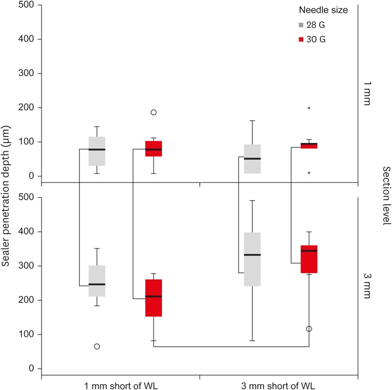
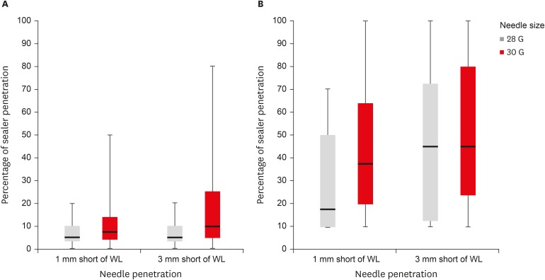
Results Inserting needles closer to the apex and using needles with wider diameters were associated with significantly more debris extrusion (p < 0.05). The position of needles and level of sections had statistically significant effects on sealer penetration depth (p < 0.05 for both).
-
Conclusions Following preparation, inserting narrower needles compatible with the final apical diameter of the prepared root canal at 3 mm short of WL during final irrigation might prevent debris extrusion and improve sealer penetration in the apical third.
INTRODUCTION
MATERIALS AND METHODS
RESULTS
The amount of extruded debris for each group of 28 G and 30 G needles according to the needle insertion depths of 1 mm and 3 mm short of working length (WL)
| Group | 28 G | 30 G |
|---|---|---|
| 1 mm short of WL (buccal roots) | 0.1090 ± 0.0268a | 0.0665 ± 0.0251c |
| 3 mm short of WL (palatal roots) | 0.0708 ± 0.0244b | 0.0296 ± 0.0138d |
DISCUSSION
CONCLUSIONS
ACKNOWLEDGEMENT
-
Conflict of Interest: No potential conflict of interest relevant to this article was reported.
-
Author Contributions:
Conceptualization: Uzunoglu-Özyürek E, Karaaslan H, Türker SA, Özçelik B.
Data curation: Uzunoglu-Özyürek E.
Formal analysis: Uzunoglu-Özyürek E, Karaaslan H, Türker SA, Özçelik B.
Funding acquisition: Uzunoglu-Özyürek E.
Investigation: Uzunoglu-Özyürek E, Karaaslan H, Türker SA.
Methodology: Uzunoglu-Özyürek E, Karaaslan H, Türker SA, Özçelik B.
Project administration: Uzunoglu-Özyürek E, Karaaslan H.
Resources: Karaaslan H, Türker SA.
Software: Uzunoglu-Özyürek E, Karaaslan H, Türker SA.
Supervision: Özçelik B; Validation: Uzunoglu-Özyürek E, Karaaslan H, Türker SA, Özçelik B.
Visualization: Uzunoglu-Özyürek E, Karaaslan H, Türker SA, Özçelik B.
Writing - original draft: Uzunoglu-Özyürek E.
Writing - review & editing: Karaaslan H, Türker SA, Özçelik B.
- 1. Haapasalo M, Endal U, Zandi H, Coil JM. Eradication of endodontic infection by instrumentation and irrigation solutions. Endod Topics 2005;10:77-102.Article
- 2. Gulabivala K, Patel B, Evans G, Ng YL. Effects of mechanical and chemical procedures on root canal surfaces. Endod Topics 2005;10:103-122.Article
- 3. Zehnder M. Root canal irrigants. J Endod 2006;32:389-398.ArticlePubMed
- 4. Paqué F, Balmer M, Attin T, Peters OA. Preparation of oval-shaped root canals in mandibular molars using nickel-titanium rotary instruments: a micro-computed tomography study. J Endod 2010;36:703-707.ArticlePubMed
- 5. Paqué F, Zehnder M, De-Deus G. Microtomography-based comparison of reciprocating single-file F2 ProTaper technique versus rotary full sequence. J Endod 2011;37:1394-1397.ArticlePubMed
- 6. Abou-Rass M, Piccinino MV. The effectiveness of four clinical irrigation methods on the removal of root canal debris. Oral Surg Oral Med Oral Pathol 1982;54:323-328.ArticlePubMed
- 7. Carrigan PJ, Morse DR, Furst ML, Sinai IH. A scanning electron microscopic evaluation of human dentinal tubules according to age and location. J Endod 1984;10:359-363.ArticlePubMed
- 8. Gu Y, Perinpanayagam H, Kum DJ, Yoo YJ, Jeong JS, Lim SM, Chang SW, Baek SH, Zhu Q, Kum KY. Effect of different agitation techniques on the penetration of irrigant and sealer into dentinal tubules. Photomed Laser Surg 2017;35:71-77.ArticlePubMed
- 9. Ørstavik D. Endodontic filling materials. Endod Topics 2014;31:53-67.Article
- 10. Mamootil K, Messer HH. Penetration of dentinal tubules by endodontic sealer cements in extracted teeth and in vivo . Int Endod J 2007;40:873-881.ArticlePubMed
- 11. Sadr S, Golmoradizadeh A, Raoof M, Tabanfar MJ. Microleakage of single-cone gutta-percha obturation technique in combination with different types of sealers. Iran Endod J 2015;10:199-203.PubMedPMC
- 12. Bolles JA, He J, Svoboda KK, Schneiderman E, Glickman GN. Comparison of Vibringe, EndoActivator, and needle irrigation on sealer penetration in extracted human teeth. J Endod 2013;39:708-711.ArticlePubMed
- 13. Kara Tuncer A, Unal B. Comparison of sealer penetration using the EndoVac irrigation system and conventional needle root canal irrigation. J Endod 2014;40:613-617.ArticlePubMed
- 14. Generali L, Cavani F, Serena V, Pettenati C, Righi E, Bertoldi C. Effect of different irrigation systems on sealer penetration into dentinal tubules. J Endod 2017;43:652-656.ArticlePubMed
- 15. Peters OA. Current challenges and concepts in the preparation of root canal systems: a review. J Endod 2004;30:559-567.ArticlePubMed
- 16. Dutner J, Mines P, Anderson A. Irrigation trends among American Association of Endodontists members: a web-based survey. J Endod 2012;38:37-40.ArticlePubMed
- 17. Chow TW. Mechanical effectiveness of root canal irrigation. J Endod 1983;9:475-479.ArticlePubMed
- 18. Usman N, Baumgartner JC, Marshall JG. Influence of instrument size on root canal debridement. J Endod 2004;30:110-112.ArticlePubMed
- 19. Falk KW, Sedgley CM. The influence of preparation size on the mechanical efficacy of root canal irrigation in vitro . J Endod 2005;31:742-745.ArticlePubMed
- 20. Sedgley CM, Nagel AC, Hall D, Applegate B. Influence of irrigant needle depth in removing bioluminescent bacteria inoculated into instrumented root canals using real-time imaging in vitro . Int Endod J 2005;38:97-104.ArticlePubMed
- 21. Khademi A, Yazdizadeh M, Feizianfard M. Determination of the minimum instrumentation size for penetration of irrigants to the apical third of root canal systems. J Endod 2006;32:417-420.ArticlePubMed
- 22. van der Sluis LW, Gambarini G, Wu MK, Wesselink PR. The influence of volume, type of irrigant and flushing method on removing artificially placed dentine debris from the apical root canal during passive ultrasonic irrigation. Int Endod J 2006;39:472-476.ArticlePubMed
- 23. Perez R, Neves AA, Belladonna FG, Silva EJ, Souza EM, Fidel S, Versiani MA, Lima I, Carvalho C, De-Deus G. Impact of needle insertion depth on the removal of hard-tissue debris. Int Endod J 2017;50:560-568.ArticlePubMedPDF
- 24. Boutsioukis C, Lambrianidis T, Kastrinakis E. Irrigant flow within a prepared root canal using various flow rates: a computational fluid dynamics study. Int Endod J 2009;42:144-155.ArticlePubMed
- 25. Munoz HR, Camacho-Cuadra K. In vivo efficacy of three different endodontic irrigation systems for irrigant delivery to working length of mesial canals of mandibular molars. J Endod 2012;38:445-448.ArticlePubMed
- 26. Aksel H, Askerbeyli S, Canbazoglu C, Serper A. Effect of needle insertion depth and apical diameter on irrigant extrusion in simulated immature permanent teeth. Braz Oral Res 2014;28:1-6.Article
- 27. Boutsioukis C, Lambrianidis T, Verhaagen B, Versluis M, Kastrinakis E, Wesselink PR, van der Sluis LW. The effect of needle-insertion depth on the irrigant flow in the root canal: evaluation using an unsteady computational fluid dynamics model. J Endod 2010;36:1664-1668.ArticlePubMed
- 28. Malentacca A, Uccioli U, Zangari D, Lajolo C, Fabiani C. Efficacy and safety of various active irrigation devices when used with either positive or negative pressure: an in vitro study. J Endod 2012;38:1622-1626.ArticlePubMed
- 29. Psimma Z, Boutsioukis C, Kastrinakis E, Vasiliadis L. Effect of needle insertion depth and root canal curvature on irrigant extrusion ex vivo. J Endod 2013;39:521-524.ArticlePubMed
- 30. Myers GL, Montgomery S. A comparison of weights of debris extruded apically by conventional filing and Canal Master techniques. J Endod 1991;17:275-279.ArticlePubMed
- 31. Boutsioukis C, Gogos C, Verhaagen B, Versluis M, Kastrinakis E, Van der Sluis LW. The effect of root canal taper on the irrigant flow: evaluation using an unsteady Computational Fluid Dynamics model. Int Endod J 2010;43:909-916.ArticlePubMed
- 32. Silva PB, Krolow AM, Pilownic KJ, Casarin RP, Lima RK, Leonardo RT, Pappen FG. Apical extrusion of debris and irrigants using different irrigation needles. Braz Dent J 2016;27:192-195.ArticlePubMed
- 33. Tanalp J, Güngör T. Apical extrusion of debris: a literature review of an inherent occurrence during root canal treatment. Int Endod J 2014;47:211-221.ArticlePubMed
- 34. Farook SA, Shah V, Lenouvel D, Sheikh O, Sadiq Z, Cascarini L, Webb R. Guidelines for management of sodium hypochlorite extrusion injuries. Br Dent J 2014;217:679-684.ArticlePubMedPDF
- 35. Boutsioukis C, Psimma Z, van der Sluis LW. Factors affecting irrigant extrusion during root canal irrigation: a systematic review. Int Endod J 2013;46:599-618.ArticlePubMed
- 36. Mandorah A. Effect of irrigation needle depth in smear layer removal: scanning electron microscope study. Saudi Endod J 2013;3:114-119.Article
- 37. Tay FR, Gu LS, Schoeffel GJ, Wimmer C, Susin L, Zhang K, Arun SN, Kim J, Looney SW, Pashley DH. Effect of vapor lock on root canal debridement by using a side-vented needle for positive-pressure irrigant delivery. J Endod 2010;36:745-750.ArticlePubMedPMC
REFERENCES
Tables & Figures
REFERENCES
Citations

- Effect of laser-induced pulpal anesthesia of single-rooted teeth with irreversible pulpitis treated by single-visit root canal therapy - A randomized clinical trial
Geeta Asthana, Dhwani Morakhia, Ravina Parmar, Rajashree Tamuli
Endodontology.2025; 37(3): 244. CrossRef - Efficacy of different irrigation needles used in endodontics: an in silico and an in vitro investigation
Maulee Sheth, Ankit Arora, Sonali Kapoor, Balraj Shukla
Biomaterial Investigations in Dentistry.2025; 12: 264. CrossRef - Preliminary insights: exploring irrigation practices during endodontic treatment among general dental practitioners in Malaysia
Kai Qi Chiew, Xin Ni Lim, Shekhar Bhatia, Naveen Chhabra
British Dental Journal.2024;[Epub] CrossRef - Efficiency of diode laser in control of post-endodontic pain: a randomized controlled trial
Hend H. Ismail, Maram Obeid, Ehab Hassanien
Clinical Oral Investigations.2023; 27(6): 2797. CrossRef - Endodontic management of an aberrant germinated composite odontome: A case report
Ankit Arora, Kavina Desai, Sonali Kapoor, Seema Gajera
Australian Endodontic Journal.2023; 49(3): 684. CrossRef - Potentials of 3D-Modeling in the Preclinical Stage of Root Needle Research
Aleksandr V. Kuligin, Larisa N. Kazakova, Oksana S. Tereshchuk, Vadim V. Bokov
I.P. Pavlov Russian Medical Biological Herald.2022; 30(1): 95. CrossRef - Effect of root canal geometry and needle type on apical extrusion of irrigant: an ex vivo study
Büşra SERÇE FİKİRLİ, Bülent ALTUNKAYNAK, Güven KAYAOĞLU
Acta Odontologica Turcica.2022; 39(3): 58. CrossRef - An in vitro radiological evaluation of irrigant penetration in the root canals using three different irrigation systems: Waterpik WP-100 device, passive irrigation, and manual dynamic irrigation systems
Suragani Hemalatha, Archana Srinivasan, A Srirekha, Lekha Santhosh, C Champa, Ashwija Shetty
Journal of Conservative Dentistry.2022; 25(4): 403. CrossRef - Preparation Ability of ProTaper Next and XP-endo Shaper Instruments in Isthmus-containing Root Canal System
Mustafa Sarıkahya, Tayfun Alaçam
Conservative Dentistry and Endodontic Journal.2021; 5(2): 28. CrossRef - Penetration depth of irrigants into root dentine after sonic, ultrasonic and photoacoustic activation
K. M. Galler, V. Grubmüller, R. Schlichting, M. Widbiller, A. Eidt, C. Schuller, M. Wölflick, K.‐A. Hiller, W. Buchalla
International Endodontic Journal.2019; 52(8): 1210. CrossRef
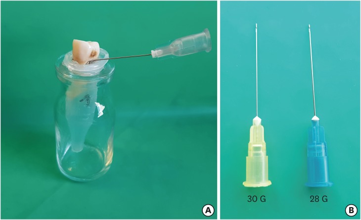


Figure 1
Figure 2
Figure 3
Figure 4
Figure 5
The amount of extruded debris for each group of 28 G and 30 G needles according to the needle insertion depths of 1 mm and 3 mm short of working length (WL)
| Group | 28 G | 30 G |
|---|---|---|
| 1 mm short of WL (buccal roots) | 0.1090 ± 0.0268a | 0.0665 ± 0.0251c |
| 3 mm short of WL (palatal roots) | 0.0708 ± 0.0244b | 0.0296 ± 0.0138d |
The values are means and standard deviations (n = 10). In the group 1 mm short of WL, needles were inserted into buccal roots; in the group 3 mm short of WL, needles were inserted into palatal roots.
Different superscript letters mean statistical significant difference for each rows and columns (p < 0.001).
The values are means and standard deviations (
Different superscript letters mean statistical significant difference for each rows and columns (

 KACD
KACD

 ePub Link
 Cite
Cite

