Articles
- Page Path
- HOME > Restor Dent Endod > Volume 49(4); 2024 > Article
-
Review Article
Influence of disinfecting solutions on the surface topography of gutta-percha cones: a systematic review of
in vitro studies -
Lora Mishra1
 , Gathani Dash1
 , Naomi Ranjan Singh1
 , Manoj Kumar2
 , Saurav Panda2
 , Franck Diemer3
 , Monika Lukomska-Szymanska4
 , Barbara Lapinska4
 , Abdul Samad Khan5
-
Restor Dent Endod 2024;49(4):e42.
DOI: https://doi.org/10.5395/rde.2024.49.e42
Published online: November 1, 2024
1Department of Conservative Dentistry and Endodontics, Institute of Dental Sciences, Siksha ‘O’ Anusandhan, Bhubaneswar, Odisha, India.
2Department of Periodontics, Institute of Dental Sciences, Siksha ‘O’ Anusandhan, Bhubaneswar, Odisha, India.
3Departement Dentaire, Université Paul Sabatier III (UPS), 3 Chemin des Maraîchers, CEDEX 9, Toulouse, France.
4Department of General Dentistry, Medical University of Lodz, Lodz, Poland.
5Department of Restorative Dental Sciences, College of Dentistry, Imam Abdulrahman Bin Faisal University, Dammam, Saudi Arabia.
- Correspondence to Lora Mishra, BDS, MDS. Department of Conservative Dentistry & Endodontics, Institute of Dental Sciences, Siksha ‘O’ Anusandhan, Bhubaneswar, Odisha 751003, India. loramishra@soa.ac.in
Copyright © 2024. The Korean Academy of Conservative Dentistry
This is an Open Access article distributed under the terms of the Creative Commons Attribution Non-Commercial License (https://creativecommons.org/licenses/by-nc/4.0/) which permits unrestricted non-commercial use, distribution, and reproduction in any medium, provided the original work is properly cited.
Abstract
- The surface integrity of gutta-percha cones is a crucial factor in the success of endodontic procedures. Disinfecting solutions play a pivotal role in sterilizing gutta-percha cones, but their influence on gutta-percha surface topography remains a subject of concern. This systematic review aimed to present a qualitative synthesis of available laboratory studies assessing the influence of disinfecting solutions on the surface topography of gutta-percha and offers insights into the implications for clinical practice. The present review followed PRISMA 2020 guidelines. An advanced database search was performed in PubMed, Google Scholar, Embase, Scopus, LILAC, non-indexed citations and reference lists of eligible studies in May 2024. Laboratory studies, in English language, were considered for inclusion. The quality (risk of bias) of the included studies was assessed using parameters for in vitro studies. A total of 28 studies were included in the qualitative synthesis. Based on the included in vitro studies, surface deposits and alterations in the physical properties of gutta-percha cones were observed after the disinfection protocol. A comprehensive review of the available literature indicates that the choice of disinfecting solution, its concentration, and immersion time significantly affect the surface topography of gutta-percha cones.
INTRODUCTION
MATERIALS AND METHODS
The inclusion and exclusion criteria
RESULTS
Concentration of disinfecting solution and device used to analyze the surface of gutta-percha in the included studies
| Study (Author, year) | Surface analysis method | % of NaOCl | % of CHX | Other disinfectants |
|---|---|---|---|---|
| Brito et al. 2013 [10] | SEM | 1, 2.5 | - | |
| Candeiro et al. 2018 [31] | 1, 2.5 | - | 17% EDTA, 10% CA and 15% MA | |
| Chandrappa et al. 2014 [17] | 5.25 | 2 | MTAD | |
| Gomes et al. 2007 [35] | 5.25 | 2 | - | |
| Nair and Bandhe 2017 [15] | 5.25 | 2 | Propolis | |
| Pang et al. 2006 [37] | 5.25 | 2 | - | |
| Pang et al. 2007 [8] | 5.25 | 2 | Chloraprep | |
| Sahinkesen et al. 2011 [34] | 2.5, 5.25 | 2 | MTAD and Octenisept | |
| Sharma et al. 2016 [26] | 3 | - | 1% PAA and Octenisept | |
| Short et al. 2003 [30] | 2.5, 5.25 | - | ||
| Srivastava 2019 [25] | 5.25 | 2 | 1% ALX | |
| Varghese et al. 2018 [23] | 5.25 | 3 | MTAD and 13% BAK | |
| Vitali et al. 2019 [9] | 1, 5.25 | - | ||
| Prado et al. 2011 (II) [36] | SEM + EDS | 5.25 | 2 | MTAD |
| Turker et al. 2015 [33] | 5.25 | 2 | 1% PAA and QMix | |
| Topuz et al. 2011 [29] | SEM + EDS + AFM | 6 | - | |
| John et al. 2017 [18] | AFM | 0.5, 2.5, 5.25 | - | |
| Karunakar et al. 2021 [27] | 5.25 | - | AgNP | |
| Mishra and Tyagi 2018 [19] | 5.25 | - | AgNP | |
| Prado et al. 2012 [7] | 5.25 | 2 | MTAD | |
| Tilakchand et al. 2014 [16] | 5.25 | 2 | - | |
| Valois et al. 2005 (I) [22] | 0.5, 2.5, 5.25 | - | - | |
| Valois et al. 2005 (II) [28] | 5.25 | 2 | - | |
| Yadav et al. 2014 [24] | 5 | 2 | 6% H2O2 and 2.2% GLD | |
| Ismail et al. 2012 [32] | Stereomicroscope | 0.5, 1.5, 5.25 | 1, 1.5, 2 | - |
| Nanda et al. 2020 [20] | 5.25 | 2 | AV | |
| Prado et al. 2011 (I) [39] | Rinehart goniometer | 5.25 | - | - |
| Nunes et al. 2019 [38] | Confocal microscopy | 5.25 | 2 | - |
1. SEM analysis
2. AFM analysis
3. Other surface analysis techniques
Immersion time and surface changes on gutta-percha in the included studies
| Study (Author, year) | Pre-treatment | Contact time with disinfecting solutions | Post-treatment rinsing done to any group | Surface changes or deposits |
|---|---|---|---|---|
| Brito et al. 2013 [10] | N | 20, 30 min, 6, 12, 24 hr | N | Y |
| Candeiro et al. 2018 [31] | Y | 1, 5, 10 min | N | Y |
| Chandrappa et al. 2014 [17] | N | 0.5, 1, 5 min | Y | Y |
| Gomes et al. 2007 [35] | Y | 1, 5, 10, 20, 30 min | Y | N |
| Ismail et al. 2012 [32] | N | 10, 15, 20 min | N | Y |
| John et al. 2017 [18] | N | 2, 5, 10 min | N | Y |
| Karunakar et al. 2021 [27] | N | 1 min | N | Y |
| Mishra and Tyagi 2018 [19] | N | 1 min | N | Y |
| Nair and Bandhe 2017 [15] | Y | 5, 10 min | Y | N |
| Nanda et al. 2020 [20] | N | 5 min | N | Y |
| Nunes et al. 2019 [38] | N | 1, 10, 15 min | N | Y |
| Pang et al. 2006 [37] | N | 1, 5 min | N | Y |
| Pang et al. 2007 [7] | Y | 5 min | N | Y |
| Prado et al. 2011 (I) [39] | N | 1 min | Y | N |
| Prado et al. 2011 (II) [36] | N | 1 min | Y | Y |
| Prado et al. 2012 [7] | Y | 1, 3, 6 min | N | Y |
| Sahinkesen et al. 2011 [34] | Y | 1, 5, 10 min | N | N |
| Sharma et al. 2016 [26] | N | 1, 5 min | N | Y |
| Short et al. 2003 [30] | N | 1 min | Y | Y |
| Srivastava 2019 [25] | N | 1 min | N | Y |
| Tilakchand et al. 2021 [16] | N | 1, 3, 10, 20, 30 min | N | Y |
| Topuz et al. 2011 [29] | N | 1, 5, 10, 20, 30 min | Y | N in SEM |
| Turker et al. 2015 [33] | N | 5, 10 min | N | N |
| Valois et al. 2005 (I) [22] | N | 1, 5 min | Y | Y |
| Valois et al. 2005 (II) [28] | N | 1, 5, 10, 20, 30 min | Y | Y |
| Varghese et al. 2018 [23] | N | 1, 5 min | N | Y |
| Vitali et al. 2019 [9] | Y | 1 min | Y | Y |
| Yadav et al. 2014 [24] | N | 1 min | N | Y |
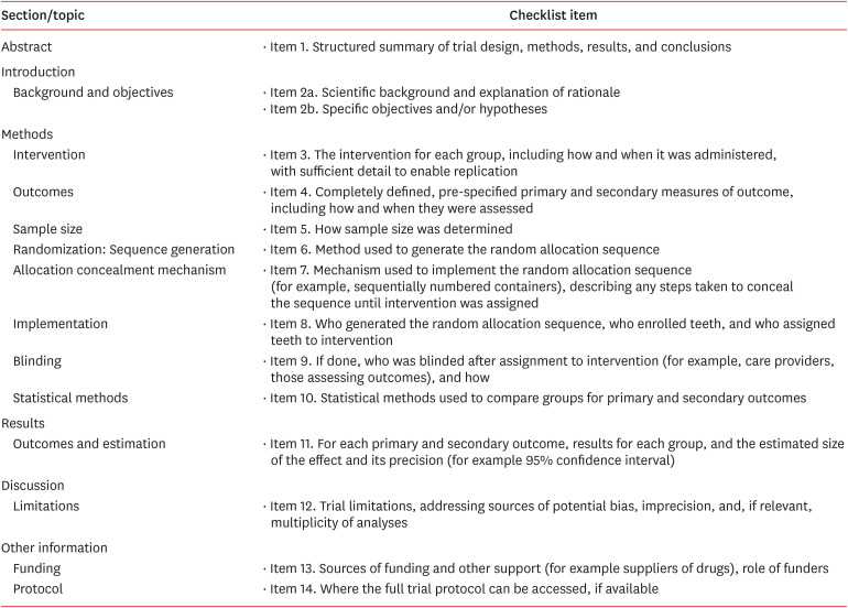
Modified CONSORT checklist of items for included in vitro studies
| Study | Item | Score | ||||||||||||||
|---|---|---|---|---|---|---|---|---|---|---|---|---|---|---|---|---|
| 1 | 2a | 2b | 3 | 4 | 5 | 6 | 7 | 8 | 9 | 10 | 11 | 12 | 13 | 14 | ||
| Brito et al. 2013 [10] | Y | Y | Y | Y | Y | N | NA | Y | Y | Y | N | NA | 8/10 | |||
| Candeiro et al. 2018 [31] | Y | Y | Y | Y | Y | N | Y | Y | N | Y | NA | 8/10 | ||||
| Chandrappa et al. 2014 [17] | Y | Y | Y | Y | Y | N | Y | Y | N | Y | NA | 8/10 | ||||
| Gomes et al. 2007 [35] | Y | Y | Y | Y | Y | N | N | N | Y | Y | NA | 7/10 | ||||
| Ismail et al. 2012 [32] | Y | Y | Y | Y | Y | N | Y | N | N | N | NA | 6/10 | ||||
| John et al. 2017 [18] | Y | Y | Y | Y | Y | N | Y | Y | Y | N | NA | 8/10 | ||||
| Karunakar et al. 2021 [27] | Y | Y | Y | Y | Y | N | Y | Y | N | Y | NA | 8/10 | ||||
| Mishra and Tyagi 2018 [19] | Y | Y | Y | Y | Y | N | Y | Y | N | Y | NA | 8/10 | ||||
| Nair and Bandhe 2017 [15] | Y | Y | Y | Y | Y | N | Y | N | N | Y | NA | 7/10 | ||||
| Nanda et al. 2020 [22] | Y | Y | Y | Y | Y | N | Y | Y | N | N | NA | 7/10 | ||||
| Nunes et al. 2019 [38] | Y | Y | Y | Y | Y | N | Y | Y | Y | Y | NA | 9/10 | ||||
| Pang et al. 2006 [37] | Y | Y | Y | Y | Y | N | Y | N | Y | N | NA | 7/10 | ||||
| Pang et al. 2007 [8] | Y | Y | Y | Y | Y | N | Y | Y | Y | N | NA | 8/10 | ||||
| Prado et al. 2011 (I) [39] | Y | Y | Y | Y | Y | N | N | N | N | Y | NA | 6/10 | ||||
| Prado et al. 2011 (II) [36] | Y | Y | Y | Y | Y | N | Y | Y | Y | Y | NA | 9/10 | ||||
| Prado et al. 2012 [7] | Y | Y | Y | Y | Y | N | Y | Y | N | Y | NA | 8/10 | ||||
| Sahinkesen et al. 2011 [34] | Y | Y | Y | Y | Y | N | N | Y | Y | Y | NA | 8/10 | ||||
| Sharma et al. 2016 [26] | Y | Y | Y | Y | Y | N | Y | N | N | N | NA | 6/10 | ||||
| Short et al. 2003 [30] | Y | Y | Y | Y | Y | N | N | N | Y | Y | NA | 8/10 | ||||
| Srivastava 2019 [25] | Y | Y | Y | Y | Y | N | Y | Y | N | Y | NA | 8/10 | ||||
| Tilakchand et al. 2014 [16] | Y | Y | Y | Y | Y | N | Y | Y | N | N | NA | 7/10 | ||||
| Topuz et al. 2011 [29] | Y | Y | Y | Y | Y | N | Y | Y | N | N | NA | 7/10 | ||||
| Turker et al. 2015 [33] | Y | Y | Y | Y | Y | N | Y | Y | N | Y | NA | 8/10 | ||||
| Valois et al. 2005 (I) [22] | Y | Y | Y | Y | Y | N | Y | Y | Y | N | NA | 8/10 | ||||
| Valois et al. 2005 (II) [28] | Y | Y | Y | Y | Y | N | Y | Y | N | N | NA | 7/10 | ||||
| Varghese et al. 2018 [23] | Y | Y | Y | Y | Y | N | N | N | N | N | NA | 5/10 | ||||
| Vitali et al. 2019 [9] | Y | Y | Y | Y | Y | N | Y | N | Y | Y | NA | 9/10 | ||||
| Yadav et al. 2014 [24] | Y | Y | Y | Y | Y | N | Y | Y | Y | N | NA | 8/10 | ||||
DISCUSSION
CONCLUSIONS
ACKNOWLEDGEMENTS
-
Conflict of Interest: No potential conflict of interest relevant to this article was reported.
-
Author Contributions:
Conceptualization: Mishra L.
Data curation: Dash G.
Investigation: Mishra L, Lukomska-Szymanska M, Lapinska B.
Formal analysis: Kumar M, Panda S.
Funding acquisition: Diemer F.
Methodology: Mishra L, Dash G.
Project administration: Diemer F, Singh NR.
Resources: Lapinska B.
Supervision: Singh NR.
Validation: Kumar M, Panda S.
Visualization: Diemer F.
Writing - original draft: Mishra L.
Writing - review & editing: Mishra L, Khan AS.
SUPPLEMENTARY MATERIAL
- 1. Siqueira JF Jr. Endodontic infections: concepts, paradigms, and perspectives. Oral Surg Oral Med Oral Pathol Oral Radiol Endod 2002;94:281-293.ArticlePubMed
- 2. Albino Souza M, Dalla Lana D, Gabrielli E, Barbosa Ribeiro M, Miyagaki DC, Cecchin D. Effectiveness of final decontamination protocols against Enterococcus faecalis and its influence on bond strength of filling material to root canal dentin. Photodiagnosis Photodyn Ther 2017;17:92-97.ArticlePubMed
- 3. Yildirim C, Altintas S, Karaarslan ES, Usumez A. Influence of different disinfection methods on bond strength of resin sealer. Mater Res Innov 2011;15:34.Article
- 4. Friedman CM, Sandrik JL, Heuer MA, Rapp GW. Composition and mechanical properties of gutta-percha endodontic points. J Dent Res 1975;54:921-925.ArticlePubMedPDF
- 5. George SW, Pichardo MR, Bergeron BE, Jeansonne BG. The effect of formalin storage on the apical microleakage of obturated canals. J Endod 2006;32:869-871.ArticlePubMed
- 6. Hörsted-Bindslev P, Andersen MA, Jensen MF, Nilsson JH, Wenzel A. Quality of molar root canal fillings performed with the lateral compaction and the single-cone technique. J Endod 2007;33:468-471.ArticlePubMed
- 7. Prado M, Gusman H, Gomes BP, Simão RA. Effect of disinfectant solutions on gutta-percha and resilon cones. Microsc Res Tech 2012;75:791-795.ArticlePubMed
- 8. Pang NS, Jung IY, Bae KS, Baek SH, Lee WC, Kum KY. Effects of short-term chemical disinfection of gutta-percha cones: identification of affected microbes and alterations in surface texture and physical properties. J Endod 2007;33:594-598.ArticlePubMed
- 9. Vitali FC, Nomura LH, Delai D, Henriques DH, Alves AM, da Fonseca Roberti Garcia L, et al. Disinfection and surface changes of gutta-percha cones after immersion in sodium hypochlorite solution containing surfactant. Microsc Res Tech 2019;82:1290-1296.ArticlePubMedPDF
- 10. Brito SM, Vasconcelos RA, Oliveira SH. Gutta-percha points surface alterations after sodium hypochlorite disinfection. Braz Dent Sci 2013;16:47-55.ArticlePDF
- 11. Patel S, Puri T, Mannocci F, Bakhsh AA. The outcome of endodontic treatment using an enhanced infection protocol in specialist practice. Br Dent J 2022;232:805-811.ArticlePubMedPDF
- 12. Page MJ, McKenzie JE, Bossuyt PM, Boutron I, Hoffmann TC, Mulrow CD, et al. The PRISMA 2020 statement: an updated guideline for reporting systematic reviews. J Clin Epidemiol 2021;134:178-189.ArticlePubMed
- 13. Page MJ, McKenzie JE, Bossuyt PM, Boutron I, Hoffmann TC, Mulrow CD, et al. The PRISMA 2020 statement: an updated guideline for reporting systematic reviews. BMJ 2021;372:n71.ArticlePubMedPMC
- 14. Faggion CM Jr. Guidelines for reporting pre-clinical in vitro studies on dental materials. J Evid Based Dent Pract 2012;12:182-189.
- 15. Nair R, Bandhe S. An in-vitro assessment of the residual antimicrobial effects and surface alterations of gutta-percha disinfected with four different solutions. J Dr NTR Univ Health Sci 2017;6:98-102.
- 16. Tilakchand M, Naik B, Shetty AS. A comparative evaluation of the effect of 5.25% sodium hypochlorite and 2% chlorhexidine on the surface texture of Gutta-percha and resilon cones using atomic force microscope. J Conserv Dent 2014;17:18-21.ArticlePubMedPMC
- 17. Chandrappa MM, Mundathodu N, Srinivasan R, Nasreen F, Kavitha P, Shetty A. Disinfection of gutta-percha cones using three reagents and their residual effects. J Conserv Dent 2014;17:571-574.ArticlePubMedPMC
- 18. John BM, Purra A, Dutta A, Zargar AW. Topographical effects of gutta percha immersed in different concentration of sodium hypochlorite disinfection at different time interval: an atomic force microscopy study. Int J Oral Health Dent 2017;3:54-58.
- 19. Mishra P, Tyagi S. Surface analysis of gutta percha after disinfecting with sodium hypochlorite and silver nanoparticles by atomic force microscopy: an in vitro study. Dent Res J 2018;15:242-247.Article
- 20. Nanda Z, Tekwani RA, Rudagi K, Reddy KK, Nausheen A, Makne SG, et al. Effect of different chemical and herbal disinfectant solutions on the mechanical and physical properties of gutta-percha: an in vitro study. J Oper Dent Endod 2020;4:84-87.
- 21. Nabeshima CK, Machado ME, Britto ML, Pallotta RC. Effectiveness of different chemical agents for disinfection of gutta-percha cones. Aust Endod J 2011;37:118-121.ArticlePubMed
- 22. Valois CR, Silva LP, Azevedo RB. Effects of 2% chlorhexidine and 5.25% sodium hypochlorite on gutta-percha cones studied by atomic force microscopy. Int Endod J 2005;38:425-429.ArticlePubMed
- 23. Varghese A, Joshua J, Shetty D, Damda A, Bhandary S. Evaluation of surface changes on gutta-percha points treated with four different disinfectants at 2 different time intervals: a SEM study. IOSR J Dent Med Sci 2018;17:30-37.
- 24. Yadav SS, Archana C, Sukhabogi JR, Ramana IV, Priyanka Y, Gopi P. Structural effects of various commonly used disinfectant solutions on gutta-percha: an atomic force microscopic study. J Orofac Res 2014;4:157-160.Article
- 25. Srivastava S. Rapid chairside disinfection and surface alterations of gutta-percha cones with 1% Alexidine, 2% Chlorhexidine and 5.25% sodium hypochlorite against enterococcus faecalis-an in vitro comparative study. Int Arch Integr Med 2019;6:1-7.
- 26. Sharma NK, Prasad A. Evaluation of disinfection of gutta-percha cones using different chemical solutions. Ind Dent J 2016;8:3-8.
- 27. Karunakar P, Ranga Reddy MS, Faizuddin U, Karteek BS, Charan Reddy CL, Rasagna M. Evaluation of surface analysis of gutta-percha after disinfecting with sodium hypochlorite, silver nanoparticles, and chitosan nanoparticles by atomic force microscopy: an in vitro study. J Conserv Dent 2021;24:63-66.ArticlePubMedPMC
- 28. Valois CR, Silva LP, Azevedo RB. Structural effects of sodium hypochlorite solutions on gutta-percha cones: atomic force microscopy study. J Endod 2005;31:749-751.ArticlePubMed
- 29. Topuz Ö, Sağlam BC, Sen F, Sen S, Gökağaç G, Görgül G. Effects of sodium hypochlorite on gutta-percha and Resilon cones: an atomic force microscopy and scanning electron microscopy study. Oral Surg Oral Med Oral Pathol Oral Radiol Endod 2011;112:e21-e26.Article
- 30. Short RD, Dorn SO, Kuttler S. The crystallization of sodium hypochlorite on gutta-percha cones after the rapid-sterilization technique: an SEM study. J Endod 2003;29:670-673.ArticlePubMed
- 31. de Miranda Candeiro GT, Akisue E, Campelo Correia F, Dos Santos Sousa E, do Vale MS, Iglecias EF, et al. Analysis of demineralized chemical substances for disinfecting gutta-percha cones. Iran Endod J 2018;13:318-322.PubMedPMC
- 32. Ismail SA, Al-Sabawi NA, Al-Askary RA. Effect of different disinfectant solutions on the properties of gutta percha cones. TJDS 2012;2:169-174.ArticlePDF
- 33. Aktemur Turker S, Aslan MH, Uzunoglu E, Ozcelik B. Antimicrobial and structural effects of different irrigation solutions on gutta-percha cones. J Istanb Univ Fac Dent 2015;49:27-32.ArticlePubMedPMC
- 34. Sahinkesen G, Oktay EA, Er Ö, Koçak MM, Kiliç A. Evaluation of residual antimicrobial effects and surface changes of gutta-percha disinfected with different solutions. J Contemp Dent Pract 2011;12:47-51.ArticlePubMed
- 35. Gomes BP, Berber VB, Montagner F, Sena NT, Zaia AA, Ferraz CC, et al. Residual effects and surface alterations in disinfected gutta-percha and Resilon cones. J Endod 2007;33:948-951.ArticlePubMed
- 36. Prado M, Gusman H, Gomes BP, Simão RA. The importance of final rinse after disinfection of gutta-percha and Resilon cones. Oral Surg Oral Med Oral Pathol Oral Radiol Endod 2011;111:e21-e24.Article
- 37. Pang NS, Jung IY, Yu YJ, Kum KY. Assessment of decontamination of gutta-percha cone and the change of surface texture after rapid chemical disinfection. Restor Dent Endod 2006;31:133-139.
- 38. Nunes AM, Gouvea JP, da Silva L. Influence of different disinfection protocols on gutta-percha cones surface roughness assessed by two different methods. J Mater Res Technol 2019;8:5464-5470.Article
- 39. Prado M, de Assis DF, Gomes BP, Simão RA. Effect of disinfectant solutions on the surface free energy and wettability of filling material. J Endod 2011;37:980-982.ArticlePubMed
REFERENCES
Tables & Figures
REFERENCES
Citations

- In Vitro Evaluation of Disinfectants on Gutta-Percha Cones: Antimicrobial Efficacy Against Enterococcus faecalis and Candida albicans
Tringa Kelmendi, Donika Bajrami Shabani, Aida Meto, Hani Ounsi
Journal of Clinical Medicine.2025; 14(19): 6846. CrossRef


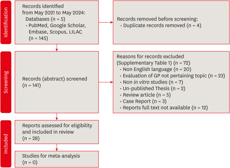
Figure 1
Figure 2
The inclusion and exclusion criteria
| Inclusion criteria | Exclusion criteria |
|---|---|
| Review articles, case reports, book chapters, grey literature, animal studies or studies, in which data is missing or incomplete | |
| Surface characteristics are analyzed using a microscope, SEM, AFM, or any other tool. | Articles, which have only assessed the antimicrobial efficacy of disinfectants and gutta-percha |
| Articles not in English |
SEM, scanning electron microscopy; AFM, atomic force microscopy.
Concentration of disinfecting solution and device used to analyze the surface of gutta-percha in the included studies
| Study (Author, year) | Surface analysis method | % of NaOCl | % of CHX | Other disinfectants |
|---|---|---|---|---|
| Brito | SEM | 1, 2.5 | - | |
| Candeiro | 1, 2.5 | - | 17% EDTA, 10% CA and 15% MA | |
| Chandrappa | 5.25 | 2 | MTAD | |
| Gomes | 5.25 | 2 | - | |
| Nair and Bandhe 2017 [ | 5.25 | 2 | Propolis | |
| Pang | 5.25 | 2 | - | |
| Pang | 5.25 | 2 | Chloraprep | |
| Sahinkesen | 2.5, 5.25 | 2 | MTAD and Octenisept | |
| Sharma | 3 | - | 1% PAA and Octenisept | |
| Short | 2.5, 5.25 | - | ||
| Srivastava 2019 [ | 5.25 | 2 | 1% ALX | |
| Varghese | 5.25 | 3 | MTAD and 13% BAK | |
| Vitali | 1, 5.25 | - | ||
| Prado | SEM + EDS | 5.25 | 2 | MTAD |
| Turker | 5.25 | 2 | 1% PAA and QMix | |
| Topuz | SEM + EDS + AFM | 6 | - | |
| John | AFM | 0.5, 2.5, 5.25 | - | |
| Karunakar | 5.25 | - | AgNP | |
| Mishra and Tyagi 2018 [ | 5.25 | - | AgNP | |
| Prado | 5.25 | 2 | MTAD | |
| Tilakchand | 5.25 | 2 | - | |
| Valois | 0.5, 2.5, 5.25 | - | - | |
| Valois | 5.25 | 2 | - | |
| Yadav | 5 | 2 | 6% H2O2 and 2.2% GLD | |
| Ismail | Stereomicroscope | 0.5, 1.5, 5.25 | 1, 1.5, 2 | - |
| Nanda | 5.25 | 2 | AV | |
| Prado | Rinehart goniometer | 5.25 | - | - |
| Nunes | Confocal microscopy | 5.25 | 2 | - |
SEM, scanning electron microscopy; EDS, energy dispersive X-ray spectrometry; AFM, atomic force microscopy; NaOCl, sodium hypochlorite; CHX, chlorhexidine; MTAD, mineral trioxide aggregate; PAA, para acetic acid; OCPT, octenisept; AV, aloe vera; H2O2, hydrogen peroxide; GLD, glutaraldehyde; ChnpP, chitosan particles; AgNP, silver nanoparticle; QMix, a solution containing 2% CHX and 17% EDTA (ethylene diamine tetraacetic acid); ALX, alexidine; CA, citric acid; MA, maleic acid; BAK, benzalkonium chloride.
Immersion time and surface changes on gutta-percha in the included studies
| Study (Author, year) | Pre-treatment | Contact time with disinfecting solutions | Post-treatment rinsing done to any group | Surface changes or deposits |
|---|---|---|---|---|
| Brito | N | 20, 30 min, 6, 12, 24 hr | N | Y |
| Candeiro | Y | 1, 5, 10 min | N | Y |
| Chandrappa | N | 0.5, 1, 5 min | Y | Y |
| Gomes | Y | 1, 5, 10, 20, 30 min | Y | N |
| Ismail | N | 10, 15, 20 min | N | Y |
| John | N | 2, 5, 10 min | N | Y |
| Karunakar | N | 1 min | N | Y |
| Mishra and Tyagi 2018 [ | N | 1 min | N | Y |
| Nair and Bandhe 2017 [ | Y | 5, 10 min | Y | N |
| Nanda | N | 5 min | N | Y |
| Nunes | N | 1, 10, 15 min | N | Y |
| Pang | N | 1, 5 min | N | Y |
| Pang | Y | 5 min | N | Y |
| Prado | N | 1 min | Y | N |
| Prado | N | 1 min | Y | Y |
| Prado | Y | 1, 3, 6 min | N | Y |
| Sahinkesen | Y | 1, 5, 10 min | N | N |
| Sharma | N | 1, 5 min | N | Y |
| Short | N | 1 min | Y | Y |
| Srivastava 2019 [ | N | 1 min | N | Y |
| Tilakchand | N | 1, 3, 10, 20, 30 min | N | Y |
| Topuz | N | 1, 5, 10, 20, 30 min | Y | N in SEM |
| Turker | N | 5, 10 min | N | N |
| Valois | N | 1, 5 min | Y | Y |
| Valois | N | 1, 5, 10, 20, 30 min | Y | Y |
| Varghese | N | 1, 5 min | N | Y |
| Vitali | Y | 1 min | Y | Y |
| Yadav | N | 1 min | N | Y |
N, no; Y, yes.
Modified CONSORT checklist of items for included in vitro studies
| Study | Item | Score | ||||||||||||||
|---|---|---|---|---|---|---|---|---|---|---|---|---|---|---|---|---|
| 1 | 2a | 2b | 3 | 4 | 5 | 6 | 7 | 8 | 9 | 10 | 11 | 12 | 13 | 14 | ||
| Brito | Y | Y | Y | Y | Y | N | NA | Y | Y | Y | N | NA | 8/10 | |||
| Candeiro | Y | Y | Y | Y | Y | N | Y | Y | N | Y | NA | 8/10 | ||||
| Chandrappa | Y | Y | Y | Y | Y | N | Y | Y | N | Y | NA | 8/10 | ||||
| Gomes | Y | Y | Y | Y | Y | N | N | N | Y | Y | NA | 7/10 | ||||
| Ismail | Y | Y | Y | Y | Y | N | Y | N | N | N | NA | 6/10 | ||||
| John | Y | Y | Y | Y | Y | N | Y | Y | Y | N | NA | 8/10 | ||||
| Karunakar | Y | Y | Y | Y | Y | N | Y | Y | N | Y | NA | 8/10 | ||||
| Mishra and Tyagi 2018 [ | Y | Y | Y | Y | Y | N | Y | Y | N | Y | NA | 8/10 | ||||
| Nair and Bandhe 2017 [ | Y | Y | Y | Y | Y | N | Y | N | N | Y | NA | 7/10 | ||||
| Nanda | Y | Y | Y | Y | Y | N | Y | Y | N | N | NA | 7/10 | ||||
| Nunes | Y | Y | Y | Y | Y | N | Y | Y | Y | Y | NA | 9/10 | ||||
| Pang | Y | Y | Y | Y | Y | N | Y | N | Y | N | NA | 7/10 | ||||
| Pang | Y | Y | Y | Y | Y | N | Y | Y | Y | N | NA | 8/10 | ||||
| Prado | Y | Y | Y | Y | Y | N | N | N | N | Y | NA | 6/10 | ||||
| Prado | Y | Y | Y | Y | Y | N | Y | Y | Y | Y | NA | 9/10 | ||||
| Prado | Y | Y | Y | Y | Y | N | Y | Y | N | Y | NA | 8/10 | ||||
| Sahinkesen | Y | Y | Y | Y | Y | N | N | Y | Y | Y | NA | 8/10 | ||||
| Sharma | Y | Y | Y | Y | Y | N | Y | N | N | N | NA | 6/10 | ||||
| Short | Y | Y | Y | Y | Y | N | N | N | Y | Y | NA | 8/10 | ||||
| Srivastava 2019 [ | Y | Y | Y | Y | Y | N | Y | Y | N | Y | NA | 8/10 | ||||
| Tilakchand | Y | Y | Y | Y | Y | N | Y | Y | N | N | NA | 7/10 | ||||
| Topuz | Y | Y | Y | Y | Y | N | Y | Y | N | N | NA | 7/10 | ||||
| Turker | Y | Y | Y | Y | Y | N | Y | Y | N | Y | NA | 8/10 | ||||
| Valois | Y | Y | Y | Y | Y | N | Y | Y | Y | N | NA | 8/10 | ||||
| Valois | Y | Y | Y | Y | Y | N | Y | Y | N | N | NA | 7/10 | ||||
| Varghese | Y | Y | Y | Y | Y | N | N | N | N | N | NA | 5/10 | ||||
| Vitali | Y | Y | Y | Y | Y | N | Y | N | Y | Y | NA | 9/10 | ||||
| Yadav | Y | Y | Y | Y | Y | N | Y | Y | Y | N | NA | 8/10 | ||||
Y, yes; N, no; NA, not applicable.
SEM, scanning electron microscopy; AFM, atomic force microscopy.
SEM, scanning electron microscopy; EDS, energy dispersive X-ray spectrometry; AFM, atomic force microscopy; NaOCl, sodium hypochlorite; CHX, chlorhexidine; MTAD, mineral trioxide aggregate; PAA, para acetic acid; OCPT, octenisept; AV, aloe vera; H2O2, hydrogen peroxide; GLD, glutaraldehyde; ChnpP, chitosan particles; AgNP, silver nanoparticle; QMix, a solution containing 2% CHX and 17% EDTA (ethylene diamine tetraacetic acid); ALX, alexidine; CA, citric acid; MA, maleic acid; BAK, benzalkonium chloride.
N, no; Y, yes.
Y, yes; N, no; NA, not applicable.

 KACD
KACD


 ePub Link
 Cite
Cite

