
Previous issues
- Page Path
- HOME > Browse articles > Previous issues
- Volume 46 (1); February 2021
-


Editorial
-
Appreciation to the Scientific Advisory Board members in 2020 for their contributions to
Restorative Dentistry and Endodontics - Kyung-San Min
- Restor Dent Endod 2021;46(1):e15. Published online February 8, 2021
- DOI: https://doi.org/10.5395/rde.2021.46.e15
- 1,392 View
- 14 Download

Review Article
- Clinical efficacy of activated irrigation in endodontics: a focused review
- Amelia Wan Tin Cheung, Angeline Hui Cheng Lee, Gary Shun Pan Cheung
- Restor Dent Endod 2021;46(1):e10. Published online January 26, 2021
- DOI: https://doi.org/10.5395/rde.2021.46.e10
-
 Abstract
 PDF
PDF PubReader
 ePub
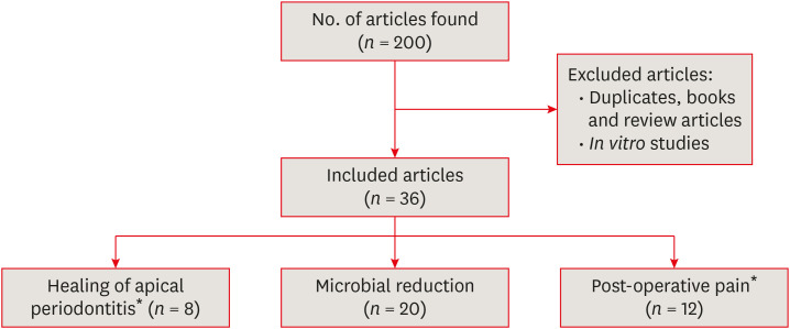
ePub Root canal debridement, which includes the removal of infected tissues and microbial biofilms, is considered the corner stone of root canal treatment. Chemical adjuncts play a multitude of functions in this regard, as tissue solvents, antimicrobial agents and for removing the smear layer. These adjuncts (irrigants) are usually delivered using a syringe and needle. With increasing knowledge of the complexity of root canal anatomy and tenacity of microbial biofilms, the need for strategies that potentiate the action of these irrigants within the root canal system cannot be overemphasized. Several such activated irrigation strategies exist. The aim of this review is to comprehensively discuss the different irrigant activation methods from the context of clinical studies.
-
Citations
Citations to this article as recorded by
- The anti-biofilm effect of ozonated water and super-oxidized water applied with different activation procedures against Enterococcus faecalis biofilm
Fatoş Albayrak, Tutku Tunç, Demet Altunbaş, Recai Zan, Muhammed Ayhan, Halil Bal
BMC Oral Health.2026;[Epub] CrossRef - Laser applications in oral precise medicine: from excision to restoration
Ouyang Siyuan, Dongxu Xie, H. M. Shakhawat Sohan, Zhou Qiunan, Luyuan Jin, Zuomin Wang, Peng Wen
International Journal of Optomechatronics.2026;[Epub] CrossRef - Clinical prospects of passive ultrasonic disinfection in endodontic practice
V.O. Demidov, R.R. Gaynutdinova, O.V. Vinogradova, R.R. Gabidullin, D.A. Guseva
Stomatology.2026; 105(2): 65. CrossRef - Protocols to evaluate the antibacterial and irrigation efficacy of new root canal irrigants: A systematic review of ex vivo studies
Atsushi Oishi, Ayako Tsubaki, Yusuke Iwabuchi, Yudai Kitaichi, Yuika Yamanaka, Kanae Wada, Asuna Sugimoto, Tsutomu Iwamoto
Japanese Dental Science Review.2026; 62: 154. CrossRef - Comparison of rotary NiTi files with ultrasonic irrigation and manual K-files with syringe irrigation in primary molar pulpectomy: A retrospective cohort study
Jing Liu, Kaixuan Yan, Yu Zheng, Feifei Wang, Xiaowei Hou
Clinical Oral Investigations.2026;[Epub] CrossRef - Antimicrobial effect and biofilm inhibition of biosurfactant as an irrigante against root canal bacteria with and without laser irradiation
Doaa Mohamed Sadony, Amira Ibrahim Mohamed, Ahmed A. Hamed, Seham F. Hasan, Heba I. Elkhouly
Lasers in Dental Science.2026;[Epub] CrossRef - Efficacy of Er: YAG, continuous-wave, and pulsed diode laser-activated irrigation on smear layer removal: a comparative microscopic study
Muhammad Mahmoud Abaza, Tarek Abdel Hamid Harhash, Ahmed Abbas Zaky
Lasers in Dental Science.2025;[Epub] CrossRef - Sodium hypochlorite accident approach with photobiomodulation during an endodontic procedure: a case report
Johanna Hernandez La Rotta, Marggie Grajales
Lasers in Dental Science.2025;[Epub] CrossRef - Antibacterial efficacy of sodium dichloroisocyanurate and 2-hydroxyisocaproic acid intracanal medicaments on Enterococcus faecalis: A comparative in-vitro study
Rasmina K. Nizar, Anju Varughese, M. Remya, V.P. Prabath Singh, Gayathri Usha, Gayathri Presannakumar
Journal of Oral Biology and Craniofacial Research.2025; 15(5): 1149. CrossRef - ВПЛИВ ХІМІЧНИХ ІРИГАНТІВ НА СТАН БІОПЛІВКИ КОРЕНЕВОГО КАНАЛУ ПРИ ЛІКУВАННІ ПЕРІОДОНТИТІВ
Р. І. Новосядлий, М. М. Рожко
Art of Medicine.2025; : 33. CrossRef - REVOLUCIONANDO LA ENDODONCIA: LA IMPORTANCIA DE IRRIGANTES MÚLTIPLES PARA UNA DESINFECCIÓN EFECTIVA DEL SISTEMA DE CONDUCTOS RADICULARES UNA REVISIÓN NARRATIVA
Irving Pablo Fernandez Calle, Edwin Macias Limachi , Abigail Marisol Vargas Ticona , Jenny Paula Aguilar Avalos , Marivel Irene Condori Escobar, Alcides Ramber Maldonado Huaycho , Jenny Claudia Apaza Cayo , Miguel Angel Espinoza Vega , Jesús Alejan
RECIMA21 - Revista Científica Multidisciplinar - ISSN 2675-6218.2024; 5(11): e5115929. CrossRef - Cleaning and disinfection of the root canal system provided by four active supplementary irrigation methods
Alessandra Timponi Goes Cruz, Adriane Antoniw Klemz, Edvaldo Antônio Ribeiro Rosa, Fabiana Soares Grecca, Bianca Mattos, Lucila Piasecki, Ricardo Machado, Sérgio Aparecido Ignácio, Ulisses Xavier da Silva Neto
Scientific Reports.2024;[Epub] CrossRef - Postendodontic Pain Using Single File System with Different Irrigation Protocols in Single-visit Root Canal Treatment: A Randomized Control Trial
Kiran Patel, Kailash Attur, Nishtha Patel, Kamal M Bagda, Karthik P Venkataraghavan, Mohammed B Mustafa, Shylaja K Attur
The Journal of Contemporary Dental Practice.2024; 25(2): 180. CrossRef - Bacteria debridement efficacy of two sonic root canal irrigant activation systems
Chang Zeng, Pei Hu, Colin P. Egan, Brian E. Bergeron, Franklin Tay, Jingzhi Ma
Journal of Dentistry.2024; 140: 104770. CrossRef - Evaluation of different activated irrigation protocols on debridement quality in various access cavity designs
Urvashi M. Ujariya, Mitul Lallubhai Gangani, Rajendra P. Bharatiya, Anjali K. Kothari
Endodontology.2024; 36(4): 400. CrossRef - Synergistic antimicrobial potential of EGCG and fosfomycin against biofilms associated with endodontic infections
Cristiane DUQUE, Amanda Caselato Andolfatto SOUZA, Kelly Limi AIDA, Jesse Augusto PEREIRA, Karina Sampaio CAIAFFA, Vanessa Rodrigues dos SANTOS, Leopoldo COSME-SILVA, Anuradha PRAKKI
Journal of Applied Oral Science.2023;[Epub] CrossRef - Insights of fluid dynamics in an optimally shaped root canal system
Kavalipurapu Venkata Teja, Sindhu Ramesh, Krishnamachari Janani
Saudi Endodontic Journal.2023; 13(2): 216. CrossRef - Diamond–coated ultrasonic tip decreases debris and uninstrumented surface after preparation of curved canals with isthmus
Maria Luiza GIOSTER–RAMOS, Mariana Mena Barreto PIVOTO–JOÃO, Jáder Camilo PINTO, Juliane Maria GUERREIRO–TANOMARU, Mário TANOMARU–FILHO
Brazilian Oral Research.2023;[Epub] CrossRef - Effectiveness of Passive Ultrasonic Irrigation Protocols in Simulated Complex Root Canal Cavities
Flávia A. Plazza, Renan Dal-Fabbro, Leopoldo Cosme-Silva, Paulo C. T. Duarte, Caroline Loureiro, Vitória Z. Custódio, Luciano T. A. Cintra, Marco A. H. Duarte, João Eduardo Gomes-Filho
Oral.2022; 3(1): 1. CrossRef - Comparison of sealer penetration of sonic activation versus conventional needle irrigation: a systematic review and meta-analysis of randomized controlled trials
Li Tan, Qiong Liu, Yun Chen, Ya-Qiong Zhao, Jie Zhao, Marie Aimee Dusenge, Yao Feng, Qin Ye, Jing Hu, Ze-Yue Ou-Yang, Ying-Hui Zhou, Yue Guo, Yun-Zhi Feng
BMC Oral Health.2022;[Epub] CrossRef - Efficacy of Photoinduced Photoacoustic Streaming and Diode Laser Irrigation Techniques on Smear Layer Removal, Sealer Penetration and Push-out Bond Strength
Latifa Mohamed Abdelgawad, Nancy Attia Ahmed ElShafei, Somaia Abdlatif Eissa, Dalia Yahia Ibrahim
Journal of Lasers in Medical Sciences.2022; 13(1): e12. CrossRef - Microbiological Aspects of Root Canal Infections and Disinfection Strategies: An Update Review on the Current Knowledge and Challenges
Jasmine Wong, Daniel Manoil, Peggy Näsman, Georgios N. Belibasakis, Prasanna Neelakantan
Frontiers in Oral Health.2021;[Epub] CrossRef - In vitro evaluation of efficacy of two endodontic sonic-powered irrigant agitation systems in killing single-species intracanal biofilms
Chang Zeng, Joseph Everett, Stephanie Sidow, Brian E. Bergeron, Fucong Tian, Jingzhi Ma, Franklin R. Tay
Journal of Dentistry.2021; 115: 103859. CrossRef - A novel three‐dimensionally printed model to assess biofilm removal by ultrasonically activated irrigation
Min‐Ji Choi, Mi‐Ah Kim, Yoorina Choi, Prasanna Neelakantan, Mi‐Kyung Yu, Kyung‐San Min
International Endodontic Journal.2021; 54(10): 1871. CrossRef
- The anti-biofilm effect of ozonated water and super-oxidized water applied with different activation procedures against Enterococcus faecalis biofilm
- 9,667 View
- 177 Download
- 18 Web of Science
- 24 Crossref
Research Articles
- Biomineralization of three calcium silicate-based cements after implantation in rat subcutaneous tissue
- Ranjdar Mahmood Talabani, Balkees Taha Garib, Reza Masaeli, Kavosh Zandsalimi, Farinaz Ketabat
- Restor Dent Endod 2021;46(1):e1. Published online December 2, 2020
- DOI: https://doi.org/10.5395/rde.2021.46.e1
-
Abstract
PDFPubReaderePub
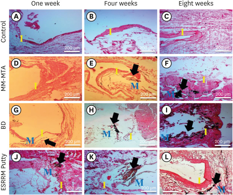
Objectives The aim of this study was to evaluate the dystrophic mineralization deposits from 3 calcium silicate-based cements (Micro-Mega mineral trioxide aggregate [MM-MTA], Biodentine [BD], and EndoSequence Root Repair Material [ESRRM] putty) over time after subcutaneous implantation into rats.
Materials and Methods Forty-five silicon tubes containing the tested materials and 15 empty tubes (serving as a control group) were subcutaneously implanted into the backs of 15 Wistar rats. At 1, 4, and 8 weeks after implantation, the animals were euthanized (
n = 5 animals/group), and the silicon tubes were removed with the surrounding tissues. Histopathological tissue sections were stained with von Kossa stain to assess mineralization. Scanning electron microscopy and energy-dispersive X-ray spectroscopy (SEM/EDX) were also used to assess the chemical components of the surface precipitates deposited on the implant and the pattern of calcium and phosphorus distribution at the material-tissue interface. The calcium-to-phosphorus ratios were compared using the non-parametric Kruskal-Wallis test at a significance level of 5%.Results The von Kossa staining showed that both BD and ESRRM putty induced mineralization starting at week 1; this mineralization increased further until the end of the study. In contrast, MM-MTA induced dystrophic calcification later, from 4 weeks onward. SEM/EDX showed no statistically significant differences in the calcium- and phosphorus-rich areas among the 3 materials at any time point (
p > 0.05).Conclusions After subcutaneous implantation, biomineralization of the 3-calcium silicate-based cements started early and increased over time, and all 3 tested cements generated calcium- and phosphorus-containing surface precipitates.
-
Citations
Citations to this article as recorded by- Comparative Biocompatibility of Portland‐Based Endodontic Cements Containing Niobium and/or Bismuth Oxides in Rat Subcutaneous Tissue
Raphael Victor Silva Andrade, Rafaela Alcindo Silva de Souza Fé, Vladimir Galdino Sabino, Carlos Augusto Galvão Barboza, Maria Luiza Diniz de Sousa Lopes, Nizyara Costa da Silva, Valkleidson Santos de Araujo, Amanda Silveira da Silva, Raimundo Fernandes d
BioMed Research International.2026;[Epub] CrossRef - Evaluating Retrieval-Augmented Large Language Models on External Cervical Resorption: A Comparative Study of Gemini and NotebookLM
Marc Garcia-Font, Nicolás Dufey-Portilla, Fernando Durán-Sindreu, José Antonio González Sánchez, Gustavo Rodríguez Millán, Venkateshbabu Nagendrababu, Paul M.H. Dummer, Francesc Abella Sans
Journal of Endodontics.2025;[Epub] CrossRef - Antibacterial, biocompatible, and mineralization‐inducing properties of calcium silicate‐based cements
Taimy Cruz Hondares, Xiaoxiao Hao, Yanfang Zhao, Yuyin Lin, Dobrawa Napierala, Janice G. Jackson, Ping Zhang
International Journal of Paediatric Dentistry.2024; 34(6): 843. CrossRef - Bioactive potential of Bio‐C Pulpo is evidenced by presence of birefringent calcite and osteocalcin immunoexpression in the rat subcutaneous tissue
Marcela Borsatto Queiroz, Rafaela Nanami Handa Inada, Camila Soares Lopes, Juliane Maria Guerreiro‐Tanomaru, Estela Sasso‐Cerri, Mário Tanomaru‐Filho, Paulo Sérgio Cerri
Journal of Biomedical Materials Research Part B: Applied Biomaterials.2022; 110(10): 2369. CrossRef
- Comparative Biocompatibility of Portland‐Based Endodontic Cements Containing Niobium and/or Bismuth Oxides in Rat Subcutaneous Tissue
- 3,377 View
- 32 Download
- 5 Web of Science
- 4 Crossref
- A micro-computed tomographic study using a novel test model to assess the filling ability and volumetric changes of bioceramic root repair materials
- Fernanda Ferrari Esteves Torres, Jader Camilo Pinto, Gabriella Oliveira Figueira, Juliane Maria Guerreiro-Tanomaru, Mario Tanomaru-Filho
- Restor Dent Endod 2021;46(1):e2. Published online December 8, 2020
- DOI: https://doi.org/10.5395/rde.2021.46.e2
-
Abstract
PDFPubReaderePub
Objectives New premixed bioceramic root repair materials require moisture for setting. Using micro-computed tomography (micro-CT), this study evaluated the filling ability and volumetric changes of calcium silicate-based repair materials (mineral trioxide aggregate repair high-plasticity [MTA HP] and Bio-C Repair, Angelus), in comparison with a zinc oxide and eugenol-based material (intermediate restorative material [IRM]; Dentsply DeTrey).
Materials and Methods Gypsum models with cavities 3 mm deep and 1 mm in diameter were manufactured and scanned using micro-CT (SkyScan 1272. Bruker). The cavities were filled with the cements and scanned again to evaluate their filling capacity. Another scan was performed after immersing the samples in distilled water for 7 days to assess the volumetric changes of the cements. The statistical significance of differences in the data was evaluated using analysis of variance and the Tukey test with a 5% significance level.
Results Bio-C Repair had a greater filling ability than MTA HP (
p < 0.05). IRM was similar to Bio-C and MTA HP (p > 0.05). MTA HP presented the largest volumetric change (p < 0.05), showing more volume loss than Bio-C and IRM, which were similar (p > 0.05).Conclusions Bio-C Repair is a new endodontic material with excellent filling capacity and low volumetric change. The gypsum model proposed for evaluating filling ability and volumetric changes by micro-CT had appropriate and reproducible results. This model may enhance the physicochemical evaluation of premixed bioceramic materials, which need moisture for setting.
-
Citations
Citations to this article as recorded by- Comparative evaluation of sealing potential of mineral trioxide aggregate, biodentine, and bio-C repair in furcation perforations: A glucose penetration study
Ashwija Shetty, Hajira Anjum Sultana, A. Srirekha, C. Champa, Suditi Pal, V. Sahithi
Journal of Conservative Dentistry and Endodontics.2025; 28(2): 144. CrossRef - Evaluation of volumetric and surface stability of calcium silicate-based repair cements at different pHs
Ana Cristina Padilha Janini, Débora Leticia Bittencourt Leite Alves, Victor Augusto Benedicto dos Santos, Brenda Fornazaro Moraes, Nilvan Alves da Silva, Matheus Barros-Costa, Luciano Augusto Cano Martins, Francisco Haiter Neto, Marina Angélica Marciano
Clinical Oral Investigations.2025;[Epub] CrossRef - Bioceramics in Endodontics: Limitations and Future Innovations—A Review
Peramune Arachchilage Amila Saman Prasad Kumara, Paul Roy Cooper, Peter Cathro, Maree Gould, George Dias, Jithendra Ratnayake
Dentistry Journal.2025; 13(4): 157. CrossRef - Physicochemical properties and periodontal ligament stem cell response to NeoMTA 2
Danilo Cassiano Ferraz, Jáder Camilo Pinto, Ariadne Letra, Renato Menezes Silva, Letícia Chaves de Souza, Juliane Maria Guerreiro-Tanomaru, Mario Tanomaru-Filho
Clinical Oral Investigations.2025;[Epub] CrossRef - Effect of Ultrasonic Condensation Time on Void Formation and Microhardness of Well-RootTM PT Apical Plugs in 3D-Printed Immature Teeth
Krasimir Hristov, Ralitsa Bogovska-Gigova
Materials.2025; 18(21): 4835. CrossRef - Effect of pH on the solubility and volumetric change of ready-to-use Bio-C Repair bioceramic material
Luana Raphael da SILVA, Jader Camilo PINTO, Juliane Maria GUERREIRO-TANOMARU, Mário TANOMARU-FILHO
Brazilian Oral Research.2024;[Epub] CrossRef - Effect of blood and artificial saliva contamination on marginal adaptation and sealing ability of different retrograde filling materials: A comparative analysis
Yantrapragada Lakshmi Sunanda, Krishna Prasad Parvathaneni, T. B. V. G. Raju, Abitha Seshadri, Gowtam Dev Dondapati
Journal of Conservative Dentistry and Endodontics.2024; 27(7): 743. CrossRef - Marginal Adaptation and Porosity of a Novel MTA Brand Applied as Root-End Filling Material: A Micro-CT Study
Yaneta Kouzmanova, Ivanka Dimitrova
Applied Sciences.2024; 14(7): 2758. CrossRef - Volumetric change of calcium silicate-based repair materials in a simulated inflammatory environment: A micro-computed tomography study
Giovanna da Cunha Mendonça, Karina Ines Medina Carita Tavares, Airton Oliveira Santos-Junior, Fernanda Ferrari Esteves Torres, Jáder Camilo Pinto, Juliane Maria Guerreiro-Tanomaru, Mário Tanomaru-Filho
Journal of Conservative Dentistry and Endodontics.2024; 27(8): 817. CrossRef - Biocompatibility, bioactivity, porosity, and sealer/dentin interface of bioceramic ready-to-use sealers using a dentin-tube model
Rafaela Nanami Handa Inada, Evelin Carine Alves Silva, Camila Soares Lopes, Marcela Borsatto Queiroz, Fernanda Ferrari Esteves Torres, Guilherme Ferreira da Silva, Paulo Sérgio Cerri, Juliane Maria Guerreiro–Tanomaru, Mário Tanomaru-Filho
Scientific Reports.2024;[Epub] CrossRef - Healing the Open Apex: A Case Report on Innovative Apexogenesis of a Maxillary Molar Using Bio-C Repair
Ashwija Shetty, Hajira A Sultana, Keerthan B V, Nithin S Reddy
Cureus.2024;[Epub] CrossRef - Evaluation the Marginal Adaptation for the Bio C Repair and Other Root end Filling Material by Using Scanning Electron Microscope (A Comparative In Vitro Study)
Fatimah HAMADHİ, Zainab M.
Cumhuriyet Dental Journal.2023; 26(3): 261. CrossRef - Biocompatibility, bioactive potential, porosity, and interface analysis calcium silicate repair cements in a dentin tube model
Rafaela Nanami Handa Inada, Marcela Borsatto Queiroz, Camila Soares Lopes, Evelin Carine Alves Silva, Fernanda Ferrari Esteves Torres, Guilherme Ferreira da Silva, Juliane Maria Guerreiro-Tanomaru, Paulo Sérgio Cerri, Mário Tanomaru-Filho
Clinical Oral Investigations.2023; 27(7): 3839. CrossRef - A new proposal for evaluating of the solubility of bioceramic materials in dentin tubes after immersion in PBS: a laboratory investigation
Giovanna da Cunha MENDONÇA, Karina Ines Medina Carita TAVARES, Airton Oliveira SANTOS-JUNIOR, Jáder Camilo PINTO, Juliane Maria GUERREIRO-TANOMARU, Mário TANOMARU-FILHO
Revista de Odontologia da UNESP.2023;[Epub] CrossRef
- Comparative evaluation of sealing potential of mineral trioxide aggregate, biodentine, and bio-C repair in furcation perforations: A glucose penetration study
- 3,318 View
- 42 Download
- 10 Web of Science
- 14 Crossref
- Interface between calcium silicate cement and adhesive systems according to adhesive families and cement maturation
- Nelly Pradelle-Plasse, Caroline Mocquot, Katherine Semennikova, Pierre Colon, Brigitte Grosgogeat
- Restor Dent Endod 2021;46(1):e3. Published online December 9, 2020
- DOI: https://doi.org/10.5395/rde.2021.46.e3
-
Abstract
PDFPubReaderePub
Objectives This study aimed to evaluate the interface between a calcium silicate cement (CSC), Biodentine and dental adhesives in terms of sealing ability.
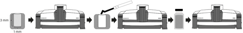
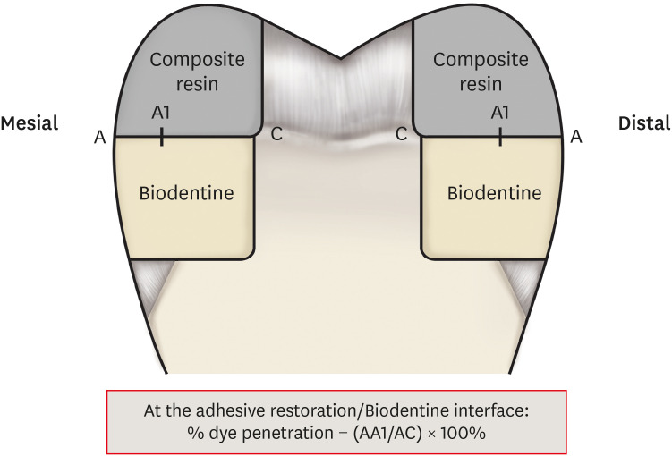
Materials and Methods Microleakage test: 160 standardized class II cavities were prepared on 80 extracted human molars. The cavities were filled with Biodentine and then divided into 2 experimental groups according to the time of restoration: composite resin obturation 15 minutes after Biodentine handling (D0); restoration after 7 days (D7). Each group was then divided into 8 subgroups (
n = 5) according to the adhesive system used: etch-and-rinse adhesive (Prime & Bond); self-etch adhesive 2 steps (Optibond XTR and Clearfil SE Bond); self-etch adhesive 1 step (Xeno III, G-aenial Bond, and Clearfil Tri-S Bond); and universal used as etch-and-rinse or self-etch (ScotchBond Universal ER or SE). After thermocycling, the teeth were immersed in a silver nitrate solution, stained, longitudinally sectioned, and the Biodentine/adhesive percolation was quantified. Scanning electron microscopic observations: Biodentine/adhesive interfaces were observed.Results A tendency towards less microleakage was observed when Biodentine was etched (2.47%) and when restorations were done without delay (D0: 4.31%, D7: 6.78%), but this was not significant. The adhesives containing 10-methacryloyloxydecyl dihydrogen phosphate monomer showed the most stable results at both times studied. All Biodentine/adhesive interfaces were homogeneous and regular.
Conclusions The good sealing of the CSC/adhesive interface is not a function of the system adhesive family used or the cement maturation before restoration. Biodentine can be used as a dentine substitute.
-
Citations
Citations to this article as recorded by- Comparison of compressive strength, surface microhardness, and surface microstructure of different types of bioceramics following varying surface treatments
Zeynep Hale Keleş, Vasfiye Işık, Soner Sismanoglu
Journal of the Australian Ceramic Society.2026; 62(2): 949. CrossRef - Temporal effects of restorative pretreatments on biodentine: an in-vitro study
Narendrakumar Akshaya, Hrudi Sundar Sahoo, Kurinji Amalavathy Ratnakaran, Adisree Ravichandran
Biomaterial Investigations in Dentistry.2026; 13: 109. CrossRef - In vitro evaluation of microleakage in a tricalcium silicate-based cement matured for 12 min or 24 h and covered with glass ionomer cement or resin composite
Wichida Chaweewannakorn, Chanoknan Karuna, Nightingale Kasemsook, Woramet Kittithummaroj, Narinee Chinajitphan, Nirada Dhanesuan, Kwanchanok Youcharoen
Journal of Oral Science.2026; 68(2): 93. CrossRef - Faster-setting calcium silicate cements–composite bonding: effect of adhesive application mode
Zeliha Gonca Bek Kürklü, Ezgi Sonkaya
BMC Oral Health.2026;[Epub] CrossRef - Effect of Er Cr YSGG laser etching procedure on the bond strength of different calcium silicate cements
Yesim Sesen Uslu, Hakan Yasin Gönder, Pinar Sesen, Gizem Gunduz Bektaş
Lasers in Dental Science.2024;[Epub] CrossRef - Managing Cracked Teeth with Root Extension: A Prospective Preliminary Study Using Biodentine™ Material
Kênia Maria Soares de Toubes, Isabella Sousa Corrêa, Regina Célia Lopes Valadares, Stephanie Quadros Tonelli, Fábio Fernandes Borém Bruzinga, Frank Ferreira Silveira, Dr Karthikeyan Ramalingam
International Journal of Dentistry.2024;[Epub] CrossRef - In Vitro Resistance of Natural Molars vs. Additive-Manufactured Simulators Treated with Pulpotomy and Endocrown
Marie-Laure Munoz-Sanchez, Alexis Gravier, Olivier Francois, Emmanuel Nicolas, Martine Hennequin, Nicolas Decerle
Journal of Functional Biomaterials.2023; 14(9): 444. CrossRef - Characterisation of the calcium silicate‐based cement–composite interface and the bonding strength with total‐etch or single/two‐stage self‐etch adhesive systems
Abidin Talha Mutluay, Merve Mutluay
Australian Endodontic Journal.2022; 48(3): 501. CrossRef - Bond Strength of Adhesive Systems to Calcium Silicate-Based Materials: A Systematic Review and Meta-Analysis of In Vitro Studies
Louis Hardan, Davide Mancino, Rim Bourgi, Alejandra Alvarado-Orozco, Laura Emma Rodríguez-Vilchis, Abigailt Flores-Ledesma, Carlos Enrique Cuevas-Suárez, Monika Lukomska-Szymanska, Ammar Eid, Maya-Line Danhache, Maryline Minoux, Youssef Haïkel, Naji Kharo
Gels.2022; 8(5): 311. CrossRef
- Comparison of compressive strength, surface microhardness, and surface microstructure of different types of bioceramics following varying surface treatments
- 3,824 View
- 67 Download
- 9 Web of Science
- 9 Crossref
- Biocompatibility and bioactive potential of the NeoMTA Plus endodontic bioceramic-based sealer
- Roberto Alameda Hoshino, Mateus Machado Delfino, Guilherme Ferreira da Silva, Juliane Maria Guerreiro-Tanomaru, Mário Tanomaru-Filho, Estela Sasso-Cerri, Paulo Sérgio Cerri
- Restor Dent Endod 2021;46(1):e4. Published online December 17, 2020
- DOI: https://doi.org/10.5395/rde.2021.46.e4
-
Abstract
PDFPubReaderePub
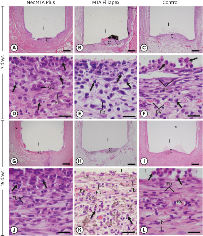
Objectives This study evaluated the biocompatibility and bioactive potential of NeoMTA Plus mixed as a root canal sealer in comparison with MTA Fillapex.
Materials and Methods Polyethylene tubes filled with NeoMTA Plus (
n = 20), MTA Fillapex (n = 20), or nothing (control group, CG;n = 20) were inserted into the connective tissue in the dorsal subcutaneous layer of rats. After 7, 15, 30 and 60 days, the specimens were processed for paraffin embedding. The capsule thickness, collagen content, and number of inflammatory cells (ICs) and interleukin-6 (IL-6) immunolabeled cells were measured. von Kossa-positive structures were evaluated and unstained sections were analyzed under polarized light. Two-way analysis of variance was performed, followed by thepost hoc Tukey test (p ≤ 0.05).Results At 7 days, the capsules around NeoMTA Plus and MTA Fillapex had more ICs and IL-6-immunostained cells than the CG. However, at 60 days, there was no significant difference in the IC number between NeoMTA Plus and the CG (
p = 0.1137) or the MTA Fillapex group (p = 0.4062), although a greater number of IL-6-immunostained cells was observed in the MTA Fillapex group (p = 0.0353). From 7 to 60 days, the capsule thickness of the NeoMTA Plus and MTA Fillapex specimens significantly decreased, concomitantly with an increase in the collagen content. The capsules around root canal sealers showed positivity to the von Kossa stain and birefringent structures.Conclusions The NeoMTA Plus root canal sealer is biocompatible and exhibits bioactive potential.
-
Citations
Citations to this article as recorded by- Retrievability of NeoMTA 2 vs AH Plus Sealer from Retreated Mesial Canals of Mandibular First Molars: A Microcomputed Tomography Ex Vivo Study
Mey A Al-Habib, Mona Alsulaiman
The Journal of Contemporary Dental Practice.2025; 26(5): 493. CrossRef - Effect of calcium silicate-based repair sealers on bone healing in rat skull defects: histological and histomorphometric study
J. M. Sauer, C. E. S. Bueno, R. A. Pelegrine, C. E. Fontana, E. F. Martinez, P. G. Montagner, W. M. Nascimento, A. G. S. Limoeiro, D. G. P. Rocha, M. F. V. Marceliano-Alves, M. P. W. Galhardi, M. Klymus, A. S. Martin
Endodontics Today.2025; 23(3): 433. CrossRef - Biocompatibility and bioactivity of bioceramic endodontic sealer: NeoSealer Flo
Evelin Carine Alves SILVA, Jéssica Arielli PRADELLI, Guilherme Ferreira da SILVA, Paulo Sérgio CERRI, Mario TANOMARU-FILHO, Juliane Maria GUERREIRO-TANOMARU
Journal of Applied Oral Science.2025;[Epub] CrossRef - The osteoinductive potential of different root-end filling materials in a rat femur model
Seçkin Aksu, Ebru Delikan, Ayşe Özcan Küçük, Zehra Demiray Asoğlu, Şakir Necat Yılmaz
Scientific Reports.2024;[Epub] CrossRef - Clinical outcomes of nonsurgical root canal treatment using NeoSealer Flo and Endosequence BC Sealer: A retrospective analysis with short-term follow-up
Christian Lepure, Ryan M. Walsh, Sayeed Attar, Casey L. Turner, Joshua Crawford, Poorya Jalali
Clinical Oral Investigations.2024;[Epub] CrossRef - Biocompatibility and bioactive potential of NeoPUTTY calcium silicate‐based cement: An in vivo study in rats
Evelin Carine Alves Silva, Jéssica Arielli Pradelli, Guilherme Ferreira da Silva, Paulo Sérgio Cerri, Mario Tanomaru‐Filho, Juliane Maria Guerreiro‐Tanomaru
International Endodontic Journal.2024; 57(6): 713. CrossRef - Carbon Nanotubes Induce Mineralization of Human Cementoblasts
Ting-Hsuan Wang, Kiyoko Watanabe, Koichiro Muromachi, Nobushiro Hamada, Nobuyuki Tani-Ishii
Journal of Endodontics.2024; 50(8): 1117. CrossRef - Tissue repair capacity of bioceramic endodontic sealers in rat subcutaneous tissue
George Sampaio Bonates dos Santos, Ceci Nunes Carvalho, Rudys Rodolfo de Jesus Tavares, Paulo Goberlânio de Barros Silva, George Táccio de Miranda Candeiro, Etevaldo Matos Maia Filho
Brazilian Dental Journal.2023; 34(3): 25. CrossRef - Participation of fibroblast growth factor‐1 and interleukin‐10 in connective tissue repair following subcutaneous implantation of bioceramic materials in rats
Mateus Machado Delfino, José Leandro de Abreu Jampani, Camila Soares Lopes, Juliane Maria Guerreiro‐Tanomaru, Mário Tanomaru‐Filho, Estela Sasso‐Cerri, Paulo Sérgio Cerri
International Endodontic Journal.2023; 56(3): 385. CrossRef - Biocompatibility and bioactive potential of an experimental tricalcium silicate‐based cement in comparison with Bio‐C repair and MTA Repair HP materials
Marcela Borsatto Queiroz, Rafaela N. H. Inada, José Leandro de Abreu Jampani, Juliane Maria Guerreiro‐Tanomaru, Estela Sasso‐Cerri, Mário Tanomaru‐Filho, Paulo Sérgio Cerri
International Endodontic Journal.2023; 56(2): 259. CrossRef - Calcium Silicate-Based Sealer Dentinal Tubule Penetration—A Systematic Review of In Vitro Studies
Israa Ashkar, José Luis Sanz, Leopoldo Forner, María Melo
Materials.2023; 16(7): 2734. CrossRef - Bioactivity Potential of Bioceramic-Based Root Canal Sealers: A Scoping Review
Mauro Schmitz Estivalet, Lucas Peixoto de Araújo, Felipe Immich, Adriana Fernandes da Silva, Nadia de Souza Ferreira, Wellington Luiz de Oliveira da Rosa, Evandro Piva
Life.2022; 12(11): 1853. CrossRef - Tricalcium silicate cement sealers
Anita Aminoshariae, Carolyn Primus, James C. Kulild
The Journal of the American Dental Association.2022; 153(8): 750. CrossRef - Bioactive potential of Bio‐C Pulpo is evidenced by presence of birefringent calcite and osteocalcin immunoexpression in the rat subcutaneous tissue
Marcela Borsatto Queiroz, Rafaela Nanami Handa Inada, Camila Soares Lopes, Juliane Maria Guerreiro‐Tanomaru, Estela Sasso‐Cerri, Mário Tanomaru‐Filho, Paulo Sérgio Cerri
Journal of Biomedical Materials Research Part B: Applied Biomaterials.2022; 110(10): 2369. CrossRef - An Updated Review on Properties and Indications of Calcium Silicate‐Based Cements in Endodontic Therapy
Fateme Eskandari, Alireza Razavian, Rozhina Hamidi, Khadije Yousefi, Susan Borzou, Zohaib Khurshid
International Journal of Dentistry.2022;[Epub] CrossRef
- Retrievability of NeoMTA 2 vs AH Plus Sealer from Retreated Mesial Canals of Mandibular First Molars: A Microcomputed Tomography Ex Vivo Study
- 3,613 View
- 49 Download
- 11 Web of Science
- 15 Crossref
- Impact of root canal curvature and instrument type on the amount of extruded debris during retreatment
- Burcu Serefoglu, Gözde Kandemir Demirci, Seniha Miçooğulları Kurt, İlknur Kaşıkçı Bilgi, Mehmet Kemal Çalışkan
- Restor Dent Endod 2021;46(1):e5. Published online December 17, 2020
- DOI: https://doi.org/10.5395/rde.2021.46.e5
-
Abstract
PDFPubReaderePub
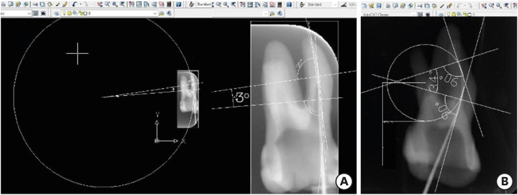
Objectives The aim of the current study was to assess whether the amount of extruded debris differs for straight and severely curved root canals during retreatment using H-files, R-Endo, Reciproc and ProTaper Universal Retreatment (PTU-R) files. Additionally, the area of residual filling material was evaluated.
Materials and Methods Severely curved (
n = 104) and straight (n = 104) root canals of maxillary molar teeth were prepared with WaveOne Primary file and obturated with gutta-percha and AH Plus sealer. Root canal filling materials were removed with one of the preparation techniques: group 1: H-file; group 2: R-Endo; group 3: Reciproc; group 4: PTU-R (n = 26). The amount of extruded material and the area of the residual filling material was measured. The data were analyzed with 2-way analysis of variance (ANOVA) and 1-way ANOVA at the 0.05 significance level.Results Except for Reciproc group (
p > 0.05), PTU-R, R-Endo, and H-file systems extruded significantly more debris in severely curved canals (p < 0.05). Each file system caused more residual filling material in severely curved canals than in straight ones (p < 0.05).Conclusions All instruments used in this study caused apical debris extrusion. Root canal curvature had an effect on extruded debris, except for Reciproc system. Clinicians should be aware that the difficult morphology of the severely curved root canals is a factor increasing the amount of extruded debris during the retreatment procedure.
-
Citations
Citations to this article as recorded by- Evaluation of the Effectiveness of MicroMega Remover, ProTaper Universal Retreatment, Reciproc, and Hedstrom Files in the Retreatment of Curved Root Canals Obturated with Different Techniques: A Micro-Computed Tomography Study
Pınar Hava Dursun, Fatma Semra Sevimay, Arda Buyuksungur, Berkan Celikten
Medicina.2026; 62(1): 188. CrossRef - Remaining Root Filling Material in Oval Canals After Retreatment Using MicroMega Remover and Reciproc Blue Systems with and Without Passive Ultrasonic Irrigation: A Micro-CT Study
Furkan Konus, Faruk Oztekin
Journal of Clinical Medicine.2026; 15(12): 4822. CrossRef - Comparative Analysis of Root Canal Curvature Measurement Methods for Permanent Mandibular Molars Distal Root: An Observational Study
Tanu Singh, Saurav Bathla, Anuraag Gurtu, Shubhi Gupta, Sana Saifi, Madhusudan Astekar
The Journal of Contemporary Dental Practice.2025; 26(10): 945. CrossRef - Do Continuous Rotating Endodontic Instruments Extrude Fewer Apical Debris Than Reciprocating Instruments in Non-Surgical Endodontic Retreatments? A Systematic Review
Francesco Puleio, Francesco Giordano, Ugo Bellezza, David Rizzo, Valentina Coppini, Roberto Lo Giudice
Applied Sciences.2024; 14(4): 1621. CrossRef - Intracanal removal and apical extrusion of filling material after retreatment using rotary or reciprocating instruments: A new approach using human cadavers
Thamyres M. Monteiro, Victor O. Cortes‐Cid, Marilia F. V. Marceliano‐Alves, Andrea F. Campello, Luan F. Bastos, Ricardo T. Lopes, José F. Siqueira, Flávio R. F. Alves
International Endodontic Journal.2024; 57(1): 100. CrossRef - Comparative analysis of methods for measuring root canal curvature based on periapical radiography: A laboratory study
Rafael Chies Hartmann, Eduardo Silva Ferraz, Theodoro Weissheimer, Jose Antônio Poli de Figueiredo, Giampiero Rossi‐Fedele, Maximiliano Schünke Gomes
International Endodontic Journal.2024; 57(12): 1848. CrossRef - Evaluation of apically extruded debris during root canal filling material removal in teeth with external apical root resorption: a comparison of different obturation techniques
Büşra Melike Çağlar, İsmail Uzun
BMC Oral Health.2024;[Epub] CrossRef - Evaluation of apically extruded debris using protaper universal, protaper next, one curve, Xp shaper, and edge file: An in vitro study
Murtada Qadir Muhaibes, Shatha Abdulkareem Alwakeel
Saudi Endodontic Journal.2024; 14(1): 31. CrossRef - A quantitative comparison of apically extruded debris during root canal preparation using NiTi full-sequence rotary and single-file rotary systems: An in vitro study
Pallavi Goel, R. Vikram, R. Anithakumari, M. S. Adarsha, M. E. Sudhanva
Endodontology.2024; 36(3): 235. CrossRef - In vitro evaluation of filling material removal and apical debris extrusion after retreatment using Reciproc blue, Hyflex EDM and ProTaper retreatment files
Passent Abdelnaby, Mohamed Ibrahim, Rania ElBackly
BMC Oral Health.2023;[Epub] CrossRef - A Comparative Study on the Shaping Ability and Cleaning Efficiency of Two Different Single-File Systems, Reciprocating Wave One Versus Continuous Rotation F360, Evaluated by Scanning Electron Microscope: An In Vitro Study
Arunkumar Samudrala, Chandrakanth Majeti, Kommineni Harika Chowdary, Lakshmi Bhavani Potru, Anusha Yaragani, Yata Prashanth Kumar, Gagandeep K Sidhu, Navneet S Kathuria
Cureus.2023;[Epub] CrossRef - COMPARATIVE EVALUATION OF THE EFFECT OF DIFFERENT ROTARY INSTRUMENT SYSTEMS ON THE AMOUNT OF APICALLY EXTRUDED DEBRIS
Recai ZAN, Bilge LENGER
Cumhuriyet Dental Journal.2022; 25(2): 172. CrossRef - A critical analysis of research methods and experimental models to study apical extrusion of debris and irrigants
Jale Tanalp
International Endodontic Journal.2022; 55(S1): 153. CrossRef - Critical analysis of research methods and experimental models to study removal of root filling materials
Mahdi A. Ajina, Pratik K. Shah, Bun San Chong
International Endodontic Journal.2022; 55(S1): 119. CrossRef
- Evaluation of the Effectiveness of MicroMega Remover, ProTaper Universal Retreatment, Reciproc, and Hedstrom Files in the Retreatment of Curved Root Canals Obturated with Different Techniques: A Micro-Computed Tomography Study
- 3,497 View
- 39 Download
- 10 Web of Science
- 14 Crossref
- Corrosion resistance assessment of nickel-titanium endodontic files with and without heat treatment
- Tatiana Dias Costa, Elison da Fonseca e Silva, Paula Liparini Caetano, Marcio José da Silva Campos, Leandro Marques Resende, André Guimarães Machado, Antônio Márcio Resende do Carmo
- Restor Dent Endod 2021;46(1):e6. Published online December 28, 2020
- DOI: https://doi.org/10.5395/rde.2021.46.e6
-
Abstract
PDFPubReaderePub
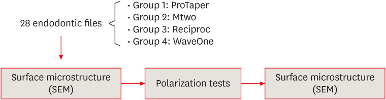
Objectives The aim of this study was to evaluate the corrosion resistance of heat-treated (Reciproc and WaveOne) and non-heat-treated (ProTaper and Mtwo) superelastic nickel-titanium endodontic files when immersed in a 5.25% sodium hypochlorite solution.
Materials and Methods Anodic polarization curves were obtained with potential sweeps that began at the open circuit potential or corrosion potential (Ecorr). The pitting potential (Epit) was identified on the anodic polarization curve as the potential at which a sudden increase in current was observed. The micromorphology of the 28 tested files was analyzed before and after the electrochemical assay using scanning electron microscope (SEM). The data were analyzed using 1-way analysis of variance with the
post hoc Bonferroni test (for Ecorr) and the Studentt -test for independent samples (for Epit).Results The mean Ecorr values were 0.506 V for ProTaper, 0.348 V for Mtwo, 0.542 V for Reciproc, and 0.321 V for WaveOne files. Only WaveOne and Protaper files exhibited pitting corrosion, with Epit values of 0.879 V and 0.904 V, respectively. On the SEM images of the ProTaper and WaveOne files, cavities suggestive of pitting corrosion were detected.
Conclusions Signs of corrosion were observed in both heat-treated and non-heat-treated files. Of the evaluated files, WaveOne (a heat-treated file) and ProTaper (a non-heat-treated file) exhibited the lowest corrosion resistance.
-
Citations
Citations to this article as recorded by- A novel approach to assess surface roughness and EDX profiling of blue rotary NiTi files following dynamic immersion in various hypochlorite concentrations
Hebatullah Ahmad Safwat, Nesreen Y. Mohammed, Asmaa Abd El-Hady
BMC Oral Health.2025;[Epub] CrossRef - Effect of phytic acid on chemical, structural, and mechanical characteristics of nickel–titanium endodontic files
Mai Samara, Mohannad Nassar, Abdullah Alqedairi, Hussam Alfawaz, Ahmed Jamleh
Scientific Reports.2024;[Epub] CrossRef - Investigating the Tribocorrosion Behaviour of NiTiNOL60 Alloy in Engineering and Biomedical Applications—An Overview
Anthony O. Okoani, Ashveen Nand, Cho-Pei Jiang, Maziar Ramezani
Metals.2024; 14(12): 1334. CrossRef - Electrochemical Properties of Nickel-Titanium Rotary Endodontic Instruments
Vidyalakshmi Subramanian, Howard W. Roberts, Shengtong Han, Stephanie J. Sidow, David W. Berzins
Journal of Endodontics.2024; 50(8): 1143. CrossRef - Effects of sodium hypochlorite on corrosion of the rotary nickel-titanium endodontic instruments - SEM analysis
Milica Jovanovic-Medojevic, Jelena Neskovic, Marijana Popovic-Bajic, Djordje Stratimirovic, Slavoljub Zivkovic
Srpski arhiv za celokupno lekarstvo.2022; 150(5-6): 254. CrossRef - Economic analysis of the different endodontic instrumentation techniques used in the Unified Health System
Laura Paredes Merchan, Livia Fernandes Probst, Ana Clara Correa Duarte Simões, Augusto Cesar Santos Raimundo, Yuri Wanderley Cavalcanti, Denise de Fátima Barros Cavalcante, João Victor Frazão Câmara, Antonio Carlos Pereira
BMC Oral Health.2022;[Epub] CrossRef
- A novel approach to assess surface roughness and EDX profiling of blue rotary NiTi files following dynamic immersion in various hypochlorite concentrations
- 2,806 View
- 36 Download
- 4 Web of Science
- 6 Crossref
- Can silver diamine fluoride or silver nanoparticle-based anticaries agents to affect enamel bond strength?
- Jaqueline Costa Favaro, Yana Cosendey Toledo de Mello Peixoto, Omar Geha, Flaviana Alves Dias, Ricardo Danil Guiraldo, Murilo Baena Lopes, Sandrine Bittencourt Berger
- Restor Dent Endod 2021;46(1):e7. Published online January 12, 2021
- DOI: https://doi.org/10.5395/rde.2021.46.e7
-
Abstract
PDFPubReaderePub
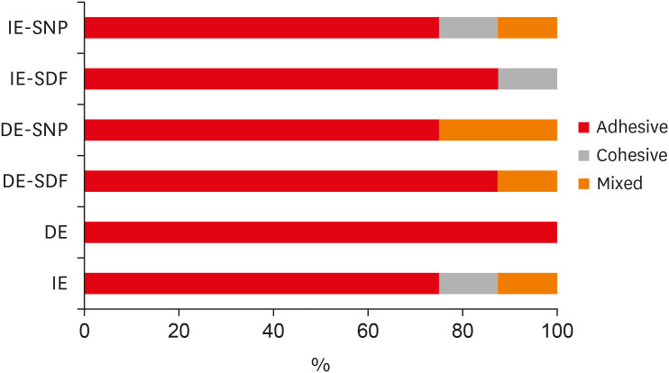
Objectives The aim of the current study is to investigate the effect of different anticaries agents, such as experimental agents based on silver nanoparticles (SNPs) and silver diamine fluoride (SDF), on the micro-shear bond strength (μ-SBS) of composite resin applied to intact enamel (IE) or demineralized enamel (DE).
Materials and Methods Sixty dental enamel fragments were collected from human third molars and categorized into 6 groups (
n = 10): positive control (IE), negative control (DE), IE + SDF, DE + SDF, IE + SNP and DE + SNP. Samples from DE, DE + SDF and DE + SNP groups were subjected to pH cycling; superficial microhardness test was performed to confirm demineralization. Resin composite build-ups were applied to the samples (0.75-mm diameter and 1-mm height) after the treatments (except for IE and DE groups); μ-SBS was also evaluated. Samples were analyzed under a stereomicroscope at 40× magnification to identify failure patterns. Data were subjected to one-way analysis of variance, followed by Tukey's and Dunnett's tests (p < 0.05).Results There was no significant difference among the IE, IE + SNP, DE + SDF, and DE + SNP groups. The IE + SDF and DE groups recorded the highest and the lowest μ-SBS values, respectively. Adhesive-type failures were the most frequent for all treatments.
Conclusions Anticaries agents did not have a negative effect on the μ-SBS of composite resin when it was used on IE or DE.
-
Citations
Citations to this article as recorded by- Impact of Incorporating Nanoparticles to Adhesive Resin on the Demineralization of Enamel: A Systematic Review
Naif Almosa
Dentistry Journal.2025; 13(3): 89. CrossRef - Preventing white spot lesions around orthodontic brackets: efficacy of pre-reacted glass-ionomer barrier coat versus silver diamine fluoride: an in vitro study
Enas A. Elshenawy, Safa B. Alawy, Wafaa Yahia Alghonemy, Ahmed Ibrahime El dosoky
BDJ Open.2025;[Epub] CrossRef - Research Status of Silver Nanoparticles for Dental Applications
Yanyan Guo, Xiaomei Hou, Sanjun Fan, Chanyuan Jin
Inorganics.2025; 13(5): 168. CrossRef - The use of silver diamine fluoride to prevent/treat enamel carious lesions: a narrative review
Rasha N. AlSheikh
PeerJ.2024; 12: e17897. CrossRef - Phosphoric Acid Etch Partially Restores the Initial Bond Strength of Composite to Silver Diamine Fluoride–Treated Enamel Using Universal Adhesives
Zaher Jabbour, Mijoo Kim, Marc Hayashi, Reuben Kim
Dentistry Journal.2023; 11(7): 161. CrossRef - Efficacy of Nano Silver Fluoride and/or Diode Laser In Enhancing Enamel Anticariogenicity around orthodontic brackets
Aya Anwar Alsherif, Mohamed Ali Farag, Mai Badreldin Helal
BDJ Open.2023;[Epub] CrossRef - Amelioration Strategies for Silver Diamine Fluoride: Moving from Black to White
Amjad Almuqrin, Inder Preet Kaur, Laurence J. Walsh, Chaminda Jayampath Seneviratne, Sobia Zafar
Antibiotics.2023; 12(2): 298. CrossRef - The Effect of Loading Time on Color Stability of Various Restorative Materials Bonded to Silver Diamine Fluoride-Treated Demineralized Dentin
Mohammed M Aldosari, Fares S Al-Sehaibany
Clinical, Cosmetic and Investigational Dentistry.2022; Volume 14: 123. CrossRef - In vitro study of the effect of nanosilver fluoride on shear bond strength of orthodontic brackets and demineralization of enamel
Mariam H. El-Toukhy, Eman M. El-Shourbagy, Neveen M. Fakhry
Tanta Dental Journal.2022; 19(4): 281. CrossRef
- Impact of Incorporating Nanoparticles to Adhesive Resin on the Demineralization of Enamel: A Systematic Review
- 2,858 View
- 35 Download
- 10 Web of Science
- 9 Crossref
- YouTube as an information source for instrument separation in root canal treatment
- Yağız Özbay, Neslihan Yılmaz Çırakoğlu
- Restor Dent Endod 2021;46(1):e8. Published online January 12, 2021
- DOI: https://doi.org/10.5395/rde.2021.46.e8
-
Abstract
PDFPubReaderePub
Objectives The reliability and educational quality of videos on YouTube for patients seeking information regarding instrument separation in root canal treatment were evaluated.
Materials and Methods YouTube was searched for videos on instrument separation in root canal treatment. Video content was scored based on reliability in terms of 3 categories (etiology, procedure, and prognosis) and based on video flow, quality, and educational usefulness using the Global Quality Score (GQS). Descriptive statistics were obtained and the data were analyzed using analysis of variance and the Kruskal-Wallis test.
Results The highest mean completeness scores were obtained for videos published by dentists or specialists (1.48 ± 1.06). There was no statistically significant difference among sources of upload in terms of content completeness. The highest mean GQS was found for videos published by dentists or specialists (1.82 ± 0.96), although there was no statistically significant correlation between GQS and the source of upload.
Conclusions Videos on YouTube have incomplete and low-quality content for patients who are concerned about instrument separation during endodontic treatment, or who experience this complication during endodontic treatment.
-
Citations
Citations to this article as recorded by- YouTube As an Information Source for Bioceramic‐Based Root Canal Sealers in Endodontics: A Cross‐Sectional Study
Sevda Durust Baris, Seyma Atli, Rumeysa Betul Gumus
Australian Endodontic Journal.2026;[Epub] CrossRef - YouTube as a Source of Information on Composite Veneer
Merve Arslan, Zeyneb Merve Ozdemır
Kahramanmaraş Sütçü İmam Üniversitesi Tıp Fakültesi Dergisi.2026; 21(2): 91. CrossRef - Quality of information in #brokenfileremoval Reels videos on Instagram: a cross-sectional study
Dilek Hancerliogullari, Eray Ceylanoglu
Journal of Public Health.2025; 33(8): 1617. CrossRef - Evaluation of the Educational Value, Reliability, and Quality of Vaginally-Assisted Natural Orifice Transluminal Endoscopic Surgery (v-NOTES) Videos on YouTube
Isa Temur
Cureus.2025;[Epub] CrossRef - Evaluating the Quality and Reliability of YouTube Videos Providing Nutritional Recommendations for Irritable Bowel Syndrome
Eda Başmısırlı, Merve Kip, Hande Altun, Neriman İnanç
Journal of Human Nutrition and Dietetics.2025;[Epub] CrossRef - Comparison of YouTube, TikTok, and Instagram as digital sources for obtaining information about pulp therapy in primary and permanent teeth
Hüseyin Gürkan Güneç, Emine Kaya, Dila Nur Okumuş, Merve Gül Erence
Restorative Dentistry & Endodontics.2025; 50(3): e26. CrossRef - Is YouTube a reliable source for learning pre-endodontic build-up? A cross-sectional study
Merve Gökyar, İdil Özden, Hesna Sazak Öveçoğlu
Restorative Dentistry & Endodontics.2025; 50(3): e27. CrossRef - Youtube As Sources of Information About Fiber-Reinforcement Composite Resin Bridge: Accuracy and Reliability Assessment
Merve Arslan, Zeyneb Merve Ozdemır
Akdeniz Diş Hekimliği Dergisi.2025; 4(2): 89. CrossRef - Evaluation of the Quality and Reliability of YouTubeTM Videos Created by Orthodontists as an Information Source for Clear Aligners
Emre Cesur, Koray Tuncer, Duygu Sevgi, Barkın Cem Balaban, Can Arslan
Turkish Journal of Orthodontics.2024; 37(1): 44. CrossRef - Is it safe to learn about vital pulp capping from YouTube™ videos? A content and quality analysis
Celalettin Topbaş, Tuğçe Paksoy, Ayşe Gülnihal İslamoğlu, Kemal Çağlar, Abdurrahman Kerim Kul
International Journal of Medical Informatics.2024; 185: 105409. CrossRef - Quality of Patient-Centered eHealth Information on Erosive Tooth Wear: Systematic Search and Evaluation of Websites and YouTube Videos
Lena Holland, Amelie Friederike Kanzow, Annette Wiegand, Philipp Kanzow
Journal of Medical Internet Research.2024; 26: e49514. CrossRef - Evaluation of YouTubeTM as an Information Source for Indirect Restorations: Cross-Sectional Evaluation
Işıl Doğruer, Merve Kütük Ömeroğlu
European Annals of Dental Sciences.2024; 51(3): 102. CrossRef - Evaluating YouTube as a Patient Information Source for the Risks of Root Canal Treatment
Stewart McLean, Neil Cook, Alexander Rovira-Wilde, Shanon Patel, Shalini Kanagasingam
Journal of Endodontics.2023; 49(2): 155. CrossRef - Evaluation of the quality of YouTube™ videos about pit and fissure sealant applications
Ayse Tugba Erturk‐Avunduk, Ebru Delikan
International Journal of Dental Hygiene.2023; 21(3): 590. CrossRef - Avülsiyon Yaralanmalarının Acil Müdahalesinde Hasta Bilgi Kaynağı Olarak Türkçe YouTube™ Videolarının Güvenilirliği: Kesitsel İçerik Analizi
Gülçin CAGAY SEVENCAN, Zeynep Şeyda YAVŞAN
Selcuk Dental Journal.2023; 10(3): 583. CrossRef - Analyzing Content and Quality of YouTube™ Videos on Removal of Amalgam Fillings
Mehmet BULDUR, Fatma AYTAÇ BAL
Clinical and Experimental Health Sciences.2022; 12(2): 423. CrossRef - Assessment of reliability and information quality of YouTube videos about root canal treatment after 2016
Myoung-jun Jung, Min-Seock Seo
BMC Oral Health.2022;[Epub] CrossRef - Are YouTube Videos Reliable Sources of Information About Devital Bleaching?
Gülbahar ERDİNÇ, Yağız ÖZBAY, Neslihan YILMAZ ÇIRAKOĞLU
Mersin Üniversitesi Tıp Fakültesi Lokman Hekim Tıp Tarihi ve Folklorik Tıp Dergisi.2022; 12(3): 637. CrossRef - Assessment of the educational value of endodontic access cavity preparation YouTube video as a learning resource for students
Ahmed Jamleh, Shouq Mohammed Aljohani, Faisal Fahad Alzamil, Shahad Muhammad Aljuhayyim, Modhi Nasser Alsubaei, Showq Raad Alali, Nawaf Munawir Alotaibi, Mohannad Nassar, MariKannan Maharajan
PLOS ONE.2022; 17(8): e0272765. CrossRef - Evaluation of YouTube videos for patients’ education on periradicular surgery
Ahmed Jamleh, Mohannad Nassar, Hamad Alissa, Abdulmohsen Alfadley, Tanay Chaubal
PLOS ONE.2021; 16(12): e0261309. CrossRef
- YouTube As an Information Source for Bioceramic‐Based Root Canal Sealers in Endodontics: A Cross‐Sectional Study
- 3,569 View
- 26 Download
- 15 Web of Science
- 20 Crossref
- Efficacy of buccal piroxicam infiltration and inferior alveolar nerve block in patients with irreversible pulpitis: a prospective, double-blind, randomized clinical trial
- Saurav Paul, Sridevi Nandamuri, Aakrati Raina, Mukta Bansal
- Restor Dent Endod 2021;46(1):e9. Published online January 26, 2021
- DOI: https://doi.org/10.5395/rde.2021.46.e9
-
Abstract
PDFPubReaderePub
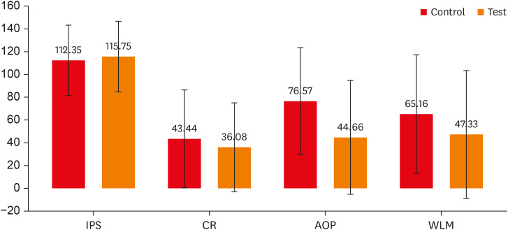
Objectives This randomized clinical trial aimed to assess the effectiveness of buccal infiltration with piroxicam on the anesthetic efficacy of inferior alveolar nerve block (IANB) with buccal infiltration in irreversible pulpitis, with pain assessed using the Heft-Parker visual analogue scale (HP-VAS).
Materials and Methods This study included 56 patients with irreversible pulpitis in mandibular molars, randomly distributed between 2 groups (
n = 28). After evaluating the initial pain score with the HP-VAS, each patient received IANB followed by buccal infiltration of 2% lignocaine with adrenaline (1:80,000). Five minutes later, the patients in groups 1 and 2 were given buccal infiltration with 40 mg/2 mL of piroxicam or normal saline, respectively. An access opening procedure (AOP) was performed 15 minutes post-IANB once the individual showed signs of lip numbness as well as 2 negative responses to electric pulp testing. The HP-VAS was used to grade the patient's pain during caries removal (CR), AOP, and working length measurement (WLM). Successful anesthesia was identified either by the absence of pain or slight pain through CR, AOP, and WLM, with no requirement of a further anesthetic dose. A statistical analysis was done using the Shapiro-Wilk and Mann-WhitneyU tests.Results The piroxicam group presented a significantly lower (
p < 0.05) mean pain score than the saline group during AOP.Conclusions Buccal infiltration with piroxicam enhanced the efficacy of anesthesia with IANB and buccal infiltration with lignocaine in patients with irreversible pulpitis.
-
Citations
Citations to this article as recorded by- Analyzing The Role of Fur-Friends in Mental Well-Being: Comparing Mental Well-Being of Pet Owners and Non-Owners
Jervin Dinglasan Quicho, Jesslaine Angela Sanchez, Alexandra Nicole P. Mendoza
International Journal of Multidisciplinary Applied Business and Education Research.2026; 7(3): 1053. CrossRef - Inferior alveolar nerve block success of 2% mepivacaine versus 4% articaine in patients with symptomatic irreversible pulpitis in mandibular molars: A randomized double‐blind single‐centre clinical trial
Mohammed Fawzy Omar Mohammed Habib, Sovana Tarek, Sara Mohamed Elsayed Teama, Khaled Ezzat, Randa Mohamed El Boghdadi, Abeer Marzouk, Manar Yehia Fouda, Shaimaa Ismail Gawdat, Marwa Mahmoud Bedier, Suzan Abdul Wanees Amin
International Endodontic Journal.2022; 55(11): 1177. CrossRef - Present status and future directions—Mechanisms and management of local anaesthetic failures
Masoud Parirokh, Paul V. Abbott
International Endodontic Journal.2022; 55(S4): 951. CrossRef
- Analyzing The Role of Fur-Friends in Mental Well-Being: Comparing Mental Well-Being of Pet Owners and Non-Owners
- 3,028 View
- 27 Download
- 2 Web of Science
- 3 Crossref
- Smear layer removal by passive ultrasonic irrigation and 2 new mechanical methods for activation of the chelating solution
- Ricardo Machado, Isadora da Silva, Daniel Comparin, Bianca Araujo Marques de Mattos, Luiz Rômulo Alberton, Ulisses Xavier da Silva Neto
- Restor Dent Endod 2021;46(1):e11. Published online January 26, 2021
- DOI: https://doi.org/10.5395/rde.2021.46.e11
-
Abstract
PDFPubReaderePub
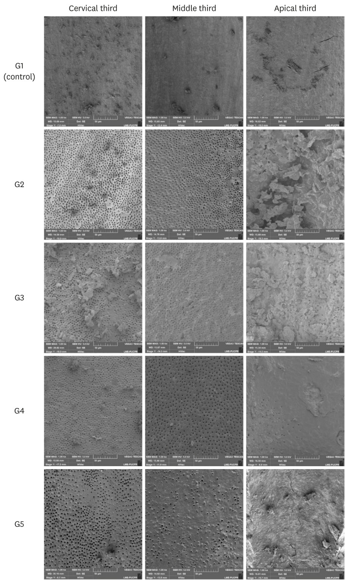
Objectives The aim of this study was to compare smear layer removal by conventional application (CA), passive ultrasonic irrigation (PUI), EasyClean (EC), and XP-Endo Finisher (XPF), using 17% ethylenediaminetetraacetic acid (EDTA) after chemomechanical preparation, as evaluated with scanning electron microscopy (SEM).
Materials and Methods Forty-five single-rooted human mandibular premolars were selected for this study. After chemomechanical preparation, the teeth were randomly divided into 5 groups according to the protocol for smear layer removal, as follows: G1 (control): CA of distilled water; G2 (CA): CA of 17% EDTA; G3 (PUI): 17% EDTA activated by PUI; G4 (EC): 17% EDTA activated by EC; and G5 (XPF): 17% EDTA activated by XPF. SEM images (×1,000) were obtained from each root third and scored by 3 examiners. Data were evaluated using the Kruskal-Wallis and Dunn tests (
p < 0.05).Results In the apical third, there were no statistically significant differences among the groups (
p > 0.05). In the cervical and middle thirds, the experimental groups performed better than the control group (p < 0.05); however, G2 presented better results than G3, G4, and G5 (p < 0.05), which showed no differences among one another (p > 0.05).Conclusions No irrigation method was able to completely remove the smear layer, especially in the apical third. Using CA for the chelating solution performed better than any form of activation.
-
Citations
Citations to this article as recorded by- Nanoscale Approaches to Oro-Dental Tissue Engineering: A Review of Strategies, Composites, and Translational Challenges
Pei Wang, Yingtong Ye, Keyi Mei, Biaoqi Chen, Ranjith Kankala, Fei Tong
International Journal of Nanomedicine.2026; Volume 21: 1. CrossRef - Comparative Evaluation of Smear Layer Removal Efficacy of Ethylenediaminetetraacetic Acid (EDTA) Gel Versus Solution With Sonic Activation: A Scanning Electron Microscopic Study
Gurkirat Singh, Rajinder Bansal, Manu Bansal, Jasvinder Kaur, Sangam Mittal, Nishant Sharma
Cureus.2026;[Epub] CrossRef - Smear layer removal comparing conventional irrigation, passive ultrasonic irrigation, EndoActivator System, and a new sonic device (Perfect Clean System) by scanning electron microscopy: An ex vivo study
Bruna Fernanda Alionço Gonçalves, Divya Reddy, Ricardo Machado, Paulo César Soares Júunior, Sérgio Aparecido Ignácio, Douglas Augusto Fernandes Couto, Karine Santos Frasquetti, Vânia Portela Ditzel Westphalen, Everdan Carneiro, Ulisses Xavier da Silva Net
PLOS ONE.2024; 19(12): e0314940. CrossRef - Impact of different agitation methods on smear layer cleaning of mesial canals with accentuated curvature
Abel Teves Cordova, Murilo Priori Alcalde, Michel Espinosa Klymus, Leonardo Rigoldi Bonjardim, Rodrigo Ricci Vivan, Marco Antonio Hungaro Duarte
Restorative Dentistry & Endodontics.2024;[Epub] CrossRef - Advances in hybridized nanoarchitectures for improved oro-dental health
Jun Guo, Pei Wang, Yuyao Li, Yifan Liu, Yingtong Ye, Yi Chen, Ranjith Kumar Kankala, Fei Tong
Journal of Nanobiotechnology.2024;[Epub] CrossRef - Cleaning and disinfection of the root canal system provided by four active supplementary irrigation methods
Alessandra Timponi Goes Cruz, Adriane Antoniw Klemz, Edvaldo Antônio Ribeiro Rosa, Fabiana Soares Grecca, Bianca Mattos, Lucila Piasecki, Ricardo Machado, Sérgio Aparecido Ignácio, Ulisses Xavier da Silva Neto
Scientific Reports.2024;[Epub] CrossRef - Scanning electron microscopic study of smear layer changes following ultrasonic endoactivator irrigation system during root canal treatment of primary teeth
Mohamed Ghaly, Aya Alsherif, Arafa Khatab
Tanta Dental Journal.2023; 20(2): 137. CrossRef - Influence of agitation methods of irrigants after methylene blue-mediated PDT on the bonding interface of a fiber post cementation system
Lucas David Galvani, Joatan Lucas de Sousa Gomes Costa, João Felipe Besegato, Joissi Ferrari Zaniboni, Wilfredo Gustavo Escalante-Otárola, Milton Carlos Kuga
Photodiagnosis and Photodynamic Therapy.2022; 37: 102708. CrossRef
- Nanoscale Approaches to Oro-Dental Tissue Engineering: A Review of Strategies, Composites, and Translational Challenges
- 4,088 View
- 44 Download
- 9 Web of Science
- 8 Crossref
- The effect of different confluence confirmation strategies on the obturation of Vertucci type II canal: micro-CT analysis
- Seungjae Do, Min-Seock Seo
- Restor Dent Endod 2021;46(1):e12. Published online January 26, 2021
- DOI: https://doi.org/10.5395/rde.2021.46.e12
-
Abstract
PDFPubReaderePub
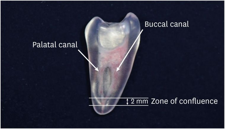
Objectives The present study aims to compare the obturation quality of 2 confluence confirmation techniques in artificial maxillary first premolars showing Vertucci type II root canal configuration.
Materials and Methods Thirty artificial maxillary premolars having Vertucci type II root canal configuration were made. They were divided into 3 groups according to the confluence confirmation technique as follows. Gutta-percha indentation (GPI) group (confluence confirmation using a gutta-percha cone and a K file); electronic apex locator (EAL) group (confluence confirmation using K files and EAL); and no confluence detection (NCD) group. In the GPI group and the EAL group, shaping and obturation were performed with the modified working length (WL). In the NCD group, shaping was performed without WL adjustment and obturation was carried out with an adjusted master cone. Micro-computed tomography was used before preparation and after obturation to calculate the percentage of gutta-percha occupied volume (%GPv) and the volume increase in the apical 4 mm. Data were analyzed using 1-way analysis of variance and
post hoc Tukey's test.Results Statistically significant difference was not found in terms of the %GPv from the apex to apical 4 mm. However, the NCD group showed a statistically significant volume increase compared with the EAL group (
p < 0.05).Conclusions In terms of gutta-percha occupied volume, no significant difference was observed among the 3 groups. Confluence confirmation using an EAL in teeth with Vertucci type II configuration showed less volume increase during canal shaping compared with no confluence confirmation.
-
Citations
Citations to this article as recorded by- ANALISE TOMOGRÁFICA DAS TERMINAÇÕES APICAIS DOS CONDUTOS MV1 e MV2 EM MOLARES SUPERIORES
Milena Matos Borges, Giulio César Moreira Manzi, Michel Sena Fernandes Faria Lima, Izabella Lucas de Abreu Lima, Diogo de Azevedo Miranda, Flávio Ricardo Manzi
ARACÊ .2026; 8(1): e11921. CrossRef - Root and root canal morphology of mandibular first and second molars in a Jordanian subpopulation: a cross-sectional cone-beam computed tomography study
Rawan Abu Zaghlan, Laith Abu Qdais, Farouq Mansour, Faisal Mansour, Faleh Sawair
Scientific Reports.2025;[Epub] CrossRef - Can the addition of surfactants to NaOCl irrigation impact on the percentage of voids of root canal filling?
Laise Pena Braga Monteiro, Marcella Yasmin Reis Guerreiro, Felipe Gonçalves Belladonna, Carolina Oliveira de Lima, Emmanuel João Nogueira Leal da Silva, Juliana Melo da Silva Brandão
Australian Endodontic Journal.2024; 50(2): 260. CrossRef
- ANALISE TOMOGRÁFICA DAS TERMINAÇÕES APICAIS DOS CONDUTOS MV1 e MV2 EM MOLARES SUPERIORES
- 3,147 View
- 71 Download
- 2 Web of Science
- 3 Crossref
- Efficacy of reciprocating and rotary retreatment nickel-titanium file systems for removing filling materials with a complementary cleaning method in oval canals
- Said Dhaimy, Hyeon-Cheol Kim, Lamyae Bedida, Imane Benkiran
- Restor Dent Endod 2021;46(1):e13. Published online February 3, 2021
- DOI: https://doi.org/10.5395/rde.2021.46.e13
-
Abstract
PDFPubReaderePub
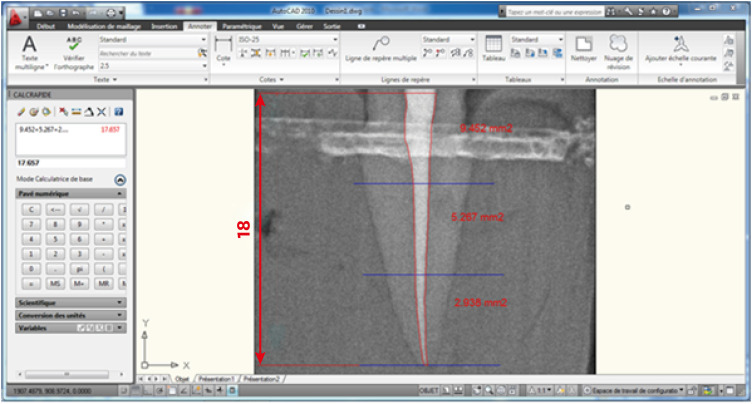
Objectives This study aimed to evaluate and compare the efficacy of the S1 reciprocating system and the D-Race retreatment rotary system for filling material removal and the apical extrusion of debris.
Materials and Methods Sixty-four freshly extracted maxillary canines were shaped with size 10 and size 15 K-files, instrumented using ProTaper Gold under irrigation with 2.5% sodium hypochlorite (NaOCl), obturated according to the principle of thermo-mechanical condensation with gutta-percha and zinc oxide eugenol sealer, and allowed to set for 3 weeks at 37°C. Subsequently, the teeth were divided into a control group (
n = 4), the D-Race rotary instrument group (n = 30), and the S1 reciprocating instrument group (n = 30). After classical retreatment, the canals were subjected to a complementary approach with the XP-Endo Shaper. Desocclusol was used as a solvent, and irrigation with 2.5% NaOCl was performed. Each group was divided into subgroups according to the timing of radiographic readings. The images were imported into a software program to measure the remaining filling material, the apical extrusion, and the root canal space. The data were statistically analyzed using the Z-test and JASP graphics software.Results No significant differences were found between the D-Race and S1 groups for primary retreatment; however, using a complementary cleaning method increased the removal of remnant filling (
p < 0.05).Conclusions Classical removal of canal filling material may not be sufficient for root canal disinfection, although a complementary finishing approach improved the results. Nevertheless, all systems left some debris and caused apical extrusion.
-
Citations
Citations to this article as recorded by- Effectiveness of different supplementary protocols for remaining filling material removal in endodontic reintervention: an integrative review
Amanda Freitas da Rosa, Bruna Venzke Fischer, Luiz Carlos de Lima Dias-Junior, Anna Victoria Costa Serique, Eduardo Antunes Bortoluzzi, Cleonice da Silveira Teixeira, Lucas da Fonseca Roberti Garcia
Odontology.2024; 112(1): 51. CrossRef - Critical analysis of research methods and experimental models to study removal of root filling materials
Mahdi A. Ajina, Pratik K. Shah, Bun San Chong
International Endodontic Journal.2022; 55(S1): 119. CrossRef - Economic analysis of the different endodontic instrumentation techniques used in the Unified Health System
Laura Paredes Merchan, Livia Fernandes Probst, Ana Clara Correa Duarte Simões, Augusto Cesar Santos Raimundo, Yuri Wanderley Cavalcanti, Denise de Fátima Barros Cavalcante, João Victor Frazão Câmara, Antonio Carlos Pereira
BMC Oral Health.2022;[Epub] CrossRef - Fabrication of a Potential Electrodeposited Nanocomposite for Dental Applications
Chun-Wei Chang, Chen-Han Tsou, Bai-Hung Huang, Kuo-Sheng Hung, Yung-Chieh Cho, Takashi Saito, Chi-Hsun Tsai, Chia-Chien Hsieh, Chung-Ming Liu, Wen-Chien Lan
Inorganics.2022; 10(10): 165. CrossRef - Influence of Filling Material Remnants on the Diffusion of Hydroxyl Ions in Endodontically Retreated Teeth: An Ex Vivo Study
Vania Portela Ditzel Westphalen, Marilisa Carneiro Leao Gabardo, Natanael Henrique Ribeiro Mattos, Camila Paiva Perin, Liliane Roskamp, Cristiano Miranda de Araújo, Luiz Fernando Fariniuk, Flares Baratto–Filho
The Journal of Contemporary Dental Practice.2022; 23(8): 768. CrossRef - Efficacy of Removing Thermafil and GuttaCore from Straight Root Canal Systems Using a Novel Non-Surgical Root Canal Re-Treatment System: A Micro-Computed Tomography Analysis
Vicente Faus-Llácer, Rubén Linero Pérez, Ignacio Faus-Matoses, Celia Ruiz-Sánchez, Álvaro Zubizarreta-Macho, Salvatore Sauro, Vicente Faus-Matoses
Journal of Clinical Medicine.2021; 10(6): 1266. CrossRef
- Effectiveness of different supplementary protocols for remaining filling material removal in endodontic reintervention: an integrative review
- 2,851 View
- 46 Download
- 7 Web of Science
- 6 Crossref
-
In vitro apical pressure created by 2 irrigation needles and a multisonic system in mandibular molars - Ronald Ordinola-Zapata, Joseph T. Crepps, Ana Arias, Fei Lin
- Restor Dent Endod 2021;46(1):e14. Published online February 8, 2021
- DOI: https://doi.org/10.5395/rde.2021.46.e14
-
Abstract
PDFPubReaderePub
Objectives The aim of this study was to evaluate the apical pressure generated by 2 endodontic irrigation needles and the GentleWave system in mandibular molars.
Materials and Methods The mesial and distal root canals of 12 mandibular molars were irrigated with a 30-gauge close-end needle or with a 30-gauge open-end needle. Procedures were performed in the mesial and distal canals. The GentleWave procedure and irrigation at 1 mm from the apex in the distal roots using an open-end needle were used, respectively, as negative and positive controls. The apical pressure was measured using a data acquisition pressure setup. Apical pressure exerted by the different needles in the 2 different canal types was statistically compared using 2-way analysis of variance.
Results Significant differences were found in the apical pressure for both needles and the canal type. The lowest values were obtained with close-end needles and in mesial canals. Negative apical pressure values were obtained using GentleWave.
Conclusions The needle and the canal type influenced the apical pressure. The GentleWave procedure produced negative apical pressure.
-
Citations
Citations to this article as recorded by- GentleWave versus Established Irrigation Techniques: Current Evidence from a Scoping Review
Mohmed Isaqali Karobari, Abdul Habeeb Adil, Niher Tabassum Snigdha, Emmanuel João Nogueira Leal da Silva
Journal of Endodontics.2026; 52(5): 713. CrossRef - Influence of kinematic motion and instrumentation strategy on apical debris extrusion during root canal preparation: An in vitro study
Amira Alghazaly, Jumanah Aljohani, Khadijah Mohabat, Rafah Ghous
Journal of Conservative Dentistry and Endodontics.2026; 29(7): 741. CrossRef - Use of the gentlewave system in endodonticsUse of the gentlewave system in endodontics
Daiana Jacobi Lazzarotto, Mayara Colpo Prado, Lara Dotto, Rafael Sarkis-Onofre
Brazilian Journal of Oral Sciences.2025; 24: e254250. CrossRef - Bibliometric analysis of the GentleWave system: trends, collaborations, and research gaps
Raimundo Sales de Oliveira Neto, Thais de Moraes Souza, João Vitor Oliveira de Amorim, Thaine Oliveira Lima, Guilherme Ferreira da Silva, Rodrigo Ricci Vivan, Murilo Priori Alcalde, Marco Antonio Hungaro Duarte
Restorative Dentistry & Endodontics.2025; 50(2): e17. CrossRef - The effect of ultrasonic and multisonic irrigation on root canal microbial communities: An ex vivo study
Ki Hong Park, Ronald Ordinola‐Zapata, W. Craig Noblett, Bruno P. Lima, Christopher Staley
International Endodontic Journal.2024; 57(7): 895. CrossRef - Efficacy of the GentleWave System in the removal of biofilm from the mesial roots of mandibular molars before and after minimal instrumentation: An ex vivo study
Kwang Ho Kim, Céline Lévesque, Gevik Malkhassian, Bettina Basrani
International Endodontic Journal.2024; 57(7): 922. CrossRef - A critical analysis of research methods and experimental models to study irrigants and irrigation systems
Christos Boutsioukis, Maria Teresa Arias‐Moliz, Luis E. Chávez de Paz
International Endodontic Journal.2022; 55(S2): 295. CrossRef - Outcomes of the GentleWave system on root canal treatment: a narrative review
Hernán Coaguila-Llerena, Eduarda Gaeta, Gisele Faria
Restorative Dentistry & Endodontics.2022;[Epub] CrossRef
- GentleWave versus Established Irrigation Techniques: Current Evidence from a Scoping Review
- 3,207 View
- 39 Download
- 7 Web of Science
- 8 Crossref
 First
First Prev
Prev

