Warning: mkdir(): Permission denied in /home/virtual/lib/view_data.php on line 81

Warning: fopen(upload/ip_log/ip_log_2024-12.txt): failed to open stream: No such file or directory in /home/virtual/lib/view_data.php on line 83

Warning: fwrite() expects parameter 1 to be resource, boolean given in /home/virtual/lib/view_data.php on line 84
Interface between calcium silicate cement and adhesive systems according to adhesive families and cement maturation
Skip Navigation
Skip to contents

Restor Dent Endod : Restorative Dentistry & Endodontics

OPEN ACCESS

Articles

Page Path
HOME > Restor Dent Endod > Volume 46(1); 2021 > Article
Research Article Interface between calcium silicate cement and adhesive systems according to adhesive families and cement maturation
Nelly Pradelle-Plasse1,2,3orcid, Caroline Mocquot1,2,3orcid, Katherine Semennikova1,2,3orcid, Pierre Colon1,2,3orcid, Brigitte Grosgogeat3,4,5orcid

DOI: https://doi.org/10.5395/rde.2021.46.e3
Published online: December 9, 2020

1Department of Conservative Dentistry - Endodontics, Faculty of Dental Surgery, University of Paris, Paris, France.

2Rothschild Hospital, Assistance Publique Hôpitaux de Paris, Paris, France.

3Multimaterials and Interfaces Laboratory (UMR 5615), Biomaterials Team, Villeurbanne, France.

4Department of Biomaterials, Faculty of Dental Surgery, University of Lyon 1, Lyon, France.

5Hospices Civils de Lyon, Lyon, France.

Correspondence to Nelly Pradelle-Plasse, DDS, MS, PhD. Associate Professor, Department of Conservative Dentistry - Endodontics, Faculty of Dental Surgery, University of Paris, 5 Rue Garancière, Paris 75006, France. nelly.pradelle@univ-paris-diderot.fr
• Received: May 23, 2020   • Revised: June 25, 2020   • Accepted: June 29, 2020

Copyright © 2021. The Korean Academy of Conservative Dentistry

This is an Open Access article distributed under the terms of the Creative Commons Attribution Non-Commercial License (https://creativecommons.org/licenses/by-nc/4.0/) which permits unrestricted non-commercial use, distribution, and reproduction in any medium, provided the original work is properly cited.

  • 30 Views
  • 1 Download
  • 5 Web of Science
prev next
  • Objectives
    This study aimed to evaluate the interface between a calcium silicate cement (CSC), Biodentine and dental adhesives in terms of sealing ability.
  • Materials and Methods
    Microleakage test: 160 standardized class II cavities were prepared on 80 extracted human molars. The cavities were filled with Biodentine and then divided into 2 experimental groups according to the time of restoration: composite resin obturation 15 minutes after Biodentine handling (D0); restoration after 7 days (D7). Each group was then divided into 8 subgroups (n = 5) according to the adhesive system used: etch-and-rinse adhesive (Prime & Bond); self-etch adhesive 2 steps (Optibond XTR and Clearfil SE Bond); self-etch adhesive 1 step (Xeno III, G-aenial Bond, and Clearfil Tri-S Bond); and universal used as etch-and-rinse or self-etch (ScotchBond Universal ER or SE). After thermocycling, the teeth were immersed in a silver nitrate solution, stained, longitudinally sectioned, and the Biodentine/adhesive percolation was quantified. Scanning electron microscopic observations: Biodentine/adhesive interfaces were observed.
  • Results
    A tendency towards less microleakage was observed when Biodentine was etched (2.47%) and when restorations were done without delay (D0: 4.31%, D7: 6.78%), but this was not significant. The adhesives containing 10-methacryloyloxydecyl dihydrogen phosphate monomer showed the most stable results at both times studied. All Biodentine/adhesive interfaces were homogeneous and regular.
  • Conclusions
    The good sealing of the CSC/adhesive interface is not a function of the system adhesive family used or the cement maturation before restoration. Biodentine can be used as a dentine substitute.
In 2010, a calcium silicate material, Biodentine (Septodont, Saint-Maur-des-Fossés, France), was launched with indications in operative dentistry [1]. Until the development of Biodentine, calcium silicate cements (CSCs) which present high bioactivity and biocompatibility, were only indicated in endodontics [2,3,4,5,6]. Local bioactivity is based on mineral repair and re-construction with a subjacent substrate within a local ion rich alkaline environment [7,8]. The main component of CSC powders is tricalcium silicate Ca3SiO5 (C3S). In the case of Biodentine, CaCO3 and ZrO2 are added to the powder and the liquid solution consists of CaCl2 with a water reducing agent. This material thus presents shorter setting times and higher mechanical properties than other CSCs. However, these mechanical properties, which do not sufficiently allow resisting occlusal force and the poor esthetic properties, suggest the use of Biodentine as a dentin substitute [9,10,11].
Marginal sealing (studied with microleakage studies) and shear-bond strength (SBS) are 2 properties that have to be evaluated to indicate the use of a dentin substitute [12,13]. Although the use of Biodentine has been validated following several studies evaluating the interfaces, SBS and marginal adaptation in an “open sandwich” technique, there is still limited information on leakage [9,14,15,16,17,18,19,20,21,22].
The current study was thus conducted to evaluate the sealing of the interface between Biodentine and adhesive systems according to the adhesive families and the maturation of the cement in order to establish the best operative sequence for weaker microleakage.
Materials

1. Materials used

The materials used in this study were Biodentine paired with 7 adhesive systems and a composite resin (Ceram X Mono, Dentsply, York, PA, USA). The 7 adhesive systems were selected according to their family (self-etching or etch-and-rinsing) and composition. In each family, an adhesive system with acetone was chosen and among the self-etching adhesive, an adhesive containing 10-methacryloyloxydecyl dihydrogen phosphate (MDP). The universal adhesive system was used with self-etching and etch-and-rinsing protocol to give information about the etching role (Table 1). For final restoration, 2 procedures were evaluated: realization of the composite restoration on the same day that Biodentine was applied (D0); realization of the composite restoration 7 days after Biodentine application (D7) since it was shown that physicochemical and mechanical properties are obtained at a minimum of 7 days [10].
Table 1

Materials used in this study

Family of material Material Components pH Protocol
Calcium silicate Biodentine (Septodont, St. Maur-des-Fossés, France) Solid: tricalcium silicate (> 80%), calcium carbonate, zirconium oxide 12.5 1. Incorporate 5 drops of liquid in the powder.
Liquid: water, CaCl2, partially modified polycarboxylate 2. Close the capsule and put it 30 sec in the oscillator.
Adhesive system
Self-etch 2 steps Optibond XTR (Kerr, Orange, CA, USA; batch: LE03355) Primer: GPDM, HEMA, water, acetone, ethanol, camphorquinone Primer: 2.4 1. Apply primer to enamel/dentin using scrubbing motion (20 sec).
Bonding: HEMA, MEHQ, ethanol, camphorquinone, loads of silica Bond: 3.3 2. Air thin with medium air pressure (5 sec).
3. Shake adhesive briefly, and apply to enamel/dentin surface using light brushing motion (15 sec).
4. Air thin with medium air pressure, and then strong air for at least 5 sec.
5. Light-cure (10 sec).
Clearfil SE Bond (Kuraray, Okayama, Japan; batch: 041838) Primer: 10-MDP, HEMA, dimethacrylate aliphatque absorbent, camphorquinone, N-diethanol-p-toluidine, water 2.0 1. Apply primer and leave for 20 sec.
Bonding:10-MDP, Bis-GMA, HEMA, dimethacrylate aliphatic hydrophobic subject, camphoroquinone, N-diethanol-p-toluidine, loads of silica 2. Do not rinse and dry with mild air flow.
3. Apply bond and distribute evenly with mild air flow.
4. Light-cure for 10 sec.
Self-etch 1 step Xeno III (Dentsply/Caulk, Mildford, DE, USA; batch: 1212000562) Liquid A: HEMA, water purified, ethanol, BHT, silicon dioxide < 1.0 1. Apply generous amounts of self-etch adhesive to wet all cavity surfaces thoroughly.
Liquid B: Pyro-EMA, PEM-F, urethan dimethacrylate, BHT, camphorquinone, ethyl-4-Dimethylaminobenzoate 2. Leave undisturbed for at least 20 sec.
3. Uniformly spread the adhesive using a gentle stream of oil free air for at least 2 sec.
4. Cure the adhesive with a light-curing unit for at least 10 sec.
G-aenial Bond (GC, Tokyo, Japan; batch: 1301161) 4-META, acetone, water, triethyleneglycol dimethacrylate, phosphoric acid ester monomer, silica loads, photo-initiators 2.0 1. Apply to the prepared enamel and dentin surfaces using the disposable applicator.
2. Leave undisturbed for 10 sec after applying.
3. Then, dry thoroughly for 5 sec with oil-free air under maximum air pressure.
4. Light-cure for 10 sec.
Clearfil Tri-S Bond (Kuraray, Okayama, Japan; batch: 1M004) MDP, Bis-GMA, HEMA, Absorbent aliphatic dimethacrylate, hydrophobic aliphatic methacrylate, silica colloidal, sodium fluoride, di-camphorquinone, accelerators, initiators, ethanol, water 2.7 1. Apply bond to the entire cavity wall with the applicator brush. Leave it in place for 10 sec.
2. Dry the entire cavity wall sufficiently by blowing mild air for more than 5 sec.
3. Light-cure for 10 sec.
Universal ScotchBond Universal (3M ESPE, Monrovia, CA, USA; batch: 70201139014) MDP, resin of dimethacrylate, HEMA, Vitrebond copolymer, loads, ethanol, water, silane 2.7 Protocol 1: self-etch 1-step
1. Apply the adhesive to the prepared tooth and rub it in for 20 sec.
2. Gently air dry the adhesive for approximately 5 sec to evaporate the solvent.
3. Light-cure for 10 sec.
Protocol 2: etch-and-rinse 2-steps
1. Apply orthophosphoric acid for 15 sec.
2. Apply the adhesive to the prepared tooth and rub it in for 20 sec.
3. Gently air dry the adhesive for approximately 5 sec to evaporate the solvent.
4. Light-cure for 10 sec.
Etch-and-rinse 2 steps Prime & Bond (Dentsply DeTrey, Konstanz, Germany; batch: 1206001168) Resin di- and trimethacrylate, silica, PENTA, photo-initiators, stabilizing, cetylamine hydrofluoride, acetone 2.0 1. Application of orthophosphoric acid 37% for at least 15 sec.
2. Rinsing and blot drying for at least 10 sec.
3. Application of Prime & Bond for 20 sec.
4. Remove excess solvent by gently drying for at least 5 sec.
5. Cure Prime & Bond adhesive for 10 sec.
Acid Orthophosphoric acid 37% (Dentsply DeTrey, Konstanz, Germany; batch: 155245) Phosphoric acid, water, thickener, methylen Blue benzalkonium chloride 0.4
Composite resin Ceram X Mono (Dentsply, York, PA, USA; batch: 1107000M97) Methacrylate modified polysiloxane, dimethacrylate resin, fluorescence pigment, UV stabilizer, stabilizer, camphorquinone, ethyl-4(dimethylamino)benzoate, barium-aluminium-borosilicate glass, methacrylate functionalised silicon dioxide nano filler, iron oxide pigments and titanium oxide aluminium sulfo silicate pigments ND The composite resin material is incrementally placed and light-cured for 20 sec.
CaCl2, calcium chloride; GPDM, glycero-phosphate dimethacrylate; HEMA, hydroxyethylmethacrylate; MEHQ, hydroquinone monomethyl ether; MDP, methacryloyloxydecyl dihydrogen phosphate; Bis-GMA, bisphenol A-glycidyl methacrylate; BHT, butylated hydroxytoluene; Pyro-EMA, tetramethacryloxyethyl pyrophosphate; PEM-F, pentamethacryloxyethyl cyclophosphazen mono fluoride; META, methacryloyloxyethy trimellitate anhydride; PENTA, dipentaerythritol penta acrylate monophosphate; UV, ultraviolet; ND, not designated.

2. Tooth collection

A total of 80 molars extracted for orthodontic reasons were collected from young adult patients. All teeth were collected after obtaining the patients' informed consent and approval from the institutional ethics committee (Art R1211 CSP). After collection, teeth were gently cleaned with tap water and kept in a 1% chloramine solution (VWR International, Radnor, PA, USA) at 4°C for up to 1 month.

3. Artificial saliva

The artificial saliva used was the Ringer solution made with sodium chloride (NaCl): 9 g/L, potassium chloride (KCl): 0.4 g/L, calcium chloride (CaCl2): 0.24 g/L and sodium bicarbonate (NaHCO3): 0.15 g/L (VWR International).
Methods

1. Microleakage method

1) Cavity preparation

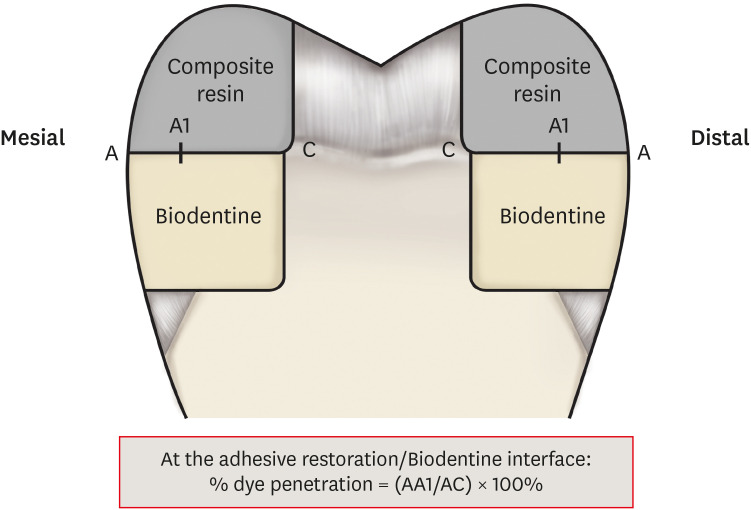
Standardized class II cavity preparations were made in the mesial and distal surfaces of the 80 selected teeth. The dimensions of the cavity were: 4 mm width (buccolingual), 5 mm high (occlusogingival), 2 mm depth (mesiodistal) and the gingival wall was placed in coronal enamel to the enamel-cement junction (1 mm to 1.5 mm width of enamel remaining in the gingival wall). This methodology allowed studying the adhesive restoration/Biodentine interface (Figure 1). The specimens were then randomly assigned to one of the 2 experimental groups (D0 or D7) to evaluate the impact of Biodentine maturation time on the quality of the adhesive interface [23].
Figure 1

Cavity preparation and measurements of the dye penetration length.

rde-46-e3-g001.jpg

2) Filling

The cavities were filled with Biodentine according to the manufacturer's instructions (Table 1). For the experimental group D0 which was divided into 8 subgroups (n = 5, 10 cavities), Biodentine was directly applied on the cavity walls. After 15 minutes, Biodentine was cut to leave a height of material of 2 mm to achieve a restoration of type “open sandwich” with the composite resin. The specimens were then restored using the adhesive system/composite resin according to the manufacturer's instructions (Table 1).
For experimental group D7 which was also divided into 8 subgroups (n = 5, 10 cavities), Biodentine was directly applied on the cavity walls, but after 15 minutes, samples were stored in an artificial Ringer solution. After 7 days, Biodentine cement was then cut to leave a height of material of 2 mm to achieve a restoration of type “open sandwich” with the composite resin. The specimens were then restored using the adhesive system/composite resin according to the manufacturer's instructions (Table 1).
Polymerization was achieved using a halogen light-curing unit (Elipar Highlight, 3M ESPE, Monrovia, CA, USA). The irradiance tested using a curing radiometer was 750 mW/cm2 and was consistent during the entire procedure. The restorations were polished with disks (Soflex, 3M ESPE) and the root apices were sealed using Ceram X Mono composite resin.

3) Thermocycling

Following resin composite obturation, each group of teeth was placed in a separate mesh bag and thermocycled together for 2,200 cycles in water between 5°C and 55°C for 10 seconds for each bath, and 10-second transfer time between baths [24].

4) Dye immersion

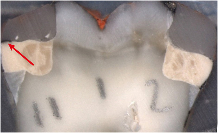
After cycling, the external surfaces of each tooth were completely coated with 2 layers of nail varnish, leaving a 1 mm wide margin around the restoration free of varnish. The specimens were then immersed in a 50 wt% silver nitrate aqueous solution (VWR International) for 2 hours in total darkness. Following retrieval, they were placed in distilled water and exposed to fluorescent light for 12 hours. Specimens were then immersed for 2 hours in a photo-developing solution (Kodak SA, Maisons-Alfort, France). After removal from the developing solution, the teeth were rinsed thoroughly in running water and immersed in acetone to dissolve the nail varnish. Each tooth was then embedded in a cold-curing epoxy resin (Epofix, Struers, Champigny-sur-Marne, France). Using a diamond blade circular disk (Accutom, Struers) at a speed of 500 rpm and with cutting lubricant (Water free cutting fluid, Struers), each specimen was sectioned longitudinally in the mesio-distal direction into 2 sections with 4 interfaces (Figure 2).
Figure 2

Tooth section: on the left, the arrow shows silver nitrate penetration along the interface; on the right no penetration.
rde-46-e3-g002.jpg
For each interface, dye penetration measurement was performed at the adhesive restoration/Biodentine interface using a binocular loop connected to a camera and analyzed using Leica Software (Leica Microsystems Imaging, Wetzlar Germany). The percentage of microleakage was defined as the measured length of dye penetration divided by the measured length of the interface. The mean percentage of microleakage was the mean of 10 cavities (10 × 4 = 40 interfaces) for each subgroup investigated (Figure 1).

5) Statistical analysis

Data were analyzed using one-way analysis of variance with Stat View software (SAS Institute, Cary, NC, USA). To compare each pair, the Mann-Whitney U test was used. A p value ≤ 0.05 was considered statistically significant.

2. Scanning electron microscope (SEM) methodology

According to the results obtained previously concerning microleakage, a tooth section from each adhesive system family of the D0 microleakage experimental group has been selected for the SEM observations [25].
Each sectioned surface was polished with abrasive discs and diamond pastes down to 1-micron particle size. The specimens were immediately immersed in 2.5% glutaraldehyde (VWR International) in 0.1 M sodium cacodylate buffer (VWR International) at pH 7.4 for 12 hours at 4°C. After fixation, the disks were rinsed with 20 mL of 0.2 M sodium cacodylate buffer at pH 7.4 for 1 hour (3 baths of 20 minutes), followed by distilled water for 1 minute. The surface of the material was then polished with abrasive discs and diamond pastes down to 1-micron particle size. The specimen was dehydrated in ascending grades of ethanol (25% for 20 minutes, 50% for 20 minutes, 75% for 20 minutes, and 100% for 20 minutes). After the final ethanol step, the specimens were placed on a filter paper inside a covered glass vial at room temperature. The sample was then mounted on copper stubs, sputter-coated with gold using a biorad sputter-coater SC 500 (Quorum Technologies Ltd., Laughton, UK) and observed under a JEOL JSM 6400 (Jeol, Tokyo, Japan) scanning electron microscope.
Microleakage at the adhesive restoration/Biodentine interface

1. Experimental group D0

The mean rates of microleakage on the interfaces ranged from 2.01% to 9.73% according to the adhesive system used. The adhesive systems may thus be classified by increasing efficiency. For adhesive systems with a penetration rate close to 2%: Prime & Bond > Scotchbond Universal etch-and-rinse (ER) system > G-aenial Bond > Scotchbond Universal self-etch (SE) system. No significant difference in terms of penetration rate was observed between these adhesive systems. For adhesive systems with a penetration rate close to 5%: Xeno III > Clearfil Tri-S Bond > Clearfil SE Bond. Once again, no significant difference was observed between these systems. The Optibond XTR adhesive system showed a penetration rate of 9.73%, which was significantly higher than that of Prime & Bond, Scotchbond Universal ER system, G-aenial Bond, Scotchbond Universal SE system, and Xeno III (Tables 2 and 3).
Table 2

Percentage of percolation according to the adhesive system used for both time points

Adhesive family Adhesive system % of percolation at D0 % of percolation at D7
Etch & rinse Prime & Bond NT 2.01% (1.072) 0.38% (0.38)
Scotchbond Universal ER 2.19% (0.611)* 5.3% (1.328)*
Self-etch 1 step G-aenial Bond 2.32% (0.821) 4.95% (1.642)
Scotchbond Universal SE 2.58% (1.084)* 12.58% (2.177)*
Xeno III 4.33% (0.930)* 18.49% (2.652)*
Clearfil Tri-S Bond 5.37% (2.274) 5.7% (1.715)
Self-etch 2 steps Clearfil SE Bond 5.95% (2.723) 5.85% (1.303)
Optibond XTR 9.73% (1.832)* 0.97% (0.464)*
Data are presented as mean % (standard error).
D0, immediate restoration; D7, delayed restoration (7 days).
*Represents significant differences for each adhesive system at D0 and D7 (p < 0.05).
Table 3

Intergroup analysis at D0

Prime & Bond NT Scotchbond Universal ER G-aenial Bond Scotchbond Universal SE Xeno III Clearfil Tri-S Bond Clearfil SE Bond Optibond XTR
Prime & Bond NT NS NS NS NS NS NS S
Scotchbond Universal ER NS NS NS NS NS NS S
G-aenial Bond NS NS NS NS NS NS S
Scotchbond Universal SE NS NS NS NS NS NS S
Xeno III NS NS NS NS NS NS S
Clearfil Tri-S Bond NS NS NS NS NS NS NS
Clearfil SE Bond NS NS NS NS NS NS NS
Optibond XTR S S S S S NS NS
NS, no significant difference; S, significant difference (p < 0.05).

2. Experimental group D7

The mean rates of microleakage on the interfaces ranged from 0.38% to 18.49% according to the adhesive system used. The adhesive systems may also be classified by increasing efficiency. For adhesive systems with a penetration rate lower than 1% without any significant difference between them: Prime & Bond > Optibond XTR. For adhesive systems with a penetration rate close to 5%, without any significant difference between them: G-aenial Bond > Scotchbond Universal ER system > Clearfil Tri-S Bond > Clearfil SE Bond. For adhesive systems with a penetration rate higher than 10%, Xeno III rate (18.49%) was significantly higher than that of Scotchbond Universal SE system (12.58%) and both showed significantly higher rates than all the others (Tables 2 and 4).
Table 4

Intergroup analysis at D7

Prime & Bond NT Scotchbond Universal ER G-aenial Bond Scotchbond Universal SE Xeno III Clearfil Tri-S Bond Clearfil SE Bond Optibond XTR
Prime & Bond NT NS NS S S S S NS
Scotchbond Universal ER S NS S S NS NS NS
G-aenial Bond NS NS S S NS NS NS
Scotchbond Universal SE S S S S S S S
Xeno III S S S S S S S
Clearfil Tri-S Bond S NS NS S S NS NS
Clearfil SE Bond S NS NS S S NS NS
Optibond XTR NS NS NS S S NS NS
NS, no significant difference; S, significant difference (p < 0.05).

3. Comparison between experimental groups D0 and D7 according to the adhesive system investigated

There was no significant difference in terms of microleakage between Prime & Bond, Clearfil SE bond, G-aenial Bond, and Tri-S Bond at D0 or D7. The microleakage of Xeno III and Scotchbond Universal was significantly higher at D7 than D0, whereas Optibond XTR showed a penetration rate significantly lower at D7 compared to D0 (Table 2).
When analyzing the mean penetration rates by the family of adhesive systems, independently from immediate or delayed restoration, results showed that etch-and-rinse systems (2.47%) present a trend towards the lowest penetration rate, followed by 2 steps self-etch systems (5.63%) and then one step self-etch systems (7.04%). Further analysis of mean penetration rates according to time of application, independently of the family of adhesives, found no significant difference in terms of microleakage between D0 (4.31%) and D7 (6.78%).
SEM observation of the adhesive restoration/Biodentine interface
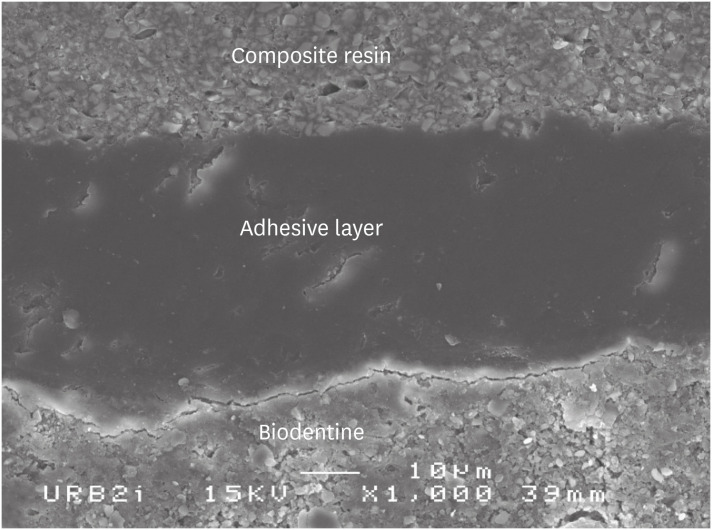
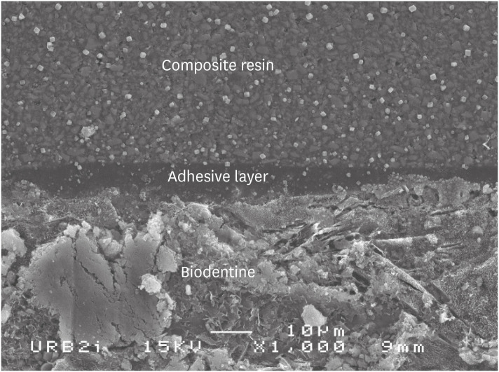
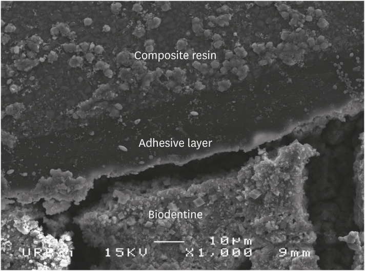
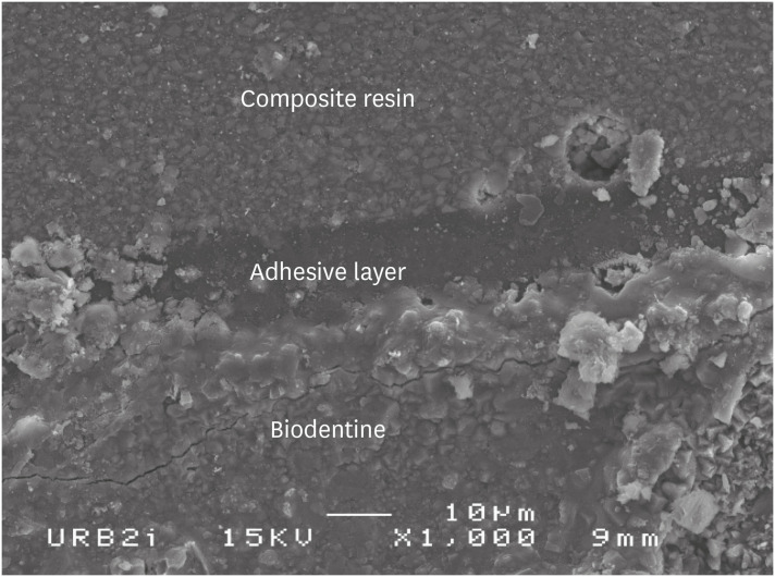
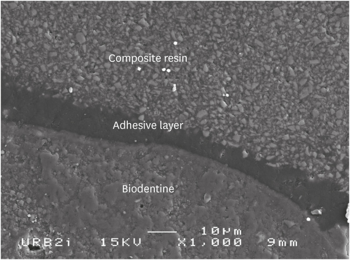
A tooth section from each adhesive system family of the D0 microleakage experimental group has been selected for the SEM observations of the interfaces: 2 steps self-etch system represented by Clearfil SE bond (Figure 3); 1 step self-etch system by Clearfil Tri-S Bond (Figure 4); universal system used in self-etching protocol represented by Scotchbond Universal (Figure 5); universal system used in etch-and-rinsing protocol represented by Scotchbond Universal (Figure 6); and etch-and-rinse system represented by Prime & Bond (Figure 7). Whatever the interfaces studied, the observations have shown that the adhesive layers were homogeneous and regular, and were adherent to the composite material. No gap was observed.
Figure 3

Scanning electron microscopic (SEM) observation of Biodentine/2 steps self-etch system (Clearfil SE Bond) interface (×1,000).
rde-46-e3-g003.jpg
Figure 4

Scanning electron microscopic (SEM) observation of Biodentine/one step self-etch system (Clearfil Tri-S Bond) interface (×1,000).
rde-46-e3-g004.jpg
Figure 5

Scanning electron microscopic (SEM) observation of Biodentine/universal system used as one step self-etch system (Scotchbond Universal) interface (×1,000).
rde-46-e3-g005.jpg
Figure 6

Scanning electron microscopic (SEM) observation of Biodentine/universal system used as etch-and-rinse system (Scotchbond Universal) interface (×1,000).
rde-46-e3-g006.jpg
Figure 7

Scanning electron microscopic (SEM) observation of Biodentine/etch-and-rinse system (Prime & Bond) interface (×1,000).
rde-46-e3-g007.jpg
This in vitro dye microleakage study showed that, in terms of sealing ability, Biodentine could be used, in a sandwich procedure, especially since silver nitrate chemical tracer allows the detection of very small defects. However, specific considerations might be given regarding the best operative sequence (immediate or delayed application time of the resin composite) for weaker microleakage according to the family of adhesive.
At D0, the chemical characteristics of the setting reaction for CSC entail that the maturation of Biodentine is incomplete when the bonding procedure is performed. Indeed the adhesive protocols were conducted only 15 minutes after the application of Biodentine. At that time, since the setting reaction is ongoing, the surface is more alkaline and presents a higher water content than at D7 [26]. From a physicochemical point of view, this means a lower acidic attack, especially when using the self-etch adhesives, during which a buffer effect occurs. From a clinical perspective, placing the dentin substitute and the definitive restoration during the same intervention is easier and time-saving. Moreover, at D0, new crystal products can form at the interface after adhesive application, improving sealing as the maturation is incomplete. Results herein showed that both etch-and-rinse systems (Prime & Bond and Scotchbond Universal ER) and self-etch adhesives except Optibond XTR all exhibit weak microleakage at D0. Of note, the results under 10% obtained using Optibond XTR, would be equivalent to a score 1 (0% to 33%), which was previously reported as satisfactory by Rengo et al. [27]. Thus, the present results indicate that the use of etch-and-rinse systems, and hence of orthophosphoric acid does not alter sealing at the Biodentine/adhesive system interface, as previously described [28]. More precisely, the 2 etch-and-rinse systems studied herein were found to be the adhesive family presenting the less leakage. Although the use of orthophosphoric acid leads to a chemical modification of the cement surface, as a poor calcium rate and a lower chloride peak, which weakens the surface structure of the material, the sealing does not appear to be degraded [14]. In line with the present results, Atabek et al. [29] showed that etch-and-rinse systems displayed a stronger adhesion force on another CSC, white mineral trioxide aggregate (WMTA) than 1-step self-etching adhesives. More precisely, the latter study showed that the fracture between the WTMA and etch-and-rinse systems is cohesive, while self-etching adhesives present adhesive fractures. The occurrence of the latter is likely to be the consequence of the low acidity of self-etching adhesives. This is further confirmed by Cengiz et al. [17], who showed that the application of etch-and-rinse adhesives might indeed improve the bond strength of composite resin on Biodentine. Conversely, when studying the effect of thermocycling on the self-etching adhesive/Biodentine interface, Meraji and Camilleri [15] noticed a total decohesion of this interface after thermocycling. However, although Hashem et al. [16] showed that there was no difference in SBS when using self-etching systems or etch-and-rinse systems, Odabas et al. [19], found improved bond strength for self-etching adhesives. Overall, the results of the present study thus confirm that orthophosphoric acid can be used on Biodentine with satisfactory marginal sealing and bond strength properties, likely increasing restoration longevity.
At D7, the setting reaction products, calcium hydroxide (CaOH2) and calcium silicate hydrate (CaOSiO2H2O) are mainly stabilized [10]. At that time, the etch-and-rinse systems and the acetone-based systems showed better results in terms of microleakage than those containing ethanol and water solvents. Indeed, the Scotchbond Universal adhesive exhibits lower leakage at D7 when used in an etch-and-rinse procedure rather than self-etch. This further confirms the performance and interest of using orthophosphoric acid on Biodentine surface similarly at D0 and D7. After maturation, the lowest percentage of microleakage being observed for Prime & Bond, Optibond XTR, and Gaenial Bond, the presence of the acetone solvent appears to be important, likely due to the high pressure of evaporation which helps the monomer create greater forces of adhesion [30].
Nevertheless, the results obtained using self-etch adhesives can also be considered satisfactory at D7, except for that of Xeno III. It is possible that the packaging of the latter which consists of 2 liquids to be mixed through a blister, could have led to an inhomogeneous mixing. This would be in accordance with the study by Karaman et al. [31], who showed that Xeno III is a very operator-dependent system. Similarly to what was reported by Palma et al. [32] and Çolak et al. [18], the results herein showed that the operative protocol did not impact the sealing on the Biodentine/adhesive restoration for half of the adhesive systems used (Prime & Bond, Clearfil SE bond, G-aenial Bond, or Clearfil Tri-S Bond). However, leakage was less important at D0 for Scotchbond Universal (ER or SE) and Xeno III. Only the interface obtained using Optibond XTR presented a better sealing at D7, this adhesive also showing the highest rate of leakage at D0. In the literature, several studies have found that applying the adhesives with delay improved bond strength, as the resin composite curing contraction can stress Biodentine in the early sensitive phase and lead to interface defects [16,29,33]. Conversely, we could also consider that the adhesive systems interact better in the porosity of the tricalcium silicate during the maturation process, leading to better sealing [10]. Overall, when considering all family of adhesives, we found no difference between D0 and D7 in terms of leakage. Consequently, if bond strength is improved in delayed time, we recommend placing the definitive resin composite restoration after maturation.
Clearfil SE Bond is a reference in terms of dentin sealing and is the gold standard of self-etching adhesives, due to the functional monomer 10-MDP. The latter behaves perfectly well in terms of degradation thanks to its stable chemical bond and the formation of nanolayering (mild self-etching system). This slightly acid monomer preserves some of the calcium of the dentinal structure and can then bind to it [34,35,36,37]. Although it is unknown whether a similar chemical union exists between Biodentine and the overlying resin composite restoration, the results found herein on the Biodentine/Clearfil SE Bond interface, at both D0 and D7, were very similar to those from a preliminary study reporting percolation rates on the dentin/Clearfil SE Bond interface [38]. This could be due to the presence of 10-MDP which could, in theory, chemically bond to the Biodentine calcium. The 10-MDP is found in Clearfil SE Bond, Scotchbond Universal, and Clearfil Tri-S Bond. Two of the self-etching systems, Clearfil SE Bond and Clearfil Tri-S Bond, presented homogeneity of their percolation rates at both D0 and D7, confirming the reproducibility and stability of these systems [18,19], which were found to be the most tolerant herein.
The good results in terms of microleakage were further confirmed by SEM observations. All interfaces showed homogeneous, regular, and adherent adhesive layers. No gap was observed. Layer thickness, however, was tighter for Prime & Bond, the only adhesive which is not loaded, and Clearfil Tri-S Bond, a self-etch adhesive. Morphology was similar when using Scotchbond Universal in either self-etch or etch-and-rinse. These considerations concerning the thickness of the interface layer may have an impact on long-term evaluation, which could be further assessed using nanoleakage evaluation.
The open sandwich technique is recommended when the conditions required for a good sealing are not obtained as the impossibility to put a watertight dental dam or a subgingival situation. In these situations, the open sandwich restoration allows to go up the cervical margin and improves bonding conditions [11,23]. Using Biodentine in this situation could also be a good alternative in the situation where the carious lesion is very deep. It may preserve the vitality of the teeth limiting endodontic treatment and prosthetic restoration, but of course, as always, benefit/risk report must be evaluated for each clinical case. The dentin/Biodentine interface is well documented in the literature and presents good sealing properties [1,8,39]. Moreover, in the cervical situation, the mechanical solicitations are weak and Biodentine leads to no dimensional changes in different environmental conditions [40,41]. However, practitioners must be vigilant about the contact point of the restoration which must necessarily be realized with the composite or ceramic restorative material. Further in vitro and in vivo studies should be investigated to confirm this data.
This in vitro dye microleakage study further confirms the ability of Biodentine, a CSC, to be used in a sandwich procedure, in terms of marginal sealing regardless of the family of adhesives used. Furthermore, concerning the adhesive used, cement maturation does not impact sealing at the Biodentine/adhesive interface.
To Stephane Legoff (URB 2I, Montrouge, France) for his contribution for the scanning electron microscopic (SEM) observations.

Conflict of Interest: No potential conflict of interest relevant to this article was reported.

Author Contributions:

  • Conceptualization: Pradelle-Plasse N, Mocquot C, Semennikova K, Colon P, Grosgogeat B.

  • Data curation: Pradelle-Plasse N, Mocquot C, Semennikova K, Colon P, Grosgogeat B.

  • Formal analysis: Pradelle-Plasse N, Semennikova K.

  • Investigation: Pradelle-Plasse N, Mocquot C, Semennikova K.

  • Methodology: Pradelle-Plasse N, Mocquot C, Semennikova K, Colon P, Grosgogeat B.

  • Project administration: Pradelle-Plasse N, Colon P, Grosgogeat B.

  • Resources: Pradelle-Plasse N, Mocquot C, Semennikova K.

  • Supervision: Pradelle-Plasse N, Colon P, Grosgogeat B.

  • Validation: Pradelle-Plasse N, Colon P, Grosgogeat B.

  • Visualization: Pradelle-Plasse N, Colon P, Grosgogeat B.

  • Writing - original draft: Pradelle-Plasse N.

  • Writing - review & editing: Pradelle-Plasse N.

  • 1. Bachoo IK, Seymour D, Brunton P. A biocompatible and bioactive replacement for dentine: Is this a reality? The properties and uses of a novel calcium-based cement. Br Dent J 2013;214:E5. PubMed
  • 2. Torabinejad M, Hong CU, McDonald F, Pitt Ford TR. Physical and chemical properties of a new root-end filling material. J Endod 1995;21:349-353. PubMed
  • 3. Schwartz RS, Mauger M, Clement DJ, Walker WA 3rd. Mineral trioxide aggregate: a new material for endodontics. J Am Dent Assoc 1999;130:967-975. ArticlePubMed
  • 4. Torabinejad M, Parirokh M. Mineral trioxide aggregate: a comprehensive literature review--part II: leakage and biocompatibility investigations. J Endod 2010;36:190-202. ArticlePubMed
  • 5. Sarkar NK, Caicedo R, Ritwik P, Moiseyeva R, Kawashima I. Physicochemical basis of the biologic properties of mineral trioxide aggregate. J Endod 2005;31:97-100. PubMed
  • 6. Bozeman TB, Lemon RR, Eleazer PD. Elemental analysis of crystal precipitate from gray and white MTA. J Endod 2006;32:425-428. PubMed
  • 7. Watson TF, Atmeh AR, Sajini S, Cook RJ, Festy F. Present and future of glass-ionomers and calcium-silicate cements as bioactive materials in dentistry: biophotonics-based interfacial analyses in health and disease. Dent Mater 2014;30:50-61. ArticlePubMedPMC
  • 8. Atmeh AR, Chong EZ, Richard G, Festy F, Watson TF. Dentin-cement interfacial interaction: calcium silicates and polyalkenoates. J Dent Res 2012;91:454-459. ArticlePubMedPMCPDF
  • 9. Pradelle-Plasse N, Tran X, Colon P. Biocompatibility or cytotoxic effects of dental composites. Oxfordshire: Coxmoor Publishing Company; 2009.
  • 10. Villat C, Tran XV, Pradelle-Plasse N, Ponthiaux P, Wenger F, Grosgogeat B, Colon P. Impedance methodology: a new way to characterize the setting reaction of dental cements. Dent Mater 2010;26:1127-1132. PubMed
  • 11. Koubi G, Colon P, Franquin JC, Hartmann A, Richard G, Faure MO, Lambert G. Clinical evaluation of the performance and safety of a new dentine substitute, Biodentine, in the restoration of posterior teeth - a prospective study. Clin Oral Investig 2013;17:243-249.PubMed
  • 12. Bergenholtz G, Cox CF, Loesche WJ, Syed SA. Bacterial leakage around dental restorations: its effect on the dental pulp. J Oral Pathol 1982;11:439-450. ArticlePubMed
  • 13. Browne RM, Tobias RS. Microbial microleakage and pulpal inflammation: a review. Endod Dent Traumatol 1986;2:177-183. ArticlePubMed
  • 14. Camilleri J. Investigation of Biodentine as dentine replacement material. J Dent 2013;41:600-610. PubMed
  • 15. Meraji N, Camilleri J. Bonding over dentin replacement materials. J Endod 2017;43:1343-1349. ArticlePubMed
  • 16. Hashem DF, Foxton R, Manoharan A, Watson TF, Banerjee A. The physical characteristics of resin composite-calcium silicate interface as part of a layered/laminate adhesive restoration. Dent Mater 2014;30:343-349. ArticlePubMed
  • 17. Cengiz E, Ulusoy N. Microshear bond strength of tri-calcium silicate-based cements to different restorative materials. J Adhes Dent 2016;18:231-237. PubMed
  • 18. Çolak H, Tokay U, Uzgur R, Uzgur Z, Ercan E, Hamidi MM. The effect of different adhesives and setting times on bond strength between Biodentine and composite. J Appl Biomater Funct Mater 2016;14:e217-e222. PubMed
  • 19. Odabaş ME, Bani M, Tirali RE. Shear bond strengths of different adhesive systems to Biodentine. ScientificWorldJournal 2013;2013:626103. PubMedPMC
  • 20. Abraham SB, Gaintantzopoulou MD, Eliades G. Cavity adaptation of water-based restoratives placed as liners under a resin composite. Int J Dent 2017;2017:5957107. ArticlePubMedPMCPDF
  • 21. Aggarwal V, Singla M, Yadav S, Yadav H, Ragini . Marginal adaptation evaluation of Biodentine and MTA plus in ‘open sandwich’ class II restorations. J Esthet Restor Dent 2015;27:167-175. ArticlePubMedPDF
  • 22. Raskin A, Eschrich G, Dejou J, About I. In vitro microleakage of Biodentine as a dentin substitute compared to Fuji II LC in cervical lining restorations. J Adhes Dent 2012;14:535-542. PubMed
  • 23. Besnault C, Attal JP. Simulated oral environment and microleakage of class II resin-based composite and sandwich restorations. Am J Dent 2003;16:186-190. PubMed
  • 24. Gale MS, Darvell BW. Thermal cycling procedures for laboratory testing of dental restorations. J Dent 1999;27:89-99. ArticlePubMed
  • 25. Cervino G, Fiorillo L, Spagnuolo G, Bramanti E, Laino L, Lauritano F, Cicciù M. Interface between MTA and dental bonding agents: scanning electron microscope evaluation. J Int Soc Prev Community Dent 2017;7:64-68. ArticlePubMedPMC
  • 26. Garrault S, Behr T, Nonat A. Formation of the C-S-H layer during early hydration of tricalcium silicate grains with different sizes. J Phys Chem B 2006;110:270-275. ArticlePubMed
  • 27. Rengo C, Goracci C, Juloski J, Chieffi N, Giovannetti A, Vichi A, Ferrari M. Influence of phosphoric acid etching on microleakage of a self-etch adhesive and a self-adhering composite. Aust Dent J 2012;57:220-226. PubMed
  • 28. Kayahan MB, Nekoofar MH, Kazandağ M, Canpolat C, Malkondu O, Kaptan F, Dummer PM. Effect of acid-etching procedure on selected physical properties of mineral trioxide aggregate. Int Endod J 2009;42:1004-1014. ArticlePubMed
  • 29. Atabek D, Sillelioğlu H, Olmez A. Bond strength of adhesive systems to mineral trioxide aggregate with different time intervals. J Endod 2012;38:1288-1292. ArticlePubMed
  • 30. Silva e Souza MH Jr, Carneiro KG, Lobato MF, Silva e Souza PA, de Góes MF. Adhesive systems: important aspects related to their composition and clinical use. J Appl Oral Sci 2010;18:207-214. ArticlePubMedPMC
  • 31. Karaman E, Yazici AR, Aksoy B, Karabulut E, Ozgunaltay G, Dayangac B. Effect of operator variability on microleakage with different adhesive systems. Eur J Dent 2013;7:S060-S065. PubMed
  • 32. Palma PJ, Marques JA, Falacho RI, Vinagre A, Santos JM, Ramos JC. Does delayed restoration improve shear bond strength of different restorative protocols to calcium silicate-based cements? Materials (Basel) 2018;11:2216.ArticlePubMedPMC
  • 33. Sultana N, Nawal RR, Chaudhry S, Sivakumar M, Talwar S. Effect of acid etching on the micro-shear bond strength of resin composite-calcium silicate interface evaluated over different time intervals of bond aging. J Conserv Dent 2018;21:194-197. ArticlePubMedPMC
  • 34. Yoshioka M, Yoshida Y, Inoue S, Lambrechts P, Vanherle G, Nomura Y, Okazaki M, Shintani H, Van Meerbeek B. Adhesion/decalcification mechanisms of acid interactions with human hard tissues. J Biomed Mater Res 2002;59:56-62. ArticlePubMed
  • 35. Yoshida Y, Nagakane K, Fukuda R, Nakayama Y, Okazaki M, Shintani H, Inoue S, Tagawa Y, Suzuki K, De Munck J, Van Meerbeek B. Comparative study on adhesive performance of functional monomers. J Dent Res 2004;83:454-458. PubMed
  • 36. Carrilho E, Cardoso M, Marques Ferreira M, Marto CM, Paula A, Coelho AS. 10-MDP based dental adhesives: adhesive interface characterization and adhesive stability-a systematic review. Materials (Basel) 2019;12:790.ArticlePubMedPMC
  • 37. Yoshihara K, Yoshida Y, Nagaoka N, Hayakawa S, Okihara T, De Munck J, Maruo Y, Nishigawa G, Minagi S, Osaka A, Van Meerbeek B. Adhesive interfacial interaction affected by different carbon-chain monomers. Dent Mater 2013;29:888-897. ArticlePubMed
  • 38. Chatel E, Pradelle-Plasse N, Colon P, Grosgogeat B. Degradation mechanisms of modern self–etching adhesives. Clin Oral Investig 2013;17:1035.
  • 39. Koubi S, Elmerini H, Koubi G, Tassery H, Camps J. Quantitative evaluation by glucose diffusion of microleakage in aged calcium silicate-based open-sandwich restorations. Inter J Dent 2012;2012:105863.ArticlePDF
  • 40. Aksel H, Küçükkaya Eren S, Askerbeyli Õrs S, Karaismailoğlu E. Surface and vertical dimensional changes of mineral trioxide aggregate and Biodentine in different environmental conditions. J Appl Oral Sci 2018;27:e20180093. ArticlePubMedPMC
  • 41. Agrafioti A, Tzimpoulas N, Chatzitheodoridis E, Kontakiotis EG. Comparative evaluation of sealing ability and microstructure of MTA and Biodentine after exposure to different environments. Clin Oral Investig 2016;20:1535-1540.ArticlePubMedPDF

Tables & Figures

REFERENCES

    Citations

    Citations to this article as recorded by  

      • ePub LinkePub Link
      • Cite
        CITE
        export Copy Download
        Close
        Download Citation
        Download a citation file in RIS format that can be imported by all major citation management software, including EndNote, ProCite, RefWorks, and Reference Manager.

        Format:
        • RIS — For EndNote, ProCite, RefWorks, and most other reference management software
        • BibTeX — For JabRef, BibDesk, and other BibTeX-specific software
        Include:
        • Citation for the content below
        Interface between calcium silicate cement and adhesive systems according to adhesive families and cement maturation
        Restor Dent Endod. 2021;46(1):e3  Published online December 9, 2020
        Close
      • XML DownloadXML Download
      Figure
      • 0
      • 1
      • 2
      • 3
      • 4
      • 5
      • 6
      Interface between calcium silicate cement and adhesive systems according to adhesive families and cement maturation
      Image Image Image Image Image Image Image
      Figure 1 Cavity preparation and measurements of the dye penetration length.
      Figure 2 Tooth section: on the left, the arrow shows silver nitrate penetration along the interface; on the right no penetration.
      Figure 3 Scanning electron microscopic (SEM) observation of Biodentine/2 steps self-etch system (Clearfil SE Bond) interface (×1,000).
      Figure 4 Scanning electron microscopic (SEM) observation of Biodentine/one step self-etch system (Clearfil Tri-S Bond) interface (×1,000).
      Figure 5 Scanning electron microscopic (SEM) observation of Biodentine/universal system used as one step self-etch system (Scotchbond Universal) interface (×1,000).
      Figure 6 Scanning electron microscopic (SEM) observation of Biodentine/universal system used as etch-and-rinse system (Scotchbond Universal) interface (×1,000).
      Figure 7 Scanning electron microscopic (SEM) observation of Biodentine/etch-and-rinse system (Prime & Bond) interface (×1,000).
      Interface between calcium silicate cement and adhesive systems according to adhesive families and cement maturation

      Materials used in this study

      Family of materialMaterialComponentspHProtocol
      Calcium silicateBiodentine (Septodont, St. Maur-des-Fossés, France)Solid: tricalcium silicate (> 80%), calcium carbonate, zirconium oxide12.51. Incorporate 5 drops of liquid in the powder.
      Liquid: water, CaCl2, partially modified polycarboxylate2. Close the capsule and put it 30 sec in the oscillator.
      Adhesive system
      Self-etch 2 stepsOptibond XTR (Kerr, Orange, CA, USA; batch: LE03355)Primer: GPDM, HEMA, water, acetone, ethanol, camphorquinonePrimer: 2.41. Apply primer to enamel/dentin using scrubbing motion (20 sec).
      Bonding: HEMA, MEHQ, ethanol, camphorquinone, loads of silicaBond: 3.32. Air thin with medium air pressure (5 sec).
      3. Shake adhesive briefly, and apply to enamel/dentin surface using light brushing motion (15 sec).
      4. Air thin with medium air pressure, and then strong air for at least 5 sec.
      5. Light-cure (10 sec).
      Clearfil SE Bond (Kuraray, Okayama, Japan; batch: 041838)Primer: 10-MDP, HEMA, dimethacrylate aliphatque absorbent, camphorquinone, N-diethanol-p-toluidine, water2.01. Apply primer and leave for 20 sec.
      Bonding:10-MDP, Bis-GMA, HEMA, dimethacrylate aliphatic hydrophobic subject, camphoroquinone, N-diethanol-p-toluidine, loads of silica2. Do not rinse and dry with mild air flow.
      3. Apply bond and distribute evenly with mild air flow.
      4. Light-cure for 10 sec.
      Self-etch 1 stepXeno III (Dentsply/Caulk, Mildford, DE, USA; batch: 1212000562)Liquid A: HEMA, water purified, ethanol, BHT, silicon dioxide< 1.01. Apply generous amounts of self-etch adhesive to wet all cavity surfaces thoroughly.
      Liquid B: Pyro-EMA, PEM-F, urethan dimethacrylate, BHT, camphorquinone, ethyl-4-Dimethylaminobenzoate2. Leave undisturbed for at least 20 sec.
      3. Uniformly spread the adhesive using a gentle stream of oil free air for at least 2 sec.
      4. Cure the adhesive with a light-curing unit for at least 10 sec.
      G-aenial Bond (GC, Tokyo, Japan; batch: 1301161)4-META, acetone, water, triethyleneglycol dimethacrylate, phosphoric acid ester monomer, silica loads, photo-initiators2.01. Apply to the prepared enamel and dentin surfaces using the disposable applicator.
      2. Leave undisturbed for 10 sec after applying.
      3. Then, dry thoroughly for 5 sec with oil-free air under maximum air pressure.
      4. Light-cure for 10 sec.
      Clearfil Tri-S Bond (Kuraray, Okayama, Japan; batch: 1M004)MDP, Bis-GMA, HEMA, Absorbent aliphatic dimethacrylate, hydrophobic aliphatic methacrylate, silica colloidal, sodium fluoride, di-camphorquinone, accelerators, initiators, ethanol, water2.71. Apply bond to the entire cavity wall with the applicator brush. Leave it in place for 10 sec.
      2. Dry the entire cavity wall sufficiently by blowing mild air for more than 5 sec.
      3. Light-cure for 10 sec.
      UniversalScotchBond Universal (3M ESPE, Monrovia, CA, USA; batch: 70201139014)MDP, resin of dimethacrylate, HEMA, Vitrebond copolymer, loads, ethanol, water, silane2.7Protocol 1: self-etch 1-step
      1. Apply the adhesive to the prepared tooth and rub it in for 20 sec.
      2. Gently air dry the adhesive for approximately 5 sec to evaporate the solvent.
      3. Light-cure for 10 sec.
      Protocol 2: etch-and-rinse 2-steps
      1. Apply orthophosphoric acid for 15 sec.
      2. Apply the adhesive to the prepared tooth and rub it in for 20 sec.
      3. Gently air dry the adhesive for approximately 5 sec to evaporate the solvent.
      4. Light-cure for 10 sec.
      Etch-and-rinse 2 stepsPrime & Bond (Dentsply DeTrey, Konstanz, Germany; batch: 1206001168)Resin di- and trimethacrylate, silica, PENTA, photo-initiators, stabilizing, cetylamine hydrofluoride, acetone2.01. Application of orthophosphoric acid 37% for at least 15 sec.
      2. Rinsing and blot drying for at least 10 sec.
      3. Application of Prime & Bond for 20 sec.
      4. Remove excess solvent by gently drying for at least 5 sec.
      5. Cure Prime & Bond adhesive for 10 sec.
      AcidOrthophosphoric acid 37% (Dentsply DeTrey, Konstanz, Germany; batch: 155245)Phosphoric acid, water, thickener, methylen Blue benzalkonium chloride0.4
      Composite resinCeram X Mono (Dentsply, York, PA, USA; batch: 1107000M97)Methacrylate modified polysiloxane, dimethacrylate resin, fluorescence pigment, UV stabilizer, stabilizer, camphorquinone, ethyl-4(dimethylamino)benzoate, barium-aluminium-borosilicate glass, methacrylate functionalised silicon dioxide nano filler, iron oxide pigments and titanium oxide aluminium sulfo silicate pigmentsNDThe composite resin material is incrementally placed and light-cured for 20 sec.

      CaCl2, calcium chloride; GPDM, glycero-phosphate dimethacrylate; HEMA, hydroxyethylmethacrylate; MEHQ, hydroquinone monomethyl ether; MDP, methacryloyloxydecyl dihydrogen phosphate; Bis-GMA, bisphenol A-glycidyl methacrylate; BHT, butylated hydroxytoluene; Pyro-EMA, tetramethacryloxyethyl pyrophosphate; PEM-F, pentamethacryloxyethyl cyclophosphazen mono fluoride; META, methacryloyloxyethy trimellitate anhydride; PENTA, dipentaerythritol penta acrylate monophosphate; UV, ultraviolet; ND, not designated.

      Percentage of percolation according to the adhesive system used for both time points

      Adhesive familyAdhesive system% of percolation at D0% of percolation at D7
      Etch & rinsePrime & Bond NT2.01% (1.072)0.38% (0.38)
      Scotchbond Universal ER2.19% (0.611)*5.3% (1.328)*
      Self-etch 1 stepG-aenial Bond2.32% (0.821)4.95% (1.642)
      Scotchbond Universal SE2.58% (1.084)*12.58% (2.177)*
      Xeno III4.33% (0.930)*18.49% (2.652)*
      Clearfil Tri-S Bond5.37% (2.274)5.7% (1.715)
      Self-etch 2 stepsClearfil SE Bond5.95% (2.723)5.85% (1.303)
      Optibond XTR9.73% (1.832)*0.97% (0.464)*

      Data are presented as mean % (standard error).

      D0, immediate restoration; D7, delayed restoration (7 days).

      *Represents significant differences for each adhesive system at D0 and D7 (p < 0.05).

      Intergroup analysis at D0

      Prime & Bond NTScotchbond Universal ERG-aenial BondScotchbond Universal SEXeno IIIClearfil Tri-S BondClearfil SE BondOptibond XTR
      Prime & Bond NTNSNSNSNSNSNSS
      Scotchbond Universal ERNSNSNSNSNSNSS
      G-aenial BondNSNSNSNSNSNSS
      Scotchbond Universal SENSNSNSNSNSNSS
      Xeno IIINSNSNSNSNSNSS
      Clearfil Tri-S BondNSNSNSNSNSNSNS
      Clearfil SE BondNSNSNSNSNSNSNS
      Optibond XTRSSSSSNSNS

      NS, no significant difference; S, significant difference (p < 0.05).

      Intergroup analysis at D7

      Prime & Bond NTScotchbond Universal ERG-aenial BondScotchbond Universal SEXeno IIIClearfil Tri-S BondClearfil SE BondOptibond XTR
      Prime & Bond NTNSNSSSSSNS
      Scotchbond Universal ERSNSSSNSNSNS
      G-aenial BondNSNSSSNSNSNS
      Scotchbond Universal SESSSSSSS
      Xeno IIISSSSSSS
      Clearfil Tri-S BondSNSNSSSNSNS
      Clearfil SE BondSNSNSSSNSNS
      Optibond XTRNSNSNSSSNSNS

      NS, no significant difference; S, significant difference (p < 0.05).

      Table 1 Materials used in this study

      CaCl2, calcium chloride; GPDM, glycero-phosphate dimethacrylate; HEMA, hydroxyethylmethacrylate; MEHQ, hydroquinone monomethyl ether; MDP, methacryloyloxydecyl dihydrogen phosphate; Bis-GMA, bisphenol A-glycidyl methacrylate; BHT, butylated hydroxytoluene; Pyro-EMA, tetramethacryloxyethyl pyrophosphate; PEM-F, pentamethacryloxyethyl cyclophosphazen mono fluoride; META, methacryloyloxyethy trimellitate anhydride; PENTA, dipentaerythritol penta acrylate monophosphate; UV, ultraviolet; ND, not designated.

      Table 2 Percentage of percolation according to the adhesive system used for both time points

      Data are presented as mean % (standard error).

      D0, immediate restoration; D7, delayed restoration (7 days).

      *Represents significant differences for each adhesive system at D0 and D7 (p < 0.05).

      Table 3 Intergroup analysis at D0

      NS, no significant difference; S, significant difference (p < 0.05).

      Table 4 Intergroup analysis at D7

      NS, no significant difference; S, significant difference (p < 0.05).


      Restor Dent Endod : Restorative Dentistry & Endodontics
      Close layer
      TOP