Warning: mkdir(): Permission denied in /home/virtual/lib/view_data.php on line 81

Warning: fopen(upload/ip_log/ip_log_2024-12.txt): failed to open stream: No such file or directory in /home/virtual/lib/view_data.php on line 83

Warning: fwrite() expects parameter 1 to be resource, boolean given in /home/virtual/lib/view_data.php on line 84
Can silver diamine fluoride or silver nanoparticle-based anticaries agents to affect enamel bond strength?
Skip Navigation
Skip to contents

Restor Dent Endod : Restorative Dentistry & Endodontics

OPEN ACCESS

Articles

Page Path
HOME > Restor Dent Endod > Volume 46(1); 2021 > Article
Research Article Can silver diamine fluoride or silver nanoparticle-based anticaries agents to affect enamel bond strength?
Jaqueline Costa Favaroorcid, Yana Cosendey Toledo de Mello Peixotoorcid, Omar Gehaorcid, Flaviana Alves Diasorcid, Ricardo Danil Guiraldoorcid, Murilo Baena Lopesorcid, Sandrine Bittencourt Bergerorcid

DOI: https://doi.org/10.5395/rde.2021.46.e7
Published online: January 12, 2021

Department of Restorative Dentistry, School of Dentistry, University of North Parana, Londrina, PR, Brazil.

Correspondence to Sandrine Bittencourt Berger, DDS, MS, PhD. Associate Professor, Department of Restorative Dentistry, School of Dentistry, University of North Parana, Rua Marselha, 183, Londrina, PR 86041-140, Brazil. berger.sandrine@gmail.com
• Received: October 2, 2019   • Revised: August 16, 2020   • Accepted: August 21, 2020

Copyright © 2021. The Korean Academy of Conservative Dentistry

This is an Open Access article distributed under the terms of the Creative Commons Attribution Non-Commercial License (https://creativecommons.org/licenses/by-nc/4.0/) which permits unrestricted non-commercial use, distribution, and reproduction in any medium, provided the original work is properly cited.

  • 20 Views
  • 1 Download
  • 5 Web of Science
prev next
  • Objectives
    The aim of the current study is to investigate the effect of different anticaries agents, such as experimental agents based on silver nanoparticles (SNPs) and silver diamine fluoride (SDF), on the micro-shear bond strength (μ-SBS) of composite resin applied to intact enamel (IE) or demineralized enamel (DE).
  • Materials and Methods
    Sixty dental enamel fragments were collected from human third molars and categorized into 6 groups (n = 10): positive control (IE), negative control (DE), IE + SDF, DE + SDF, IE + SNP and DE + SNP. Samples from DE, DE + SDF and DE + SNP groups were subjected to pH cycling; superficial microhardness test was performed to confirm demineralization. Resin composite build-ups were applied to the samples (0.75-mm diameter and 1-mm height) after the treatments (except for IE and DE groups); μ-SBS was also evaluated. Samples were analyzed under a stereomicroscope at 40× magnification to identify failure patterns. Data were subjected to one-way analysis of variance, followed by Tukey's and Dunnett's tests (p < 0.05).
  • Results
    There was no significant difference among the IE, IE + SNP, DE + SDF, and DE + SNP groups. The IE + SDF and DE groups recorded the highest and the lowest μ-SBS values, respectively. Adhesive-type failures were the most frequent for all treatments.
  • Conclusions
    Anticaries agents did not have a negative effect on the μ-SBS of composite resin when it was used on IE or DE.
The use of chemical agents to delay or stop caries progression without lesion removal has been documented in the literature since 1969 [1]. Silver diamine fluoride (SDF) is a chemical agent that, once applied to the dental surface, increases enamel resistance, inhibits biofilm formation, decreases acid production by microorganisms, reduces the Streptococcus mutans population and promote dentinal tubule obliteration [2,3]. However, SDF changes the color of areas affected by caries into black due to the silver ion reduction process triggered by its formulation [4]. Such adverse effect restricts SDF use, although it is highly successful in stopping carious lesions [3,5,6,7].
Studies have investigated the use of silver nanoparticle (SNP) anticaries agents to prevent the aesthetic damage caused by SDF. Results suggested that SNP was potentially effective in preventing and stopping dental caries without staining the demineralized dental enamel [6,7]. SNPs have a large surface area available to interact with microorganisms, and it makes them more effective antimicrobial agents than other particles [7]. It is interesting to use fluoride associated with SNP solution due to their synergistic effect, which promotes enamel remineralization together with a bactericidal action against cariogenic microorganisms [6,8,9].
Studies have reported that the application of anticaries agents and fluoride on the dental surface can change the bond strength of restorative materials [10,11,12,13,14,15,16,17,18]. However, the literature lacks information about the adhesion of resinous materials to enamel treated with SNP-based anticaries agents. Hence, it is necessary to investigate the interaction of materials used to reconstruct the lost dental structure on substrates modified by anticaries agents. Thus, the aim of the present study was to investigate the effect of anticaries agents (SNP or SDF) on the micro-shear bond strength (μ-SBS) of composite resin applied to intact enamel (IE) or demineralized enamel (DE). The null hypothesis tested is that the use of SDF or SNP on IE or DE did not affect μ-SBS values.
Sample selection and preparation
The current study was approved by the local ethics committee (protocol number 92548818.0.0000.0108). Thirty human molars, caries and enamel defects free, extracted for therapeutic purposes were used in this study. The teeth were cleaned right after their extraction and stored in 0.5% chloramine T solution for 7 days; next, they were stored in distilled water at 4°C until they were used in the experiment.
Enamel specimens were collected from the vestibular and lingual surfaces of each tooth (7 mm width × 4 mm length × 7 mm height). The enamel surfaces were fixed with acrylic resin (JET, Clássico, São Paulo, SP, Brazil) in PVC tube rings (Odeme Dental Research, Luzerna, SC, Brazil).
Enamel surfaces were abraded with 400, 600, 1,000, 1,200, and 1,500-grit silicon carbide paper and polished with abrasive paper and 1-µm diamond paste in electric polisher (APL4 Arotec S/A Ind. e Comércio, Cotia, SP, Brazil). Samples were then subjected to an ultrasonic bath in deionized water for 10 minutes (Model Ultrasonic Cleaner, Odontobras, Ribeirão Preto, SP, Brazil) to remove debris. Prepared specimens were examined under a stereomicroscope (Model Bel Photonics STM Pro, Bel Microimage Analyzer, Bel Photonics, Monza, Italy) at 40× magnification to confirm the absence of cracks or other surface defects. Specimens were stored in deionized water until the time to be used to avoid dehydration.
Enamel specimens were randomly categorized into 4 experimental and 2 control groups (n = 10). Table 1 shows the experimental groups and the composition of the anticaries agents.
Table 1

Experimental groups and anticaries agent composition

Experimental group Anticaries agent composition
Positive control: IE NA
Negative control: DE NA
IE + 30% SDF* Silver nitrate, ammonium hydroxide, fluoroboric acid, deionized water
DE + SDF* Silver nitrate, ammonium hydroxide, fluoroboric acid, deionized water
IE + experimental solution with SNP SNPs
DE + SNP SNPs
IE, intact enamel; DE, demineralized enamel; SDF, silver diamine fluoride; SNP, silver nanoparticle; NA, not available.
*Cariestop, Biodiâmica Química e Farmacêutica LTDA, Londrina, PR, Brazil; Experimental solution, not commercially available.
pH-cycling regimen used to simulate initial caries
Samples from the DE, DE + SDF and DE + SNP groups were subjected to pH cycling at 37°C for 8 days. They were immersed 2 hours in demineralizing solution (0.05 mol/L acetate buffer, pH 5.0, 2 mL per sample comprising 1.28 mmol/L Ca, 0.74 mmol/L P and 0.03 μg/mL F) followed by 22 hours immersion in remineralizing solution (0.1 mol/L Tris buffer, pH 7.0, 2 mL per sample comprising 1.5 mmol/L Ca, 0.9 mmol/L P, 150 mmol/L KCl and 0.05 μg/mL) [11]. Solutions were renewed on a daily basis.
Microhardness test after pH cycling
The microhardness of samples subjected to pH cycling was analyzed in Shimadzu Micro Hardness Tester (Model HMV-G 21S, Shimadzu Corporation, Kyoto, Japan) by using a Knoop-type indenter with 50 g load for 10 seconds [19]. Three indentations were made 100 μm away from each other outward the center of the fragments. The mean values of the 3 measurements were calculated, and the results were used to represent the sample.
Application of anticaries agents
All samples were cleaned with water, pumice and Robinson's brush. They were also washed and dried using compressed air for 5 seconds. Two drops of SDF (Cariestop Biodinâmica Química e Farmacêutica LTDA, Ibiporã, PR, Brazil) or SNP (experimental solution based on SNPs) was applied using a micro brush for 3 minutes on each sample. Subsequently, they were washed and stored in distilled water at 37°C. Treatments were performed only once.
Adhesive procedure
A 2-steps total-etch bonding system was used (Adper Single Bond 2, 3M ESPE, St. Paul, MN, USA) for adhesive procedures. Thirty-seven percent (37%) phosphoric acidic (Dentsply, Petropolis, RJ, Brazil) was applied on the flat enamel surface for 30 seconds; next, samples were washed for additional 30 seconds and air-dried for 5 seconds. The bonding agent was applied with the aid of a brush; light-curing was carried out by using a LED curing unit (Radii-cal, SDI, Baywater, VIC, Australia) at a light intensity of 1,200 mW/cm2. Subsequently, 3 Tygon matrices (TYG-030, Saint-Gobain Performance Plastic, Maime Lakes, FL, USA)—0.75-mm diameter and 1-mm height—were positioned on each sample using a clinical clamp. Composite resin (Filtek Z350, 3M ESPE) was applied to the matrix in a single increment with the aid of a calcium hydroxide applicator and light-cured for 40 seconds (Radii-cal, SDI) to produce resin composite cylinders. The samples were stored at 37 °C for 24 hours until bond strength measurements.
μ-SBS
The Tygon matrix was carefully removed 24 hours after the adhesive procedure. The excess of composite and bonding agents in the enamel was removed with scalpel blade No. 11. Each resin cylinder was individually wrapped in steel wire (0.2-mm diameter), which was fixed to the shear device coupled to a universal test machine (EMIC DL 2000, Equipment and Testing Systems, EMIC, São José dos Pinhais, PR, Brazil). The μ-SBS test was performed at a speed of 0.5 mm/min until a fracture was produced. The shear strength value was transformed into megapascal (MPa) by using the load value indicated at fracture time (in Newtons) and divided by the inner surface area of the cylinder.
Assessment of failure modes
All samples were observed under a stereomicroscope (Bel Microimage Analyser, Bel Photonics) at 40× magnification in order to identify failure patterns. Failure modes were classified into the following 3 groups: adhesive (lack of adhesion), cohesive (tooth substrate or resin composite failure) or mixed (adhesive and cohesive failures).
Statistical analysis
Data were tabulated; normality and homoscedasticity of all data were assessed through Kolmogorov-Smirnov and Bartlett's tests, respectively, in the Minitab 16 for Windows 8 software (Minitab Inc., Pennsylvania State College, Philadelphia, PA, USA). Data presented normal distribution (p > 0.05) and homoscedasticity (p > 0.05). Thus, one-way analysis of variance was applied, followed by Tukey's test at 5% significance level. Dunnett's test was used to compare the experimental groups with the control groups (positive and negative control groups). The power analysis was 0.92 considering 6 groups, 10 samples per group, the maximum difference between the averages of 14.5 MPa and the standard deviation of 7.3 MPa.
μ-SBS
Table 2 shows the mean μ-SBS values (in MPa) and the standard deviation of the groups. The μ-SBS values recorded for demineralized groups treated with anticaries agents (DE + SDF: 30.10 MPa and DE + SNP: 26.10 MPa) were similar to those recorded for the positive control (IE: 33.00 MPa). Furthermore, IE groups treated with SDF (47.50 MPa) or SNP (42.50 MPa) have shown a significant μ-SBS increase in comparison to the demineralized group. Moreover, the μ-SBS value recorded for the IE + SDF group was statistically higher than that of the IE group. In addition, the IE group (33.00 MPa) recorded a significantly higher µ-SBS value than DE (18.90 MPa).
Table 2

Mean ± standard deviation micro-shear bond strength (μ-SBS) values according to the experimental groups

Substrate SDF SNP
IE 47.50 ± 11.60Aa* 42.50 ± 12.10Aa*
DE 30.10 ± 9.50Ba† 26.10 ± 8.10Ba†
Positive control 33.00 ± 11.80*
Negative control 18.90 ± 2.50
IE, intact enamel; DE, demineralized enamel; SDF, silver diamine fluoride; SNP, silver nanoparticle.
Mean values followed by different upper case letters, in columns, comparing the treatments, are statistically different according to Tukey's test (p < 0.05). Mean values followed by lower case letters, in rows, comparing the enamel surface, are statistically different according to Tukey's test (p < 0.05).
*Statistically different from the negative control group according to Dunnett's test (p < 0.05); Statistically different from the positive control group according to Dunnett's test (p < 0.05).
Failure modes
Figure 1 depicts the failure modes of the resin composite bonded to the enamel from each group. Adhesive failure was the prevalent fracture mode observed in all groups, and it was followed by mixed and cohesive failures, respectively.
Figure 1

Details of all the fracture modes according to the experimental groups.
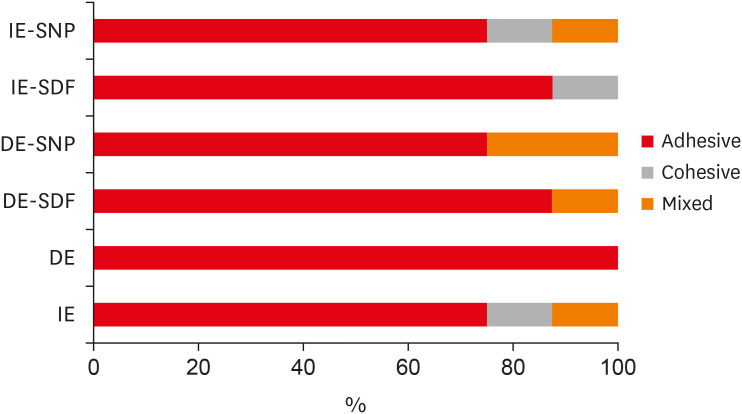
IE, intact enamel; DE, demineralized enamel; SDF, silver diamine fluoride; SNP, silver nanoparticle.
rde-46-e7-g001.jpg
This in vitro study investigated the effect of an experimental agent based on SNPs and SDF on the μ-SBS of IE and artificially DE. The experimental agent based on SNPs was the material of choice because this solution presents anticaries actions similar to those of SDF [5,6,7,9,11]. Based on the results obtained in the current study, the null hypothesis was accepted since there was no significant difference in μ-SBS between resin composite and dental structure when the treatments were used.
The μ-SBS test was used to measure resin composite adhesion resistance to human dental enamel (IE and DE) and to avoid the non-uniform distribution of interfacial tension during the micro-shear test [20,21]. Moreover, results have shown that the IE-surface pretreatment with anticaries agents did not decrease the μ-SBS of the resin composite in comparison to that of the untreated IE group. Furthermore, the use of SDF led to improved bond strength values; the same effect was reported by Pérez-Hernández et al. [22]. This may have happened because SDF reacts to the mineral hydroxyapatite in the tooth in order to form calcium fluoride and silver phosphate, which are responsible for preventing and remineralizing caries [2]. This reaction changes the enamel surface, which may explain the greater bond strength in the IE pretreated with SDF.
However, demineralized groups treated with anticaries agents before the adhesive procedure have shown lower μ-SBS values than the IE groups previously treated with SDF or SNP. It may have happened because enamel surface demineralization decreases the μ-SBS in composites due to its morphological changes that occurred on the surface [10,12,13].
Kucukyilmaz et al. [13] reported decreased resin composite bond strength in intact and demineralized dental surfaces treated with SDF. These results can be explained by several factors such as the effect of enamel demineralization on the reduction of resin composite μ-SBS, as reported above, and the negative effect of fluoride on mechanisms of adhesion to the tooth structure. Fluoride remineralization action changes enamel surface, which can decrease the adhesion of restorative materials to the dental enamel [6,10]. This phenomenon may have happened in this study, as reported by William et al. [14], who observed a significant decrease of resin composite μ-SBS on hypomineralized enamel.
Despite the reduced μ-SBS observed for DE groups previously treated with anticaries agents, values found (SDF: 30.10 MPa and SNP: 26.10 MPa) in the current study were higher than the bond strength required to assure resin composite retention to anterior or posterior teeth (17–24 MPa) [10,23]. These values suggest the possibility of using remineralizing agents such as SDF, as well as the experimental agent used in the present study, before restorations with resins composite. This effect can be explained by SNPs' size and shape in this study. The SNPs presented spherical size (7 ± 30 nm) dispersed in a colloidal solution. Failure pattern analysis indicated the prevalence of adhesive failures over cohesive or mixed failures, which indicates that the bond strength values observed in the micro-shear test were obtained from the adhesive interface (dental structure and resin composite) [16].
The bond strength effectiveness between SDF pretreated demineralized tooth surface to resin composite has already been reported in the literature. However, such studies have reported dark staining on the surface of the samples, SNP anti-caries does not compromise esthetics [17,18,24]. The disadvantage of using SDF is the appearance of black spots on the tooth structure, which restricts its use despite their high success rate [3,5,6,7,18]. Results of this study suggest that composite restorations on decayed teeth treated with the experimental anticaries agent are possible. Its bond strength was similar to SDF, without the disadvantage of staining the dental surface.
The limitation of the current in vitro study lies on the impossibility to confirm the effect of different anticaries agents on the resin composite in the oral cavity. Nevertheless, further in situ and clinical studies should be conducted to confirm these in vitro findings.
The use of anticaries agents (SDF and SNP) did not reduce the SBS of resin composite when they are used on the intact or artificially demineralized dental enamel. Thus, anticaries agents, tested in this study, can be used as a pretreatment prior to resin restoration contributing to caries prevention.
This study was financed in part by the Coordenação de Aperfeiçoamento de Pessoal de Nível Superior - Brazil (CAPES) - Finance Code 001 and National Foundation for the Development of Private Higher Education (FUNADESP).

Conflict of Interest: No potential conflict of interest relevant to this article was reported.

Author Contributions:

  • Conceptualization: Berger SB.

  • Data curation: Favaro JC, Geha O.

  • Formal analysis: Favaro JC, Geha O, Guiraldo RD, Lopes MB, Berger SB.

  • Investigation: Favaro JC, Mello-Peixoto YCT, Geha O, Guiraldo RD.

  • Methodology: Favaro JC, Geha O, Guiraldo RD, Lopes MB.

  • Project administration: Berger SB.

  • Resources: Guiraldo RD.

  • Supervision: Berger SB.

  • Validation: Favaro JC, Geha O, Dias FA.

  • Visualization: Favaro JC, Geha O.

  • Writing - original draft: Favaro JC, Dias FA.

  • Writing - review & editing: Favaro JC, Geha O, Guiraldo RD, Lopes MB, Dias FA, Berger SB.

  • 1. Yamaga R, Nishino M, Yoshida S, Yokomizo I. Diammine silver fluoride and its clinical application. J Osaka Univ Dent Sch 1972;12:1-20. PubMed
  • 2. Lou YL, Botelho MG, Darvell BW. Reaction of silver diamine fluoride with hydroxyapatite and protein. J Dent 2011;39:612-618. PubMed
  • 3. Shah S, Bhaskar V, Venkatraghavan K, Choudhary P, Ganesh M, Trivedi K. Silver diamine fluoride: a review and current applications. J Adv Oral Res 2018;5:25-35.ArticlePDF
  • 4. Rosenblatt A, Stamford TC, Niederman R. Silver diamine fluoride: a caries “silver-fluoride bullet”. J Dent Res 2009;88:116-125. ArticlePubMedPDF
  • 5. Burgess JO, Vaghela PM. Silver diamine fluoride: a successful anticarious solution with limits. Adv Dent Res 2018;29:131-134. ArticlePubMedPDF
  • 6. Santos VE Jr, Vasconcelos Filho A, Targino AG, Flores MA, Galembeck A, Caldas AF Jr, Rosenblatt A. A new “silver-bullet” to treat caries in children--nano silver fluoride: a randomised clinical trial. J Dent 2014;42:945-951. PubMed
  • 7. Targino AG, Flores MA, dos Santos Junior VE, de Godoy Bené Bezerra F, de Luna Freire H, Galembeck A, Rosenblatt A. An innovative approach to treating dental decay in children. A new anti-caries agent. J Mater Sci Mater Med 2014;25:2041-2047. ArticlePubMedPDF
  • 8. Mei ML, Nudelman F, Marzec B, Walker JM, Lo EC, Walls AW, Chu CH. Formation of fluorohydroxyapatite with silver diamine fluoride. J Dent Res 2017;96:1122-1128. ArticlePubMedPMCPDF
  • 9. Nozari A, Ajami S, Rafiei A, Niazi E. Impact of nano hydroxyapatite, nano silver fluoride and sodium fluoride varnish on primary teeth enamel remineralization: an in vitro study. J Clin Diagn Res 2017;11:ZC97-ZC100.
  • 10. Ortiz-Ruiz AJ, Muñoz-Gómez IJ, Pérez-Pardo A, Germán-Cecilia C, Martínez-Beneyto Y, Vicente A. Influence of fluoride varnish on shear bond strength of a universal adhesive on intact and demineralized enamel. Odontology 2018;106:460-468. PubMed
  • 11. Scarpelli BB, Punhagui MF, Hoeppner MG, Almeida RS, Juliani FA, Guiraldo RD, Berger SB. In vitro evaluation of the remineralizing potential and antimicrobial activity of a cariostatic agent with silver nanoparticles. Braz Dent J 2017;28:738-743. ArticlePubMed
  • 12. Akin M, Baka ZM, Ileri Z, Basciftci FA. Can demineralized enamel surfaces be bonded safely? Acta Odontol Scand 2014;72:283-289. ArticlePubMed
  • 13. Kucukyilmaz E, Savas S, Akcay M, Bolukbasi B. Effect of silver diamine fluoride and ammonium hexafluorosilicate applications with and without Er:YAG laser irradiation on the microtensile bond strength in sound and caries-affected dentin. Lasers Surg Med 2016;48:62-69. ArticlePubMedPDF
  • 14. William V, Burrow MF, Palamara JE, Messer LB. Microshear bond strength of resin composite to teeth affected by molar hypomineralization using 2 adhesive systems. Pediatr Dent 2006;28:233-241. PubMed
  • 15. Puwanawiroj A, Trairatvorakul C, Dasanayake AP, Auychai P. Microtensile bond strength between glass ionomer cement and silver diamine fluoride-treated carious primary dentin. Pediatr Dent 2018;40:291-295. PubMed
  • 16. Shimaoka AM, de Andrade AP, Cardoso MV, de Carvalho RC. The importance of adhesive area delimitation in a microshear bond strength experimental design. J Adhes Dent 2011;13:307-314. PubMed
  • 17. Quock RL, Barros JA, Yang SW, Patel SA. Effect of silver diamine fluoride on microtensile bond strength to dentin. Oper Dent 2012;37:610-616. ArticlePubMedPDF
  • 18. Selvaraj K, Sampath V, Sujatha V, Mahalaxmi S. Evaluation of microshear bond strength and nanoleakage of etch-and-rinse and self-etch adhesives to dentin pretreated with silver diamine fluoride/potassium iodide: an in vitro study. Indian J Dent Res 2016;27:421-425. ArticlePubMed
  • 19. Noronha MS, Romão DA, Cury JA, Tabchoury CP. Effect of fluoride concentration on reduction of enamel demineralization according to the cariogenic challenge. Braz Dent J 2016;27:393-398. ArticlePubMed
  • 20. Cheng YL, Musonda J, Cheng H, Attin T, Zheng M, Yu H. Effect of surface removal following bleaching on the bond strength of enamel. BMC Oral Health 2019;19:50. ArticlePubMedPMCPDF
  • 21. De Munck J, Van Landuyt K, Peumans M, Poitevin A, Lambrechts P, Braem M, Van Meerbeek B. A critical review of the durability of adhesion to tooth tissue: methods and results. J Dent Res 2005;84:118-132. ArticlePubMedPDF
  • 22. Pérez-Hernández J, Aguilar-Díaz FC, Venegas-Lancón RD, Gayosso CA, Villanueva-Vilchis MC, de la Fuente-Hernández J. Effect of silver diamine fluoride on adhesion and microleakage of a pit and fissure sealant to tooth enamel: in vitro trial. Eur Arch Paediatr Dent 2018;19:411-416. PubMed
  • 23. Swift EJ Jr, Perdigão J, Heymann HO. Bonding to enamel and dentin: a brief history and state of the art, 1995. Quintessence Int 1995;26:95-110. PubMed
  • 24. Espíndola-Castro LF, Rosenblatt A, Galembeck A, Monteiro G. Dentin staining caused by nano-silver fluoride: a comparative study. Oper Dent 2020;45:435-441. PubMed

Tables & Figures

REFERENCES

    Citations

    Citations to this article as recorded by  

      • ePub LinkePub Link
      • Cite
        CITE
        export Copy Download
        Close
        Download Citation
        Download a citation file in RIS format that can be imported by all major citation management software, including EndNote, ProCite, RefWorks, and Reference Manager.

        Format:
        • RIS — For EndNote, ProCite, RefWorks, and most other reference management software
        • BibTeX — For JabRef, BibDesk, and other BibTeX-specific software
        Include:
        • Citation for the content below
        Can silver diamine fluoride or silver nanoparticle-based anticaries agents to affect enamel bond strength?
        Restor Dent Endod. 2021;46(1):e7  Published online January 12, 2021
        Close
      • XML DownloadXML Download
      Figure
      • 0
      Can silver diamine fluoride or silver nanoparticle-based anticaries agents to affect enamel bond strength?
      Image
      Figure 1 Details of all the fracture modes according to the experimental groups.IE, intact enamel; DE, demineralized enamel; SDF, silver diamine fluoride; SNP, silver nanoparticle.
      Can silver diamine fluoride or silver nanoparticle-based anticaries agents to affect enamel bond strength?

      Experimental groups and anticaries agent composition

      Experimental groupAnticaries agent composition
      Positive control: IENA
      Negative control: DENA
      IE + 30% SDF*Silver nitrate, ammonium hydroxide, fluoroboric acid, deionized water
      DE + SDF*Silver nitrate, ammonium hydroxide, fluoroboric acid, deionized water
      IE + experimental solution with SNPSNPs
      DE + SNPSNPs

      IE, intact enamel; DE, demineralized enamel; SDF, silver diamine fluoride; SNP, silver nanoparticle; NA, not available.

      *Cariestop, Biodiâmica Química e Farmacêutica LTDA, Londrina, PR, Brazil; Experimental solution, not commercially available.

      Mean ± standard deviation micro-shear bond strength (μ-SBS) values according to the experimental groups

      SubstrateSDFSNP
      IE47.50 ± 11.60Aa*42.50 ± 12.10Aa*
      DE30.10 ± 9.50Ba†26.10 ± 8.10Ba†
      Positive control33.00 ± 11.80*
      Negative control18.90 ± 2.50

      IE, intact enamel; DE, demineralized enamel; SDF, silver diamine fluoride; SNP, silver nanoparticle.

      Mean values followed by different upper case letters, in columns, comparing the treatments, are statistically different according to Tukey's test (p < 0.05). Mean values followed by lower case letters, in rows, comparing the enamel surface, are statistically different according to Tukey's test (p < 0.05).

      *Statistically different from the negative control group according to Dunnett's test (p < 0.05); Statistically different from the positive control group according to Dunnett's test (p < 0.05).

      Table 1 Experimental groups and anticaries agent composition

      IE, intact enamel; DE, demineralized enamel; SDF, silver diamine fluoride; SNP, silver nanoparticle; NA, not available.

      *Cariestop, Biodiâmica Química e Farmacêutica LTDA, Londrina, PR, Brazil; Experimental solution, not commercially available.

      Table 2 Mean ± standard deviation micro-shear bond strength (μ-SBS) values according to the experimental groups

      IE, intact enamel; DE, demineralized enamel; SDF, silver diamine fluoride; SNP, silver nanoparticle.

      Mean values followed by different upper case letters, in columns, comparing the treatments, are statistically different according to Tukey's test (p < 0.05). Mean values followed by lower case letters, in rows, comparing the enamel surface, are statistically different according to Tukey's test (p < 0.05).

      *Statistically different from the negative control group according to Dunnett's test (p < 0.05); Statistically different from the positive control group according to Dunnett's test (p < 0.05).


      Restor Dent Endod : Restorative Dentistry & Endodontics
      Close layer
      TOP