
Search
- Page Path
- HOME > Search
Research Articles
- The effect of limonene extract on the adhesion of different endodontic cements to root dentin: an in vitro experimental study
- Nayara Lima Ferraz Aguiar, Eduardo José Soares, Guilherme Nilson Alves dos Santos, Anna Luísa Araújo Pimenta, Laryssa Karla Romano, Ricardo Gariba Silva, Fernanda de Carvalho Panzeri
- Restor Dent Endod 2025;50(2):e16. Published online May 12, 2025
- DOI: https://doi.org/10.5395/rde.2025.50.e16
-
 Abstract
 PDF
PDF PubReader
 ePub
ePub - Objectives
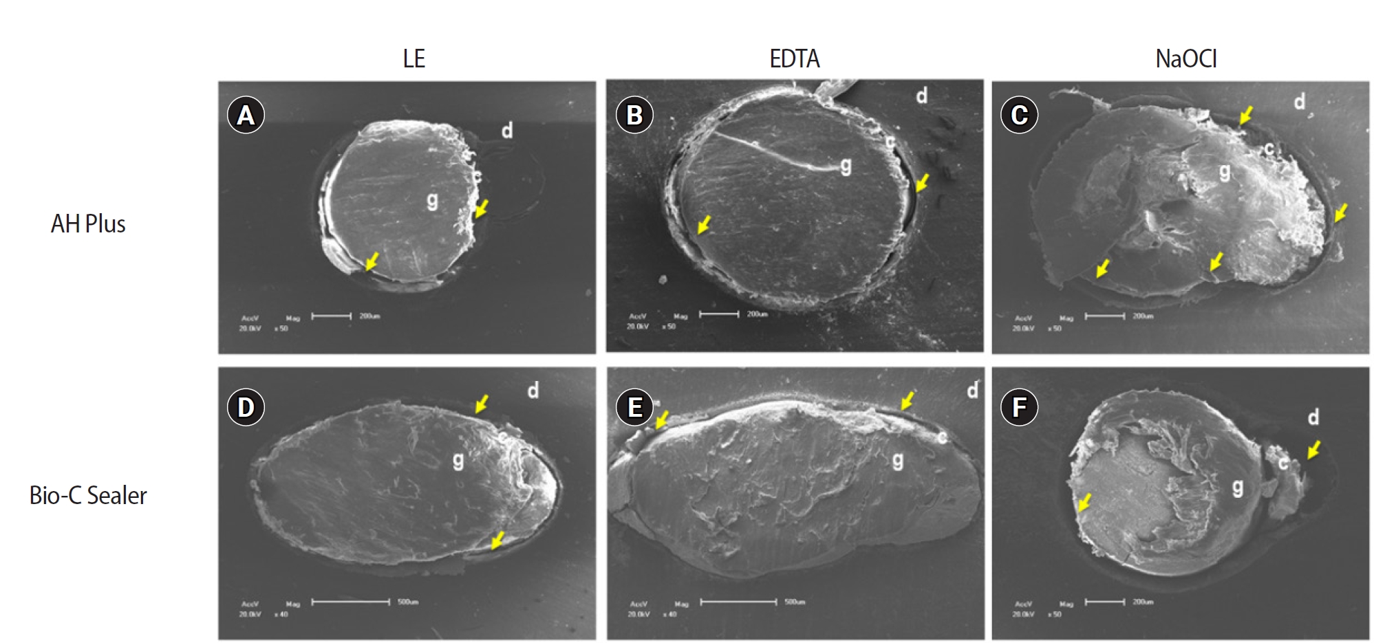
The study aimed to evaluate the effect of limonene extract (LE) on push-out bond strength (BS) to root dentin in endodontically treated teeth.
Methods
Single-rooted teeth were selected and instrumented using the reciprocating technique, then divided into three groups based on the final irrigating solution: 2.5% sodium hypochlorite (NaOCl), 17% ethylenediaminetetraacetic acid (EDTA), and 5% LE. The roots were further divided (n = 12) and obturated using the single-cone technique with epoxy resin-based (ERB) or bioceramic sealer (Bio-C). After 3 days, the roots were sectioned into 2-mm slices, obtaining two slices from each root third. Push-out BS testing was conducted at 0.5 mm/min, followed by failure pattern and adhesive interface analysis using scanning electron microscopy. Push-out BS data were analyzed by three-way analysis of variance and Tukey post-hoc test (p < 0.05).
Results
ERB showed higher BS when irrigated with EDTA (5.0 ± 2.3 MPa) compared to NaOCl (1.8 ± 1.1 MPa) (p = 0.0005), particularly in the cervical third. LE yielded intermediate values without significant differences from the other irrigants (3.5 ± 1.9 MPa) (p > 0.05). For Bio-C, the highest BS was observed in the apical third, especially with LE (9.4 ± 5.0 MPa), differing from other thirds and final irrigating solutions (p < 0.05). Mixed failure patterns were most prevalent, regardless of the irrigant solutions.
Conclusions
The combination of LE with Bio-C demonstrated superior BS in the apical third, suggesting its potential as a final irrigating solution in endodontic treatments.
- 3,095 View
- 237 Download

- The status of clinical trials regarding root canal sealers
- Ahmad AL Malak, Yasmina EL Masri, Mira Al Ziab, Nancy Zrara, Tarek Baroud, Pascale Salameh
- Restor Dent Endod 2024;49(1):e5. Published online January 15, 2024
- DOI: https://doi.org/10.5395/rde.2024.49.e5
-
Abstract
PDFPubReaderePub
Objectives This study aimed to present the results and analyses of clinical trials, including updates on the different functions of root canal sealers.
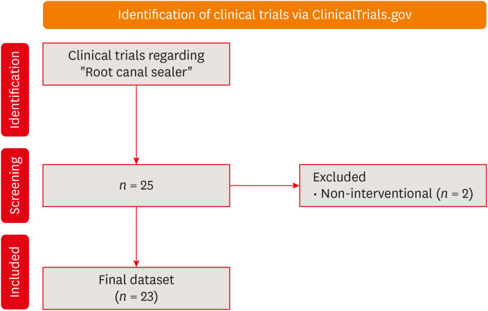
Materials and Methods In June 2023, we performed a comprehensive search of ClinicalTrials.gov to identify interventional clinical trials pertaining to root canal sealers. In total, 23 clinical trials conducted up to June 2023 were included in this study.
Results Approximately half of the trials (11 out of 23) were completed, while none were terminated or withdrawn. Each included trial had a minimum of 10 participants, with 11 trials having more than 100 participants. None of the assessed trials provided outcomes, and the majority (17 out of 23) lacked associated publications. In terms of geographic distribution, the USA and Canada did not contribute to any root canal sealer trials.
Conclusions This study highlights the lack of diversity in trial locations, the absence of reported results, and a scarcity of clinical trials examining the physicochemical properties of different sealers. Most published trials primarily focused on assessing the post-operative pain effect of these sealers, but no significant difference was found regarding post-operative pain control.
- 4,591 View
- 69 Download
- Bone repair in defects filled with AH Plus sealer and different concentrations of MTA: a study in rat tibiae
- Jessica Emanuella Rocha Paz, Priscila Oliveira Costa, Albert Alexandre Costa Souza, Ingrid Macedo de Oliveira, Lucas Fernandes Falcão, Carlos Alberto Monteiro Falcão, Maria Ângela Area Leão Ferraz, Lucielma Salmito Soares Pinto
- Restor Dent Endod 2021;46(4):e48. Published online September 2, 2021
- DOI: https://doi.org/10.5395/rde.2021.46.e48
-
Abstract
PDFPubReaderePub
Objectives This study aimed to evaluate the effects on bone repair of different concentrations of mineral trioxide aggregate (MTA) added to AH Plus.
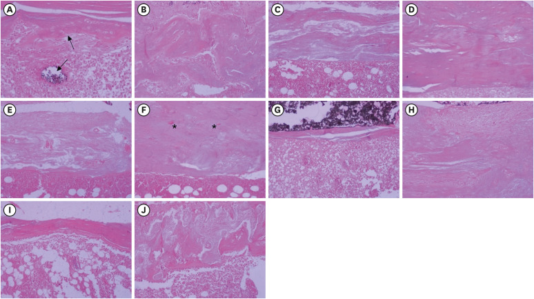
Materials and Methods Bone tissue reactions were evaluated in 30 rats (
Rattus norvegicus ) after 7 and 30 days. In the AH + MTA10, AH + MTA20, and AH + MTA30 groups, defects in the tibiae were filled with AH Plus with MTA in proportions of 10%, 20% and 30%, respectively; in the MTA-FILL group, MTA Fillapex was used; and in the control group, no sealer was used. The samples were histologically analyzed to assess bone union and maturation. The Kruskal-Wallis and Mann-Whitney tests were performed for multiple pairwise comparisons (p ≤ 0.05).Results At the 7-day time point, AH + MTA10 was superior to MTA-FILL with respect to bone union, and AH + MTA20 was superior to MTA-FILL with respect to bone maturity (
p < 0.05). At the 30-day time point, both the AH + MTA10 and AH + MTA20 experimental sealers were superior not only to MTA-FILL, but also to AH + MTA30 with respect to both parameters (p < 0.05). The results of the AH + MTA10 and AH + MTA20 groups were superior to those of the control group for both parameters and experimental time points (p < 0.05).Conclusions The results suggest the potential benefit of using a combination of these materials in situations requiring bone repair.
-
Citations
Citations to this article as recorded by
- Analysis of the cytotoxicity and bioactivity of CeraSeal, BioRoot™ and AH Plus® sealers in pre-osteoblast lineage cells
Luciano Aparecido de Almeida-Junior, Giuliana de Campos Chaves Lamarque, Henry Herrera, Maya Fernanda Manfrin Arnez, Francine Lorencetti-Silva, Raquel Assed Bezerra Silva, Léa Assed Bezerra Silva, Francisco Wanderley Garcia Paula-Silva
BMC Oral Health.2024;[Epub] CrossRef - A Review of the research methods and progress of biocompatibility evaluation of root canal sealers
Xiliang Yang, Tianxia Zheng, Nuoya Yang, Zihan Yin, Wuliang Wang, Yuhong Bai
Australian Endodontic Journal.2023; 49(S1): 508. CrossRef - Effect of Vitapex Combined with AH-Plus Paste on Inflammation in Middle-Aged and Elderly Patients with Periodontal-Endodontic Disease
Rong Hu, Fulan Zhang, Xiangyu Guo, Youren Jing, Xiaowan Lin, Liping Tian, Min Tang
Computational and Mathematical Methods in Medicine.2022; 2022: 1. CrossRef
- Analysis of the cytotoxicity and bioactivity of CeraSeal, BioRoot™ and AH Plus® sealers in pre-osteoblast lineage cells
- 2,525 View
- 15 Download
- 4 Web of Science
- 3 Crossref
- Effects of radiation therapy on the dislocation resistance of root canal sealers applied to dentin and the sealer-dentin interface: a pilot study
- Pallavi Yaduka, Rubi Kataki, Debosmita Roy, Lima Das, Shachindra Goswami
- Restor Dent Endod 2021;46(2):e22. Published online March 29, 2021
- DOI: https://doi.org/10.5395/rde.2021.46.e22
-
Abstract
PDFPubReaderePub
Objectives This study evaluated and compared the effects of radiation therapy on the dislocation resistance of AH Plus and BioRoot RCS applied to dentin and the sealer-dentin interface.
Materials and Methods Thirty single-rooted teeth were randomly assigned to 2 groups (
n = 15 each): AH Plus (Dentsply DeTrey) and BioRoot RCS (Septodont). Each group was subdivided into control and experimental groups. The experimental group was subjected to a total radiation dose of 60 Gy. The root canals of all samples were cleaned, shaped, and obturated using the single-cone technique. Dentin slices (1 mm) were sectioned from each root third for the push-out test and scanning electron microscopy (SEM) was done to examine the sealer-dentin interface. The failure mode was determined using stereomicroscopy. Bond strength data were analyzed by the independentt -test, 1-way analysis of variance, and the Tukeypost hoc test (α = 0.05).Results Significantly lower bond strength was observed in irradiated teeth than non-irradiated teeth in the AH Plus group (
p < 0.05). The BioRoot RCS group showed no significant reduction in bond strength after irradiation (p > 0.05) and showed a higher post-irradiation bond strength (209.92 ± 172.26 MPa) than the AH Plus group. SEM revealed slightly larger gap-containing regions in irradiated specimens from both groups.Conclusions The dislocation resistance of BioRoot RCS was not significantly changed by irradiation and was higher than that of AH Plus. BioRoot RCS may be the sealer of choice for root canal treatment in patients undergoing radiation therapy.
-
Citations
Citations to this article as recorded by- The impact of radiotherapy on endodontic treatment: a scoping review
Guilherme Pauletto, Giovanna Isabel Mittmann Voigt, Sidnei Flores de Pellegrin, Yasmin Padoin, Carlos Alexandre Souza Bier
Odontology.2026; 114(1): 24. CrossRef - Effects of radiotherapy dose and endodontic irrigants on universal resin cement bonding to root dentin: mechanical and interfacial analyses
Lívia Ribeiro, Luíz Carlos de Lima Dias-Júnior, Paulo Henrique dos Santos, Mariana Comparotto Minamisako, Paulo Marcelo Rodrigues, Vicente Ribeiro Netto, Bruno Alexandre Pacheco de Castro Henriques, Renata Gondo Machado, Cleonice da Silveira Teixeira, Luc
International Journal of Adhesion and Adhesives.2026; 146: 104252. CrossRef - Push-out bond strength of bioceramic-based sealers following different irrigation activation techniques
Fatma Begüm Peker, Hümeyra Çapkın, Ahsen Narbay
Journal of Applied Biomaterials & Functional Materials.2026;[Epub] CrossRef - Endodontic Management Before, During and After Head and Neck Radiotherapy: Biological, Diagnostic and Clinical Considerations in Head and Neck Cancer Patients
Jing‐zhi Ma, Franklin Tay
International Endodontic Journal.2026;[Epub] CrossRef - Impact of radiation therapy regimen on the dislodgement resistance of endodontic sealers: A micro push-out test
Marcos Testa Magoga, Rafaela Lourdes de Sousa, Luiz Carlos Lima Dias-Junior, Rayssa Sabino-Silva, Mariana Comparotto Minamisako, Paulo Marcelo Rodrigues, Vicente Ribeiro Netto, Ricardo Machado, Cleonice da Silveira Teixeira, Lucas da Fonseca Roberti Garci
International Journal of Adhesion and Adhesives.2025; 136: 103894. CrossRef - Evaluation of the root dentin bond strength and intratubular biomineralization of a premixed calcium aluminate-based hydraulic bioceramic endodontic sealer
Yu-Na Lee, Min-Kyeong Kim, Hee-Jin Kim, Mi-Kyung Yu, Kwang-Won Lee, Kyung-San Min
Journal of Oral Science.2024; 66(2): 96. CrossRef - Effects of radiotherapy dose and application time on the load-to-failure values of teeth filled with different sealers
Ozgun Gulderen, Esma Saricam, Sedef Gökhan Açikgöz, Yılmaz Tezcan
BMC Oral Health.2024;[Epub] CrossRef - Ultrasonic activation of the endodontic sealer enhances its intratubular penetration and bond strength to irradiated root dentin
Luana Duart Jordani, Amanda Freitas da Rosa, Luiz Carlos de Lima Dias-Junior, Julia Menezes Savaris, Mariana Comparotto Minamisako, Luciano Roberto da Silva, Marcio Toshio Umeda Takashima, Eduardo Antunes Bortoluzzi, Cleonice da Silveira Teixeira, Lucas d
Odontology.2024; 112(3): 917. CrossRef - Effect of the timing of primary endodontic treatment and dosage of radiation therapy on the filling material removal
Bruna Venzke Fischer, Luiz Carlos de Lima Dias‐Junior, Mariana Comparotto Minamisako, Cristiane Maria Almeida, Luciano Roberto da Silva, Eduardo Antunes Bortoluzzi, Cleonice da Silveira Teixeira, Lucas da Fonseca Roberti Garcia
Australian Endodontic Journal.2024; 50(2): 321. CrossRef - Does radiation therapy affect adhesion of tricalcium silicate cements to root dentin?
Lochan KHULLAR, Nidambur Vasudev BALLAL, Tan Fırat EYÜBOĞLU, Mutlu ÖZCAN
Journal of Applied Oral Science.2023;[Epub] CrossRef - Effect of the timing of radiation therapy on the push‐out strength of resin cement to root dentine
Patrícia da Agostim Cancelier, Renata Gondo Machado, Júlia Menezes Savaris, Eduardo Antunes Bortoluzzi, Cleonice da Silveira Teixeira, Mariana Comparotto Minamisako, Paulo Marcelo Rodrigues, Vicente Ribeiro Netto, Kamile Leonardi Dutra‐Horstmann, Lucas da
Australian Endodontic Journal.2023; 49(S1): 122. CrossRef - Influence of irrigation and laser assisted root canal disinfection protocols on dislocation resistance of a bioceramic sealer
Ivona Bago, Ana Sandrić, Katarina Beljic-Ivanovic, Boris Pažin
Photodiagnosis and Photodynamic Therapy.2022; 40: 103067. CrossRef - Influence of 2% chlorhexidine on the dislodgement resistance of AH plus, bioroot RCS, and GuttaFlow 2 sealer to dentin and sealer-dentin interface
Debosmita Roy, Rubi Kataki, Lima Das, Khushboo Jain
Journal of Conservative Dentistry.2022; 25(6): 642. CrossRef
- The impact of radiotherapy on endodontic treatment: a scoping review
- 2,984 View
- 30 Download
- 13 Web of Science
- 13 Crossref
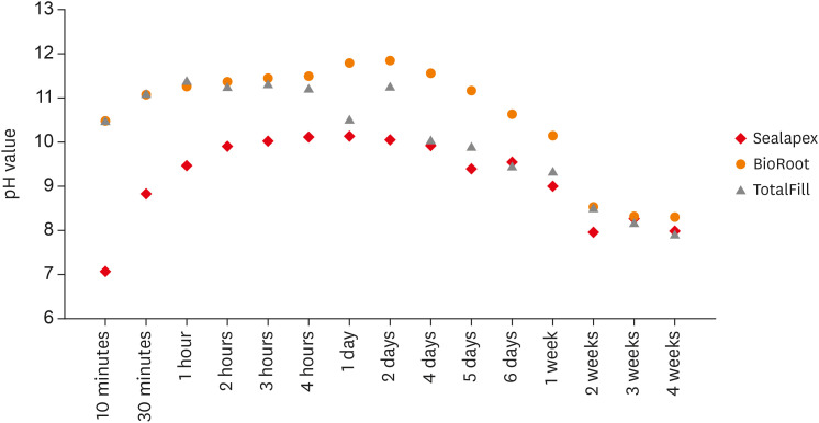
- Flow characteristics and alkalinity of novel bioceramic root canal sealers
- Anastasios Katakidis, Konstantinos Sidiropoulos, Elisabeth Koulaouzidou, Christos Gogos, Nikolaos Economides
- Restor Dent Endod 2020;45(4):e42. Published online August 18, 2020
- DOI: https://doi.org/10.5395/rde.2020.45.e42
-
Abstract
PDFPubReaderePub
Objective This study aimed to examine the physical properties (pH and flow) of 2 novel bioceramic sealers.
Materials and Methods The tested sealers were a calcium hydroxide sealer (Sealapex) and 2 bioceramic sealers (BioRoot RCS and TotalFill BC Sealer). Flow measurements were conducted according to ISO 6876/2012, with a press method of 0.05 mL of sealer. The pH of fresh samples was tested immediately after manipulation, while set samples were stored for 3 times the recommended setting time. The predetermined time intervals ranged from 3 minutes to 24 hours for fresh samples and from 10 minutes to 7 days and 4 weeks for the set samples. Analysis of variance was performed, with
p = 0.05 considered indicating significance.Results The mean flow values were 26.99 mm for BioRoot, 28.19 for Sealapex, and 30.8 mm for TotalFill BC Sealer, satisfying the ISO standard. In the set samples, BioRoot RCS had higher pH values at 24 hours to 1 week after immersion in distilled water. At 2 weeks, both bioceramic sealers had similar pH values, greater than that of Sealapex. In the fresh samples, the bioceramic sealers had significantly higher initial pH values than Sealapex (
p < 0.05). At 24 hours post-immersion, all sealers showed an alkaline pH, with the highest pH observed for TotalFill.Conclusions The TotalFill BC Sealer demonstrated the highest flow. The bioceramic sealers initially presented higher alkaline activity than the polymeric calcium hydroxide sealer. However, at 3 and 4 weeks post-immersion, all sealers had similar pH values.
-
Citations
Citations to this article as recorded by- In vitro comparative evaluation of physicochemical and mechanical properties, cytocompatibility, and antimicrobial efficacy of various bioceramic root canal sealers
Fushi Wang, Jiaxing Li, Jingjing Wan, Siyuan Li, Shijia Tang, Li Wang, Liuyan Meng
Ceramics International.2026; 52(7): 9561. CrossRef - Comparative analysis between resin-based root canal sealer and recent bioceramic-based root canal sealers using MicroCT, film thickness, and solubility
Amira Galal Ismail, Manar M. Galal, Tamer M. Hamdy
Journal of Oral Biology and Craniofacial Research.2026; 16(2): 101400. CrossRef - Setting Characteristics, Solubility, Bioactivity and Interaction with Dentin of Four Calcium Silicate-Based Endodontic Sealers
Areti Dimitra Vrochari, Anastasia Agrafioti, Maria Dimitriadi, George Eliades
Journal of Functional Biomaterials.2026; 17(4): 192. CrossRef - Cryotherapy-Driven Modulation of Postoperative Pain in Single-Visit Endodontic Treatment Across Different Obturation Materials: A Retrospective Study
Kaan Ilıcalı, Ahter Şanal Çıkman, Özge Başar
Journal of Clinical Medicine.2026; 15(10): 3899. CrossRef - Functional and Bioactive Performance of Premixed Bioceramic Sealers with Warm Obturation: A Scoping Review
Patryk Wiśniewski, Stanisław Krokosz, Małgorzata Pietruska, Anna Zalewska
Gels.2025; 11(11): 932. CrossRef - Physicochemical properties of AH plus bioceramic sealer, Bio-C Sealer, and ADseal root canal sealer
Tamer M. Hamdy, Manar M. Galal, Amira Galal Ismail, Shehabeldin Saber
Head & Face Medicine.2024;[Epub] CrossRef - Characterization and Assessment of Physical Properties of 3 Single Syringe Hydraulic Cement–based Sealers
Veksina Raman, Josette Camilleri
Journal of Endodontics.2024; 50(3): 381. CrossRef - The Impact of Silver Nanoparticles on Dentinal Tubule Penetration of Endodontic Bioceramic Sealer
Sundus Bukhary, Sarah Alkahtany, Amal Almohaimede, Nourah Alkhayatt, Shahad Alsulaiman, Salma Alohali
Applied Sciences.2024; 14(24): 11639. CrossRef - Influence of root canal moisture on the penetration of TotalFill bioceramic sealer into the dentinal tubules: A confocal laser scanning microscopy study
Archika M Singh, Tarek M Elsewify, Walid S El-Sayed, Husam H Nuawafleh, Ranya F Elemam, Bassem M Eid
Saudi Endodontic Journal.2024; 14(2): 187. CrossRef - Unusual Canal Morphology in Mandibular Premolars With Two Distal and One Mesial Canal: A Case Series
Jinesh A, Sanjana Jayakumar Nair, Saurabh Gupta, Harsh Chansoria, Gaurav Rawat
Cureus.2024;[Epub] CrossRef - A scientometric, bibliometric, and thematic map analysis of hydraulic calcium silicate root canal sealers
Anastasios Katakidis, Konstantinos Kodonas, Anastasia Fardi, Christos Gogos
Restorative Dentistry & Endodontics.2023;[Epub] CrossRef - Thermal, chemical and physical analysis of VDW.1Seal, Fill Root ST, and ADseal root canal sealers
Shehabeldin Saber, Manar M. Galal, Amira Galal Ismail, Tamer M. Hamdy
Scientific Reports.2023;[Epub] CrossRef - α-tricalcium phosphate/fluorapatite-based cement - promising dental root canal filling material
Abdul Kazuz, Zeljko Radovanovic, Djordje Veljovic, Vesna Kojic, Dimitar Jakimov, Tamara Vlajic-Tovilovic, Vesna Miletic, Rada Petrovic, Djordje Janackovic
Processing and Application of Ceramics.2022; 16(1): 22. CrossRef
- In vitro comparative evaluation of physicochemical and mechanical properties, cytocompatibility, and antimicrobial efficacy of various bioceramic root canal sealers
- 3,707 View
- 43 Download
- 13 Crossref
- A micro-computed tomographic study of remaining filling materials of two bioceramic sealers and epoxy resin sealer after retreatment
- KyungJae Kim, Da Vin Kim, Sin-Young Kim, SungEun Yang
- Restor Dent Endod 2019;44(2):e18. Published online April 26, 2019
- DOI: https://doi.org/10.5395/rde.2019.44.e18
-
Abstract
PDFPubReaderePub
Objective This study evaluated the presence of residual root canal filling material after retreatment using micro-computed tomography (micro-CT).
Materials and Methods Extracted human teeth (single- and double-rooted,
n = 21/each; C-shaped,n = 15) were prepared with ProFile and randomly assigned to three subgroups for obturation with gutta-percha and three different sealers (EndoSeal MTA, EndoSequence BC sealer, and AH Plus). After 10 days, the filling material was removed and the root canals were instrumented one size up from the previous master apical file size. The teeth were scanned using micro-CT before and after retreatment. The percentage of remaining filling material after retreatment was calculated at the coronal, middle, and apical thirds. Data were analyzed using the Kruskal-Wallis test and Mann-WhitneyU test with Bonferronipost hoc correction.Results The tested sealers showed no significant differences in the percentage of remaining filling material in single- and double-rooted teeth, although EndoSeal MTA showed the highest value in C-shaped roots (
p < 0.05). The percentage of remaining filling material of AH Plus and EndoSeal MTA was significantly higher in C-shaped roots than in single- or double-roots (p < 0.05), while that of BC sealer was similar across all root types. EndoSeal MTA showed the highest values at the apical thirds of single- and double-roots (p < 0.05); otherwise, no significant differences were observed among the coronal, middle, and apical thirds.Conclusions Within the limitations of this study, a large amount of EndoSeal MTA remained after retreatment, especially in C-shaped root canals.
-
Citations
Citations to this article as recorded by- A laboratory study comparing two methods for removing plastic carrier obturators from severely curved root canals
Tania Gancedo-Gancedo, Patricia Pereira-Lores, Venkateshbabu Nagendrababu, Paul MH Dummer, Jenifer Martín-González, Alba Bello-Castro, Inmaculada Tomás, Benjamín Martín-Biedma, Pablo Castelo-Baz
BMC Oral Health.2026;[Epub] CrossRef - Development of a deep neural network and empirical model for predicting local gas holdup profiles in bubble columns
Sebastián Uribe, Ahmed Alalou, Mario E. Cordero, Muthanna Al‐Dahhan
The Canadian Journal of Chemical Engineering.2025; 103(6): 2918. CrossRef - An In Vitro Comparison of Epoxy Resin Sealer Removal During Endodontic Retreatment
Prashant A Bondarde, Aditi S Patkar, Aishwarya R Pawar, Rukmini Pande, Akshata Deshpande, Rachana S Agrawal, Seema Gupta
Cureus.2025;[Epub] CrossRef - Calcium silicate-based sealers remnants in isthmuses of mesial roots of mandibular molars: an in vitro evaluation
David Saldanha de Brito Alencar, Ana Cristina Padilha Janini, Lauter Eston Pelepenko, Brenda Fornazaro Moraes, Francisco Haiter Neto, Marco Antonio Hungaro Duarte, Marina Angélica Marciano
Restorative Dentistry & Endodontics.2025; 50(3): e25. CrossRef - Push-out bond strength of two endodontic sealers in retreated canals using different solvents
Sara Gamal Ghanem, Walaa M. Ghoneim, Ahmed H. Labib
Tanta Dental Journal.2025; 22(3): 504. CrossRef - Assessing Volume of Two Sealers’ Remnants after Reinstrumentation Using 3D Imaging Technology: An In Vitro Comparative Study
Khalel Mutaz Dawod, Raghad Abdulrazzaq Al-Hashimi
The Journal of Contemporary Dental Practice.2025; 26(8): 743. CrossRef - Removal efficacy of two different root canal sealers in retrograde cavities: a micro-CT study
Özge Başar, Ahter Şanal Çıkman, Cangül Keskin
BMC Oral Health.2025;[Epub] CrossRef - Evaluation of the retreatability of bioceramic root canal sealers with various formulations in simulated grooves
Meltem Sümbüllü, Afzal Ali, Abdulaziz Bakhsh, Hakan Arslan
PeerJ.2025; 13: e20398. CrossRef - Correlation of Bond Strength and Dentinal Tubule Penetration Evaluation of Four Different Endodontic Sealers: AH Plus, MTA Fillapex, Endoseal MTA, and Endoseal TCS (Maruchi): An In Vitro Study
Arezoo Mirzaei Sadeghloo, Seyedali Seyedmajidi, Akam Saeidi, Elham Mahmoudi, Murilo Baena Lopes
International Journal of Dentistry.2025;[Epub] CrossRef - Root canal cleanliness and debris extrusion following retreatment of thermoplastic injection technique and bioceramic-based root canal sealer
Deniz Bender, Mert Ocak, Emel Uzunoğlu Özyürek
Clinical Oral Investigations.2024;[Epub] CrossRef - The Effect of Different Obturation Techniques Using Different Root Canal Sealers on the Residual Filling Material After Retreatment Procedures
M Sarı, K Yılmaz
Nigerian Journal of Clinical Practice.2024; 27(2): 174. CrossRef - Effect of Different Obturation Techniques on the Amount of Debris Extrusion During Endodontic Retreatment Using XP Endo Retreatment Set Files (In vitro Study)
Pawan Mohamad Amin, Hawzhen Mohammed Saeed
Sulaimani Dental Journal.2023; 10: 49. CrossRef - The efficiency of different irrigation activation techniques in the removal of calcium silicate‐based endodontic sealer from artificially created groove
Meltem Sümbüllü, Afzal Ali, Mine Büker, Hakan Arslan
Australian Endodontic Journal.2023; 49(S1): 238. CrossRef - Efficiency of diode laser and ultrasonic‐activated irrigation in retreatment of gutta percha and bioceramic sealer: An in vitro study
Rahaf A. Almohareb, Reem M. Barakat, Noor Aljarallah, Halah Mudhish, Amjaad Almutairi, Fahda N. Algahtani
Australian Endodontic Journal.2023; 49(2): 318. CrossRef - Efficiency of the new reciprocating and rotary systems with or without ultrasonics in removing root-canals filling with calcium silicate-based sealer (MTA)
Ahmad A. Madarati, Aya M. N. Sammani, Ahmad A. Alnazzawi, Ali Alrahlah
BMC Oral Health.2023;[Epub] CrossRef - Retreatability of calcium silicate‐based root canal sealer using reciprocating instrumentation with different irrigation activation techniques in single‐rooted canals
Daniele Angerame, Matteo De Biasi, Davide Porrelli, Lorenzo Bevilacqua, Riccardo Zanin, Matteo Olivi, Vassilios Kaitsas, Giovanni Olivi
Australian Endodontic Journal.2022; 48(3): 415. CrossRef - Critical analysis of research methods and experimental models to study removal of root filling materials
Mahdi A. Ajina, Pratik K. Shah, Bun San Chong
International Endodontic Journal.2022; 55(S1): 119. CrossRef - An Updated Review on Properties and Indications of Calcium Silicate‐Based Cements in Endodontic Therapy
Fateme Eskandari, Alireza Razavian, Rozhina Hamidi, Khadije Yousefi, Susan Borzou, Zohaib Khurshid
International Journal of Dentistry.2022;[Epub] CrossRef - How do imaging protocols affect the assessment of root-end fillings?
Fernanda Ferrari Esteves Torres, Reinhilde Jacobs, Mostafa EzEldeen, Karla de Faria-Vasconcelos, Juliane Maria Guerreiro-Tanomaru, Bernardo Camargo dos Santos, Mário Tanomaru-Filho
Restorative Dentistry & Endodontics.2022;[Epub] CrossRef - The Efficacy of Er:YAG Laser-Activated Shock Wave-Enhanced Emission Photoacoustic Streaming Compared to Ultrasonically Activated Irrigation and Needle Irrigation in the Removal of Bioceramic Filling Remnants from Oval Root Canals—An Ex Vivo Study
Gabrijela Kapetanović Petričević, Marko Katić, Valentina Brzović Rajić, Ivica Anić, Ivona Bago
Bioengineering.2022; 9(12): 820. CrossRef - An in vitro comparative evaluation of retreatability of a bioceramic and resin sealer using cone-beam computed tomography analysis
Sumit Sharma, Ramya Raghu, Ashish Shetty, Subhashini Rajasekhara, Harika Lakshmisetty, G. Bharath
Endodontology.2022; 34(3): 173. CrossRef - Positive and negative properties of four endodontic sealant groups: a systematic review
E. V. Chestnyh, I. O. Larichkin, M. V. Iusufova, D. I. Oreshkina, E. I. Oreshkina, V. S. Minakova, S. V. Plekhanova
Kuban Scientific Medical Bulletin.2021; 28(3): 130. CrossRef - Retrievability of bioceramic-based sealers in comparison with epoxy resin-based sealer assessed using microcomputed tomography: A systematic review of laboratory-based studies
Buvaneshwari Arul, Aswathi Varghese, Anisha Mishra, Subashini Elango, Sairathna Padmanaban, Velmurugan Natanasabapathy
Journal of Conservative Dentistry.2021; 24(5): 421. CrossRef - Micro CT pilot evaluation of removability of two endodontic sealers
David Colmenar, Tenzin Tamula, Qiang Zhu, Chul Ahn, Carolyn Primus, Takashi Komabayashi
Journal of Oral Science.2021; 63(4): 306. CrossRef - Comparison of Obturation Quality between Calcium Silicate-Based Sealers and Resin-Based Sealers for Endodontic Re-treatment
Hye-Ryeon Jin, Young-Eun Jang, Yemi Kim
Materials.2021; 15(1): 72. CrossRef - Micro-computed tomographic evaluation of a new system for root canal filling using calcium silicate-based root canal sealers
Mario Tanomaru-Filho, Fernanda Ferrari Esteves Torres, Jader Camilo Pinto, Airton Oliveira Santos-Junior, Karina Ines Medina Carita Tavares, Juliane Maria Guerreiro-Tanomaru
Restorative Dentistry & Endodontics.2020;[Epub] CrossRef - Micro-computed tomographic evaluation of the flow and filling ability of endodontic materials using different test models
Fernanda Ferrari Esteves Torres, Juliane Maria Guerreiro-Tanomaru, Gisselle Moraima Chavez-Andrade, Jader Camilo Pinto, Fábio Luiz Camargo Villela Berbert, Mario Tanomaru-Filho
Restorative Dentistry & Endodontics.2020;[Epub] CrossRef - Retreatment efficacy of hydraulic calcium silicate sealers used in single cone obturation
M. Garrib, J. Camilleri
Journal of Dentistry.2020; 98: 103370. CrossRef
- A laboratory study comparing two methods for removing plastic carrier obturators from severely curved root canals
- 2,964 View
- 37 Download
- 28 Crossref
- Bacterial leakage and micro-computed tomography evaluation in round-shaped canals obturated with bioceramic cone and sealer using matched single cone technique
- Kallaya Yanpiset, Danuchit Banomyong, Kanet Chotvorrarak, Ratchapin Laovanitch Srisatjaluk
- Restor Dent Endod 2018;43(3):e30. Published online July 5, 2018
- DOI: https://doi.org/10.5395/rde.2018.43.e30
-
Abstract
PDFPubReaderePub
Objectives To evaluate sealing ability of root canals obturated with bioceramic-impregnated gutta percha cone (BCC) or gutta percha (GP), with bioceramic sealer (BCS) or AH Plus (AH; Dentsply-Maillefer), in roundly-prepared canals using matched single-cone technique, based on bacterial leakage test, and to analyze obturation quality using micro-computed tomography (CT) analysis.
Materials and Methods Ninety-two distobuccal roots of maxillary molars were prepared using nickel-titanium files to apical size 40/0.06. The roots were divided into 4 groups (
n = 20) that were obturated with a master cone and sealer: GP/AH, BCC/AH, GP/BCS, and BCC/BCS. Bacterial leakage model usingEnterococcus faecalis was used to evaluate sealing ability for 60-day period. Obturated samples from each group (n = 4) were analyzed using micro-CT.Results All groups showed bacterial leakage at 20%–45% of samples with mean leakage times of 42–52 days. There were no significant differences in bacterial leakage among the groups. Micro-CT showed minimal gaps and voids in all groups at less than 1%.
Conclusions In roundly-prepared canals, the single cone obturation with BCC/BCS was comparable to GP/AH for bacterial leakage at 60 days.
-
Citations
Citations to this article as recorded by- Time-Dependent Volumetric and Porosity Changes of Bioceramic, Silicone Bioactive Glass-Based, and Epoxy Resin-Based Root Canal Sealers: A Micro-CT Analysis
Thanh Quang Nguyen, Chantida Pawaputanon Na Mahasarakham, Pinpana Thaweesit, Kanet Chotvorrarak, Angsana Jainaen
European Journal of Dentistry.2026;[Epub] CrossRef - Effect of Root Dentin Moisture on the Apical Sealing Ability of Root Canal Sealers: In vitro Study
Zahraa Khalil Alani, Manal Hussain Abd-alla
Al-Rafidain Journal of Medical Sciences ( ISSN 2789-3219 ).2025; 8(2): 122. CrossRef - Synthesis, physical properties, and root canal sealing of experimental MTA- and salicylate-based root canal sealers
Rafael Pino Vitti, Kusai Baroudi, Tarun Walia, Raghavandra M. Shetty, Flávia Goulart da Rosa Cardoso, Flávia de Moura Pereira, Evandro Piva, Cesar Henrique Zanchi, Gabriel Flores Abuna, Carolina Oliveira de Lima, Emmanuel João Nogueira Leal Silva, Flávio
PLOS One.2025; 20(7): e0329476. CrossRef - Impact of cone system compatibility on single cone bioceramic obturation in canals prepared with variable taper NiTi rotary files
Reem M. Barakat, Rahaf A. Almohareb, Njoom Aleid, Hoor Almowais, Aljawhara Alharbi, Meshal Al-Sharafa, Ali Alrahlah
Scientific Reports.2025;[Epub] CrossRef - Estudio de la obturación con selladores biocerámicos de conductos radiculares de premolares inferiores
Alicia Beatriz Bonafé, Cecilia Inés Rourera, Carla Pedraza, Yamila Victoria Zanoni, Soledad Salduna, Cecilia Noemi De Caso, Gabriela Martín
Methodo Investigación Aplicada a las Ciencias Biológicas.2025; 10(3): 31. CrossRef - Sealing ability of mineral trioxide aggregate: A scoping review of laboratory assessment methods
Kenta Tsuchiya, Salvatore Sauro, Jukka P. Matinlinna, Hidehiko Sano, Monica Yamauti, Deepak Mehta, Kyung‐San Min, Atsushi Tomokiyo
European Journal of Oral Sciences.2025;[Epub] CrossRef - Bacterial Leakage Testing in Dentistry: A Comprehensive Review on Methods, Models, and Clinical Relevance
Niher Tabassum Snigdha, Mohmed Isaqali Karobari, Sukhamoy Gorai
Scientifica.2025;[Epub] CrossRef - In vitro comparative evaluation of apical leakage using a bioceramic sealer with three different obturating techniques: A glucose leakage model
Tanvi S Agrawal, Shishir Singh, Rajesh S Podar, Gaurav Kulkarni, Anuprita Gadkari, Navin Agarwal
Journal of Conservative Dentistry and Endodontics.2024; 27(1): 76. CrossRef - In Vitro Microscopical and Microbiological Assessment of the Sealing Ability of Calcium Silicate-Based Root Canal Sealers
Karin Christine Huth, Sabina Noreen Wuersching, Leander Benz, Stefan Kist, Maximilian Kollmuss
Journal of Functional Biomaterials.2024; 15(11): 341. CrossRef - Comparison between AH plus sealer and total fill bioceramic sealer performance in previously untreated and retreatment cases of maxillary incisors with large-sized periapical lesion: a randomized controlled trial
Eisa Wahbi, Hassan Achour, Yasser Alsayed Tolibah
BDJ Open.2024;[Epub] CrossRef - Bacterial sealing ability of calcium silicate-based sealer for endodontic surgery: an in-vitro study
Mai M. Mansour, Sybel M. Moussa, Marwa A. Meheissen, Mahmoud R. Aboelseoud
BMC Oral Health.2024;[Epub] CrossRef - Assessment the bioactivity of zinc oxid eugenol sealer after the addition of different concentrations of nano hydroxyapatite-tyrosine amino acid
Rasha M. Al-Shamaa, Raghad A. Al-Askary
Brazilian Journal of Oral Sciences.2024; 23: e243733. CrossRef - Assessment of Bacterial Sealing Ability of Two Different Bio-Ceramic Sealers in Single-Rooted Teeth Using Single Cone Obturation Technique: An In Vitro Study
Doaa M. AlEraky, Ahmed M. Rahoma, Hatem M. Abuohashish, Abdullh AlQasser, Abbas AlHamali, Hussain M. AlHussain, Hussain M. AlShoalah, Zakrya AlSaghah, Abdulrahman Khattar, Shimaa Rifaat
Applied Sciences.2023; 13(5): 2906. CrossRef - How do imaging protocols affect the assessment of root-end fillings?
Fernanda Ferrari Esteves Torres, Reinhilde Jacobs, Mostafa EzEldeen, Karla de Faria-Vasconcelos, Juliane Maria Guerreiro-Tanomaru, Bernardo Camargo dos Santos, Mário Tanomaru-Filho
Restorative Dentistry & Endodontics.2022;[Epub] CrossRef - The impact of Morse taper implant design on microleakage at implant-healing abutment interface
Soyeon KIM, Joo Won LEE, Jae-Heon KIM, Van Mai TRUONG, Young-Seok PARK
Dental Materials Journal.2022; 41(5): 767. CrossRef - A critical analysis of research methods and experimental models to study root canal fillings
Gustavo De‐Deus, Erick Miranda Souza, Emmanuel João Nogueira Leal Silva, Felipe Gonçalves Belladonna, Marco Simões‐Carvalho, Daniele Moreira Cavalcante, Marco Aurélio Versiani
International Endodontic Journal.2022; 55(S2): 384. CrossRef - Micro‐CT assessment of gap‐containing areas along the gutta‐percha‐sealer interface in oval‐shaped canals
Gustavo De‐Deus, Gustavo O. Santos, Iara Zamboni Monteiro, Daniele M. Cavalcante, Marco Simões‐Carvalho, Felipe G. Belladonna, Emmanuel J. N. L. Silva, Erick M. Souza, Raphael Licha, Carla Zogheib, Marco A. Versiani
International Endodontic Journal.2022; 55(7): 795. CrossRef - Comparison of Sealing Ability of Bioceramic Sealer, AH Plus, and GuttaFlow in Conservatively Prepared Curved Root Canals Obturated with Single-Cone Technique: An In vitro Study
Shalan Kaul, Ajay Kumar, Bhumika Kamal Badiyani, Laxmi Sukhtankar, M. Madhumitha, Amit Kumar
Journal of Pharmacy and Bioallied Sciences.2021; 13(Suppl 1): S857. CrossRef - Micro-CT Evaluation of Four Root Canal Obturation Techniques
Mahmood Reza Kalantar Motamedi, Amin Mortaheb, Maryam Zare Jahromi, Brett E. Gilbert, Marilena Vivona
Scanning.2021; 2021: 1. CrossRef - Effects of Both Fiber Post/Core Resin Construction System and Root Canal Sealer on the Material Interface in Deep Areas of Root Canal
Hiroki Miura, Shinji Yoshii, Masataka Fujimoto, Ayako Washio, Takahiko Morotomi, Hiroshi Ikeda, Chiaki Kitamura
Materials.2021; 14(4): 982. CrossRef - Sealing ability and microbial leakage of root-end filling materials: MTA versus epoxy resin: A systematic review and meta-analysis
Mario Dioguardi, Mario Alovisi, Diego Sovereto, Giuseppe Troiano, Giancarlo Malagnino, Michele Di Cosola, Angela Pia Cazzolla, Luigi Laino, Lorenzo Lo Muzio
Heliyon.2021; 7(7): e07494. CrossRef - Development of A Nano-Apatite Based Composite Sealer for Endodontic Root Canal Filling
Angelica Bertacci, Daniele Moro, Gianfranco Ulian, Giovanni Valdrè
Journal of Composites Science.2021; 5(1): 30. CrossRef - BIOCERAMIC-BASED ROOT CANAL SEALERS
L Somolová, Z Zapletalová, M Rosa, B Novotná, I Voborná, Y Morozova
Česká stomatologie a praktické zubní lékařství.2021; 121(4): 116. CrossRef - Calcium Silicate-Based Root Canal Sealers: A Narrative Review and Clinical Perspectives
Germain Sfeir, Carla Zogheib, Shanon Patel, Thomas Giraud, Venkateshbabu Nagendrababu, Frédéric Bukiet
Materials.2021; 14(14): 3965. CrossRef - Physico-Chemical Properties of Calcium-Silicate vs. Resin Based Sealers—A Systematic Review and Meta-Analysis of Laboratory-Based Studies
Viresh Chopra, Graham Davis, Aylin Baysan
Materials.2021; 15(1): 229. CrossRef - Comparison of apical sealing ability of bioceramic sealer and epoxy resin-based sealer using the fluid filtration technique and scanning electron microscopy
Widcha Asawaworarit, Thitapa Pinyosopon, Kanittha Kijsamanmith
Journal of Dental Sciences.2020; 15(2): 186. CrossRef - Micro-computed tomographic evaluation of a new system for root canal filling using calcium silicate-based root canal sealers
Mario Tanomaru-Filho, Fernanda Ferrari Esteves Torres, Jader Camilo Pinto, Airton Oliveira Santos-Junior, Karina Ines Medina Carita Tavares, Juliane Maria Guerreiro-Tanomaru
Restorative Dentistry & Endodontics.2020;[Epub] CrossRef - A micro-computed tomographic evaluation of root canal filling with a single gutta-percha cone and calcium silicate sealer
Jong Cheon Kim, Maung Maung Kyaw Moe, Sung Kyo Kim
Restorative Dentistry & Endodontics.2020;[Epub] CrossRef - Comparative evaluation of sealing ability of gutta percha and resilon as root canal filling materials- a systematic review
Pragya Pandey, Himanshi Aggarwal, A.P. Tikku, Arpit Singh, Rhythm Bains, Shambhavi Mishra
Journal of Oral Biology and Craniofacial Research.2020; 10(2): 220. CrossRef - Micro-computed tomographic evaluation of the flow and filling ability of endodontic materials using different test models
Fernanda Ferrari Esteves Torres, Juliane Maria Guerreiro-Tanomaru, Gisselle Moraima Chavez-Andrade, Jader Camilo Pinto, Fábio Luiz Camargo Villela Berbert, Mario Tanomaru-Filho
Restorative Dentistry & Endodontics.2020;[Epub] CrossRef - Root fillings with a matched-taper single cone and two calcium silicate–based sealers: an analysis of voids using micro-computed tomography
Eugenio Pedullà, Roula S. Abiad, Gianluca Conte, Giusy R. M. La Rosa, Ernesto Rapisarda, Prasanna Neelakantan
Clinical Oral Investigations.2020; 24(12): 4487. CrossRef - Influence of different disinfection protocols on gutta-percha cones surface roughness assessed by two different methods
A.M. Nunes, J.P. Gouvea, L. da Silva
Journal of Materials Research and Technology.2019; 8(6): 5464. CrossRef - Endodontic sealers based on calcium silicates: a systematic review
David Donnermeyer, Sebastian Bürklein, Till Dammaschke, Edgar Schäfer
Odontology.2019; 107(4): 421. CrossRef
- Time-Dependent Volumetric and Porosity Changes of Bioceramic, Silicone Bioactive Glass-Based, and Epoxy Resin-Based Root Canal Sealers: A Micro-CT Analysis
- 3,475 View
- 53 Download
- 33 Crossref
- Push-out bond strength and dentinal tubule penetration of different root canal sealers used with coated core materials
- Derya Deniz Sungur, Nuhan Purali, Erdal Coşgun, Semra Calt
- Restor Dent Endod 2016;41(2):114-120. Published online May 4, 2016
- DOI: https://doi.org/10.5395/rde.2016.41.2.114
-
Abstract
PDFPubReaderePub
Objectives The aim of this study was to compare the push-out bond strength and dentinal tubule penetration of root canal sealers used with coated core materials and conventional gutta-percha.
Materials and Methods A total of 72 single-rooted human mandibular incisors were instrumented with NiTi rotary files with irrigation of 2.5% NaOCl. The smear layer was removed with 17% ethylenediaminetetraacetic acid (EDTA). Specimens were assigned into four groups according to the obturation system: Group 1, EndoRez (Ultradent Product Inc.); Group 2, Activ GP (Brasseler); Group 3, SmartSeal (DFRP Ltd. Villa Farm); Group 4, AH 26 (Dentsply de Trey)/gutta-percha (GP). For push-out bond strength measurement, two horizontal slices were obtained from each specimen (
n = 20). To compare dentinal tubule penetration, remaining 32 roots assigned to 4 groups as above were obturated with 0.1% Rhodamine B labeled sealers. One horizontal slice was obtained from the middle third of each specimen (n = 8) and scanned under confocal laser scanning electron microscope. Tubule penetration area, depth, and percentage were measured. Kruskall-Wallis test was used for statistical analysis.Results EndoRez showed significantly lower push-out bond strength than the others (
p < 0.05). No significant difference was found amongst the groups in terms of percentage of sealer penetration. SmartSeal showed the least penetration than the others (p < 0.05).Conclusions The bond strength and sealer penetration of resin-and glass ionomer-based sealers used with coated core was not superior to resin-based sealer used with conventional GP. Dentinal tubule penetration has limited effect on bond strength. The use of conventional GP with sealer seems to be sufficient in terms of push-out bond strength.
-
Citations
Citations to this article as recorded by- Does the Combination of Calcium Hydroxide and Chlorhexidine Gel Affect Dentinal Tubule Penetration of Epoxy Resin and Tricalcium Silicate‐Based Sealers?
Ecem Kuscu Baran, Aysenur Oncu, Berkan Celikten
Australian Endodontic Journal.2026;[Epub] CrossRef - Comparative evaluation of pushout bond strength of three different sealers: An in vitro study
J. S. Beautlin, M. S. Ravisankar, Arvind Kumar Alexander, R. C. Neil Ananth, V. Jevina Christy, K. T. Manoj
Journal of Conservative Dentistry and Endodontics.2025; 28(7): 637. CrossRef - Comparative analysis of dentinal tubule penetration: effects of irrigation activation methods and root canal sealers: in-vitro study
Yagmur Kilic, Mustafa Mert Tulgar, Emrah Karataslioglu, Tugba Turk
BMC Oral Health.2025;[Epub] CrossRef - Comparative evaluation of bioactive calcium silicate coating on functionalized gutta-percha and its effect on bioceramic sealer wettability – An in vitro study
Bollineni Swetha, B. Devi Priya, K. Hanisha Reddy, G. Prasanthi, T. Murali Mohan, Dumpa Tejaswi
Journal of Conservative Dentistry and Endodontics.2025; 28(7): 613. CrossRef - In Vitro and In Vivo Evaluation of a New Experimental Polydimethylsiloxane-Based Endodontic Sealer
Fabiola Cardoso Maldonado, Cesar Gaitan Fonseca, Carlos Bermudez Jimenez, Luis Alejandro Aguilera Galaviz, Margarita L. Martinez-Fierro, Lorena Troncoso Vazquez, Martha Eugenia Reyes Ortiz
Journal of Functional Biomaterials.2025; 16(11): 402. CrossRef - A Comparative Evaluation of Push-Out Bond Strength of Fiber-Reinforced Composite Posts Luted with Two Different Cement Systems: An Ex vivo Study
Shahnaz Shahnawaz, R. P. Shanoj, K. Nandakumar, M. C. Juraise, Smrithi Sudhakaran, R. Shaheena
Journal of Pharmacy and Bioallied Sciences.2025; 17(Suppl 4): S3084. CrossRef - Efficacy of Different Irrigation Activation Techniques on Dentinal Tubule Penetration of the Novel AH-Plus Bioceramic Sealer
Alhasan Almasri, Mohamad Abduljalil, Umut Aksoy
Applied Sciences.2024; 14(2): 701. CrossRef - In Vitro Microscopical and Microbiological Assessment of the Sealing Ability of Calcium Silicate-Based Root Canal Sealers
Karin Christine Huth, Sabina Noreen Wuersching, Leander Benz, Stefan Kist, Maximilian Kollmuss
Journal of Functional Biomaterials.2024; 15(11): 341. CrossRef - Comparative evaluation of dentinal tubule penetration and push-out bond strength of new injectable hydraulic calcium disilicate based root canal sealer: A single blinded in vitro study
Aman Verma, Anshul Arora, Sonali Taneja
Journal of Oral Biology and Craniofacial Research.2024; 14(2): 143. CrossRef - Scanning Electron Microscopy Analysis of the Intratubular Radicular Dentin Penetration of Calcium Hydroxide, Triple Antibiotic Paste, and Nitrofurantoin
Unmesh Khanvilkar, Sanika Pawar, Siddhesh Bandekar, Vaishnavi Dhok, Suraj Arora, Ajinkya M. Pawar, Francesco Pagnoni, Rodolfo Reda, Luca Testarelli
Journal of Personalized Medicine.2023; 13(11): 1554. CrossRef - Influence of access cavity design on calcium hydroxide removal using different cleaning protocols: a confocal laser scanning microscopy study
Seda Falakaloğlu, Merve Yeniçeri Özata, Betül Güneş, Emmanuel João Nogueira Leal Silva, Mustafa Gündoğar, Burcu Güçyetmez Topal
Restorative Dentistry & Endodontics.2023;[Epub] CrossRef - Efficacy of antimicrobial photodynamic therapy and Er,Cr:YSGG laser-activated irrigation on dentinal tubule penetration of MTA-based root canal sealer: a confocal microscopy study
Gül Keskin, Mehmet Çiloğlu
Photodiagnosis and Photodynamic Therapy.2021; 36: 102584. CrossRef - Smear layer removal and sealer penetration with different tapers after using photon-initiated photoacoustic streaming technique
Islam Mohamed Eldeeb, Nawar Naguib Nawar, Shehabeldin Mohamed Saber, Ehab El-Sayed Hassanein, Edgar Schäfer
Clinical Oral Investigations.2021; 25(8): 5025. CrossRef - The Effect of Different Final Irrigation Regimens on the Dentinal Tubule Penetration of Three Different Root Canal Sealers: A Confocal Laser Scanning Microscopy Study In Vitro
Tufan Ozasir, Birgul Eren, Kamran Gulsahi, Mete Ungor, Stefan G. Stanciu
Scanning.2021; 2021: 1. CrossRef - Effect of Vehicle and Agitation Methods on the Penetration of Calcium Hydroxide Paste in the Dentinal Tubules
Mariana de Almeida Barbosa, Kauhanna Vianna de Oliveira, Vinícius Rodrigues dos Santos, Wander José da Silva, Flávia Sens Fagundes Tomazinho, Flares Baratto-Filho, Marilisa Carneiro Leão Gabardo
Journal of Endodontics.2020; 46(7): 980. CrossRef - Micro-computed tomographic evaluation of a new system for root canal filling using calcium silicate-based root canal sealers
Mario Tanomaru-Filho, Fernanda Ferrari Esteves Torres, Jader Camilo Pinto, Airton Oliveira Santos-Junior, Karina Ines Medina Carita Tavares, Juliane Maria Guerreiro-Tanomaru
Restorative Dentistry & Endodontics.2020;[Epub] CrossRef - Dentinal tubule penetration of endodontic sealers after nonthermal plasma treatment: A confocal laser scanning microscopy study
Betul Gunes, Kubra Y. Yeter, Arslan Terlemez, Basak Seker, Yasin Altay
Microscopy Research and Technique.2019; 82(6): 903. CrossRef - Tooth crown discoloration induced by endodontic sealers: a 3-year ex vivo evaluation
Mügem Aslı Ekici, Adil Ekici, Tolga Kaskatı, Bağdagül Helvacıoğlu Kıvanç
Clinical Oral Investigations.2019; 23(5): 2097. CrossRef - Effect of Calcium Hydroxide Dressing on the Dentinal Tubule Penetration of 2 Different Root Canal Sealers: A Confocal Laser Scanning Microscopic Study
Emel Uzunoglu-Özyürek, Özge Erdoğan, Sevinç Aktemur Türker
Journal of Endodontics.2018; 44(6): 1018. CrossRef - Design Variability of the Push-out Bond Test in Endodontic Research: A Systematic Review
James Brichko, Michael F. Burrow, Peter Parashos
Journal of Endodontics.2018; 44(8): 1237. CrossRef - Evaluation of dentinal tubule penetration depth and push-out bond strength of AH 26, BioRoot RCS, and MTA Plus root canal sealers in presence or absence of smear layer
Sevinç Aktemur Türker, Emel Uzunoğlu, Nuhan Purali
Journal of Dental Research, Dental Clinics, Dental Prospects.2018; 12(4): 294. CrossRef - Comparison of Tubular Penetration of AH26, EasySeal, and SureSeal Root Canal Sealers in Single-Rooted Teeth Using Scanning Electron Microscopy
S Toursavadkohi, F Zameni, M Afkar
Journal of Research in Dental and Maxillofacial Sciences.2018; 3(3): 27. CrossRef - Effect of a Low Surface Tension Vehicle on the Dentinal Tubule Penetration of Calcium Hydroxide and Triple Antibiotic Paste
Derya Deniz Sungur, Hacer Aksel, Nuhan Purali
Journal of Endodontics.2017; 43(3): 452. CrossRef
- Does the Combination of Calcium Hydroxide and Chlorhexidine Gel Affect Dentinal Tubule Penetration of Epoxy Resin and Tricalcium Silicate‐Based Sealers?
- 3,090 View
- 19 Download
- 23 Crossref
-
A comparative evaluation of cytotoxicity of root canal sealers: an
in vitro study - Gautam Pyarelal Badole, Manjusha Madhukar Warhadpande, Ganesh Kothiramji Meshram, Rakesh Namdeoraoji Bahadure, Shubha Gopal Tawani, Gopal Tawani, Shital Gautam Badole
- Restor Dent Endod 2013;38(4):204-209. Published online November 12, 2013
- DOI: https://doi.org/10.5395/rde.2013.38.4.204
-
Abstract
PDFPubReaderePub
Objectives The objective of this
in vitro study was to evaluate and compare the cytotoxicity of four different root canal sealersi.e. Apexit Plus (Ivoclar Vivadent), Endomethasone N (Septodont), AH-26 (Dentsply) and Pulpdent Root Canal Sealer (Pulpdent), on a mouse fibroblast cell line (L929).Materials and Methods Thirty two discs for each sealer (5 mm in diameter and 2 mm in height) were fabricated in Teflon mould. The sealer extraction was made in cell culture medium (Dulbecco's Modified Eagle's Medium, DMEM) using the ratio 1.25 cm2/mL between the surface of the sealer samples and the volume of medium in a shaker incubator. Extraction of each sealer was obtained at 24 hr, 7th day, 14th day, and one month of interval. These extracts were incubated with L929 cell line and 3-(4,5-dimethylthiazol-2yl)-2,5-diphenyltetrazolium bromide (MTT) assay was done. Two-way ANOVA for interaction effects between sealer and time and Post-hoc multiple comparison using Tukey's test across all the 16 different groups were used for statistical analysis.
Results Apexit Plus root canal sealer was significantly less toxic than other sealers (
p < 0.05) and showed higher cellular growth than control. Endomethasone N showed mild cytotoxicity. AH-26 showed severe toxicity which became mild after one month while Pulpdent Root Canal Sealer showed severe to moderate toxicity.Conclusions Apexit Plus was relatively biocompatible sealer as compared to other three sealers which were cytotoxic at their initial stages, however, they became biocompatible with time.
-
Citations
Citations to this article as recorded by- The Evolution of In Vitro Toxicity Assessment Methods for Oral Cavity Tissues—From 2D Cell Cultures to Organ-on-a-Chip
Alexandra Jităreanu, Luminița Agoroaei, Ioana-Cezara Caba, Florina-Daniela Cojocaru, Liliana Vereștiuc, Mădălina Vieriu, Ioana Mârțu
Toxics.2025; 13(3): 195. CrossRef - Comparative analysis of the inflammatory response of human gingival fibroblasts to NeoSEALER Flo and CeraSeal bioceramic sealers: an in vitro study
Sarah Salah Gaafar, Abdel Rahman O. El Mekkawi, Rehab Ali Farag, Mohamed H. A. Gadelmawla, Ahmad Mostafa Hussein Mohamad Hussein, Mohamed Sayed, Mohammad Rayyan, Doaa Gamal AbdelMouez Basta
BMC Oral Health.2025;[Epub] CrossRef - Surface characteristics of nanostructured titanium surface modified via UV irradiation during SLA
S. Khorasani, G. Faraji
Applied Physics A.2025;[Epub] CrossRef - Cytotoxicity and Cell Viability of Two Bioactive Root Canal Sealers, Mineral Trioxide Aggregate, and BioRoot Root Canal Sealer: An In Vitro Study
Emmanuel Samson, Lata B Gangurde, Jaiprakash R Rathod, Pradnya S Jadhav, Sangeeta Ambhore, Pranav S Jadhav
CODS - Journal of Dentistry.2023; 14(2): 57. CrossRef - Human Gingival Fibroblasts Response to Different Endodontic Sealers: An In Vitro Study
Rita Noites, Inês Tavares, Miguel Cardoso, Isabel M. Carreira, Maria Bartolomeu, Ana S. Duarte, Ilda P. Ribeiro
Applied Sciences.2023; 13(19): 10976. CrossRef - Antibacterial and Cytotoxicity of Root Canal Sealer with the Addition of Chitosan Nanoparticle at Various Concentrations
Diatri Nari Ratih, Ema Mulyawati, Rika Kurnia Santi, Yulita Kristanti
European Journal of Dentistry.2023; 17(02): 398. CrossRef - Transforaminal and systemic diffusion of an active agent from a zinc oxide eugenol-based endodontic sealer containing hydrocortisone—in an in vivo model
Davy Aubeux, Anne Valot-Salengro, Gaelle Gautier, Arnaud Malet, Fabienne Pérez
Clinical Oral Investigations.2020; 24(12): 4395. CrossRef - PEGylated curcumin-loaded nanofibrous mats with controlled burst release through bead knot-on-spring design
Mahdi Saeed, Hamid Mirzadeh, Mojgan Zandi, Jalal Barzin
Progress in Biomaterials.2020; 9(4): 175. CrossRef - A New Calcium Silicate-Based Root Canal Dressing: Physical and Chemical Properties, Cytotoxicity and Dentinal Tubule Penetration
Natália Villa, Vanessa Valgas Dos Santos, Ubirajara Maciel da Costa, Aline Teixeira Mendes, Pedro Henrique Marks Duarte, Ricardo Abreu da Rosa, Jefferson Ricardo Pereira, Marcus Vinícius Reis Só
Brazilian Dental Journal.2020; 31(6): 598. CrossRef - Evaluation of the Antimicrobial Efficacy of Herbal Extracts Added to Root Canal Sealers of Different Bases: An In Vitro Study
Abhay M Tripathi, Minarani T Devi, Sonali K Kalra, Ujjala Ghoshal
International Journal of Clinical Pediatric Dentistry.2019; 12(5): 398. CrossRef - Endodontic-related inferior alveolar nerve injuries: A review and a therapeutic flow chart
R. Castro, M. Guivarc'h, J.M. Foletti, J.H. Catherine, C. Chossegros, L. Guyot
Journal of Stomatology, Oral and Maxillofacial Surgery.2018; 119(5): 412. CrossRef - Comparison between direct contact and extract exposure methods for PFO cytotoxicity evaluation
Girish K. Srivastava, Maria L. Alonso-Alonso, Ivan Fernandez-Bueno, Maria T. Garcia-Gutierrez, Fernando Rull, Jesús Medina, Rosa M. Coco, J. Carlos Pastor
Scientific Reports.2018;[Epub] CrossRef - Novel endodontic sealers induce cell cytotoxicity and apoptosis in a dose-dependent behavior and favorable response in mice subcutaneous tissue
L. A. B. Silva, L. U. Azevedo, A. Consolaro, F. Barnett, Y. Xu, R. A. Battaglino, P. S. Cañadas, Katharina Morant Holanda de Oliveira, R. A. B. Silva
Clinical Oral Investigations.2017; 21(9): 2851. CrossRef - Designing and fabrication of curcumin loaded PCL/PVA multi-layer nanofibrous electrospun structures as active wound dressing
Seyed Mahdi Saeed, Hamid Mirzadeh, Mojgan Zandi, Jalal Barzin
Progress in Biomaterials.2017; 6(1-2): 39. CrossRef
- The Evolution of In Vitro Toxicity Assessment Methods for Oral Cavity Tissues—From 2D Cell Cultures to Organ-on-a-Chip
- 2,553 View
- 7 Download
- 14 Crossref
Original Articles
- A comparative study on radiopacity of root canal sealers
- Tae-Min Kim, Seo-Kyoung Kim, In-Nam Hwang, Yun-Chan Hwang, Byung-Cheol Kang, Suk-Ja Yoon, Jae-Seo Lee, Won-Mann Oh
- J Korean Acad Conserv Dent 2009;34(1):61-68. Published online January 31, 2009
- DOI: https://doi.org/10.5395/JKACD.2009.34.1.061
-
Abstract
PDFPubReaderePub
This study was performed to assess the radiopacity of a variety of root canal sealers according to the specification concerning root canal sealers.
Ten materials including Tubli-Seal™, Kerr Pulp Canal Sealer™, AH 26®, AH plus®, AH plus jet™, Ad sea l™, Sealapex™, NOGENOL™, ZOB seal™, Epiphany™ and dentin were evaluated in this study. In the first part, densitometric reading of an each step of aluminum step wedge on occlusal film was performed at different voltage and exposure time. In the second part, ten specimens were radiographed simultaneously with an aluminum step wedges on the occlusal films under decided condition. The mean radiographic den sity values of the materials were transformed into radiopacity expressed equivalent thickness of aluminum (mm Al).
The following results were obtained.
1. Among the various conditions, the appropriate voltage and exposure time that meet the requirement density was 60 kVp at 0.2 s
2. All of the materials had greater radiopacity than 3 mm Al requirement of ANSI/ADA specification No. 57 (2000) and ISO No. 6876 (2001) standards.
3. The radiopacity of materials increased as thickness of materials increased.
4. The mm Al value of each specimen at 1mm in thickness has a significant difference in the statistics.
It suggests that root canal sealers have a sufficient radiopacity that meet the requirement.
-
Citations
Citations to this article as recorded by- Evaluation of radiopacity and discriminability of various fiber reinforced composite posts
Eun-Hye Lee, Hang-Moon Choi, Se-Hee Park, Jin-Woo Kim, Kyung-Mo Cho
Journal of Korean Academy of Conservative Dentistry.2010; 35(3): 188. CrossRef
- Evaluation of radiopacity and discriminability of various fiber reinforced composite posts
- 1,824 View
- 4 Download
- 1 Crossref
- Microleakage of resilon by methacrylate-based sealer and self-adhesive resin cement
- Sun-Young Ham, Jin-Woo Kim, Hye-Jin Shin, Kyung-Mo Cho, Se-Hee Park
- J Korean Acad Conserv Dent 2008;33(3):204-212. Published online May 31, 2008
- DOI: https://doi.org/10.5395/JKACD.2008.33.3.204
-
Abstract
PDFPubReaderePub
The purpose of this study was to compare the apical microleakage in root canal filled with Resilon by methacrylate-based root canal sealer or 2 different self-adhesive resin cements. Seventy single-rooted extracted human teeth were sectioned at the CEJ perpendicular to the long axis of the roots with diamond disk. Canal preparation was performed with crown-down technique using Profile NiTi rotary instruments and GG drill. Each canal was prepared to ISO size 40, .04 taper and 1 mm short from the apex. The prepared roots were randomly divided into 4 experimental groups of 15 roots each and 5 roots each for positive and negative control group. The root canals were filled by lateral condensation as follows. Group 1: Guttapercha with AH-26, Group 2: Resilon with RealSeal primer & sealer, Group 3: Resilon with Rely-X Unicem, Group 4: Resilon with BisCem. After stored in 37℃, 100% humidity chamber for 7 days, the roots were coated with 2 layers of nail varnish except apical 3 mm. The roots were then immersed in 1% methylene blue dye for 7 days. Apical microleakage was measured by a maximum length of linear dye penetration after roots were separated longitudinally. One way ANOVA and Scheffe's post-hoc test were performed for statistical analysis. Group 1 showed the least apical leakage and there was no statistical significance between Group 2, 3, 4. According to the results, the self adhesive resin cement is possible to use as sealer instead of primer & sealant when root canal filled by Resilon.
- 1,149 View
- 1 Download
- Microleakage of resilon: Effects of several self-etching primer
- Jong-Hyeon O, Se-Hee Park, Hye-Jin Shin, Kyung-Mo Cho, Jin-Woo Kim
- J Korean Acad Conserv Dent 2008;33(2):133-140. Published online March 31, 2008
- DOI: https://doi.org/10.5395/JKACD.2008.33.2.133
-
Abstract
PDFPubReaderePub
The purpose of this study was to compare the apical microleakage in root canal filled with Resilon by several self-etching primers and methacrylate-based root canal sealer. Seventy single-rooted human teeth were used in this study. The canals were instrumented by a crown-down manner with Gate-Glidden drills and .04 Taper Profile to ISO #40. The teeth were randomly divided into four experimental groups of 15 teeth each according to root canal filling material and self-etching primers and two control groups (positive and negative) of 5 teeth each as follows: group 1 - gutta percha and AH26® sealer; group 2 - Resilon, RealSeal™ primer and RealSeal™ sealer; group 3 - Resilon, Clearfil SE Bond® primer and RealSeal™ sealer group 4 - Resilon, AdheSe® primer and RealSeal™ sealer. Apical leakage was measured by a maximum length of linear dye penetration of roots sectioned longitudinally by diamond disk. Statistical analysis was performed using the One-way ANOVA followed by Scheffe's test. There were no statistical differences in the mean apical dye penetration among the groups 2, 3 and 4 of self-etching primers. And group 1, 2 and 3 had also no statistical difference in apical dye penetration. But, there was statistical difference between group 1 and 4 (p < 0.05). The group 1 showed the least dye penetration. According to the results of this study, Resilon with self-etching primer was not sealed root canal better than gutta precha with AH26® at sealing root canals. And there was no significant difference in apical leakage among the three self-etching primers.
- 1,103 View
- 1 Download
- Evaluation of the radiopacity and cytotoxicity of resinous root canal sealers
- Chang-Kyu Kim, Hyun-Wook Ryu, Hoon-Sang Chang, Byung-Do Lee, Kyung-San Min, Chan-Ui Hong
- J Korean Acad Conserv Dent 2007;32(5):419-425. Published online September 30, 2007
- DOI: https://doi.org/10.5395/JKACD.2007.32.5.419
-
Abstract
PDFPubReaderePub
The aim of this study was to evaluate the radiopacity and cytotoxicity of three resin-based (AH 26, EZ fill and AD Seal), a zinc oxide-eugenol-based (ZOB Seal), and a calcium hydroxide-based (Sealapex) root canal sealers. Specimens, 10 mm in diameter and 1 mm in thickness, were radiographed simultaneously with an aluminum step wedge using occlusal films, according to ISO 6876/2001 standards. Radiographs were digitized, and the radiopacity of sealers was compared to the different thicknesses of the aluminum step wedge, using the Scion image software. Using the 3-(4,5-dimethylthiazol-2-yl)-2,5-diphenyltetrazolium bromide (MTT) assay, the cytotoxicity of each material was determined in immortalized human periodontal ligament (IPDL) cells.
The results demonstrated that EZ fill was the most radiopaque sealer, while Sealapex was the least radiopaque (p < 0.05). AH 26, AD Seal and ZOB Seal presented intermediate radiopacity values. All the materials evaluated, except for Sealapex, presented the minimum radiopacity required by ISO standards. The cell viabilities of resin-based root canal sealers were statistically higher than that of other type of root canal sealers through the all experimental time. Further, EZ fill showed statistically lower cell viability in 24 and 48 hours compared to AD Seal and in 72 hours compared to all other resin-based root canal sealers. However, there was no correlation between the radiopacity and cytotoxicity of three resin-based root canals sealers (p > 0.05).
These results indicate that resin-based root canal sealer is more biocompatible and has advantage in terms of radiopacity.
-
Citations
Citations to this article as recorded by- A Comparative Evaluation of Two Commonly Used GP Solvents on Different Epoxy Resin-based Sealers: An In Vitro Study
Sakshi Tyagi, Ekta Choudhary, Rajat Chauhan, Ashish Choudhary
International Journal of Clinical Pediatric Dentistry.2020; 13(1): 35. CrossRef - Evaluation of softening ability of Xylene & Endosolv-R on three different epoxy resin based sealers within 1 to 2 minutes - anin vitrostudy
Pratima Ramakrishna Shenoi, Gautam Pyarelal Badole, Rajiv Tarachand Khode
Restorative Dentistry & Endodontics.2014; 39(1): 17. CrossRef - A comparative evaluation of cytotoxicity of root canal sealers: anin vitrostudy
Gautam Pyarelal Badole, Manjusha Madhukar Warhadpande, Ganesh Kothiramji Meshram, Rakesh Namdeoraoji Bahadure, Shubha Gopal Tawani, Gopal Tawani, Shital Gautam Badole
Restorative Dentistry & Endodontics.2013; 38(4): 204. CrossRef - Evaluation of radiopacity and discriminability of various fiber reinforced composite posts
Eun-Hye Lee, Hang-Moon Choi, Se-Hee Park, Jin-Woo Kim, Kyung-Mo Cho
Journal of Korean Academy of Conservative Dentistry.2010; 35(3): 188. CrossRef - A comparative study on radiopacity of root canal sealers
Tae-Min Kim, Seo-Kyoung Kim, In-Nam Hwang, Yun-Chan Hwang, Byung-Cheol Kang, Suk-Ja Yoon, Jae-Seo Lee, Won-Mann Oh
Journal of Korean Academy of Conservative Dentistry.2009; 34(1): 61. CrossRef - A comparative study on radiopacity of canal filling and retrograde root-end filling materials
Yong-Sang Kim, Seo-Kyong Kim, Yun-Chan Hwang, In-Nam Hwang, Won-Mann Oh
Journal of Korean Academy of Conservative Dentistry.2008; 33(2): 107. CrossRef
- A Comparative Evaluation of Two Commonly Used GP Solvents on Different Epoxy Resin-based Sealers: An In Vitro Study
- 2,498 View
- 6 Download
- 6 Crossref
- The influence of AH-26 and zinc oxide-eugenol root canal sealer on the shear bond strength of composite resin to dentin
- Ju-Yeon Cho, Myoung-Uk Jin, Young-Kyung Kim, Sung Kyo Kim
- J Korean Acad Conserv Dent 2006;31(3):147-152. Published online May 31, 2006
- DOI: https://doi.org/10.5395/JKACD.2006.31.3.147
-
Abstract
PDFPubReaderePub
The purpose of this study was to evaluate the influence of the AH-26 root canal sealer on the shear bond strength of composite resin to dentin.
One hundred and forty four (144) extracted, sound human molars were used. After embedding in a cylindrical mold, the occlusal part of the anatomical crown was cut away and trimmed in order to create a flat dentin surface. The teeth were randomly divided into three groups; the AH-26 sealer was applied to the AH-26 group, and zinc-oxide eugenol (ZOE) paste was applied to the ZOE group. The dentin surface of the control group did not receive any sealer.
A mount jig was placed against the surface of the teeth and the One-step dentin bonding agent was applied after acid etching. Charisma composite resin was packed into the mold and light cured. After polymerization, the alignment tube and mold were removed and the specimens were placed in distilled water at 37℃ for twenty four hours. The shear bond strength was measured by an Instron testing machine. The data for each group were subjected to one-way ANOVA and Tukey's studentized rank test so as to make comparisons between the groups.
The AH-26 group and the control group showed significantly higher shear bond strength than the ZOE group (
p < 0.05).There were no significant differences between the AH-26 group and the control one (
p > 0.05).Under the conditions of this study, the AH-26 root canal sealer did not seem to affect the shear bond strength of the composite resin to dentin while the ZOE sealer did. Therefore, there may be no decrease in bond strength when the composite resin core is built up immediately after a canal filling with AH-26 as a root canal sealer.
-
Citations
Citations to this article as recorded by- Is Zinc Oxide Eugenol Cement Still Impeding the Use of Resin-based Restoration? A Systematic Review
Fawaz Pullishery, Hajer Ayed Alhejoury, Mohammed Turkistani, Yasser Refay Souror
Dentistry and Medical Research.2021; 9(2): 59. CrossRef - A comparative evaluation of removal of gutta percha using two retreatment file system: An in vitro study
Shruthi Mary Sunil, Balakrishnan Rajkumar, Vishesh Gupta, Akanksha Bhatt, Pragyan Paliwal
IP Indian Journal of Conservative and Endodontics.2020; 5(2): 53. CrossRef - Evaluation of softening ability of Xylene & Endosolv-R on three different epoxy resin based sealers within 1 to 2 minutes - anin vitrostudy
Pratima Ramakrishna Shenoi, Gautam Pyarelal Badole, Rajiv Tarachand Khode
Restorative Dentistry & Endodontics.2014; 39(1): 17. CrossRef - Influence of Sodium Ascorbate on Microtensile Bond Strengths to Pulp Chamber Dentin treated with NaOCl
Soo-Yeon Jeon, Kwang-Won Lee, Mi-Kyung Yu
Journal of Korean Academy of Conservative Dentistry.2008; 33(6): 545. CrossRef
- Is Zinc Oxide Eugenol Cement Still Impeding the Use of Resin-based Restoration? A Systematic Review
- 2,934 View
- 6 Download
- 4 Crossref
- Cytotoxicity and genotoxicity of newly developed calcium phosphate-based root canal sealers
- Hee-Jung Kim, Seung-Ho Baek, Kwang-Shik Bae
- J Korean Acad Conserv Dent 2006;31(1):36-49. Published online January 31, 2006
- DOI: https://doi.org/10.5395/JKACD.2006.31.1.036
-
Abstract
PDFPubReaderePub
The purpose of this study was to compare the cytotoxicity by MTT test and genotoxicity by Ames test of new calcium phosphate-based root canal sealers (CAPSEAL I, CAPSEAL II) with commercially available resin-based sealers (AH 26, AH Plus), zinc oxide eugenol-based sealers (Tubliseal EWT, Pulp Canal Sealer EWT), calcium hydroxide-based sealer (Sealapex), and tricalcium phosphate based sealers (Sankin Apatite Root Canal Sealer I, II, III).
According to this study, the results were as follows:
The extracts of freshly mixed group showed higher toxicity than those of 24 h set group in MTT assay (p < 0.001).
CAPSEAL I and CAPSEAL II were less cytotoxic than AH 26, AH Plus, Tubliseal EWT, Pulp Canal Sealer EWT, Sealapex and SARCS II in freshly mixed group (p < 0.01).
AH 26 in freshly mixed group showed mutagenicity to TA98 and TA100 with and without S9 mix and AH Plus extracts also were mutagenic to TA100 with and without S9 mix.
Tubliseal EWT, Pulp Canal Sealer EWT and Sealapex in freshly mixed group were mutagenic to TA100 with S9 mix.
Among those of 24 h set groups, the extracts of SARCS II were mutagenic to TA98 with and without S9 mix and AH 26 showed mutagenic effects to TA98 with S9 mix.
No mutagenic effect of CAPSEAL I and CAPSEAL II was detected.
There is no statistically significant difference between CAPSEAL I and CAPSEAL II at MTT assay and Ames test in both freshly mixed group and 24 h set group.
-
Citations
Citations to this article as recorded by- In vitrocytotoxicity of four calcium silicate-based endodontic cements on human monocytes, a colorimetric MTT assay
Sedigheh Khedmat, Somayyeh Dehghan, Jamshid Hadjati, Farimah Masoumi, Mohammad Hossein Nekoofar, Paul Michael Howell Dummer
Restorative Dentistry & Endodontics.2014; 39(3): 149. CrossRef - A comparative evaluation of cytotoxicity of root canal sealers: anin vitrostudy
Gautam Pyarelal Badole, Manjusha Madhukar Warhadpande, Ganesh Kothiramji Meshram, Rakesh Namdeoraoji Bahadure, Shubha Gopal Tawani, Gopal Tawani, Shital Gautam Badole
Restorative Dentistry & Endodontics.2013; 38(4): 204. CrossRef - Comparison of biocompatibility of four root perforation repair materials
Min-Kyung Kang, In-Ho Bae, Jeong-Tae Koh, Yun-Chan Hwang, In-Nam Hwang, Won-Mann Oh
Journal of Korean Academy of Conservative Dentistry.2009; 34(3): 192. CrossRef
- In vitrocytotoxicity of four calcium silicate-based endodontic cements on human monocytes, a colorimetric MTT assay
- 2,240 View
- 8 Download
- 3 Crossref
- Cytotoxicity of resin-based root canal sealer, adseal
- Hee-Jung Kim, Seung-Ho Baek, Woo-Cheol Lee, Han-Soo Park, Kwang-Shik Bae
- J Korean Acad Conserv Dent 2004;29(6):498-503. Published online November 30, 2004
- DOI: https://doi.org/10.5395/JKACD.2004.29.6.498
-
Abstract
PDFPubReaderePub
The properties of ideal root canal sealers include the ability of sealing the total root canal system and no toxic effects to periradicular tissues. Cytotoxicity test using cell culture is a common screening method for evaluation of the biocompatibility of root canal sealers. The purpose of this study was to investigate the cytotoxic effect of newly developed resin-based sealer (Adseal 1, 2, and 3) comparing with those commercial resin-based sealers (AH26 and AH Plus), ZOE-based sealers (Tubliseal EWT, Pulp Canal Sealer EWT) and calcium hydroxide based sealer (Sealapex). An indirect contact test of cytotoxicity by agar diffusion was performed according to the international standard ISO 10993-5. L929 fibroblast cells were incubated at 37℃ in humidified 5% CO2-containing air atmosphere. The freshly mixed test materials were inserted into glass rings of internal diameter 5 mm and height 5 mm placed on the agar. After the 24 hrs incubation period, the decolorization zones around the test materials were assessed using an inverted microscope with a calibrated screen. A Decolorization Index was determined for each specimen. Adseal 1, 2, and 3 did not exert any cytotoxic effects, whereas AH26, AH Plus, Tubliseal EWT, Pulp Canal Sealer EWT, and Sealapex produced mild cytotoxicity.
-
Citations
Citations to this article as recorded by- Cytotoxicity and subcutaneous tissue response of beta RCS in comparison with ADSeal and AH plus endodontic sealers: in vitro/in vivo study
Mehdi Dastorani, Mohammad Reza Babaie, Babak Farzaneh, Atousa Haghdoost
The Saudi Dental Journal.2025;[Epub] CrossRef - Cytotoxicity Comparison of Sure-seal root and Adseal Sealers on mouse fibroblast Cells:Invitro study
Azam haddadikohsar, Mohammad shokrzade, Marjan Fallah, Fatemeh Shakeri
journal of research in dental sciences.2024; 21(1): 46. CrossRef - Antimicrobial efficacy of Kerr pulp canal sealer (EWT) in combination with 10% amoxicillin on Enterococcus faecalis: A confocal laser scanning microscopic study
Madhureema De Sarkar, Kundabala Mala, Suchitra Shenoy Mala, Shama Prasada Kabekkodu, Srikant Natarajan, Neeta Shetty, Priyanka Madhav Kamath, Manuel Thomas
F1000Research.2023; 12: 725. CrossRef - Antimicrobial efficacy of Kerr pulp canal sealer (EWT) in combination with 10% amoxicillin on Enterococcus faecalis: A confocal laser scanning microscopic study
Madhureema De Sarkar, Kundabala Mala, Suchitra Shenoy Mala, Shama Prasada Kabekkodu, Srikant Natarajan, Neeta Shetty, Priyanka Madhav Kamath, Manuel Thomas
F1000Research.2023; 12: 725. CrossRef - Cytotoxicity and Genotoxicity of Epoxy Resin-Based Root Canal Sealers before and after Setting Procedures
Mijoo Kim, Marc Hayashi, Bo Yu, Thomas K. Lee, Reuben H. Kim, Deuk-won Jo
Life.2022; 12(6): 847. CrossRef - Biocompatibility and Bioactivity of Four Different Root Canal Sealers in Osteoblastic Cell Line MC3T3-El
Nu-Ri Jun, Sun-Kyung Lee, Sang-Im Lee
Journal of Dental Hygiene Science.2021; 21(4): 243. CrossRef
- Cytotoxicity and subcutaneous tissue response of beta RCS in comparison with ADSeal and AH plus endodontic sealers: in vitro/in vivo study
- 1,697 View
- 7 Download
- 6 Crossref
- Influence of plugger penetration depth on the apical extrusion of root canal sealer in Continuous Wave of Condensation Technique
- Ho-Young So, Young-Mi Lee, Kwang-Keun Kim, Ki-Ok Kim, Young-Kyung Kim, Sung-Kyo Kim
- J Korean Acad Conserv Dent 2004;29(5):439-445. Published online January 14, 2004
- DOI: https://doi.org/10.5395/JKACD.2004.29.5.439
-
Abstract
PDFPubReaderePub
ABSTRACT The purpose of this study was to evaluate the influence of plugger penetration depth on the apical extrusion of root canal sealer during root canal obturation with Continuous Wave of Condensation Technique.
Root canals of forty extracted human teeth were divided into four groups and were prepared up to size 40 of 0.06 taper with ProFile. After drying, canals of three groups were filled with Continuous Wave of Condensation Technique with System B™ and different plugger penetration depths of 3, 5, and 7 mm from the apex. Canals of one group were filled with cold lateral compaction technique as a control. Canals were filled with non-standardized master gutta-percha cones and 0.02 mL of Sealapex. Apical extruded sealer was collected in a container and weighed. Data was analyzed with one-way ANOVA and Duncan’s Multiple Range Test. 3 and 5 mm penetration depth groups in Continuous Wave of Condensation Technique showed significantly more extrusion of root canal sealer than 7 mm penetration depth group (
p < 0.05). However, there was no significant difference between 7 mm depth group in Continuous Wave of Condensation Technique and cold lateral compaction group (p < 0.05).The result of this study demonstrates that deeper plugger penetration depth causes more extrusion of root canal sealer in root canal obturation by Continuous Wave of Condensation Technique. Therefore, special caution is needed when plugger penetration is deeper in the canal in Continuous Wave of Condensation Technique to minimize the amount of sealer extrusion beyond apex.
-
Citations
Citations to this article as recorded by- Influence of plugger penetration depth on the area of the canal space occupied by gutta-percha
Young Mi Lee, Ho-young So, Young Kyung Kim, Sung Kyo Kim
Journal of Korean Academy of Conservative Dentistry.2006; 31(1): 66. CrossRef
- Influence of plugger penetration depth on the area of the canal space occupied by gutta-percha
- 1,838 View
- 9 Download
- 1 Crossref
 First
First Prev
Prev

