Search
- Page Path
- HOME > Search
- Phase transformation temperatures influence the reduction ratio of fatigue resistance of nickel-titanium reciprocating files at body temperature: an in vitro experimental study
- Walid Nehme, Alfred Naaman, Lola Pedèches, Sylvie Lê, Marie Georgelin-Gurgel, Sang Won Kwak, Hyeon-Cheol Kim, Franck Diemer
- Restor Dent Endod 2025;50(4):e35. Published online November 5, 2025
- DOI: https://doi.org/10.5395/rde.2025.50.e35
-
 Abstract
 PDF
PDF PubReader
 ePub
ePub - Objectives
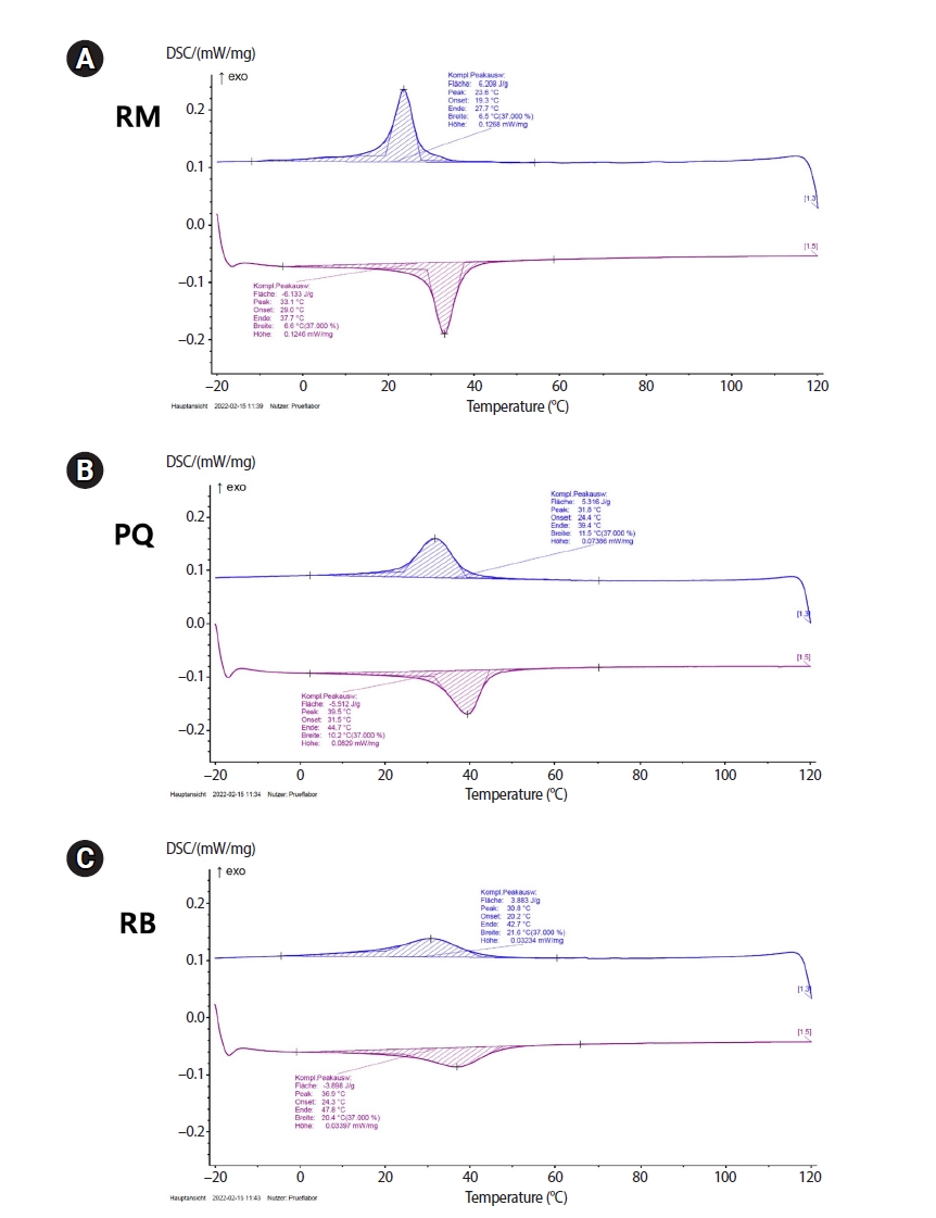
The objective of this study was to evaluate the effects of transformational temperatures on the cyclic fatigue resistance at body temperature of reciprocating file systems: R motion (RM), Procodile Q (PQ), and Reciproc Blue.
Methods
Resistance test was done in a custom-made device at room (20°C ± 1°C) and body (37°C ± 1°C) temperatures within a 60° angle of curvature and 5 mm radius of the artificial canal. The time to fracture (TTF) was recorded. The scanning electron microscope observation and differential scanning calorimetry analyses were performed. Two-way analysis of variance and Tukey post-hoc comparison were applied at a significance level of 0.05.
Results
The results showed a significant influence of temperature on instrumental breakage, regardless of the file systems (p < 0.05). The TTF is significantly decreased at body temperature (p < 0.05). PQ showed the longest TTF in both temperature conditions (p < 0.05). RM demonstrated a significantly higher TTF reduction ratio compared to the other files (p < 0.05).
Conclusions
Within the limitations of this study, the heat-treated files with reciprocating kinetics may have different reduction ratios of the fatigue resistance of the file systems under different temperature conditions. This characteristic is an important point of consideration when clinicians select the file system to reduce potential file fracture.
- 1,086 View
- 47 Download

- Multidisciplinary management of an endo-perio lesion complicated by a cemental tear: a case report
- Nishanth D. Sadhak, Akshaya Pallod, Shreyas Oza
- Restor Dent Endod 2025;50(3):e31. Published online August 22, 2025
- DOI: https://doi.org/10.5395/rde.2025.50.e31
-
 Abstract
 PDF
PDF PubReader
 ePub
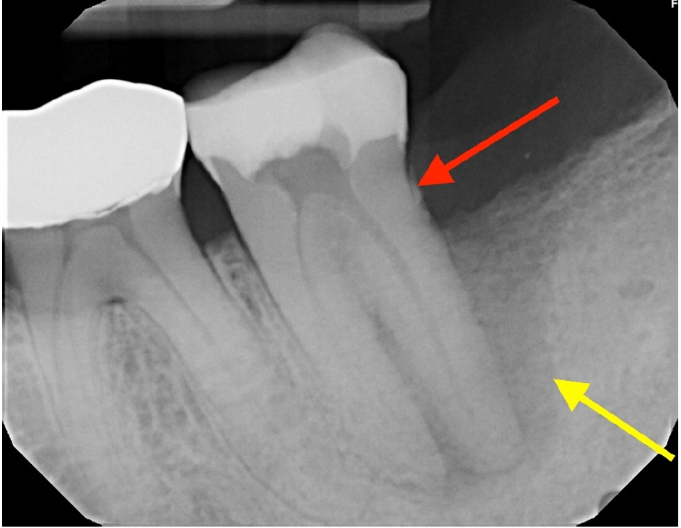
ePub - Endodontic-periodontal lesions (EPLs) complicated by cemental tears present a diagnostic and therapeutic challenge. This case report describes the successful management of a 66-year-old male patient with a mandibular second molar (#18) exhibiting an EPL complicated by a cemental tear. Clinical examination revealed a draining sinus tract, deep periodontal pockets, and radiographic evidence of a “J-shaped” lesion and a radiopaque cemental fragment. The tooth had previously initiated endodontic treatment. A multidisciplinary approach involving endodontic treatment and surgical removal of the cemental tear was implemented. At 24-month follow-up, clinical and radiographic examination revealed significant improvement in periodontal health, bone regeneration, and resolution of the lesion. This case highlights the importance of considering cemental tears in the differential diagnosis of EPLs and demonstrates the efficacy of a combined endodontic-periodontal approach for achieving predictable outcomes.
- 2,846 View
- 196 Download

- How protocol, posts, and experience affect fracture detection in multi-rooted teeth using cone-beam computed tomography: an ex vivo experimental study
- Gleica Dal’ Ongaro Savegnago, Gabriela Marzullo de Abreu, Carolina Baumgratz Spiger, Lucas Machado Maracci, Wislem Miranda de Mello, Gabriela Salatino Liedke
- Restor Dent Endod 2025;50(3):e23. Published online July 24, 2025
- DOI: https://doi.org/10.5395/rde.2025.50.e23
-
 Abstract
 PDF
PDF PubReader
 ePub
ePub - Objectives
This study aimed to evaluate the influence of cone-beam computed tomography (CBCT) acquisition protocol, the presence of intraradicular metal post, and examiner experience on the detection of complete root fractures in multi-rooted teeth.
Methods
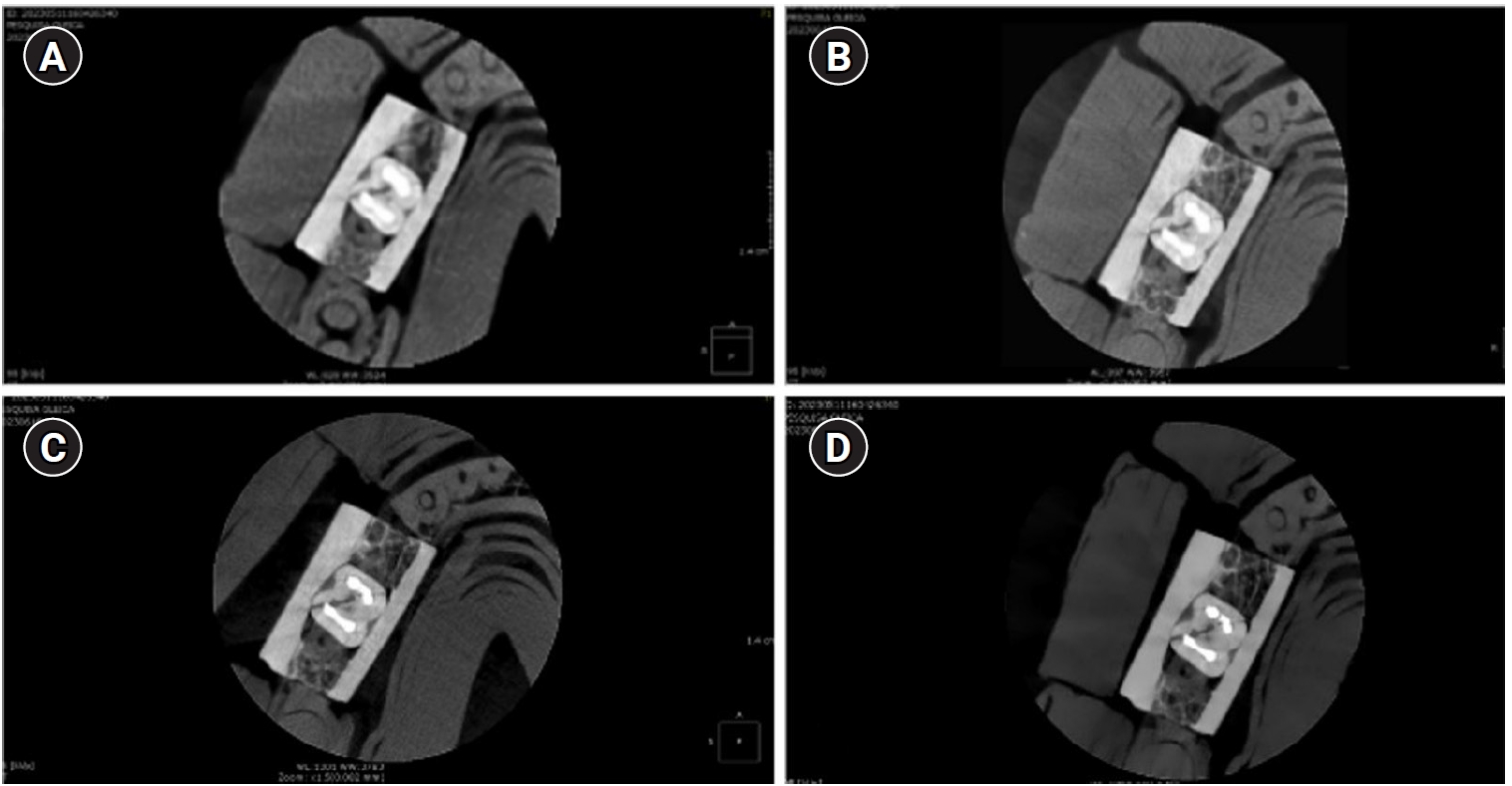
Twenty human molar teeth filled with gutta-percha were placed into artificial alveoli created in bovine ribs. The sample was divided into two groups based on the presence or absence of intraradicular posts in the distal roots. CBCT scans were obtained using four acquisition protocols with varying voxel sizes (0.28, 0.2, 0.125, and 0.80 mm). Following the creation of controlled fractures using a chisel and hammer, CBCT imaging was repeated, resulting in 160 images. Five examiners assessed the images using OnDemand software (KaVo Dental GmbH). Sensitivity, specificity, and accuracy were calculated for each examiner, CBCT protocol, and post-condition. Statistical comparisons were performed using Cochran’s Q test and McNemar test, and a significance level of 5%.
Results
In teeth without metallic posts, sensitivity, specificity, and accuracy values exceeded 0.70, 0.70, and 0.80, respectively. However, the presence of metallic posts significantly reduced diagnostic performance, particularly in low-resolution protocols evaluated by less-experienced examiners.
Conclusions
CBCT acquisition protocols should be selected based on the presence of metallic posts to optimize root fracture detection in multi-rooted teeth. Examiner experience also plays a critical role in diagnostic accuracy.
- 2,171 View
- 94 Download

- Fracture resistance and failure modes of endodontically-treated permanent teeth restored with Ribbond posts vs other post systems: a systematic review and meta-analysis of in vitro studies
- Meghana Aditya Vartak, Vibha Rahul Hegde, Sanitra Rahul Hegde, Ushaina Fanibunda
- Restor Dent Endod 2025;50(1):e5. Published online February 17, 2025
- DOI: https://doi.org/10.5395/rde.2025.50.e5

-
 Abstract
 PDF
PDF PubReader
 ePub
ePub - Objectives
This systematic review aimed to investigate the fracture resistance and mode of failure of endodontically-treated permanent teeth restored with Ribbond posts (Ribbond, Inc.) compared with endodontically-treated permanent teeth restored with other post systems.
Methods
A comprehensive, systematic literature search was carried out using several electronic databases: MEDLINE/PubMed, Google Scholar, and Cochrane Library. Two separate researchers were appointed to identify the studies meeting the eligibility criteria, and to perform the data extraction, risk of bias, and quality assessment.
Results
Twelve studies were included in the quantitative analysis. Meta-analysis was performed with 11 of the 12 included articles. The meta-analysis showed that Ribbond posts have a fracture strength less than prefabricated metal posts, cast metal posts, and prefabricated fiber posts and greater than custom e-glass fiber posts. Mode of failure analysis revealed that Ribbond posts have the most favorable non-catastrophic fractures.
Conclusions
Although Ribbond posts have lower fracture resistance, their favorable mode of failure makes them potentially the most biomimetic post system. -
Citations
Citations to this article as recorded by- Effect of Short and Long Fiber-Reinforced Composite Resins Used as Post and Core on Fracture Resistance of Premolars: An in vitro Study
Manal Hussian Abd-alla, Tuqa Jameel Ebrahim, Ahmed Sleibi Mustafa
Al-Rafidain Journal of Medical Sciences ( ISSN 2789-3219 ).2026; 10(1): 66. CrossRef - Análise comparativa dos aspectos biomecânicos dos pinos de fibra de vidro e fibra de polietileno (RIBBOND) - revisão de literatura
Ana Kamily da Cunha Silva, Tânia Regina Carvalho de Sá, Livia Duarte Santos Lopes de Carvalho, Lilian Gomes Soares Pires, Marconi Raphael de Siqueira Rego, Matheus Araújo Brito Santos Lopes
RCMOS - Revista Científica Multidisciplinar O Saber.2025;[Epub] CrossRef - Biomimetic Strategies for the Rehabilitation of Compromised Anterior Teeth
Aakansha Puri, M.S. Prathap
Contemporary Clinical Dentistry.2025; 16(3): 218. CrossRef - Clinical Outcomes of Nonmetallic Customized Post-and-Core Systems: A Systematic Review
Jonathan Jun Xian Yuen, Yew Hin Beh, Zhi Kuan Saw, Hock Siang Chua
Journal of Endodontics.2025;[Epub] CrossRef - Evaluation of fracture resistance and crack propensity of bulk-fill composite restorations reinforced by polyethylene fiber
Ayşe Aslı Şenol, Aybike Manav, Bengü Doğu Kaya, Pınar Yılmaz Atalı, Erkut Kahramanoğlu, Bilge Tarçın, Cafer Türkmen
BMC Oral Health.2025;[Epub] CrossRef - A Comparative Study on the Fracture Resistance of CAD/CAM–Fabricated Single‐Piece Post‐Crowns
Ali Erdem, Mehmet Selim Bilgin, Ibrahim Ersoy, Erhan Dilber, Ebru Nur Işık, Tan Fırat Eyüboğlu, Mutlu Özcan
Clinical and Experimental Dental Research.2025;[Epub] CrossRef - Fracture Resistance of Extensively Compromised Anterior Teeth Restored With Fiberglass Posts and Biomimetic Protocols: An In Vitro Study
Chiu Tzyy Haur, Emanuel Ewerton Mendonça Vasconcelos, Natália Gomes de Oliveira, Gabriela Queiroz de Melo Monteiro, Luís Felipe Espíndola‐Castro
Journal of Esthetic and Restorative Dentistry.2025;[Epub] CrossRef - CAD/CAM Technologies in Post and Core Restoration of Endodontically Treated Teeth: Current Evidence, Clinical Applications, and Interdisciplinary Perspectives
Rawabi Abdulrahman Ahmed, Faris Ali Aseri, Ahmed Saleh Alammari, Zaher Saleh Asiri, Fahad Oudah Al Matir, Sami Safar Al Shahrani, Abdullah Ali Alharthi, Abdulaziz Ahmed Alfaifi, Hassan Yahya Hassan Asiri, Hassan Manea Ali Al Fotais, Amal Mali Almutairi, A
Journal of Clinical Practice and Medical Research.2025; 1(3): 178. CrossRef
- Effect of Short and Long Fiber-Reinforced Composite Resins Used as Post and Core on Fracture Resistance of Premolars: An in vitro Study
- 12,071 View
- 457 Download
- 4 Web of Science
- 8 Crossref

-
Fracture resistance after root canal filling removal using ProTaper Next, ProTaper Universal Retreatment or hybrid instrumentation: an
ex vivo study - Hadeel Hassan Hanafy, Marwa Mahmoud Bedier, Suzan Abdul Wanees Amin
- Restor Dent Endod 2024;49(4):e38. Published online October 11, 2024
- DOI: https://doi.org/10.5395/rde.2024.49.e38
-
 Abstract
 PDF
PDF PubReader
 ePub
ePub Objectives This study evaluated the effect of ProTaper Next (PTN), ProTaper Universal Retreatment (PTR) and hybrid instrumentation (HI) for canal filling removal on the fracture resistance (FR), mode of failure (MoF), and filling removal time.
Materials and Methods Ninety-six, mandibular premolars were decoronated and randomly divided into 6 groups (
n = 16), as follows: sound (S), untreated canals; prepared teeth (P), canals only prepared to ProTaper Universal finishing instrument (F4); endodontically-treated (ET), prepared and obturated canals using the single-cone technique; and groups PTN, PTR, and HI where filling was removed using PTN, PTR, or HI respectively. FR under vertical loading; MoF and time were assessed. Data were analyzed (Significance level [α] = 0.05).Results There was a significant difference in FR among all groups (
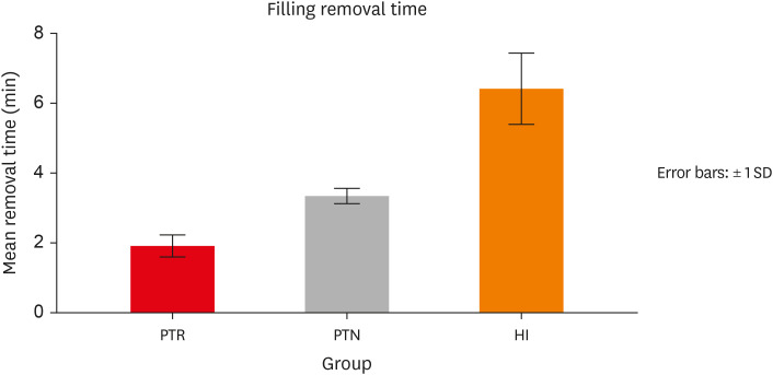
p < 0.001) (HI < P < PTN < S < ET < PTR). HI showed lower FR than S, ET and PTR, and P showed lower FR than PTR (p < 0.05). For experimental groups, there was a significant difference between every group pair (p < 0.05) No significant difference was found regarding MoF distribution (p > 0.05). HI required the highest filling removal time, while PTR required the least (p < 0.05 between every group pair).Conclusions The effect of filling removal on FR may depend on the filling removal technique/system used. PTR could be faster and protect against fracture followed by PTN; HI could adversely affect FR. FR may be associated with filling removal time.
- 2,818 View
- 122 Download

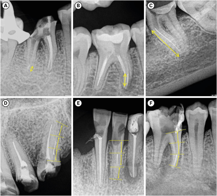
- Predictive factors in the retrieval of endodontic instruments: the relationship between the fragment length and location
- Ricardo Portigliatti, Eugenia Pilar Consoli Lizzi, Pablo Alejandro Rodríguez
- Restor Dent Endod 2024;49(4):e35. Published online September 9, 2024
- DOI: https://doi.org/10.5395/rde.2024.49.e35
-
 Abstract
 PDF
PDF PubReader
 ePub
ePub Objectives This study aimed to relate the file fragment length and location in the root canal to the retrieval chances, the clinical time required and the occurrence of secondary fractures.
Materials and Methods Sixty clinical cases of fractured instruments were included in this study. They were classified according to the instrument length and the location of the root canal. In each group, the success rate in the instrument retrieval, the clinical time required and the occurrence of secondary fractures were evaluated. The collected data were analyzed using the Kruskal-Wallis test on the basis of a 0.05 significance level.
Results The fragment length showed no significant influence on the assessed variables (
p > 0.05). The root third where the instrument was located resulted in an increased clinical time, with statistically significant differences (p < 0.05). However, the procedure success rate and the occurrence of secondary fractures showed no association with these variables.Conclusions In accordance with the findings of this study, the fractured fragment length did not influence any of the variables assessed, but it is suggested to focus on the fragment location inside the root canal to decide the retrieval of a fractured instrument.
-
Citations
Citations to this article as recorded by- Neodymium-Doped Yttrium Aluminum Perovskite (Nd:YAP) Laser in the Elimination of Endodontic Nickel-Titanium Files Fractured in Rooted Canals (Part 2: Teeth With Significant Root Curvature)
Amaury Namour, Marwan El Mobadder, Clément Cerfontaine, Patrick Matamba, Lucia Misoaga, Delphine Magnin , Praveen Arany, Samir Nammour
Cureus.2025;[Epub] CrossRef
- Neodymium-Doped Yttrium Aluminum Perovskite (Nd:YAP) Laser in the Elimination of Endodontic Nickel-Titanium Files Fractured in Rooted Canals (Part 2: Teeth With Significant Root Curvature)
- 3,026 View
- 251 Download
- 1 Crossref

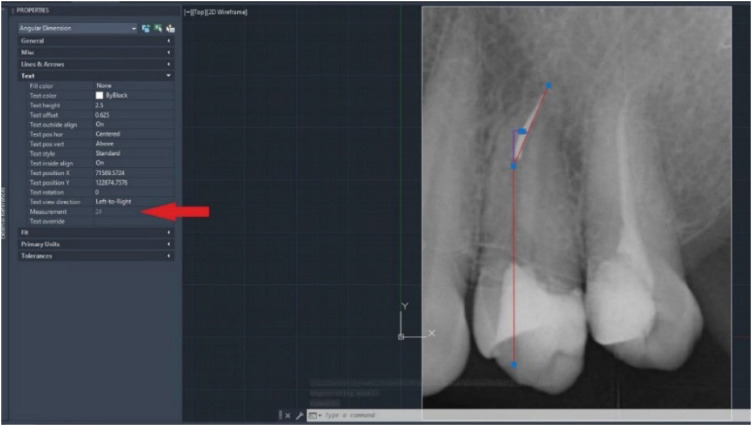
- Investigation of fracture prevalence of instruments used in root canal treatments at a faculty of dentistry: a prospective study
- Mehmet Eskibağlar, Merve Yeniçeri Özata, Mevlüt Sinan Ocak, Faruk Öztekin
- Restor Dent Endod 2023;48(4):e38. Published online November 1, 2023
- DOI: https://doi.org/10.5395/rde.2023.48.e38
-
 Abstract
 PDF
PDF PubReader
 ePub
ePub Objectives The aim of this study was to examine the use of hand or rotary files by pre-graduation (fourth- and fifth-year) and postgraduate students in endodontic treatments and to determine the incidence of file fracture and the management of cases with broken instruments.
Materials and Methods A total of 2,168 teeth undergoing primary endodontic treatment were included in this study. It was determined that 79 of these teeth resulted in broken tools. In the case of broken tools, the education level of the treating clinician, the tooth that was being treated, the canal and fracture level, the curvature of the tooth and the management of the broken instrument were recorded. Periapical radiographs of the patients were used to calculate curvature following the Schneider method.
Results There was no significant difference in the incidence of broken tools according to education level (
p > 0.05). The incidence of file fracture in molar teeth (73.4%) was higher than in other teeth (p < 0.05). More files were broken in the mandibular molar MB canal (20.25%) and in the apical third of the canals (72.1%). The risk of instrument fracture was high in teeth with moderate (44.3%) and severe (38%) curvature canals. The management of apically broken (80%) files mostly involved lefting (p < 0.05).Conclusions There was no statistically significant difference between fourth-year students, fifth-year students and postgraduate students in terms of instrument fracture.
-
Citations
Citations to this article as recorded by- Case Study of a Broken Instrument in a Primary Tooth and Literature Review
Masashi Nakano, Tatsuya Akitomo, Masashi Ogawa, Mariko Kametani, Momoko Usuda, Satoru Kusaka, Chieko Mitsuhata, Ryota Nomura
Children.2025; 12(2): 149. CrossRef - Neodymium-Doped Yttrium Aluminum Perovskite (Nd:YAP) Laser in the Elimination of Endodontic Nickel-Titanium Files Fractured in Rooted Canals (Part 2: Teeth With Significant Root Curvature)
Amaury Namour, Marwan El Mobadder, Clément Cerfontaine, Patrick Matamba, Lucia Misoaga, Delphine Magnin , Praveen Arany, Samir Nammour
Cureus.2025;[Epub] CrossRef - Pattern of endodontic instrument separation and factors affecting its retrieval: a 10-year retrospective observational study in a postgraduate institute
Velmurugan Natanasabapathy, Aswathi Varghese, Paul Kevin Abishek Karthikeyan, Srinivasan Narasimhan
Restorative Dentistry & Endodontics.2025; 50(1): e7. CrossRef - Remoção de instrumentos fraturados nos canais radiculares: Desafios, estratégias e perspectivas clínicas
João Victor da Fonseca Barbosa, Eduardo Kitto Miranda Teixeira , Laura Rodrigues Barbosa, Martinelle Ferreira da Rocha Taranto, Jáder Camilo Pinto
Research, Society and Development.2025; 14(10): e98141049749. CrossRef - Comparative evaluation of curvature severity of mesiobuccal canals in permanent maxillary 1st molar using multiple complexity risk criteria: A cone-beam computed tomography-based cross-sectional study of central Indian subpopulation
Mahima Mathur, Suparna Ganguly Saha, Rolly S. Agarwal, Shakti Singh, Manasi Kewlani, Shaurya Sahu
Endodontology.2025; 37(4): 393. CrossRef - Methods for preventing fragmentation of endodontic instruments: a systematic review
A. V. Mitronin, D. A. Ostanina, K. A. Archakov, Yu. A. Mitronin
Endodontics Today.2025; 23(4): 672. CrossRef - Perception of Dental Interns About Intracanal Fracture of Endodontic Instruments in the Central Region of Saudi Arabia: A Cross-Sectional Study
Abdullah Ahmad A. Aloyouni, Muhammad Atif Saleem Agwan, Saleh Suliman S. Almuzaini, Faris Saleh A. Alqazlan, Abdulaziz Abdulrhman A. Alshumaym, Khalid Abdullah G. Alfuryah
Journal of Pharmacy and Bioallied Sciences.2024; 16(Suppl 4): S3890. CrossRef - Predictive factors in the retrieval of endodontic instruments: the relationship between the fragment length and location
Ricardo Portigliatti, Eugenia Pilar Consoli Lizzi, Pablo Alejandro Rodríguez
Restorative Dentistry & Endodontics.2024;[Epub] CrossRef - Causes and prevention of endodontic file fractures: a review of the literature
Erkal Damla, Er Kürşat
Acta Stomatologica Marisiensis Journal.2024; 7(2): 33. CrossRef - PREVALENCE AND ENDODONTIC MANAGEMENT OF SEPARATED INSTRUMENTS INSIDE THE ROOT CANAL
Cristina Coralia Nistor, Ana Maria Țâncu , Elena Claudia Coculescu , Albu Cristina Crenguta , Stefan Milicescu , Bogdan Dimitriu
Romanian Journal of Oral Rehabilitation.2024; 16(1): 96. CrossRef
- Case Study of a Broken Instrument in a Primary Tooth and Literature Review
- 2,906 View
- 81 Download
- 4 Web of Science
- 10 Crossref

-
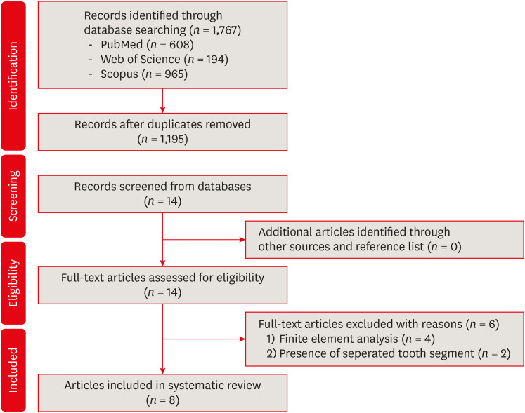
Does minimally invasive canal preparation provide higher fracture resistance of endodontically treated teeth? A systematic review of
in vitro studies - Sıla Nur Usta, Emmanuel João Nogueira Leal Silva, Seda Falakaloğlu, Mustafa Gündoğar
- Restor Dent Endod 2023;48(4):e34. Published online October 17, 2023
- DOI: https://doi.org/10.5395/rde.2023.48.e34
-
 Abstract
 PDF
PDF PubReader
 ePub
ePub This systematic review aimed to investigate whether minimally invasive root canal preparation ensures higher fracture resistance compared to conventional root canal preparation in endodontically treated teeth (ETT). A comprehensive search strategy was conducted on the “PubMed, Web of Science, and Scopus” databases, alongside reference and hand searches, with language restrictions applied. Two independent reviews selected pertinent laboratory studies that explored the effect of minimally invasive root canal preparation on fracture resistance, in comparison to larger preparation counterparts. The quality of the studies was assessed, and the risk of bias was categorized as low, moderate, or high. The electronic search yielded a total of 1,767 articles. After applying eligibility criteria, 8 studies were included. Given the low methodological quality of these studies and the large variability of fracture resistance values, the impact of reduced apical size and/or taper on the fracture resistance of the ETT can be considered uncertain. This systematic review could not reveal sufficient evidence regarding the effect of minimally invasive preparation on increasing fracture resistance of ETT, primarily due to the inherent limitations of the studies and the moderate risk of bias.
-
Citations
Citations to this article as recorded by- Impact of conservative versus conventional instrumentation on the release of inflammatory mediators and post‐operative pain in mandibular molars with asymptomatic irreversible pulpitis: A randomized clinical trial
Sıla Nur Usta, Ana Arias, Emre Avcı, Emmanuel João Nogueira Leal Silva
International Endodontic Journal.2025; 58(6): 862. CrossRef - Mapping risk of bias criteria in systematic reviews of in vitro endodontic studies: an umbrella review
Rafaella Rodrigues da Gama, Lucas Peixoto de Araújo, Evandro Piva, Leandro Perello Duro, Adriana Fernandes da Silva, Wellington Luiz de Oliveira da Rosa
Evidence-Based Dentistry.2025; 26(4): 179. CrossRef - Micro‐computed tomography evaluation of minimally invasive root canal preparation in 3D‐printed C‐shaped canal
Nutcha Supavititpattana, Siriwan Suebnukarn, Panupat Phumpatrakom, Kamon Budsaba
Australian Endodontic Journal.2024; 50(3): 621. CrossRef - Ex vivo investigation on the effect of minimally invasive endodontic treatment on vertical root fracture resistance and crack formation
Andreas Rathke, Henry Frehse, Maria Bechtold
Scientific Reports.2024;[Epub] CrossRef
- Impact of conservative versus conventional instrumentation on the release of inflammatory mediators and post‐operative pain in mandibular molars with asymptomatic irreversible pulpitis: A randomized clinical trial
- 4,192 View
- 117 Download
- 4 Web of Science
- 4 Crossref

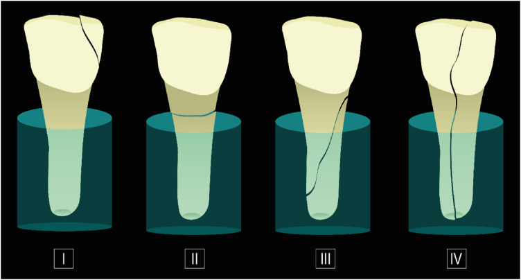
- Effects of different calcium-silicate based materials on fracture resistance of immature permanent teeth with replacement root resorption and osteoclastogenesis
- Gabriela Leite de Souza, Gabrielle Alves Nunes Freitas, Maria Tereza Hordones Ribeiro, Nelly Xiomara Alvarado Lemus, Carlos José Soares, Camilla Christian Gomes Moura
- Restor Dent Endod 2023;48(2):e21. Published online May 5, 2023
- DOI: https://doi.org/10.5395/rde.2023.48.e21
-
 Abstract
 PDF
PDF Supplementary Material
 PubReader
 ePub
ePub Objectives This study evaluated the effects of Biodentine (BD), Bio-C Repair (BCR), and mineral trioxide aggregate (MTA) plug on the fracture resistance of simulated immature teeth with replacement root resorption (RRR) and
in vitro -induced osteoclastogenesis.Materials and Methods Sixty bovine incisors simulating immature teeth and RRR were divided into 5 groups: BD and BCR groups, with samples completely filled with the respective materials; MTA group, which utilized a 3-mm apical MTA plug; RRR group, which received no root canal filling; and normal periodontal ligament (PL) group, which had no RRR and no root canal filling. All the teeth underwent cycling loading, and compression strength testing was performed using a universal testing machine. RAW 264.7 macrophages were treated with 1:16 extracts of BD, BCR, and MTA containing receptor activator of nuclear factor-kappa B ligand (RANKL) for 5 days. RANKL-induced osteoclast differentiation was assessed by staining with tartrate-resistant acid phosphatase. The fracture load and osteoclast number were analyzed using 1-way ANOVA and Tukey’s test (α = 0.05).
Results No significant difference in fracture resistance was observed among the groups (
p > 0.05). All materials similarly inhibited osteoclastogenesis (p > 0.05), except for BCR, which led to a lower percentage of osteoclasts than did MTA (p < 0.0001).Conclusions The treatment options for non-vital immature teeth with RRR did not strengthen the teeth and promoted a similar resistance to fractures in all cases. BD, MTA, and BCR showed inhibitory effects on osteoclast differentiation, with BCR yielding improved results compared to the other materials.
-
Citations
Citations to this article as recorded by- In vitro comparison of fracture strength of maxillary incisors with the simulated external root resorption cavities repaired with BioMTA or Biodentine
Tufan Ozasir, Birgul Ozasir, Nagihan Aribal, Derin Bugu Yuzer, Baris Kandemir, Kamran Gulsahi
Journal of Dental Sciences.2025; 20(3): 1532. CrossRef - Comparative Analysis of Gene Expression in Periodontal Ligament Stem Cells Exposed to Biodentine and Bio-C Repair: Implications for Cementogenesis—An In Vitro Study
Mahmoud M. Bakr, Mahmoud Al Ankily, Mohammed Meer, Mohamed Shamel
Oral.2025; 5(1): 19. CrossRef - Efficacy of Mineral Trioxide Aggregate Versus Biodentine as a Direct Pulp Capping Material in Carious Human Mature Permanent Teeth: A Systematic Review
Rashmi Misra, Nikita Toprani, Sumita Bhagwat, Aashaka Vaishnav, Aastha Dureja, Omkar Bhosale
Cureus.2025;[Epub] CrossRef - Effect of Restoration Strategy and Cavity Location on the Fracture Resistance of Teeth with External Cervical Resorption
Saadet Elpe, Öznur Sarıyılmaz
Journal of Endodontics.2025;[Epub] CrossRef - Evaluation of Different Techniques and Materials for Filling in 3-dimensional Printed Teeth Replicas with Perforating Internal Resorption by Means of Micro–Computed Tomography
Angelo J.S. Torres-Carrillo, Helena C. Assis, Rodrigo E. Salazar-Gamarra, Leonardo Moreira Teodosio, Alice C. Silva-Sousa, Jardel F. Mazzi-Chaves, Priscila B. Ferreira-Soares, Manoel D. Sousa-Neto, Fabiane C. Lopes-Olhê
Journal of Endodontics.2024; 50(2): 205. CrossRef
- In vitro comparison of fracture strength of maxillary incisors with the simulated external root resorption cavities repaired with BioMTA or Biodentine
- 2,464 View
- 65 Download
- 3 Web of Science
- 5 Crossref

-
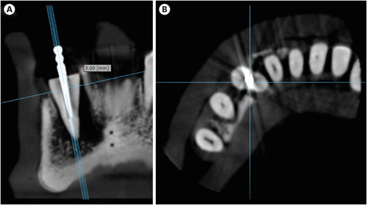
Influence of CBCT parameters on image quality and the diagnosis of vertical root fractures in teeth with metallic posts: an
ex vivo study - Larissa Pereira Lagos de Melo, Polyane Mazucatto Queiroz, Larissa Moreira-Souza, Mariana Rocha Nadaes, Gustavo Machado Santaella, Matheus Lima Oliveira, Deborah Queiroz Freitas
- Restor Dent Endod 2023;48(2):e16. Published online April 27, 2023
- DOI: https://doi.org/10.5395/rde.2023.48.e16
-
 Abstract
 PDF
PDF PubReader
 ePub
ePub Objectives The aim of this study was to evaluate the influence of peak kilovoltage (kVp) and a metal artifact reduction (MAR) tool on image quality and the diagnosis of vertical root fracture (VRF) in cone-beam computed tomography (CBCT).
Materials and Methods Twenty single-rooted human teeth filled with an intracanal metal post were divided into 2 groups: control (
n = 10) and VRF (n = 10). Each tooth was placed into the socket of a dry mandible, and CBCT scans were acquired using a Picasso Trio varying the kVp (70, 80, 90, or 99), and the use of MAR (with or without). The examinations were assessed by 5 examiners for the diagnosis of VRF using a 5-point scale. A subjective evaluation of the expression of artifacts was done by comparing random axial images of the studied protocols. The results of the diagnoses were analyzed using 2-way analysis of variance and the Tukeypost hoc test, the subjective evaluations were compared using the Friedman test, and intra-examiner reproducibility was evaluated using the weighted kappa test (α = 5%).Results The kVp and MAR did not influence the diagnosis of VRF (
p > 0.05). According to the subjective classification, the 99 kVp protocol with MAR demonstrated the least expression of artifacts, while the 70 kVp protocol without MAR led to the most artifacts.Conclusions Protocols with higher kVp combined with MAR improved the image quality of CBCT examinations. However, those factors did not lead to an improvement in the diagnosis of VRF.
-
Citations
Citations to this article as recorded by- Photon‐Counting CT for Diagnosing Vertical Root Fractures in Teeth With Metal Posts: An Ex Vivo Comparative Analysis With Four CBCT Devices
Renata M. S. Leal, Fernanda B. Fagundes, Maria F. S. A. Bortoletto, Samuel C. Kluthcovsky, Walter Coudyzer, Bruno C. Cavenago, Reinhilde Jacobs, Rocharles Cavalcante Fontenele
International Endodontic Journal.2026;[Epub] CrossRef - Diagnostic Performance of Iterative Reconstruction of Cone-beam Computed Tomography for Detecting Vertical Root Fractures in the Presence of Metal Artifacts
Matheus Barros-Costa, Gustavo Santaella, Christiano Oliveira-Santos, Deborah Queiroz Freitas, William C. Scarfe, Francisco Carlos Groppo
Journal of Endodontics.2025; 51(6): 715. CrossRef - Radiographic and Clinical Outcomes of Laser-Enhanced Disinfection in Endodontic Therapy
Janos Kantor, Sorana Maria Bucur, Eugen Silviu Bud, Victor Nimigean, Ioana Maria Crișan, Mariana Păcurar
Journal of Clinical Medicine.2025; 14(12): 4055. CrossRef - Exploring Diagnostic Reliability of CBCT for Vertical Root Fractures: A Systematic Review and Meta‐Analytical Approach
Luiz Carlos de Lima Dias-Junior, Diego Leonardo de Souza, Adriana Pinto Bezerra, Marcio Correa, Cleonice da Silveira Teixeira, Eduardo Antunes Bortoluzzi, Lucas da Fonseca Roberti Garcia, Stefano Corbella
International Journal of Dentistry.2025;[Epub] CrossRef - Deep learning for dentomaxillofacial cone-beam computed tomography image quality enhancement: A pilot study
Ali Nazari, Seyed Mohammad Yousef Najafi, Reza Abbasi, Hossein Mohammad-Rahimi, Parisa Motie, Mina Iranparvar Alamdari, Mehdi Hosseinzadeh, Ruben Pauwels, Falk Schwendicke
Imaging Science in Dentistry.2025; 55(3): 271. CrossRef - Diagnostic Accuracy of Intraoral, Extraoral and Cone Beam Computed Tomography (CBCT)-Generated Bitewings for Detecting Approximal Caries and Periodontal Bone Loss
Jyoti Mago, Alan G Lurie, Aadarsh Gopalakrishna, Aditya Tadinada
Cureus.2025;[Epub] CrossRef - Digital Dentistry Society Quality Forum: Clinical recommendations on cone-beam computed tomography for the digital dentistry workflow
Hugo Gaêta-Araujo, Rocharles Cavalcante Fontenele, Reinhilde Jacobs
Digital Dentistry Journal.2025; : 100065. CrossRef - Vertical root fracture diagnosis in teeth with metallic posts: Impact of metal artifact reduction and sharpening filters
Débora Costa Ruiz, Lucas P. Lopes Rosado, Rocharles Cavalcante Fontenele, Amanda Farias-Gomes, Deborah Queiroz Freitas
Imaging Science in Dentistry.2024; 54(2): 139. CrossRef - Comparing standard- and low-dose CBCT in diagnosis and treatment decisions for impacted mandibular third molars: a non-inferiority randomised clinical study
Kuo Feng Hung, Andy Wai Kan Yeung, May Chun Mei Wong, Michael M. Bornstein, Yiu Yan Leung
Clinical Oral Investigations.2024;[Epub] CrossRef
- Photon‐Counting CT for Diagnosing Vertical Root Fractures in Teeth With Metal Posts: An Ex Vivo Comparative Analysis With Four CBCT Devices
- 2,875 View
- 47 Download
- 7 Web of Science
- 9 Crossref

- Fracture incidence of Reciproc instruments during root canal retreatment performed by postgraduate students: a cross-sectional retrospective clinical study
- Liliana Machado Ruivo, Marcos de Azevedo Rios, Alexandre Mascarenhas Villela, Alexandre Sigrist de Martin, Augusto Shoji Kato, Rina Andrea Pelegrine, Ana Flávia Almeida Barbosa, Emmanuel João Nogueira Leal Silva, Carlos Eduardo da Silveira Bueno
- Restor Dent Endod 2021;46(4):e49. Published online September 9, 2021
- DOI: https://doi.org/10.5395/rde.2021.46.e49
-
 Abstract
 PDF
PDF PubReader
 ePub
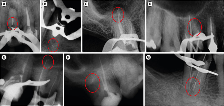
ePub Objectives To evaluate the fracture incidence of Reciproc R25 instruments (VDW) used during non-surgical root canal retreatments performed by students in a postgraduate endodontic program.
Materials and Methods From the analysis of clinical record cards and periapical radiographs of root canal retreatments performed by postgraduate students using the Reciproc R25, a total of 1,016 teeth (2,544 root canals) were selected. The instruments were discarded after a single use. The general incidence of instrument fractures and its frequency was analyzed considering the group of teeth and the root thirds where the fractures occurred. Statistical analysis was performed using the χ2 test (
p < 0.01).Results Seven instruments were separated during the procedures. The percentage of fracture in relation to the number of instrumented canals was 0.27% and 0.68% in relation to the number of instrumented teeth. Four fractures occurred in maxillary molars, 1 in a mandibular molar, 1 in a mandibular premolar and 1 in a maxillary incisor. A greater number of fractures was observed in molars when compared with the number of fractures observed in the other dental groups (
p < 0.01). Considering all of the instrument fractures, 71.43% were located in the apical third and 28.57% in the middle third (p < 0.01). One instrument fragment was removed, one bypassed, while in 5 cases, the instrument fragment remained inside the root canal.Conclusions The use of Reciproc R25 instruments in root canal retreatments carried out by postgraduate students was associated with a low incidence of fractures.
-
Citations
Citations to this article as recorded by- Reciprocating Torsional Fatigue and Mechanical Tests of Thermal-Treated Nickel Titanium Instruments
Victor Talarico Leal Vieira, Alejandro Jaime, Carlos Garcia Puente, Giuliana Soimu, Emmanuel João Nogueira Leal Silva, Carlos Nelson Elias, Gustavo de Deus
Journal of Endodontics.2025; 51(3): 359. CrossRef - Neodymium-Doped Yttrium Aluminum Perovskite (Nd:YAP) Laser in the Elimination of Endodontic Nickel-Titanium Files Fractured in Rooted Canals (Part 2: Teeth With Significant Root Curvature)
Amaury Namour, Marwan El Mobadder, Clément Cerfontaine, Patrick Matamba, Lucia Misoaga, Delphine Magnin , Praveen Arany, Samir Nammour
Cureus.2025;[Epub] CrossRef - Temperature-Dependent Effects on Cyclic Fatigue Resistance in Three Reciprocating Endodontic Systems: An In Vitro Study
Marcela Salamanca Ramos, José Aranguren, Giulia Malvicini, Cesar De Gregorio, Carmen Bonilla, Alejandro R. Perez
Materials.2025; 18(5): 952. CrossRef - The Cost of Instrument Retrieval on the Root Integrity
Marco A. Versiani, Hugo Sousa Dias, Emmanuel J. N. L. Silva, Felipe G. Belladonna, Jorge N. R. Martins, Gustavo De‐Deus
International Endodontic Journal.2025; 58(12): 1948. CrossRef - Multimethod analysis of large‐ and low‐tapered single file reciprocating instruments: Design, metallurgy, mechanical performance, and irrigation flow
Emmanuel João Nogueira Leal Silva, Fernando Peña‐Bengoa, Natasha C. Ajuz, Victor T. L. Vieira, Jorge N. R. Martins, Duarte Marques, Ricardo Pinto, Mario Rito Pereira, Francisco Manuel Braz‐Fernandes, Marco A. Versiani
International Endodontic Journal.2024; 57(5): 601. CrossRef - Nd: YAP Laser in the Elimination of Endodontic Nickel-Titanium Files Fractured in Rooted Canals (Part 1: Teeth With Minimal Root Curvature)
Amaury Namour, Marwan El Mobadder, Patrick Matamba, Lucia Misoaga, Delphine Magnin , Praveen Arany, Samir Nammour
Cureus.2024;[Epub] CrossRef - Cyclic Fatigue of Different Reciprocating Endodontic Instruments Using Matching Artificial Root Canals at Body Temperature In Vitro
Sebastian Bürklein, Paul Maßmann, Edgar Schäfer, David Donnermeyer
Materials.2024; 17(4): 827. CrossRef - Endodontic Orthograde Retreatments: Challenges and Solutions
Alessio Zanza, Rodolfo Reda, Luca Testarelli
Clinical, Cosmetic and Investigational Dentistry.2023; Volume 15: 245. CrossRef - Design, metallurgy, mechanical properties, and shaping ability of 3 heat-treated reciprocating systems: a multimethod investigation
Emmanuel J. N. L. Silva, Jorge N. R. Martins, Natasha C. Ajuz, Henrique dos Santos Antunes, Victor Talarico Leal Vieira, Francisco Manuel Braz-Fernandes, Felipe Gonçalves Belladonna, Marco Aurélio Versiani
Clinical Oral Investigations.2023; 27(5): 2427. CrossRef - Noncontact 3D evaluation of surface topography of reciprocating instruments after retreatment procedures
Miriam Fatima Zaccaro-Scelza, Renato Lenoir Cardoso Henrique Martinez, Sandro Oliveira Tavares, Fabiano Palmeira Gonçalves, Marcelo Montagnana, Emmanuel João Nogueira Leal da Silva, Pantaleo Scelza
Brazilian Dental Journal.2022; 33(3): 38. CrossRef
- Reciprocating Torsional Fatigue and Mechanical Tests of Thermal-Treated Nickel Titanium Instruments
- 2,377 View
- 21 Download
- 7 Web of Science
- 10 Crossref

- Traditional and minimally invasive access cavities in endodontics: a literature review
- Ioanna Kapetanaki, Fotis Dimopoulos, Christos Gogos
- Restor Dent Endod 2021;46(3):e46. Published online August 13, 2021
- DOI: https://doi.org/10.5395/rde.2021.46.e46
-
 Abstract
 PDF
PDF PubReader
 ePub
ePub The aim of this review was to evaluate the effects of different access cavity designs on endodontic treatment and tooth prognosis. Two independent reviewers conducted an unrestricted search of the relevant literature contained in the following electronic databases: PubMed, Science Direct, Scopus, Web of Science, and OpenGrey. The electronic search was supplemented by a manual search during the same time period. The reference lists of the articles that advanced to second-round screening were hand-searched to identify additional potential articles. Experts were also contacted in an effort to learn about possible unpublished or ongoing studies. The benefits of minimally invasive access (MIA) cavities are not yet fully supported by research data. There is no evidence that this approach can replace the traditional approach of straight-line access cavities. Guided endodontics is a new method for teeth with pulp canal calcification and apical infection, but there have been no cost-benefit investigations or time studies to verify these personal opinions. Although the purpose of MIA cavities is to reflect clinicians' interest in retaining a greater amount of the dental substance, traditional cavities are the safer method for effective instrument operation and the prevention of iatrogenic complications.
-
Citations
Citations to this article as recorded by- Benefits of Using Magnification in Access Cavity Preparation by Undergraduate Dental Students: A Micro‐Computed Tomography Study
Manal Almaslamani, Okba Mahmoud, Aya Ali, Mawada Abdelmagied
European Journal of Dental Education.2025;[Epub] CrossRef - Effect of access cavity design on canal instrumentation efficiency and fracture resistance in mandibular molars: A cone-beam computed tomography study
Dalia Al-Harith, Rawan Meshal AlOtaibi
Saudi Journal of Oral Sciences.2025; 12(1): 72. CrossRef - Full-coverage Porcelain-fused-to-metal Crown with Guided Access for Future Endodontic Treatment: A Comparative Pilot In Vitro Study
Mohammed Mashyakhy, Hemant Chourasia, Hafiz Adawi, Abdulaziz Abu-Melha, Elham Khudhayr, Rafif Bakri, Taif Kameli, Khalid Moashy, Hitesh Chohan
The Journal of Contemporary Dental Practice.2025; 26(3): 234. CrossRef - Comparative evaluation of root canal morphology in mandibular first premolars with deep radicular grooves using direct vision, dental operating microscope, 2D radiographic visualisation and micro-computed tomography
Mohmed Isaqali Karobari, Hany Mohamed Aly Ahmed, Mohd Fadhli Bin Khamis, Norliza Ibrahim, Tahir Yusuf Noorani, Miriam Fatima Zaccaro Scelza
PLOS One.2025; 20(7): e0329439. CrossRef - Impact of conservative and traditional endodontic accesses on the strength of maxillary zirconia crowns
Carlos A. Jurado, Gustavo Morrice, Mark Antal, Silvia Rojas‐Rueda, Francisco X. Azpiazu‐Flores, Brian R. Morrow, Franklin Garcia‐Godoy, Damian J. Lee
Journal of Prosthodontics.2025;[Epub] CrossRef - A Finite Element Method Study of Stress Distribution in Dental Hard Tissues: Impact of Access Cavity Design and Restoration Material
Mihaela-Roxana Boțilă, Dragos Laurențiu Popa, Răzvan Mercuț, Monica Mihaela Iacov-Crăițoiu, Monica Scrieciu, Sanda Mihaela Popescu, Veronica Mercuț
Bioengineering.2024; 11(9): 878. CrossRef - Impact of Access Cavity Design on Fracture Resistance of Endodontically Treated Maxillary First Premolar: In Vitro
Anju Daniel, Abdul Rahman Saleh, Anas Al-Jadaa, Waad Kheder
Brazilian Dental Journal.2024;[Epub] CrossRef - Management of Traumatized Teeth With Severely Calcified Canals and Minimally Invasive Access Cavity Using the AReneto® System: A Case Report
Pucha Sai Manaswini, Varun Prabhuji, Champa C, Srirekha A, Veena S Pai
Cureus.2024;[Epub] CrossRef - Exploring the Impact of Access Cavity Designs on Canal Orifice Localization and Debris Presence: A Scoping Review
Mario Dioguardi, Davide La Notte, Diego Sovereto, Cristian Quarta, Andrea Ballini, Vito Crincoli, Riccardo Aiuto, Mario Alovisi, Angelo Martella, Lorenzo Lo Muzio
Clinical and Experimental Dental Research.2024;[Epub] CrossRef - The effect of computer aided navigation techniques on the precision of endodontic access cavities: A systematic review and meta-analysis
P. R. Kesharani, S. D. Aggarwal, N. K. Patel, J. A. Patel, D. A. Patil, S. H. Modi
Endodontics Today.2024; 22(3): 244. CrossRef - Minimally Invasive Access Cavity Designs: A Review
Sushmita Rane, Varsha Pandit, Ashwini Gaikwad, Shivani Chavan, Rajlaxmi Patil, Mrunal Shinde
Journal of Pharmacy and Bioallied Sciences.2024; 16(Suppl 3): S1971. CrossRef - Influence of Cavity Designs on Fracture Resistance: Analysis of the Role of Different Access Techniques to the Endodontic Cavity in the Onset of Fractures: Narrative Review
Mario Dioguardi, Davide La Notte, Diego Sovereto, Cristian Quarta, Angelo Martella, Andrea Ballini, Cornelis H. Pameijer
The Scientific World Journal.2024;[Epub] CrossRef - Digital precision meets dentin preservation: PriciGuide™ system for guided access opening
Varun Prabhuji, A. Srirekha, Veena Pai, Archana Srinivasan, S. M. Laxmikanth, Shwetha Shanbhag
Journal of Conservative Dentistry and Endodontics.2024; 27(8): 884. CrossRef - Minimal Invasive Endodontics: A Comprehensive Narrative Review
Jaydip Marvaniya, Kishan Agarwal, Dhaval N Mehta, Nirav Parmar, Ritwik Shyamal , Jenee Patel
Cureus.2022;[Epub] CrossRef
- Benefits of Using Magnification in Access Cavity Preparation by Undergraduate Dental Students: A Micro‐Computed Tomography Study
- 6,641 View
- 178 Download
- 8 Web of Science
- 14 Crossref

- Retrospective study of fracture survival in endodontically treated molars: the effect of single-unit crowns versus direct-resin composite restorations
- Kanet Chotvorrarak, Warattama Suksaphar, Danuchit Banomyong
- Restor Dent Endod 2021;46(2):e29. Published online May 6, 2021
- DOI: https://doi.org/10.5395/rde.2021.46.e29
-
 Abstract
 PDF
PDF PubReader
 ePub
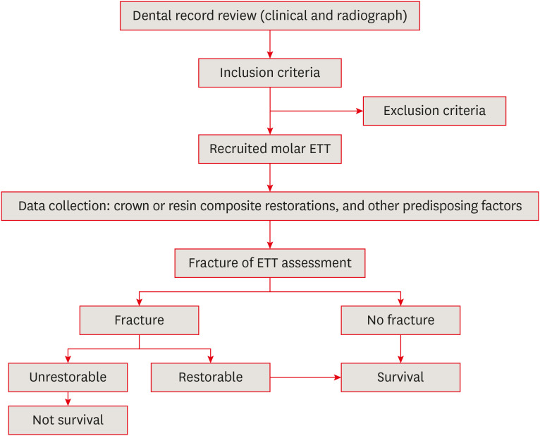
ePub Objectives This study was conducted to compare the post-fracture survival rate of endodontically treated molar endodontically treated teeth (molar ETT) restored with resin composites or crowns and to identify potential risk factors, using a retrospective cohort design.
Materials and Methods Dental records of molar ETT with crowns or composite restorations (recall period, 2015–2019) were collected based on inclusion and exclusion criteria. The incidence of unrestorable fractures was identified, and molar ETT were classified according to survival. Information on potential risk factors was collected. Survival rates and potential risk factors were analyzed using the Kaplan-Meier log-rank test and Cox regression model.
Results The overall survival rate of molar ETT was 87% (mean recall period, 31.73 ± 17.56 months). The survival rates of molar ETT restored with composites and crowns were 81.6% and 92.7%, reflecting a significant difference (
p < 0.05). However, ETT restored with composites showed a 100% survival rate if only 1 surface was lost, which was comparable to the survival rate of ETT with crowns. The survival rates of ETT with composites and crowns were significantly different (97.6% vs. 83.7%) in the short-term (12–24 months), but not in the long-term (> 24 months) (87.8% vs. 79.5%).Conclusions The survival rate from fracture was higher for molar ETT restored with crowns was higher than for ETT restored with composites, especially in the first 2 years after restoration. Molar ETT with limited tooth structure loss only on the occlusal surface could be successfully restored with composite restorations.
-
Citations
Citations to this article as recorded by- Effect of Conventional Filler, Short Fiber-reinforced, and Polyethylene Woven Fiber-reinforced Composite on the Fracture Toughness of Extracted Premolar Teeth
Leeza Bharati, Chandrakar Chaman, Udai P Singh, Asib Ahmad, Siddharth Anand, Aparna Singh
The Journal of Contemporary Dental Practice.2025; 26(7): 693. CrossRef - Risk factors for the appearance of cracks and fractures of teeth according to a survey of dentists
Emilia A. Olesova, Alexander A. Ilyin, Sergey D. Arutyunov, Elena V. Glazkova, Arsen A. Popov, Svetlana P. Iarilkina
Russian Journal of Dentistry.2024; 28(6): 562. CrossRef - Performance of Bonded Lithium Disilicate Partial-coverage Crowns in the Restoration of Endodontically Treated Posterior Teeth: An Up to Seven-Year Retrospective Study
Q Jiang, Z Wang, S Zhang, X Liu, B Fu
Operative Dentistry.2024; 49(4): 365. CrossRef - In Vitro Bond Strength of Dentin Treated with Sodium Hypochlorite: Effects of Antioxidant Solutions
Guillermo Grazioli, Elisa de León Cáceres, Romina Tessore, Rafael Lund, Ana Monjarás-Ávila, Monika Lukomska-Szymanska, Louis Hardan, Rim Bourgi, Carlos Cuevas-Suárez
Antioxidants.2024; 13(9): 1116. CrossRef - Stress Analysis on Mesiolingual Cavity of Endodontically Treated Molar Restored Using Bidirectional Fiber-Reinforced Composite (Wallpapering Technique)
Harnia Neri, Dudi Aripin, Anna Muryani, Hendra Dharsono, Yolanda Yolanda, Andi Mahyuddin
Clinical, Cosmetic and Investigational Dentistry.2024; Volume 16: 75. CrossRef - Effect of Luting Cement Film Thickness on the Pull-Out Bond Strength of Endodontic Post Systems
Khalil Aleisa, Syed Rashid Habib, Abdul Sadekh Ansari, Ragad Altayyar, Shahad Alharbi, Sultan Ali S. Alanazi, Khalid Tawfik Alduaiji
Polymers.2021; 13(18): 3082. CrossRef
- Effect of Conventional Filler, Short Fiber-reinforced, and Polyethylene Woven Fiber-reinforced Composite on the Fracture Toughness of Extracted Premolar Teeth
- 2,972 View
- 48 Download
- 8 Web of Science
- 6 Crossref

- Effect of number of uses and sterilization on the instrumented area and resistance of reciprocating instruments
- Victor de Ornelas Peraça, Samantha Rodrigues Xavier, Fabio de Almeida Gomes, Luciane Geanini Pena dos Santos, Erick Miranda Souza, Fernanda Geraldo Pappen
- Restor Dent Endod 2021;46(2):e28. Published online April 29, 2021
- DOI: https://doi.org/10.5395/rde.2021.46.e28
-
 Abstract
 PDF
PDF PubReader
 ePub
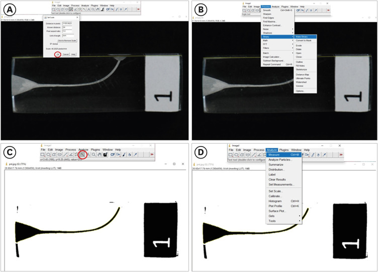
ePub Objectives This study evaluated the effect of repeated uses and autoclaving in the instrumented area, fracture resistance, and time of instrumentation of thermally treated nickel-titanium reciprocating systems.
Materials and Methods Two hundred simulated canals were instrumented using Reciproc Blue and WaveOne Gold. Each file was used up to 10 times or until fracture. The instrumented area was measured in pre- and post-operative images, using ImageJ software. Kaplan-Meier survival analysis evaluated the number of uses of instruments before fracture. Instrumented area and time of instrumentation were analyzed by Mann-Whitney U test and Kruskal-Wallis. Correlations among the number of uses and instrumented area were measured. The level of statistical significance was set at
p < 0.05.Results Reciproc Blue presented a higher estimated number of uses in comparison with WaveOne Gold (
p = 0.026), but autoclaving did not affect the resistance to fracture of instruments (p > 0.05). The instrumented area was different among the evaluated groups (p = 0.039), and the instrumented area along the uses of both tested instruments was reduced. With the time of instrumentation, there was also a significant difference among the evaluated groups; the groups without sterilization cycles were faster, in comparison to those submitted to autoclaving (p = 0.010).Conclusions Reciproc Blue was more resistant than WaveOne Gold, suffering later fracture. Additionally, the sterilization cycles did not influence the estimated number of uses of thermally treated reciprocating instruments, but the instrumented area of root canals was reduced along with the repeated uses of both instruments.
-
Citations
Citations to this article as recorded by- Effect of sterilization on the cutting efficiency of two different rotary NiTi instruments (An In- vitro Study)
Merna Mamdouh Botros, Kariem Mostafa ElBatouty, Tariq Yehia Abdelrahman
BMC Oral Health.2025;[Epub] CrossRef - Fracture Risk of Endodontic Files: Clinical Analysis of Reciproc and X1 Blue After Multiple Uses
Elisa Korte Fortes Gollo, Fábio de Almeida Gomes, Katerine Jahnecke Pilownic, Daiana Elisabeth Böttcher, Carolina Clasen Vieira, Fernanda Geraldo Pappen
Australian Endodontic Journal.2025; 51(3): 699. CrossRef - The influence of autoclave sterilization on the cyclic fatigue of M-wire rotary endodontic instruments
Nenad Stosic, Jelena Popovic, Antonije Stankovic, Aleksandar Mitic, Marija Nikolic, Kosta Todorovic
Vojnosanitetski pregled.2024; 81(10): 642. CrossRef
- Effect of sterilization on the cutting efficiency of two different rotary NiTi instruments (An In- vitro Study)
- 1,417 View
- 22 Download
- 2 Web of Science
- 3 Crossref

- Effect of post space preparation drills on the incidence of root dentin defects
- Thaíse Ayres Bezerra Zuli, Orlando Aguirre Guedes, Gislaine Figueiredo Zarza Arguello Gonçalves, Aurélio Rosa da Silva Júnior, Álvaro Henrique Borges, Andreza Maria Fábio Aranha
- Restor Dent Endod 2020;45(4):e53. Published online October 16, 2020
- DOI: https://doi.org/10.5395/rde.2020.45.e53

-
 Abstract
 PDF
PDF PubReader
 ePub
ePub Objectives This study investigated the incidence of root dentin defects after the use of different post space preparation (PSP) drills.
Materials and Methods Seventy-two bovine incisors were selected and obtained 14-mm-long root sections. Twelve roots served as controls with no intervention (G1). The 60 root canals remaining were instrumented using the crown-down technique with the ProTaper Next system and obturated using the lateral condensation technique. Specimens were randomly distributed into 5 groups (
n = 12) according to the operative steps performed: G2, root canal instrumentation and filling (I+F); G3, I+F and PSP with Gates-Glidden drills; G4, I+F and PSP with Largo-Peeso reamers; G5, I+F and PSP with Exacto drill; and G6, I+F and PSP with WhitePost drill. Roots were sectioned at 3, 6, 9, and 12 mm from the apex, and digital images were captured. The presence of root dentin defects was recorded. Data were analyzed by the χ2 test, withp < 0.05 considered to indicate statistical significance.Results Root dentin defects were observed in 39.6% of the root sections. No defects were observed in G1. G5 had significantly more cracks and craze lines than G1, G2, and G3 (
p < 0.05), and more fractures than G1, G2, G3, and G4 (p < 0.05). When all root sections were analyzed together, significantly more defects were observed at the 12-mm level than at the 3-mm level (p < 0.05).Conclusions PSP drills caused defects in the root dentin. Gates-Glidden drills caused fewer root defects than Largo-Peeso reamers and Exacto drills.
-
Citations
Citations to this article as recorded by- Evaluation of dentinal crack formation during post space preparation using different fiber post systems with micro-computed tomography
Ayşe Nur Kuşuçar, Damla Kırıcı
BMC Oral Health.2025;[Epub] CrossRef - Fracture and Crack Behavior of Weakened Incisors Restored With Fiber Posts, Polyethylene Reinforcement, or 3D-Printed Endocrowns
Diana Codas-Duarte, Laís L Pelozo, Jardel F Mazzi-Chaves, Fabiane C Lopes-Olhê, Manoel D Sousa-Neto, Aline E Souza-Gabriel
Cureus.2025;[Epub] CrossRef - Selecting drill size for post space preparation based on final endodontic radiographs: An in vitro study
Farzaneh Farid, Julfikar Haider, Marjan Sadeghpour Shahab, Nika Rezaeikalantari
Technology and Health Care.2024; 32(4): 2575. CrossRef - Cone Beam Computed Tomography Analysis of Post Space in Bifurcated Premolars Using ParaPost and Peeso Reamer Drills
Abdulaziz Saleh Alqahtani, Omar Nasser Almonabhi, Abdulmajeed Moh. Almutairi, Reem R. Alnatsha
The Open Dentistry Journal.2024;[Epub] CrossRef - A Comparative Evaluation of Real-Time Guided Dynamic Navigation and Conventional Techniques for Post Space Preparation During Post Endodontic Management: An In Vitro Study
Sherifa Shervani, Sihivahanan Dhanasekaran, Vijay Venkatesh
Cureus.2024;[Epub] CrossRef - The effect of ultrasonic vibration protocols for cast post removal on the incidence of root dentin defects
Giulliano C. Serpa, Orlando A. Guedes, Neurinelma S. S. Freitas, Julio A. Silva, Carlos Estrela, Daniel A. Decurcio
Journal of Oral Science.2023; 65(3): 190. CrossRef
- Evaluation of dentinal crack formation during post space preparation using different fiber post systems with micro-computed tomography
- 2,522 View
- 31 Download
- 6 Crossref

- Fiber-reinforced composite resin bridges: an alternative method to treat root-fractured teeth
- Gun Heo, Eun-Hye Lee, Jin-Woo Kim, Kyung-Mo Cho, Se-Hee Park
- Restor Dent Endod 2020;45(1):e8. Published online December 27, 2019
- DOI: https://doi.org/10.5395/rde.2020.45.e8

-
 Abstract
 PDF
PDF PubReader
 ePub
ePub The replacement of missing teeth, especially in the anterior region, is an essential part of dental practice. Fiber-reinforced composite resin bridges are a conservative alternative to conventional fixed dental prostheses or implants. It is a minimally invasive, reversible technique that can be completed in a single visit. The two cases presented herein exemplify the treatment of root-fractured anterior teeth with a natural pontic immediately after extraction.
-
Citations
Citations to this article as recorded by- Prosthodontic Aspects of Splinting the Mandibular Anterior Teeth by Fiber Reinforced Composites
Hrelja Miroslav, Laškarin Mirko, Čimić Samir, Kraljević Sonja, Dulčić Nikša, Badel Tomislav
Journal of Dental Problems and Solutions.2025; 12(1): 004. CrossRef - Current Evidence on the Fiber-reinforced Composite Bridges
Ramesh Chowdhary, Sunil Kumar Mishra
International Journal of Prosthodontics and Restorative Dentistry.2023; 12(4): 159. CrossRef - Bridging the Gap: A Case Report of Tooth Replacement using Resin-Bonded Fiber- Reinforced Composite Resin
Vineet Sharma, Sumit Bhansali, Sonal Priya Bhansali
Journal of Pierre Fauchard Academy (India Section).2023; : 66. CrossRef - Reconstruction of Natural Smile and Splinting with Natural Tooth Pontic Fiber‐Reinforced Composite Bridge
Maryam S. Tavangar, Fatemeh Aghaei, Massoumeh Nowrouzi, Andrea Scribante
Case Reports in Dentistry.2022;[Epub] CrossRef
- Prosthodontic Aspects of Splinting the Mandibular Anterior Teeth by Fiber Reinforced Composites
- 1,767 View
- 12 Download
- 4 Crossref

-
Cyclic fatigue resistance of M-Pro and RaCe Ni-Ti rotary endodontic instruments in artificial curved canals: a comparative
in vitro study - Hadeer Mostafa El Feky, Khalid Mohammed Ezzat, Marwa Mahmoud Ali Bedier
- Restor Dent Endod 2019;44(4):e44. Published online November 7, 2019
- DOI: https://doi.org/10.5395/rde.2019.44.e44
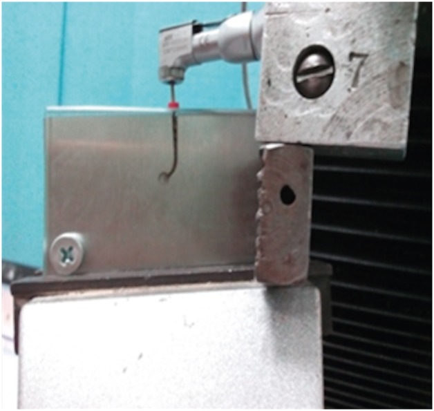
-
 Abstract
 PDF
PDF PubReader
 ePub
ePub Objectives To compare the flexural cyclic fatigue resistance and the length of the fractured segments (FLs) of recently introduced M-Pro rotary files with that of RaCe rotary files in curved canals and to evaluate the fracture surface by scanning electron microscopy (SEM).
Materials and Methods Thirty-six endodontic files with the same tip size and taper (size 25, 0.06 taper) were used. The samples were classified into 2 groups (n = 18): the M-Pro group (M-Pro IMD) and the RaCe group (FKG). A custom-made simulated canal model was fabricated to evaluate the total number of cycles to failure and the FL. SEM was used to examine the fracture surfaces of the fragmented segments. The data were statistically analyzed and comparisons between the 2 groups for normally distributed numerical variables were carried out using the independent Student's
t -test. Ap value less than 0.05 was considered to indicate statistical significance.Results The M-Pro group showed significantly higher resistance to flexural cyclic fatigue than the RaCe group (
p < 0.05), but there was no significant difference in the FLs between the 2 groups (p ≥ 0.05).Conclusions Thermal treatment of nickel-titanium instruments can improve the flexural cyclic fatigue resistance of rotary endodontic files, and the M-Pro rotary system seems to be a promising rotary endodontic file.
-
Citations
Citations to this article as recorded by- The Effect of Canal Curvature and Different Manufacturing Processes of Five Different NiTi Rotary Files on Cyclic Fatigue Resistance
Panupat Phumpatrakom, Awiruth Klaisiri, Sukitti Techapatiphandee, Thippawan Saekow, Panuroot Aguilar
European Journal of General Dentistry.2025; 14(03): 264. CrossRef - EndoMagic Gold M06 Eğelerinde Boyut ve Konikliğin Döngüsel Yorgunluğa Etkisi: Bir İn Vitro Çalışma
Bircan Kuloğlu, Ayşe Çoban, Hatice Büyüközer Özkan
Akdeniz Diş Hekimliği Dergisi.2025; 4(3): 212. CrossRef - Evaluatation of two nickle-titanium systems’ (Neolix and X Pro Gold) resistance to fracture after immersion in sodium hypochlorite.
Solmaz Araghi, Abbas Delvarani, Faeze dehghan, Parisa Kaghazloo
journal of research in dental sciences.2024; 21(1): 17. CrossRef - Endodontic Ni–Ti Rotary Instruments for Glide-path, Are They Still Necessary and How to Think about the Ideal Instrument?
Shilpa Bhandi, Rodolfo Reda, Luca Testarelli, Elisa Maccari
The Journal of Contemporary Dental Practice.2024; 25(6): 505. CrossRef - Comparative evaluation of cyclic fatigue resistance of thermomechanically treated NiTi rotary instruments in simulated curved canals with two different radii of curvature: An in vitro Study
Tahira Hamid, Azhar Malik, Ajay Kumar, Shamim Anjum
Journal of Conservative Dentistry and Endodontics.2024; 27(4): 393. CrossRef - New heat-treated vs electropolished nickel-titanium instruments used in root canal treatment: Influence of autoclave sterilization on surface roughness
Rahaf A. Almohareb, Reem Barakat, Fatimah Albohairy, Hannes C. Schniepp
PLOS ONE.2022; 17(3): e0265226. CrossRef - The Effect of Taper and Apical Diameter on the Cyclic Fatigue Resistance of Rotary Endodontic Files Using an Experimental Electronic Device
Vicente Faus-Llácer, Nirmine Hamoud Kharrat, Celia Ruiz-Sánchez, Ignacio Faus-Matoses, Álvaro Zubizarreta-Macho, Vicente Faus-Matoses
Applied Sciences.2021; 11(2): 863. CrossRef
- The Effect of Canal Curvature and Different Manufacturing Processes of Five Different NiTi Rotary Files on Cyclic Fatigue Resistance
- 2,104 View
- 10 Download
- 7 Crossref

- Pulp revascularization with and without platelet-rich plasma in two anterior teeth with horizontal radicular fractures: a case report
- Edison Arango-Gómez, Javier Laureano Nino-Barrera, Gustavo Nino, Freddy Jordan, Henry Sossa-Rojas
- Restor Dent Endod 2019;44(4):e35. Published online August 20, 2019
- DOI: https://doi.org/10.5395/rde.2019.44.e35
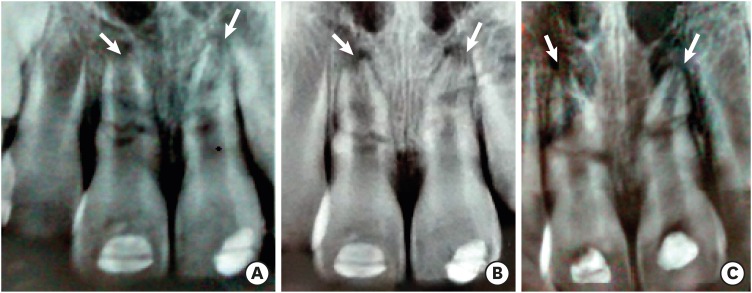
-
 Abstract
 PDF
PDF PubReader
 ePub
ePub Pulp revascularization is an alternative treatment in immature traumatized teeth with necrotic pulp. However, this procedure has not been reported in horizontal root fractures. This is a case report of a 9-year-old patient with multiple horizontal root fractures in 2 upper central incisors that were successfully treated with pulp revascularization. The patient presented for treatment 2 years after the initial trauma, and revascularization was attempted after the initial treatment with calcium hydroxide had failed. Prior to pulp revascularization, cone-beam computed tomography and autoradiograms demonstrated multiple horizontal fractures in the middle and apical thirds of the roots of the 2 affected teeth. Revascularization was performed in both teeth; platelet-rich plasma (PRP) was used in one tooth (#11) and the conventional method (blood clot) was used in the other tooth (#21). Clinical and radiographic follow-up over 4 years demonstrated pulp calcification in the PRP-treated tooth. Neither of the 2 teeth were lost, and the root canal calcification of tooth #11 was greater than that of tooth #21. This case suggests that PRP-based pulp revascularization may be an alternative for horizontal root fractures.
-
Citations
Citations to this article as recorded by- Platelet-Rich Plasma and Platelet-Rich Fibrin in Endodontics: A Scoping Review
Simão Rebimbas Guerreiro, Carlos Miguel Marto, Anabela Paula, Joana Rita de Azevedo Pereira, Eunice Carrilho, Manuel Marques-Ferreira, Siri Vicente Paulo
International Journal of Molecular Sciences.2025; 26(12): 5479. CrossRef - Dental pulp mesenchymal stem cells-response to fibrin hydrogel reveals ITGA2 and MMPs expression
David Tong, Stéphanie Gobert, Alicia Reuzeau, Jean-Christophe Farges, Marianne Leveque, Marie Bolon, Arthur Costantini, Marielle Pasdeloup, Jérôme Lafont, Maxime Ducret, Mourad Bekhouche
Heliyon.2024; 10(13): e32891. CrossRef - Pulp regeneration treatment using different bioactive materials in permanent teeth of pediatric subjects
Dina Abdellatif, Alfredo Iandolo, Giuseppina De Benedetto, Francesco Giordano, Davide Mancino, Edouard Euvrard, Massimo Pisano
Journal of Conservative Dentistry and Endodontics.2024; 27(5): 458. CrossRef - Retreatment of a Failed Regenerative Endodontic Treatment in an Immature Tooth with a Horizontal Root Fracture: A Case Report
Zaher Marjy, Iris Slutzky-Goldberg
International Journal of Clinical Pediatric Dentistry.2024; 17(10): 1168. CrossRef - The Impact of the Preferred Reporting Items for Case Reports in Endodontics (PRICE) 2020 Guidelines on the Reporting of Endodontic Case Reports
Sofian Youssef, Phillip Tomson, Amir Reza Akbari, Natalie Archer, Fayjel Shah, Jasmeet Heran, Sunmeet Kandhari, Sandeep Pai, Shivakar Mehrotra, Joanna M Batt
Cureus.2023;[Epub] CrossRef - Evaluation of postoperative pain and healing following regenerative endodontics using platelet‐rich plasma versus conventional endodontic treatment in necrotic mature mandibular molars with chronic periapical periodontitis. A randomized clinical trial
Yassmin Elsayed Ahmed, Geraldine Mohamed Ahmed, Angie Galal Ghoneim
International Endodontic Journal.2023; 56(4): 404. CrossRef - Regenerative endodontic procedures for two traumatized mature anterior teeth with transverse root fractures
Jing Lu, Bill Kahler
BMC Oral Health.2022;[Epub] CrossRef - Are platelet concentrate scaffolds superior to traditional blood clot scaffolds in regeneration therapy of necrotic immature permanent teeth? A systematic review and meta-analysis
Qianwei Tang, Hua Jin, Song Lin, Long Ma, Tingyu Tian, Xiurong Qin
BMC Oral Health.2022;[Epub] CrossRef - Platelet-Rich Fibrin Used as a Scaffold in Pulp Regeneration: Case Series
Ceren ÇİMEN, Selin ŞEN, Elif ŞENAY, Tuğba BEZGİN
Cumhuriyet Dental Journal.2021; 24(1): 113. CrossRef - Plasma rico en plaquetas en Odontología: Revisión de la literatura
Hugo Anthony Rosas Rozas, Hugo Leoncio Rosas Cisneros
Yachay - Revista Científico Cultural.2021; 10(1): 536. CrossRef
- Platelet-Rich Plasma and Platelet-Rich Fibrin in Endodontics: A Scoping Review
- 2,432 View
- 41 Download
- 10 Crossref

- Effect of glide path preparation with PathFile and ProGlider on the cyclic fatigue resistance of WaveOne nickel-titanium files
- Gülşah Uslu, Uğur İnan
- Restor Dent Endod 2019;44(2):e22. Published online May 9, 2019
- DOI: https://doi.org/10.5395/rde.2019.44.e22
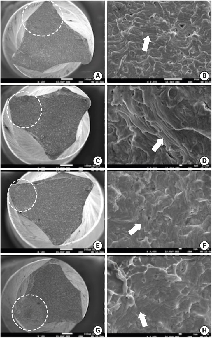
-
 Abstract
 PDF
PDF PubReader
 ePub
ePub Objectives The aim of this study was to investigate the effect of glide path preparation with PathFile and ProGlider nickel-titanium (NiTi) files on the cyclic fatigue resistance of WaveOne NiTi files.
Materials and Methods Forty-four WaveOne Primary files were used and divided into four groups (
n = 11). In the first group (0 WaveOne), the WaveOne Primary files served as a control group and were not used on acrylic blocks. In the 1 WaveOne Group, acrylic blocks were prepared using only WaveOne Primary files, and in the PF+WaveOne group and PG+WaveOne groups, acrylic blocks were first prepared with PathFile or ProGlider NiTi files, respectively, followed by the use of WaveOne Primary files. All the WaveOne Primary files were then subjected to cyclic fatigue testing. The number of cycles to failure was calculated and the data were statistically analyzed using one-way analysis of variance (ANOVA) and the Tukey honest significant difference multiple-comparison test at a 5% significance level.Results The highest number of cycles to failure was found in the control group, and the lowest numbers were found in the 1 WaveOne group and the PF+WaveOne group. Significant differences were found among the 1 WaveOne, PF+WaveOne, and control groups (
p < 0.05). No statistically significant differences were found between the PG+WaveOne group and the other three groups (p > 0.05).Conclusion Glide path preparation with NiTi rotary files did not affect the cyclic fatigue resistance of WaveOne Primary files used on acrylic blocks.
-
Citations
Citations to this article as recorded by- Screw-in force, torque generation, and performance of glide-path files with three rotation kinetics
Jee-Yeon Woo, Ji-Hyun Jang, Seok Woo Chang, Soram Oh
Odontology.2024; 112(3): 761. CrossRef - Glide Path in Endodontics: A Literature Review of Current Knowledge
Vlad Mircea Lup, Giulia Malvicini, Carlo Gaeta, Simone Grandini, Gabriela Ciavoi
Dentistry Journal.2024; 12(8): 257. CrossRef - Comparative Evaluation of the Cyclic Fatigue Resistance of WaveOne Gold in Reciprocation, ProGlider in Rotary Motion, and Manual Files in a Reciprocating Handpiece Within Simulated Curved Canals: An In Vitro Study
Shivangi M Pujara, Hardik B Shah, Leena H Jobanputra
Cureus.2024;[Epub] CrossRef - Effect of glide path instruments in cyclic fatigue resistance of reciprocating instruments after three uses
André Schroder Scherer, Carlos Alexandre Souza Bier, José Roberto Vanni
Brazilian Dental Journal.2023; 34(2): 27. CrossRef - An Investigation of the Accuracy and Reproducibility of 3D Printed Transparent Endodontic Blocks
Martin Smutný, Martin Kopeček, Aleš Bezrouk
Acta Medica (Hradec Kralove, Czech Republic).2022; 65(2): 59. CrossRef - Evaluation of Cyclic Fatigue of Hyflex EDM, Twisted Files, and ProTaper Gold Manufactured with Different Processes: An In Vitro Study
Pooja D. Khandagale, Prashant P. Shetty, Saleem D. Makandar, Pradeep A. Bapna, Mohmed Isaqali Karobari, Anand Marya, Pietro Messina, Giuseppe Alessandro Scardina, Antonino Lo Giudice
International Journal of Dentistry.2021; 2021: 1. CrossRef
- Screw-in force, torque generation, and performance of glide-path files with three rotation kinetics
- 1,539 View
- 10 Download
- 6 Crossref

- Critical evaluation of fracture strength testing for endodontically treated teeth: a finite element analysis study
- Emel Uzunoglu-Özyürek, Selen Küçükkaya Eren, Oğuz Eraslan, Sema Belli
- Restor Dent Endod 2019;44(2):e15. Published online April 18, 2019
- DOI: https://doi.org/10.5395/rde.2019.44.e15
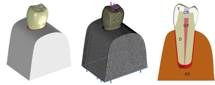
-
 Abstract
 PDF
PDF PubReader
 ePub
ePub Objectives The aim of this study was to investigate whether the diameter and direction of the plunger and simulation of the periodontal ligament (PDL) affected the stress distribution in endodontically treated premolars.
Methods A fracture strength test was simulated via finite element analysis. A base model was set up, and the following parameters were modified: plunger diameter (3 mm vs. 6 mm), plunger direction (vertical vs. 135° angular to the central fossa), and PDL simulation. The analysis was conducted using the CosmosWorks structural analysis program, and the results are presented in terms of von Mises stresses.
Results The smaller plunger increased the stresses at the contact area of the crown, but the plunger diameter had no effect on the stress distribution within the root. An angular plunger direction increased stresses within the root, as well as at the buccal cusp of the crown, compared with the vertical direction. Simulation of the PDL caused higher stress accumulation, especially in the cervical region of the root.
Conclusions The plunger diameter had no effect on the stress distribution in the roots, whereas the plunger direction and PDL simulation did affect the stress distribution. More stringent standards can be established by taking such parameters into account when performing fracture testing in future studies.
-
Citations
Citations to this article as recorded by- Access cavity in endodontics: Balancing precision, preservation, and clinical needs
Dina Abdellatif, Ismail Davut Capar, De Fontaine Sarah, Alfredo Iandolo, Christophe Meyer, Davide Mancino
Journal of Conservative Dentistry and Endodontics.2025; 28(6): 573. CrossRef - Assessment of Stress Distribution with 3 Taper Design Preparation of Root Canal Using Finite Element Analysis
Tejasree Rathod, G. Durgabhavani, Pudu Tirupathi, Nusrath Parveen, Yelloji Paramesh, Prabhakar Dharavattu
Journal of Pharmacy and Bioallied Sciences.2024; 16(Suppl 1): S112. CrossRef - The impact of the filling technique with two sealers in bulk or associated with gutta-percha on the fatigue behavior and failure patterns of endodontically treated teeth
Isabella Marian Lena, Luiza Colpo Chiaratti, Rafaela Oliveira Pilecco, Renan Vaz Machry, João Paulo Mendes Tribst, Cornelis Johannes Kleverlaan, Gabriel Kalil Rocha Pereira, Renata Dornelles Morgental
PeerJ.2024; 12: e18221. CrossRef - Stronger than Ever: Multifilament Fiberglass Posts Boost Maxillary Premolar Fracture Resistance
Naji Kharouf, Eugenio Pedullà, Gianluca Plotino, Hamdi Jmal, Mohammed-El-Habib Alloui, Philippine Simonis, Patrice Laquerriere, Valentina Macaluso, Dina Abdellatif, Raphaël Richert, Youssef Haikel, Davide Mancino
Journal of Clinical Medicine.2023; 12(8): 2975. CrossRef - Neural network approach to evaluate the physical properties of dentin
Mohammad Ali Saghiri, Ali Mohammad Saghiri, Elham Samadi, Devyani Nath, Julia Vakhnovetsky, Steven M. Morgano
Odontology.2023; 111(1): 68. CrossRef - Modelling and evaluating periodontal ligament mechanical behaviour and properties: A scoping review of current approaches and limitations
Enaiyat Ghani Ovy, Dan L. Romanyk, Carlos Flores Mir, Lindsey Westover
Orthodontics & Craniofacial Research.2022; 25(2): 199. CrossRef - FEAr no more! Finite element analysis in orthodontics
Shilpa Chawla, Shailesh Deshmukh
Journal of the International Clinical Dental Research Organization.2022; 14(1): 6. CrossRef - Influence of Methodological Variables on Fracture Strength Tests Results of Premolars with Different Number of Residual Walls. A Systematic Review with Meta-Analysis
Carlo Gaeta, Crystal Marruganti, Emanuele Mignosa, Giovanni Franciosi, Edoardo Ferrari, Simone Grandini
Dentistry Journal.2021; 9(12): 146. CrossRef
- Access cavity in endodontics: Balancing precision, preservation, and clinical needs
- 2,203 View
- 36 Download
- 8 Crossref

- The top 10 most-cited articles on the management of fractured instruments: a bibliometric analysis
- Lora Mishra, Hyeon-Cheol Kim, Naomi Ranjan Singh, Priti Pragati Rath
- Restor Dent Endod 2019;44(1):e2. Published online December 26, 2018
- DOI: https://doi.org/10.5395/rde.2019.44.e2
-
 Abstract
 PDF
PDF PubReader
 ePub
ePub Objectives The purpose of this research was to identify the top 10 most-cited articles on the management of fractured or broken instruments and to perform a bibliometric analysis thereof.
Materials and Methods Published articles related to fractured instruments were screened from online databases, such as Web of Science, Scopus, PubMed, and ScienceDirect, and highly cited papers, with at least 50 citations since publication, were identified. The most-cited articles were selected and analysed with regard to publication title, authorship, the journal of publication, year, institution, country of origin, article type, and number of citations.
Results The top 10 most-cited articles were from various journals. Most were published in the
Journal of Endodontics , followed by theInternational Endodontic Journal , andDental Traumatology . The leading countries were Australia, Israel, Switzerland, the USA, and Germany, and the leading institution was the University of Melbourne. The majority of articles among the top 10 articles were clinical research studies (n = 8), followed by a basic research article and a non-systematic review article.Conclusions This bibliometric analysis revealed interesting information about scientific progress in endodontics regarding fractured instruments. Overall, clinical research studies and basic research articles published in high-impact endodontic journals had the highest citation rates.
-
Citations
Citations to this article as recorded by- Bibliometric analysis of the publications that list the most-cited articles in endodontics
Oscar Alejandro Gutiérrez-Alvarez, Luis Alberto Pantoja-Villa, Benigno Miguel Calderón-Rojas
Endodontology.2025; 37(2): 128. CrossRef - A Bibliometric Analysis of the 100 Top-Cited Articles on Vertical Root Fractures
Pillai Arun Gopinathan , Ikram UI Haq, Nawaf Alfahad, Saleh Alwatban, Abdullah Alghamdi, Amal Alamri, Kiran Iyer
Cureus.2024;[Epub] CrossRef - Predictive factors in the retrieval of endodontic instruments: the relationship between the fragment length and location
Ricardo Portigliatti, Eugenia Pilar Consoli Lizzi, Pablo Alejandro Rodríguez
Restorative Dentistry & Endodontics.2024;[Epub] CrossRef - A bibliometric analysis of the top 100 most‐cited case reports and case series in Endodontic journals
Venkateshbabu Nagendrababu, Jelena Jacimovic, Aleksandar Jakovljevic, Giampiero Rossi‐Fedele, Paul M. H. Dummer
International Endodontic Journal.2022; 55(3): 185. CrossRef - The Most Highly Cited Publications on Basketball Originate From English-Speaking Countries, Are Published After 2000, Are Focused on Medicine-Related Topics, and Are Level III Evidence
Zachary D. Griffin, Jordan R. Pollock, M. Lane Moore, Kade S. McQuivey, Jaymeson R. Arthur, Anikar Chhabra
Arthroscopy, Sports Medicine, and Rehabilitation.2022; 4(3): e891. CrossRef - Ten years of minimally invasive access cavities in Endodontics: a bibliometric analysis of the 25 most-cited studies
Emmanuel João Nogueira Leal Silva, Karem Paula Pinto, Natasha C. Ajuz, Luciana Moura Sassone
Restorative Dentistry & Endodontics.2021;[Epub] CrossRef - Publication trends in micro‐CT endodontic research: a bibliometric analysis over a 25‐year period
U. Aksoy, M. Küçük, M. A. Versiani, K. Orhan
International Endodontic Journal.2021; 54(3): 343. CrossRef
- Bibliometric analysis of the publications that list the most-cited articles in endodontics
- 1,417 View
- 10 Download
- 7 Crossref

- Survival rates against fracture of endodontically treated posterior teeth restored with full-coverage crowns or resin composite restorations: a systematic review
- Warattama Suksaphar, Danuchit Banomyong, Titalee Jirathanyanatt, Yaowaluk Ngoenwiwatkul
- Restor Dent Endod 2017;42(3):157-167. Published online July 31, 2017
- DOI: https://doi.org/10.5395/rde.2017.42.3.157
-
 Abstract
 PDF
PDF PubReader
 ePub
ePub This systematic review aims to summarize the current clinical studies that investigated survival rates against fracture of endodontically treated posterior teeth restored with crowns or resin composite restorations. Literature search were performed using keywords. Publications from 1980 to 2016 were searched in PubMed, ScienceDirect, Web of Science, MEDLINE, and SCOPUS. Included studies were selected based on inclusion and exclusion criteria. Three clinical studies were included: 1 randomized controlled trial and 1 prospective and 1 retrospective cohort studies. Pooled survival rates ranged from 94%–100% and 91.9%–100% for crowns and resin composite, respectively. The majority of teeth had no more than 3 surface loss of tooth structure. The studies included were heterogeneous, and were not appropriate for further meta-analysis. Current evidence suggested that the survival rates against the fracture of endodontically treated posterior teeth restored with crowns or resin composites were not significantly different in the teeth with minimum to moderate loss of tooth structure.
-
Citations
Citations to this article as recorded by- Effect of using different materials and restorative techniques on cuspal deflection and microleakage in endodontically treated teeth
Ceyda Sari, Oya Bala, Sinem Akgul, Cemile Kedici Alp
BMC Oral Health.2025;[Epub] CrossRef - Direct restorations versus full crowns in endodontically treated molar teeth: A three-year randomized clinical trial
Motasum Abu-Awwad, Ruba Halasa, Laila Haikal, Ahmad El-Ma'aita, Mohammad Hammad, Haralampos Petridis
Journal of Dentistry.2025; 156: 105699. CrossRef - Is the use of an intraradicular post essential for reducing failures in restoring endodontically treated teeth? A systematic review and meta-analysis
Jacqueline Salomão Jardim, Vinicius de Menezes Félix Ferreira, Hiskell Francine Fernandes e Oliveira, Daniele Sorgatto Faé, Cleidiel Aparecido Araujo Lemos
Journal of Dentistry.2025; 159: 105739. CrossRef - Systematic Reviews Comparing Direct and Indirect Restorations: An Umbrella Review That Examines Restoration Type and Confidence in Results
Mona Kimmel, Clovis Mariano Faggion
Clinical and Experimental Dental Research.2025;[Epub] CrossRef - Knowledge, Awareness, and Perceptions on Root Canal Treatment among Patients Reporting with Dental Pain to Conservative Dentistry and Endodontics Department: An Institution-based Survey
Abdu Semeer Palottil, Moopil Midhun Mohanan, N. T. Nishad, S. Jayasree
Journal of Primary Care Dentistry and Oral Health.2025; 6(2): 99. CrossRef - One-year clinical performance of restorations with and without a bulk-fill flowable base in endodontically treated premolars: a pilot randomized controlled trial
Brenda Leyton, Jullyana Dezanetti, Rodrigo Rached, Sérgio Ignácio, Evelise Souza
BMC Oral Health.2025;[Epub] CrossRef - One-piece endodontic crown fixed partial denture: Is it possible?
João Paulo M. Tribst, Amanda Maria de O. Dal Piva, Joris Muris, Cornelis J. Kleverlaan, Albert J. Feilzer
The Journal of Prosthetic Dentistry.2024; 131(6): 1118. CrossRef - Survival Rate Against Fracture of Endodontically Treated Premolars Restored with Crowns and Resin Composites: A Retrospective Study
Enas Khamakhim, Farida Alsayeh
AlQalam Journal of Medical and Applied Sciences.2024; : 398. CrossRef - Knowledge and Awareness of Root Canal Treatment among Patients in Tripoli: A Survey-Based Study
Sumaya Aghila
AlQalam Journal of Medical and Applied Sciences.2024; : 532. CrossRef - Clinical performance of polyethylenefiber reinforced resin composite restorations in endodontically treated teeth: (a randomized controlled clinical trial)
Ahmed Abdelsattar Metwaly, Amira Farid Elzoghby, Rawda Hesham Abd ElAziz
BMC Oral Health.2024;[Epub] CrossRef - Direct Versus Indirect Treatment Options of Endodontically Treated Posterior Teeth: A Narrative Review
Mai M Alhamdan, Rodina F Aljamaan, Munira M Abuthnain, Shahd A Alsumikhi, Ghada S Alqahtani, Reem A Alkharaiyef
Cureus.2024;[Epub] CrossRef - Single crown vs. composite for glass fiber post-retained restorations: An 8-year randomized clinical trial
Victório Poletto-Neto, Luiz Alexandre Chisini, Wietske Fokkinga, Cees Kreulen, Bas Loomans, Maximiliano Sérgio Cenci, Tatiana Pereira-Cenci
Journal of Dentistry.2024; 142: 104837. CrossRef - Factors influencing the clinical performance of the restoration of endodontically treated teeth: An assessment of systematic reviews of clinical studies
Lara Dotto, Luiza Paloma S. Girotto, Yara Teresinha Correa Silva Sousa, Gabriel Kalil Rocha Pereira, Ataís Bacchi, Rafael Sarkis-Onofre
The Journal of Prosthetic Dentistry.2024; 131(6): 1043. CrossRef - Influence of technical quality and coronal restoration on periapical health of root canal treatment performed by Malaysian undergraduate students
Norazlina Mohammad, Faizah Abdul Fatah, Azlan Jaafar, Siti Hajar Omar, Aimi Amalina Ahmad, Abdul Azim Asy Abdul Aziz, Aws Hashim Ali Al-Kadhim
Saudi Endodontic Journal.2023; 13(1): 63. CrossRef - The success rate of indirect adhesive restorations in the distal dentition fabricated with chairside CAD/CAM system
Marek Šupler, Andrej Jenča, Michal Straka, Juraj Deglovič, Janka Jenčová
Stomatológ.2023; 33(2): 10. CrossRef - A Comparative Study of the Marginal Fit of Endocrowns Fabricated From Three Different Computer-Aided Design/Computer-Aided Manufacturing (CAD/CAM) Ceramic Materials: An In Vitro Study
Esraa Attar, Shatha Alshali, Tariq Abuhaimed
Cureus.2023;[Epub] CrossRef - Evaluation of titanium mesh and fibers in reinforcing endodontically treated molars: An in vitro study
Hemalatha Hiremath, Devansh Verma, Sheetal Khandelwal, AishwaryaSingh Solanki, Sonam Patidar
Journal of Conservative Dentistry.2022; 25(2): 189. CrossRef - Effect of surface treatment, ferrule height, and luting agent type on pull-out bond strength of monolithic zirconia endocrowns
Emine B. Buyukerkmen, Durmuş A. Bozkurt, Arslan Terlemez
Journal of Oral Science.2022; 64(4): 279. CrossRef - An Umbrella Review of Systematic Reviews and Meta‐Analyses Evaluating the Success Rate of Prosthetic Restorations on Endodontically Treated Teeth
Amirhossein Fathi, Behnaz Ebadian, Sara Nasrollahi Dezaki, Nahal Mardasi, Ramin Mosharraf, Sabire Isler, Shiva Sadat Tabatabaei, Stefano Pagano
International Journal of Dentistry.2022;[Epub] CrossRef - Survival and success of endocrowns: A systematic review and meta-analysis
Raghad A. Al-Dabbagh
The Journal of Prosthetic Dentistry.2021; 125(3): 415.e1. CrossRef - Fracture strength of non-invasively reinforced MOD cavities on endodontically treated teeth
René Daher, Stefano Ardu, Enrico Di Bella, Giovanni T. Rocca, Albert J. Feilzer, Ivo Krejci
Odontology.2021; 109(2): 368. CrossRef - Retrospective study of fracture survival in endodontically treated molars: the effect of single-unit crowns versus direct-resin composite restorations
Kanet Chotvorrarak, Warattama Suksaphar, Danuchit Banomyong
Restorative Dentistry & Endodontics.2021;[Epub] CrossRef - An insight into patient's perceptions regarding root canal treatment: A questionnaire-based survey
Ramta Bansal, Aditya Jain
Journal of Family Medicine and Primary Care.2020; 9(2): 1020. CrossRef - Endodontically treated posterior teeth restored with or without crown restorations: A 5‐year retrospective study of survival rates from fracture
Titalee Jirathanyanatt, Warattama Suksaphar, Danuchit Banomyong, Yaowaluk Ngoenwiwatkul
Journal of Investigative and Clinical Dentistry.2019;[Epub] CrossRef - Fracture resistance, gap and void formation in root‐filled mandibular molars restored with bulk‐fill resin composites and glass‐ionomer cement base
Nathamon Thongbai‐on, Kanet Chotvorrarak, Danuchit Banomyong, Michael F. Burrow, Sittichoke Osiri, Nattha Pattaravisitsate
Journal of Investigative and Clinical Dentistry.2019;[Epub] CrossRef - Current options concerning the endodontically-treated teeth restoration with the adhesive approach
Marco Aurélio de Carvalho, Priscilla Cardoso Lazari, Marco Gresnigt, Altair Antoninha Del Bel Cury, Pascal Magne
Brazilian Oral Research.2018;[Epub] CrossRef
- Effect of using different materials and restorative techniques on cuspal deflection and microleakage in endodontically treated teeth
- 5,365 View
- 64 Download
- 26 Crossref

- Effect of adhesive luting on the fracture resistance of zirconia compared to that of composite resin and lithium disilicate glass ceramic
- Myung-Jin Lim, Kwang-Won Lee
- Restor Dent Endod 2017;42(1):1-8. Published online October 14, 2016
- DOI: https://doi.org/10.5395/rde.2017.42.1.1

-
 Abstract
 PDF
PDF PubReader
 ePub
ePub Objectives The purpose of this study was to evaluate the effect of adhesive luting on the fracture resistance of zirconia compared to that of a composite resin and a lithium disilicate glass ceramic.
Materials and Methods The specimens (dimension: 2 mm × 2 mm × 25 mm) of the composite resin, lithium disilicate glass ceramic, and yttria-stabilized tetragonal zirconia polycrystal (Y-TZP) were prepared. These were then divided into nine groups: three non-luting groups, three non-adhesive luting groups, and three adhesive luting groups, for each restorative material. In the non-luting groups, specimens were placed on the bovine tooth without any luting agents. In the non-adhesive luting groups, only zinc phosphate cement was used for luting the specimen to the bovine tooth. In the adhesive luting groups, specimens were pretreated, and the adhesive luting procedure was performed using a self-adhesive resin cement. For all the groups, a flexural test was performed using universal testing machine, in which the fracture resistance was measured by recording the force at which the specimen was fractured.
Results The fracture resistance after adhesive luting increased by approximately 29% in the case of the composite resin, 26% in the case of the lithium disilicate glass ceramic, and only 2% in the case of Y-TZP as compared to non-adhesive luting.
Conclusions The fracture resistance of Y-TZP did not increased significantly after adhesive luting as compared to that of the composite resin and the lithium disilicate glass ceramic.
-
Citations
Citations to this article as recorded by- The influence of endodontic access preparation on the mechanical strength of zirconia crowns: A literature review
Abdulrahman Almalki
Saudi Endodontic Journal.2026; 16(1): 20. CrossRef - Cyclic fatigue of a repaired 4 YSZ ceramic: Effect of the repair protocol on the adhesive and mechanical behavior
Pablo Machado Soares, Lucas Saldanha da Rosa, Rafaela Oliveira Pilecco, Gabriel Kalil Rocha Pereira, Amanda Maria de Oliveira Dal Piva, João Paulo Mendes Tribst, Luiz Felipe Valandro, Cornelis Johannes Kleverlaan, Marilia Pivetta Rippe
Heliyon.2024; 10(1): e23709. CrossRef - Effect of wall thickness on shape accuracy of hollow zirconia artificial teeth fabricated by a 3D printer
Hiro Kobayashi, Franz Sebastian Schwindling, Akinori Tasaka, Peter Rammelsberg, Shuichiro Yamashita, Stefan Rues
Journal of Prosthodontic Research.2024; 69(2): 233. CrossRef - Effect of powder air polishing and ultrasonic scaling on the marginal and internal interface (tooth-veneer) of ceramic veneers: an in vitro study
Florian Fuchs, Laura Antonia Mayer, Lena Unterschütz, Dirk Ziebolz, Nadia Oberueck, Ellen Schulz‑Kornas, Sebastian Hahnel, Andreas Koenig
Clinical Oral Investigations.2024;[Epub] CrossRef - Clinical performance of two onlay designs for molars after root canal treatment
Shujiang Chen, Meng Lu, Zhimin Zhu, Wenchuan Chen
Journal of Oral Science.2023; 65(3): 171. CrossRef - Tooth‐cusp preservation with lithium disilicate onlay restorations: A fatigue resistance study
Elizabeth Griffis, Islam Abd Alraheam, Lee Boushell, Terrence Donovan, Dennis Fasbinder, Taiseer A. Sulaiman
Journal of Esthetic and Restorative Dentistry.2022; 34(3): 512. CrossRef - Cyclic fatigue vs static loading for shear bond strength test of lithium disilicate and dentin substrates: A comparison of resin cement viscosities
Kiara Serafini Dapieve, Renan Vaz Machry, Ana Carolina Cadore-Rodrigues, Jessica Klöckner Knorst, Gabriel Kalil Rocha Pereira, Niek De Jager, Luiz Felipe Valandro, Cornelis Johannes Kleverlaan
Dental Materials.2022; 38(12): 1910. CrossRef - Fracture resistance and 3D finite element analysis of machined ceramic crowns bonded to endodontically treated molars with two planes versus flat occlusal preparation designs: an in vitro study
Omnia Nabil, Carl Hany Halim, Ashraf Hassan Mokhtar
F1000Research.2021; 8: 1020. CrossRef - Establishment of optimal variable elastic modulus distribution in the design of full-crown restorations by finite element analysis
Jianghai CHEN, Yutao JIAN, Shumin CHEN, Xiaodong WANG, Li DAO, Ke ZHAO
Dental Materials Journal.2021; 40(6): 1403. CrossRef - Load-bearing capacity of CAD/CAM 3D-printed zirconia, CAD/CAM milled zirconia, and heat-pressed lithium disilicate ultra-thin occlusal veneers on molars
A. Ioannidis, D. Bomze, C.H.F. Hämmerle, J. Hüsler, O. Birrer, S. Mühlemann
Dental Materials.2020; 36(4): e109. CrossRef - Fracture resistance and 3D finite element analysis of machined ceramic crowns bonded to endodontically treated molars with two planes versus flat occlusal preparation designs: an in vitro study
Omnia Nabil, Carl Hany Halim, Ashraf Hassan Mokhtar
F1000Research.2019; 8: 1020. CrossRef - The effect of adhesive failure and defects on the stress distribution in all-ceramic crowns
Yonggang Liu, Yuanzhi Xu, Bo Su, Dwayne Arola, Dongsheng Zhang
Journal of Dentistry.2018; 75: 74. CrossRef
- The influence of endodontic access preparation on the mechanical strength of zirconia crowns: A literature review
- 1,424 View
- 8 Download
- 12 Crossref

- Microsurgical re-treatment of an endodontically treated tooth with an apically located incomplete vertical root fracture: a clinical case report
- Silvio Taschieri, Massimo Del Fabbro, Ahmed El Kabbaney, Igor Tsesis, Eyal Rosen, Stefano Corbella
- Restor Dent Endod 2016;41(4):316-321. Published online June 21, 2016
- DOI: https://doi.org/10.5395/rde.2016.41.4.316
-
 Abstract
 PDF
PDF PubReader
 ePub
ePub Although it is challenging, the early diagnosis of a vertical root fracture (VRF) is crucial in order to ensure tooth preservation. The purpose of this clinical case report was to describe reparative surgery performed to treat a tooth affected by an incomplete VRF. A 26 year old male patient was suspected to have a VRF in a maxillary left central incisor, and an exploratory flap was performed in order to confirm the diagnosis. After detecting the fracture, the lesion was surgically treated, the fracture and the infected root-end were removed, and a platelet-rich plasma membrane was used to cover the defect in order to prevent bacterial migration. A 24 month clinical and radiological follow-up examination showed that the tooth was asymptomatic and that the healing process was in progress. The surgical approach described here may be considered an effective treatment for a combined endodontic-periodontal lesion originating from an incomplete VRF and a recurrent periapical lesion.
-
Citations
Citations to this article as recorded by- Biomechanical perspectives on dentine cracks and fractures: Implications in their clinical management
Sishi Chen, Dwayne Arola, Domenico Ricucci, Brian E. Bergeron, John A. Branton, Li-sha Gu, Franklin R. Tay
Journal of Dentistry.2023; 130: 104424. CrossRef - Efficacy of Autologous Platelet Concentrates in Regenerative Endodontic Treatment: A Systematic Review of Human Studies
Joanna Metlerska, Irini Fagogeni, Alicja Nowicka
Journal of Endodontics.2019; 45(1): 20. CrossRef - The preservation of teeth with root-originated fractures
Eyal Rosen, Ilan Beitlitum, Igor Tsesis
Evidence-Based Endodontics.2018;[Epub] CrossRef
- Biomechanical perspectives on dentine cracks and fractures: Implications in their clinical management
- 2,140 View
- 24 Download
- 3 Crossref

- Esthetic enhancement of a traumatized anterior tooth with a combination of forced eruption and tooth alignment: a case report
- So-Hee Kang, Jung-Hong Ha, Myoung-Uk Jin, Sung-Kyo Kim, Young-Kyung Kim
- Restor Dent Endod 2016;41(3):210-217. Published online June 1, 2016
- DOI: https://doi.org/10.5395/rde.2016.41.3.210
-
 Abstract
 PDF
PDF PubReader
 ePub
ePub Exposing sound structure of a subgingivally fractured tooth using orthodontic extrusion is considered to be a conservative way to re-establish biologic width without sacrificing esthetics or jeopardizing periodontal support of neighboring teeth. When a misaligned tooth is traumatically involved, a more comprehensive approach combining tooth extrusion and re-alignment may be necessary for a successful restorative outcome. This case report describes a successful esthetic management of a patient with complicated crown-root fracture on the maxillary right central incisor and pre-existing malocclusion in the maxillary anterior region. Forced eruption along with re-alignment of teeth by orthodontic movement seems to allow re-positioning of the fracture line to a favorable position and correction of crowding, providing a better esthetic result.
-
Citations
Citations to this article as recorded by- Effects of systematic bisphosphonate use in patients under orthodontic treatment: a systematic review
Vasileios F Zymperdikas, Maria P Yavropoulou, Eleftherios G Kaklamanos, Moschos A Papadopoulos
European Journal of Orthodontics.2020; 42(1): 60. CrossRef - In vitro retention efficiency of temporary type zinc oxide cement for orthodontic forced eruption
Renato Nieto-Aguilar, Deyanira Serrato-Ochoa, Rafael Medina-Navarro, Asdrúbal Aguilera-Méndez, Karina Denisse Morales-Soto, Juan Pablo Loyola-Rodriguez, Antonio Campos, Miguel Alaminos
International Orthodontics.2019; 17(1): 96. CrossRef
- Effects of systematic bisphosphonate use in patients under orthodontic treatment: a systematic review
- 1,848 View
- 17 Download
- 2 Crossref

- Fracture resistance of upper central incisors restored with different posts and cores
- Maryam Rezaei Dastjerdi, Kamran Amirian Chaijan, Saeid Tavanafar
- Restor Dent Endod 2015;40(3):229-235. Published online July 24, 2015
- DOI: https://doi.org/10.5395/rde.2015.40.3.229
-
 Abstract
 PDF
PDF PubReader
 ePub
ePub Objectives To determine and compare the fracture resistance of endodontically treated maxillary central incisors restored with different posts and cores.
Materials and Methods Forty-eight upper central incisors were randomly divided into four groups: cast post and core (group 1), fiber-reinforced composite (FRC) post and composite core (group 2), composite post and core (group 3), and controls (group 4). Mesio-distal and bucco-lingual dimensions at 7 and 14 mm from the apex were compared to ensure standardization among the groups. Twelve teeth were prepared for crown restoration (group 4). Teeth in other groups were endodontically treated, decoronated at 14 mm from the apex, and prepared for posts and cores. Resin-based materials were used for cementation in groups 1 and 2. In group 3, composite was used directly to fill the post space and for core build-up. All samples were restored by standard metal crowns using glass ionomer cement, mounted at 135° vertical angle, subjected to thermomechanical aging, and then fractured using a universal testing machine. Kruskal-Wallis and Mann-Whitney
U tests were used to analyze the data.Results Fracture resistance of the groups was as follows: Control (group 4) > cast post and core (group 1) > fiber post and composite core (group 2) > composite post and core (group 3). All samples in groups 2 and 3 fractured in restorable patterns, whereas most (58%) in group 1 were non-restorable.
Conclusions Within the limitations of this study, FRC posts showed acceptable fracture resistance with favorable fracture patterns for reconstruction of upper central incisors.
-
Citations
Citations to this article as recorded by- Effect of Ferrule Height on the Fracture Resistance of Endodontically Treated Teeth Restored With Glass Fiber Posts: An In Vitro Study
Sneha Rathaur, Pankaj K Gupta, Sonal Dhote, Kumari S Pravin, Komal Kishlay, Seema Gupta
Cureus.2025;[Epub] CrossRef - Influence of Contracted Endodontic Cavity Design on the Debridement Efficacy of Three Different Irrigant Activation Systems in Human Permanent Mandibular Molars: A Scanning Electron Microscopy Analysis
Srilekha Jayakumar, Vignesh Srinivasan, Janani Karunakaran, Jwaalaa Rajkumar, Vashni Solomon, Aarthi Thiagarajan
World Journal of Dentistry.2025; 16(1): 62. CrossRef - Can Coronal Restorative Choices Influence Root Strength After RET? A Fracture Resistance Evaluation in Simulated Immature Teeth In Vitro
Emel Uzunoglu Ozyurek, Betül Eren Kaya, Ceren Bayraktutan, Uzay Koç Vural
Dental Traumatology.2025;[Epub] CrossRef - The Effect of Additional Silane Pre-Treatment on the Microtensile Bond Strength of Resin-Based Composite Post-and-Core Build-Up Material
Chia-Ying Wu, Keigo Nakamura, Aya Miyashita-Kobayashi, Akiko Haruyama, Yukiko Yokoi, Akihiro Kuroiwa, Nobuo Yoshinari, Atsushi Kameyama
Applied Sciences.2024; 14(15): 6637. CrossRef - The Influence on Fracture Resistance of Different Composite Resins and Prefabricated Posts to Restore Endodontically Treated Teeth
Saulo Pamato, Weber Adad Ricci, Milton Carlos Kuga, Eliane Cristina Gulin de Oliveira, João Carlos Silos Moraes, Marcus Vinicius Reis Só, Tamara Carolina Trevisan, Newton Fahl Júnior, Jefferson Ricardo Pereira
Polymers.2023; 15(1): 236. CrossRef - Comparison of the pull-out bond strength of endodontically treated anterior teeth with monolithic zirconia endocrown and post-and-core crown restorations
Durmus A. Bozkurt, Emine B. Buyukerkmen, Arslan Terlemez
Journal of Oral Science.2023; 65(1): 1. CrossRef - Minimally invasive access cavities in endodontics
Lubna A Abdulrazaq, Ahmed H Ali, Federico Foschi
Journal of Baghdad College of Dentistry.2023; 35(2): 65. CrossRef - Comparison of Fracture Resistance of Endodontically Treated Teeth With Traditional Endodontic Access Cavity, Conservative Endodontic Access Cavity, Truss Endodontic Access Cavity, and Ninja Endodontic Access Cavity Designs: An In Vitro Study
Prasad Patil , Pooja Newase, Swapnil Pawar, Hasmukh Gosai , Dharmendra Shah, Sameer M Parhad
Cureus.2022;[Epub] CrossRef - Effect of different access cavity designs on fracture toughness of endodontically treated teeth: a systematic review and network meta-analysis
Momina A. Motiwala, Meisha Gul, Robia Ghafoor
Evidence-Based Dentistry.2022;[Epub] CrossRef - Fracture resistance of polyetheretherketone, Ni-Cr, and fiberglass postcore systems: An in vitro study
Hossein Pourkhalili, Donya Maleki
Dental Research Journal.2022; 19(1): 20. CrossRef - The effect of bulk-fill composites: Activa and Smart Dentin Replacement on cuspal deflection in endodontically treated teeth with different access cavity designs
Rupali Karale, BJ Prathima, BR Prashanth, NS Shivaranjan, Neha Jain
Journal of Conservative Dentistry.2022; 25(4): 375. CrossRef - Efficacy of Root Canal Instrumentation and Fracture Strength Assessment in Primary Molars after Preparing Two Different Shapes of Access Cavity: An Ex Vivo Histological Study
Yashika Singhal, Vandana Reddy
International Journal of Clinical Pediatric Dentistry.2021; 14(4): 518. CrossRef - MANAGEMENT OF ELISS CLASS IV FRACTURE USING FIBER POST : A CASE REPORT
Shivangi Shreya, Neha Verma
INTERNATIONAL JOURNAL OF SCIENTIFIC RESEARCH.2021; : 13. CrossRef - Effect of intracanal diode laser irradiation on fracture resistance of roots restored with CAD/CAM posts
Flavia Florentino Teixeira da Silva, André Hayato Saguchi, Sidnea Aparecida Freitas Paiva, Guilherme Espósito Pires, Mariana Isidoro, Aldo Brugnera Junior, Paulo Francisco Cesar, Ângela Toshie Araki
Brazilian Journal of Oral Sciences.2021;[Epub] CrossRef - Does ultraconservative access affect the efficacy of root canal treatment and the fracture resistance of two‐rooted maxillary premolars?
A. A. Silva, F. G. Belladonna, G. Rover, R. T. Lopes, E. J. L. Moreira, G. De‐Deus, E. J. N. L. Silva
International Endodontic Journal.2020; 53(2): 265. CrossRef - Effect of Glass Fiber Post Diameter on Fracture Resistance of Endodontically Treated Teeth
Saied Nokar, Mahsa Sadat Mortazavi, Somayeh Niakan
Pesquisa Brasileira em Odontopediatria e Clínica Integrada.2020;[Epub] CrossRef - One‐step fiber post cementation and core build‐up in endodontically treated tooth: A clinical case report
José Mauricio dos Santos Nunes Reis, Carlos R. de Moura Oliveira, Erica G. J. Reis, Bruno A. Mascaro, Filipe de Oliveira Abi‐Rached
Journal of Esthetic and Restorative Dentistry.2020; 32(1): 5. CrossRef - Impact of contracted endodontic cavities on fracture resistance of endodontically treated teeth: a systematic review of in vitro studies
Emmanuel João Nogueira Leal Silva, Gabriela Rover, Felipe Gonçalves Belladonna, Gustavo De-Deus, Cleonice da Silveira Teixeira, Tatiana Kelly da Silva Fidalgo
Clinical Oral Investigations.2018; 22(1): 109. CrossRef - Fracture Strength of Endodontically Treated Teeth with Different Access Cavity Designs
Gianluca Plotino, Nicola Maria Grande, Almira Isufi, Pietro Ioppolo, Eugenio Pedullà, Rossella Bedini, Gianluca Gambarini, Luca Testarelli
Journal of Endodontics.2017; 43(6): 995. CrossRef - Comparison of push-out bond strength of fiber-reinforced composite resin posts according to cement thickness
Jun-Seong Park, Jeong-Sub Lee, Jeong-Won Park, Won-Gyun Chung, Eun-Hee Choi, Yoon Lee
The Journal of Prosthetic Dentistry.2017; 118(3): 372. CrossRef - Tratamiento restaurador de lesiones dentales traumáticas. Reporte de tres casos clínicos
Johann Vladimir Uzcátegui Quintero, Alinne Hernández Ayala, Ricardo González Plata, Enrique Ríos Szalay
Revista Odontológica Mexicana.2017; 21(3): 185. CrossRef - The effects of post and core material combination on the surface strain of the 4-unit zirconia fixed partial denture margins
Yoko ISHIKAWA, Wataru KOMADA, Tasuku INAGAKI, Reina NEMOTO, Satoshi OMORI, Hiroyuki MIURA
Dental Materials Journal.2017; 36(6): 798. CrossRef - Restorative treatment of traumatic dental injuries. Report of three clinical cases
Johann Vladimir Uzcátegui Quintero, Alinne Hernández Ayala, Ricardo González Plata, Enrique Ríos Szalay
Revista Odontológica Mexicana.2017; 21(3): e179. CrossRef
- Effect of Ferrule Height on the Fracture Resistance of Endodontically Treated Teeth Restored With Glass Fiber Posts: An In Vitro Study
- 2,342 View
- 25 Download
- 23 Crossref

- Healing after horizontal root fractures: 3 cases with 2-year follow-up
- Yoorina Choi, Sung-Ok Hong, Seok-Ryun Lee, Kyung-San Min, Su-Jung Park
- Restor Dent Endod 2014;39(2):126-131. Published online March 21, 2014
- DOI: https://doi.org/10.5395/rde.2014.39.2.126
-
 Abstract
 PDF
PDF PubReader
 ePub
ePub Among dental traumas, horizontal root fractures are relatively uncommon injuries. Proper initial management and periodical evaluation is essential for the successful treatment of a root-fractured tooth. If pulpal necrosis develops, endodontic treatment is indicated, exclusively for the coronal fragment. Fragment diastases exert a great influence on healing at the fracture line and on pulpal necrosis. An adequately treated root-fractured tooth has a good prognosis. This case report describes the treatment and 2-yr follow up of 3 maxillary central incisors, first with horizontal root fracture, second with horizontal root fracture and avulsion, and third with horizontal root fracture and lateral luxation. All three cases were treated with mineral trioxide aggregate (ProRoot, Dentsply). During 2 yr of follow-up evaluation, the root-fractured teeth of the present patients were well retained in the arch, showing periodontal healing, even after endodontic treatment.
-
Citations
Citations to this article as recorded by- Clinical applications of calcium silicate‐based materials: a narrative review
S Küçükkaya Eren
Australian Dental Journal.2023;[Epub] CrossRef - A three-dimensional finite element analysis of stress distribution in maxillary central incisor with a horizontal mid root fracture after various management protocols
Kavitha Anantula, Bhavana Vankayala, SarjeevSingh Yadav
Journal of Conservative Dentistry.2021; 24(5): 470. CrossRef - : The Use of Mineral Trioxide Aggregate in The Treatment of Horizontal Root Fractures: A Case Presentation and Literature Update
Elif BALLIKAYA, Hamdi GÜNGÖR
Selcuk Dental Journal.2021; 8(3): 850. CrossRef - Mineral trioxide aggregate and other bioactive endodontic cements: an updated overview – part II: other clinical applications and complications
M. Torabinejad, M. Parirokh, P. M. H. Dummer
International Endodontic Journal.2018; 51(3): 284. CrossRef
- Clinical applications of calcium silicate‐based materials: a narrative review
- 2,085 View
- 17 Download
- 4 Crossref

- Cyclic fatigue resistance tests of Nickel-Titanium rotary files using simulated canal and weight loading conditions
- Ok-In Cho, Antheunis Versluis, Gary SP Cheung, Jung-Hong Ha, Bock Hur, Hyeon-Cheol Kim
- Restor Dent Endod 2013;38(1):31-35. Published online February 26, 2013
- DOI: https://doi.org/10.5395/rde.2013.38.1.31
-
 Abstract
 PDF
PDF PubReader
 ePub
ePub Objectives This study compared the cyclic fatigue resistance of nickel-titanium (NiTi) files obtained in a conventional test using a simulated canal with a newly developed method that allows the application of constant fatigue load conditions.
Materials and Methods ProFile and K3 files of #25/.06, #30/.06, and #40/.04 were selected. Two types of testing devices were built to test their fatigue performance. The first (conventional) device prescribed curvature inside a simulated canal (C-test), the second new device exerted a constant load (L-test) whilst allowing any resulting curvature. Ten new instruments of each size and brand were tested with each device. The files were rotated until fracture and the number of cycles to failure (NCF) was determined. The NCF were subjected to one-way ANOVA and Duncan's
post-hoc test for each method. Spearman's rank correlation coefficient was computed to examine any association between methods.Results Spearman's rank correlation coefficient (ρ = -0.905) showed a significant negative correlation between methods. Groups with significant difference after the L-test divided into 4 clusters, whilst the C-test gave just 2 clusters. From the L-test, considering the negative correlation of NCF, K3 gave a significantly lower fatigue resistance than ProFile as in the C-test. K3 #30/.06 showed a lower fatigue resistance than K3 #25/.06, which was not found by the C-test. Variation in fatigue test methodology resulted in different cyclic fatigue resistance rankings for various NiTi files.
Conclusions The new methodology standardized the load during fatigue testing, allowing determination fatigue behavior under constant load conditions.
-
Citations
Citations to this article as recorded by- Effect of autoclave sterilization on the cyclic fatigue resistance of EdgeFile X7, 2Shape, and F-one nickel–titanium endodontic instruments
ArkanH Al-Amidi, HikmetAbdul-Rahim Al-Gharrawi
Journal of Conservative Dentistry.2023; 26(1): 26. CrossRef - Investigation of cyclic fatigue of rotary endodontic instruments
Z. S. Khabadze, F. R. Ismailov
Endodontics Today.2022; 20(1): 28. CrossRef - Torsional Resistance of Heat-Treated Nickel-Titanium Instruments under Different Temperature Conditions
Hyo Jin Jo, Sang Won Kwak, Hyeon-Cheol Kim, Sung Kyo Kim, Jung-Hong Ha
Materials.2021; 14(18): 5295. CrossRef - Effect of surface treatment on the mechanical properties of nickel-titanium files with a similar cross-section
Sang Won Kwak, Joo Yeong Lee, Hye-Jin Goo, Hyeon-Cheol Kim
Restorative Dentistry & Endodontics.2017; 42(3): 216. CrossRef - Effect from Rotational Speed on Torsional Resistance of the Nickel-titanium Instruments
Jung-Hong Ha, Sang Won Kwak, Sung Kyo Kim, Asgeir Sigurdsson, Hyeon-Cheol Kim
Journal of Endodontics.2017; 43(3): 443. CrossRef - Mechanical Properties of Various Heat-treated Nickel-titanium Rotary Instruments
Hye-Jin Goo, Sang Won Kwak, Jung-Hong Ha, Eugenio Pedullà, Hyeon-Cheol Kim
Journal of Endodontics.2017; 43(11): 1872. CrossRef - Cyclic Fatigue Resistance and Force Generated by OneShape Instruments during Curved Canal Preparation
Zhuyu Wang, Wen Zhang, Xiaolei Zhang, Luigi F. Rodella
PLOS ONE.2016; 11(8): e0160815. CrossRef - Conditioning of root canal anatomy on static and dynamics of nickel-titanium rotary instruments
Italo Di Giuseppe, Davide Di Giuseppe, Vito Antonio Malagnino, Enrico Paolo Silla, Francesco Somma
Giornale Italiano di Endodonzia.2015; 29(2): 58. CrossRef - Effect from surface treatment of nickel‐titanium rotary files on the fracture resistance
Bo Hoon Kim, Jung‐Hong Ha, Woo Cheol Lee, Sang‐Won Kwak, Hyeon‐Cheol Kim
Scanning.2015; 37(1): 82. CrossRef - Effect of alloy type on the life‐time of torsion‐preloaded nickel‐titanium endodontic instruments
Jung‐Hong Ha, Sung Kyo Kim, Gary Shun‐Pan Cheung, Seong Hwa Jeong, Yong Chul Bae, Hyeon‐Cheol Kim
Scanning.2015; 37(3): 172. CrossRef - ‘Screw‐in’ tendency of rotary nickel–titanium files due to design geometry
J. H. Ha, G. S. P. Cheung, A. Versluis, C. J. Lee, S. W. Kwak, H. C. Kim
International Endodontic Journal.2015; 48(7): 666. CrossRef - Elastic Limits in Torsion of Reciprocating Nickel-Titanium Instruments
Jung-Hong Ha, Seo-Ryeong Kim, Antheunis Versluis, Gary Shun-Pan Cheung, Jin-Woon Kim, Hyeon-Cheol Kim
Journal of Endodontics.2015; 41(5): 715. CrossRef - Buckling resistance, bending stiffness, and torsional resistance of various instruments for canal exploration and glide path preparation
Sang-Won Kwak, Jung-Hong Ha, WooCheol Lee, Sung-Kyo Kim, Hyeon-Cheol Kim
Restorative Dentistry & Endodontics.2014; 39(4): 270. CrossRef - Safety of the Factory Preset Rotation Angle of Reciprocating Instruments
Jin-Woon Kim, Jung-Hong Ha, Gary Shun-Pan Cheung, Antheunis Versluis, Sang-Won Kwak, Hyeon-Cheol Kim
Journal of Endodontics.2014; 40(10): 1671. CrossRef - Cyclic Fatigue Resistance of Rotary NiTi Instruments after Simulated Clinical Use in Curved Root Canals
Oscar Faciola Pessoa, Juliana Melo da Silva, Giulio Gavini
Brazilian Dental Journal.2013; 24(2): 117. CrossRef - Methods and models to study nickel–titanium instruments
Ya Shen, Gary S.P. Cheung
Endodontic Topics.2013; 29(1): 18. CrossRef - An overview of the mechanical properties of nickel–titanium endodontic instruments
Huimin Zhou, Bin Peng, Yu‐Feng Zheng
Endodontic Topics.2013; 29(1): 42. CrossRef
- Effect of autoclave sterilization on the cyclic fatigue resistance of EdgeFile X7, 2Shape, and F-one nickel–titanium endodontic instruments
- 1,418 View
- 5 Download
- 17 Crossref

- A survey of experience-based preference of Nickel-Titanium rotary files and incidence of fracture among general dentists
- WooCheol Lee, Minju Song, Euiseong Kim, Hyojin Lee, Hyeon-Cheol Kim
- Restor Dent Endod 2012;37(4):201-206. Published online November 21, 2012
- DOI: https://doi.org/10.5395/rde.2012.37.4.201
-
 Abstract
 PDF
PDF PubReader
 ePub
ePub Objectives The purpose was to investigate the preference and usage technique of NiTi rotary instruments and to retrieve data on the frequency of re-use and the estimated incidence of file separation in the clinical practice among general dentists.
Materials and Methods A survey was disseminated via e-mail and on-site to 673 general dentists. The correlation between the operator's experience or preferred technique and frequency of re-use or incidence of file fracture was assessed.
Results A total of 348 dentists (51.7%) responded. The most frequently used NiTi instruments was ProFile (39.8%) followed by ProTaper. The most preferred preparation technique was crown-down (44.6%). 54.3% of the respondents re-used NiTi files more than 10 times. There was a significant correlation between experience with NiTi files and the number of reuses (
p = 0.0025). 54.6% of the respondents estimated experiencing file separation less than 5 times per year. The frequency of separation was significantly correlated with the instrumentation technique (p = 0.0003).Conclusions A large number of general dentists in Korea prefer to re-use NiTi rotary files. As their experience with NiTi files increased, the number of re-uses increased, while the frequency of breakage decreased. Operators who adopt the hybrid technique showed less tendency of separation even with the increased number of re-use.
-
Citations
Citations to this article as recorded by- Migration of a Separated Endodontic File Into the Mandibular Canal: An 8-Year Follow-up Case Report
Chiaki Akiba Katz, Misaki Fujimoto, Hidetaka Kuroda, Koichiro Muromachi
Journal of Endodontics.2025;[Epub] CrossRef - Adoption of rotary instrumentation among general practitioners in Qassim region, Saudi Arabia: A cross-sectional survey
Badi B. Alotaibi
Saudi Endodontic Journal.2024; 14(2): 181. CrossRef - Undergraduate Endodontic Training and Its Relation to Contemporary Practice: Multicenter Cross-Sectional Study in Saudi Arabia
Fahda N. Algahtani, Reem M. Barakat, Lujain M. Alqarni, Alanoud F. Alqabbani, Manal F. Alkadi, Rahaf A. Almohareb, André Luiz Ferreira Costa
International Journal of Clinical Practice.2023; 2023: 1. CrossRef - Fracture Incidence of Kedo-S Square Pediatric Rotary Files: A Prospective Clinical Study
Lakshimi Lakshmanan, Ganesh Jeevanandan, Prabhadevi C Maganur, Satish Vishwanathaiah
European Journal of Dentistry.2022; 16(03): 594. CrossRef - Prevention and management of fractured instruments in endodontic treatment
Wei-Rong Tang
World Journal of Surgical Procedures.2015; 5(1): 82. CrossRef - Influence of operator's experience level on lifespan of the WaveOne Primary file in extracted teeth
Abdulrahman Mohammed Saleh, Saeid Tavanafar, Pouyan Vakili-Gilani, Noor Jamal Al Sammerraie, Faahim Rashid
Restorative Dentistry & Endodontics.2013; 38(4): 222. CrossRef
- Migration of a Separated Endodontic File Into the Mandibular Canal: An 8-Year Follow-up Case Report
- 1,845 View
- 8 Download
- 6 Crossref

- Management of horizontal root fractures by fabrication of canine protected occlusion using composite resin
- Joo-Hee Shin, Ryan Jin-Young Kim
- Restor Dent Endod 2012;37(3):180-184. Published online August 29, 2012
- DOI: https://doi.org/10.5395/rde.2012.37.3.180
-
 Abstract
 PDF
PDF PubReader
 ePub
ePub Traumatic injuries of the face often involve root fractures especially in anterior teeth. The prognosis and the treatment of the root fracture depend on the extent of the fracture line, general health and patient compliance. This case report outlines a new conservative trial treatment modality to stabilize the maxillary central incisors with horizontal root fracture on the cervical to middle third by fabricating canine guidance to remove loading on the traumatized maxillary central incisors during eccentric movements and thus inducing spontaneous healing of the fractured line between the fragments. Radiographs after thirty months showed adequate healing with no signs of pathological changes including root resorption, ankylosis or displacement. Long term follow-up revealed that vitality, stability and aesthetics were maintained and the patient was satisfied with the outcome.
-
Citations
Citations to this article as recorded by- Healing after horizontal root fractures: 3 cases with 2-year follow-up
Yoorina Choi, Sung-Ok Hong, Seok-Ryun Lee, Kyung-San Min, Su-Jung Park
Restorative Dentistry & Endodontics.2014; 39(2): 126. CrossRef
- Healing after horizontal root fractures: 3 cases with 2-year follow-up
- 1,240 View
- 4 Download
- 1 Crossref

- Re-establishment of occlusion after unilateral condylar fracture
- Yookyung Kim, Sung-Ho Park, Byoung-Duck Roh
- Restor Dent Endod 2012;37(2):110-113. Published online May 18, 2012
- DOI: https://doi.org/10.5395/rde.2012.37.2.110
-
 Abstract
 PDF
PDF PubReader
 ePub
ePub Complications resulting from condylar fracture include occlusal disturbance due to loss of leverage from temporomandibular joint (TMJ). In general, closed reduction with active physical training has been performed, and under favorable circumstances, adaptation occurs in attempt to restore the articulation. The patient in this case report had unilateral condylar fracture accompanied with multiple teeth injuries, but he was left without any dental treatment for 1 mon which led to unrestorable occlusal collapse. Fortunately, delayed surgical repositioning of dislocated maxillary anterior teeth followed by consistent long-term physical training has been proved successful. Normal occlusion and satisfactory remodeling of condyle were obtained on 10 mon follow-up.
- 822 View
- 2 Download

- The effects of short-term application of calcium hydroxide on dentin fracture strength
- Eun-Jung Shin, Yeong-Joon Park, Bin-Na Lee, Ji-Hyun Jang, Hoon-Sang Chang, In-Nam Hwang, Won-Mann Oh, Yun-Chan Hwang
- J Korean Acad Conserv Dent 2011;36(5):425-430. Published online September 30, 2011
- DOI: https://doi.org/10.5395/JKACD.2011.36.5.425
-
 Abstract
 PDF
PDF PubReader
 ePub
ePub Objectives This
in vitro study investigated whether short-term application of calcium hydroxide in the root canal system for 1 and 4 wk affects the fracture strength of human permanent teeth.Materials and Methods Thirty two mature human single rooted mandibular premolars in similar size and dentin thickness without decay or restorations were hand and rotary instrumented and 16 teeth vertically packed with calcium hydroxide paste and sealed coronally with caviton to imitate the endodontic procedure and the other 16 teeth was left empty as a control group. The apicies of all the samples were sealed with resin, submerged in normal saline and put in a storage box at 37℃ to mimic the oral environment. After 1 and 4 wk, 8 samples out of 16 samples from each group were removed from the storage box and fracture strength test was performed. The maximum load required to fracture the samples was recorded and data were analysed statistically by the two way ANOVA test at 5% significance level.
Results The mean fracture strengths of two groups after 1 wk and 4 wk were similar. The intracanal placement of calcium hydroxide weakened the fracture strength of teeth by 8.2% after 4 wk: an average of 39.23 MPa for no treatment group and 36.01 MPa for CH group. However there was no statistically significant difference between experimental groups and between time intervals.
Conclusions These results suggest that short term calcium hydroxide application is available during endodontic treatment.
-
Citations
Citations to this article as recorded by- Effect of niobium pentoxide incorporated calcium hydroxide as an intracanal medicament on fracture resistance of root canal dentin
Nadimpalli Teja Varma, Venkatappan Sujatha, Kittappa Karthikeyan, Sekar Mahalaxmi
Journal of Conservative Dentistry and Endodontics.2025; 28(12): 1211. CrossRef - Effect of Calcium Hydroxide as an Intracanal Medication on Dentine Fracture Resistance: A Systematic Review and Network Meta-Analysis
Chayanit Sunlakawit, Chitpol Chaimanakarn, Natchalee Srimaneekarn, Sittichoke Osiri
Journal of Endodontics.2024; 50(12): 1714. CrossRef
- Effect of niobium pentoxide incorporated calcium hydroxide as an intracanal medicament on fracture resistance of root canal dentin
- 3,056 View
- 23 Download
- 2 Crossref

- Mechanical and geometric features of endodontic instruments and its clinical effect
- Hyeon-Cheol Kim
- J Korean Acad Conserv Dent 2011;36(1):1-11. Published online January 14, 2011
- DOI: https://doi.org/10.5395/JKACD.2011.36.1.1
-
 Abstract
 PDF
PDF PubReader
 ePub
ePub Abstract Introduction: The aim of this paper is to discuss the mechanical and geometric features of Nickel-titanium (NiTi) rotary files and its clinical effects. NiTi rotary files have been introduced to the markets with their own geometries and claims that they have better ability for the root canal shaping than their competitors. The contents of this paper include the (possible) interrelationship between the geometries of NiTi file (eg. tip, taper, helical angle, etc) and clinical performance of the files as follows;
- Fracture modes of NiTi rotary files
- Non-cutting guiding tip and glide path
- Taper and clinical effects
- Cross-sectional area and clinical effects
- Heat treatments and surface characteristics
- Screw-in effect and preservation of root dentin integrity
- Designs for reducing screw-in effect
Conclusions: Based on the reviewed contents, clinicians may have an advice to use various brands of NiTi rotary instruments regarding their advantages which would fit for clinical situation.
-
Citations
Citations to this article as recorded by- Cyclic fatigue resistance tests of Nickel-Titanium rotary files using simulated canal and weight loading conditions
Ok-In Cho, Antheunis Versluis, Gary SP Cheung, Jung-Hong Ha, Bock Hur, Hyeon-Cheol Kim
Restorative Dentistry & Endodontics.2013; 38(1): 31. CrossRef
- Cyclic fatigue resistance tests of Nickel-Titanium rotary files using simulated canal and weight loading conditions
- 2,628 View
- 69 Download
- 1 Crossref

- The study of fractural behavior of repaired composite
- Sang-Soon Park, Wook Nam, Ah-Hyang Eom, Duck-Su Kim, Gi-Woon Choi, Kyoung-Kyu Choi
- J Korean Acad Conserv Dent 2010;35(6):461-472. Published online November 30, 2010
- DOI: https://doi.org/10.5395/JKACD.2010.35.6.461
-
 Abstract
 PDF
PDF PubReader
 ePub
ePub Objectives This study evaluated microtensile bond strength (µTBS) and short-rod fracture toughness to explain fractural behavior of repaired composite restorations according to different surface treatments.
Materials and Methods Thirty composite blocks for µTBS test and sixty short-rod specimens for fracture toughness test were fabricated and were allocated to 3 groups according to the combination of surface treatment (none-treated, sand blasting, bur roughening). Each group was repaired immediately and 2 weeks later. Twenty-four hours later from repair, µTBS and fracture toughness test were conducted. Mean values analyzed with two-way ANOVA / Tukey's B test (α = 0.05) and correlation analysis was done between µTBS and fracture toughness. FE-SEM was employed on fractured surface to examine the crack propagation.
Results The fresh composite resin showed higher µTBS than the aged composite resin (
p < 0.001). Mechanically treated groups showed higher bond strength than non-mechanically treated groups except none-treated fresh group in µTBS (p < 0.05). The fracture toughness value of mechanically treated surface was higher than that of non-mechanically treated surface (p < 0.05). There was no correlation between fracture toughness and microtensile bond strength values. Specimens having high KIC showed toughening mechanism including crack deviation, microcracks and crack bridging in FE-SEM.Conclusions Surface treatment by mechanical interlock is more important for effective composite repair, and the fracture toughness test could be used as an appropriate tool to examine the fractural behavior of the repaired composite with microtensile bond strength.
- 913 View
- 1 Download

- Treatment of crown-root fracture with a modified crown fragment reattachment technique
- Chang-Won Song, Min-Ju Song, Su-Jung Shin, Jeong-Won Park
- J Korean Acad Conserv Dent 2010;35(5):395-401. Published online September 30, 2010
- DOI: https://doi.org/10.5395/JKACD.2010.35.5.395
-
 Abstract
 PDF
PDF PubReader
 ePub
ePub The development of adhesive dentistry has allowed that the crown fragment reattachment can be another option in the treatment of crown fracture. However, additional crown lengthening procedure or extrusion of the tooth may be necessary in the treatment of crown root fracture because subgingival fracture line in close proximity to the alveolar bone leads to challenges for restorative procedure and the violation of the biologic width. This case report presents a modified crown fragment reattachment technique of crown root fracture with pulp exposure, which was done without additional crown lengthening procedures. After the endodontic treatment, the patient was treated using a post insertion and the fragment reattachment technique, which made it possible to preserve the space for the biologic width and maintain a dry surgical field for adequate adhesion through the modification of the fractured coronal fragment. Since a coronal fracture was occurred and reattached afterward, it was observed that the coronal fragment was well maintained without the additional loss of periodontal attachment through 2-year follow up.
- 959 View
- 11 Download

- Influence of post types and sizes on fracture resistance in the immature tooth model
- Jong-Hyun Kim, Sung-Ho Park, Jeong-Won Park, Il-Young Jung
- J Korean Acad Conserv Dent 2010;35(4):257-266. Published online July 31, 2010
- DOI: https://doi.org/10.5395/JKACD.2010.35.4.257
-
 Abstract
 PDF
PDF PubReader
 ePub
ePub The purpose of this study was to determine the effect of post types and sizes on fracture resistance in immature tooth model with various restorative techniques. Bovine incisors were sectioned 8 mm above and 12 mm below the cementoenamel junction to simulate immature tooth model. To compare various post-and-core restorations, canals were restored with gutta-percha and resin core, or reinforced dentin wall with dual-cured resin composite, followed by placement of D.T. LIGHT-POST, ParaPost XT, and various sizes of EverStick Post individually. All of specimens were stored in the distilled water for 72 hours and underwent 6,000 thermal cycles. After simulation of periodontal ligament structure with polyether impression material, compressive load was applied at 45 degrees to the long axis of the specimen until fracture was occurred.
Experimental groups reinforced with post and composite resin were shown significantly higher fracture strength than gutta-percha group without post placement (p < 0.05). Most specimens fractured limited to cervical third of roots. Post types did not influence on fracture resistance and fracture level significantly when cement space was filled with dual-cured resin composite. In addition, no statistically significant differences were seen between customized and standardized glass fiber posts, which cement spaces were filled with resin cement or composite resin individually. Therefore, root reinforcement procedures as above in immature teeth improved fracture resistance regardless of post types and sizes.
-
Citations
Citations to this article as recorded by- Effect of Dental Posts used in Restoring Badly Broken Primary teeth
Tebra Alkayakh, Abdulrahim Aldarewesh
Libyan Journal of Medical Research.2024; 18(1): 65. CrossRef - An in vitro comparison of fracture resistance of immature teeth subjected to apexification using three different bioactive materials
Aarshati Vyas, Shilpa Shah, Nishtha K Patel, Krushnangi Yagnik, Vyoma Hirpara, Rajvi Shah
IP Indian Journal of Conservative and Endodontics.2023; 7(4): 172. CrossRef - Comparative Evaluation of the Fracture Resistance of Simulated Immature Teeth Reinforced with a Novel Anatomic Post and MTA or Biodentine as an Apical Barrier: An In Vitro Study
Shivani H Dholakia, Mrunalini J Vaidya
Journal of Operative Dentistry & Endodontics.2020; 4(2): 62. CrossRef - Rehabilitation of compromised permanent incisors with anatomically adjustable fiber post
Talat M. Beltagy
Tanta Dental Journal.2018; 15(1): 52. CrossRef - Fracture resistance of upper central incisors restored with different posts and cores
Maryam Rezaei Dastjerdi, Kamran Amirian Chaijan, Saeid Tavanafar
Restorative Dentistry & Endodontics.2015; 40(3): 229. CrossRef - Retentive strength of different intracanal posts in restorations of anterior primary teeth: anin vitrostudy
Mahtab Memarpour, Fereshteh Shafiei, Maryam Abbaszadeh
Restorative Dentistry & Endodontics.2013; 38(4): 215. CrossRef
- Effect of Dental Posts used in Restoring Badly Broken Primary teeth
- 1,318 View
- 4 Download
- 6 Crossref

- Fracture resistance of crown-root fractured teeth repaired with dual-cured composite resin and horizontal posts
- Seok-Woo Chang, Yong-Keun Lee, Seung-Hyun Kyung, Hyun-Mi Yoo, Tae-Seok Oh, Dong-Sung Park
- J Korean Acad Conserv Dent 2009;34(5):383-389. Published online September 30, 2009
- DOI: https://doi.org/10.5395/JKACD.2009.34.5.383
-
 Abstract
 PDF
PDF PubReader
 ePub
ePub The purpose of this study was to investigate the fracture resistance of crown-root fractured teeth repaired with dual-cured composite resin and horizontal posts. 48 extracted human premolars were assigned to control group and three experimental groups. Complete crown-root fractures were experimentally induced in all control and experimental teeth. In the control group, the teeth (n=12) were bonded with resin cement and endodontically treated. Thereafter, the access cavities were sealed with dual-cured composite resin. In composite resin core - post group (n=12), the teeth were endodontically treated and access cavities were sealed with dual-cured composite resin. In addition, the fractured segments in this group were fixed using horizontal posts. In composite resin core group (n=12), the teeth were endodontically treated and the access cavities were filled with dual-cured composite resin without horizontal posts. In bonded amalgam group (n=12), the teeth were endodontically treated and the access cavities were sealed with bonded amalgam. Experimental complete crown-root fractures were induced again on repaired control and experimental teeth. The ratio of fracture resistance to original fracture resistance was analyzed with Kruskal-Wallis test. The results showed that teeth in control and composite resin core - post group showed significantly higher resistance to re-fracture than those in amalgam core group (
p < 0.05). The resistance to refracture was high in the order of composite resin - post group, control group, composite resin group and bonded amalgam group. Within the scope of this study, the use of horizontal post could be beneficial in increasing the fracture resistance of previously fractured teeth.
- 1,012 View
- 2 Download

- Fracture resistance of the three types of undermined cavity filled with composite resin
- Hoon-Soo Choi, Dong-Hoon Shin
- J Korean Acad Conserv Dent 2008;33(3):177-183. Published online May 31, 2008
- DOI: https://doi.org/10.5395/JKACD.2008.33.3.177
-
 Abstract
 PDF
PDF PubReader
 ePub
ePub It was reported that esthetic composite resin restoration reinforces the strength of remaining tooth structure with preserving the natural tooth structure. However, it is unknown how much the strength would be recovered. The purpose of this study was to compare the fracture resistance of three types of undermined cavity filled with composite resin with that of non-cavitated natural tooth.
Forty sound upper molars were allocated randomly into four groups of 10 teeth. After flattening occlusal enamel, undermined cavities were prepared in thirty teeth to make three types of specimens with various thickness of occlusal structure (Group 1 ~ 3). All the cavity have the 5 mm width mesiodistally and 7 mm depth bucco-lingually. Another natural 10 teeth (Group 4) were used as a control group. Teeth in group 1 have remaining occlusal structure about 1 mm thickness, which was composed of mainly enamel and small amount of dentin. In Group 2, remained thickness was about 1.5 mm, including 0.5 mm thickness dentin. In Group 3, thickness was about 2.0 mm, including 1 mm thickness dentin. Every effort was made to keep the remaining dentin thickness about 0.5 mm from the pulp space in cavitated groups. All the thickness was evaluated with radiographic Length Analyzer program.
After acid etching with 37% phosphoric acid, one-bottle adhesive (Single Bond™, 3M/ESPE, USA) was applied following the manufacturer's recommendation and cavities were incrementally filled with hybrid composite resin (Filtek Z-250™, 3M/ESPE, USA). Teeth were stored in distilled water for one day at room temperature, after then, they were finished and polished with Sof-Lex system.
All specimens were embedded in acrylic resin and static load was applied to the specimens with a 3 mm diameter stainless steel rod in an Universal testing machine and cross-head speed was 1 mm/min. Maximum load in case of fracture was recorded for each specimen.
The data were statistically analyzed using one-way analysis of variance (ANOVA) and a Tukey test at the 95% confidence level.
The results were as follows:
Fracture resistance of the undermined cavity filled with composite resin was about 75% of the natural tooth.
No significant difference in fracture loads of composite resin restoration was found among the three types of cavitated groups.
Within the limits of this study, it can be concluded the fracture resistance of the undermined cavity filled with composite resin was lower than that of natural teeth, however remaining tooth structure may be supported and saved by the reinforcement with adhesive restoration, even if that portion consists of mainly enamel and a little dentin structure.
-
Citations
Citations to this article as recorded by- Fracture resistance of crown-root fractured teeth repaired with dual-cured composite resin and horizontal posts
Seok-Woo Chang, Yong-Keun Lee, Seung-Hyun Kyung, Hyun-Mi Yoo, Tae-Seok Oh, Dong-Sung Park
Journal of Korean Academy of Conservative Dentistry.2009; 34(5): 383. CrossRef
- Fracture resistance of crown-root fractured teeth repaired with dual-cured composite resin and horizontal posts
- 1,337 View
- 4 Download
- 1 Crossref

- Anterior esthetic improvement through orthodontic extrusive remodeling and single-unit implantation in a fractured upper lateral incisor with alveolar bone loss: A case report
- Soo-Youn Hwang, Won-Jun Shon, Young-Chul Han, Kwang-Shik Bae, Seung-Ho Back, WooCheol Lee, Kee-Yeon Kum
- J Korean Acad Conserv Dent 2008;33(1):39-44. Published online January 31, 2008
- DOI: https://doi.org/10.5395/JKACD.2008.33.1.039
-
 Abstract
 PDF
PDF PubReader
 ePub
ePub The treatment of esthetic areas with single-tooth implants represents a new challenge for the clinician. In 1993, a modification of the forced eruption technique, called "orthodontic extrusive remodelling," was proposed as a way to augment both soft- and hard-tissue profiles at potential implant sites. This case report describes augmentation of the coronal soft and hard tissues around a fractured maxillary lateral incisor associated with alveolar bone loss, which was achieved by forced orthodontic extrusion before implant placement. Through these procedures we could reconstruct esthetics and function in a hopeless tooth diagnosed with subgingival root fracture by trauma.
- 720 View
- 1 Download

- CYCLIC FATIGUE OF THE SODIUM HYPOCHLORITE TREATED AND /OR STEAM AUTOCLAVED NICKEL-TITANIUM ENDODONTIC FILES
- Hye-Young Cho, Il-Young Jung, Chan-Young Lee, Euiseong Kim
- J Korean Acad Conserv Dent 2008;33(1):54-65. Published online January 14, 2008
- DOI: https://doi.org/10.5395/JKACD.2008.33.1.054
-
 Abstract
 PDF
PDF PubReader
 ePub
ePub Abstract The purpose of this study was to determine the effect of sodium hypochlorite and steam autoclaving on the cyclic fatigue of nickel-titanium endodontic files.
Two types of files with a .06 taper and #30 were used, K3® (SybronEndo, Glendora, California, USA) and Hero642®(Micro-Mega, Besançon, France).
The files were divided into 6 experimental groups containing 10 files each group depending the soaking time in 6% sodium hypochlorite solution and number of cycles of steam autoclave. After sterilization, a cyclic fatigue test was performed on each file, and the fracture time was recorded in seconds. The control group underwent the cyclic fatigue test only. After the test, the surface characteristics of the files were observed using scanning electron microscopy (SEM).
All groups containing the Hero 642® files showed a similar cyclic fatigue fracture time. However, the cyclic fatigue fracture time with the K3® files was significantly shorter in groups which were treated with sodium hypochlorite than in the control group (P < 0.05). SEM revealed both Hero642® and K3® files to have significant corrosion on the file surface in groups treated with sodium hypochlorite, compared with the sharp and regular blades of the control group. K3® files showed more corrosion than the Hero642® files. Bluntness of the blades of the K3® file was observed in groups treated with steam autoclave. Although there was no obvious destruction on the surface of steam autoclaved Hero642® files, slight bluntness was observed.
Sterilizing with a steam autoclave is much less destructive to K3® files than sodium hypochlorite. The longer time exposed to sodium hypochlorite, the more destructive pattern was shown on the blades of the files. Therefore, when using sodium hypochlorite solution, the exposure time should be as short as possible in order to prevent corrosion and increase the cyclic fatigue fracture time.
- 730 View
- 1 Download

- A study on fractural behavior of dentin-resin interface
- Gil-Joo Ryu, Gi-Woon Choi, Sang-Jin Park, Kyung-Kyu Choi
- J Korean Acad Conserv Dent 2007;32(3):208-221. Published online May 31, 2007
- DOI: https://doi.org/10.5395/JKACD.2007.32.3.208
-
 Abstract
 PDF
PDF PubReader
 ePub
ePub The fracture toughness test is believed as a clinically relevant method for assessing the fracture resistance of the dentinal restoratives. The objectives of this study were to measure the fracture toughness (K1C) and microtensile bond strength of dentin-resin composite interface and compare their relationship for their use in evaluation of the integrity of the dentin-resin bond.
A minimum of six short-rod specimens for fracture toughness test and fifteen specimens for microtensile bond strength test was fabricated for each group of materials used. After all specimens storing for 24 hours in distilled water at 37℃, they were tensile-loaded with an EZ tester universal testing machin. Statistical analysis was performed using ANOVA and Tukey's test at the 95% confidence level, Pearson's coefficient was used to verify the correlation between the mean of fracture toughness and microtensile bond strength. FE-SEM was employed on fractured surface to describe the crack propagation.
Fracture toughness value of Clearfil SE Bond (SE) was the highest, followed by Adper Single Bond 2 (SB), OptiBond Solo (OB), ONE-STEP PLUS (OS), ScotchBond Multi-purpose (SM) and there was significant difference between SE and other 4 groups (p < 0.05). There were, however, no significant difference among SB, OB, OS, SM (p > 0.05). Microtensile bond strength of SE was the highest, followed by SB, OB, SM, OS and OS only showed significant lower value (p < 0.05). There was no correlation between fracture toughness and microtensile bond strength values. FE-SEM examination revealed that dentin bonding agent showed different film thickness and different failure pattern according to the film thickness.
From the limited results of this study, it was noted that there was statistically no correlation between K1C and µTBS. We can conclude that for obtaining the reliability of bond strength test of dentin bonding agent, we must pay more attention to the test procedure and its profound scrutiny.
-
Citations
Citations to this article as recorded by- The study of fractural behavior of repaired composite
Sang-Soon Park, Wook Nam, Ah-Hyang Eom, Duck-Su Kim, Gi-Woon Choi, Kyoung-Kyu Choi
Journal of Korean Academy of Conservative Dentistry.2010; 35(6): 461. CrossRef
- The study of fractural behavior of repaired composite
- 988 View
- 4 Download
- 1 Crossref

- The Effect of Surface Defects on the Cyclic Fatigue Fracture of HEROShaper Ni-Ti rotary files in a Dynamic Model: A Fractographic Analysis
- Jung-Kyu Lee, Eui-Sung Kim, Myoung-Whai Kang, Kee-Yeon Kum
- J Korean Acad Conserv Dent 2007;32(2):130-137. Published online March 31, 2007
- DOI: https://doi.org/10.5395/JKACD.2007.32.2.130
-
 Abstract
 PDF
PDF PubReader
 ePub
ePub This
in vitro study examined the effect of surface defects on cutting blades on the extent of the cyclic fatigue fracture of HEROShaper Ni-Ti rotary files using fractographic analysis of the fractured surfaces. A total of 45 HEROShaper (MicroMega) Ni-Ti rotary files with a #30/.04 taper were divided into three groups of 15 each. Group 1 contained new HEROShapers without any surface defects. Group 2 contained HEROShapers with manufacturing defects such as metal rollover and machining marks. Group 3 contained HEROShapers that had been clinically used for the canal preparation of 4-6 molars. A fatigue-testing device was designed to allow cyclic tension and compressive stress on the tip of the instrument whilst maintaining similar conditions to those experienced in a clinic. The level of fatigue fracture time was measured using a computer connected the system. Statistical analysis was performed using a Tukey's test. Scanning electron microscopy (SEM) was used for fractographic analysis of the fractured surfaces. The fatigue fracture time between groups 1 and 2, and between groups 1 and 3 was significantly different (p < 0.05) but there was no significant difference between groups 2 and 3 (p > 0.05). A low magnification SEM views show brittle fracture as the main initial failure mode. At higher magnification, the brittle fracture region showed clusters of fatigue striations and a large number of secondary cracks. These fractures typically led to a central region of catastrophic ductile failure. Qualitatively, the ductile fracture region was characterized by the formation of microvoids and dimpling. The fractured surfaces of the HEROShapers in groups 2 and 3 were always associated with pre-existing surface defects. Typically, the fractured surface in the brittle fracture region showed evidence of cleavage (transgranular) facets across the grains, as well as intergranular facets along the grain boundaries. These results show that surface defects on cutting blades of Ni-Ti rotary files might be the preferred sites for the origin of fatigue fracture under experimental conditions. Furthermore, this work demonstrates the utility of fractography in evaluating the failure of Ni-Ti rotary files.-
Citations
Citations to this article as recorded by- The Effect of Canal Curvature and Different Manufacturing Processes of Five Different NiTi Rotary Files on Cyclic Fatigue Resistance
Panupat Phumpatrakom, Awiruth Klaisiri, Sukitti Techapatiphandee, Thippawan Saekow, Panuroot Aguilar
European Journal of General Dentistry.2025; 14(03): 264. CrossRef - Effect of internal stress on cyclic fatigue failure in .06 taper ProFile
Hye-Rim Jung, Jin-Woo Kim, Kyung-Mo Cho, Se-Hee Park
Restorative Dentistry & Endodontics.2012; 37(2): 79. CrossRef - Effect of internal stress on cyclic fatigue failure in K3
Jun-Young Kim, Jin-Woo Kim, Kyung-Mo Cho, Se-Hee Park
Restorative Dentistry & Endodontics.2012; 37(2): 74. CrossRef - Stress distribution for NiTi files of triangular based and rectangular based cross-sections using 3-dimensional finite element analysis
Hyun-Ju Kim, Chan-Joo Lee, Byung-Min Kim, Jeong-Kil Park, Bock Hur, Hyeon-Cheol Kim
Journal of Korean Academy of Conservative Dentistry.2009; 34(1): 1. CrossRef - Comparative analysis of various corrosive environmental conditions for NiTi rotary files
Ji-Wan Yum, Jeong-Kil Park, Bock Hur, Hyeon-Cheol Kim
Journal of Korean Academy of Conservative Dentistry.2008; 33(4): 377. CrossRef
- The Effect of Canal Curvature and Different Manufacturing Processes of Five Different NiTi Rotary Files on Cyclic Fatigue Resistance
- 1,404 View
- 4 Download
- 5 Crossref

- The effect of reinforcing methods on fracture strength of composite inlay bridge
- Chang-Won Byun, Sang-Hyuk Park, Sang-Jin Park, Kyoung-Kyu Choi
- J Korean Acad Conserv Dent 2007;32(2):111-120. Published online March 31, 2007
- DOI: https://doi.org/10.5395/JKACD.2007.32.2.111
-
 Abstract
 PDF
PDF PubReader
 ePub
ePub The purpose of this study is to evaluate the effects of surface treatment and composition of reinforcement material on fracture strength of fiber reinforced composite inlay bridges.
The materials used for this study were I-beam, U-beam TESCERA ATL system and ONE STEP(Bisco, IL, USA). Two kinds of surface treatments were used; the silane and the sandblast. The specimens were divided into 11 groups through the composition of reinforcing materials and the surface treatments.
On the dentiform, supposing the missing of Maxillary second pre-molar and indirect composite inlay bridge cavities on adjacent first pre-molar disto-occlusal cavity, first molar mesio-occlusal cavity was prepared with conventional high-speed inlay bur.The reinforcing materials were placed on the proximal box space and build up the composite inlay bridge consequently. After the curing, specimen was set on the testing die with ZPC. Flexural force was applied with universal testing machine (EZ-tester; Shimadzu, Japan). at a cross-head speed of 1 mm/min until initial crack occurred. The data wasanalyzed using one-way ANOVA/Scheffes' post-hoc test at 95% significance level.
Groups using I-beam showed the highest fracture strengths (p < 0.05) and there were no significant differences between each surface treatment (p > 0.05). Most of the specimens in groups that used reinforcing material showed delamination.
The use of I-beam represented highest fracture strengths (p < 0.05).
In groups only using silane as a surface treatment showed highest fracture strength, but there were no significant differences between other surface treatments (p > 0.05).
The reinforcing materials affect the fracture strength and pattern of composites inlay bridge.
The holes at the U-beam did not increase the fracture strength of composites inlay bridge.
-
Citations
Citations to this article as recorded by- Esthetic rehabilitation of single anterior edentulous space using fiber-reinforced composite
Hyeon Kim, Min-Ju Song, Su-Jung Shin, Yoon Lee, Jeong-Won Park
Restorative Dentistry & Endodontics.2014; 39(3): 220. CrossRef
- Esthetic rehabilitation of single anterior edentulous space using fiber-reinforced composite
- 1,035 View
- 3 Download
- 1 Crossref

- Aging effect on the microtensile bond strength of self-etching adhesives
- JS Park, JS Kim Kim, HH Son, HC Kwon, BH Cho
- J Korean Acad Conserv Dent 2006;31(6):415-426. Published online November 30, 2006
- DOI: https://doi.org/10.5395/JKACD.2006.31.6.415
-
 Abstract
 PDF
PDF PubReader
 ePub
ePub In this study, the changes in the degree of conversion (DC) and the microtensile bond strength (MTBS) of self-etching adhesives to dentin was investigated according to the time after curing. The MTBS of Single Bond (SB, 3M ESPE, USA), Clearfil SE Bond (SE, Kuraray, Japan), Xeno-III (XIII, Dentsply, Germany), and Adper Prompt (AP, 3M ESPE, USA) were measured at 48h, at 1 week and after thermocycling for 5,000 cycles between 5℃ and 55℃. The DC of the adhesives were measured immediately, at 48h and at 7 days after curing using a Fourier Transform Infra-red Spectrometer. The fractured surfaces were also evaluated with scanning electron microscope. The MTBS and DC were significantly increased with time and there was an interaction between the variables of time and material (MTBS, 2-way ANOVA, p = 0.018; DC, Repeated Measures ANOVA, p < 0.001). The low DC was suggested as a cause of the low MTBS of self-etching adhesives, XIII and AP, but the increase in the MTBS of SE and AP after 48h could not be related with the changes in the DC. The microscopic maturation of the adhesive layer might be considered as the cause of increasing bond strength.
-
Citations
Citations to this article as recorded by- Effect of Plasma Deposition Using Low-Power/Non-thermal Atmospheric Pressure Plasma on Promoting Adhesion of Composite Resin to Enamel
Geum-Jun Han, Jae-Hoon Kim, Sung-No Chung, Bae-Hyeock Chun, Chang-Keun Kim, Byeong-Hoon Cho
Plasma Chemistry and Plasma Processing.2014; 34(4): 933. CrossRef - The effect of priming etched dentin with solvent on the microtensile bond strength of hydrophobic dentin adhesive
Eun-Sook Park, Ji-Hyun Bae, Jong-Soon Kim, Jae-Hoon Kim, In-Bog Lee, Chang-Keun Kim, Ho-Hyun Son, Byeong-Hoon Cho
Journal of Korean Academy of Conservative Dentistry.2009; 34(1): 42. CrossRef - Effect of curing methods of resin cements on bond strength and adhesive interface of post
Mun-Hong Kim, Hae-Jung Kim, Young-Gon Cho
Journal of Korean Academy of Conservative Dentistry.2009; 34(2): 103. CrossRef - Difference in bond strength according to filling techniques and cavity walls in box-type occlusal composite resin restoration
Eun-Joo Ko, Dong-Hoon Shin
Journal of Korean Academy of Conservative Dentistry.2009; 34(4): 350. CrossRef - The effect of various bonding systems on the microtensile bond strength of immediate and delayed dentin sealing
Jin-hee Ha, Hyeon-Cheol Kim, Bock Hur, Jeong-Kil Park
Journal of Korean Academy of Conservative Dentistry.2008; 33(6): 526. CrossRef
- Effect of Plasma Deposition Using Low-Power/Non-thermal Atmospheric Pressure Plasma on Promoting Adhesion of Composite Resin to Enamel
- 1,075 View
- 0 Download
- 5 Crossref

- Comparative study on morphology of cross-section and cyclic fatigue test with different rotary NiTi files and handling methods
- Jae-Gwan Kim, Kee-Yeon Kum, Eui-Seong Kim
- J Korean Acad Conserv Dent 2006;31(2):96-102. Published online March 31, 2006
- DOI: https://doi.org/10.5395/JKACD.2006.31.2.096
-
 Abstract
 PDF
PDF PubReader
 ePub
ePub There are various factors affecting the fracture of NiTi rotary files. This study was performed to evaluate the effect of cross sectional area, pecking motion and pecking distance on the cyclic fatigue fracture of different NiTi files. Five different NiTi files-Profile®(Maillefer, Ballaigue, Switzerland), ProTaper™ (Maillefer, Ballaigue, Switzerland), K3® (SybronEndo, Orange, CA), Hero 642® (Micro-mega, Besancon, France), Hero Shaper®(Micro-mega, Besancon, France)-were used. Each file was embedded in temporary resin, sectioned horizontally and observed with scanning electron microscope. The ratio of cross-sectional area to the circumscribed circle was calculated. Special device was fabricated to simulate the cyclic fatigue fracture of NiTi file in the curved canal,. On this device, NiTi files were rotated (300rpm) with different pecking distances (3 mm or 6 mm) and with different motions (static motion or dynamic pecking motion). Time until fracture occurs was measured. The results demonstrated that cross-sectional area didn't have any effect on the time of file fracture. Among the files, Profile® took the longest time to be fractured. Between the pecking motions, dynamic motion took the longer time to be fractured than static motion. There was no significant difference between the pecking distances with dynamic motion, however with static motion, the longer time was taken at 3mm distance. In this study, we could suggest that dynamic pecking motion would lengthen the time for NiTi file to be fractured from cyclic fatigue.
-
Citations
Citations to this article as recorded by- Comparative Study Assessing the Canal Cleanliness Using Automated Device and Conventional Syringe Needle for Root Canal Irrigation—An Ex-Vivo Study
Keerthika Rajamanickam, Kavalipurapu Venkata Teja, Sindhu Ramesh, Abdulaziz S. AbuMelha, Mazen F. Alkahtany, Khalid H. Almadi, Sarah Ahmed Bahammam, Krishnamachari Janani, Sahil Choudhari, Jerry Jose, Kumar Chandan Srivastava, Deepti Shrivastava, Shankarg
Materials.2022; 15(18): 6184. CrossRef - Vibrations Generated by Several Nickel-titanium Endodontic File Systems during Canal Shaping in an Ex Vivo Model
Dong-Min Choi, Jin-Woo Kim, Se-Hee Park, Kyung-Mo Cho, Sang Won Kwak, Hyeon-Cheol Kim
Journal of Endodontics.2017; 43(7): 1197. CrossRef - Effect of cross-sectional area of 6 nickel-titanium rotary instruments on the fatigue fracture under cyclic flexural stress: A fractographic analysis
Soo-Youn Hwang, So-Ram Oh, Yoon Lee, Sang-Min Lim, Kee-Yeon Kum
Journal of Korean Academy of Conservative Dentistry.2009; 34(5): 424. CrossRef
- Comparative Study Assessing the Canal Cleanliness Using Automated Device and Conventional Syringe Needle for Root Canal Irrigation—An Ex-Vivo Study
- 1,081 View
- 0 Download
- 3 Crossref

- Effect of additional coating of bonding resin on the microtensile bond strength of self-etching adhesives to dentin
- Moon-Kyung Jung, Byeong-Hoon Cho, Ho-Hyun Son, Chung-Moon Um, Young-Chul Han, Sae-Joon Choung
- J Korean Acad Conserv Dent 2006;31(2):103-112. Published online January 14, 2006
- DOI: https://doi.org/10.5395/JKACD.2006.31.2.103
-
 Abstract
 PDF
PDF PubReader
 ePub
ePub Abstract This study investigated the hypothesis that the dentin bond strength of self-etching adhesive (SEA) might be improved by applying additional layer of bonding resin that might alleviate the pH difference between the SEA and the restorative composite resin. Two SEAs were used in this study; Experimental SEA (Exp, pH: 1.96) and Adper Prompt (AP, 3M ESPE, USA, pH: 1.0). In the control groups, they were applied with two sequential coats. In the experimental groups, after applying the first coat of assigned SEAs, the D/E bonding resin of All-Bond 2 (Bisco Inc., USA, pH: 6.9) was applied as the intermediate adhesive. Z-250 (3M ESPE, USA) composite resin was built-up in order to prepare hourglass-shaped specimens. The microtensile bond strength (MTBS) was measured and the effect of the intermediate layer on the bond strength was analyzed for each SEA using t-test. The fracture mode of each specimen was inspected using stereomicroscope and Field Emission Scanning Electron Microscope (FE-SEM). When D/E bonding resin was applied as the second coat, MTBS was significantly higher than that of the control groups. The incidence of the failure between the adhesive and the composite or between the adhesive and dentin decreased and that of the failure within the adhesive layer increased. According to the results, applying the bonding resin of neutral pH can increase the bond strength of SEAs by alleviating the difference in acidity between the SEA and restorative composite resin.
-
Citations
Citations to this article as recorded by- Effect of an intermediate bonding resin and flowable resin on the compatibility of two-step total etching adhesives with a self-curing composite resin
Sook-Kyung Choi, Ji-Wan Yum, Hyeon-Cheol Kim, Bock Hur, Jeong-Kil Park
Journal of Korean Academy of Conservative Dentistry.2009; 34(5): 397. CrossRef - Aging effect on the microtensile bond strength of self-etching adhesives
JS Park, JS Kim, MS Kim, HH Son, HC Kwon, BH Cho
Journal of Korean Academy of Conservative Dentistry.2006; 31(6): 415. CrossRef
- Effect of an intermediate bonding resin and flowable resin on the compatibility of two-step total etching adhesives with a self-curing composite resin
- 1,206 View
- 2 Download
- 2 Crossref

- The influence of different access cavity designs on the fracture strength in endodontically treated mandibular anterior teeth
- Young-Gyun Lee, Hye-Jin Shin, Se-Hee Park, Kyung-Mo Cho, Jin-Woo Kim
- J Korean Acad Conserv Dent 2004;29(6):515-519. Published online November 30, 2004
- DOI: https://doi.org/10.5395/JKACD.2004.29.6.515
-
 Abstract
 PDF
PDF PubReader
 ePub
ePub Straight access cavity design allows the operator to locate all canals, helps in proper cleaning and shaping, ultimately facilitates the obturation of the canal system. However, change in the fracture strength according to the access cavity designs was not clearly demonstrated yet. The purpose of this study was to determine the influence of different access cavity designs on the fracture strength in endodontically treated mandibular anterior teeth.
Recently extracted mandibular anterior teeth that have no caries, cervical abrasion, and fracture were divided into three groups (Group 1 : conventional lingual access cavity, Group 2 : straight access cavity, Group 3 : extended straight access cavity) according to the cavity designs. After conventional endodontic treatment, cavities were filled with resin core material. Compressive loads parallel to the long axis of the teeth were applied at a crosshead speed of 2mm/min until the fracture occurred. The fracture strength analyzed with ANOVA and the Scheffe test at the 95% confidence level.
The results of this study were as follows :
1. The mean fracture strength decrease in following sequence Group 1 (558.90 ± 77.40 N), Group 2 (494.07 ± 123.98 N) and Group 3 (267.33 ± 27.02 N).
2. There was significant difference between Group 3 and other groups (P = 0.00).
Considering advantage of direct access to apical third and results of this study, straight access cavity is recommended for access cavity form of the mandibular anterior teeth.
-
Citations
Citations to this article as recorded by- Fracture resistance of crown-root fractured teeth repaired with dual-cured composite resin and horizontal posts
Seok-Woo Chang, Yong-Keun Lee, Seung-Hyun Kyung, Hyun-Mi Yoo, Tae-Seok Oh, Dong-Sung Park
Journal of Korean Academy of Conservative Dentistry.2009; 34(5): 383. CrossRef
- Fracture resistance of crown-root fractured teeth repaired with dual-cured composite resin and horizontal posts
- 980 View
- 2 Download
- 1 Crossref

- Effect of surface defects and cross-sectional configuration on the fatigue fracture of NiTi rotary files under cyclic loading
- Yu-Mi Shin, Eui-Sung Kim, Kwang-Man Kim, Kee-Yeon Kum
- J Korean Acad Conserv Dent 2004;29(3):267-272. Published online May 31, 2004
- DOI: https://doi.org/10.5395/JKACD.2004.29.3.267
-
 Abstract
 PDF
PDF PubReader
 ePub
ePub The purpose of this in vitro study was to evaluate the effect of surface defects and cross-sectional configuration of NiTi rotary files on the fatigue life under cyclic loading. Three NiTi rotary files (K3™, ProFile®, and HERO 642®) with #30/.04 taper were evaluated. Each rotary file was divided into 2 subgroups: control (no surface defects) and experimental group (artificial surface defects). A total of six groups of each 10 were tested. The NiTi rotary files were rotated at 300rpm using the apparatus which simulated curved canal (40 degree of curvature) until they fracture. The number of cycles to fracture was calculated and the fractured surfaces were observed with a scanning electron microscope. The data were analyzed statistically. The results showed that experimental groups with surface defects had lower number of cycles to fracture than control group but there was only a statistical significance between control and experimental group in the K3™ (p<0.05). There was no strong correlation between the cross-sectional configuration area and fracture resistance under experimental conditions. Several of fractured files demonstrated characteristic patterns of brittle fracture consistent with the propagation of pre-existing cracks.
This data indicate that surface defects of NiTi rotary files may significantly decrease fatigue life and it may be one possible factor for early fracture of NiTi rotary files in clinical practice.
-
Citations
Citations to this article as recorded by- Torsional behavior and microstructure characterization of additively manufactured NiTi shape memory alloy tubes
Keyvan Safaei, Mohammadreza Nematollahi, Parisa Bayati, Hediyeh Dabbaghi, Othmane Benafan, Mohammad Elahinia
Engineering Structures.2021; 226: 111383. CrossRef - Effect of internal stress on cyclic fatigue failure in K3
Jun-Young Kim, Jin-Woo Kim, Kyung-Mo Cho, Se-Hee Park
Restorative Dentistry & Endodontics.2012; 37(2): 74. CrossRef - An evaluation of rotational stability in endodontic electronic motors
Se-Hee Park, Hyun-Woo Seo, Chan-Ui Hong
Journal of Korean Academy of Conservative Dentistry.2010; 35(4): 246. CrossRef - Effect of cross-sectional area of 6 nickel-titanium rotary instruments on the fatigue fracture under cyclic flexural stress: A fractographic analysis
Soo-Youn Hwang, So-Ram Oh, Yoon Lee, Sang-Min Lim, Kee-Yeon Kum
Journal of Korean Academy of Conservative Dentistry.2009; 34(5): 424. CrossRef - The Effect of Surface Defects on the Cyclic Fatigue Fracture of HEROShaper Ni-Ti rotary files in a Dynamic Model: A Fractographic Analysis
Jung-Kyu Lee, Eui-Sung Kim, Myoung-Whai Kang, Kee-Yeon Kum
Journal of Korean Academy of Conservative Dentistry.2007; 32(2): 130. CrossRef - Comparative study on morphology of cross-section and cyclic fatigue test with different rotary NiTi files and handling methods
Jae-Gwan Kim, Kee-Yeon Kum, Eui-Seong Kim
Journal of Korean Academy of Conservative Dentistry.2006; 31(2): 96. CrossRef
- Torsional behavior and microstructure characterization of additively manufactured NiTi shape memory alloy tubes
- 1,079 View
- 2 Download
- 6 Crossref

- The effect of cavity wall property on the shear bond strength test using iris method
- Dong-Hwan Kim, Ji-Hyun Bae, Byeong-Hoon Cho, In-Bog Lee, Seung-Ho Baek, Hyun-Mi Ryu, Ho-Hyun Son, Chung-Moon Um, Hyuck-Choon Kwon
- J Korean Acad Conserv Dent 2004;29(2):170-176. Published online March 31, 2004
- DOI: https://doi.org/10.5395/JKACD.2004.29.2.170
-
 Abstract
 PDF
PDF PubReader
 ePub
ePub Objectives In the unique metal iris method, the developing interfacial gap at the cavity floor resulting from the cavity wall property during polymerizing composite resin might affect the nominal shear bond strength values. The aim of this study is to evaluate that the iris method reduces the cohesive failure in the substrates and the cavity wall property effects on the shear bond strength tests using iris method.
Materials and Methods The occlusal dentin of 64 extracted human molars were randomly divided into 4 groups to simulate two different levels of cavity wall property (metal and dentin iris) and two different materials (ONE-STEP® and ALL-BOND® 2) for each wall property. After positioning the iris on the dentin surface, composite resin was packed and light-cured. After 24 hours the shear bond strength was measured at a crosshead speed of 0.5 mm/min. Fracture analysis was performed using a microscope and SEM. The data was analyzed statistically by a two-way ANOVA and t-test.
Results The shear bond strength with metal iris was significant higher than those with dentin iris (p = 0.034). Using ONE-STEP®, the shear bond strength with metal iris was significant higher than those with dentin iris (p = 0.005), but not in ALL-BOND® 2 (p = 0.774). The incidence of cohesive failure was very lower than other shear bond strength tests that did not use iris method.
Conclusions The iris method may significantly reduce the cohesive failures in the substrates. According to the bonding agent systems, the shear bond strength was affected by the cavity wall property.
-
Citations
Citations to this article as recorded by- Effect of infection control barrier thickness on light curing units
Hoon-Sang Chang, Seok-Ryun Lee, Sung-Ok Hong, Hyun-Wook Ryu, Chang-Kyu Song, Kyung-San Min
Journal of Korean Academy of Conservative Dentistry.2010; 35(5): 368. CrossRef
- Effect of infection control barrier thickness on light curing units
- 1,257 View
- 0 Download
- 1 Crossref


 KACD
KACD

 First
First Prev
Prev


