Articles
- Page Path
- HOME > Restor Dent Endod > Volume 46(4); 2021 > Article
- Research Article Fracture incidence of Reciproc instruments during root canal retreatment performed by postgraduate students: a cross-sectional retrospective clinical study
-
Liliana Machado Ruivo1
 , Marcos de Azevedo Rios2
 , Alexandre Mascarenhas Villela3
 , Alexandre Sigrist de Martin1
 , Augusto Shoji Kato4
 , Rina Andrea Pelegrine1
 , Ana Flávia Almeida Barbosa5
 , Emmanuel João Nogueira Leal Silva5,6
 , Carlos Eduardo da Silveira Bueno1
-
Restor Dent Endod 2021;46(4):e49.
DOI: https://doi.org/10.5395/rde.2021.46.e49
Published online: September 9, 2021
1Department of Endodontics, São Leopoldo Mandic Dental Research Center, Campinas, SP, Brazil.
2Department of Endodontics, Universidade Estadual de Feira de Santana, Feira de Santana, BA, Brazil.
3Department of Endodontics, Centro Baiano de Estudos Odontológicos, Salvador, BA, Brazil.
4Department of Restorative Dentistry, Endodontics and Dental Materials, Bauru Dental School, University of São Paulo, Bauru, SP, Brazil.
5Department of Endodontics, Rio de Janeiro State University (UERJ), Rio de Janeiro, RJ, Brazil.
6Department of Endodontic, Grande Rio University (UNIGRANRIO), Rio de Janeiro, RJ, Brazil.
- Correspondence to Emmanuel João Nogueira Leal Silva, DDS, MSc, PhD. Associate Professor, Department of Endodontics, Grande Rio University (UNIGRANRIO) School of Dentistry, Rua Herotides de Oliveira, 61/902 Icaraí, Niterói, RJ 24230-230, Brazil. nogueiraemmanuel@hotmail.com
Copyright © 2021. The Korean Academy of Conservative Dentistry
This is an Open Access article distributed under the terms of the Creative Commons Attribution Non-Commercial License (https://creativecommons.org/licenses/by-nc/4.0/) which permits unrestricted non-commercial use, distribution, and reproduction in any medium, provided the original work is properly cited.
Abstract
-
Objectives To evaluate the fracture incidence of Reciproc R25 instruments (VDW) used during non-surgical root canal retreatments performed by students in a postgraduate endodontic program.
-
Materials and Methods From the analysis of clinical record cards and periapical radiographs of root canal retreatments performed by postgraduate students using the Reciproc R25, a total of 1,016 teeth (2,544 root canals) were selected. The instruments were discarded after a single use. The general incidence of instrument fractures and its frequency was analyzed considering the group of teeth and the root thirds where the fractures occurred. Statistical analysis was performed using the χ2 test (p < 0.01).
-
Results Seven instruments were separated during the procedures. The percentage of fracture in relation to the number of instrumented canals was 0.27% and 0.68% in relation to the number of instrumented teeth. Four fractures occurred in maxillary molars, 1 in a mandibular molar, 1 in a mandibular premolar and 1 in a maxillary incisor. A greater number of fractures was observed in molars when compared with the number of fractures observed in the other dental groups (p < 0.01). Considering all of the instrument fractures, 71.43% were located in the apical third and 28.57% in the middle third (p < 0.01). One instrument fragment was removed, one bypassed, while in 5 cases, the instrument fragment remained inside the root canal.
-
Conclusions The use of Reciproc R25 instruments in root canal retreatments carried out by postgraduate students was associated with a low incidence of fractures.
INTRODUCTION
MATERIALS AND METHODS
Number of total teeth and root canals used in this study
RESULTS
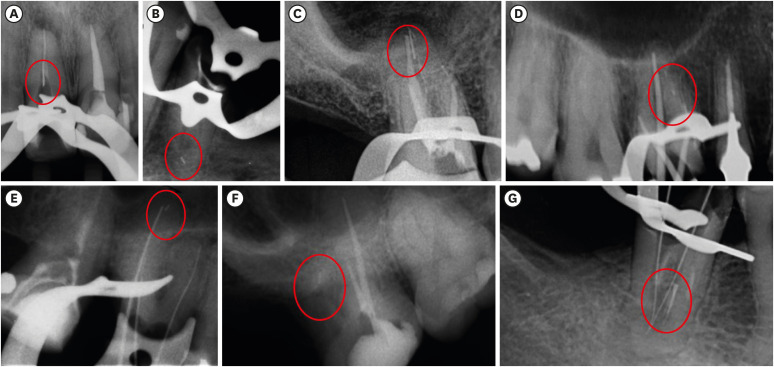
Radiograph images of the 7 Reciproc R25 instruments fractured during root canal retreatments in each tooth and respective root canal. A maxillary central incisor with a fragment of fractured instrument in its middle third (A); a mandibular second premolar with a fragment of fractured instrument in the apical third of its vestibular root canal (B); a maxillary second molar with a fragment of fractured instrument in the apical third of its palatal root canal (C); a maxillary first molar with a fragment of fractured instrument in the apical third of its mesiobuccal root canal (D); a maxillary first molar with a fragment of fractured instrument in the apical third of its palatal root canal (E); a maxillary second molar with a fragment of fractured instrument in the apical third of its distobuccal root canal (F); a mandibular first molar with a fragment of fractured instrument in the middle third of its mesiobuccal root canal (G).

Number of fractures in each group of teeth according to the respective tooth and root canal, fracture position, fragment size, and fragment management
DISCUSSION
CONCLUSIONS
-
Conflict of Interest: No potential conflict of interest relevant to this article was reported.
-
Author Contributions:
Conceptualization: Ruivo LM, Rios MA, Vilela AM, Bueno CES.
Formal analysis: Martin AS.
Investigation: Ruivo LM, Pelegrine RA, Kato AS.
Methodology: Ruivo LM, Pelegrine RA, Rios MA, Vilela AM, Bueno CES.
Project administration: Bueno CES.
Supervision: Bueno CES.
Writing - original draft: Barbosa AFA.
Writing - review & editing: Silva EJNL.
- 1. Siqueira JF Jr, Rôças IN. Clinical implications and microbiology of bacterial persistence after treatment procedures. J Endod 2008;34:1291-1301.e3.ArticlePubMed
- 2. Mollo A, Botti G, Prinicipi Goldoni N, Randellini E, Paragliola R, Chazine M, Ounsi HF, Grandini S. Efficacy of two Ni-Ti systems and hand files for removing gutta-percha from root canals. Int Endod J 2012;45:1-6.ArticlePubMed
- 3. Saad AY, Al-Hadlaq SM, Al-Katheeri NH. Efficacy of two rotary NiTi instruments in the removal of gutta-percha during root canal retreatment. J Endod 2007;33:38-41.ArticlePubMed
- 4. Marques da Silva B, Baratto-Filho F, Leonardi DP, Henrique Borges A, Volpato L, Branco Barletta F. Effectiveness of ProTaper, D-RaCe, and Mtwo retreatment files with and without supplementary instruments in the removal of root canal filling material. Int Endod J 2012;45:927-932.ArticlePubMed
- 5. Akbulut MB, Akman M, Terlemez A, Magat G, Sener S, Shetty H. Efficacy of Twisted File Adaptive, Reciproc and ProTaper Universal Retreatment instruments for root-canal-filling removal: a cone-beam computed tomography study. Dent Mater J 2016;35:126-131.ArticlePubMed
- 6. Alves FR, Marceliano-Alves MF, Sousa JC, Silveira SB, Provenzano JC, Siqueira JF Jr. Removal of root canal fillings in curved canals using either reciprocating single- or rotary multi-instrument systems and a supplementary step with the XP-Endo Finisher. J Endod 2016;42:1114-1119.ArticlePubMed
- 7. Crozeta BM, Silva-Sousa YT, Leoni GB, Mazzi-Chaves JF, Fantinato T, Baratto-Filho F, Sousa-Neto MD. Micro-computed tomography study of filling material removal from oval-shaped canals by using rotary, reciprocating, and adaptive motion systems. J Endod 2016;42:793-797.ArticlePubMed
- 8. Zuolo AS, Mello JE Jr, Cunha RS, Zuolo ML, Bueno CES. Efficacy of reciprocating and rotary techniques for removing filling material during root canal retreatment. Int Endod J 2013;46:947-953.ArticlePubMed
- 9. Rios Mde A, Villela AM, Cunha RS, Velasco RC, De Martin AS, Kato AS, Bueno CES. Efficacy of 2 reciprocating systems compared with a rotary retreatment system for gutta-percha removal. J Endod 2014;40:543-546.ArticlePubMed
- 10. Kırıcı D, Demirbuga S, Karataş E. Micro-computed tomographic assessment of the residual filling volume, apical transportation, and crack formation after retreatment with Reciproc and Reciproc Blue systems in curved root canals. J Endod 2020;46:238-243.ArticlePubMed
- 11. Silva EJNL, Vieira VTL, Hecksher F, Dos Santos Oliveira MRS, Dos Santos Antunes H, Moreira EJL. Cyclic fatigue using severely curved canals and torsional resistance of thermally treated reciprocating instruments. Clin Oral Investig 2018;22:2633-2638.ArticlePubMedPDF
- 12. Ferreira F, Adeodato C, Barbosa I, Aboud L, Scelza P, Zaccaro Scelza M. Movement kinematics and cyclic fatigue of NiTi rotary instruments: a systematic review. Int Endod J 2017;50:143-152.ArticlePubMedPDF
- 13. Varela-Patiño P, Ibañez-Párraga A, Rivas-Mundiña B, Cantatore G, Otero XL, Martin-Biedma B. Alternating versus continuous rotation: a comparative study of the effect on instrument life. J Endod 2010;36:157-159.ArticlePubMed
- 14. Cunha RS, Junaid A, Ensinas P, Nudera W, Bueno CES. Assessment of the separation incidence of reciprocating WaveOne files: a prospective clinical study. J Endod 2014;40:922-924.ArticlePubMed
- 15. Bueno CSP, Oliveira DP, Pelegrine RA, Fontana CE, Rocha DGP, Bueno CES. Fracture incidence of WaveOne and Reciproc files during root canal preparation of up to 3 posterior teeth: a prospective clinical study. J Endod 2017;43:705-708.ArticlePubMed
- 16. Shen Y, Coil JM, Mo AJ, Wang Z, Hieawy A, Yang Y, Haapasalo M. WaveOne rotary instruments after clinical use. J Endod 2016;42:186-189.ArticlePubMed
- 17. Plotino G, Grande NM, Porciani PF. Deformation and fracture incidence of Reciproc instruments: a clinical evaluation. Int Endod J 2015;48:199-205.PubMed
- 18. Caballero-Flores H, Nabeshima CK, Binotto E, Machado ME. Fracture incidence of instruments from a single-file reciprocating system by students in an endodontic graduate programme: a cross-sectional retrospective study. Int Endod J 2019;52:13-18.ArticlePubMedPDF
- 19. Schneider SW. A comparison of canal preparations in straight and curved root canals. Oral Surg Oral Med Oral Pathol 1971;32:271-275.ArticlePubMed
- 20. Özyürek T, Demiryürek EÖ. Efficacy of different nickel-titanium instruments in removing gutta-percha during root canal retreatment. J Endod 2016;42:646-649.ArticlePubMed
- 21. De-Deus G, Belladonna FG, Zuolo AS, Simões-Carvalho M, Santos CB, Oliveira DS, Cavalcante DM, Silva EJNL. Effectiveness of Reciproc Blue in removing canal filling material and regaining apical patency. Int Endod J 2019;52:250-257.ArticlePubMedPDF
- 22. Iqbal MK, Kohli MR, Kim JS. A retrospective clinical study of incidence of root canal instrument separation in an endodontics graduate program: a PennEndo database study. J Endod 2006;32:1048-1052.ArticlePubMed
- 23. Ehrhardt IC, Zuolo ML, Cunha RS, De Martin AS, Kherlakian D, Carvalho MC, Bueno CES. Assessment of the separation incidence of mtwo files used with preflaring: prospective clinical study. J Endod 2012;38:1078-1081.ArticlePubMed
- 24. Yared GM, Dagher FE, Machtou P, Kulkarni GK. Influence of rotational speed, torque and operator proficiency on failure of Greater Taper files. Int Endod J 2002;35:7-12.ArticlePubMed
- 25. Al-Omari MA, Aurich T, Wirtti S. Shaping canals with ProFiles and K3 instruments: does operator experience matter? Oral Surg Oral Med Oral Pathol Oral Radiol Endod 2010;110:e50-e55.ArticlePubMed
- 26. Plotino G, Grande NM, Testarelli L, Gambarini G. Cyclic fatigue of Reciproc and WaveOne reciprocating instruments. Int Endod J 2012;45:614-618.ArticlePubMed
- 27. Di Fiore PM, Genov KA, Komaroff E, Li Y, Lin L. Nickel-titanium rotary instrument fracture: a clinical practice assessment. Int Endod J 2006;39:700-708.ArticlePubMed
- 28. Knowles KI, Hammond NB, Biggs SG, Ibarrola JL. Incidence of instrument separation using LightSpeed rotary instruments. J Endod 2006;32:14-16.ArticlePubMed
- 29. Wolcott S, Wolcott J, Ishley D, Kennedy W, Johnson S, Minnich S, Meyers J. Separation incidence of ProTaper rotary instruments: a large cohort clinical evaluation. J Endod 2006;32:1139-1141.ArticlePubMed
- 30. Muñoz E, Forner L, Llena C. Influence of operator's experience on root canal shaping ability with a rotary nickel-titanium single-file reciprocating motion system. J Endod 2014;40:547-550.ArticlePubMed
- 31. Machado R, Júnior CS, Colombelli MF, Picolli AP, Junior JS, Cosme-Silva L, Garcia LDFR, Alberton LR. Incidence of ProTaper universal system instrument fractures - a retrospective clinical study. Eur Endod J 2018;3:77-81.ArticlePubMedPMC
- 32. Cheung GS. Instrument fracture: mechanisms, removal of fragments, and clinical outcomes. Endod Topics 2009;16:1-26.Article
- 33. Parashos P, Messer HH. Rotary NiTi instrument fracture and its consequences. J Endod 2006;32:1031-1043.ArticlePubMed
- 34. Ungerechts C, Bårdsen A, Fristad I. Instrument fracture in root canals - where, why, when and what? A study from a student clinic. Int Endod J 2014;47:183-190.ArticlePubMed
REFERENCES
Tables & Figures
REFERENCES
Citations

- Reciprocating Torsional Fatigue and Mechanical Tests of Thermal-Treated Nickel Titanium Instruments
Victor Talarico Leal Vieira, Alejandro Jaime, Carlos Garcia Puente, Giuliana Soimu, Emmanuel João Nogueira Leal Silva, Carlos Nelson Elias, Gustavo de Deus
Journal of Endodontics.2025; 51(3): 359. CrossRef - Neodymium-Doped Yttrium Aluminum Perovskite (Nd:YAP) Laser in the Elimination of Endodontic Nickel-Titanium Files Fractured in Rooted Canals (Part 2: Teeth With Significant Root Curvature)
Amaury Namour, Marwan El Mobadder, Clément Cerfontaine, Patrick Matamba, Lucia Misoaga, Delphine Magnin , Praveen Arany, Samir Nammour
Cureus.2025;[Epub] CrossRef - Temperature-Dependent Effects on Cyclic Fatigue Resistance in Three Reciprocating Endodontic Systems: An In Vitro Study
Marcela Salamanca Ramos, José Aranguren, Giulia Malvicini, Cesar De Gregorio, Carmen Bonilla, Alejandro R. Perez
Materials.2025; 18(5): 952. CrossRef - The Cost of Instrument Retrieval on the Root Integrity
Marco A. Versiani, Hugo Sousa Dias, Emmanuel J. N. L. Silva, Felipe G. Belladonna, Jorge N. R. Martins, Gustavo De‐Deus
International Endodontic Journal.2025; 58(12): 1948. CrossRef - Multimethod analysis of large‐ and low‐tapered single file reciprocating instruments: Design, metallurgy, mechanical performance, and irrigation flow
Emmanuel João Nogueira Leal Silva, Fernando Peña‐Bengoa, Natasha C. Ajuz, Victor T. L. Vieira, Jorge N. R. Martins, Duarte Marques, Ricardo Pinto, Mario Rito Pereira, Francisco Manuel Braz‐Fernandes, Marco A. Versiani
International Endodontic Journal.2024; 57(5): 601. CrossRef - Nd: YAP Laser in the Elimination of Endodontic Nickel-Titanium Files Fractured in Rooted Canals (Part 1: Teeth With Minimal Root Curvature)
Amaury Namour, Marwan El Mobadder, Patrick Matamba, Lucia Misoaga, Delphine Magnin , Praveen Arany, Samir Nammour
Cureus.2024;[Epub] CrossRef - Cyclic Fatigue of Different Reciprocating Endodontic Instruments Using Matching Artificial Root Canals at Body Temperature In Vitro
Sebastian Bürklein, Paul Maßmann, Edgar Schäfer, David Donnermeyer
Materials.2024; 17(4): 827. CrossRef - Endodontic Orthograde Retreatments: Challenges and Solutions
Alessio Zanza, Rodolfo Reda, Luca Testarelli
Clinical, Cosmetic and Investigational Dentistry.2023; Volume 15: 245. CrossRef - Design, metallurgy, mechanical properties, and shaping ability of 3 heat-treated reciprocating systems: a multimethod investigation
Emmanuel J. N. L. Silva, Jorge N. R. Martins, Natasha C. Ajuz, Henrique dos Santos Antunes, Victor Talarico Leal Vieira, Francisco Manuel Braz-Fernandes, Felipe Gonçalves Belladonna, Marco Aurélio Versiani
Clinical Oral Investigations.2023; 27(5): 2427. CrossRef - Noncontact 3D evaluation of surface topography of reciprocating instruments after retreatment procedures
Miriam Fatima Zaccaro-Scelza, Renato Lenoir Cardoso Henrique Martinez, Sandro Oliveira Tavares, Fabiano Palmeira Gonçalves, Marcelo Montagnana, Emmanuel João Nogueira Leal da Silva, Pantaleo Scelza
Brazilian Dental Journal.2022; 33(3): 38. CrossRef

Figure 1
Number of total teeth and root canals used in this study
| Variables | Incisors | Canines | Premolars | Molars | ||||
|---|---|---|---|---|---|---|---|---|
| No. of teeth | No. of root canals | No. of teeth | No. of root canals | No. of teeth | No. of root canals | No. of teeth | No. of root canals | |
| Maxillary | 140 | 140 | 26 | 26 | 171 | 342 | 194 | 747 |
| Mandibular | 76 | 76 | 15 | 15 | 115 | 138 | 279 | 1,060 |
Number of fractures in each group of teeth according to the respective tooth and root canal, fracture position, fragment size, and fragment management
| Group of teeth | No. of fractures | Tooth | Root canal | Fracture position | Fragment size | Fragment management |
|---|---|---|---|---|---|---|
| Incisor | 1 | 11 | - | Middle third | 6 mm | Removed |
| Premolar | 1 | 35 | Vestibular | Apical third | 2 mm | NRNB |
| Maxillary molar | 4 | 16 | Palatal | Apical third | 5 mm | NRNB |
| 16 | Mesiobuccal | Apical third | 1 mm | NRNB | ||
| 16 | Palatal | Apical third | 2 mm | NRNB | ||
| 17 | Distobuccal | Apical third | 2 mm | NRNB | ||
| Mandibular molar | 1 | 46 | Mesiobuccal | Middle third | 4 mm | Bypassed |
DB, distobuccal; MB, mesiobuccal; P, palatal. NRNB, neither removed nor bypassed.
DB, distobuccal; MB, mesiobuccal; P, palatal. NRNB, neither removed nor bypassed.

 KACD
KACD
 ePub Link
 Cite
Cite

