Previous issues
- Page Path
- HOME > Browse articles > Previous issues
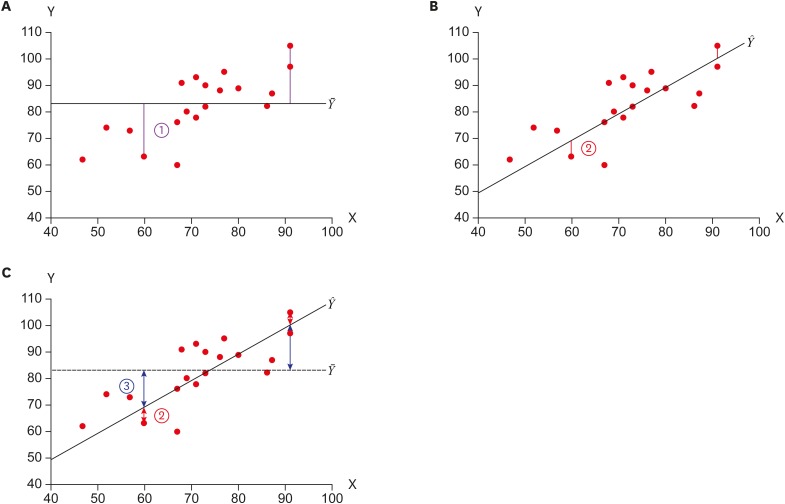
- Statistical notes for clinical researchers: simple linear regression 2 – evaluation of regression line
- Hae-Young Kim
- Restor Dent Endod 2018;43(3):e34. Published online August 9, 2018
- DOI: https://doi.org/10.5395/rde.2018.43.e34
-
 PDF
PDF PubReader
 ePub
ePub -
Citations
Citations to this article as recorded by- Exploring soil pollution patterns in Ghana's northeastern mining zone using machine learning models
Daniel Kwayisi, Raymond Webrah Kazapoe, Seidu Alidu, Samuel Dzidefo Sagoe, Aliyu Ohiani Umaru, Ebenezer Ebo Yahans Amuah, Prosper Kpiebaya
Journal of Hazardous Materials Advances.2024; 16: 100480. CrossRef - Influence of the radius of Monson’s sphere and excursive occlusal contacts on masticatory function of dentate subjects
Dominique Ellen Carneiro, Luiz Ricardo Marafigo Zander, Carolina Ruppel, Giancarlo De La Torre Canales, Rubén Auccaise-Estrada, Alfonso Sánchez-Ayala
Archives of Oral Biology.2024; 159: 105879. CrossRef - Application of hot air-derived RSM conditions and shading for solar drying of avocado pulp and its properties
Sitanan Kowarit, Kitti Sathapornprasath, Surachai Narrat Jansri
Solar Energy.2024; 278: 112768. CrossRef - Corrosion Behavior of Alloy 22 According to Hydrogen Sulfide, Chloride, and pH in an Anaerobic Environment
Yun-Ho Lee, Jin-Seok Yoo, Yong-Won Kim, Jung-Gu Kim
Metals and Materials International.2024; 30(7): 1878. CrossRef - Classification of Male Athletes Based on Critical
Power
Javier Olaya-Cuartero, Basilio Pueo, Alfonso Penichet-Tomas, Jose M. Jimenez-Olmedo
International Journal of Sports Medicine.2024; 45(09): 678. CrossRef
- Exploring soil pollution patterns in Ghana's northeastern mining zone using machine learning models
- 1,875 View
- 31 Download
- 5 Crossref

- Triple antibiotic paste: momentous roles and applications in endodontics: a review
- Ardavan Parhizkar, Hanieh Nojehdehian, Saeed Asgary
- Restor Dent Endod 2018;43(3):e28. Published online June 20, 2018
- DOI: https://doi.org/10.5395/rde.2018.43.e28
-
 Abstract
 PDF
PDF PubReader
 ePub
ePub This study investigated the latest findings and notions regarding ‘triple antibiotic paste’ (TAP) and its applications in dentistry, particularly endodontics. TAP is a combination of 3 antibiotics, ciprofloxacin, metronidazole, and minocycline. Despite the problems and pitfalls research pertaining to this paste has unveiled, it has been vastly used in endodontic treatments. The paste's applications vary, from vital pulp therapy to the recently introduced regeneration and revascularisation protocol. Studies have shown that the paste can eliminate the root canal microorganisms and prepare an appropriate matrix for further treatments. This combination is able to remove diverse groups of obligate and facultative gram-positive and gram-negative bacteria, providing an environment for healing. In regeneration protocol cases, this allows the development, disinfection, and possible sterilization of the root canal system, so that new tissue can infiltrate and grow into the radicular area. Moreover, TAP is capable of creating a discipline in which other wanted and needed treatments can be successfully performed. In conclusion, TAP, as an antibacterial intracanal medication, has diverse uses. Nevertheless, despite its positive effects, the paste has shown drawbacks. Further research concerning the combined paste and other intracanal medications to control microbiota is a must.
-
Citations
Citations to this article as recorded by- Endodontic Intracanal Medicaments and Agents
Anu Priya Guruswamy Pandian, Depti Bellani, Ritya Mary Jibu, Varsha Agnihotri
Dental Clinics of North America.2026; 70(1): 45. CrossRef - Evaluation of Effect of Laser in Root Canal Disinfection in Pulp Regenerative Therapy: A Systematic Review
Kiran Kumar N, Abhishek M, Savitha B. Naik, Biji Brigit, Swetha Geervani V, M Manimozhi
Photobiomodulation, Photomedicine, and Laser Surgery.2025; 43(2): 53. CrossRef - Assessing Cell Viability: Comparative Analysis of Calcium Hydroxide, Triple Antibiotic Paste, and Their Synergistic Impact on human Dental Pulp Stem Cells
Dini Asrianti Bagio, Ibramanto Warganegara, Ike Dwi Maharti, Anggraini Margono, Citra Kusumasari, Sylva Dinie Alinda, Valeria Widita Wairooy
European Journal of General Dentistry.2025; 14(01): 073. CrossRef - Cytotoxic and Antibiofilm Properties of Antibiotic-Loaded Thermoresponsive Hydrogels for Root Canal Therapy
Cristiane Duque, Gabriela Pacheco de Almeida Braga, Juliana Machado de Carvalho, Karina Sampaio Caiaffa, Gabriel Pereira Nunes, Rafaela Laruzo Rabelo, Vanessa Rodrigues dos Santos, Geórgia Rondó Peres, Lucas da Silva Ribeiro, Emerson Rodrigues de Camargo
Processes.2025; 13(3): 661. CrossRef - Antibiofilm properties, cytotoxicity, and effect on protease activity of antibiotics and EGCG-based medications for endodontic purposes
Daniela Alvim Chrisostomo, Jesse Augusto Pereira, Polliana Mendes Candia Scaffa, Zach Gouveia, Gabriel Flores Abuna, Sergey V. Plotnikov, Anuradha Prakki, Cristiane Duque
Journal of Dentistry.2025; 156: 105660. CrossRef - The use of three-dimensional-printed guides, static navigation, and bioactive materials to treat bilateral and double dens invaginatus
Parth Patel, Nidhi Bharti, Ankit Arora, C. Nimisha Shah
Saudi Endodontic Journal.2025; 15(2): 207. CrossRef - To Assess the Cell Viability of Triple Antibiotic Paste under Confocal Microscope: An In Vitro Study
Elanthendral Saravanan, Mahesh Ramakrishnan
Journal of South Asian Association of Pediatric Dentistry.2025; 8(2): 81. CrossRef - Eficacia de la pasta triantibiótica en endodoncia: actividad antibacteriana frente a cepas resistentes de Enterococcus faecalis. Una revisión exhaustiva
Elena Patricia Cevallos Fernández, Katherine de los Ángeles Cuenca León
Anatomía Digital.2025; 8(3.1): 88. CrossRef - Efectividad de diferentes antimicóticos, junto con la pasta triantibiotica, para el tratamiento de Candida albicans en conductos radiculares
Carlos Andrés Rodríguez Tapia, Jessica María Sarmiento Ordoñez
Anatomía Digital.2025; 8(3.2): 45. CrossRef - Comparative Evaluation of Calcium Hydroxide, Triple Antibiotic Paste, and Calcium Hydroxide with 2% Chlorhexidine as Intracanal Medicaments in Reducing Interappointment Pain during Endodontic Treatment: An In Vivo Study
Rachit Mathur, Shaista Gazal, Itika Jain, Shyam Agrawal, Akshada Mungee, Babra Khan
International Journal of Clinical Pediatric Dentistry.2025; 18(6): 628. CrossRef - In Vitro Effectiveness of Endodontic Triple Antibiotic Paste Associated With Daptomycin
Sabrina S Azevedo, Gabriela C Chianca, Bruna A Thurler, Raiane C Chamon, Helvécio C Corrêa Póvoa, Leonardo S Antunes, Natalia L Pontes Póvoa Iorio
Cureus.2025;[Epub] CrossRef - Effect of Chitosan and bioactive glass nanomaterials as intracanal medicaments on TGF-β1 release from intraradicular dentin
Sarah Salah Hashem, Mohammed M. Khalefa, Mahmoud Hassan Mohamed, Hemat M. ELSheikh, Fatma Abd El-Rahman Taher
BMC Oral Health.2025;[Epub] CrossRef - Clinical and radiographic success of lesion sterilization and tissue repair in primary teeth: a systematic review and meta-analysis
Khlood Baghlaf, Rana A. Alamoudi
The Saudi Dental Journal.2025;[Epub] CrossRef - A Comparative Evaluation of Herbal Extracts and Triple Antibiotic Paste as Intracanal Medicament against Enterococcus faecalis: A Microbiological Study
Divya Singh, Rashi Singh, Nidhi Gupta, Natasha Gambhir, Saritha Golla
International Journal of Clinical Pediatric Dentistry.2024; 17(3): 285. CrossRef - Comparative Evaluation of Push-Out Bond Strength of Conventional Mineral Trioxide Aggregate, Biodentine, a Modified Mineral Trioxide Aggregate, and Two Novel Antibacterial-Enhanced Mineral Trioxide Aggregates
Arokia Rajkumar Shancy Merlin, Vignesh Ravindran, Ganesh Jeevanandan, Rajalakshmanan Eswaramoorthy, Abirami Arthanari
Cureus.2024;[Epub] CrossRef - In Vitro Evaluation of the Effect of Oleanolic Acid as a Potential Root Canal Medicament on Viability and Proliferation of Dental Pulp Stem Cells
Khalifah A. Alhaila, Manal Farouk Badawi, Mohamed G. Elbeltagy, Amany E. Badr
European Journal of General Dentistry.2024; 13(01): 051. CrossRef - Regenerative Endodontic Treatment in Dentinogenesis Imperfecta‐Induced Apical Periodontitis
Ying Liao, Ting Pan, Xianghui Xing, Sivakumar Nuvvula
Case Reports in Dentistry.2024;[Epub] CrossRef - Efecto antimicrobiano como medicación intraconducto de la pasta triantibiótica.
Paúl Sebastián Ulloa Amores, Diana Álvarez Álvarez, María Elizabeth Moscoso Abad, Magda Zulay Bastidas Calva
Revista de la Asociación Dental Mexicana.2024; 81(4): 211. CrossRef - Synthesis and Characterization of Nanosilica-Coated Antibiotics, TAP: A Comprehensive Study Utilizing XRD, EDS, FTIR, SEM, and TEM – Invitro Study
Mahaboob S. Hameed, S. Delphine P. Antony, Rajeshkumar Shanmugam, Sandhya Raghu
Journal of International Oral Health.2024; 16(5): 386. CrossRef - Association between host defence peptide IDR‐1002 and ciprofloxacin: Effects on human dental pulp cells
Danilo César Mota Martins, Maurício Gonçalves da Costa Sousa, Poliana Amanda Oliveira Silva, Lana Ribeiro Aguiar, Rosângela Vieira de Andrade, Amandda Évellin Silva‐Carvalho, Felipe Saldanha‐Araújo, Octávio Luiz Franco, Taia Maria Berto Rezende
Australian Endodontic Journal.2024; 50(3): 547. CrossRef - Comparative evaluation of antimicrobial efficacy of triple antibiotic paste and amoxicillin clavulanate paste as an intracanal medicament against Enterococcus faecalis: An in vitro study
Dhandayuthapani Sasikala, Parisa Norouzi Baghkomeh, Jamaluddin Mohammed Farzan
Dental Research Journal.2024;[Epub] CrossRef - Triple Antibiotic Paste: A Game Changer in Endodontics
Kalagi G. Panchal, Karima Virani, Vraj Patel, Aquib Ali Khan, Anam Pettiwala, Srikala S. Puranik, Srushti Joshi
Journal of Pharmacy and Bioallied Sciences.2024; 16(Suppl 3): S1913. CrossRef - Non-surgical Management of a Large Periapical Lesion: A Case Study of the Successful Application of a Modified Triple Antibacterial Paste
Srushti Awghad, Joyeeta Mahapatra, Amit Reche, Ankita Burse, Aradhana Kibe
Cureus.2024;[Epub] CrossRef - Enhancing Antimicrobial Efficacy and Synergistic Effects of Nano-Silica-Based Combinations With Doxycycline, Metronidazole, and Ciprofloxacin Against Enterococcus faecalis Biofilms
Shahul Hameed, Delphine P Antony, Rajeshkumar Shanmugam, Sandhya Raghu, Hima Sandeep Adimulapu
Cureus.2024;[Epub] CrossRef - Healing of periapical lesion by non-surgical endodontic therapy: A case series
Athira Ramesh, Rajesh Pillai, Afzal A, Anakha Santhosh, Arunima G.S, Sandeep K. V
IP Indian Journal of Conservative and Endodontics.2024; 9(2): 99. CrossRef - Effect of Calcium Hydroxide Versus Double Antibiotic Paste on Endodontic Treatment Outcomes in Teeth With Large Periapical Lesions: A Triple‐Blind Randomized Clinical Trial
Afsaneh Rahmati, Farshad Seyedein, Omid Dianat, Sara Saedi, Golriz Rostami, Alireza Akbarzadeh Baghban, Shima Sabertahan, Majid Kazem, Kee Y. Kum
International Journal of Dentistry.2024;[Epub] CrossRef - Can antimicrobial photodynamic therapy serve as an effective adjunct protocol for disinfecting the necrotic root canal system? A randomized controlled study
Remy Barazy, Hisham Alafif, Hassan Achour, Ahmad Al-Aloul, Yasser Alsayed Tolibah
BDJ Open.2024;[Epub] CrossRef - Drain and Treat: A Rubber Dam Technique for Acute Periapical Abscess
S. Grover, K. Mala, J. D’Souza
Acta Medica Bulgarica.2024; 51(s2): 143. CrossRef - Microbial Dynamics in Endodontic Pathology—From Bacterial Infection to Therapeutic Interventions—A Narrative Review
Klara Wieczorkiewicz, Anna Jarząbek, Estera Bakinowska, Kajetan Kiełbowski, Andrzej Pawlik
Pathogens.2024; 14(1): 12. CrossRef - An in vitro comparative evaluation of the effect of three intracanal medicaments – chlorhexidine gel, triple antibiotic paste, and calcium hydroxide paste on the push-out bond strength of MTA Plus, Biodentine, and calcium-enriched mixture
Gouthami Datta, Ramya Raghu, Ashish Shetty, Gautham P Manjunath, Dishant Patel, Subhashini Rajasekhara
Endodontology.2023; 35(1): 60. CrossRef - The cytotoxic effect of cysteamine and its combinations with various endodontic intracanal medications on fibroblast cells: in vitro study
Esraa Adel Mohamed Abd Elhameed ElGammal, Abeer Hashem Mahran, Salma Hassan El Ashry, Sara Hossam Fahmy
Bulletin of the National Research Centre.2023;[Epub] CrossRef - Healing of large endodontic lesions using a new combination of triple antibiotics: A case report
Saeed Asgary, Ardavan Parhizkar
Clinical Case Reports.2023;[Epub] CrossRef - Effects of Intracanal Medicaments on the Measurement Accuracy of Four Apex Locators: An In Vitro Study
Hamza Cudal, Tuğrul Aslan, Bertan Kesim
Meandros Medical and Dental Journal.2023; 24(3): 215. CrossRef - Comparative evaluation of the antibacterial efficacy of herbal agents as intracanal medicaments individually or in combination with chitosan: An in vitro RTPCR study
Gaurav Patri, Kotni Sheetal, PrasantiKumar Pradhan, Pratik Agrawal, S Lata
Journal of International Oral Health.2023; 15(1): 89. CrossRef - Recent progress in carbon dots for anti-pathogen applications in oral cavity
Yuying Jiang, Chuqiang Yin, Jianning Mo, Xiaoyu Wang, Ting Wang, Guotai Li, Qihui Zhou
Frontiers in Cellular and Infection Microbiology.2023;[Epub] CrossRef - A Comprehensive Review of Lesion Sterilization and Tissue Repair: An Alternative for Pulpectomy in Deciduous Teeth
Aparna Achanta, Amit Reche, Rishika Dakhale, Rudra R Bharate
Cureus.2023;[Epub] CrossRef - Healing of Large Endodontic Lesions Using Long‐Term Application of a New Combination of Triple Antibiotics: A Series of Cases
Saeed Asgary, Ardavan Parhizkar, Maria Beatriz Duarte Gavião
Case Reports in Dentistry.2023;[Epub] CrossRef - Non-cytotoxic Root Canal Dressing with Improved Antimicrobial Efficacy
Farzad Koosha, Jerome Cymerman, Thomas Manders, Marcia Simon, Stephen Walker, Miriam Rafailovich
Journal of Endodontics.2023; 49(2): 205. CrossRef - Triple antibiotics: A synergistic approach to combating infection
Hemant Sawhney, Anukriti Kumari, Ritik Kashwani, Geetanjali Gupta, SJ Das
IP Indian Journal of Conservative and Endodontics.2023; 8(4): 189. CrossRef - A 1-year Clinical and Radiographic Assessment of Regenerative Endodontic Therapy for Necrotic Primary Molars: A Randomized controlled Trial
Dina D Abdelmoneim, Amr M Abdelaziz, Gehan G Allam, Amira S Badran
International Journal of Clinical Pediatric Dentistry.2023; 16(2): 295. CrossRef - “BIODENTINE” THE DENTINE IN A CAPSULE AS AN APICAL BARRIER IN TRAUMATIZED MAXILLARY CENTRAL INCISOR WITH TWO YEARS FOLLOW UP.
Savita Thakur, Udai Bhanu, Gurkirat Singh Grewal
INTERNATIONAL JOURNAL OF SCIENTIFIC RESEARCH.2022; : 64. CrossRef - Long-term treatment of dentine with triple antibiotic paste promotes stem cell viability and attachment
Samiya Riaz, Ahmad Azlina, Zuliani Mahmood, Aung T. Htun
Journal of Taibah University Medical Sciences.2022; 17(4): 630. CrossRef - Non surgical management of trauma induced root resorption and large periapical
lesion using bioactive material- A case report
Tejasvini Prajapati, Sonali Kapoor, Purnil Shah, Ankit Arora, Hardik Rana
Clinical Dentistry.2022;[Epub] CrossRef - Antibacterial efficacy of antibiotic pastes versus calcium hydroxide intracanal dressing: A systematic review and meta-analysis of ex vivo studies
Mohammadreza Vatankhah, Kamyar Khosravi, Nazanin Zargar, Armin Shirvani, MohammadHossein Nekoofar, Omid Dianat
Journal of Conservative Dentistry.2022; 25(5): 463. CrossRef - Triple Antibiotic Paste: A Suitable Medicament for Intracanal Disinfection
Krutika Malu, Monika Khubchandani
Cureus.2022;[Epub] CrossRef - Present status and future directions of intracanal medicaments
Ronald Ordinola‐Zapata, W. Craig Noblett, Alejandro Perez‐Ron, Zhou Ye, Jorge Vera
International Endodontic Journal.2022; 55(S3): 613. CrossRef - The effect of four different intracanal medicaments on the push-out bond strength of root canal sealers
Shalu Maan, Vijaya Dhar Bhatt, Rohit Singh, Sayak Gupta, Syed Alay Noorain, Aashna Gill, Pradeep Kumar, Sushil Yadav, Preeti Sharma
Journal of Medicine and Life.2022; 15(4): 448. CrossRef - A Comparative Evaluation of Microhardness and Chemical Structure of Radicular Dentin with Two Combinations of TAP and MTAP: An In Vitro Study
P Niharika, Saigeeta Kondamadugu, Nagireddy Venugopal Reddy, Muthumula Daneswari, Annie P Chris, Nikhila V Reddy
International Journal of Clinical Pediatric Dentistry.2022; 15(S2): S151. CrossRef - Comparative Evaluation of Two Antibiotic Pastes for Root Canal Disinfection
Sadhna Sharma, Urvashi Bhushan, Mridula Goswami, CP Baveja
International Journal of Clinical Pediatric Dentistry.2022; 15(S1): S12. CrossRef - Management of External Inflammatory Root Resorption following Tooth Autotransplantation Using a Modified Combination of Triple Antibiotics
Saeed Asgary, Ardavan Parhizkar, Paulo J. Palma
Case Reports in Dentistry.2022;[Epub] CrossRef - Effect of triple antibiotic paste on the bond strength of epoxy and methacrylate resin-based sealers to root canal dentin
Fereshte Sobhnamayan, Alireza Adl, Mahdi Sedigh-Shams, Hossein Mirkhaghani
Journal of Conservative Dentistry.2022; 25(4): 426. CrossRef - Progress of Research on the Application of Triple Antibiotic Paste and Hydrogel Scaffold Materials in Endodontic Revascularization: A Systematic Review
Jia Zhao, Tian Jiao Wang
Evidence-Based Complementary and Alternative Medicine.2022; 2022: 1. CrossRef - The effect of different intracanal medicaments on the dislodgement resistance of mineral trioxide aggregate
Farzaneh Afkhami, Shahrzad Razavi, Sholeh Ghabraei
BMC Oral Health.2022;[Epub] CrossRef - Efficacy of Ciprofloxacin, Metronidazole and Minocycline in Ordered Mesoporous Silica against Enterococcus faecalis for Dental Pulp Revascularization: An In-Vitro Study
Cintia Micaela Chamorro-Petronacci, Beatriz Santos Torres, Rocío Guerrero-Nieves, Mario Pérez-Sayáns, Marcia Carvalho-de Abreu Fantini, Luis Carlos Cides-da-Silva, Beatriz Magariños, Berta Rivas-Mundiña
Materials.2022; 15(6): 2266. CrossRef - Antibiotic Mixtures in Noninstrumental Endodontic Treatment of Primary Teeth with Necrotic Pulps: A Systematic Review
Farah Chouchene, Fatma Masmoudi, Ahlem Baaziz, Fethi Maatouk, Hichem Ghedira, Sivakumar Nuvvula
International Journal of Dentistry.2021; 2021: 1. CrossRef - Potential apply of hydrogel-carried chlorhexidine and metronidazole in root canal disinfection
Yanhong YAN, Peng ZHOU, Haibing LU, Yun GUAN, Ming MA, Juan WANG, Guangwei SHANG, Beizhan JIANG
Dental Materials Journal.2021; 40(4): 986. CrossRef - Evaluation of the Effect of Nitrofurantoin Paste as an Intracanal Medicament on the Chemical Structure of Radicular Dentine
Mewan Abdulrahman, Bestoon Faraj, Kawa Dizaye
Sulaimani Dental Journal.2021; 8(2): 8. CrossRef - Local Drug Delivery Systems for Vital Pulp Therapy: A New Hope
Ardavan Parhizkar, Saeed Asgary, Carlo Galli
International Journal of Biomaterials.2021; 2021: 1. CrossRef - Antibiofilm and cytotoxic effect of 3,3′-dihydroxycurcumin (DHC) as photosensitizer agent in antimicrobial photodynamic therapy for endodontic purposes
Jesse Augusto Pereira, Carlos Roberto Polaquini, VanessaRodrigues dos Santos, Karina Sampaio Caiaffa, Rafaela Laruzo Rabelo, Reinaldo dos Santos Theodoro, Letícia Helena Theodoro, Luis Octavio Regasini, Cristiane Duque
Photodiagnosis and Photodynamic Therapy.2021; 36: 102534. CrossRef - An in-vitro Comparative Evaluation of Quantitative Release of Transforming Growth Factor β-1 from Dentin upon the Action of Endodontic Irrigants, Medicaments, Ultrasonic Activation, and Low-Level Laser Irradiation
Anilkumar Akhila, V. P. Prabath Singh, Kerala R. Varma, Senthil V. Vasudevan, V. Sukhithasri, Salu Sasikumar
Amrita Journal of Medicine.2021; 17(2): 34. CrossRef - Effect of triple antibiotic paste and calcium hydroxide on the rate of healing of periapical lesions: A systematic review
NKiran Kumar, Biji Brigit, BS Annapoorna, SavithaB Naik, Seema Merwade, K Rashmi
Journal of Conservative Dentistry.2021; 24(4): 307. CrossRef - Comparison of the efficacy of CanalBrush, EndoActivator, and Passive Ultrasonic Irrigation on the removal of triple antibiotic paste from root canal walls: An in vitro study
Santosh Kumar, Kavisha Desai, Aparna Palekar, Baswaraj Biradar, Ananjan Chatterjee, Khushboo Kumari
Journal of International Society of Preventive and Community Dentistry.2020; 10(4): 424. CrossRef - Apexification with Calcium Hydroxide vs. Revascularization
H. Boufdil, M. Mtalsi, S. El Arabi, B. Bousfiha, Jose López-López
Case Reports in Dentistry.2020;[Epub] CrossRef - Effect of Intracanal Medicaments and Irrigants on the Release of Transforming Growth Factor Beta 1 and Vascular Endothelial Growth Factor from Cervical Root Dentin
Lívia Nazareth Ferreira, Regina Maria Puppin-Rontani, Fernanda Miori Pascon
Journal of Endodontics.2020; 46(11): 1616. CrossRef - Incorporation of amoxicillin-loaded microspheres in mineral trioxide aggregate cement: an in vitro study
Fábio Rocha Bohns, Vicente Castelo Branco Leitune, Isadora Martini Garcia, Bruna Genari, Nélio Bairros Dornelles, Silvia Stanisçuaski Guterres, Fabrício Aulo Ogliari, Mary Anne Sampaio de Melo, Fabrício Mezzomo Collares
Restorative Dentistry & Endodontics.2020;[Epub] CrossRef - Assessment of Nitrofurantoin as an Experimental Intracanal Medicament in Endodontics
Mewan Salahalddin A. Alrahman, Bestoon Muhammed Faraj, Kawa F. Dizaye, Abdelwahab Omri
BioMed Research International.2020;[Epub] CrossRef - An in vitro assessment of effect on microhardness of dentin using vicker's hardness method
Manoj Chandak, Richa Modi, Rakesh Gogiya, Rakhi Chandak, Anuja Ikhar, Nikhil Mankar
Journal of Datta Meghe Institute of Medical Sciences University.2020; 15(2): 251. CrossRef - Inspection of the Microbiota in Endodontic Lesions
Mario Dioguardi, Giovanni Di Gioia, Gaetano Illuzzi, Claudia Arena, Vito Carlo Alberto Caponio, Giorgia Apollonia Caloro, Khrystyna Zhurakivska, Iolanda Adipietro, Giuseppe Troiano, Lorenzo Lo Muzio
Dentistry Journal.2019; 7(2): 47. CrossRef - Materials for pulpotomy in immature permanent teeth: a systematic review and meta-analysis
Yuan Chen, Xinlei Chen, Yali Zhang, Fangjie Zhou, Jiaxin Deng, Jing Zou, Yan Wang
BMC Oral Health.2019;[Epub] CrossRef - Adjunctive antimicrobial photodynamic therapy to conventional chemo-mechanical debridement of infected root canal systems: A systematic review and meta-analysis
Maryam Pourhajibagher, Abbas bahador
Photodiagnosis and Photodynamic Therapy.2019; 26: 19. CrossRef - Comparison of Antibacterial Effects of Photodynamic Therapy, Modified Triple Antibiotic Paste and Calcium Hydroxide on Root Canals Infected With Enterococcus faecalis: An In Vitro Study
Mohammad Asnaashari, Mohammad Jafar Eghbal, Amirali Sahba Yaghmayi, Mehdi Shokri, Saranaz Azari-Marhabi
Journal of Lasers in Medical Sciences.2019; 10(5): S23. CrossRef
- Endodontic Intracanal Medicaments and Agents
- 14,439 View
- 156 Download
- 71 Crossref

-
Retention of BioAggregate and MTA as coronal plugs after intracanal medication for regenerative endodontic procedures: an
ex vivo study - Suzan Abdul Wanees Amin, Shaimaa Ismail Gawdat
- Restor Dent Endod 2018;43(3):e18. Published online April 26, 2018
- DOI: https://doi.org/10.5395/rde.2018.43.e18

-
 Abstract
 PDF
PDF PubReader
 ePub
ePub Objectives This study compared the retention of BioAggregate (BA; Innovative BioCeramix) and mineral trioxide aggregate (MTA; Angelus) as coronal plugs after applying different intracanal medications (ICMs) used in regenerative endodontics.
Materials and Methods One-hundred human maxillary central incisors were used. The canals were enlarged to a diameter of 1.7 mm. Specimens were divided into 5 groups (
n = 20) according to the ICM used: calcium hydroxide (CH), 2% chlorhexidine (CHX), triple-antibiotic paste (TAP), double-antibiotic paste (DAP), and no ICM (control; CON). After 3 weeks of application, ICMs were removed and BA or MTA were placed as the plug material (n = 10). The push-out bond strength and the mode of failure were assessed. The data were analyzed using 2-way analysis of variance, the Tukey's test, and the χ2 test;p values < 0.05 indicated statistical significance.Results The type of ICM and the type of plug material significantly affected bond strength (
p < 0.01). Regardless of the type of ICM, BA showed a lower bond strength than MTA (p < 0.05). For MTA, CH showed a higher bond strength than CON, TAP and DAP; CHX showed a higher bond strength than DAP (p < 0.01). For BA, CH showed a higher bond strength than DAP (p < 0.05). The mode of failure was predominantly cohesive for BA (p < 0.05).Conclusions MTA may show better retention than BA. The mode of bond failure with BA can be predominantly cohesive. BA retention may be less affected by ICM type than MTA retention.
-
Citations
Citations to this article as recorded by- An in vitro comparative evaluation of the effect of three intracanal medicaments – chlorhexidine gel, triple antibiotic paste, and calcium hydroxide paste on the push-out bond strength of MTA Plus, Biodentine, and calcium-enriched mixture
Gouthami Datta, Ramya Raghu, Ashish Shetty, Gautham P Manjunath, Dishant Patel, Subhashini Rajasekhara
Endodontology.2023; 35(1): 60. CrossRef - Effects of calcium hydroxide intracanal medicament on push‐out bond strength of endodontic sealers: A systematic review and meta‐analysis
Mohammed Nasser Alhajj, Fadhilah Daud, Sadeq Ali Al‐Maweri, Yanti Johari, Zuryati Ab‐Ghani, Mariatti Jaafar, Yoshihito Naito, Widyasri Prananingrum, Zaihan Ariffin
Journal of Esthetic and Restorative Dentistry.2022; 34(8): 1166. CrossRef - A Breakthrough in the Era of Calcium Silicate-Based Cements: A Critical Review
Payal S Chaudhari, Manoj G Chandak, Akshay A Jaiswal, Nikhil P Mankar, Priyanka Paul
Cureus.2022;[Epub] CrossRef - Modern Medicaments for Endodontic Treatment in Children
Н.В. Шаковец, О.С. Романова
Стоматология. Эстетика. Инновации.2021; (4): 408. CrossRef - Do intracanal medications used in regenerative endodontics affect the bond strength of powder-to-liquid and ready-to-use cervical sealing materials?
MarinaCarvalho Prado, Kevillin Martiniano, AndreaCardoso Pereira, KarineL Cortellazzi, MarinaA Marciano, Gabriel Abuna, Adriana de-Jesus-Soares
Journal of Conservative Dentistry.2021; 24(5): 464. CrossRef - In vivo Biocompatibility and Bioactivity of Calcium Silicate-Based Bioceramics in Endodontics
Wencheng Song, Wei Sun, Lili Chen, Zhenglin Yuan
Frontiers in Bioengineering and Biotechnology.2020;[Epub] CrossRef - Effect of Hydrogel-Based Antibiotic Intracanal Medicaments on Push-Out Bond Strength
Rayan B. Yaghmoor, Jeffrey A. Platt, Kenneth J. Spolnik, Tien Min Gabriel Chu, Ghaeth H. Yassen
European Journal of Dentistry.2020; 14(04): 575. CrossRef - Regenerative Endodontic Procedures, Disinfectants and Outcomes: A Systematic Review
Adam S. Kharchi, Nara Tagiyeva-Milne, Shalini Kanagasingam
Primary Dental Journal.2020; 9(4): 65. CrossRef
- An in vitro comparative evaluation of the effect of three intracanal medicaments – chlorhexidine gel, triple antibiotic paste, and calcium hydroxide paste on the push-out bond strength of MTA Plus, Biodentine, and calcium-enriched mixture
- 1,652 View
- 9 Download
- 8 Crossref

- Effects of four novel root-end filling materials on the viability of periodontal ligament fibroblasts
- Makbule Bilge Akbulut, Pembegul Uyar Arpaci, Ayce Unverdi Eldeniz
- Restor Dent Endod 2018;43(3):e24. Published online May 25, 2018
- DOI: https://doi.org/10.5395/rde.2018.43.e24
-
 Abstract
 PDF
PDF PubReader
 ePub
ePub Objectives The aim of this
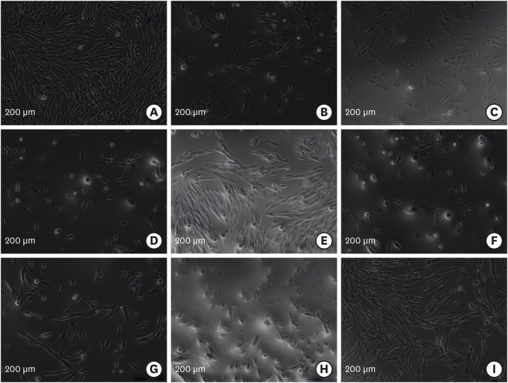
in vitro study was to evaluate the biocompatibility of newly proposed root-end filling materials, Biodentine, Micro-Mega mineral trioxide aggregate (MM-MTA), polymethylmethacrylate (PMMA) bone cement, and Smart Dentin Replacement (SDR), in comparison with contemporary root-end filling materials, intermediate restorative material (IRM), Dyract compomer, ProRoot MTA (PMTA), and Vitrebond, using human periodontal ligament (hPDL) fibroblasts.Materials and Methods Ten discs from each material were fabricated in sterile Teflon molds and 24-hour eluates were obtained from each root-end filling material in cell culture media after 1- or 3-day setting. hPDL fibroblasts were plated at a density of 5 × 10³/well, and were incubated for 24 hours with 1:1, 1:2, 1:4, and 1:8 dilutions of eluates. Cell viability was evaluated by XTT assay. Data was statistically analysed. Apoptotic/necrotic activity of PDL cells exposed to material eluates was established by flow cytometry.
Results The Vitrebond and IRM were significantly more cytotoxic than the other root-end filling materials (
p < 0.05). Those cells exposed to the Biodentine and Dyract compomer eluates showed the highest survival rates (p < 0.05), while the PMTA, MM-MTA, SDR, and PMMA groups exhibited similar cell viabilities. Three-day samples were more cytotoxic than 1-day samples (p < 0.05). Eluates from the cements at 1:1 dilution were significantly more cytotoxic (p < 0.05). Vitrebond induced cell necrosis as indicated by flow cytometry.Conclusions This
in vitro study demonstrated that Biodentine and Compomer were more biocompatible than the other root-end filling materials. Vitrebond eluate caused necrotic cell death.-
Citations
Citations to this article as recorded by- Effects of Three Retrograde Filling Materials on Production of Inflammatory Cytokines and Resorbing Mediators
Samaneh Arab, Marjan Bahraminasab, Masoumeh Motamedi, Jamshid Hadjati, Alaviye Vahid
Journal of Microbiota.2024;[Epub] CrossRef - Apoptotic effects of biodentine, calcium-enriched mixture (CEM) cement, ferric sulfate, and mineral trioxide aggregate (MTA) on human mesenchymal stem cells isolated from the human pulp of exfoliated deciduous teeth
Bahareh NAZEMI SALMAN, Mahshid MOHEBBI RAD, Ehsan SABURI
Minerva Dental and Oral Science.2024;[Epub] CrossRef - Assessment of Mechanical/Chemical Properties and Cytotoxicity of Resin-Modified Glass Ionomer Cements Containing Sr/F-Bioactive Glass Nanoparticles and Methacrylate Functionalized Polyacids
Wisitsin Potiprapanpong, Parichart Naruphontjirakul, Chutikarn Khamsuk, Somruethai Channasanon, Arnit Toneluck, Siriporn Tanodekaew, Naruporn Monmaturapoj, Anne M. Young, Piyaphong Panpisut
International Journal of Molecular Sciences.2023; 24(12): 10231. CrossRef - Comparative biological properties of resin-free and resin-based calcium silicate-based endodontic repair materials on human periodontal ligament stem cells
Shehabeldin M. Saber, Shaimaa M. Gomaa, Mohamed M. Elashiry, Ahmed El-Banna, Edgar Schäfer
Clinical Oral Investigations.2023; 27(11): 6757. CrossRef - Comparison of root end sealing ability of three retrograde filling materials in teeth with root apices resected at 900 using dye penetration method under fluorescent microscope
Dr. Payal Chaudhari, Manoj Chandak, Dr. Aditya Patel
F1000Research.2023; 12: 1049. CrossRef - The Effects of Tricalcium-Silicate-Nanoparticle-Containing Cement: In Vitro and In Vivo Studies
Naho Ezawa, Yoshihiko Akashi, Kei Nakajima, Katsutoshi Kokubun, Masahiro Furusawa, Kenichi Matsuzaka
Materials.2023; 16(12): 4451. CrossRef - Evaluation of the cytotoxic effects of a new Harvard MTA compared to MTA Flow and ProRoot MTA on human gingival fibroblasts
Abdel-Rahman Youssef, Samia Elsherief
The Saudi Dental Journal.2021; 33(7): 679. CrossRef - Cytotoxicity and Bioactivity of Mineral Trioxide Aggregate and Bioactive Endodontic Type Cements: A Systematic Review
Uma Dixit, Rucha Shivajirao Bhise Patil, Rupanshi Parekh
International Journal of Clinical Pediatric Dentistry.2021; 14(1): 30. CrossRef - MTT versus other cell viability assays to evaluate the biocompatibility of root canal filling materials: a systematic review
A. V. B. Pintor, L. D. Queiroz, R. Barcelos, L. S. G. Primo, L. C. Maia, G. G. Alves
International Endodontic Journal.2020; 53(10): 1348. CrossRef - Micro-computed tomographic evaluation of the flow and filling ability of endodontic materials using different test models
Fernanda Ferrari Esteves Torres, Juliane Maria Guerreiro-Tanomaru, Gisselle Moraima Chavez-Andrade, Jader Camilo Pinto, Fábio Luiz Camargo Villela Berbert, Mario Tanomaru-Filho
Restorative Dentistry & Endodontics.2020;[Epub] CrossRef - Long-term Follow-up for Apical Microsurgery of Teeth with Core and Post Restorations
Astrid Truschnegg, Petra Rugani, Barbara Kirnbauer, Lumnije Kqiku, Norbert Jakse, Robert Kirmeier
Journal of Endodontics.2020; 46(2): 178. CrossRef - Comparison of Cytotoxic Effects of Calcium Silicate-based Materials on Human Pulp Fibroblasts
Mehmet Adıgüzel, Fuat Ahmetoğlu, Ayçe Ünverdi Eldeniz, Mehmet Gökhan Tekin, Bülent Göğebakan
Journal of Dental Research, Dental Clinics, Dental Prospects.2019; 13(4): 241. CrossRef
- Effects of Three Retrograde Filling Materials on Production of Inflammatory Cytokines and Resorbing Mediators
- 1,591 View
- 5 Download
- 12 Crossref

- Light transmittance of CAD/CAM ceramics with different shades and thicknesses and microhardness of the underlying light-cured resin cement
- Zahra Jafari, Homayoon Alaghehmand, Yasaman Samani, Mina Mahdian, Soraya Khafri
- Restor Dent Endod 2018;43(3):e27. Published online June 4, 2018
- DOI: https://doi.org/10.5395/rde.2018.43.e27
-
 Abstract
 PDF
PDF PubReader
 ePub
ePub Objectives The aim of this
in vitro study was to evaluate the effects of the thickness and shade of 3 types of computer-aided design/computer-aided manufacturing (CAD/CAM) materials.Materials and Methods A total of 120 specimens of 2 shades (A1 and A3) and 2 thicknesses (1 and 2 mm) were fabricated using VITA Mark II (VM; VITA Zahnfabrik), IPS e.max CAD (IE; IvoclarVivadent), and VITA Suprinity (VS; VITA Zahnfabrik) (
n = 10 per subgroup). The amount of light transmission through the ceramic specimens was measured by a radiometer (Optilux, Kerr). Light-cured resin cement samples (Choice 2, Bisco) were fabricated in a Teflon mold and activated through the various ceramics with different shades and thicknesses using an LED unit (Bluephase, IvoclarVivadent). In the control group, the resin cement sample was directly light-cured without any ceramic. Vickers microhardness indentations were made on the resin surfaces (KoopaPazhoohesh) after 24 hours of dark storage in a 37°C incubator. Data were analyzed using analysis of variance followed by the Tukeypost hoc test (α = 0.05).Results Ceramic thickness and shade had significant effects on light transmission and the microhardness of all specimens (
p < 0.05). The mean values of light transmittance and microhardness of the resin cement in the VM group were significantly higher than those observed in the IE and VS groups. The lowest microhardness was observed in the VS group, due to the lowest level of light transmission (p < 0.05).Conclusion Greater thickness and darker shades of the 3 types of CAD/CAM ceramics significantly decreased the microhardness of the underlying resin cement.
-
Citations
Citations to this article as recorded by- Investigating the Ability to Mask Dental Discoloration by CAD/CAM Bleach Shade Ceramics in Different Thicknesses
Shervin Reybod, Fariba Ezoji, Ghazaleh Ahmadizenouz, Behnaz Esmaeili
Clinical and Experimental Dental Research.2025;[Epub] CrossRef - Impact of Ultrasonic Scaling on Microleakage in Lithium Disilicate Crowns Luted With Different Resin Cements
Waleed AL-Mutairi, Marwa Eltayeb I. Elagra, Hannah Wesley
International Journal of Dentistry.2025;[Epub] CrossRef - Light Polymerization through Glass-ceramics: Influence of Light-polymerizing Unit’s Emitted Power and Restoration Parameters (Shade, Translucency, and Thickness) on Transmitted Radiant Power
Ra’fat I. Farah, Ibrahim A. Alblihed, Alhareth A. Aljuoie, Bandar Alresheedi
Contemporary Clinical Dentistry.2024; 15(1): 35. CrossRef - Effect of computer-aided design/computer-aided manufacturing bleach shade ceramic thickness on its light transmittance and microhardness of light-cured resin cement
Pardis Sheibani, Ghazaleh Ahmadizenous, Behnaz Esmaeili, Ali Bijani
Dental Research Journal.2024;[Epub] CrossRef - Effects of shade and thickness on the translucency parameter of anatomic-contour zirconia, transmitted light intensity, and degree of conversion of the resin cement
Noppamath Supornpun, Molly Oster, Kamolphob Phasuk, Tien-Min G. Chu
The Journal of Prosthetic Dentistry.2023; 129(1): 213. CrossRef - The Effect of Different Surface Treatments on the Color Stabilities of Lithium Disilicate Material
Onur Doğan DAĞ, Göknil ALKAN DEMETOĞLU, Ayşegül KURT
Selcuk Dental Journal.2023; 10(2): 395. CrossRef - Effect of thickness of CAD/CAM materials on light transmission and resin cement polymerization using a blue light‐emitting diode light‐curing unit
Eduardo Fernandes de Castro, Bruna Marin Fronza, Jorge Soto‐Montero, Marcelo Giannini, Carlos Tadeu dos‐Santos‐Dias, Richard Bengt Price
Journal of Esthetic and Restorative Dentistry.2023; 35(2): 368. CrossRef - Effect of Optical Properties of Lithium Disilicate Glass Ceramics and Light-Curing Protocols on the Curing Performance of Resin Cement
Kejing Meng, Lu Wang, Jintao Wang, Zhuoqun Yan, Bin Zhao, Bing Li
Coatings.2022; 12(6): 715. CrossRef - Effect of the thickness of CAD‐CAM materials on the shear bond strength of light‐polymerized resin cement
Yener Okutan, Banucicek Kandemir, Mustafa Borga Donmez, Munir Tolga Yucel
European Journal of Oral Sciences.2022;[Epub] CrossRef - Influence of inhomogeneity of the polymerization light beam on the microhardness of resin cement under a CAD-CAM block
Yu-Ra Go, Kwang-Man Kim, Sung-Ho Park
The Journal of Prosthetic Dentistry.2022; 127(5): 802.e1. CrossRef - Evaluation of microhardness and water sorption/solubility of dual-cure resin cement through monolithic zirconia in different shades
Elham Ansarifard, Zahra Panbehzan, Rashin Giti
The Journal of Indian Prosthodontic Society.2021; 21(1): 50. CrossRef - Comparison between Different Shades of Monolithic Zirconia over Microhardness and Water Solubility and Sorption of Dual-cure Resin Cement
Sarika Sharma, Soni Kumari, Nikita Raman, Ashish K Srivastava, Gunja LNU, Arunendra S Chauhan
The Journal of Contemporary Dental Practice.2021; 22(9): 1019. CrossRef - Effect of light intensity, light-curing unit exposure time, and porcelain thickness of ips e.max press and vintage LD press on the hardness of resin cement
Silvia Naliani, Suzan Elias, Rosalina Tjandrawinata
Scientific Dental Journal.2020; 4(1): 21. CrossRef
- Investigating the Ability to Mask Dental Discoloration by CAD/CAM Bleach Shade Ceramics in Different Thicknesses
- 1,813 View
- 6 Download
- 13 Crossref

- Do universal adhesives promote bonding to dentin? A systematic review and meta-analysis
- Ali A. Elkaffas, Hamdi H. H. Hamama, Salah H. Mahmoud
- Restor Dent Endod 2018;43(3):e29. Published online June 18, 2018
- DOI: https://doi.org/10.5395/rde.2018.43.e29
-
 Abstract
 PDF
PDF PubReader
 ePub
ePub Objectives The aims of this study were to conduct a systematic review of the microtensile bond strength (µTBS) of multi-mode adhesives to dentin and to perform a meta-analysis to assess the significance of differences in the µTBS of one of the most commonly used universal adhesives (Scotchbond Universal, 3M ESPE) depending on whether the etch-and-rinse or self-etch mode was used.
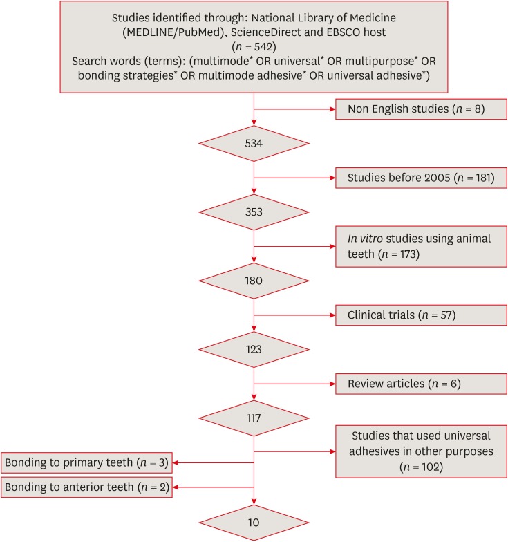
Materials and Methods An electronic search was performed of MEDLINE/PubMed, ScienceDirect, and EBSCOhost. Laboratory studies that evaluated the µTBS of multi-mode adhesives to dentin using either the etch-and-rinse or self-etch mode were selected. A meta-analysis was conducted of the reviewed studies to quantify the differences in the µTBS of Scotchbond Universal adhesive.
Results Only 10 studies fulfilled the inclusion criteria for the systematic review. Extensive variation was found in the restorative materials, testing methodologies, and failure mode in the reviewed articles. Furthermore, variation was also observed in the dimensions of the microtensile testing beams. The meta-analysis showed no statistically significant difference between the etch-and-rinse and self-etch modes for Scotchbond Universal adhesive (
p > 0.05).Conclusions Multi-mode ‘universal’ adhesives can achieve substantial bonding to dentin, regardless of the used modes (either etch-and-rinse or self-etch).
-
Citations
Citations to this article as recorded by- Influence of Proximal-Cervical Undermined Enamel Areas on Marginal Quality and Enamel Integrity of Laboratory and CAD/CAM Ceramic Inlays and Partial Crowns
Roland Frankenberger, Katharina Friedrich, Marie-Christine Dudek, Julia Winter, Norbert Krämer, Matthias J. Roggendorf
Journal of Functional Biomaterials.2025; 16(3): 82. CrossRef - Improving Bonding Protocols: The Effect of Selective Dentin Etching with Two Different Universal Adhesives—An In Vitro Study
Sandro Ferreira, Tiago Rodrigues, Mariana Nunes, Ana Mano Azul, José João Mendes, Ana Filipa Chasqueira, Joana Costa
Polymers.2025; 17(9): 1215. CrossRef - Effect of surface treatment on glass ionomers in sandwich restorations: a systematic review and meta-analysis of laboratory studies
Hoda S. Ismail, Ashraf Ibrahim Ali, Franklin Garcia-Godoy
Restorative Dentistry & Endodontics.2025; 50(2): e13. CrossRef - Wet vs. Dry Dentin Bonding: A Systematic Review and Meta-Analysis of Adhesive Performance and Hybrid Layer Integrity
Mircea Popescu, Mădălina Malița, Andrei Vorovenci, Andreea Angela Ștețiu, Viorel Ștefan Perieanu, Radu Cătălin Costea, Mihai David, Raluca Mariana Costea, Maria Antonia Ștețiu, Andi Ciprian Drăguș, Cristina Maria Șerbănescu, Andrei Burlibașa, Oana Eftene,
Oral.2025; 5(3): 63. CrossRef - The Effect of Different Multimode Adhesives On Microleakage of Class V Composite Restorations in Three Etching Modes
Fatma Yılmaz, Sevgi Kurşun, Zeliha Öztürk
ADO Klinik Bilimler Dergisi.2025; 14(3): 177. CrossRef - Controversies about refrigeration of dental adhesives: a review
Omar Abd El-Maksoud, Hamdi Hosni Hamdan Hamama, Ramy Ahmed Wafaie, Salah Hasab Mahmoud
BDJ Open.2025;[Epub] CrossRef - Tooth-composite bond failure with a universal and an etch-and-rinse adhesive depending on mode and frequency of application
Ellen Schulz-Kornas, Mathilde Tittel, Hartmut Schneider, Maximilian Bemmann, Marco Pellino, Tobias Meissner, Florian Fuchs, Christian Hannig, Florian Tetschke, Kyung-Jin Park, Michaela Strumpski, Rainer Haak
Dental Materials.2024; 40(2): 359. CrossRef - Comparison of postoperative hypersensitivity between Total-etch and Universal adhesive system: a randomized clinical trial
Kiran Javed, Nouman Noor, Muhammad Zubair Nasir, Manzoor Ahmed Manzoor
Scientific Reports.2024;[Epub] CrossRef - Adhesion and sealing of different universal adhesive systems associated with bulk‐fill resins after using endodontic irrigation solutions: An in vitro study
Érika Mayumi Omoto, Anderson Catelan, Paulo Henrique dos Santos, Luciano Tavares Angelo Cintra, Fernanda de Souza e Silva Ramos, Caio César Pavani, André Luiz Fraga Briso, Ticiane Cestari Fagundes
Australian Endodontic Journal.2024; 50(2): 309. CrossRef - Evaluation of the effects of combined application of dimethylaminohexadecyl methacrylate and MDP on dentin bonding and antimicrobial properties
Jiadi Shen, Ming Ma, Yun Huang, Haochen Miao, Xin Wei
Journal of Materials Science.2023; 58(31): 12685. CrossRef - Efficacy of adhesive strategies for restorative dentistry: A systematic review and network meta-analysis of double-blind randomized controlled trials over 12 months of follow-up
Kevin Sheng-Kai Ma, Li-Tzu Wang, Markus B. Blatz
Journal of Prosthodontic Research.2023; 67(1): 35. CrossRef - Impact of Preceded Tumor Therapeutic Irradiation on the Microtensile Bond Strength of Universal Adhesives Applied in Self-Etch Mode to Human Dentin In Vitro
Sina Broscheit, Dirk Vordermark, Reinhard Gerlach, Christian Ralf Gernhardt
Applied Sciences.2023; 13(13): 7873. CrossRef - Effect of the Adhesive Strategy on Clinical Performance and Marginal Integrity of a Universal Adhesive in Non-Carious Cervical Lesions in a Randomized 36-Month Study
Rainer Haak, Gesa Stache, Hartmut Schneider, Matthias Häfer, Gerhard Schmalz, Ellen Schulz-Kornas
Journal of Clinical Medicine.2023; 12(18): 5776. CrossRef - Universal Adhesives in Clinical Dentistry
Fusun Ozer, Shilpa Patnaikuni
Science, Art and Religion.2023; 2(1--2): 6. CrossRef - Deep proximal margin rebuilding with direct esthetic restorations: a systematic review of marginal adaptation and bond strength
Hoda S. Ismail, Ashraf I. Ali, Rabab El. Mehesen, Jelena Juloski, Franklin Garcia-Godoy, Salah H. Mahmoud
Restorative Dentistry & Endodontics.2022;[Epub] CrossRef - Improving Properties of an Experimental Universal Adhesive by Adding a Multifunctional Dendrimer (G-IEMA): Bond Strength and Nanoleakage Evaluation
Joana Vasconcelos e Cruz, António H. S. Delgado, Samuel Félix, José Brito, Luísa Gonçalves, Mário Polido
Polymers.2022; 14(7): 1462. CrossRef - Scoping review of trials evaluating adhesive strategies in pediatric dentistry: where do simplified strategies lie?
António H. S. Delgado, Hasan Jamal, Anne Young, Paul Ashley
BMC Oral Health.2021;[Epub] CrossRef - Does acid etching prior to applying universal adhesives affect the bond strength of glass fiber post to root dentin?
Helder Callegaro Velho, Eduardo Trindade Dalence, Pablo Soares Machado, Marília Pivetta Rippe, Jovito Adiel Skupien, Vinícius Felipe Wandscher
International Journal of Adhesion and Adhesives.2021; 105: 102795. CrossRef - Does Adhesive Layer Thickness and Tag Length Influence Short/Long-Term Bond Strength of Universal Adhesive Systems? An In-Vitro Study
Naji Kharouf, Tarek Ashi, Ammar Eid, Levi Maguina, Jihed Zghal, Nairy Sekayan, Rim Bourgi, Louis Hardan, Salvatore Sauro, Youssef Haikel, Davide Mancino
Applied Sciences.2021; 11(6): 2635. CrossRef - Chronological history and current advancements of dental adhesive systems development: a narrative review
Maicon Sebold, Carolina Bosso André, Beatriz Ometto Sahadi, Lorenzo Breschi, Marcelo Giannini
Journal of Adhesion Science and Technology.2021; 35(18): 1941. CrossRef - Laboratory methods for measuring adhesive bond strength between restoration materials and hard tooth tissues
I.Ya. Poyurovskaya, A.P. Polikarpova, F.S. Rusanov
Stomatologiya.2021; 100(5): 88. CrossRef - Effect of Curcumin Suspension and Vitamin C on Dentin Shear Bond Strength and Durability. A Pilot Study
Dalia A. Abuelenain, Ensanya A. Abou Neel, Tariq S. Abuhaimed, Amal M. Alamri, Hanan S. Ammar, Sahar M. N. Bukhary
The Open Dentistry Journal.2021; 15(1): 540. CrossRef - Effect of 9.3 μm CO2 and 2.94 μm Er:YAG Laser vs. Bur Preparations on Marginal Adaptation in Enamel and Dentin of Mixed Class V Cavities Restored With Different Restorative Systems
Clara Isabel Anton y Otero, Enrico Di Bella, Ivo Krejci, Tissiana Bortolotto
Frontiers in Dental Medicine.2021;[Epub] CrossRef - Adhesion strategy and curing mode of a universal adhesive influence the bonding of dual-cured core build-up resin composite to dentin
Ahmed Eid Elsayed, Mohamed Amr Kamel, Farid Sabry El-Askary
Journal of Adhesion Science and Technology.2021; 35(1): 52. CrossRef - Influence of etching mode and composite resin type on bond strength to dentin using universal adhesive system
Stefan Dačić, Milan Miljković, Aleksandar Mitić, Goran Radenković, Marija Anđelković‐Apostolović, Milica Jovanović
Microscopy Research and Technique.2021; 84(6): 1212. CrossRef - Universal adhesives - a new direction in the development of adhesive systems
A. Tichý, K. Hosaka, J. Tagami
Česká stomatologie a praktické zubní lékařství.2020; 120(1): 4. CrossRef - Effect of Over-Etching and Prolonged Application Time of a Universal Adhesive on Dentin Bond Strength
Phoebe Burrer, Hoang Dang, Matej Par, Thomas Attin, Tobias T. Tauböck
Polymers.2020; 12(12): 2902. CrossRef - Profile of a 10-MDP-based universal adhesive system associated with chlorhexidine: Dentin bond strength and in situ zymography performance
Marina Ciccone Giacomini, Polliana Mendes Candia Scaffa, Rafael Simões Gonçalves, Giovanna Speranza Zabeu, Cristina de Mattos Pimenta Vidal, Marcela Rocha de Oliveira Carrilho, Heitor Marques Honório, Linda Wang
Journal of the Mechanical Behavior of Biomedical Materials.2020; 110: 103925. CrossRef - Universal dental adhesives: Current status, laboratory testing, and clinical performance
Sanket Nagarkar, Nicole Theis‐Mahon, Jorge Perdigão
Journal of Biomedical Materials Research Part B: Applied Biomaterials.2019; 107(6): 2121. CrossRef - Modifying Adhesive Materials to Improve the Longevity of Resinous Restorations
Wen Zhou, Shiyu Liu, Xuedong Zhou, Matthias Hannig, Stefan Rupf, Jin Feng, Xian Peng, Lei Cheng
International Journal of Molecular Sciences.2019; 20(3): 723. CrossRef
- Influence of Proximal-Cervical Undermined Enamel Areas on Marginal Quality and Enamel Integrity of Laboratory and CAD/CAM Ceramic Inlays and Partial Crowns
- 5,175 View
- 45 Download
- 30 Crossref

- Bacterial leakage and micro-computed tomography evaluation in round-shaped canals obturated with bioceramic cone and sealer using matched single cone technique
- Kallaya Yanpiset, Danuchit Banomyong, Kanet Chotvorrarak, Ratchapin Laovanitch Srisatjaluk
- Restor Dent Endod 2018;43(3):e30. Published online July 5, 2018
- DOI: https://doi.org/10.5395/rde.2018.43.e30
-
 Abstract
 PDF
PDF PubReader
 ePub
ePub Objectives To evaluate sealing ability of root canals obturated with bioceramic-impregnated gutta percha cone (BCC) or gutta percha (GP), with bioceramic sealer (BCS) or AH Plus (AH; Dentsply-Maillefer), in roundly-prepared canals using matched single-cone technique, based on bacterial leakage test, and to analyze obturation quality using micro-computed tomography (CT) analysis.
Materials and Methods Ninety-two distobuccal roots of maxillary molars were prepared using nickel-titanium files to apical size 40/0.06. The roots were divided into 4 groups (
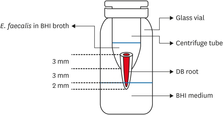
n = 20) that were obturated with a master cone and sealer: GP/AH, BCC/AH, GP/BCS, and BCC/BCS. Bacterial leakage model usingEnterococcus faecalis was used to evaluate sealing ability for 60-day period. Obturated samples from each group (n = 4) were analyzed using micro-CT.Results All groups showed bacterial leakage at 20%–45% of samples with mean leakage times of 42–52 days. There were no significant differences in bacterial leakage among the groups. Micro-CT showed minimal gaps and voids in all groups at less than 1%.
Conclusions In roundly-prepared canals, the single cone obturation with BCC/BCS was comparable to GP/AH for bacterial leakage at 60 days.
-
Citations
Citations to this article as recorded by- Effect of Root Dentin Moisture on the Apical Sealing Ability of Root Canal Sealers: In vitro Study
Zahraa Khalil Alani, Manal Hussain Abd-alla
Al-Rafidain Journal of Medical Sciences ( ISSN 2789-3219 ).2025; 8(2): 122. CrossRef - Synthesis, physical properties, and root canal sealing of experimental MTA- and salicylate-based root canal sealers
Rafael Pino Vitti, Kusai Baroudi, Tarun Walia, Raghavandra M. Shetty, Flávia Goulart da Rosa Cardoso, Flávia de Moura Pereira, Evandro Piva, Cesar Henrique Zanchi, Gabriel Flores Abuna, Carolina Oliveira de Lima, Emmanuel João Nogueira Leal Silva, Flávio
PLOS One.2025; 20(7): e0329476. CrossRef - Impact of cone system compatibility on single cone bioceramic obturation in canals prepared with variable taper NiTi rotary files
Reem M. Barakat, Rahaf A. Almohareb, Njoom Aleid, Hoor Almowais, Aljawhara Alharbi, Meshal Al-Sharafa, Ali Alrahlah
Scientific Reports.2025;[Epub] CrossRef - Estudio de la obturación con selladores biocerámicos de conductos radiculares de premolares inferiores
Alicia Beatriz Bonafé, Cecilia Inés Rourera, Carla Pedraza, Yamila Victoria Zanoni, Soledad Salduna, Cecilia Noemi De Caso, Gabriela Martín
Methodo Investigación Aplicada a las Ciencias Biológicas.2025; 10(3): 31. CrossRef - Sealing ability of mineral trioxide aggregate: A scoping review of laboratory assessment methods
Kenta Tsuchiya, Salvatore Sauro, Jukka P. Matinlinna, Hidehiko Sano, Monica Yamauti, Deepak Mehta, Kyung‐San Min, Atsushi Tomokiyo
European Journal of Oral Sciences.2025;[Epub] CrossRef - Bacterial Leakage Testing in Dentistry: A Comprehensive Review on Methods, Models, and Clinical Relevance
Niher Tabassum Snigdha, Mohmed Isaqali Karobari, Sukhamoy Gorai
Scientifica.2025;[Epub] CrossRef - In vitro comparative evaluation of apical leakage using a bioceramic sealer with three different obturating techniques: A glucose leakage model
Tanvi S Agrawal, Shishir Singh, Rajesh S Podar, Gaurav Kulkarni, Anuprita Gadkari, Navin Agarwal
Journal of Conservative Dentistry and Endodontics.2024; 27(1): 76. CrossRef - In Vitro Microscopical and Microbiological Assessment of the Sealing Ability of Calcium Silicate-Based Root Canal Sealers
Karin Christine Huth, Sabina Noreen Wuersching, Leander Benz, Stefan Kist, Maximilian Kollmuss
Journal of Functional Biomaterials.2024; 15(11): 341. CrossRef - Comparison between AH plus sealer and total fill bioceramic sealer performance in previously untreated and retreatment cases of maxillary incisors with large-sized periapical lesion: a randomized controlled trial
Eisa Wahbi, Hassan Achour, Yasser Alsayed Tolibah
BDJ Open.2024;[Epub] CrossRef - Bacterial sealing ability of calcium silicate-based sealer for endodontic surgery: an in-vitro study
Mai M. Mansour, Sybel M. Moussa, Marwa A. Meheissen, Mahmoud R. Aboelseoud
BMC Oral Health.2024;[Epub] CrossRef - Assessment the bioactivity of zinc oxid eugenol sealer after the addition of different concentrations of nano hydroxyapatite-tyrosine amino acid
Rasha M. Al-Shamaa, Raghad A. Al-Askary
Brazilian Journal of Oral Sciences.2024; 23: e243733. CrossRef - Assessment of Bacterial Sealing Ability of Two Different Bio-Ceramic Sealers in Single-Rooted Teeth Using Single Cone Obturation Technique: An In Vitro Study
Doaa M. AlEraky, Ahmed M. Rahoma, Hatem M. Abuohashish, Abdullh AlQasser, Abbas AlHamali, Hussain M. AlHussain, Hussain M. AlShoalah, Zakrya AlSaghah, Abdulrahman Khattar, Shimaa Rifaat
Applied Sciences.2023; 13(5): 2906. CrossRef - How do imaging protocols affect the assessment of root-end fillings?
Fernanda Ferrari Esteves Torres, Reinhilde Jacobs, Mostafa EzEldeen, Karla de Faria-Vasconcelos, Juliane Maria Guerreiro-Tanomaru, Bernardo Camargo dos Santos, Mário Tanomaru-Filho
Restorative Dentistry & Endodontics.2022;[Epub] CrossRef - The impact of Morse taper implant design on microleakage at implant-healing abutment interface
Soyeon KIM, Joo Won LEE, Jae-Heon KIM, Van Mai TRUONG, Young-Seok PARK
Dental Materials Journal.2022; 41(5): 767. CrossRef - A critical analysis of research methods and experimental models to study root canal fillings
Gustavo De‐Deus, Erick Miranda Souza, Emmanuel João Nogueira Leal Silva, Felipe Gonçalves Belladonna, Marco Simões‐Carvalho, Daniele Moreira Cavalcante, Marco Aurélio Versiani
International Endodontic Journal.2022; 55(S2): 384. CrossRef - Micro‐CT assessment of gap‐containing areas along the gutta‐percha‐sealer interface in oval‐shaped canals
Gustavo De‐Deus, Gustavo O. Santos, Iara Zamboni Monteiro, Daniele M. Cavalcante, Marco Simões‐Carvalho, Felipe G. Belladonna, Emmanuel J. N. L. Silva, Erick M. Souza, Raphael Licha, Carla Zogheib, Marco A. Versiani
International Endodontic Journal.2022; 55(7): 795. CrossRef - Comparison of Sealing Ability of Bioceramic Sealer, AH Plus, and GuttaFlow in Conservatively Prepared Curved Root Canals Obturated with Single-Cone Technique: An In vitro Study
Shalan Kaul, Ajay Kumar, Bhumika Kamal Badiyani, Laxmi Sukhtankar, M. Madhumitha, Amit Kumar
Journal of Pharmacy and Bioallied Sciences.2021; 13(Suppl 1): S857. CrossRef - Micro-CT Evaluation of Four Root Canal Obturation Techniques
Mahmood Reza Kalantar Motamedi, Amin Mortaheb, Maryam Zare Jahromi, Brett E. Gilbert, Marilena Vivona
Scanning.2021; 2021: 1. CrossRef - Effects of Both Fiber Post/Core Resin Construction System and Root Canal Sealer on the Material Interface in Deep Areas of Root Canal
Hiroki Miura, Shinji Yoshii, Masataka Fujimoto, Ayako Washio, Takahiko Morotomi, Hiroshi Ikeda, Chiaki Kitamura
Materials.2021; 14(4): 982. CrossRef - Sealing ability and microbial leakage of root-end filling materials: MTA versus epoxy resin: A systematic review and meta-analysis
Mario Dioguardi, Mario Alovisi, Diego Sovereto, Giuseppe Troiano, Giancarlo Malagnino, Michele Di Cosola, Angela Pia Cazzolla, Luigi Laino, Lorenzo Lo Muzio
Heliyon.2021; 7(7): e07494. CrossRef - Development of A Nano-Apatite Based Composite Sealer for Endodontic Root Canal Filling
Angelica Bertacci, Daniele Moro, Gianfranco Ulian, Giovanni Valdrè
Journal of Composites Science.2021; 5(1): 30. CrossRef - BIOCERAMIC-BASED ROOT CANAL SEALERS
L Somolová, Z Zapletalová, M Rosa, B Novotná, I Voborná, Y Morozova
Česká stomatologie a praktické zubní lékařství.2021; 121(4): 116. CrossRef - Calcium Silicate-Based Root Canal Sealers: A Narrative Review and Clinical Perspectives
Germain Sfeir, Carla Zogheib, Shanon Patel, Thomas Giraud, Venkateshbabu Nagendrababu, Frédéric Bukiet
Materials.2021; 14(14): 3965. CrossRef - Physico-Chemical Properties of Calcium-Silicate vs. Resin Based Sealers—A Systematic Review and Meta-Analysis of Laboratory-Based Studies
Viresh Chopra, Graham Davis, Aylin Baysan
Materials.2021; 15(1): 229. CrossRef - Comparison of apical sealing ability of bioceramic sealer and epoxy resin-based sealer using the fluid filtration technique and scanning electron microscopy
Widcha Asawaworarit, Thitapa Pinyosopon, Kanittha Kijsamanmith
Journal of Dental Sciences.2020; 15(2): 186. CrossRef - Micro-computed tomographic evaluation of a new system for root canal filling using calcium silicate-based root canal sealers
Mario Tanomaru-Filho, Fernanda Ferrari Esteves Torres, Jader Camilo Pinto, Airton Oliveira Santos-Junior, Karina Ines Medina Carita Tavares, Juliane Maria Guerreiro-Tanomaru
Restorative Dentistry & Endodontics.2020;[Epub] CrossRef - A micro-computed tomographic evaluation of root canal filling with a single gutta-percha cone and calcium silicate sealer
Jong Cheon Kim, Maung Maung Kyaw Moe, Sung Kyo Kim
Restorative Dentistry & Endodontics.2020;[Epub] CrossRef - Comparative evaluation of sealing ability of gutta percha and resilon as root canal filling materials- a systematic review
Pragya Pandey, Himanshi Aggarwal, A.P. Tikku, Arpit Singh, Rhythm Bains, Shambhavi Mishra
Journal of Oral Biology and Craniofacial Research.2020; 10(2): 220. CrossRef - Micro-computed tomographic evaluation of the flow and filling ability of endodontic materials using different test models
Fernanda Ferrari Esteves Torres, Juliane Maria Guerreiro-Tanomaru, Gisselle Moraima Chavez-Andrade, Jader Camilo Pinto, Fábio Luiz Camargo Villela Berbert, Mario Tanomaru-Filho
Restorative Dentistry & Endodontics.2020;[Epub] CrossRef - Root fillings with a matched-taper single cone and two calcium silicate–based sealers: an analysis of voids using micro-computed tomography
Eugenio Pedullà, Roula S. Abiad, Gianluca Conte, Giusy R. M. La Rosa, Ernesto Rapisarda, Prasanna Neelakantan
Clinical Oral Investigations.2020; 24(12): 4487. CrossRef - Influence of different disinfection protocols on gutta-percha cones surface roughness assessed by two different methods
A.M. Nunes, J.P. Gouvea, L. da Silva
Journal of Materials Research and Technology.2019; 8(6): 5464. CrossRef - Endodontic sealers based on calcium silicates: a systematic review
David Donnermeyer, Sebastian Bürklein, Till Dammaschke, Edgar Schäfer
Odontology.2019; 107(4): 421. CrossRef
- Effect of Root Dentin Moisture on the Apical Sealing Ability of Root Canal Sealers: In vitro Study
- 2,278 View
- 34 Download
- 32 Crossref

- Effects of a bleaching agent on properties of commercial glass-ionomer cements
- Fernanda Lúcia Lago de Camargo, Ailla Carla Lancellotti, Adriano Fonseca de Lima, Vinícius Rangel Geraldo Martins, Luciano de Souza Gonçalves
- Restor Dent Endod 2018;43(3):e32. Published online July 5, 2018
- DOI: https://doi.org/10.5395/rde.2018.43.e32
-
 Abstract
 PDF
PDF PubReader
 ePub
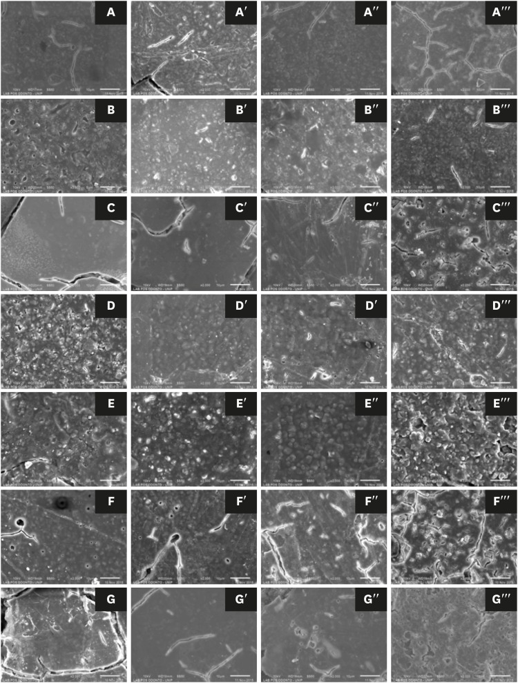
ePub Objectives This study evaluated the effects of a bleaching agent on the composition, mechanical properties, and surface topography of 6 conventional glass-ionomer cements (GICs) and one resin-modified GIC.
Materials and Methods For 3 days, the specimens were subjected to three 20-minute applications of a 37% H2O2-based bleaching agent and evaluated for water uptake (WTK), weight loss (WL), compressive strength (CS), and Knoop hardness number (KHN). Changes in surface topography and chemical element distribution were also analyzed by energy-dispersive X-ray spectroscopy and scanning electron microscopy. For statistical evaluation, the Kruskal-Wallis and Wilcoxon paired tests (
α = 0.05) were used to evaluate WTK and WL. CS specimens were subjected to 2-way analysis of variance (ANOVA) and the Tukeypost hoc test (α = 0.05), and KH was evaluated by one-way ANOVA, the Holm-Sidakpost hoc test (α = 0.05), and thet -test for independent samples (α = 0.05).Results The bleaching agent increased the WTK of Maxxion R, but did not affect the WL of any GICs. It had various effects on the CS, KHN, surface topography, and the chemical element distribution of the GICs.
Conclusions The bleaching agent with 37% H2O2 affected the mechanical and surface properties of GICs. The extent of the changes seemed to be dependent on exposure time and cement composition.
-
Citations
Citations to this article as recorded by- Multidisciplinary conservative management of a severely discolored nonvital tooth
Álvaro Ferrando Cascales, Francesc Abella Sans, Rubén Agustín-Panadero, José Amengual Lorenzo
The Journal of Prosthetic Dentistry.2025; 133(4): 941. CrossRef - The effects of bleaching products on the color stability of ion-releasing restoratives
Jian Sheng Lee, Noor Azlin Yahya, Azwatee Abdul Aziz, Adrian U-Jin Yap
BMC Oral Health.2025;[Epub] CrossRef - Physical-mechanical, chemical and biological properties of graphene-reinforced glass ionomer cements
Tatiane Ramos dos Santos Jordão, Laura Soares Viana Fernandes, Karla Lorene de França Leite, Adílis Alexandria, Emmanuel João Nogueira Leal Silva, Lucianne Cople Maia, Tatiana Kelly da Silva Fidalgo
Restorative Dentistry & Endodontics.2024;[Epub] CrossRef - An In Vitro Exploration of Interaction Mechanisms of Intracoronal Bleaching on the Compressive Strength of Conventional and Calcium Silicate–Based Self‐Adhesive Resins and Their Bonding to Composite Resin Restorative Material
Fereshteh Shafiei, Paria Dehghanian, Shadi Tivay, Yasamin Ghahramani, Luca Testarelli
International Journal of Dentistry.2024;[Epub] CrossRef - Éclaircissement dentaire
V. Pilliol, B. Ballester, T. Baudinet, G. Aboudharama, E. Terrer
EMC - Odontologie.2023; 39(2): 1. CrossRef - The Effect of Home and In-Office Bleaching on Microhardness and Color of Different CAD/CAM Ceramic Materials
Ruwaida Z. Alshali, Mohammed A. Alqahtani
Materials.2022; 15(17): 5948. CrossRef - Éclaircissement dentaire
V. Pilliol, B. Ballester, T. Baudinet, G. Aboudharama, E. Terrer
EMC - Médecine buccale.2022; 15(4): 1. CrossRef - Éclaircissement dentaire
V. Pilliol, B. Ballester, T. Baudinet, G. Aboudharam, E. Terrer
EMC - Orthopédie dentofaciale.2022; 34(1): 1. CrossRef - Comparative Evaluation of Two Glass Polyalkenoate Cements: An In Vivo Pilot Study Using a Sheep Model
Leyla Hasandoost, Daniella Marx, Paul Zalzal, Oleg Safir, Mark Hurtig, Cina Mehrvar, Stephen D. Waldman, Marcello Papini, Mark R. Towler
Journal of Functional Biomaterials.2021; 12(3): 44. CrossRef - The Effect of Simulated Field Storage Conditions on Dental Restorative Materials for Military Field Use
David J Lemon, Wen Chen, Trevor Smith, April A Ford, Steven X Moffett, Jeffrey T Hoyle, Nicholas J Hamlin, Yoon Y Hwang
Military Medicine.2020; 185(5-6): e831. CrossRef
- Multidisciplinary conservative management of a severely discolored nonvital tooth
- 1,403 View
- 5 Download
- 10 Crossref

- CBCT study of mandibular first molars with a distolingual root in Koreans
- Hee-Ho Kim, Hyoung-Hoon Jo, Jeong-Bum Min, Ho-Keel Hwang
- Restor Dent Endod 2018;43(3):e33. Published online July 30, 2018
- DOI: https://doi.org/10.5395/rde.2018.43.e33
-
 Abstract
 PDF
PDF PubReader
 ePub
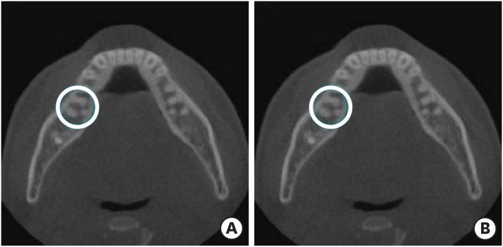
ePub Objectives This study aimed to investigate the prevalence of a separate distolingual root and to measure the thickness of the buccal cortical bone in mandibular first molars in Koreans using cone-beam computed tomography (CBCT) images.
Materials and Methods High-quality CBCT data from 432 patients were analyzed in this study. The prevalence of a separate distolingual root of the mandibular first molar was investigated. The distance from the distobuccal and distolingual root apices to the outer surface of the buccal cortical bone was measured. We also evaluated the thickness of the buccal cortical bone.
Results The prevalence of a separate distolingual root (2 separate distal roots with 1 canal in each root; 2R2C) was 23.26%. In mandibular first molars with 2R2C, the distance from the distobuccal root apex to the outer surface of the buccal cortical bone was 5.51 mm. Furthermore, the distance from the distolingual root apex to the outer surface of the buccal cortical bone was 12.09 mm. In mandibular first molars with 2R2C morphology, the thickness of the buccal cortical bone at the distobuccal root apex of the mandibular first molar was 3.30 mm. The buccal cortical bone at the distobuccal root apex was significantly thicker in the right side (3.38 mm) than the left side (3.09 mm) (
p < 0.05).Conclusions A separate distolingual root is not rare in mandibular first molars in the Korean population. Anatomic and morphologic knowledge of the mandibular first molar can be useful in treatment planning, including surgical endodontic treatment.
-
Citations
Citations to this article as recorded by- The association between complex root canal morphology of mandibular anteriors and distolingual roots in mandibular first molars in a Turkish population
Özge Kurt, Elif Solakoğlu
BMC Oral Health.2025;[Epub] CrossRef - Radix molaris is a hidden truth of mandibular first permanent molars: A descriptive- analytic study using cone beam computed tomography
Mohammed A. Alobaid, Saurabh Chaturvedi, Ebtihal Mobarak S. Alshahrani, Ebtsam M. Alshehri, Amal S. Shaiban, Mohamed Khaled Addas, Giuseppe Minervini
Technology and Health Care.2023; 31(5): 1957. CrossRef - Prevalence of radix entomolaris in India and its comparison with the rest of the world
Sumit MOHAN, Jyoti THAKUR
Minerva Dental and Oral Science.2022;[Epub] CrossRef - A critical analysis of laboratory and clinical research methods to study root and canal anatomy
Hany Mohamed Aly Ahmed
International Endodontic Journal.2022; 55(S2): 229. CrossRef - Three‐Rooted Permanent Mandibular First Molars: A Meta‐Analysis of Prevalence
Nyan M. Aung, Kyaw K. Myint, Luca Testarelli
International Journal of Dentistry.2022;[Epub] CrossRef - Reproducibilidad en el diagnóstico imagenológico de periodontitis apical a partir de CBCT
Sandra Milena Buitrago Rojas, Yeny Zulay Castellanos Dominguez, Jhonny Alexander Contreras Vargas, Yosdi Tomás Solano Diaz, Eder Fabián Gutierrez Argote
Acta Odontológica Colombiana.2020;[Epub] CrossRef - Assessment of Root and Root Canal Morphology of Human Primary Molars using CBCT
Yoomin Choi, Seonmi Kim, Namki Choi
THE JOURNAL OF THE KOREAN ACADEMY OF PEDTATRIC DENTISTRY.2020; 47(1): 25. CrossRef - The prevalence of radix molaris in the mandibular first molars of a Saudi subpopulation based on cone-beam computed tomography
Hassan AL-Alawi, Saad Al-Nazhan, Nassr Al-Maflehi, Mazen A. Aldosimani, Mohammed Nabil Zahid, Ghadeer N. Shihabi
Restorative Dentistry & Endodontics.2020;[Epub] CrossRef - Preferred Reporting Items for Epidemiologic Cross-sectional Studies on Root and Root Canal Anatomy Using Cone-beam Computed Tomographic Technology: A Systematized Assessment
Jorge N.R. Martins, Anil Kishen, Duarte Marques, Emmanuel João Nogueira Leal Silva, João Caramês, António Mata, Marco A. Versiani
Journal of Endodontics.2020; 46(7): 915. CrossRef - Evaluation of roots and canal systems of mandibular first molars in a vietnamese subpopulation using cone-beam computed tomography
KhoaVan Pham, AnhHoang Lan Le
Journal of International Society of Preventive and Community Dentistry.2019; 9(4): 356. CrossRef
- The association between complex root canal morphology of mandibular anteriors and distolingual roots in mandibular first molars in a Turkish population
- 1,667 View
- 10 Download
- 10 Crossref

- Revitalization of necrotic mature permanent incisors with apical periodontitis: a case report
- Emre Nagas, M. Ozgur Uyanik, Zafer C. Cehreli
- Restor Dent Endod 2018;43(3):e31. Published online July 5, 2018
- DOI: https://doi.org/10.5395/rde.2018.43.e31
-
 Abstract
 PDF
PDF PubReader
 ePub
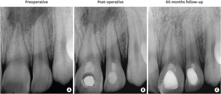
ePub Despite considerable focus on the regenerative endodontic treatment of immature teeth with necrotic infected pulps and apical periodontitis, little data exist with regard to its possible implementation in necrotic permanent teeth with complete apical and radicular development. The present report describes the procedures and outcome of a regenerative endodontic treatment approach in 2 previously-traumatized incisors with closed apex with apical periodontitis. A 2-visit treatment procedure was employed. At initial visit, the root canals were copiously irrigated, followed by placement of a triple antibiotic paste containing ciprofloxacin, metronidazole, and clindamycin into the root canals. After 4 weeks, the antibiotic paste was removed, and apical bleeding was initiated with size 10 hand files beyond the apices. The root canals were coronally sealed with mineral trioxide aggregate, and the access cavities were restored with bonded resin composite. At post-operative 60 months, both teeth were remained asymptomatic, with the recall radiographs showing complete resolution of apical radiolucency and reestablishment of periradicular tissues. In both teeth, the dimensions of root space remained unchanged as verified by image analysis. The revitalization protocol utilizing root canal disinfection and induced apical bleeding in necrotic, closed-apex incisors may offer a clinically acceptable alternative to conventional root canal treatment.
-
Citations
Citations to this article as recorded by- Regenerative potential of concentrated growth factor compared to platelet-rich fibrin in treatment of necrotic mature teeth: a randomized clinical trial
Taghreed Salah, Wael Hussein, Heba Abdelkafy
BDJ Open.2025;[Epub] CrossRef - Efficacy of Pulp Revascularization in the Treatment of Apical Periodontitis in Mature Necrotic Teeth: An Umbrella Review
Wanderson Limeira de Sousa Barbosa, Luiz Renato Paranhos, Márcia Valente de Brito Dantas, Rômulo Dias Jesuino, João Marcos da Costa Ribeiro, Walbert A. Vieira, Felipe de Souza Matos
Australian Endodontic Journal.2025; 51(2): 495. CrossRef - Clinical and radiographic outcomes of non-surgical retreatment of mature maxillary incisors using two regenerative endodontic techniques in adolescents: a 24-month randomized clinical trial
Ahmad Abdel Hamid Elheeny, Sherif Shafik EL Bahnasy, Yassmin Mohamed ElMakawi, Mohammed Turky, Eman Farouk Ahmed, Norhan Khaled Omar Wahba
BDJ Open.2025;[Epub] CrossRef - Effect of chitosan medicaments loaded with green-synthesized silver nanoparticles on basic fibroblast growth factor release from infected dentin
Dilek Hancerliogullari, Zehra Gun Gok, Nebahat Aytuna Cerci, Eray Ceylanoglu, Bengisu Ozturk, Ozum Hekim Harput, Sevda Durust Baris, Filiz Kiper, Ali Erdemir
Odontology.2025;[Epub] CrossRef - Revolutionizing Endodontics: Innovative Approaches for Treating Mature Teeth With Closed Apices and Apical Lesions: A Report of Two Cases
Claudia Brizuela, Gastón Meza, Maroun Khoury
Journal of Endodontics.2024; 50(5): 596. CrossRef - Current Aspects of Regenerative Endodontics: A Systematic Review
A. V. Mitronin, K. A. Archakov, D. A. Ostanina, Yu. A. Mitronin, T. V. Khizrieva
Endodontics Today.2024; 21(4): 287. CrossRef - Correlation between pulp sensibility and magnetic resonance signal intensity following regenerative endodontic procedures in mature necrotic teeth- a retrospective cohort study
Noha Mohamed El-Kateb, Amr Mohamed Abdallah, Rania Noaman ElBackly
BMC Oral Health.2024;[Epub] CrossRef - Influence of periapical lesion size on healing outcome following regenerative endodontic procedures: a clinical investigation
Noha Mohamed El Kateb, Mahmoud Mostafa Fata
Oral Radiology.2022; 38(4): 480. CrossRef - Do alternative scaffolds used in regenerative endodontics promote better root development than that achieved with blood clots?
Letícia de Araújo, Taynara Santos Goulart, Ana Clara Kuerten Gil, Daniela Peressoni Vieira Schuldt, Beatriz Serrato Coelho, Daniela de Rossi Figueiredo, Lucas da Fonseca Roberti Garcia, Josiane de Almeida
Brazilian Dental Journal.2022; 33(2): 22. CrossRef - Endodontic Regenerative Procedures in Necrotic Adult Teeth
Sara Garrido-Parada, Pablo Castelo-Baz, Nancy Feijoo-Pato, José Gaviño-Orduña, Benjamín Martín-Biedma
Applied Sciences.2022; 12(9): 4212. CrossRef - Combined conventional and regenerative treatment in molars with coexistent closed and open apices: A case series
Zafer C. Cehreli, Gizem Erbas Unverdi, Pinar Eymirli, Irem Mergen, Ezgihan Arslan, Gulce Esenturk
Australian Endodontic Journal.2022; 48(1): 197. CrossRef - Regenerative Endodontic Procedures for the Treatment of Necrotic Mature Teeth with Apical Periodontitis: A Systematic Review and Meta-analysis of Randomized Controlled Trials
Antonios Glynis, Federico Foschi, Ismini Kefalou, Despina Koletsi, Giorgos N. Tzanetakis
Journal of Endodontics.2021; 47(6): 873. CrossRef - Different Approaches to the Regeneration of Dental Tissues in Regenerative Endodontics
Anna M. Krupińska, Katarzyna Skośkiewicz-Malinowska, Tomasz Staniowski
Applied Sciences.2021; 11(4): 1699. CrossRef - Quantitative Assessment of Intracanal Regenerated Tissues after Regenerative Endodontic Procedures in Mature Teeth Using Magnetic Resonance Imaging: A Randomized Controlled Clinical Trial
Noha Mohamed El-Kateb, Rania Noaman El-Backly, Wessam Mohamed Amin, Amr Mohamed Abdalla
Journal of Endodontics.2020; 46(5): 563. CrossRef
- Regenerative potential of concentrated growth factor compared to platelet-rich fibrin in treatment of necrotic mature teeth: a randomized clinical trial
- 2,225 View
- 26 Download
- 14 Crossref

- Management of a permanent maxillary first molar with unusual crown and root anatomy: a case report
- Prateeksha Chowdhry, Pallavi Reddy, Mamta Kaushik
- Restor Dent Endod 2018;43(3):e35. Published online August 7, 2018
- DOI: https://doi.org/10.5395/rde.2018.43.e35
-
 Abstract
 PDF
PDF PubReader
 ePub
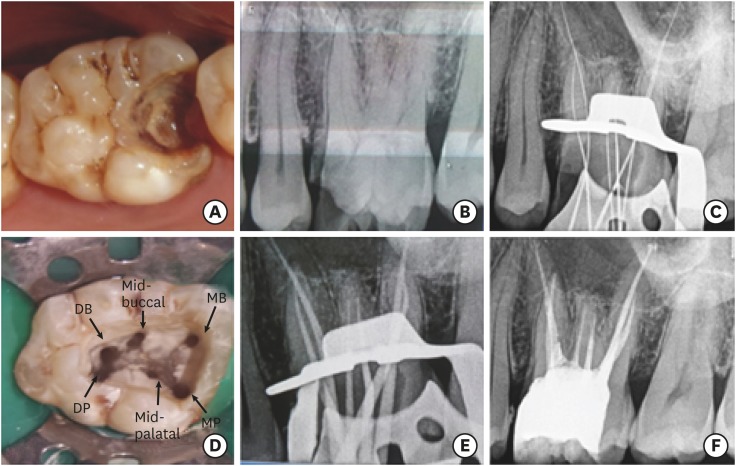
ePub The aim of this article was to showcase the endodontic management of a maxillary first molar with an unusual crown and root anatomy. Clinical diagnosis of the roots and root canal configuration was confirmed by a cone-beam computed tomography (CBCT) and the detection of the canals was made using a dental operating microscope. CBCT images revealed the presence of 5 roots with Vertucci type I canal configuration in all, except, in the middle root which had 2 canals with type IV configuration. The 6 canal orifices were clinically visualized under the dental operating microscope. Clinicians should familiarize themselves with the latest technologies to get additional information in endodontic practice in order to enhance the outcomes of endodontic therapy.
-
Citations
Citations to this article as recorded by- Endodontic management of maxillary first molar with unusual anatomy
MadhuriSai Battula, Mamta Kaushik, Neha Mehra, Ankeeta Singh
Journal of Conservative Dentistry.2022; 25(5): 569. CrossRef - Diversity of root canal morphology of maxillary first molars
Juhász Kincső-Réka, Kovács Mónika, Pop Mihai, Pop Silvia, Kerekes-Máthé Bernadette
Bulletin of Medical Sciences.2021; 94(1): 63. CrossRef
- Endodontic management of maxillary first molar with unusual anatomy
- 1,392 View
- 10 Download
- 2 Crossref


 KACD
KACD


 First
First Prev
Prev


