-
Calcium silicate-based root canal sealers: a literature review
-
Miyoung Lim, Chanyong Jung, Dong-Hoon Shin, Yong-bum Cho, Minju Song
-
Restor Dent Endod 2020;45(3):e35. Published online June 9, 2020
-
DOI: https://doi.org/10.5395/rde.2020.45.e35
-
-
 Abstract Abstract
 PDF PDF PubReader PubReader ePub ePub
Epoxy resin-based sealers are currently widely used, and several studies have considered AH Plus to be the gold-standard sealer. However, it still has limitations, including possible mutagenicity, cytotoxicity, inflammatory response, and hydrophobicity. Drawing upon the advantages of mineral trioxide aggregate, calcium silicate-based sealers were introduced with high levels of biocompatibility and hydrophilicity. Because of the hydrophilic environment in root canals, water resorption and solubility of root canal sealers are important factors contributing to their stability. Sealers displaying lower microleakage and stronger push-out bond strength are also needed to endure the dynamic tooth environment. Although the physical properties of calcium silicate-based sealers meet International Organization for Standardization recommendations, and they have consistently reported to be biocompatible, they have not overcome conventional resin-based sealers in actual practice. Therefore, further studies aiming to improve the physical properties of calcium silicate-based sealers are needed. -
Citations
Citations to this article as recorded by  - Does the Use of a Bioceramic Sealer Reduce Postoperative Pain Compared With an Epoxy Resin‐Based Sealer After Primary Root Canal Treatment and Retreatment?—An Umbrella Review
Lokhasudhan Govindaraju, Rajeswari Kalaiselvam, Mathan Rajan Rajendran, Aleksandar Jakovljevic, Jelena Jacimovic, Henry F. Duncan, Venkateshbabu Nagendrababu
International Endodontic Journal.2026; 59(3): 341. CrossRef - Evidence synthesis of postoperative pain with bioceramic vs. epoxy resin sealers: umbrella review of randomized trials within existing systematic reviews
Mrunali Dahikar, Ashish Mandwe, Kulvinder Singh Banga, Alexander Maniangat Luke, Suraj Arora, Unmesh Khanvilkar, Ajinkya M. Pawar
Frontiers in Dental Medicine.2026;[Epub] CrossRef - Effects of interactions between sealers and irrigants on the physicochemical and surface characteristics of endodontic sealers
Hye-In Kim, Yeon-Ju You, Yemi Kim, Jin-Woo Kim, Young-Eun Jang
Clinical Oral Investigations.2026;[Epub] CrossRef - Physical and mechanical properties of a strontium silicate-based sealer
Shannon Wong, Xiaofei Zhu, Tun-Yi Hsu, Sami Chogle, Russell A. Giordano, Yuwei Fan
Odontology.2026;[Epub] CrossRef - Assessment of Release of Calcium Ions from Apical Plugs of Different Bioceramic Sealers in Regenerative Endodontics for Immature Permanent Teeth
Alok Dubey, Nischitha Naik, Bhavana Sujanamulk, Mushir Mulla, Munaz Mulla, Alkananda Sahoo, Mohamed T Salama, Murali Patla S Bhat
World Journal of Dentistry.2026; 17(1): 69. CrossRef - Push-out bond strength of bioceramic-based sealers following different irrigation activation techniques
Fatma Begüm Peker, Hümeyra Çapkın, Ahsen Narbay
Journal of Applied Biomaterials & Functional Materials.2026;[Epub] CrossRef - In Vitro Assessment of Osteogenic Modulation and Molecular Responses Induced by Contemporary Endodontic Sealers in MC3T3-E1 Pre-Osteoblasts
Yuka Miyamoto, Yuka Kato, Ryan Needle, Julie Yongsook Kim, Jin Koo Kim, Paul H. Krebsbach, Insoon Chang
Dentistry Journal.2026; 14(3): 160. CrossRef - The Versatile Role of Rare Earth Elements in Biomaterials
Alfred Ngowi, Thywill Cephas Dzogbewu, Mohammad Rezwan Habib
Advances in Materials Science and Engineering.2026;[Epub] CrossRef - Thermal stability and setting dynamics of nanozinc oxide and AH Plus sealers: An in vitro study
Maryam Gharechahi, Amirreza Panjtanian, Pooya Ahmadi, Arsalan Shahri
Saudi Endodontic Journal.2026; 16(2): 167. CrossRef - Evaluación in vitro de la solubilidad, radiopacidad y pH de un sellador endodóntico biocerámico experimental preparado con diferentes proporciones de silicato de calcio y radiopacificador // In vitro evaluation of solubility, radiopacity and pH of an exp
Alejandro Leonhardt, Osvaldo Zmener, Nicolás Paduli, Roberto Della Porta, Miguel Chantiri
Revista de la Asociación Odontológica Argentina.2026;[Epub] CrossRef - Physiochemical properties of premixed bioceramic-based root canal sealer reinforced with copper oxide nanoparticles
Prasanti Kumari Pradhan, Neelanjana Majee, Gaurav Patri, Prahlad Saraf, Debkant Jena, Akansha Tilokani, Pratik Agrawal
Journal of Conservative Dentistry and Endodontics.2026; 29(6): 672. CrossRef - Effect of Different Tapered Gutta-Percha Points on Push-Out Bond Strength of Two Root Canal Sealers
Warattama Suksaphar, Pakit Tungsawat, Ninnita Wongwatanasanti, Siripat Lertnantapanya, Prattana Yodmanothum
European Journal of General Dentistry.2025; 14(03): 285. CrossRef - Effect of Electrical Heat Carrier Temperature on Bacterial Leakage of Endodontically Treated Teeth Using a Bioceramic Sealer
Mir Ahmad Nabavi, Mahmood Reza Kalantar Motamedi, Pedram Fattahi, Saber Khazaei
Clinical and Experimental Dental Research.2025;[Epub] CrossRef - Nanoparticles modified bioceramic sealers on solubility, antimicrobial efficacy, pushout bond strength and marginal adaptation at apical-third of canal dentin
Basil Almutairi, Fahad Alkhudhairy
PeerJ.2025; 13: e18840. CrossRef - Assessing the antimicrobial properties of bioceramic sealers enhanced with herbal extracts against E. faecalis
KS Sachin, K Shibani Shetty, KB Jeyalakshmi, S Harishma, S Harshini
Folia Medica.2025;[Epub] CrossRef - Estudio comparativo de la solubilidad de dos selladores endodónticos biocerámicos y un sellador a base de resinas
//Comparative study of the solubility of two bioceramic endodontic sealers and one epoxi-resin based sealer
Alejandro Leonhardt, Nicolás Paduli, Osvaldo Zmener, Miguel Chantiri
Revista de la Asociación Odontológica Argentina.2025; : 1. CrossRef - Enhancing root canal sealing: Exploring the sealing potential of epoxy and calcium silicate-based sealers with chitosan nanoparticle enhancement
S. Harishma, Srilekha Jayakumar, K Shibani Shetty, Barkavi Panchatcharam, Jwaalaa Rajkumar, S. Harshini
Endodontology.2025; 37(3): 306. CrossRef - Evaluation of the Genotoxicity and Cytotoxicity of Bioceramic Endodontic Sealers in HepG2 and V79 Cell Lines: An In Vitro Study Using the Comet and Micronucleus Assays
Antonija Tadin, Marija Badrov, Danijela Juric Kacunic, Nada Galic, Matea Macan, Ivan Kovacic, Davor Zeljezic
Journal of Functional Biomaterials.2025; 16(5): 169. CrossRef - In Vitro Apatite-Forming Ability of Different Root Canal Sealers (A Comparative Study)
Raghad A Al-Askary, Wiaam M. O. Al-Ashou, Sawsan H. Al-Jubori
Journal of International Society of Preventive and Community Dentistry.2025; 15(2): 173. CrossRef - Microstructural and elemental characterization of novel bioactive glass bioceramic sealer using Fourier transform infrared and X-ray diffraction analysis
Poulomi Guha, Pradeep Solete, Delphine Antony, Nishitha Arun, Mohmed Isaqali Karobari, Surendar Ramamoorthi
Journal of Conservative Dentistry and Endodontics.2025; 28(5): 412. CrossRef - Microstructural and Elemental Characterization of Calcium Silicate-Based Sealers
Mateusz Radwanski, Ireneusz Piwonski, Tomasz Szmechtyk, Salvatore Sauro, Monika Lukomska-Szymanska
Nanomaterials.2025; 15(10): 756. CrossRef - Apical negative pressure-enhanced sealer infiltration for obturating long oval-shaped root canals with the single-cone technique
Yaxu Feng, Brian E. Bergeron, Shijin Zhang, Danyang Sun, Kole Fisher, Franklin R. Tay, Bing Fan
Journal of Dentistry.2025; 160: 105909. CrossRef - Effects of different apical preparation sizes and root canal sealers on the fracture resistance of roots aged for 12 months in endodontically retreated mandibular premolars
Dilek Hancerliogullari, Sevda Durust Baris, Ali Turkyilmaz, Ali Erdemir
British Dental Journal.2025;[Epub] CrossRef - Influence of different endodontic treatment protocols on tooth survival: A retrospective cohort study with multistate analysis and group balancing
Ahmed Elmaasarawi, Mohamed Mekhemar, Andreas Bartols
International Endodontic Journal.2025; 58(10): 1529. CrossRef - Evaluation of 2,6-xylidine precipitate on sealer penetration of calcium silicate-based sealer and resin-based sealer: An in vitro study
M. B. Kalpana, Divya Shetty, Rajaram Naik
Endodontology.2025; 37(2): 183. CrossRef - Translational Advances in Regenerative Dentistry: Functional Biomaterials and Emerging Technologies
Seher Yaylacı, Hacer Eberliköse, Hakan Ceylan
Current Oral Health Reports.2025;[Epub] CrossRef - Marginal adaptation of heat and non-heat compatible bioceramic sealers in warm obturation: an in vitro SEM study
Thanomsuk Jearanaiphaisarn, Thanida Leelayuttakarn, Panisara Amatamahuthana, Pinmanus Chenpairojsakul, Keskanya Subbalekha, Pavena Chivatxaranukul
Scientific Reports.2025;[Epub] CrossRef - Influence of irrigating solutions on the hydration of calcium silicate-based dental biomaterials: An in vitro study
Pradeep M. Divya, Amit Jena, Saumyakanta Mohanty, Govind Shashirekha, Rashmi Rekha Mallick, Priyanka Sarangi
Journal of Conservative Dentistry and Endodontics.2025; 28(8): 758. CrossRef - Multispecies Biofilms Treated With Endodontic Sealers or Calcium Hydroxide: Antimicrobial Activity and Changes in Community Composition
Steven K. Uttech, Ronald Ordinola‐Zapata, W. Craig Noblett, Maria Martell, Bruno Lima, Christopher Staley
International Endodontic Journal.2025; 58(11): 1764. CrossRef - A comparative analysis of adhesion abilities between AH Plus® Bioceramic, Ceraseal® and AH Plus® on root canal dentine surfaces
Ike Dwi Maharti, Indira Larasputri, Nendar Herdianto, Anggraini Margono, Riesma Tasomara, Romilda Rosseti
Journal of Conservative Dentistry and Endodontics.2025; 28(9): 881. CrossRef - Clinical and radiographic success of single-cone bioceramic obturation versus traditional techniques: a systematic review and meta-analysis of randomized controlled trials
Firas Elmsmari, Yousef Elsayed, Abdelrahman Aboubakr, Mahdi Kaafarani, Osama Nour, Ajinkya M. Pawar
Journal of Oral Biology and Craniofacial Research.2025; 15(6): 1422. CrossRef - The Effect of Irrigation Solutions on the Setting Time, Solubility, and pH of Three Types of Premixed Bioceramic‐Based Root Canal Sealers
Kitichai Singharat, Ninnita Wongwatanasanti, Warattama Suksaphar, Pakit Tungsawat, Zhengrui Li
International Journal of Dentistry.2025;[Epub] CrossRef - Endodontie – State of the Art von A bis Z
Will Qian, Andreas Bartols
Zahnmedizin up2date.2025; 19(04): 281. CrossRef - Assessing Volume of Two Sealers’ Remnants after Reinstrumentation Using 3D Imaging Technology: An In Vitro Comparative Study
Khalel Mutaz Dawod, Raghad Abdulrazzaq Al-Hashimi
The Journal of Contemporary Dental Practice.2025; 26(8): 743. CrossRef - Functional and Bioactive Performance of Premixed Bioceramic Sealers with Warm Obturation: A Scoping Review
Patryk Wiśniewski, Stanisław Krokosz, Małgorzata Pietruska, Anna Zalewska
Gels.2025; 11(11): 932. CrossRef - Correlation of Bond Strength and Dentinal Tubule Penetration Evaluation of Four Different Endodontic Sealers: AH Plus, MTA Fillapex, Endoseal MTA, and Endoseal TCS (Maruchi): An In Vitro Study
Arezoo Mirzaei Sadeghloo, Seyedali Seyedmajidi, Akam Saeidi, Elham Mahmoudi, Murilo Baena Lopes
International Journal of Dentistry.2025;[Epub] CrossRef - Osteogenic Potential of Various Premixed Hydraulic Calcium Silicate-Based Sealers on Human Bone Marrow Stem Cells
Na-Hyun You, Donghee Lee, Yemi Kim, Sieun Nam, Sin-Young Kim
Materials.2025; 18(23): 5326. CrossRef - Polydopamine‐Functionalized Zinc Oxide Nanoparticles as a Root Canal Sealer: Characterization, Biological, and Physicochemical Properties
Arul Nayagi Raj, Aditya Shetty, Lakshmi Nidhi Rao, Giuseppe Ciccarella
Bioinorganic Chemistry and Applications.2025;[Epub] CrossRef - Management of rarely seen internal tunnelling root resorption associated with a maxillary permanent incisor
Kirsty A. Carney, Thibault N. E. Colloc, Julie K. Kilgariff
British Dental Journal.2024; 236(12): 955. CrossRef - Top tips for treatment planning: tooth-by-tooth prognosis - Part 3: endodontic prognosis
Prashanti Eachempati, Andrew Harris, Guy Lambourn, Tony Francis, Ewen McColl
British Dental Journal.2024; 237(9): 686. CrossRef - Retreatability of calcium silicate-based sealers based on micro-computed tomographic evaluation − A systematic review
Sundus Mohammed Bukhary
The Saudi Dental Journal.2024; 36(10): 1278. CrossRef - Evaluation of Setting Time, Flowability, Film Thickness, and Radiopacity of Experimental Monocalcium Silicate‐Based Root Canal Sealers
Sukanya Juntha, Pakit Tungsawat, Ninnita Wongwatanasanti, Warattama Suksaphar, Siripat Lertnantapanya, Carlos M. Ardila
International Journal of Dentistry.2024;[Epub] CrossRef - Root Canal Treatment and Demand for Continuing Education among Thai Dental Practitioners
Ninnita Wongwatanasanti, Pakit Tungsawat, Warattama Suksaphar, Siripat Lertnantapanya, Prattana Yodmanotham
The Open Dentistry Journal.2024;[Epub] CrossRef - Clinical outcome of non-surgical root canal treatment using different sealers and techniques of obturation in 237 patients: A retrospective study
Mateusz Radwanski, Krystyna Pietrzycka, Tan Fırat Eyüboğlu, Mutlu Özcan, Monika Lukomska-Szymanska
Clinical Oral Investigations.2024;[Epub] CrossRef - Endodontic sealers after exposure to chlorhexidine digluconate: An assessment of physicochemical properties
Vasileios Kapralos, Josette Camilleri, Andreas Koutroulis, Håkon Valen, Dag Ørstavik, Pia Titterud Sunde
Dental Materials.2024; 40(3): 420. CrossRef - Assessment the bioactivity of zinc oxid eugenol sealer after the addition of different concentrations of nano hydroxyapatite-tyrosine amino acid
Rasha M. Al-Shamaa, Raghad A. Al-Askary
Brazilian Journal of Oral Sciences.2024; 23: e243733. CrossRef - Interfacial adaptation of newly prepared nano-tricalcium silicate-58s bioactive glass-based endodontic sealer
Nawal A. Al-Sabawi, Sawsan Hameed Al-Jubori
Journal of Dental Research, Dental Clinics, Dental Prospects.2024; 18(2): 115. CrossRef - Marginal adaptation of customized gutta percha cone with calcium silicate based sealer versus MTA and biodentine apical plugs in simulated immature permanent teeth (an in vitro study)
Mary M. Mina, Sybel M. Moussa, Mahmoud R. Aboelseoud
BMC Oral Health.2024;[Epub] CrossRef - Solubility of Endoseal and AH26 Root Canal Sealers
Nooshin Fakhari, Ali Reza Mirjani, Abbas Bagheri, Jalil Modaresi
Journal of Research in Dental and Maxillofacial Sciences.2024; 9(1): 1. CrossRef - Novel bioactive nanospheres show effective antibacterial effect against multiple endodontic pathogens
Jin Liu, Haoze Wu, Jun Qiu, Sirui Yang, Doudou Xiang, Xinhua Zhang, Jinxin Kuang, Min Xiao, Qing Yu, Xiaogang Cheng
Heliyon.2024; 10(7): e28266. CrossRef - Evaluation of canal patency and cleanliness following retreatment of bioceramic sealer‐obturated root canals using three different irrigant activation protocols
Daiasharailang Lyngdoh, Sharique Alam, Huma Iftekhar, Surendra Kumar Mishra
Australian Endodontic Journal.2024; 50(3): 475. CrossRef - Antibiofilm Efficacy of Calcium Silicate-Based Endodontic Sealers
Matilde Ruiz-Linares, Vsevolod Fedoseev, Carmen Solana, Cecilia Muñoz-Sandoval, Carmen María Ferrer-Luque
Materials.2024; 17(16): 3937. CrossRef - Enhancing the Biological Properties of Organic–Inorganic Hybrid Calcium Silicate Cements: An In Vitro Study
Minji Choi, Jiyoung Kwon, Ji-Hyun Jang, Duck-Su Kim, Hyun-Jung Kim
Journal of Functional Biomaterials.2024; 15(11): 337. CrossRef - Cytotoxicity and cell migration evaluation of a strontium silicate-based root canal sealer on stem cells from rat apical papilla: an in vitro study
Guanglei Zhou, Yu Zhao, Liangjing Cai, Liwei Liu, Xu Li, Lu Sun, Jiayin Deng
BMC Oral Health.2024;[Epub] CrossRef - An In Vitro Comparative Analysis of Physico–Mechanical Properties of Commercial and Experimental Bioactive Endodontic Sealers
Abdulmajeed Kashaf, Faisal Alonaizan, Khalid S. Almulhim, Dana Almohazey, Deemah Abdullah Alotaibi, Sultan Akhtar, Ashwin C. Shetty, Abdul Samad Khan
Bioengineering.2024; 11(11): 1079. CrossRef - Chemical, Antibacterial, and Cytotoxic Properties of Four Different Endodontic Sealer Leachates Over Time
Jo-Hsun Chen, Veksina Raman, Sarah A. Kuehne, Josette Camilleri, Josefine Hirschfeld
Journal of Endodontics.2024; 50(11): 1612. CrossRef - Comparative Analysis of Fracture Resistance of Endodontic Sealer Types and Filling Methods
Yun Song, Kee-Deog Kim, Bock-Young Jung, Wonse Park, Nan-Sim Pang
Materials.2024; 18(1): 40. CrossRef - Comparative Evaluation of Removal of Bioceramic Sealers Using Rotary Retreatment Files Supplemented with Passive Ultrasonic Activation: An In Vitro Study
Anuradha B Patil, Amrut Bambawale, Pooja R Barghare, Sumanthini V Margasahayam, Divya Naik, Jayeeta S Verma
World Journal of Dentistry.2024; 15(4): 292. CrossRef - Nonsurgical Endodontic Management of Nonperforating Internal Root Resorption in a Maxillary Central Incisor: A Case Report with a 4-Year Follow-Up
Paras M. Gehlot, Divya S. Rajkumar, Annapoorna B. Mariswamy, Upendra Natha N. Reddy, Chaitanya Chappidi
Journal of Pharmacy and Bioallied Sciences.2024; 16(Suppl 3): S3005. CrossRef - Evaluating the Sealing Performance of Endodontic Sealers: Insights Into Achieving Complete Sealing
Ajay Chhabra, Ramya K P., Saravana Prathap, Priyanka Yadav, Himani Mehra, Sona J Parvathy
Cureus.2024;[Epub] CrossRef - Effects of vehicles on the physical properties and biocompatibility of premixed calcium silicate cements
Gitae SON, Gyeung Mi SEON, Sang Hoon CHOI, Hyeong-Cheol YANG
Dental Materials Journal.2024; 43(2): 276. CrossRef - Comparative cytotoxicity study of putty- and powder-type calcium silicate cements
Sora Park, Dohyun Cho, Ji Hyeon Yoon, Yeonjoo Kang, Quang Canh Vo, Gitae Son, Hongjoo Park, Hyeong-Cheol Yang
Korean Journal of Dental Materials.2024; 51(4): 259. CrossRef - Physical-chemical properties and acellular bioactivity of newly prepared nano-tricalcium silicate-58s bioactive glass-based endodontic sealer
Nawal A. Al-Sabawi, Sawsan Hameed Al-Jubori
Journal of Oral Biosciences.2023; 65(4): 305. CrossRef - Dentinal Tubule Penetrability and Bond Strength of Two Novel Calcium Silicate-Based Root Canal Sealers
Karissa Shieh, Jack Yang, Elsa Heng Zhu, Ove Andreas Peters, Sepanta Hosseinpour
Materials.2023; 16(9): 3309. CrossRef - Cytotoxicity and Mineralization Activity of Calcium Silicate-Based Root Canal Sealers Compared to Conventional Resin-Based Sealer in Human Gingival Fibroblast Cells
Mohammad Shokrzadeh, Farzaneh Sadat Motafeghi, Anahita Lotfizadeh, Mohammad Ghorbani, Azam Haddadi Kohsar, Cesar Rogério Pucci
International Journal of Dentistry.2023; 2023: 1. CrossRef - Effect of three different photosensitizers in photodynamic therapy on bond strength of a calcium silicate‐based sealer to radicular dentin
Cihan Küden, Seda Nur Karakaş
Australian Endodontic Journal.2023; 49(S1): 265. CrossRef - Effect of endodontic sealer on postoperative pain: a network meta-analysis
Cynthia Maria Chaves Monteiro, Ana Cristina Rodrigues Martins, Alessandra Reis, Juliana Larocca de Geus
Restorative Dentistry & Endodontics.2023;[Epub] CrossRef - Antimicrobial Activity of Five Calcium Silicate Based Root Canal Sealers against a Multispecies Engineered Biofilm: An In Vitro Study
Carla Zogheib, Issam Khalil, Wajih Hage, Dolla Karam Sarkis, Mireille Kallasy, Germain Sfeir, May Mallah, Roula El Hachem
The Journal of Contemporary Dental Practice.2023; 24(9): 707. CrossRef - Calcium silicate sealers in endodontics
Archana Chavan, Nidambur Ballal
Acta stomatologica Naissi.2023; 39(87): 2624. CrossRef - Assessing the Sealing Performance and Clinical Outcomes of Endodontic Treatment in Patients with Chronic Apical Periodontitis Using Epoxy Resin and Calcium Salicylate Seals
Razvan Mihai Horhat, Bogdan Andrei Bumbu, Laura Orel, Oana Velea-Barta, Laura Cirligeriu, Gratiana Nicoleta Chicin, Marius Pricop, Mircea Rivis, Stefania Dinu, Delia Ioana Horhat, Felix Bratosin, Roxana Manuela Fericean, Rodica Anamaria Negrean, Luminita
Medicina.2023; 59(6): 1137. CrossRef -
In Vitro Cytotoxicity and Mineralization Potential of an Endodontic Bioceramic Material
Soumya Sheela, Mohannad Nassar, Fatma M. AlGhalban, Mehmet O. Gorduysus
European Journal of Dentistry.2023; 17(02): 548. CrossRef - Dislodgment Resistance, Adhesive Pattern, and Dentinal Tubule Penetration of a Novel Experimental Algin Biopolymer-Incorporated Bioceramic-Based Root Canal Sealer
Galvin Sim Siang Lin, Norhayati Luddin, Huwaina Abd Ghani, Josephine Chang Hui Lai, Tahir Yusuf Noorani
Polymers.2023; 15(5): 1317. CrossRef - Impact of Final Irrigation Protocol on the Push-Out Bond Strength of Two Types of Endodontic Sealers
Germain Sfeir, Frédéric Bukiet, Wajih Hage, Roula El Hachem, Carla Zogheib
Materials.2023; 16(5): 1761. CrossRef - Clinical Approaches to the Three-Dimensional Endodontic Obturation Protocol for Teeth with Periapical Bone Lesions
Angela Gusiyska, Elena Dyulgerova
Applied Sciences.2023; 13(17): 9755. CrossRef - Evaluating the bioactivity of endodontic sealers with respect to their thermo-nanomechanical properties
Andreea Marica, Luminita Fritea, Florin Banica, Iosif Hulka, Gerlinde Rusu, Cosmin Sinescu, Traian Octavian Costea, Simona Cavalu
Materials Science-Poland.2023; 41(3): 126. CrossRef - Advances and challenges in regenerative dentistry: A systematic review of calcium phosphate and silicate-based materials on human dental pulp stem cells
B. Christie, N. Musri, N. Djustiana, V. Takarini, N. Tuygunov, M.N. Zakaria, A. Cahyanto
Materials Today Bio.2023; 23: 100815. CrossRef - Radiographic Evaluation of Periapical Healing Rates Between Bio-Ceramic Sealer and AH+ Sealer: A Retrospective Study
Dalia Nayil Alharith, Iman T. Mansi, YoumnaElsaid Abdulmotalib, HebaFuad Amous, TagreedSuliman Aljulban, Haifa Mohammed Al Aiban, Sali Mohamad Haffar
Annals of Dental Specialty.2023; 11(2): 124. CrossRef - Obturation canalaire
N. Linas, M.-L. Munoz-Sanchez, N. Decerle, P.-Y. Cousson
EMC - Médecine buccale.2023; 16(5): 1. CrossRef - Biodentine Inhibits the Initial Microbial Adhesion of Oral Microbiota In Vivo
Ali Al-Ahmad, Michael Haendel, Markus Altenburger, Lamprini Karygianni, Elmar Hellwig, Karl Wrbas, Kirstin Vach, Christian Tennert
Antibiotics.2022; 12(1): 4. CrossRef - Pilot Evaluation of Sealer-Based Root Canal Obturation Using Epoxy-Resin-Based and Calcium-Silicate-Based Sealers: A Randomized Clinical Trial
Minju Song, Min-Gyu Park, Sang-Won Kwak, Ruben H. Kim, Jung-Hong Ha, Hyeon-Cheol Kim
Materials.2022; 15(15): 5146. CrossRef - The antibacterial activity of mineral trioxide aggregate containing calcium fluoride
Miyoung Lim, Seunghoon Yoo
Journal of Dental Sciences.2022; 17(2): 836. CrossRef - Physicochemical and Mechanical Properties of Premixed Calcium Silicate and Resin Sealers
Naji Kharouf, Salvatore Sauro, Ammar Eid, Jihed Zghal, Hamdi Jmal, Anta Seck, Valentina Macaluso, Frédéric Addiego, Francesco Inchingolo, Christine Affolter-Zbaraszczuk, Florent Meyer, Youssef Haikel, Davide Mancino
Journal of Functional Biomaterials.2022; 14(1): 9. CrossRef - Comparison of Fracture Resistance between Single-cone and Warm Vertical Compaction Technique Using Bio-C Sealer® in Mandibular Incisors: An In Vitro Study
Raphael Lichaa, George Deeb, Rami Mhanna, Carla Zogheib
The Journal of Contemporary Dental Practice.2022; 23(2): 143. CrossRef - In vitro physicochemical characterization of five root canal sealers and their influence on an ex vivo oral multi‐species biofilm community
Flavia M. Saavedra, Lauter E. Pelepenko, William S. Boyle, Anqi Zhang, Christopher Staley, Mark C. Herzberg, Marina A. Marciano, Bruno P. Lima
International Endodontic Journal.2022; 55(7): 772. CrossRef - Premixed Calcium Silicate-Based Root Canal Sealer Reinforced with Bioactive Glass Nanoparticles to Improve Biological Properties
Min-Kyung Jung, So-Chung Park, Yu-Jin Kim, Jong-Tae Park, Jonathan C. Knowles, Jeong-Hui Park, Khandmaa Dashnyam, Soo-Kyung Jun, Hae-Hyoung Lee, Jung-Hwan Lee
Pharmaceutics.2022; 14(9): 1903. CrossRef - A critical analysis of research methods and experimental models to study root canal fillings
Gustavo De‐Deus, Erick Miranda Souza, Emmanuel João Nogueira Leal Silva, Felipe Gonçalves Belladonna, Marco Simões‐Carvalho, Daniele Moreira Cavalcante, Marco Aurélio Versiani
International Endodontic Journal.2022; 55(S2): 384. CrossRef - Bioactivity Potential of Bioceramic-Based Root Canal Sealers: A Scoping Review
Mauro Schmitz Estivalet, Lucas Peixoto de Araújo, Felipe Immich, Adriana Fernandes da Silva, Nadia de Souza Ferreira, Wellington Luiz de Oliveira da Rosa, Evandro Piva
Life.2022; 12(11): 1853. CrossRef - The influence of humidity on bond strength of AH Plus, BioRoot RCS, and Nanoseal-S sealers
Sunanda Laxman Gaddalay, Damini Vilas Patil, Ramchandra Kabir
Endodontology.2022; 34(3): 202. CrossRef - The Effect of Bioceramic HiFlow and EndoSequence Bioceramic Sealers on Increasing the Fracture Resistance of Endodontically Treated Teeth: An In Vitro Study
Mohamad Khir Abdulsamad Alskaf, Hassan Achour, Hasan Alzoubi
Cureus.2022;[Epub] CrossRef - Unravelling the effects of ibuprofen-acetaminophen infused copper-bioglass towards the creation of root canal sealant
Chitra S, Riju Chandran, Ramya R, Durgalakshmi D, Balakumar S
Biomedical Materials.2022; 17(3): 035001. CrossRef - A Micro-CT Analysis of Initial and Long-Term Pores Volume and Porosity of Bioactive Endodontic Sealers
Mateusz Radwanski, Michal Leski, Adam K. Puszkarz, Jerzy Sokolowski, Louis Hardan, Rim Bourgi, Salvatore Sauro, Monika Lukomska-Szymanska
Biomedicines.2022; 10(10): 2403. CrossRef - A comprehensive in vitro comparison of the biological and physicochemical properties of bioactive root canal sealers
Sabina Noreen Wuersching, Christian Diegritz, Reinhard Hickel, Karin Christine Huth, Maximilian Kollmuss
Clinical Oral Investigations.2022; 26(10): 6209. CrossRef - Stability and solubility test of endodontic materials
Ivan Matovic, Jelena Vucetic
Stomatoloski glasnik Srbije.2022; 69(4): 169. CrossRef - Antimicrobial effectiveness of root canal sealers againstEnterococcus faecalis
Paola Castillo-Villagomez, Elizabeth Madla-Cruz, Fanny Lopez-Martinez, Idalia Rodriguez-Delgado, Jorge Jaime Flores-Treviño, Guadalupe Ismael Malagon-Santiago, Myriam Angelica de La Garza-Ramos
Biomaterial Investigations in Dentistry.2022; 9(1): 47. CrossRef - Tricalcium silicate cement sealers
Anita Aminoshariae, Carolyn Primus, James C. Kulild
The Journal of the American Dental Association.2022; 153(8): 750. CrossRef - Influence of variations in the environmental pH on the solubility and water sorption of a calcium silicate‐based root canal sealer
E. J. N. L. Silva, C. M. Ferreira, K. P. Pinto, A. F. A. Barbosa, M. V. Colaço, L. M. Sassone
International Endodontic Journal.2021; 54(8): 1394. CrossRef - Calcium Silicate-Based Root Canal Sealers: A Narrative Review and Clinical Perspectives
Germain Sfeir, Carla Zogheib, Shanon Patel, Thomas Giraud, Venkateshbabu Nagendrababu, Frédéric Bukiet
Materials.2021; 14(14): 3965. CrossRef - Development of A Nano-Apatite Based Composite Sealer for Endodontic Root Canal Filling
Angelica Bertacci, Daniele Moro, Gianfranco Ulian, Giovanni Valdrè
Journal of Composites Science.2021; 5(1): 30. CrossRef - Bone repair in defects filled with AH Plus sealer and different concentrations of MTA: a study in rat tibiae
Jessica Emanuella Rocha Paz, Priscila Oliveira Costa, Albert Alexandre Costa Souza, Ingrid Macedo de Oliveira, Lucas Fernandes Falcão, Carlos Alberto Monteiro Falcão, Maria Ângela Area Leão Ferraz, Lucielma Salmito Soares Pinto
Restorative Dentistry & Endodontics.2021;[Epub] CrossRef - Characterization, Antimicrobial Effects, and Cytocompatibility of a Root Canal Sealer Produced by Pozzolan Reaction between Calcium Hydroxide and Silica
Mi-Ah Kim, Vinicius Rosa, Prasanna Neelakantan, Yun-Chan Hwang, Kyung-San Min
Materials.2021; 14(11): 2863. CrossRef - Synthesis and Characterization of Novel Calcium-Silicate Nanobioceramics with Magnesium: Effect of Heat Treatment on Biological, Physical and Chemical Properties
Konstantina Kazeli, Ioannis Tsamesidis, Anna Theocharidou, Lamprini Malletzidou, Jonathan Rhoades, Georgia K. Pouroutzidou, Eleni Likotrafiti, Konstantinos Chrissafis, Theodoros Lialiaris, Lambrini Papadopoulou, Eleana Kontonasaki, Evgenia Lymperaki
Ceramics.2021; 4(4): 628. CrossRef - Calcium Silicate Cements vs. Epoxy Resin Based Cements: Narrative Review
Mario Dioguardi, Cristian Quarta, Diego Sovereto, Giuseppe Troiano, Khrystyna Zhurakivska, Maria Bizzoca, Lorenzo Lo Muzio, Lucio Lo Russo
Oral.2021; 1(1): 23. CrossRef - In Vitro Microleakage Evaluation of Bioceramic and Zinc-Eugenol Sealers with Two Obturation Techniques
Francesco De Angelis, Camillo D’Arcangelo, Matteo Buonvivere, Rachele Argentino, Mirco Vadini
Coatings.2021; 11(6): 727. CrossRef - Efficacy Of Calcium Silicate-Based Sealers In Root Canal Treatment: A Systematic Review
Hattan Mohammed Omar Baismail, Mohammed Ghazi Moiser Albalawi, Alaa Mofareh Thoilek Alanazi, Muhannad Atallah Saleem Alatawi, Badr Soliman Alhussain
Annals of Dental Specialty.2021; 9(1): 87. CrossRef - Apical Sealing Ability of Two Calcium Silicate-Based Sealers Using a Radioactive Isotope Method: An In Vitro Apexification Model
Inês Raquel Pereira, Catarina Carvalho, Siri Paulo, José Pedro Martinho, Ana Sofia Coelho, Anabela Baptista Paula, Carlos Miguel Marto, Eunice Carrilho, Maria Filomena Botelho, Ana Margarida Abrantes, Manuel Marques Ferreira
Materials.2021; 14(21): 6456. CrossRef
-
15,978
View
-
307
Download
-
105
Crossref
-
Antibacterial effect of urushiol on E. faecalis as a root canal irrigant
-
Sang-Wan Kim, Dong-Hoon Shin
-
Restor Dent Endod 2017;42(1):54-59. Published online January 25, 2017
-
DOI: https://doi.org/10.5395/rde.2017.42.1.54
-
-
Abstract
PDFPubReaderePub
- Objectives
The purpose of this study was to compare the antibacterial activity of urushiol against Enterococcus faecalis (E. faecalis) to that of NaOCl. Materials and MethodsThe canals of thirty two single rooted human teeth were instrumented with Ni-Ti files (ProTaper Next X1, X2, X3, Dentsply). A pure culture of E. faecalis ATCC 19433 was prepared in sterile brain heart infusion (BHI) broth. The teeth were submerged in the suspension of E. faecalis and were incubated at 37℃ for 7 days to allow biofilm formation. The teeth were randomly divided into three experimental groups according to the irrigant used, and a negative control group where no irrigant was used (n = 8). Group 1 used physiologic normal saline, group 2 used 6% NaOCl, and group 3 used 10 wt% urushiol solution. After canal irrigation, each sample was collected by the sequential placement of 2 sterile paper points (ProTaper NEXT paper points, size X3, Dentsply). Ten-fold serial dilutions on each vials, and 100 µL were cultured on a BHI agar plate for 8 hours, and colony forming unit (CFU) analysis was done. The data were statistically analyzed using Kruskal-Wallis and Mann-whitney U tests. ResultsSaline group exhibited no difference in the CFU counts with control group, while NaOCl and urushiol groups showed significantly less CFU counts than saline and control groups (p < 0.05). ConclusionsThe result of this study suggests 10% urushiol and 6% NaOCl solution had powerful antibacterial activity against E. faecalis when they were used as root canal irrigants.
-
Citations
Citations to this article as recorded by - Synthesis, Characterisation and Antibacterial Activity of Reduced Graphene Oxide‐Cuprous Oxide Nanocomposite Against Dual Species Biofilms Using Clsm
Aashna Dinesh, M. S. Prathap, Pradeep Kumar, Jeslee Ann Jose, Samreena Kalander, Satwik Varadraj
Australian Endodontic Journal.2026;[Epub] CrossRef - A critical review on innovative targets for signal disruption in Enterococcus faecalis infection management
Kayeen Vadakkan, Gajanan Sampatrao Ghodake, Chin Wei Lai, Selvaraj Vijayanand, Janarthanam Hemapriya
Microbial Pathogenesis.2025; 207: 107876. CrossRef - Effect of proanthocyanidins application on push-out bond strength of root canal filling after different final irrigation procedures
Funda Fundaoğlu Küçükekenci, Ahmet Serkan Küçükekenci
BMC Oral Health.2025;[Epub] CrossRef - Evaluation of Silver-Ion-Coated Rotary Nickel Titanium Files - An In Vitro Study
Jhanvi H. Sadaria, Kondas V. Venkatesh, Dhanasekaran Sihivahanan
Indian Journal of Dental Research.2025; 36(3): 344. CrossRef - Antimicrobial efficacy of natural-based endodontic solutions: a systematic review with a network meta-analysis
Danilo Cassiano FERRAZ, Anahi de Paula MELO, Felipe de Souza MATOS, Luiz Renato PARANHOS, Camilla Christian Gomes MOURA, Cauane BLUMENBERG, Juliane Maria GUERREIRO-TANOMARU, Mário TANOMARU-FILHO
Brazilian Oral Research.2024;[Epub] CrossRef - Irrigants and irrigation activation systems in Endodontics
Brenda P. F. A. Gomes, Emelly Aveiro, Anil Kishen
Brazilian Dental Journal.2023; 34(4): 1. CrossRef - Effectiveness of sodium hypochlorite gel and solutions in endodontics: A systematic review
Sourabh Barbhi, SR Srinidhi, Rajesh Shetty, Poonam Joshi, Vini Mehta, Sanket Aras
Endodontology.2023; 35(4): 290. CrossRef - Antibiofilm activity of phytochemicals against Enterococcus faecalis: A literature review
Islam A. A. Ali, Prasanna Neelakantan
Phytotherapy Research.2022; 36(7): 2824. CrossRef - Chemical compounds Anti-bacterial of Citrus aurantifolia Ethanol Extract to Inhibit the Early Biofilm Formation and Growth of Enterococcus faecalis Root Canal Isolate
Nur Asmah, Dewi Fatma Suniarti, Endang Winiati Bachtiar, Dewi Angraini Margono, Basri A. Gani
Research Journal of Pharmacy and Technology.2022; : 2667. CrossRef - Antibacterial efficacy of silver diamine fluoride as a root canal irrigant
Ebtissam M. Al‐Madi, Manar A. Al‐Jamie, Noura M. Al‐Owaid, Amal A. Almohaimede, Albandary M. Al‐Owid
Clinical and Experimental Dental Research.2019; 5(5): 551. CrossRef
-
2,669
View
-
21
Download
-
10
Crossref
-
Effects of applying antioxidants on bond strength of bleached bovine dentin
-
Hyo-Jin Whang, Dong-Hoon Shin
-
Restor Dent Endod 2015;40(1):37-43. Published online October 13, 2014
-
DOI: https://doi.org/10.5395/rde.2015.40.1.37
-
-
Abstract
PDFPubReaderePub
- Objectives
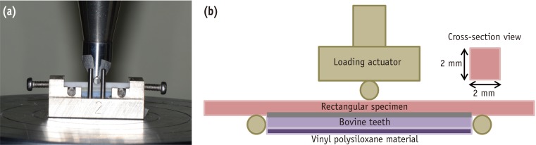
Some antioxidants are believed to restore dentin bond strength after dental bleaching. This study was done to evaluate the influence of antioxidants on the bond strength of bleached bovine dentin. Materials and MethodsThirty incisors were randomly assigned to 10 groups (two unbleached control and eight bleached groups: immediate bonding IB, 4 wk delayed bonding DB, 10% sodium ascorbate treated SA, 10% α-tocopherol treated TP groups). Teeth in half of groups were subjected to thermal stress, whereas the remaining groups were not. Resin-dentin rods with a cross-sectional area of 2.25 mm2 were obtained and microtensile bond strength was determined at a crosshead speed of 1 mm/min. Fifteen specimens were prepared for SEM to compare the surface characteristics of each group. The change in dentin bond strength from thermal stress and antioxidant treatment was evaluated using two-way analysis of variance (ANOVA) and Sheffe's post hoc test at a significance level of 95%. ResultsThe control group exhibited the highest bond strength values, whereas IB group showed the lowest value before and after thermocycling. The DB group recovered its bond strength similar to that of the control group. The SA and TP groups exhibited similar bond strength values with those of the control and DB groups before thermocycling. However, The TP group did not maintain bond strength with thermal stress, whereas the SA group did. ConclusionsApplying a 10% sodium ascorbate solution rather than 10% α-tocopherol solution for 60 sec is recommended to maintain dentin bond strength when restoring non-vitally bleached teeth.
-
Citations
Citations to this article as recorded by - Shear bond strength analysis of self-etch and total-etch adhesive systems on antioxidant-treated dentin in post-endodontic restoration
Suvarna Kavya Harish, Shruthi H. Attavar
Frontiers in Dental Medicine.2026;[Epub] CrossRef - Evaluation of the effect of the application of Quercus cerris extract and the use of fluoride bonding material on the bonding strength of orthodontic brackets after tooth bleaching with hydrogen peroxide
Ezgi Ay, Derya Dursun
PeerJ.2025; 13: e19335. CrossRef - Antioxidant effect on shear bond strength of resin composite to in-office versus home bleached enamel surface
Maha Mosaad Mohamed, Magda E. -A. Shalaby, Eman A. E. -G. Shebl
Tanta Dental Journal.2025; 22(3): 409. CrossRef - Comparative analysis of the impact of modern cavity disinfectants on dentin bond strength
Simge Gümüş Ayaz, Ezgi Sonkaya, Gökçe Keçeci
Frontiers in Materials.2025;[Epub] CrossRef - Effect of glutathione on bond strength of composite resin to enamel following extracoronal bleaching
Nair Devika, Chandrasekaran Charanya, K Athira, James Vandana, Sundaresan Balagopal
Journal of Conservative Dentistry and Endodontics.2024; 27(11): 1110. CrossRef - Effect of Chitosan Nanoparticle as an Antioxidant Material on Shear Bond Strength of Composite Resin to Enamel after External Bleaching
Diatri Nari Ratih, Shintatika Erlagista, Tunjung Nugraheni
Open Access Macedonian Journal of Medical Sciences.2024; 12: 1. CrossRef - Effects of alpha‐tocopherol antioxidant on fracture strength and adhesion of endodontically treated teeth restored after dental bleaching
Natália Marcomini, Maria Carolina da Costa Albaricci, Joatan Lucas de Sousa Gomes Costa, João Felipe Besegato, Eduardo Fernández Godoy, Andréa Abi Rached Dantas, Milton Carlos Kuga
European Journal of Oral Sciences.2024;[Epub] CrossRef - Alpha-tocopherol: An alternative solution for the adverse effects of dental bleaching on dentin adhesion
Maria Carolina da Costa Albaricci, Natália Marcomini, Joatan Lucas de Sousa Gomes Costa, Antonia Patricia Oliveira Barros, Lucas David Galvani, Milton Carlos Kuga, Andréa Abi Rached Dantas
International Journal of Adhesion and Adhesives.2024; 131: 103655. CrossRef - Efficacy of organic and antioxidant agents to regain bond strength to bleached enamel in different dental adhesive solvents
Satheesh B Haralur, Renad Mohammed Al-Ibrahim, Faten Abdullah Al-Shahrani, Rahaf Abdullah Al-Qahtani, Saurabh Chaturvedi, Naseer M Alqahtani
Journal of Applied Biomaterials & Functional Materials.2023;[Epub] CrossRef - Present status and future directions – Managing discoloured teeth
Bill Kahler
International Endodontic Journal.2022; 55(S4): 922. CrossRef - Comparative evaluation of the postbleaching application of sodium ascorbate, alpha‐tocopherol, and quercetin on shear bond strength of composite resin to enamel
Marzieh Moradian, Maryam Saadat, Mohammad Hossein S. Shiri, Fatemeh Sohrabniya
Clinical and Experimental Dental Research.2022; 8(6): 1598. CrossRef - Use of antioxidants to restore bond strength after tooth bleaching with peroxides
Dorcas E. R. P. Olmedo, Matheus Kury, Bruna A. Resende, Vanessa Cavalli
European Journal of Oral Sciences.2021;[Epub] CrossRef - Elemental and morphological analysis of enamel following the application of two bleaching systems with amorphous calcium phosphate: effect on enamel erosion susceptibility
Shaymaa M. Nagi, Shahinaz H. Nabil, Mohamed H. Zaazou
Bulletin of the National Research Centre.2021;[Epub] CrossRef - Pomegranate (Punica granatum L.) gel extract as an antioxidant on the shear bond strength of a resin composite post-bleaching application with 40% hydrogen peroxide
Indes Rosmalisa Suratno, Irfan Dwiandhono, Ryana Budi Purnama
Dental Journal.2021; 54(2): 87. CrossRef - In Vitro Re-Hardening of Bleached Enamel Using Mineralizing Pastes: Toward Preventing Bacterial Colonization
Andrea Scribante, Claudio Poggio, Simone Gallo, Paolo Riva, Antonella Cuocci, Manuel Carbone, Carla Arciola, Marco Colombo
Materials.2020; 13(4): 818. CrossRef - DİŞ BEYAZLATMA İŞLEMİNİN LİTYUM DİSİLİKAT SERAMİĞİN BAĞLANMA DAYANIMINA ETKİSİ
Merve YILDIRAK, Rıfat GÖZNELİ
Atatürk Üniversitesi Diş Hekimliği Fakültesi Dergisi.2020; : 1. CrossRef - The improvement of biocompatibility of adhesives
Cigdem Atalayin, Huseyin Tezel, Zeynep Ergucu, Nimet Unlu, Guliz Armagan, Taner Dagci, Timur Kose
Clinical Oral Investigations.2019; 23(8): 3213. CrossRef - Dentin bond strength and nanoleakage of the adhesive interface after intracoronal bleaching
Vanessa Cavalli, Maicon Sebold, Mirela Sanae Shinohara, Patrícia Nóbrega Rodrigues Pereira, Marcelo Giannini
Microscopy Research and Technique.2018; 81(4): 428. CrossRef - Composite resin shear bond strength on bleached dentin increased by 35% sodium ascorbate application
Tunjung Nugraheni, N Nuryono, Siti Sunarintyas, Ema Mulyawati
Dental Journal (Majalah Kedokteran Gigi).2017; 50(4): 178. CrossRef - Comparative Evaluation of Immediate Bond Strength to Bleached Enamel Following Application of Various Antioxidant Solutions
Anshu Minocha, Ashu K. Gupta, Alisha Dhingra, Nayantara Sen
Dental Journal of Advance Studies.2017; 5(2): 84. CrossRef - Effects of Erbium Family Laser on Shear Bond Strength of Composite to Dentin After Internal Bleaching
Nazanin Kiomarsi, Yasaman Arjmand, Mohammad Javad Kharrazi Fard, Nasim Chiniforush
Journal of Lasers in Medical Sciences.2017; 9(1): 58. CrossRef - Antioxidant therapy enhances pulpal healing in bleached teeth
Adriano Fonseca Lima, Marcelo Rocha Marques, Diana Gabriela Soares, Josimeri Hebling, Giselle Maria Marchi, Carlos Alberto de Souza Costa
Restorative Dentistry & Endodontics.2016; 41(1): 44. CrossRef - Effects of alpha-tocopherol on fracture resistance after endodontic treatment, bleaching and restoration
Keren Cristina Fagundes JORDÃO-BASSO, Milton Carlos KUGA, Andrea Abi Rached DANTAS, Mateus Rodrigues TONETTO, Suellen Nogueira Linhares LIMA, Matheus Coêlho BANDÉCA
Brazilian Oral Research.2016;[Epub] CrossRef - Resin Bonding of Self-Etch Adhesives to Bovine Dentin Bleached from Pulp Chamber
Akiko Haruyama, Atsushi Kameyama, Junji Kato, Shinji Takemoto, Yutaka Oda, Eiji Kawada, Toshiyuki Takahashi, Masahiro Furusawa
BioMed Research International.2016; 2016: 1. CrossRef
-
2,058
View
-
8
Download
-
24
Crossref
-
Antibacterial effect of self-etching adhesive systems on Streptococcus mutans
-
Seung-Ryong Kim, Dong-Hoon Shin
-
Restor Dent Endod 2014;39(1):32-38. Published online January 20, 2014
-
DOI: https://doi.org/10.5395/rde.2014.39.1.32
-
-
Abstract
PDFPubReaderePub
- Objectives
In this study, we evaluated the antibacterial activity of self-etching adhesive systems against Streptococcus mutans using the agar diffusion method. Materials and MethodsThree 2-step systems, Clearfil SE Bond (SE, Kuraray), Contax (CT, DMG), and Unifil Bond (UnB, GC), and three 1-step systems, Easy Bond (EB, 3M ESPE), U-Bond (UB, Vericom), and All Bond SE (AB, BISCO) were used. 0.12% chlorhexidine (CHX, Bukwang) and 37% phosphoric acid gel (PA, Vericom) were used as positive controls. ResultsThe antibacterial activity of CHX and PA was stronger than that of the other groups, except SE. After light activation, the inhibition zone was reduced in the case of all 2-step systems except CT. However, all 1-step systems did not exhibit any inhibition zone upon the light activation. ConclusionsSE may be better than CT or UnB among the 2-step systems with respect to antibacterial activity, however, 1-step systems do not exhibit any antibacterial activity after light curing.
-
Citations
Citations to this article as recorded by - Comparative Evaluation of Antibacterial Activity of Three Universal Bonding Agents Against Streptococcus mutans on Demineralized Dentin: An In Vitro Study
Kirti Rathee, Charu Dayal, Reena Rani, A Anukriti, Anjum Zia, Ankita Sundan, Seema Gupta
Cureus.2026;[Epub] CrossRef - Incorporation of chlorhexidine in self-adhesive resin cements
Idris M. MEHDAWI, Ranna KITAGAWA, Haruaki KITAGAWA, Satoshi YAMAGUCHI, Nanako HIROSE, Tomoki KOHNO, Satoshi IMAZATO
Dental Materials Journal.2022; 41(5): 675. CrossRef - Antibacterial and Bonding Properties of Universal Adhesive Dental Polymers Doped with Pyrogallol
Naji Kharouf, Ammar Eid, Louis Hardan, Rim Bourgi, Youri Arntz, Hamdi Jmal, Federico Foschi, Salvatore Sauro, Vincent Ball, Youssef Haikel, Davide Mancino
Polymers.2021; 13(10): 1538. CrossRef - Influence of Protease Inhibitors on Bond Degradation of Self-Etch Adhesive Systems to Caries-Affected Dentin: An <i>in Vitro</i> Study
Diana Roberta Pereira Grandizoli, Sérgio Luiz Pinheiro
Advances in Biological Chemistry.2018; 08(01): 15. CrossRef - Epigallocatechin-3-gallate and Epigallocatechin-3-O-(3-O-methyl)-gallate Enhance the Bonding Stability of an Etch-and-Rinse Adhesive to Dentin
Hao-Han Yu, Ling Zhang, Fan Yu, Fang Li, Zheng-Ya Liu, Ji-Hua Chen
Materials.2017; 10(2): 183. CrossRef - An In vitro Assessment of Antibacterial Activity of Three Self-etching Primers Against Oral Microflora
Sneha Dipak Shinde, Vikram Pai, R. Vijay Naik
APOS Trends in Orthodontics.2017; 7: 181. CrossRef - Functional Dental Restorative Materials That Hinder Oral Biofilm
Hércules Bezerra Dias, Victor Trassi Fernandes da Silva Souza, Rafael Amorim Martins, Ana Carolina Bosco Mendes, Monica Irma Aparecida Valdeci de Souza, Ângela Cristina Cilense Zuanon, Alessandra Nara de Souza Rastelli
Current Oral Health Reports.2017; 4(1): 22. CrossRef - In vitroantibacterial activity of various adhesive materials against oral streptococci
Emre Ozel, Fetiye Kolayli, Elif Bahar Tuna, Doganhan Er
Biotechnology & Biotechnological Equipment.2016; 30(1): 121. CrossRef - A systematic review about antibacterial monomers used in dental adhesive systems: Current status and further prospects
Alexandra Rubin Cocco, Wellington Luiz de Oliveira da Rosa, Adriana Fernandes da Silva, Rafael Guerra Lund, Evandro Piva
Dental Materials.2015; 31(11): 1345. CrossRef
-
1,985
View
-
6
Download
-
9
Crossref
-
Inhibitory effect on Streptococcus mutans and mechanical properties of the chitosan containing composite resin
-
Ji-Sun Kim, Dong-Hoon Shin
-
Restor Dent Endod 2013;38(1):36-42. Published online February 26, 2013
-
DOI: https://doi.org/10.5395/rde.2013.38.1.36
-
-
Abstract
PDFPubReaderePub
- Objectives
This study evaluated the antibacterial effect and mechanical properties of composite resins (LCR, MCR, HCR) incorporating chitosan with three different molecular weights (L, Low; M, Medium; H, High). Materials and MethodsStreptococcus (S). mutans 100 mL and each chitosan powder were inoculated in sterilized 10 mL Brain-Heart Infusion (BHI) solution, and was centrifuged for 12 hr. Absorbance of the supernatent was measured at OD660 to estimate the antibacterial activities of chitosan. After S. mutans was inoculated in the disc shaped chitosan-containing composite resins, the disc was cleansed with BHI and diluted with serial dilution method. S. mutans was spread on Mitis-salivarius bacitracin agar. After then, colony forming unit (CFU) was measured to verify the inhibitory effect on S. mutans biofilm. To ascertain the effect on the mechanical properties of composite resin, 3-point bending and Vickers hardness tests were done after 1 and 3 wk water storage, respectively. Using 2-way analysis of variance (ANOVA) and Scheffe test, statistical analysis was done with 95% significance level. ResultsAll chitosan powder showed inhibition effect against S. mutans. CFU number in chitosan-containing composite resins was smaller than that of control resin without chitosan. The chitosan containing composite resins did not show any significant difference in flexural strength and Vickers hardness in comparison with the control resin. However, the composite resin, MCR showed a slightly decreased flexural strength and the maximum load than those of control and the other composite resins HCR and LCR. ConclusionsLCR and HCR would be recommended as a feasible antibacterial restorative due to its antibacterial nature and mechanical properties.
-
Citations
Citations to this article as recorded by - Antibacterial activity, cytotoxicity, and microshear bond strength of an experimental adhesive system containing chitosan-based silver oxide particles
Hamideh Sadat Mohammadipour, Alireza Boruziniat, Seyedeh Azam Hoseini, Hosein Bagheri, Navid Ramezanian, Abbas Tanhaieian, Solmaz Pourgonabadi, Arsalan Shahri
Odontology.2026; 114(2): 494. CrossRef - Antibacterial efficacy of chitosan-coated thermoplastic orthodontic aligners against oral pathogens: An in vivo study – a randomized controlled trial
K. Gobinath, S. Saravana Kumar, Prema Anbarasu, R. Kanmani, Varalakshmi Raja Kuppusamy, R. Arunkumar
APOS Trends in Orthodontics.2026; 0: 1. CrossRef - Physicochemical and antibacterial properties of composite resin incorporating glycidyl methacrylate-chitosan derivative
Haiying Zhang, Shen Zhao, Yaoxin Wang, Mangnan Liu, Luxuan Wang, Chen Zhang, Benxiang Hou
Materials Research Express.2026; 13(8): 085401. CrossRef - Dental Resin Composites Modified with Chitosan: A Systematic Review
Wojciech Dobrzyński, Paweł J. Piszko, Jan Kiryk, Sylwia Kiryk, Mateusz Michalak, Agnieszka Kotela, Julia Kensy, Witold Świenc, Natalia Grychowska, Jacek Matys, Maciej Dobrzyński
Marine Drugs.2025; 23(5): 199. CrossRef - Bactericidal Effects of Ultraviolet-C Light-Emitting Diode Prototype Device Through Thin Optical Fiber
Mi-Jeong Jeon, Yu-Sung Choi, Deog-Gyu Seo
Applied Sciences.2025; 15(8): 4504. CrossRef - Synthesis and evaluation of a novel antibacterial nanocomposite resin restorative material
Rasha Abd El Rahman El Naggar, Manal A. ElEbiary, ElRefaie Kenawy, Gehan A. Elolimy
Tanta Dental Journal.2025; 22(3): 439. CrossRef - The impact of chitosan in experimental resin with different photoinitiator systems
Isaías Donizeti Silva, Letícia Cristina Cidreira Boaro, Bruno Vilela Muniz, Karina Cogo-Muller, Flávia Gonçalves, William Cunha Brandt
Journal of the Mechanical Behavior of Biomedical Materials.2024; 150: 106323. CrossRef - The effect of adding chitosan nanoparticles on different properties of the adhesive and high-filled composite resin
Mahan Masoumi, Sara Valizadeh, Ricardo M. Carvalho, Alireza Akbari Moghaddam, Safoura Ghodsi
International Journal of Adhesion and Adhesives.2024; 134: 103766. CrossRef - Prospective and applications of bacterial nanocellulose in dentistry
Yasmin Alimardani, Esmaeel Mirzakhani, Fereshteh Ansari, Hadi Pourjafar, Nadia Sadeghi
Cellulose.2024; 31(13): 7819. CrossRef - BNN/TiO2 nanocomposite system–modified dental flow resins and the mechanism of the enhancement of mechanical and antibacterial properties
Xinzi Kong, Qize Han, Axue Jiang, Yurui Wang, Ruizhi Li, Yuting Wang, Shengjie Xiao, Rong Wei, Yu Ma
Biomaterials Science.2023; 11(8): 2775. CrossRef - Influence of the Loading with Newly Green Silver Nanoparticles Synthesized Using Equisetum sylvaticum on the Antibacterial Activity and Surface Hardness of a Composite Resin
Ionuț Tărăboanță, Ana Flavia Burlec, Simona Stoleriu, Andreia Corciovă, Adrian Fifere, Denisa Batir-Marin, Monica Hăncianu, Cornelia Mircea, Irina Nica, Andra Claudia Tărăboanță-Gamen, Sorin Andrian
Journal of Functional Biomaterials.2023; 14(8): 402. CrossRef - The Impact of Adding Chitosan Nanoparticles on Biofilm Formation, Cytotoxicity, and Certain Physical and Mechanical Aspects of Directly Printed Orthodontic Clear Aligners
Botan Barzan Taher, Tara Ali Rasheed
Nanomaterials.2023; 13(19): 2649. CrossRef - Synthesis of Submicrometric Chitosan Particles Loaded with Calcium Phosphate for Biomedical Applications
Diana Pereira Lopes, Selma Regina Muniz Freitas, Carina Baptiston Tanaka, Giovanne Delechiave, Lucia Nobuco Takamori Kikuchi, Roberto R. Braga, Jamie J. Kruzic, Maria Stella Moreira, Leticia Cristina Cidreira Boaro, Luiz Henrique Catalani, Flávia Gonçalve
AAPS PharmSciTech.2023;[Epub] CrossRef - Biodegradable Nonwoven Materials with Antipathogenic Layer
Longina Madej-Kiełbik, Karolina Gzyra-Jagieła, Jagoda Jóźwik-Pruska, Maria Wiśniewskia-Wrona, Marzena Dymel
Environments.2022; 9(7): 79. CrossRef - Comparative evaluation of eighth-generation bonding agent modified with 7% arginine and 0.12% chitosan for antibacterial property and microtensile bond strength
HimaliRajan Desai, SanjyotA Mulay, RonitR Shinde, PradeepK Shetty, SoumyaS Shetty
Journal of Conservative Dentistry.2022; 25(4): 440. CrossRef - Effects of the crosslinking of chitosan/DCPA particles in the antimicrobial and mechanical properties of dental restorative composites
Lucia Nobuco Takamori Kikuchi, Selma Regina Muniz Freitas, Aldo Ferreira Amorim, Giovanne Delechiave, Luiz Henrique Catalani, Roberto Ruggiero Braga, Maria Stella Moreira, Leticia Cristina Cidreira Boaro, Flávia Gonçalves
Dental Materials.2022; 38(9): 1482. CrossRef - Antimicrobial activity of lactoferrin-chitosan-gellan nanoparticles and their influence on strawberry preservation
Larissa G.R. Duarte, Carolina S.F. Picone
Food Research International.2022; 159: 111586. CrossRef - Polyphenol-Enriched Extract of Lacquer Sap Used as a Dentine Primer with Benefits of Improving Collagen Cross-Linking and Antibacterial Functions
Ying Zhao, Xi He, Han Wang, Huimin Wang, Zuosen Shi, Song Zhu, Zhanchen Cui
ACS Biomaterials Science & Engineering.2022; 8(9): 3741. CrossRef - Evaluation of the changes in physical properties and mineral content of enamel exposed to radiation after treating with remineralization agent
Merve Pelin Dur, Neslihan Celik, Nilgun Seven
Clinical Oral Investigations.2022; 26(9): 5673. CrossRef - Evaluation of Immediate and Delayed Microleakage of Class V Cavities Restored with Chitosan-incorporated Composite Resins: An In Vitro Study
Arpita Deb, Veena Pai, Roopa R Nadig
International Journal of Clinical Pediatric Dentistry.2021; 14(5): 621. CrossRef - Evaluation of Microleakage of Micro Hybrid Composite Resins versus Chitosan-Incorporated Composite Resins When Restored in Class V Cavities Using Total Etch and Self-Etch Adhesives
Arpita Deb, Veena Pai, Aesha Akhtar, Roopa R. Nadig
Contemporary Clinical Dentistry.2021; 12(4): 346. CrossRef - Nanomaterials Application in Orthodontics
Wojciech Zakrzewski, Maciej Dobrzynski, Wojciech Dobrzynski, Anna Zawadzka-Knefel, Mateusz Janecki, Karolina Kurek, Adam Lubojanski, Maria Szymonowicz, Zbigniew Rybak, Rafal J. Wiglusz
Nanomaterials.2021; 11(2): 337. CrossRef - Antibacterial Effect on Enterococcus Faecalis and Physical Properties of Chitosan Added Calcium Hydroxide Canal Filling Material
Sol Song, Yu-Jin Kim, Jung-Hwan Lee, Joonhaeng Lee, Jisun Shin, Jongbin Kim
THE JOURNAL OF THE KOREAN ACADEMY OF PEDTATRIC DENTISTRY.2021; 48(2): 198. CrossRef - Antibacterial and Bonding Properties of Universal Adhesive Dental Polymers Doped with Pyrogallol
Naji Kharouf, Ammar Eid, Louis Hardan, Rim Bourgi, Youri Arntz, Hamdi Jmal, Federico Foschi, Salvatore Sauro, Vincent Ball, Youssef Haikel, Davide Mancino
Polymers.2021; 13(10): 1538. CrossRef - Efficacy of chitosan-based chewing gum on reducing salivary S. mutans counts and salivary pH: a randomised clinical trial
Zahra Khamverdi, Fatemeh Farhadian, Salman Khazaei, Maryam Adabi
Acta Odontologica Scandinavica.2021; 79(4): 268. CrossRef - Effect of antiseptic gels in the microbiologic colonization of the suture threads after oral surgery
Samuel Rodríguez Zorrilla, Andrés Blanco Carrión, Abel García García, Pablo Galindo Moreno, Xabier Marichalar Mendía, Rafael Seoane Prado, Antonio J. Pérez Estévez, Mario Pérez-Sayáns
Scientific Reports.2020;[Epub] CrossRef - Evaluating antibacterial and surface mechanical properties of chitosan modified dental resin composites
Shahid Ali, Laila Sangi, Naresh Kumar, Bharat Kumar, Zohaib Khurshid, Muhammad S. Zafar
Technology and Health Care.2020; 28(2): 165. CrossRef - Comparison of antibacterial effects of orthodontic composites containing different nanoparticles on Streptococcus mutans at different times
Soghra Yassaei, Ali Nasr, Hengameh Zandi, Mohammad Nima Motallaei
Dental Press Journal of Orthodontics.2020; 25(2): 52. CrossRef - Development of novel dental restorative composites with dibasic calcium phosphate loaded chitosan fillers
Carina B. Tanaka, Diana P. Lopes, Lucia N.T. Kikuchi, Maria Stella Moreira, Luiz H. Catalani, Roberto R. Braga, Jamie J. Kruzic, Flávia Gonçalves
Dental Materials.2020; 36(4): 551. CrossRef - Effect of iodonium salt and chitosan on the physical and antibacterial properties of experimental infiltrants
Mariana Dias FLOR-RIBEIRO, Talita Signoreti GRAZIANO, Flávio Henrique Baggio AGUIAR, Rafael Nóbrega STIPP, Giselle Maria MARCHI
Brazilian Oral Research.2019;[Epub] CrossRef - Chitosan/Fluoride Nanoparticles for Preventing Dental Caries
Niousha Ebrahimi, Ali Asghar Soleimani, Jamal Rashidiani, Beheshteh Malekafzali, Fatemeh Abedini, Hossein Hosseinkhani
Current Dentistry.2019; 1(1): 61. CrossRef - Physical and chemical properties of model composites containing quaternary ammonium methacrylates
Marina Lermenn Vidal, Guilherme Ferreira Rego, Gil Mendes Viana, Lucio Mendes Cabral, Juliana Primo Basílio Souza, Nick Silikas, Luis Felipe Schneider, Larissa Maria Cavalcante
Dental Materials.2018; 34(1): 143. CrossRef - Chitosan—PRP nanosphere as a growth factors slow releasing device with superior antibacterial capability
Radyum Ikono, Etik Mardliyati, Iis Tentia Agustin, Muhammad Mufarrij Fuad Ulfi, Dimas Andrianto, Uswatun Hasanah, Boy Muchlis Bachtiar, Nofa Mardianingsih, Endang Winiati Bachtiar, Nurwenda Novan Maulana, Nurul Taufiqu Rochman, Li Xianqi, Hideaki Kagami,
Biomedical Physics & Engineering Express.2018; 4(4): 045026. CrossRef - The Antibacterial Effect of Two Cavity Disinfectants against One of Cariogenic Pathogen: An In vitro Comparative Study
Hanaa M. Elgamily, Hoda S. El-Sayed, Ali Abdelnabi
Contemporary Clinical Dentistry.2018; 9(3): 457. CrossRef - Chitosan-Properties and Applications in Dentistry
Kmiec M
Advances in Tissue Engineering & Regenerative Medicine: Open Access.2017;[Epub] CrossRef - Analysis of the shelf life of chitosan stored in different types of packaging, using colorimetry and dentin microhardness
Antonio Miranda da Cruz-Filho, Angelo Rafael de Vito Bordin, Luis Eduardo Souza-Flamini, Débora Fernandes da Costa Guedes, Paulo César Saquy, Ricardo Gariba Silva, Jesus Djalma Pécora
Restorative Dentistry & Endodontics.2017; 42(2): 87. CrossRef - Restorative materials containing antimicrobial agents: is there evidence for their antimicrobial and anticaries effects? A systematic review
GS do Amaral, T Negrini, M Maltz, RA Arthur
Australian Dental Journal.2016; 61(1): 6. CrossRef - Antibacterial capacity of cavity disinfectants against Streptococcus mutans and their effects on shear bond strength of a self-etch adhesive
Han-Sol CHA, Dong-Hoon SHIN
Dental Materials Journal.2016; 35(1): 147. CrossRef - Antibacterial Effect and Physical-Mechanical Properties of Temporary Restorative Material Containing Antibacterial Agents
Amanda Mahammad Mushashe, Carla Castiglia Gonzaga, Paulo Henrique Tomazinho, Leonardo Fernandes da Cunha, Denise Piotto Leonardi, Janes Francio Pissaia, Gisele Maria Correr
International Scholarly Research Notices.2015; 2015: 1. CrossRef - Antimicrobial properties of conventional restorative filling materials and advances in antimicrobial properties of composite resins and glass ionomer cements—A literature review
Cher Farrugia, Josette Camilleri
Dental Materials.2015; 31(4): e89. CrossRef - Antibacterial effect of self-etching adhesive systems onStreptococcus mutans
Seung-Ryong Kim, Dong-Hoon Shin
Restorative Dentistry & Endodontics.2014; 39(1): 32. CrossRef - Dental materials with antibiofilm properties
Zhejun Wang, Ya Shen, Markus Haapasalo
Dental Materials.2014; 30(2): e1. CrossRef - Antibacterial properties of composite resins incorporating silver and zinc oxide nanoparticles onStreptococcus mutansandLactobacillus
Shahin Kasraei, Lida Sami, Sareh Hendi, Mohammad-Yousef AliKhani, Loghman Rezaei-Soufi, Zahra Khamverdi
Restorative Dentistry & Endodontics.2014; 39(2): 109. CrossRef - Antibakterielle fyllinger - hvor står vi i dag?
Nils Jacobsen
Den norske tannlegeforenings Tidende.2014; 124(8): 616. CrossRef
-
2,572
View
-
15
Download
-
44
Crossref
-
Effect of chlorhexidine application on the bond strength of resin core to axial dentin in endodontic cavity
-
Yun-Hee Kim, Dong-Hoon Shin
-
Restor Dent Endod 2012;37(4):207-214. Published online November 21, 2012
-
DOI: https://doi.org/10.5395/rde.2012.37.4.207
-
-
Abstract
PDFPubReaderePub
- Objectives
This study evaluated the influence of chlorhexidine (CHX) on the microtensile bonds strength (µTBS) of resin core with two adhesive systems to dentin in endodontic cavities. Materials and MethodsFlat dentinal surfaces in 40 molar endodontic cavities were treated with self-etch adhesive system, Contax (DMG) and total-etch adhesive system, Adper Single Bond 2 (3M ESPE) after the following surface treatments: (1) Priming only (Contax), (2) CHX for 15 sec + rinsing + priming (Contax), (3) Etching with priming (Adper Single Bond 2), (4) Etching + CHX for 15 sec + rinsing + priming (Adper Single Bond 2). Resin composite build-ups were made with LuxaCore (DMG) using a bulk method and polymerized for 40 sec. For each condition, half of specimens were submitted to µTBS after 24 hr storage and half of them were submitted to thermocycling of 10,000 cycles between 5℃ and 55℃ before testing. The data were analyzed using ANOVA and independent t-test at a significance level of 95%. ResultsCHX pre-treatment did not affect the bond strength of specimens tested at the immediate testing period, regardless of dentin surface treatments. However, after 10,000 thermocycling, all groups showed reduced bond strength. The amount of reduction was greater in groups without CHX treatments than groups with CHX treatment. These characteristics were the same in both self-etch adhesive system and total-etch adhesive system. Conclusions2% CHX application for 15 sec proved to alleviate the decrease of bond strength of dentin bonding systems. No significant difference was shown in µTBS between total-etching system and self-etching system.
-
Citations
Citations to this article as recorded by - Physical characterization and bond performance of a non-methacrylate dental adhesive in long-term biochemical and thermal aging models
Zach Gouveia, Rastin Rahiminejad, Lingyun Zhu, Jesse Barker, Yoav Finer, J. Paul Santerre
Dental Materials.2026; 42(5): 877. CrossRef - Micro Tensile bond strength and microleakage assessment of total-etch and self-etch adhesive bonded to carious affected dentin disinfected with Chlorhexidine, Curcumin, and Malachite green
Zeeshan Qamar, Nishath Sayed Abdul, R Naveen Reddy, Mahesh Shenoy, Saleh Alghufaili, Yousef Alqublan, Ali Barakat
Photodiagnosis and Photodynamic Therapy.2023; 43: 103636. CrossRef - The Classification and Selection of Adhesive Agents; an Overview for the General Dentist
Naji Ziad Arandi
Clinical, Cosmetic and Investigational Dentistry.2023; Volume 15: 165. CrossRef - Influence of chlorhexidine 2% and sodium hypochlorite 5.25% on micro-tensile bond strength of universal adhesive system (G-Premio Bond)
Nafiseh Fazelian, Abbas Rahimi Dashtaki, MohammadAmin Eftekharian, Batool Amiri
Brazilian Journal of Oral Sciences.2022;[Epub] CrossRef - Comparative evaluation of the effects of different methods of post space preparation in primary anterior teeth on the fracture resistance of tooth restorations
Bahman Seraj, Sara Ghadimi, Ebrahim Najafpoor, Fatemeh Abdolalian, razieh khanmohammadi
Journal of Dental Research, Dental Clinics, Dental Prospects.2019; 13(2): 141. CrossRef - Chemical, microbial, and host‐related factors: effects on the integrity of dentin and the dentin–biomaterial interface
Marcela T. Carrilho, Fabiana Piveta, Leo Tjäderhane
Endodontic Topics.2015; 33(1): 50. CrossRef - MMP Inhibitors on Dentin Stability
A.F. Montagner, R. Sarkis-Onofre, T. Pereira-Cenci, M.S. Cenci
Journal of Dental Research.2014; 93(8): 733. CrossRef - Thermal cycling for restorative materials: Does a standardized protocol exist in laboratory testing? A literature review
Anna Lucia Morresi, Maurizio D'Amario, Mario Capogreco, Roberto Gatto, Giuseppe Marzo, Camillo D'Arcangelo, Annalisa Monaco
Journal of the Mechanical Behavior of Biomedical Materials.2014; 29: 295. CrossRef
-
1,835
View
-
5
Download
-
8
Crossref
-
Effect of CQ-amine ratio on the degree of conversion in resin monomers with binary and ternary photoinitiation systems
-
Ho-Jin Moon, Dong-Hoon Shin
-
Restor Dent Endod 2012;37(2):96-102. Published online May 18, 2012
-
DOI: https://doi.org/10.5395/rde.2012.37.2.96
-
-
Abstract
PDFPubReaderePub
-
Objectives
This study evaluated the effect of camphorquinone (CQ)-amine ratio on the C=C double bond conversion of resins with binary and ternary photoinitiation systems.
Materials and Methods
Two monomer mixtures (37.5 Bis-GMA/37.5 Bis-EMA/25 TEGDMA) with binary systems (CQ/DMAEMA in weight ratio, group A [0.5/1.0] and B [1.0/0.5]) and four mixtures with ternary system (CQ/OPPI/DMAEMA, group C [0.1/1.0/0.1], D [0.1/1.0/0.2], E [0.2/1.0/0.1] and F [0.2/1.0/0.2]) were tested: 1 : 2 or 2 : 1 CQ-amine ratio in binary system, while 1 : 1 ratio was added in ternary system. The monomer mixture was cured for 5, 20, 40, and 300 sec with a Demetron 400 curing unit (Demetron). After each exposure time, degree of conversion (DC) was estimated using Fourier transform infrared (FTIR) spectrophotometer (Nicolet 520, Nicolet Instrument Corp.). The results were analyzed by ANOVA followed by Scheffe test, with p = 0.05 as the level of significance.
Results
DC (%) was expressed in the order of curing time (5, 20, 40, and 300 sec). Group A (14.63 ± 10.42, 25.23 ± 6.32, 51.62 ± 2.69, 68.52 ± 2.77); Group B (4.04 ± 6.23, 16.56 ± 3.38, 37.95 ± 2.79, 64.48 ± 1.21); Group C (16.87 ± 5.72, 55.47 ± 2.75, 60.83 ± 2.07, 68.32 ± 3.31); Group D (23.77 ± 1.64, 61.05 ± 1.82, 65.13 ± 2.09, 71.87 ± 1.17); Group E (28.66 ± 2.92, 56.68 ± 1.33, 60.66 ± 1.17, 68.78 ± 1.30); Group F (39.74 ± 6.31, 61.07 ± 2.58, 64.22 ± 2.29, 69.94 ± 2.15).
Conclusion
All the monomers with ternary photoinitiation system showed higher DC than the ones with binary system, until 40 sec. Concerning about the effect of CQ-amine ratio on the DC, group A converted into polymer more than group B in binary system. However, there was no significant difference among groups with ternary system, except group C when cured for 5 sec only.
-
Citations
Citations to this article as recorded by - The power of light – From dental materials processing to diagnostics and therapeutics
Mohammed A. Hadis, Adrian C. Shortall, William M. Palin
Biomaterial Investigations in Dentistry.2024; : 1. CrossRef - Mechanical Properties of Experimental Composites with Different Photoinitiator
Luis Felipe Marques de Resende, Anderson Catelan, Kusai Baroudi, Alan Rodrigo Muniz Palialol, Alexandre Marques de Resende, Ana Carolina Andreucci, Rayssa Ferreira Zanatta, Priscila Christiane Suzy Liporoni
European Journal of Dentistry.2022; 16(01): 167. CrossRef - Long-term bonding efficacy of adhesives containing benzodioxioles as alternative co-initiators
Giana da Silveira LIMA, Andressa Goicochea MOREIRA, Carine Tais Welter MEEREIS, Ginia Brito LIMA, Fernanda Barbosa LEAL, Rafael Ratto de MORAES, Fabrício A OGLIARI, Cesar Liberato PETZHOLD, Evandro PIVA
Brazilian Oral Research.2018;[Epub] CrossRef - Piperonyl methacrylate: Copolymerizable coinitiator for adhesive compositions
Andressa Goicochea Moreira, Carlos Enrique Cuevas-Suárez, Wellington Luiz de Oliveira da Rosa, Aline Oliveira Ogliari, Cesar Liberato Petzhold, Evandro Piva, Fabrício Aulo Ogliari, Giana da Silveira Lima
Journal of Dentistry.2018; 79: 31. CrossRef - Kinetics of bulk photo-initiated copper(i)-catalyzed azide–alkyne cycloaddition (CuAAC) polymerizations
Han Byul Song, Austin Baranek, Christopher N. Bowman
Polymer Chemistry.2016; 7(3): 603. CrossRef - 1,3-Butadiene as an Adhesion Promoter Between Composite Resin and Dental Ceramic in a Dielectric Barrier Discharge Jet
Geum-Jun Han, Sung-No Chung, Bae-Hyeock Chun, Chang-Keun Kim, Kyu Hwan Oh, Byeong-Hoon Cho
Plasma Chemistry and Plasma Processing.2013; 33(2): 539. CrossRef
-
2,541
View
-
11
Download
-
6
Crossref
-
Effect of different chlorhexidine application times on microtensile bond strength to dentin in Class I cavities
-
Hyun-Jung Kang, Ho-Jin Moon, Dong-Hoon Shin
-
Restor Dent Endod 2012;37(1):9-15. Published online March 2, 2012
-
DOI: https://doi.org/10.5395/rde.2012.37.1.9
-
-
Abstract
PDFPubReaderePub
-
Objectives
This study evaluated the effect of 2% chlorhexidine digluconate (CHX) with different application times on microtensile bonds strength (MTBS) to dentin in class I cavities and intended to search for ideal application time for a simplified bonding protocol.
Materials and Methods
Flat dentinal surfaces with class I cavities (4 mm × 4 mm × 2 mm) in 40 molar teeth were bonded with etch-and-rinse adhesive system, Adper Single Bond 2 (3M ESPE) after: (1) etching only as a control group; (2) etching + CHX 5 sec + rinsing; (3) etching + CHX 15 sec + rinsing; (4) etching + CHX 30 sec + rinsing; and (5) etching + CHX 60 sec + rinsing. Resin composite was built-up with Z-250 (3M ESPE) using a bulk method and polymerized for 40 sec. For each condition, half of the specimens were immediately submitted to MTBS test and the rest of them were assigned to thermocycling of 10,000 cycles between 5℃ and 55℃ before testing. The data were analyzed using two-way ANOVA, at a significance level of 95%.
Results
There was no significant difference in bond strength between CHX pre-treated group and control group at the immediate testing period. After thermocycling, all groups showed reduced bond strength irrespective of the CHX use. However, groups treated with CHX maintained significantly higher MTBS than control group (p < 0.05). In addition, CHX application time did not have any significant influence on the bond strength among groups treated with CHX.
Conclusion
Application of 2% CHX for a short time period (5 sec) after etching with 37% phosphoric acid may be sufficient to preserve dentin bond strength.
-
Citations
Citations to this article as recorded by - Evaluation of Fracture Resistance of Tooth Fragment Reattached with Cavity Disinfectants in Primary and Permanent Teeth: An In Vitro Study
Komal P Bhosale, Vishnu R Chamarthi, Dhanraj Kalaivanan, Santham Krishnamoorthy, Sumaiyya Saleem, Santhosh Priya AKR, Sai SK Kothimbakkam
The Journal of Contemporary Dental Practice.2026; 27(3): 269. CrossRef - Effect of nonthermal atmospheric plasma application at different time intervals on the dentinal shear bond strength pretreated with 2% chlorhexidine as cavity disinfectant: An in vitro study
Roopadevi Garlapati, Nagesh Bolla, Gali Praveen Kumar, Mayana Aameena Banu, Bandlapally Sreenivasa Guptha Anila, Shaik Afreen Kamal
Journal of Conservative Dentistry and Endodontics.2024; 27(7): 769. CrossRef - Comparative evaluation ofEmblica officinalisas an etchant and an MMP inhibitor with orthophosphoric acid and chlorhexidine on the microshear bond strength of composite resin: anex vivostudy
Divya Sangeetha Rajkumar, Annapoorna Ballagere Mariswamy
Restorative Dentistry & Endodontics.2021;[Epub] CrossRef - Effect of Cavity Disinfectants on Adhesion to Primary Teeth—A Systematic Review
Ana Coelho, Inês Amaro, Ana Apolónio, Anabela Paula, José Saraiva, Manuel Marques Ferreira, Carlos Miguel Marto, Eunice Carrilho
International Journal of Molecular Sciences.2021; 22(9): 4398. CrossRef - Effect of Different Matrix Metalloproteinase Inhibitors on Shear Bond Strength of Composite Attached to Primary Teeth Dentin
Najmeh Mohammadi, Zahra Parsaie, Dana Jafarpour, Fatemeh Bizolm
European Journal of General Dentistry.2020; 9(03): 147. CrossRef
-
2,492
View
-
6
Download
-
5
Crossref
-
Optimal combination of 3-component photoinitiation system to increase the degree of conversion of resin monomers
-
Chang-Gyu Kim, Ho-Jin Moon, Dong-Hoon Shin
-
J Korean Acad Conserv Dent 2011;36(4):313-323. Published online July 31, 2011
-
DOI: https://doi.org/10.5395/JKACD.2011.36.4.313
-
-
Abstract
PDFPubReaderePub
-
Objectives
This study investigated the optimal combination of 3-component photoinitiation system, consisting of CQ, p-octyloxy-phenyl-phenyl iodonium hexafluoroantimonate (OPPI), and 2-dimethylaminoethyl methacrylate (DMAEMA) to increase the degree of conversion of resin monomers, and analyze the effect of the ratio of the photoinitiator to the co-initiator.
Materials and Methods
Each photoinitiators (CQ and OPP) and co-initiator (DMAEMA) were mixed in three levels with 0.2 wt.% (low concentration, L), 1.0 wt.% (medium concentration, M), and 2.0 wt.% (high concentration, H). A total of nine groups using the Taguchi method were tested according to the following proportion of components in the photoinitiator system: LLL, LMM, LHH, MLM, MMH, MHL, HLH, HML, HHM. Each monomer was polymerized using a quartz-tungsten-halogen curing unit (Demetron 400, USA) for 5, 20, 40, 60, 300 sec and the degree of conversion (DC) was determined at each exposure time using FTIR.
Results
Significant differences were found for DC values in groups. MMH group and HHM group exhibited greater initial DC than the others. No significant difference was found with the ratio of the photoinitiators (CQ, OPPI) to the co-initiator (DMAEMA). The concentrations of CQ didn't affect the DC values, but those of OPPI did strongly.
Conclusions
MMH and HHM groups seem to be best ones to get increased DC. MMH group is indicated for bright, translucent color and HHM group is good for dark, opaque colored-resin.
-
Citations
Citations to this article as recorded by - Evaluation of Surface Roughness of Composite, Compomer and Carbomer After Curing Through Mylar Strip and Glycerin: A Comparative Study
Asli Topaloglu-Ak, Dilara Çayırgan, Melisa Uslu
Journal of Advanced Oral Research.2020; 11(1): 12. CrossRef - Effect of CQ-amine ratio on the degree of conversion in resin monomers with binary and ternary photoinitiation systems
Ho-Jin Moon, Dong-Hoon Shin
Restorative Dentistry & Endodontics.2012; 37(2): 96. CrossRef - Effect of glycerin on the surface hardness of composites after curing
Hyun-Hee Park, In-Bog Lee
Journal of Korean Academy of Conservative Dentistry.2011; 36(6): 483. CrossRef
-
1,474
View
-
1
Download
-
3
Crossref
-
Microleakage of the experimental composite resin with three component photoinitiator systems
-
Ji-Hoon Kim, Dong-Hoon Shin
-
J Korean Acad Conserv Dent 2009;34(4):333-339. Published online July 31, 2009
-
DOI: https://doi.org/10.5395/JKACD.2009.34.4.333
-
-
Abstract
PDFPubReaderePub
This study was done to determine if there is any difference in microleakage between experimental composite resins, in which various proportions of three component photoinitiators (Camphoroquinone, OPPI, Amine) were included.
Four kinds of experimental composite resin were made by mixing 3.2% silanated barium glass (78 wt.%, average size; 1 µm) with each monomer system including variously proportioned photoinitiator systems used for photoinitiating BisGMA/BisEMA/TEGDMA monomer blend (37.5:37.5:25 wt.%). The weight percentage of each component were as follows (in sequence Camphoroquinone, OPPI, Amine): Group A - 0.5%, 0%, 1% / Group B - 2%, 0.2%, 2% / Group C - 0.2%, 1%, 0.2% / Group D - 1%, 1%, 2%.
Each composite resin was used as a filling material for round class V cavities (diameter: 2/3 of mesiodistal width; depth: 1.5 mm) made on extracted human premolars and they were polymerized using curing light unit (XL 2500, 3M ESPE) for 40 s with an intensity of 600 mW/cm2. Teeth were thermocycled five-hundred times between 50℃ and 550℃ for 30s at each temperature.
Electrical conductivity (µA) was recorded two times (just after thermocycling and after three-month storage in saline solution) by electrochemical method.
Microleakage scores of each group according to evaluation time were as follows [Group: at first record / at second record; unit (µA)]: A: 3.80 (0.69) / 13.22 (4.48), B: 3.42 (1.33) / 18.84 (5.53), C: 4.18 (2.55) / 28.08 (7.75), D: 4.12 (1.86) / 7.41 (3.41).
Just after thermocycling, there was no difference in microleakage between groups, however, group C showed the largest score after three-month storage. Although there seems to be no difference in microleakage between groups just after thermocycling, composite resin with highly concentrated initiation system or classical design (Camphoroquinone and Amine system) would be more desirable for minimizing microleakage after three-month storage. -
Citations
Citations to this article as recorded by - Comparison of polymerization shrinkage of dual-cure core build-up resin according to shade and curing mode
Yoorina Choi, Karl Lee, Hoon-Sang Chang
Oral Biology Research.2019; 43(4): 243. CrossRef - Optimal combination of 3-component photoinitiation system to increase the degree of conversion of resin monomers
Chang-Gyu Kim, Ho-Jin Moon, Dong-Hoon Shin
Journal of Korean Academy of Conservative Dentistry.2011; 36(4): 313. CrossRef
-
1,346
View
-
1
Download
-
2
Crossref
-
Difference in bond strength according to filling techniques and cavity walls in box-type occlusal composite resin restoration
-
Eun-Joo Ko, Dong-Hoon Shin
-
J Korean Acad Conserv Dent 2009;34(4):350-355. Published online July 31, 2009
-
DOI: https://doi.org/10.5395/JKACD.2009.34.4.350
-
-
Abstract
PDFPubReaderePub
Bond strength depends on characteristics of bonding surface and restorative technique. The majority of studies dealing with dentin bond strength were carried out on flat bonding surface, therefore, difference of bond strength between axial wall and pulpal wall is not clear yet. This study evaluated bonding difference between cavity walls in class I composite resin restoration with different filling techniques.
Twenty extracted caries-free human third molars were used. Cavities were prepared in 6 × 4 × 3 mm box-type and divided into four groups according to filling technique and bonding surface: Group I; bulk filling - pulpal wall, Group II; bulk filling - axial wall, Group III; incremental filling - pulpal wall, Group IV; incremental filling - axial wall.
Cavities were filled with Filtek Z250®(3M/ESPE., USA) and Clearfill SE bond®(Kuraray, Japan). After 24 hour-storage in 37℃ water, the resin bonded teeth were sectioned bucco-lingualy at the center of cavity.
Specimens were vertically sectioned into 1.0 × 1.0 mm thick serial sticks perpendicular to the bond surface using a low-speed diamond saw (Accutom 50, Struers, Copenhagen, Denmark) under water cooling. The trimmed specimens were then attached to the testing device and in turn, was placed in a universal testing machine (EZ test, Shimadzu Co., Kyoto, Japan) for micro-tensile testing at a cross-head speed of 1 mm/min. The results obtained were statistically analyzed using 2-way ANOVA and t-test at a significance level of 95%.
The results were as follows:
1. There was no significant difference between bulk filling and incremental filling.
2. There was no significant difference between pulpal wall and axial wall, either.
Within the limit of this study, it was concluded that microtensile bond strength was not affected by the filling technique and the site of cavity walls. -
Citations
Citations to this article as recorded by - The effect of the strength and wetting characteristics of Bis-GMA/TEGDMA-based adhesives on the bond strength to dentin
Eun-Sook Park, Chang-Keun Kim, Ji-Hyun Bae, Byeong-Hoon Cho
Journal of Korean Academy of Conservative Dentistry.2011; 36(2): 139. CrossRef
-
1,388
View
-
1
Download
-
1
Crossref
-
Fracture resistance of the three types of undermined cavity filled with composite resin
-
Hoon-Soo Choi, Dong-Hoon Shin
-
J Korean Acad Conserv Dent 2008;33(3):177-183. Published online May 31, 2008
-
DOI: https://doi.org/10.5395/JKACD.2008.33.3.177
-
-
Abstract
PDFPubReaderePub
It was reported that esthetic composite resin restoration reinforces the strength of remaining tooth structure with preserving the natural tooth structure. However, it is unknown how much the strength would be recovered. The purpose of this study was to compare the fracture resistance of three types of undermined cavity filled with composite resin with that of non-cavitated natural tooth.
Forty sound upper molars were allocated randomly into four groups of 10 teeth. After flattening occlusal enamel, undermined cavities were prepared in thirty teeth to make three types of specimens with various thickness of occlusal structure (Group 1 ~ 3). All the cavity have the 5 mm width mesiodistally and 7 mm depth bucco-lingually. Another natural 10 teeth (Group 4) were used as a control group. Teeth in group 1 have remaining occlusal structure about 1 mm thickness, which was composed of mainly enamel and small amount of dentin. In Group 2, remained thickness was about 1.5 mm, including 0.5 mm thickness dentin. In Group 3, thickness was about 2.0 mm, including 1 mm thickness dentin. Every effort was made to keep the remaining dentin thickness about 0.5 mm from the pulp space in cavitated groups. All the thickness was evaluated with radiographic Length Analyzer program.
After acid etching with 37% phosphoric acid, one-bottle adhesive (Single Bond™, 3M/ESPE, USA) was applied following the manufacturer's recommendation and cavities were incrementally filled with hybrid composite resin (Filtek Z-250™, 3M/ESPE, USA). Teeth were stored in distilled water for one day at room temperature, after then, they were finished and polished with Sof-Lex system.
All specimens were embedded in acrylic resin and static load was applied to the specimens with a 3 mm diameter stainless steel rod in an Universal testing machine and cross-head speed was 1 mm/min. Maximum load in case of fracture was recorded for each specimen.
The data were statistically analyzed using one-way analysis of variance (ANOVA) and a Tukey test at the 95% confidence level.
The results were as follows:
Fracture resistance of the undermined cavity filled with composite resin was about 75% of the natural tooth.
No significant difference in fracture loads of composite resin restoration was found among the three types of cavitated groups.
Within the limits of this study, it can be concluded the fracture resistance of the undermined cavity filled with composite resin was lower than that of natural teeth, however remaining tooth structure may be supported and saved by the reinforcement with adhesive restoration, even if that portion consists of mainly enamel and a little dentin structure. -
Citations
Citations to this article as recorded by - Fracture resistance of crown-root fractured teeth repaired with dual-cured composite resin and horizontal posts
Seok-Woo Chang, Yong-Keun Lee, Seung-Hyun Kyung, Hyun-Mi Yoo, Tae-Seok Oh, Dong-Sung Park
Journal of Korean Academy of Conservative Dentistry.2009; 34(5): 383. CrossRef
-
2,023
View
-
6
Download
-
1
Crossref
-
Microleakage of composite resin restoration according to the number of thermocycling
-
Chang-Youn Kim, Dong-Hoon Shin
-
J Korean Acad Conserv Dent 2007;32(4):377-384. Published online July 31, 2007
-
DOI: https://doi.org/10.5395/JKACD.2007.32.4.377
-
-
Abstract
PDFPubReaderePub
Present tooth bonding system can be categorized into total etching bonding system (TE) and self-etching boding system (SE) based on their way of smear layer treatment. The purposes of this study were to compare the effectiveness between these two systems and to evaluate the effect of number of themocycling on microleakage of class V composite resin restorations.
Total forty class V cavities were prepared on the single-rooted bovine teeth and were randomly divided into four experimental groups: two kinds of bonding system and another two kinds of thermocycling groups. Half of the cavities were filled with Z250 follwing the use of TE system, Single Bond and another twenty cavities were filled with Metafil and AQ Bond, SE system. All composite restoratives were cured using light curing unit (XL2500, 3M ESPE, St. Paul, MN, USA) for 40 seconds with a light intensity of 600 mW/cm2.
Teeth were stored in distilled water for one day at room temperature and were finished and polished with Sof-Lex system. Half of teeth were thermocycled 500 times and the other half were thermocycled 5,000 times between 5℃ and 55℃ for 30 second at each temperature.
Teeth were isolated with two layers of nail varnish except the restoration surface and 1 mm surrounding margins. Electrical conductivity (µA) was recorded in distilled water by electrochemical method. Microleakage scores were compared and analyzed using two-way ANOVA at 95% level.
From this study, following results were obtained: There was no interaction between variables of bonding system and number of thermocycling (p = 0.485). Microleakage was not affected by the number of thermocycling either (p = 0.814). However, Composite restoration of Metafil and AQ Bond, SE bond system showed less microleakage than composite restoration of Z250 and Single Bond, TE bond system (p = 0.005). -
Citations
Citations to this article as recorded by - Evaluation of Shear Bond Strength and Microleakage of Bulk-fill Resin Composites
Hanbyeol Lee, Hyunwoo Seo, Juhyun Lee, Howon Park
THE JOURNAL OF THE KOREAN ACADEMY OF PEDTATRIC DENTISTRY.2015; 42(4): 281. CrossRef - Effect of Er:YAG lasing on the dentin bonding strength of two-step adhesives
Byeong-Choon Song, Young-Gon Cho, Myung-Seon Lee
Journal of Korean Academy of Conservative Dentistry.2011; 36(5): 409. CrossRef - Microleakage of the experimental composite resin with three component photoinitiator systems
Ji-Hoon Kim, Dong-Hoon Shin
Journal of Korean Academy of Conservative Dentistry.2009; 34(4): 333. CrossRef
-
1,442
View
-
2
Download
-
3
Crossref
-
The effect of C-factor and volume on microleakage of composite resin restorations with enamel margins
-
Bong-Joo Koo, Dong-Hoon Shin
-
J Korean Acad Conserv Dent 2006;31(6):452-459. Published online November 30, 2006
-
DOI: https://doi.org/10.5395/JKACD.2006.31.6.452
-
-
Abstract
PDFPubReaderePub
Competition will usually develop between the opposing walls as the restorative resin shrinks during polymerization. Magnitude of this phenomenon may be depended upon cavity configuration and volume.
The purpose of this sturdy was to evaluate the effect of cavity configuration and volume on microleakage of composite resin restoration that has margins on the enamel site only.
The labial enamel of forty bovine teeth was ground using a model trimmer to expose a flat enamel surface. Four groups with cylindrical cavities were defined, according to volume and configuration factor (Depth × Diameter / C-factor) - Group I: 1.5 mm × 2.0 mm / 4.0, Group II: 1.5 mm × 6.0 mm / 2.0, Group III: 2.0 mm × 1.72 mm / 5.62, Group IV: 2.0 mm × 5.23 mm / 2.54.
After treating with fifth-generation one-bottle adhesive - BC Plus™ (Vericom, AnYang, Korea), cavities were bulk filled with microhybrid composite resin - Denfill™ (Vericom). Teeth were stored in distilled water for one day at room temperature and were finished and polished with Sof-Lex system. Specimens were thermocycled 500 times between 5℃ and 55℃ for 30 second at each temperature.
Teeth were isolated with two layers of nail varnish except the restoration surface and 1 mm surrounding margins. Electrical conductivity (µA) was recorded in distilled water by electrochemical method. Microleakage scores were compared and analyzed using two-way ANOVA at 95% level.
The results were as follows:
1. Small cavity volume showed lower microleakage score than large one, however, there was no statistically significant difference.
2. There was no relationship between cavity configuration and microleakage.
Factors of cavity configuration and volume did not affect on microleakage of resin restorations with enamel margins only. -
Citations
Citations to this article as recorded by - Influence of rebonding procedures on microleakage of composite resin restorations
Mi-Ae Lee, Duck-Kyu Seo, Ho-Hyun Son, Byeong-Hoon Cho
Journal of Korean Academy of Conservative Dentistry.2010; 35(3): 164. CrossRef - Microleakage of the experimental composite resin with three component photoinitiator systems
Ji-Hoon Kim, Dong-Hoon Shin
Journal of Korean Academy of Conservative Dentistry.2009; 34(4): 333. CrossRef - A survey on the use of composite resin in Class II restoration in Korea
Dong-Ho Shin, Se-Eun Park, In-Seok Yang, Juhea Chang, In-Bog Lee, Byeong-Hoon Cho, Ho-Hyun Son
Journal of Korean Academy of Conservative Dentistry.2009; 34(2): 87. CrossRef - Difference in bond strength according to filling techniques and cavity walls in box-type occlusal composite resin restoration
Eun-Joo Ko, Dong-Hoon Shin
Journal of Korean Academy of Conservative Dentistry.2009; 34(4): 350. CrossRef
-
1,845
View
-
8
Download
-
4
Crossref
-
Estimation of relation between techniques of dye penetration for microleakage and SEM evaluation for marginal adaptation of the restoration
-
Soon-Joo Hwang, Dong-Hoon Shin
-
J Korean Acad Conserv Dent 2006;31(5):337-343. Published online September 30, 2006
-
DOI: https://doi.org/10.5395/JKACD.2006.31.5.337
-
-
Abstract
PDFPubReaderePub
The purpose of this study was to estimate the relation between techniques used for microleakage from dye penetration and for marginal adaptation from SEM evaluation of the restoration.
Using high speed #330 bur, class V cavities (4 × 3 × 1.5 mm around CEJ) were prepared on the buccal surface of 20 extracted human molars. Six dimples as reference points for SEM and dye penetration evaluation were made with 1/2 round bur. Cavity was bulk filled with microhybrid composite resin (Esthet X) and all-in-one adhesive (Xeno III). Teeth were stored in saline solution for one day, after then, they were finished and polished using Sof-Lex system.
Fifty percent silver nitrate dye solution was used for the evaluation of microleakage and resin replica was used for marginal adaptation. All of these were done after 1000 times thermocycling between 5 and 55℃.
Vertical sections were made through three dimples of restoration to obtain samples for the evaluation of dye penetration and inner marginal adaptation. Outer adaptational estimation was done with an intact restoration before sectioning. Dye penetration was determined in three degrees and percentage of outer and inner leaky margin was estimated from SEM image.
The data were analysed statistically: Spearman's rho test were used to check relationships between two methods.
The result were as follows:
There were significant relationships between degree of dye penetration and inner and outer marginal adaptations each (p < 0.01).
However, there was no significant relationship between the results of inner and outer marginal adaptation.
Within the results of this study, relationship between the percentage of marginal adaptation and microleakage shows significant relationship. However, inner and outer marginal adaptation did not show any significant relationship mutually. -
Citations
Citations to this article as recorded by - Evaluation of Shear Bond Strength and Microleakage of Bulk-fill Resin Composites
Hanbyeol Lee, Hyunwoo Seo, Juhyun Lee, Howon Park
THE JOURNAL OF THE KOREAN ACADEMY OF PEDTATRIC DENTISTRY.2015; 42(4): 281. CrossRef - Comparison of marginal microleakage between low and high flowable resins in class V cavity
Sang-Bae Bae, Young-Gon Cho, Myeong-Seon Lee
Journal of Korean Academy of Conservative Dentistry.2009; 34(6): 477. CrossRef
-
1,572
View
-
10
Download
-
2
Crossref
-
Microleakage of the class V cavity according to restoration site and cavity size using SEM and three-dimensional reconstruction techniques
-
In-Seo Yang, Dong-Hoon Shin
-
J Korean Acad Conserv Dent 2005;30(2):112-120. Published online March 31, 2005
-
DOI: https://doi.org/10.5395/JKACD.2005.30.2.112
-
-
Abstract
PDFPubReaderePub
This study was done to evaluate whether there were any differences in microleakage of class V composite restorations according to restoration site and cavity size.
Total sixty-four restorations were made in molar teeth using Esthet-X. Small (2 × 2 × 1.5 mm) and large (4 × 2 × 1.5 mm) restorations were made at the buccal/lingual surface and the proximal surface each. After 1,000 times of thermocycling (5℃ - 55℃), resin replica was made and the percentage of marginal gap to the whole periphery of the restoration was estimated from SEM evaluation.
Thermocycled tooth was dye penetrated with 50% silver nitrate solution. After imbedding in an auto-curing resin, it was serially ground with a thickness of 0.25 mm. Volumetric microleakage was estimated after reconstructing three dimensionally.
Two-way ANOVA and independent T-test for dye volume, Mann-Whitney U test for the percentage of marginal gap, Spearman's rho test for the relationship between two techniques were used.
The results were as follows:
1. The site and size of the restoration affected on the microleakage of restoration. Namely, much more leakage was seen in the proximal and the large restorations rather than the buccal/lingual and the small restorations.
2. Close relationship was found between two techniques (Correlation coefficient = 0.614 / P = 0.000).
Within the limits of this study, it was noted that proximal and the large restorations leaked more than buccal/lingual and the small restorations. Therefore, it should be strictly recommended large exposure of margins should be avoided by reducing unnecessary tooth reduction.
-
New quantitative measuring technique for microleakage of the restored tooth through 3D reconstruction
-
Sang-Yoon Ha, Dong-Hoon Shin
-
J Korean Acad Conserv Dent 2004;29(5):413-422. Published online September 30, 2004
-
DOI: https://doi.org/10.5395/JKACD.2004.29.5.413
-
-
Abstract
PDFPubReaderePub
Established microleakage tests have their own disadvantages. In this study, 3D reconstruction method was tried to overcome these disadvantages.
Four types of microleakage tests were used and relationships among them were estimated: penetrated dye volume; marginal adaptability; degree of dye penetration and relative penetrated length to cavity wall.
Twenty-four Class V cavities were bulk filled with composite (Esthet X) following surface treatments: N group (no treatment); E group (etching only); T group (etching + Prime & Bond NT). 50% silver nitrate was used as a dye solution after thermocycling (5℃ & 55℃, 1,000 times). Teeth were serially ground with a thickness of 0.2 mm. Volume of dye penetration was estimated from a three-dimensionally reconstructed image with a software (3D-DOCTOR). Percentage of margin without gap was estimated from SEM and degree of dye penetration and the relative length of dye penetration to overall cavity wall were also estimated.
ANOVA and Scheffe test for dye volume, Kruskal-Wallis and Mann-Whitney test for marginal quality, Spearman's rho test for checking of relationships among methods were used.
The results were as follows:
1. Dye penetration could be seen from several directions, furthermore, its volumetric estimation was possible.
2. Reverse relationship was found between dye volume and marginal quality (r = -0.881 / p = 0.004).
3. Very low relationship was seen between dye volume and two-dimensional tests (degree of dye penetration and relative length). However, 2D evaluation methods showed high relationship (p = 0.002-0.054) each other.
4. Three times vertical section could be recommended as a 2D test. -
Citations
Citations to this article as recorded by - Estimation of relation between techniques of dye penetration for microleakage and SEM evaluation for marginal adaptation of the restoration
Soon-Joo Hwang, Dong-Hoon Shin
Journal of Korean Academy of Conservative Dentistry.2006; 31(5): 337. CrossRef - Microleakage of the class V cavity according to restoration site and cavity size using SEM and three-dimensional reconstruction techniques
In-Seo Yang, Dong-Hoon Shin
Journal of Korean Academy of Conservative Dentistry.2005; 30(2): 112. CrossRef
-
1,564
View
-
0
Download
-
2
Crossref
-
A study on the material properties of various composite resins for core build-up
-
Soo-Il Shin, Dong-Hoon Shin
-
J Korean Acad Conserv Dent 2004;29(2):191-199. Published online March 31, 2004
-
DOI: https://doi.org/10.5395/JKACD.2004.29.2.191
-
-
Abstract
PDFPubReaderePub
The purposes of this study were to estimate the material properties of the recently developed domestic composite resins for core filling material (Chemical, Dual A, Dual B; Vericom, Korea) and to compare them with other marketed foreign products (CorePaste, Den-Mat, USA; Ti-Core, Essential Dental Systems, USA; Support, SCI-Pharm, USA). Six assessments were made; working time, setting time, depth of polymerization, flexural strength, bonding strength, and marginal leakage. All items were compared to ISO standards.
All domestic products satisfied the minimum requirements from ISO standards (working time: above 90 seconds, setting time: within 5 minutes), and showed significantly higher flexural strength than Core Paste. Dual A and B could, especially, reduce the setting time to 60 seconds when cured with 600 mW/cm2 light intensity. All experimental materials showed 6 mm depth of polymerization.
Bond strengths of Ti-Core and Dual B materials were significantly higher than the other materials. Furthermore, three domestic products and Ti-Core could reduce the microleakage effectively. -
Citations
Citations to this article as recorded by - Effect of surface treatments of fiber posts on bond strength to composite resin cores
Hye-Jo Keum, Hyun-Mi Yoo
Journal of Korean Academy of Conservative Dentistry.2010; 35(3): 173. CrossRef
-
1,637
View
-
3
Download
-
1
Crossref
-
A quantitative analysis about microleakage of all-in-one adhesives
-
Yong-Hee Kang, Soo-Il Shin, Dong-Hoon Shin
-
J Korean Acad Conserv Dent 2004;29(1):66-72. Published online January 31, 2004
-
DOI: https://doi.org/10.5395/JKACD.2004.29.1.066
-
-
Abstract
PDFPubReaderePub
All-in-one adhesives were recently developed for reducing the techique sensitivity and chair time, but lots of concerns were made on bondability, longevity, and microleakage.
The object of this study was to evaluate microleakage and marginal quality of all-in-one adhesives using electrochemical method and SEM analysis quantitatively.
After making Class V cavities, they were bulk filled with Heliomolar(#A1) after surface treatment with three adhesives: Adper Prompt (Group A), One up bond F (Group O), Xeno III (Group X). Electrical conductivity (microamphere, µA) was checked two times: before and after cavity filling.
Percentage of leaky margin was estimated from SEM image (×1,000).
The data were statistically analysed: ANOVA and Paired T test for electrical conductivity, Kruskal-Wallis and Mann-Whitney test for marginal quality, Spearman's rho test for checking of relationships between 2 methods.
The result were as follows:
1. There was no difference in microleakage between adhesive systems and every specimen showed some of microleakage after filling.
2. Microleakage was reduced about 70% with composite resin filling.
3. Marginal quality was the best in group A, decreasing among groups in the following order: group O, followed by group X. There were significant differences between group A and group X (p=0.015), and between group O and group X (p=0.019).
4. There was no relationship between the microleakage measured by electrochemical method and marginal quality measured by SEM analysis.
Within the results of this study, there was no difference in microleakage among groups by electrical conductivity. However, significant difference in marginal quality was seen among groups. It was believed that these dissimilar results might be induced because of their own characteristics. Analysis of microleakage needs various methods for accuracy.
-
Three dimensional reconstruction of teeth using x-ray microtomography
-
Dong-Hoon Shin
-
J Korean Acad Conserv Dent 2003;28(6):485-490. Published online November 30, 2003
-
DOI: https://doi.org/10.5395/JKACD.2003.28.6.485
-
-
Abstract
PDFPubReaderePub
Complete understanding of the exterior and interior structure of the tooth would be prerequisite to the successful clinical results, especially in the restorative and endodontic treatment.
Although three-dimensional reconstruction method using x-ray microtomography could not be used in clinical cases, it may be the best way to reconstruct the morphologic characteristics of the tooth structure in detail without destructing the tooth itself. This study was done to three dimensionally reconstruct every teeth in the arch in order to increase the understanding about the endodontic treatment and to promote the effective restorative treatment by upgrading the knowledge of the tooth morphology.
After placing tooth between the microfocus x-ray tube and the image intensifier to obtain two-dimensional images of each level, scanning was done under the condition of 80 keV, 100 µA, 16.8 magnification with the spot size of 8 µm. Cross-section pixel size of 16.28 µm and 48.83 cross-section to cross-section distance were also used.
From the results of this study, precise three dimensional reconstructed images of every teeth could be obtained. Furthermore, it was possible to see image that showed interested area only, for example, enamel portion only, pulp and dentin area without enamel structure, pulp only, combination image of enamel and pulp, etc.
It was also possible to see transparent image without some part of tooth structure. This image might be used as a guide when restoring and preparing the full and partial crown by showing the positional and morphological relationship between the pulp and the outer tooth structure.
Another profit may be related with the fact that it would promote the understanding of the interior structure by making observation of the auto-rotating image of .AVI file from the various direction possible. -
Citations
Citations to this article as recorded by - Fracture Flow of Radionuclides in Unsaturated Conditions at LILW Disposal Facility
Won-Seok Kim, Jungjin Kim, Jinmo Ahn, Seongsik Nam, Wooyong Um
Journal of Korean Society of Environmental Engineers.2015; 37(8): 465. CrossRef
-
1,481
View
-
1
Download
-
1
Crossref
-
Surface roughness of universal composites after polishing procedures
-
Jae-Yong Lee, Dong-Hoon Shin
-
J Korean Acad Conserv Dent 2003;28(5):369-377. Published online September 30, 2003
-
DOI: https://doi.org/10.5395/JKACD.2003.28.5.369
-
-
Abstract
PDFPubReaderePub
The aim of this study was to evaluate the effect of two polishing methods and chemical conditioning on the surface of hybrid composites.
Ninety cylindrical specimens (diameter: 8 mm, depth: 2 mm) were made with three hybrid composites - Filtek Z250, Tetric Ceram, DenFil. Specimens for each composite were randomly divided into three treatment subgroups - ① Mylar strip (no treatment), ② Sof-Lex XT system, ③ PoGo system. Average surface roughness(Ra) was taken using a surface profilometer at the time of setting and after immersion into 0.02N lactic acid for 1 week and 1 month. Representative specimens were examined by scanning electron microscopy. The data were analyzed using ANOVA and Scheffe's tests at 0.05% significance level.
The results were as follows:
Mylar strip resulted in smoother surface than PoGo and Sof-Lex system(p<0.001). Sof-Lex system gave the worst results.
Tetric Ceram was smoother than DenFil and Z250 when cured under only mylar strip. However, it was significantly rougher than other materials when polished with PoGo system.
All materials showed rough surface after storage in 0.02N lactic acid, except groups polished with a PoGo system.
The PoGo system gave a superior polish than Sof-Lex system for the three composites. However, the correlation to clinical practice may be limited, since there are several processes, such as abrasive, fatigue, and corrosive mechanisms. Thus, further studies are needed for polishing technique under in vivo conditions. -
Citations
Citations to this article as recorded by - The evaluation of surface roughness and polishing time between polishing systems
Ye-Mi Kim, Su-Jung Shin, Min-Ju Song, Jeong-Won Park
Journal of Korean Academy of Conservative Dentistry.2011; 36(2): 119. CrossRef - Surface roughness of experimental composite resins using confocal laser scanning microscope
JH Bae, MA Lee, BH Cho
Journal of Korean Academy of Conservative Dentistry.2008; 33(1): 1. CrossRef - Evaluation on the abrasion resistance of a surface sealant
Soo-Mee Kim, Sae-Hee Han, Young-Gon Cho
Journal of Korean Academy of Conservative Dentistry.2007; 32(3): 180. CrossRef - Surface roughness of composite resin according to finishing methods
Jeong-Bum Min, Kong-Chul Cho, Young-Gon Cho
Journal of Korean Academy of Conservative Dentistry.2007; 32(2): 138. CrossRef - Influence of the Surface roughness on translucency and surface color of the dental composite resins
Kyu-Jeong Cho, Su-Jung Park, Hyun-Gu Cho, Dong-Jun Kim, Yun-Chan Hwang, Won-Mann Oh, In-Nam Hwang
Journal of Korean Academy of Conservative Dentistry.2006; 31(4): 312. CrossRef
-
1,487
View
-
3
Download
-
5
Crossref
-
Microleakage of various composite resin systems
-
In-Soo Kim, Kyung-San Min, Dong-Hoon Shin
-
J Korean Acad Conserv Dent 2003;28(2):127-133. Published online March 31, 2003
-
DOI: https://doi.org/10.5395/JKACD.2003.28.2.127
-
-
Abstract
PDFPubReaderePub
The object of this study was to compare the microleakage between various composite resin systems of multistep, one-bottle, and self-etching systems using electrical conductivity.
After making class V cavities (4×3×1.5 mm around CEJ), they were bulk filled with three kinds of resins of A3. Teeth were storaged in a saline solution for one day, after then, they were finished and polished using Sof-Lex system. Another stress of thermocycling was made for 500 times from 5° to 55℃ with each dwelling time of 10 seconds. Electrical conductivity (microamphere, µA) was checked four times: before and after cavity preparation, after filling, after thermocycling.
One-way ANOVA and 95% Scheffe Post Hoc test was used for checking any statistical difference among groups. Another 95% Paired Samples T-test was also used for estimating any significant difference within group after cavity filling or thermocycling.
The results were as follows:
Every specimen showed various range of microleakage after filling.
There was, however, no difference between composite resin systems.
All composite resin systems showed marked increase in microleakage with a thermocycling (p<0.05), there was, however, no difference between composite resin systems.
Although there was no significant difference between groups (p=0.078), one-bottle and self-etching systems seemed to be unstable than multistep system.
Within the limits of this study, it was concluded that much more consideration should be needed when using thermally unstable one-bottle and self-etching systems that have multi-advantages from simplified step. More studies will be needed to solve these kinds of problems. -
Citations
Citations to this article as recorded by - Effect of a new resin monomer on the microleakage of composite resin restorations
JH Bae, YK Kim, PY Yoon, MA Lee, BH Cho
Journal of Korean Academy of Conservative Dentistry.2007; 32(5): 469. CrossRef - Microhardness and microleakage of composite resin cured by visible light with various band of wavelength
Soo-Man Park, Jae-Yong Lee, Seung-Ryul Han, Sang-Yoon Ha, Dong-Hoon Shin
Journal of Korean Academy of Conservative Dentistry.2002; 27(4): 403. CrossRef
-
1,494
View
-
0
Download
-
2
Crossref
-
Mechanical properties and microleakage of composite resin materials cured by variable light intensities
-
Seung-Ryul Han, Kyung-San Min, Dong-Hoon Shin
-
J Korean Acad Conserv Dent 2003;28(2):134-145. Published online March 31, 2003
-
DOI: https://doi.org/10.5395/JKACD.2003.28.2.134
-
-
Abstract
PDFPubReaderePub
Mechanical properties and microleakage of two composites [conventional hybrid type DenFil (VERICOM Co., Anyang, Korea) / micro matrix hybrid type Esthet X (Dentsply Caulk, Milford, DE, U.S.A.)] were evaluated to assess whether variable light intensity curing is better than conventional curing technique.
Curing was done for 40 seconds in two ways of 2 step soft-start technique and 5 step ramping technique. Three kinds of light intensities of 50, 100, 200 mW/cm2 were initially used for 10, 20, 30 seconds each and the maximum intensity of 600 mW/cm2 was used for the rest of curing time in a soft-start curing technique. In a ramping technique, curing was done with the same initial intensities and the light intensity was increased 5 times with the same rate to the maximum intensity of 600 mW/cm2.
After determining conditions that showed no different mechanical properties with conventional technique, Esthet X composite was filled in a class V cavity, which dimension was 4×3×1.5 mm and cured under those conditions.
Microleakage was evaluated in two ways of dye penetration and maximum gap estimation through SEM observation. ANOVA and Spearman's rho test were used to confirm any statistical significance among groups.
The results were as follows:
Several curing conditions of variable light intensities resulted in the similar mechanical properties with a conventional continuous curing technique, except conditions that start curing with an initial light intensity of 50 mW/cm2,
Conventional and ramping techniques were better than soft-start technique in mechanical properties of microhardness and compressive strength.
Soft-start group that started curing with an initial light intensity of 100 mW/cm2 for 10 seconds showed the least dye penetration. Soft-start group that started curing with an initial light intensity of 200 mW/cm2 for 10 seconds showed the smallest marginal gap, if there was no difference among groups.
Soft-start technique resulted in better dye-proof margin than conventional technique (p=0.014) and ramping technique(p=0.002).
There was a very low relationship(p=0.157) between the methods of dye penetration and marginal gap determination through SEM evaluation.
From the results of this study, it was revealed that ramping technique would be better than conventional technique in mechanical properties, however, soft-start technique might be better than conventional one in microleakage.
It was concluded that much endeavor should be made to find out the curing conditions, which have advantages of both aspects or to solve these kinds of problems through a novel idea of polymerization.
-
MICROHARDNESS AND MICROLEAKAGE OF COMPOSITE RESIN CURED BY VISIBLE LIGHT WITH VARIOUS BAND OF WAVELENGTH
-
Soo-Man Park, Jae-Yong Lee, Seung-Ryul Han, Sang-Yoon Ha, Dong-Hoon Shin
-
J Korean Acad Conserv Dent 2002;27(4):403-410. Published online January 14, 2002
-
DOI: https://doi.org/10.5395/JKACD.2002.27.4.403
-
-
Abstract
PDFPubReaderePub
- ABSTRACT
Several ways of curing are being tried to improve material’s properties and reduce marginal gap. However, all are considering about the pattern of light intensity. It was noted from the preliminary study the change of light wavelength from filter changing may give an impact on material’s property and microleakage.
The object of this study was to verify the effect of filters with various wavelength width on the microhardness and microleakage of composite resin; hybrid type of DenFil and submicron hybrid type of Esthet X. Composite resins were cured using 3 kinds of filter; narrow-banded(465-475 nm), mid-banded(430-470 nm), wide-banded(400-500 nm). After the estimation of microhardness, degree of dye penetration and the maximum gap from SEM evaluation were done between 4 groups that showed no difference in microhardness value of the lower surface.
The results were as follows:
Adequate microhardness could not be gained with a narrow-banded filter irrespective of curing time. At the upper surface, DenFil should be polymerized with middle or wide-banded filter for 20 seconds at least, while Esthet X be cured with middle or wide-banded filter for 30 seconds at least to get similar hardness value to control group.
There was little dye penetration in enamel margin, but all dentin margins showed much more dye penetration irrespective of curing conditions. Although there was no statistical difference, groups cured with mid-banded filter for 40 seconds and with wide-width filter for 20 seconds showed relatively less dye penetration.
It was revealed from the SEM examination that group cured with wide-banded filter had the smallest gap without statistical significance. Spearman’s rho test showed that the correlation between the results of dye penetration and SEM examination was very low.
From these results, it could be concluded that curing with wide-width filter would be better than the other techniques, even though the curing technique using mid-width filter seems to have its own unique advantage.
-
Citations
Citations to this article as recorded by - The Effect of Two Different Light-Curing Units and Curing Times on Bulk-Fill Restorative Materials
Gokcen Deniz Bayrak, Elif Yaman-Dosdogru, Senem Selvi-Kuvvetli
Polymers.2022; 14(9): 1885. CrossRef
-
1,538
View
-
4
Download
-
1
Crossref
-
Morphologic analysis of C-shaped root using 3-D reconstruction
-
Eun-Hee Jung, Dong-Hoon Shin
-
J Korean Acad Conserv Dent 2002;27(4):421-431. Published online July 31, 2002
-
DOI: https://doi.org/10.5395/JKACD.2002.27.4.421
-
-
Abstract
PDFPubReaderePub
C-shaped canal configuration is very difficult to treat because that clues about preoperative canal anatomy cannot be ascertained from clinical crown morphology and limited information can be derived from radiographic examination.
This study was done to get more informations about the root and canal configuration of C-shape root by 3-dimensionally reconstructing for the purpose of enhancing success rate of endodontic treatment.
30 mandibular molars with C-shaped root were selected. Six photo images from occlusal, apical, mesial, distal, buccal, lingual directions and radiographic view were taken as preoperative ones to compare them with 3-D image. After crown reduction to the level of 1-2mm over pulpal floor was performed, teeth were stored in 5.25% sodium hypochlorite solution for the removal of pulp tissue and debris. They were cleaned under running water, allowed to bench dry and embedded in a self-curing resin. This resin block was serially ground with a microtome (Accutom-50, Struers, Denmark) and the image of each level was recorded by digital camera (FinePix S1-pro, Fuji Co., Japan). The thickness of each section was 0.25mm. Photographs of serial sections through all root canal were digitized using Adobe Photoshop 5.0 and then minimum thickness of open and closed sites were measured (open site is the surface containing occluso-apical groove : closed site is oppsite). After dizitization using 3-D Doctor (Able software Corp, USA), 3D reconstruction of the outer surface of tooth and the inner surface of pulp space was made. Canal classsification of C-shaped roots was performed from this 3-D reconstructed image.
The results were as follows:
1. Most C-shape rooted teeth showed lingual groove (28/30).
2. According to Vertuccis'calssification, type I, II, III, IV, VII were observed, but also new canal types such as 2-3-2, 1-2-3-2, 2-3-2-1, 2-3-2-3 were shown.
3. There was little difference in minimum thickness on coronal and apical portions, but open site were thinner than closed site on mid portion.
Conclusively, 3D reconstruction method could make the exact configurations of C-shape root possible to be visualized and analyzed from multi-directions. Data from minimum thickness recommend cleaning and shaping be more carefully done on dangerous mid portion. -
Citations
Citations to this article as recorded by - Microleakage of the class V cavity according to restoration site and cavity size using SEM and three-dimensional reconstruction techniques
In-Seo Yang, Dong-Hoon Shin
Journal of Korean Academy of Conservative Dentistry.2005; 30(2): 112. CrossRef - New quantitative measuring technique for microleakage of the restored tooth through 3D reconstruction
Sang-Yoon Ha, Dong-Hoon Shin
Journal of Korean Academy of Conservative Dentistry.2004; 29(5): 413. CrossRef - Three dimensional reconstruction of teeth using x-ray microtomography
Dong-Hoon Shin
Journal of Korean Academy of Conservative Dentistry.2003; 28(6): 485. CrossRef
-
1,428
View
-
1
Download
-
3
Crossref
|