Search
- Page Path
- HOME > Search
- Impact of different agitation methods on smear layer cleaning of mesial canals with accentuated curvature
- Abel Teves Cordova, Murilo Priori Alcalde, Michel Espinosa Klymus, Leonardo Rigoldi Bonjardim, Rodrigo Ricci Vivan, Marco Antonio Hungaro Duarte
- Restor Dent Endod 2024;49(2):e12. Published online March 4, 2024
- DOI: https://doi.org/10.5395/rde.2024.49.e12

-
 Abstract
 PDF
PDF PubReader
 ePub
ePub Objectives This study evaluated the impact of different methods of irrigant agitation on smear layer removal in the apical third of curved mesial canals of 3 dimensionally (D) printed mandibular molars.
Materials and Methods Sixty 3D-printed mandibular second molars were used, presenting a 70° curvature and a Vertucci type II configuration in the mesial root. A round cavity was cut 2 mm from the apex using a trephine of 2 mm in diameter, 60 bovine dentin disks were made, and a smear layer was formed. The dentin disks had the adaptation checked in the apical third of the teeth with wax. The dentin disks were evaluated in environmental scanning electron microscope before and after the following irrigant agitation methods: G1(PIK Ultrasonic Tip), G2 (Passive Ultrasonic Irrigation with Irrisonic– PUI), G3 (Easy Clean), G4 (HBW Ultrasonic Tip), G5 (Ultramint X Ultrasonic tip), and G6 (conventional irrigation-CI) (
n = 10). All groups were irrigated with 2.5% sodium hypochlorite and 17% ethylenediaminetetraacetic acid.Results All dentin disks were 100% covered by the smear layer before treatment, and all groups significantly reduced the percentage of the smear layer after treatment. After the irrigation protocols, the Ultra-X group showed the lowest coverage percentage, statistically differing from the conventional, PIK, and HBW groups (
p < 0.05). There was no significant difference among Ultramint X, PUI-Irrisonic, and Easy Clean (p > 0.05). None of the agitation methods could remove the smear layer altogether.Conclusions Ultramint X resulted in the most significant number of completely clean specimens.
-
Citations
Citations to this article as recorded by- A new cleaning protocol in minimally invasive endodontic surgery: RUA (“retro irrigant activation”)
Dina Abdellatif, Davide Mancino, Massimo Pisano, Sara De Fontaine, Alfredo Iandolo
Journal of Conservative Dentistry and Endodontics.2025; 28(3): 297. CrossRef - Impact of the use of high-power 810-nm diode laser as monotherapy on the clinical and tomographic success of the treatment of teeth with periapical lesions: an observational clinical study
Fabricio Hinojosa Pedraza, Abel Victor Isidro Teves-Cordova, Murilo Priori Alcalde, Marco Antonio Hungaro Duarte
Restorative Dentistry & Endodontics.2025; 50(2): e15. CrossRef - Smear layer removal comparing conventional irrigation, passive ultrasonic irrigation, EndoActivator System, and a new sonic device (Perfect Clean System) by scanning electron microscopy: An ex vivo study
Bruna Fernanda Alionço Gonçalves, Divya Reddy, Ricardo Machado, Paulo César Soares Júunior, Sérgio Aparecido Ignácio, Douglas Augusto Fernandes Couto, Karine Santos Frasquetti, Vânia Portela Ditzel Westphalen, Everdan Carneiro, Ulisses Xavier da Silva Net
PLOS ONE.2024; 19(12): e0314940. CrossRef
- A new cleaning protocol in minimally invasive endodontic surgery: RUA (“retro irrigant activation”)
- 2,261 View
- 126 Download
- 2 Web of Science
- 3 Crossref

- Dentinal tubule penetration of sodium hypochlorite in root canals with and without mechanical preparation and different irrigant activation methods
- Renata Aqel de Oliveira, Theodoro Weissheimer, Gabriel Barcelos Só, Ricardo Abreu da Rosa, Matheus Albino Souza, Rodrigo Gonçalves Ribeiro, Marcus Vinicius Reis Só
- Restor Dent Endod 2023;48(1):e1. Published online December 1, 2022
- DOI: https://doi.org/10.5395/rde.2023.48.e1

-
 Abstract
 PDF
PDF PubReader
 ePub
ePub Objectives This study evaluated the dentinal penetration depth of 2.5% sodium hypochlorite (NaOCl) in root canals with and without preparation and different irrigant activation protocols.
Materials and Methods Sixty-three bovine mandibular incisors were randomly allocated to 6 groups (
n = 10): G1, preparation + conventional needle irrigation (CNI); G2, preparation + passive ultrasonic irrigation (PUI); G3, preparation + Odous Clean (OC); G4, no preparation + CNI; G5, no preparation + PUI; G6, no preparation + OC; and CG (negative control;n = 3). Samples were filled with crystal violet for 72 hours. Irrigant activation was performed. Samples were sectioned perpendicularly along the long axis, 3 mm and 7 mm from the apex. Images of the root thirds of each block were captured with a stereomicroscope and analyzed with an image analysis software. One-way analysis of variance, followed by the Tukeypost hoc test, and the Student’st -test were used for data analysis, with a significance level of 5%.Results The NaOCl penetration depth was similar when preparation was performed, regardless of the method of irrigation activation (
p > 0.05). In the groups without preparation, G6 showed greater NaOCl penetration depth (p < 0.05). The groups without preparation had a greater NaOCl penetration depth than those with preparation (p = 0.0019).Conclusions The NaOCl penetration depth was similar in groups with root canal preparation. Without root canal preparation, OC allowed deeper NaOCl penetration. The groups without preparation had greater NaOCl penetration than those undergoing root canal preparation.
-
Citations
Citations to this article as recorded by- Novel approaches involving curcumin in endodontic and periodontal diseases: a scoping review
Yuxi Xing, Yanbing Zhu, Yukai Shen, Yuou Xu, Ziman Xu, Mengxue Wang, Xudong Ma, Lehua Liu, Shu Chen
BMC Oral Health.2026;[Epub] CrossRef - Influence of passive ultrasonic irrigation cycles on the penetration depth of sodium hypochlorite into root dentin
Hüseyin Gündüz, Esin Özlek, Züleyha Baş
Scientific Reports.2025;[Epub] CrossRef - Evaluating the Effects of Various Antioxidants on Dentinal Tubule Penetrability of a Resin-Based Sealer: A Confocal Laser Microscopic Study
Sanjeev Srivastava, Shijita Sinha, Abhishek Singh, Aditya Singh, Pragyan Paliwal, Syed H Mehdii
Cureus.2025;[Epub] CrossRef - Impact of different activation procedures on sodium hypochlorite penetration into dentinal tubules after endodontic retreatment via confocal laser scanning microscopy
Betul Gunes, Kübra Yeşildal Yeter, Yasin Altay
BMC Oral Health.2024;[Epub] CrossRef - Debridement ability of the WaveOne Gold and TruNatomy systems in the apical third of root canals: ex vivo assessment
Sara Carvalho Avelar de Oliveira, Carlos Eduardo da Silveira Bueno, Rina Andréa Pelegrine, Carlos Eduardo Fontana, Alexandre Sigrist de Martin, Carolina Pessoa Stringheta
Brazilian Dental Journal.2024;[Epub] CrossRef - Combined effect of electrical energy and graphene oxide on Enterococcus faecalis biofilms
Myung-Jin LEE, Mi-Ah KIM, Kyung-San MIN
Dental Materials Journal.2023; 42(6): 844. CrossRef
- Novel approaches involving curcumin in endodontic and periodontal diseases: a scoping review
- 2,363 View
- 63 Download
- 4 Web of Science
- 6 Crossref

- Effects of dentin surface preparations on bonding of self-etching adhesives under simulated pulpal pressure
- Chantima Siriporananon, Pisol Senawongse, Vanthana Sattabanasuk, Natchalee Srimaneekarn, Hidehiko Sano, Pipop Saikaew
- Restor Dent Endod 2022;47(1):e4. Published online December 28, 2021
- DOI: https://doi.org/10.5395/rde.2022.47.e4

-
 Abstract
 PDF
PDF PubReader
 ePub
ePub Objectives This study evaluated the effects of different smear layer preparations on the dentin permeability and microtensile bond strength (µTBS) of 2 self-etching adhesives (Clearfil SE Bond [CSE] and Clearfil Tri-S Bond Universal [CTS]) under dynamic pulpal pressure.
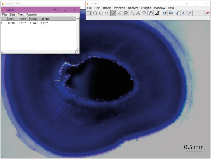
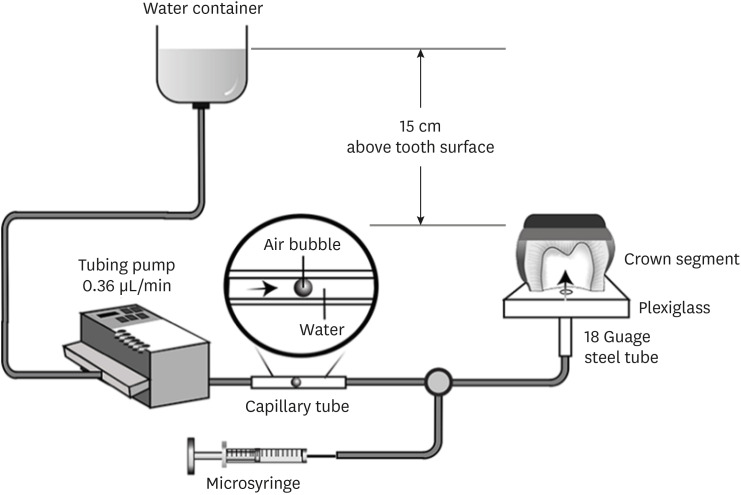
Materials and Methods Human third molars were cut into crown segments. The dentin surfaces were prepared using 4 armamentaria: 600-grit SiC paper, coarse diamond burs, superfine diamond burs, and carbide burs. The pulp chamber of each crown segment was connected to a dynamic intra-pulpal pressure simulation apparatus, and the permeability test was done under a pressure of 15 cmH2O. The relative permeability (%P) was evaluated on the smear layer-covered and bonded dentin surfaces. The teeth were bonded to either of the adhesives under pulpal pressure simulation, and cut into sticks after 24 hours water storage for the µTBS test. The resin-dentin interface and nanoleakage observations were performed using a scanning electron microscope. Statistical comparisons were done using analysis of variance and
post hoc tests.Results Only the method of surface preparation had a significant effect on permeability (
p < 0.05). The smear layers created by the carbide and superfine diamond burs yielded the lowest permeability. CSE demonstrated a higher µTBS, with these values in the superfine diamond and carbide bur groups being the highest. Microscopic evaluation of the resin-dentin interface revealed nanoleakage in the coarse diamond bur and SiC paper groups for both adhesives.Conclusions Superfine diamond and carbide burs can be recommended for dentin preparation with the use of 2-step CSE.
-
Citations
Citations to this article as recorded by- The effect of different adhesive strategies and diamond burs on dentin bond strength of universal resin cements
Chavakorn Atsavathavornset, Pipop Saikaew, Choltacha Harnirattisai, Hidehiko Sano
Clinical Oral Investigations.2025;[Epub] CrossRef - Universal adhesive systems in dentistry: A narrative review
Svetlana N. Razumova, Anzhela S. Brago, Oxana R. Ruda, Zoya A. Guryeva, Elvira V. Adzhieva
Russian Journal of Dentistry.2024; 28(5): 512. CrossRef - Delayed light activation of resin composite affects the bond strength of adhesives under dynamic simulated pulpal pressure
Nattaporn Sukprasert, Choltacha Harnirattisai, Pisol Senawongse, Hidehiko Sano, Pipop Saikaew
Clinical Oral Investigations.2022; 26(11): 6743. CrossRef
- The effect of different adhesive strategies and diamond burs on dentin bond strength of universal resin cements
- 2,869 View
- 42 Download
- 2 Web of Science
- 3 Crossref

-
Effect of QMix irrigant in removal of smear layer in root canal system: a systematic review of
in vitro studies - Margaret Soo Yee Chia, Abhishek Parolia, Benjamin Syek Hur Lim, Jayakumar Jayaraman, Isabel Cristina Celerino de Moraes Porto
- Restor Dent Endod 2020;45(3):e28. Published online May 21, 2020
- DOI: https://doi.org/10.5395/rde.2020.45.e28

-
 Abstract
 PDF
PDF PubReader
 ePub
ePub Objectives To evaluate the outcome of
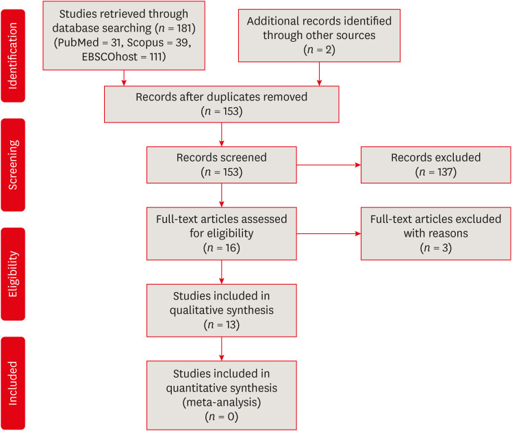
in vitro studies comparing the effectiveness of QMix irrigant in removing the smear layer in the root canal system compared with other irrigants.Materials and Methods The research question was developed by using Population, Intervention, Comparison, Outcome and Study design framework. Literature search was performed using 3 electronic databases PubMed, Scopus, and EBSCOhost until October 2019. Two reviewers were independently involved in the selection of the articles and data extraction process. Risk of bias of the studies was independently appraised using revised Cochrane Risk of Bias tool (RoB 2.0) based on 5 domains.
Results Thirteen studies fulfilled the selection criteria. The overall risk of bias was moderate. QMix was found to have better smear layer removal ability than mixture of tetracycline isonomer, an acid and a detergent (MTAD), sodium hypochlorite (NaOCl), and phytic acid. The efficacy was less effective than 7% maleic acid and 10% citric acid. No conclusive results could be drawn between QMix and 17% ethylenediaminetetraacetic acid due to conflicting results. QMix was more effective when used for 3 minutes than 1 minute.
Conclusions QMix has better smear layer removal ability compared to MTAD, NaOCl, Tubulicid Plus, and Phytic acid. In order to remove the smear layer more effectively with QMix, it is recommended to use it for a longer duration.
-
Citations
Citations to this article as recorded by- Biological and chemical properties of new multi-functional root canal irrigants
Nidambur Vasudev Ballal, Rajkumar Narkedamalli, Padmaja A Shenoy, Shubhankar Das, Saravana Karthikeyan Balasubramanian, Jothi Varghese, Herman Sunil Dsouza, Kevin Epps, Theodroe Ravenel, Franklin R. Tay
Journal of Dentistry.2025; 153: 105551. CrossRef - Chitosan’s Ability to Remove the Smear Layer—A Systematic Review of Ex Vivo Studies
Ana Ferreira-Reguera, Inês Ferreira, Irene Pina-Vaz, Benjamín Martín-Biedma, José Martín-Cruces
Medicina.2025; 61(1): 114. CrossRef - Enhancing Endodontic Outcomes with the Synergistic Microbicidal and Activated Root-Cleansing Technique (SMART): A Novel Approach to Root Canal Irrigation
Max Foroughi, Sara Abolmaali, Hamid Abedi, Theodore Ravenel
Medicina.2025; 61(5): 874. CrossRef - Cleaning efficiency of different root canal irrigating solutions
Mna E. Elshalkamy, Neveen A. Shaheen, Dalia A. Sherif
Tanta Dental Journal.2025; 22(2): 222. CrossRef - Comparison of human pulpal dissolution and removal of smear layer by triton and Qmix 2 in 1 solution: An in vitro study
Rahul Halkai, S. Syed Ishaq, Kiran R Halkai
Saudi Endodontic Journal.2025; 15(3): 245. CrossRef - Mapping risk of bias criteria in systematic reviews of in vitro endodontic studies: an umbrella review
Rafaella Rodrigues da Gama, Lucas Peixoto de Araújo, Evandro Piva, Leandro Perello Duro, Adriana Fernandes da Silva, Wellington Luiz de Oliveira da Rosa
Evidence-Based Dentistry.2025; 26(4): 179. CrossRef - The advancement in irrigation solution within the field of endodontics, A Review
Fatima Fahad , Raghad A Al-Hashimi , Munther J Hussain
Journal of Baghdad College of Dentistry.2024; 36(1): 54. CrossRef - The effect of final irrigation with different solutions on smear layer removal and dentin erosion: A scanning electron microscope study
Mohammed AlBatati, Ammar AbuMostafa, Miriam Fatima Zaccaro Scelza
PLOS ONE.2024; 19(8): e0308606. CrossRef - Effect of QMix as final irrigation protocol on periapical healing after single‐visit root canal treatment: A randomised controlled clinical trial
Cemre Sapmaz Ucan, Aysin Dumani, Ilker Unal, Sehnaz Yilmaz, Oguz Yoldas
Australian Endodontic Journal.2023; 49(S1): 113. CrossRef - Effectiveness of Different Final Irrigation Procedures on Enterococcus faecalis Infected Root Canals: An In Vitro Evaluation
Sanda Ileana Cîmpean, Ioana-Sofia Pop-Ciutrila, Sebastian-Roberto Matei, Ioana Alina Colosi, Carmen Costache, Gheorghe Zsolt Nicula, Iulia Clara Badea, Loredana Colceriu Burtea
Materials.2022; 15(19): 6688. CrossRef - Evaluation of a Novel Tool for Apical Plug Formation during Apexification of Immature Teeth
Yasser Alsayed Tolibah, Line Droubi, Saleh Alkurdi, Mohammad Tamer Abbara, Nada Bshara, Thuraya Lazkani, Chaza Kouchaji, Ibrahim Ali Ahmad, Ziad D. Baghdadi
International Journal of Environmental Research and Public Health.2022; 19(9): 5304. CrossRef - Influence of chelating solutions on tubular dentin sealer penetration: A systematic review with network meta‐analysis
Felipe de Souza Matos, Camila Maria Peres de Rosatto, Thaís Christina Cunha, Maria Tereza Campos Vidigal, Cauane Blumenberg, Luiz Renato Paranhos, Camilla Christian Gomes Moura
Australian Endodontic Journal.2021; 47(3): 715. CrossRef
- Biological and chemical properties of new multi-functional root canal irrigants
- 3,232 View
- 43 Download
- 12 Crossref

- Effect of smear layer deproteinization on bonding of self-etch adhesives to dentin: a systematic review and meta-analysis
- Khaldoan H. Alshaikh, Hamdi H. H. Hamama, Salah H. Mahmoud
- Restor Dent Endod 2018;43(2):e14. Published online March 6, 2018
- DOI: https://doi.org/10.5395/rde.2018.43.e14

-
 Abstract
 PDF
PDF PubReader
 ePub
ePub Objectives The aim of this systematic review was to critically analyze previously published studies of the effects of dentin surface pretreatment with deproteinizing agents on the bonding of self-etch (SE) adhesives to dentin. Additionally, a meta-analysis was conducted to quantify the effects of the above-mentioned surface pretreatment methods on the bonding of SE adhesives to dentin.
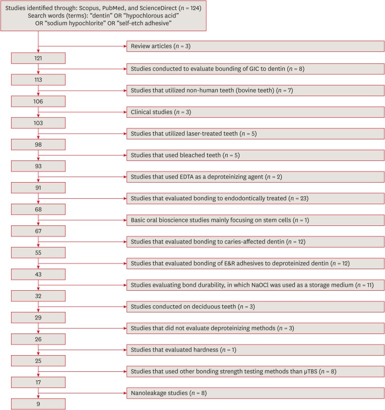
Materials and Methods An electronic search was performed using the following databases: Scopus, PubMed and ScienceDirect. The online search was performed using the following keywords: ‘dentin’ or ‘hypochlorous acid’ or ‘sodium hypochlorite’ and ‘self-etch adhesive.’ The following categories were excluded during the assessment process: non-English articles, randomized clinical trials, case reports, animal studies, and review articles. The reviewed studies were subjected to meta-analysis to quantify the effect of the application time and concentration of sodium hypochlorite (NaOCl) and hypochlorous acid (HOCl) deproteinizing agents on bonding to dentin.
Results Only 9 laboratory studies fit the inclusion criteria of this systematic review. The results of the meta-analysis revealed that the pooled average microtensile bond strength values to dentin pre-treated with deproteinizing agents (15.71 MPa) was significantly lower than those of the non-treated control group (20.94 MPa).
Conclusions In light of the currently available scientific evidence, dentin surface pretreatment with deproteinizing agents does not enhance the bonding of SE adhesives to dentin. The HOCl deproteinizing agent exhibited minimal adverse effects on bonding to dentin in comparison with NaOCl solutions.
-
Citations
Citations to this article as recorded by- Is the Percentage of Collagen in Coronal Dentin Related to Microtensile Strength? An In Vitro Study
Taíssa Cássia de Souza Furtado, Gilberto Antonio Borges, Vinícius Rangel Geraldo-Martins, Bruno Henrique dos Reis Souza Oliveira, Renata Margarida Etchebehere, Sanívia Aparecida de Lima Pereira
Pesquisa Brasileira em Odontopediatria e Clínica Integrada.2026;[Epub] CrossRef -
Evaluating the remnants of Al
2
O
3
particles on different dentine substrate after sandblasting and various cleaning protocols
Faeze Hamze, Khotan Aflatoonian, Mahshid Mohammadibassir, Mohammad-Bagher Rezvani
Journal of Adhesion Science and Technology.2025; 39(6): 869. CrossRef - Preservation Strategies for Interfacial Integrity in Restorative Dentistry: A Non-Comprehensive Literature Review
Carmem S. Pfeifer, Fernanda S. Lucena, Fernanda M. Tsuzuki
Journal of Functional Biomaterials.2025; 16(2): 42. CrossRef - Outcome of Er, Cr:YSGG laser and antioxidant pretreatments on bonding quality to caries-induced dentin
Lamiaa M. Moharam, Haidy N. Salem, Ahmed Abdou, Rasha H. Afifi
BMC Oral Health.2025;[Epub] CrossRef - Advancing Adhesive Strategies for Endodontically Treated Teeth—Part II: Dentin Sealing Before Irrigation Increases Long‐Term Microtensile Bond Strength to Coronal Dentin
Joana A. Marques, Rui I. Falacho, Gabriela Almeida, Francisco Caramelo, João Miguel Santos, João Rocha, Markus B. Blatz, João Carlos Ramos, Paulo J. Palma
Journal of Esthetic and Restorative Dentistry.2025; 37(7): 1865. CrossRef - Effect of finishing protocols on dentin surface characteristics and bond strength after tooth preparation for indirect restorations
Paola Bernardes, Amanda das Graças Soares, Bárbara Inácio de Melo, Leandro Maruki Pereira, Regina Guenka Palma-Dibb, Rafael Rocha Pacheco, Marcel Santana Prudente, Luís Henrique Araújo Raposo
The Journal of Prosthetic Dentistry.2025;[Epub] CrossRef - A comparison of different cleaning approaches for blood contamination after curing universal adhesives on the dentine surface
Ting Liu, Haifeng Xie, Chen Chen
Dental Materials.2024; 40(11): 1786. CrossRef - Effect of fiber-reinforced direct restorative materials on the fracture resistance of endodontically treated mandibular molars restored with a conservative endodontic cavity design
Merve Nezir, Beyza Arslandaş Dinçtürk, Ceyda Sarı, Cemile Kedici Alp, Hanife Altınışık
Clinical Oral Investigations.2024;[Epub] CrossRef - Effect of the use of bromelain associated with bioactive glass-ceramic on dentin/adhesive interface
Rocio Geng Vivanco, Ana Beatriz Silva Sousa, Viviane de de Cássia Oliveira, Mário Alexandre Coelho Sinhoreti, Fernanda de Carvalho Panzeri Pires-de-Souza
Clinical Oral Investigations.2024;[Epub] CrossRef - Experimental and Chitosan-Infused Adhesive with Dentin Pretreated with Femtosecond Laser, Methylene Blue-Activated Low-Level Laser, and Phosphoric Acid
Fahad Alkhudhairy
Photobiomodulation, Photomedicine, and Laser Surgery.2024; 42(10): 634. CrossRef - Evaluation of Effective Bond Strength of Composite Resin to Etched Dentin after Dentin Pretreatment: An In-vitro Study
Muhammed Bilal, Shiraz Pasha, Arathi S. Nair
Journal of the Scientific Society.2024; 51(4): 545. CrossRef - Comparison of Different Dentin Deproteinizing Agents on Bond Strength and Microleakage of Universal Adhesive to Dentin
Fatih Bedir, Gül Yıldız Telatar
Journal of Advanced Oral Research.2023; 14(1): 44. CrossRef - Addition of metal chlorides to a HOCl conditioner can enhance bond strength to smear layer deproteinized dentin
Kittisak Sanon, Antonin Tichy, Takashi Hatayama, Ornnicha Thanatvarakorn, Taweesak Prasansuttiporn, Takahiro Wada, Yasushi Shimada, Keiichi Hosaka, Masatoshi Nakajima
Dental Materials.2022; 38(8): 1235. CrossRef - Internal and Marginal Adaptation of Adhesive Resin Cements Used for Luting Inlay Restorations: An In Vitro Micro-CT Study
Linah M. Ashy, Hanadi Marghalani
Materials.2022; 15(17): 6161. CrossRef - Collagen-depletion strategies in dentin as alternatives to the hybrid layer concept and their effect on bond strength: a systematic review
António H. S. Delgado, Madalena Belmar Da Costa, Mário Cruz Polido, Ana Mano Azul, Salvatore Sauro
Scientific Reports.2022;[Epub] CrossRef - NaOCl Application after Acid Etching and Retention of Cervical Restorations: A 3-Year Randomized Clinical Trial
M Favetti, T Schroeder, AF Montagner, RR Moraes, T Pereira-Cenci, MS Cenci
Operative Dentistry.2022; 47(3): 268. CrossRef - Resin infiltrant protects deproteinized dentin against erosive and abrasive wear
Ana Theresa Queiroz de Albuquerque, Bruna Oliveira Bezerra, Isabelly de Carvalho Leal, Maria Denise Rodrigues de Moraes, Mary Anne S. Melo, Vanara Florêncio Passos
Restorative Dentistry & Endodontics.2022;[Epub] CrossRef - Bis[2-(Methacryloyloxy) Ethyl] Phosphate as a Primer for Enamel and Dentine
R. Alkattan, G. Koller, S. Banerji, S. Deb
Journal of Dental Research.2021; 100(10): 1081. CrossRef - Influence of Dentine Pre-Treatment by Sandblasting with Aluminum Oxide in Adhesive Restorations. An In Vitro Study
Bruna Sinjari, Manlio Santilli, Gianmaria D’Addazio, Imena Rexhepi, Alessia Gigante, Sergio Caputi, Tonino Traini
Materials.2020; 13(13): 3026. CrossRef - A novel prime-&-rinse mode using MDP and MMPs inhibitors improves the dentin bond durability of self-etch adhesive
Jingqiu Xu, Mingxing Li, Wenting Wang, Zhifang Wu, Chaoyang Wang, Xiaoting Jin, Ling Zhang, Wenxiang Jiang, Baiping Fu
Journal of the Mechanical Behavior of Biomedical Materials.2020; 104: 103698. CrossRef - The effects of deproteinization and primer treatment on microtensile bond strength of self-adhesive resin cement to dentin
In-Hye Bae, Sung-Ae Son, Jeong-Kil Park
Korean Journal of Dental Materials.2019; 46(2): 99. CrossRef - Effect of Papain and Bromelain Enzymes on Shear Bond Strength of Composite to Superficial Dentin in Different Adhesive Systems
Farahnaz Sharafeddin, Mina Safari
The Journal of Contemporary Dental Practice.2019; 20(9): 1077. CrossRef
- Is the Percentage of Collagen in Coronal Dentin Related to Microtensile Strength? An In Vitro Study
- 2,492 View
- 23 Download
- 22 Crossref

- Smear layer removal by different chemical solutions used with or without ultrasonic activation after post preparation
- Daniel Poletto, Ana Claudia Poletto, Andressa Cavalaro, Ricardo Machado, Leopoldo Cosme-Silva, Cássia Cilene Dezan Garbelini, Márcio Grama Hoeppner
- Restor Dent Endod 2017;42(4):324-331. Published online November 1, 2017
- DOI: https://doi.org/10.5395/rde.2017.42.4.324
-
 Abstract
 PDF
PDF PubReader
 ePub
ePub Objectives This study evaluated smear layer removal by different chemical solutions used with or without ultrasonic activation after post preparation.
Materials and Methods Forty-five extracted uniradicular human mandibular premolars with single canals were treated endodontically. The cervical and middle thirds of the fillings were then removed, and the specimens were divided into 9 groups: G1, saline solution (NaCl); G2, 2.5% sodium hypochlorite (NaOCl); G3, 2% chlorhexidine (CHX); G4, 11.5% polyacrylic acid (PAA); G5, 17% ethylenediaminetetraacetic acid (EDTA). For the groups 6, 7, 8, and 9, the same solutions used in the groups 2, 3, 4, and 5 were used, respectively, but activated with ultrasonic activation. Afterwards, the roots were analyzed by a score considering the images obtained from a scanning electron microscope.
Results EDTA achieved the best performance compared with the other solutions evaluated regardless of the irrigation method (
p < 0.05).Conclusions Ultrasonic activation did not significantly influence smear layer removal.
-
Citations
Citations to this article as recorded by- O papel do ultrassom no tratamento e retratamento de canais radiculares: Revisão de literatura
Carlos Roberto Souza Hipp, Joaquim Carlos Fest da Silveira, Luiz Felipe Gilson de Oliveira Rangel, Tatiana Federici de Souza Fest da Silveira, Carla Minozzo Mello, Rodrigo Simões de Oliveira
Research, Society and Development.2025; 14(8): e1314849323. CrossRef - Effect of sodium hypochlorite, ethylenediaminetetraacetic acid, and dual-rinse irrigation on dentin adhesion using an etch-and-rinse or self-etch approach
Matej Par, Tobias Steffen, Selinay Dogan, Noah Walser, Tobias T. Tauböck
Scientific Reports.2024;[Epub] CrossRef - Evaluation of Effect of Poloxamer on Smear Layer Removal Using Apical Negative Pressure: An In Vitro Scanning Electron Microscopy Study
Chandra Prabha, Chitharanjan Shetty, Aditya Shetty
Journal of International Oral Health.2024; 16(6): 498. CrossRef - Laboratory Assessment of Antibacterial Efficacy of Five Different Herbal-based Potential Endodontic Irrigants
Anjali A Oak, Kailash Attur, Kamal Bagda, Nitish Mathur, Lubna Mohammad, Nikhat M Attar
Advances in Human Biology.2023; 13(4): 350. CrossRef - Dental Surface Conditioning Techniques to Increase the Micromechanical Retention to Fiberglass Posts: A Literature Review
Paulina Leticia Moreno-Sánchez, Maricela Ramírez-Álvarez, Alfredo del Rosario Ayala-Ham, Erika de Lourdes Silva-Benítez, Miguel Ángel Casillas-Santana, Diana Leyva del Rio, León Francisco Espinosa-Cristóbal, Erik Lizárraga-Verdugo, Mariana Melisa Avendaño
Applied Sciences.2023; 13(14): 8083. CrossRef - Effect of irrigation protocols on smear layer removal, bond strength and nanoleakage of fiber posts using a self-adhesive resin cement
Rodrigo Stadler Alessi, Renata Terumi Jitumori, Bruna Fortes Bittencourt, Giovana Mongruel Gomes, João Carlos Gomes
Restorative Dentistry & Endodontics.2023;[Epub] CrossRef - Effects of using different root canal sealers and protocols for cleaning intraradicular dentin on the bond strength of a composite resin used to reinforce weakened roots
Luiz Pascoal Vansan, Ricardo Machado, Celso Bernardes de Souza, Ricardo Gariba, Antônio Miranda da Cruz, Cinara Muniz, Jardel FranciscoX Jardel Francisco Mazzi-Chaves, Lucas da Fonseca Roberti Garcia
Journal of Oral Research.2022; 11(6): 1. CrossRef - Influence of the use of chelating agents as final irrigant on the push‐out bond strength of epoxy resin‐based root canal sealers: A systematic review
Carla M. Augusto, Miguel A. Cunha Neto, Karem P. Pinto, Ana Flavia A. Barbosa, Emmanuel J. N. L. Silva, Ana Paula P. dos Santos, Luciana M. Sassone
Australian Endodontic Journal.2022; 48(2): 347. CrossRef - Adhesion and whitening efficacy of P11-4 self-assembling peptide and HAP suspension after using NaOCl as a pre-treatment agent
Niloofar Hojabri, Karl-Heinz Kunzelmann
BMC Oral Health.2022;[Epub] CrossRef - Influence of resin cements and root canal disinfection techniques on the adhesive bond strength of fibre reinforced composite post to radicular dentin
Zaid A. Al Jeaidi
Photodiagnosis and Photodynamic Therapy.2021; 33: 102108. CrossRef - The Antibacterial Efficacy and In Vivo Toxicity of Sodium Hypochlorite and Electrolyzed Oxidizing (EO) Water-Based Endodontic Irrigating Solutions
Sung-Chih Hsieh, Nai-Chia Teng, Chia Chun Chu, You-Tai Chu, Chung-He Chen, Liang-Yu Chang, Chieh-Yun Hsu, Ching-Shuan Huang, Grace Ying-Wen Hsiao, Jen-Chang Yang
Materials.2020; 13(2): 260. CrossRef
- O papel do ultrassom no tratamento e retratamento de canais radiculares: Revisão de literatura
- 2,422 View
- 17 Download
- 11 Crossref

- The use of auxiliary devices during irrigation to increase the cleaning ability of a chelating agent
- Marina Carvalho Prado, Fernanda Leal, Renata Antoun Simão, Heloisa Gusman, Maíra do Prado
- Restor Dent Endod 2017;42(2):105-110. Published online February 3, 2017
- DOI: https://doi.org/10.5395/rde.2017.42.2.105
-
 Abstract
 PDF
PDF PubReader
 ePub
ePub Objectives This study investigated the cleaning ability of ultrasonically activated irrigation (UAI) and a novel activation system with reciprocating motion (EC, EasyClean, Easy Equipamentos Odontológicos) when used with a relatively new chelating agent (QMix, Dentsply). In addition, the effect of QMix solution when used for a shorter (1 minute) and a longer application time (3 minutes) was investigated.
Materials and Methods Fifty permanent human teeth were prepared with K3 rotary system and 6% sodium hypochlorite. Samples were randomly assigned to five groups (
n = 10) according to the final irrigation protocol: G1, negative control (distilled water); G2, positive control (QMix 1 minute); G3, QMix 1 minute/UAI; G4, QMix 1 minute/EC; G5, QMix 3 minutes. Subsequently the teeth were prepared and three photomicrographs were obtained in each root third of root walls, by scanning electron microscopy. Two blinded and pre-calibrated examiners evaluated the images using a four-category scoring system. Data were statistically analyzed using Kruskal-Wallis and Dunn tests (p < 0.05).Results There were differences among groups (
p < 0.05). UAI showed better cleaning ability than EC (p < 0.05). There were improvements when QMix was used with auxiliary devices in comparison with conventional irrigation (p < 0.05). Conventional irrigation for 3 minutes presented significantly better results than its use for 1 minute (p < 0.05).Conclusions QMix should be used for 1 minute when it is used with UAI, since this final irrigation protocol showed the best performance and also allowed clinical optimization of this procedure.
-
Citations
Citations to this article as recorded by- Comparative Evaluation of Different Methods of Activation of Chelating Solution for Smear Layer Removal in the Apical Portion of the Root Canal Using a Scanning Electron Microscopy: An In Vitro Study
Mrunal B Alhat, Sudha B Mattigatti, Rushikesh R Mahaparale, Kapil D Wahane, Apoorva Jadhav
Cureus.2024;[Epub] CrossRef - The Impact of Laser-Activated and Conventional Irrigation Techniques on Sealer Penetration into Dentinal Tubules
Dilara Koruk, Fatma Basmacı, Dilan Kırmızı, Umut Aksoy
Photobiomodulation, Photomedicine, and Laser Surgery.2022; 40(8): 565. CrossRef - Utilização dos atuais métodos de agitação de soluções endodônticas no canal radicular
Lívia Rodrigues Schneider, Larissa Giovanella
Revista Científica Multidisciplinar Núcleo do Conhecimento.2022; : 135. CrossRef - Smear layer removal by passive ultrasonic irrigation and 2 new mechanical methods for activation of the chelating solution
Ricardo Machado, Isadora da Silva, Daniel Comparin, Bianca Araujo Marques de Mattos, Luiz Rômulo Alberton, Ulisses Xavier da Silva Neto
Restorative Dentistry & Endodontics.2021;[Epub] CrossRef - Proteomic analysis of human dental pulp in different clinical diagnosis
Poliana Amanda Oliveira Silva, Stella Maris de Freitas Lima, Mirna de Souza Freire, André Melro Murad, Octávio Luiz Franco, Taia Maria Berto Rezende
Clinical Oral Investigations.2021; 25(5): 3285. CrossRef - Effect of QMix irrigant in removal of smear layer in root canal system: a systematic review of in vitro studies
Margaret Soo Yee Chia, Abhishek Parolia, Benjamin Syek Hur Lim, Jayakumar Jayaraman, Isabel Cristina Celerino de Moraes Porto
Restorative Dentistry & Endodontics.2020;[Epub] CrossRef - The effect of 17% EDTA and QMiX ultrasonic activation on smear layer removal and sealer penetration: ex vivo study
Felipe de Souza Matos, Fabrício Rutz da Silva, Luiz Renato Paranhos, Camilla Christian Gomes Moura, Eduardo Bresciani, Marcia Carneiro Valera
Scientific Reports.2020;[Epub] CrossRef - Micro-CT evaluation of different final irrigation protocols on the removal of hard-tissue debris from isthmus-containing mesial root of mandibular molars
Emmanuel João Nogueira Leal Silva, Carla Rodrigues Carvalho, Felipe Gonçalves Belladonna, Marina Carvalho Prado, Ricardo Tadeu Lopes, Gustavo De-Deus, Edson Jorge Lima Moreira
Clinical Oral Investigations.2019; 23(2): 681. CrossRef
- Comparative Evaluation of Different Methods of Activation of Chelating Solution for Smear Layer Removal in the Apical Portion of the Root Canal Using a Scanning Electron Microscopy: An In Vitro Study
- 1,223 View
- 6 Download
- 8 Crossref

- Effects of canal enlargement and irrigation needle depth on the cleaning of the root canal system at 3 mm from the apex
- Ho-Jin Moon, Chan-Ui Hong
- Restor Dent Endod 2012;37(1):24-28. Published online March 2, 2012
- DOI: https://doi.org/10.5395/rde.2012.37.1.24
-
 Abstract
 PDF
PDF PubReader
 ePub
ePub Objectives The aim of this study was to test the hypothesis, that the effectiveness of irrigation in removing smear layer in the apical third of root canal system is dependent on the depth of placement of the irrigation needle into the root canal and the enlargement size of the canal.
Materials and Methods Eighty sound human lower incisors were divided into eight groups according to the enlargement size (#25, #30, #35 and #40) and the needle penetration depth (3 mm from working length, WL-3 mm and 9 mm from working length, WL-9 mm). Each canal was enlarged to working length with Profile.06 Rotary Ni-Ti files and irrigated with 5.25% NaOCl. Then, each canal received a final irrigation with 3 mL of 3% EDTA for 4 min, followed by 5 mL of 5.25% NaOCl at different level (WL-3 mm and WL-9 mm) from working length. Each specimen was prepared for the scanning electron microscope (SEM). Photographs of the 3mm area from the apical constriction of each canal with a magnification of ×250, ×500, ×1,000, ×2,500 were taken for the final evaluation.
Results Removal of smear layer in WL-3 mm group showed a significantly different effect when the canal was enlarged to larger than #30. There was a significant difference in removing apical smear layer between the needle penetration depth of WL-3 mm and WL-9 mm.
Conclusions Removal of smear layer from the apical portion of root canals was effectively accomplished with apical instrumentation to #35/40 06 taper file and 3 mm needle penetration from the working length.
-
Citations
Citations to this article as recorded by- Numerical Evaluation of Flow Pattern for Root Canal Irrigation Including icrobubbles
Joon Hyun Kim, Chan U Lee, Inwhan Lee, Jaeyong Sung
Journal of the Korean Society of Manufacturing Technology Engineers.2023; 32(5): 251. CrossRef
- Numerical Evaluation of Flow Pattern for Root Canal Irrigation Including icrobubbles
- 1,031 View
- 5 Download
- 1 Crossref

- Effect of soft chelating irrigation on the sealing ability of GP/AH Plus root fillings
- Yi-Suk Yu, Tae-Gun Kim, Kwang-Won Lee, Mi-Kyung Yu
- J Korean Acad Conserv Dent 2009;34(6):484-490. Published online November 30, 2009
- DOI: https://doi.org/10.5395/JKACD.2009.34.6.484
-
 Abstract
 PDF
PDF PubReader
 ePub
ePub The purpose of this study was to evaluate the effect of soft chelating irrigant on the sealing ability of root fillings by using a glucose leakage test.
A total of 45 single-rooted teeth were selected for the study. The teeth were decoronated leaving a total length of 13mm. The root canals prepared using K3 NiTi rotary instruments to an apical dimension of size 45(0.06 taper). The specimens were then randomly divided into 3 experimental groups of 13 roots each and 2 control groups of 3 roots each. Specimen in each group were prepared with different irrigation protocols : group 1, 2.5% NaOCl; group 2, 2.5% NaOCl and 17% EDTA; group 3, 2.5% NaOCl and 15% HEBP. The root canals were filled with gutta-percha and AH Plus sealer using lateral condensation. After 7 days in 37℃, 100% humidity, the coronal-to-apical microleakage was evaluated quantitatively using a glucose leakage model. The leaked glucose concentration was measured with spectrophotometry at 1, 4, 7, 14, 21 and 28 days.
There was a tendency of increase in leakage in all experimental groups during experimental period. HEBP-treated dentin showed no significant difference with EDTA-treated dentin during experimental period. From the 21th day onward, HEBP-treated dentin showed significantly lower leakage than smear-covered dentin. HEBP-treated dentin displayed a similar sealing pattern to EDTA-treated dentin and a better sealing ability than smear-covered dentin. Consequently, a soft chelator(HEBP) could be considered as the possible alternative to EDTA.
-
Citations
Citations to this article as recorded by- Effect of moisture on sealing ability of root canal filling with different types of sealer through the glucose penetration model
Jin-Ah Jang, Hee-Lyang Kim, Mi-Ja Her, Kwang-Won Lee, Mi-Kyung Yu
Journal of Korean Academy of Conservative Dentistry.2010; 35(5): 335. CrossRef
- Effect of moisture on sealing ability of root canal filling with different types of sealer through the glucose penetration model
- 996 View
- 3 Download
- 1 Crossref

- Effect of microleakage of a self-etching primer adhesive according to types of cutting instruments
- Yong-Hee Kim, Jae-Gu Park, Young-Gon Cho
- J Korean Acad Conserv Dent 2007;32(4):327-334. Published online July 31, 2007
- DOI: https://doi.org/10.5395/JKACD.2007.32.4.327
-
 Abstract
 PDF
PDF PubReader
 ePub
ePub The purpose of this study was to evaluate the effect of burs on microleakage of Class V resin restorations when a self-etching primer adhesive was used.
Forty Class V cavities were prepared with four different cutting burs on extracted third molars, and divided into one of four equal groups (n = 10); Group 1-plain cut carbide bur (no. 245), Group 2-cross cut carbide bur (no. 557), Group 3-fine diamond bur (TF-21F), Group 4-standard diamond bur (EX-41).
The occlusal and gingival margin of cavities was located in enamel and dentin, respectively. Cavities were treated with Clearfil SE Bond and restored with Clearfil AP-X. Specimens were thermocycled, immersed in a 2% methylene blue solution for 24 hours, and bisected longitudinally. They were observed leakages at enamel and dentinal margins. Data were analyzed using Mann-Whitney and Wilcoxon signed ranked test.
The results of this study were as follows;
1. At enamel margin, microleakage of group 4 was statistically higher than those of group 1, 2 and 3 (p < 0.01).
2. At dentinal margin, microleakage of group 4 was statistically higher than group 3 (p < 0.01), but group 1 and 2 were not statistically different with group 3 and 4.
3. Enamel microleakage was statistically higher than dentinal microleakage in group 1, 2 and 3 (p < 0.05), but statistical difference between the microleakage of enamel and dentinal margin was not in group 4.
In conclusion, the use of coarse diamond bur showed high microleakage at both enamel and dentinal margin when Clearfil SE Bond was used in class V cavity.
-
Citations
Citations to this article as recorded by- Microshear bond strength of a self-etching primer adhesive to enamel according to the type of bur
Jin-Ho Jeong, Young-Gon Cho, Myung-Seon Lee
Journal of Korean Academy of Conservative Dentistry.2011; 36(6): 477. CrossRef - Effect of cutting instruments on the dentin bond strength of a self-etch adhesive
Young-Gon Lee, So-Ra Moon, Young-Gon Cho
Journal of Korean Academy of Conservative Dentistry.2010; 35(1): 13. CrossRef
- Microshear bond strength of a self-etching primer adhesive to enamel according to the type of bur
- 916 View
- 1 Download
- 2 Crossref

- Time-dependent effects of EDTA application on removal of smear layer in the root canal system
- Ja-Kyong Lee, Sang-Hyuk Park, Gi-Woon Choi
- J Korean Acad Conserv Dent 2006;31(3):169-178. Published online May 31, 2006
- DOI: https://doi.org/10.5395/JKACD.2006.31.3.169
-
 Abstract
 PDF
PDF PubReader
 ePub
ePub This study was to verify that the combined application of NaOCl and EDTA was more effective in removal of smear layer than the application of NaOCl alone. Furthermore it was aimed to find out the optimal time for the application of EDTA.
Thirty five single rooted teeth were cleaned and shaped. NaOCl solution was used as an irrigant during instrumentation. After instrumentation, root canals of the control group were irrigated with 5 ml of NaOCl for 2 minutes. 30 sec, 1 min, and 2 min group were irrigated with 5 ml of 17% EDTA for 30 sec, 1 min, and 2 min respectively. Then the roots were examined with scanning electron microscopy for evaluating removal of smear layer and erosion of dentinal tubule.
The results were as follows;
The control group:
The smear layer was not removed at all.
The other groups:
1) Middle⅓: All groups showed almost no smear layer. And the erosion occurred more frequently as increasing irrigation time.
2) Apical⅓: The cleaning effect of 2 min group was better than the others.
The results suggest that 2 min application of 17% EDTA should be adequate to remove smear layer on both apical⅓ and middle⅓.
-
Citations
Citations to this article as recorded by- Enhancing the Antibacterial Effect of Erythrosine-Mediated Photodynamic Therapy with Ethylenediamine Tetraacetic Acid
MinKi Choi, Haeni Kim, Siyoung Lee, Juhyun Lee
THE JOURNAL OF THE KOREAN ACADEMY OF PEDTATRIC DENTISTRY.2024; 51(1): 32. CrossRef - Apical foramen morphology according to the length of merged canal at the apex
Hee-Ho Kim, Jeong-Bum Min, Ho-Keel Hwang
Restorative Dentistry & Endodontics.2013; 38(1): 26. CrossRef - Effect of moisture on sealing ability of root canal filling with different types of sealer through the glucose penetration model
Jin-Ah Jang, Hee-Lyang Kim, Mi-Ja Her, Kwang-Won Lee, Mi-Kyung Yu
Journal of Korean Academy of Conservative Dentistry.2010; 35(5): 335. CrossRef
- Enhancing the Antibacterial Effect of Erythrosine-Mediated Photodynamic Therapy with Ethylenediamine Tetraacetic Acid
- 1,524 View
- 5 Download
- 3 Crossref

- The effect of MTAD on the apical leakage of obturated root canals: an electrochemical study
- Dong-Sung Park
- J Korean Acad Conserv Dent 2006;31(2):119-124. Published online March 31, 2006
- DOI: https://doi.org/10.5395/JKACD.2006.31.2.119
-
 Abstract
 PDF
PDF PubReader
 ePub
ePub The purpose of this study was to evaluate the effect of newly developed endodontic root canal cleanser (MTAD) on the apical leakage of obturated root canal using an electrochemical method.
Canals of 60 extracted single-rooted human teeth were prepared by using a crown-down technique with rotary nickel-titanium files. In Group 1 (positive control group) and 2 (negative control group), 5.25% NaOCl was used as a canal irrigant and no canal wall treatment was done. In group 3, only 5.25% NaOCl were used as canal irrigant, canal wall treatment and final rinse. In group 4, specimens were irrigated with 5.25% NaOCl, treated with 5
ml of 17% EDTA for 5 minutes and final rinsed with 5.25% NaOCl. Specimens of group 5 were irrigated with 1.3% NaOCl and treated with 5 ml of MTAD for 5 minutes. All root canals are dried with paper points and obtuated with gutta-percha and AH plus as a sealer using a continuous wave of condensation technique except in the group 1. The electrical resistance between the standard and experimental electrodes in canals was measured over a period of 10 days. Rising of apical leakage with time was observed for all the groups. Group 4 and 5 showed lower apical leakage than group 3 but differences between the group 3, 4 and 5 were no statistical significance at any measurement time.-
Citations
Citations to this article as recorded by- Effect of soft chelating irrigation on the sealing ability of GP/AH Plus root fillings
Yi-Suk Yu, Tae-Gun Kim, Kwang-Won Lee, Mi-Kyung Yu
Journal of Korean Academy of Conservative Dentistry.2009; 34(6): 484. CrossRef
- Effect of soft chelating irrigation on the sealing ability of GP/AH Plus root fillings
- 1,034 View
- 0 Download
- 1 Crossref

- THE EFFECT OF SMEAR LAYER TREATMENT ON THE MICROLEAKAGE
- Jung-Min Lee, Sang-Hyuk Park, Gi-Woon Choi
- J Korean Acad Conserv Dent 2006;31(5):378-389. Published online January 14, 2006
- DOI: https://doi.org/10.5395/JKACD.2006.31.5.378
-
 Abstract
 PDF
PDF PubReader
 ePub
ePub Abstract The purpose of this study was to compare the sealing ability of root canal obturation with or without the treatment of smear layer. Eighty extracted human teeth with one canal were selected. Instrumentation was performed with crown-down technique. After instrumentation, root canals of the NaOCl group and NaOCl-6 group were irrigated with 3% NaOCl. EDTA group and EDTA-6 group were irrigated with 17% EDTA. Then all teeth were obturated using continuous wave obturation technique.
NaOCl group and EDTA group were immersed in methylene blue solution for 84hours. NaOCl-6 group and EDTA-6 group were immersed in methylene blue solution for 6months. The teeth were sectioned at 1.5 mm (Level 1), 3.0 mm (Level 2) and 4.5 mm (Level 3) from the root apex. The length of dye-penetrated interface and the circumferential length of canal at each level were measured using Sigma-Scan Pro 5.0.
The mean leakage ratio was decreased cervically.
NaOCl group showed higher mean leakage ratio than EDTA group at each level. But there was significant difference at level 1 only (p < 0.05).
NaOCl-6 group showed higher mean leakage ratio than EDTA-6 group at each level. But there was significant difference at level 1 only (p < 0.05).
NaOCl-6 group showed higher mean leakage ratio than NaOCl group at each level. But there was significant difference at level 1 only (p < 0.05).
EDTA-6 group showed higher mean leakage ratio than EDTA group at each level. But there was no significant difference.
In NaOCl group and NaOCl-6 group, scanning electron micrographs of tooth sections generally covered with smear layer. In EDTA group and EDTA-6 group, tooth sections showing the penetration of sealers to opened dentinal tubules. The results suggest that removal of smear layer was effective to reduce the apical microleakage of the root canal.
-
Citations
Citations to this article as recorded by- Effect of soft chelating irrigation on the sealing ability of GP/AH Plus root fillings
Yi-Suk Yu, Tae-Gun Kim, Kwang-Won Lee, Mi-Kyung Yu
Journal of Korean Academy of Conservative Dentistry.2009; 34(6): 484. CrossRef - The effect of MTAD as a final root canal irrigants on the coronal bacterial leakage of obturated root canals
Tae Woo Kim, Seok Woo Chang, Dong Sung Park
Journal of Korean Academy of Conservative Dentistry.2008; 33(4): 397. CrossRef
- Effect of soft chelating irrigation on the sealing ability of GP/AH Plus root fillings
- 959 View
- 5 Download
- 2 Crossref

- A sem observation on the efficiency preparation of oval canals using hand and engine-driven instruments
- Uk Song, Bock Hur, Hee-Joo Lee
- J Korean Acad Conserv Dent 2004;29(2):141-146. Published online March 31, 2004
- DOI: https://doi.org/10.5395/JKACD.2004.29.2.141
-
 Abstract
 PDF
PDF PubReader
 ePub
ePub The purpose of this study was to evaluate the efficiency of the preparation of oval canals using hand and engine-driven instruments with SEM observation. Thirty single-rooted teeth with oval canal were used in this study. The teeth were divided into 3 groups. In group A, the teeth were instrumented up to a size 35 K-file using RC-prep and irrigated with 5% NaOCl between each file size. In group B, the teeth were instrumented with Profile according to the manufacture's instructions using RC-Prep and irrigated with 5% NaOCl between each file size. In group C, the teeth were instrumented with GT file according to the manufacture's instructions using RC-prep and irrigated with 5% NaOCl between each file size. Then, in all teeth, a final flush of 5ml of distilled water delivered for 30s. Canals were dried with sterile standardized paper points. After preparing the canals, the teeth were sectioned along their mesial and diatal surfaces by using low-speed diamond disc, chisel and mallet. Each root section was then dehydrated in graded concentration of alcohol (70, 80, 90, 100%), mounted on an aluminum stub, sputter-coated with gold-palladium and observed with scanning electron microscope (HITACHI S-4200) in middle and apical area.
The results of this study were as follows:
In the middle area, group B and group C showed less smear layer than group A, and it was statistically significant (p < 0.05).
In the middle area, group B showed greater smear layer than group C, but it was not statistically significant (p > 0.05).
In the apical area, group C showed less smear layer than group A, and it was statistically significant (p < 0.05).
In the apical area, group A showed greater smear layer than group B, but it was not statistically significant (p > 0.05).
In the apical area, group B showed greater smear layer than group C, but it was not statistically significant (p > 0.05).
In all groups, the middle area was less smear layer than the apical area, and it was statistically significant (p < 0.05).
- 629 View
- 0 Download

- The effects of EDTA and pulsed Nd:YAG laser on apical leakage of canal obturation
- Jin-Soo Kwon, Hee-Joo Lee, Bock Hur
- J Korean Acad Conserv Dent 2003;28(1):50-56. Published online January 31, 2003
- DOI: https://doi.org/10.5395/JKACD.2003.28.1.050
-
 Abstract
 PDF
PDF PubReader
 ePub
ePub The purpose of this study was to evaluate the effects of EDTA and pulsed Nd:YAG laser on apical leakage of canal obturation. Forty-eight single-rooted teeth were used in this study. The teeth were instrumented up to a size 40 K-file and irrigated with 2.5% NaOCl between each file size. And the teeth were divided into 4 groups. In group A, the root canals were irrigated with a final flush of 5ml 2.5% NaOCl as a control group. The teeth in group B were irrigated with a final flush of 5ml 17% EDTA. The teeth in group C and D were irradiated by pulsed Nd:YAG laser(laser parameters were set at 1W, 100mJ, 10Hz, and 2W, 100mJ, 20Hz respectively).
The results were as follows:
1. Apical leakage was observed in 50% of samples in group A, 30% of samples in group B, 20% of samples in group C, and 10% of samples in group D.
2. The teeth in group B had less leakage than group A, but there was no statistically significant differences(p>0.05).
3. The teeth in group C, D had less leakage than group A, and there was statistically significant differences(p<0.05).
4. The teeth in group C, D had less leakage than group B, but there was no statistically significant differences(p>0.05).
5. There was no significant differences in apical leakage between group C and group D(p>0.05).
- 692 View
- 1 Download

- Scanning electron microscopic study on the efficacy of root canal wall debridement of rotary Ni-Ti instruments with different cutting angle
- In-soo Jeon, Kee-yeon Kum, Seong-ho Park, Tai-cheol Yoon
- J Korean Acad Conserv Dent 2002;27(6):577-586. Published online November 30, 2002
- DOI: https://doi.org/10.5395/JKACD.2002.27.6.577
-
 Abstract
 PDF
PDF PubReader
 ePub
ePub The purpose of this in vitro study was to compare the effects of root canal debridement following rotary Ni-Ti instruments with positive versus negative rake angle. Seventy sound, extracted human anterior teeth & premolars were randomly divided into four groups. The used rotary instruments were Ni-Ti HERO 642(Micro-Mega in France, 20 specimen), Ni-Ti ProFile(Maillefer, Ballaigues, Switzerland, 20 specimen), stainless steel engine reamer(Mani, Matsutani Seisakusho Co.,Japan, 20 specimen) and negative control group(10 specimen) was only extirpated with barbed broach(Mani, Matsutani Seisakusho Co.,Japan)
Group 1 & 2 teeth were prepared to a #40 at the apex followed by 1 mm using crown-down technique. Group 3 teeth were instrumented from a #15 to a #40 in sequential order. After preparation and final irrigation, the roots split longitudinally into a bucco-lingual direction. Root halves were cross-sectioned in apical third portion again. all root specimens were prepared for SEM investigation & photographed. Separate evaluations were undertaken for smear layer on prepared walls with a five score-index for each using reference photograph in root halves. the penetration depth of smear layer into dentinal tubules was also estimated in the other halves. the following results were obtained :
1. Smear layer was observed on all the prepared walls with three experimental groups except negative control group
2. Smear layer characteristics
1) HERO 642 groups showed snowy & dusty appearance & were observed only few some dentinal tubuli open on the prepared walls, and the penetration depth of it into dentinal tubules may be 1-2 µm thick.
2) ProFile groups showed shiny & burnished appearance & complete root canal wall covered by a homogenous smear layer with no open dentinal tubuli and penetration depth of it into dentinal tubules may be 1-2 µm thick.
3) Engine reamer groups showed obviously file's passed tracks on the prepared walls & were observed complete root canal wall covered by a homogenous smear layer with no open dentinal tubuli.
The results revealed that a completely clean root canal could not be achieved regardless of positive & negative rake angle, which is in accordance with the majority of studies on root canal cleanliness.
In conclusion, throughout irrigation with antibacterial solutions or chelating agents is recommended to remove the smear layer on prepared canal walls.
-
Citations
Citations to this article as recorded by- Comparative evaluation of dentin volume removal and centralization of the root canal after shaping with the ProTaper Universal, ProTaper Gold, and One-Curve instruments using micro-CT
Hatice Yalniz, Mehrdad Koohnavard, Aysenur Oncu, Berkan Celikten, Ayse Isil Orhan, Kaan Orhan
Journal of Dental Research, Dental Clinics, Dental Prospects.2021; 15(1): 47. CrossRef - Microorganism penetration in dentinal tubules of instrumented and retreated root canal walls.In vitroSEM study
Saad Al-Nazhan, Alaa Al-Sulaiman, Fellwa Al-Rasheed, Fatimah Alnajjar, Bander Al-Abdulwahab, Abdulhakeem Al-Badah
Restorative Dentistry & Endodontics.2014; 39(4): 258. CrossRef - Shaping characteristics of two different motions nickel titanium file: a preliminary comparative study of surface profile and dentin chip
So-Ra Park, Se-Hee Park, Kyung-Mo Cho, Jin-Woo Kim
Journal of Dental Rehabilitation and Applied Science.2014; 30(2): 121. CrossRef
- Comparative evaluation of dentin volume removal and centralization of the root canal after shaping with the ProTaper Universal, ProTaper Gold, and One-Curve instruments using micro-CT
- 984 View
- 0 Download
- 3 Crossref


 KACD
KACD

 First
First Prev
Prev


