Articles
- Page Path
- HOME > Restor Dent Endod > Volume 43(2); 2018 > Article
- Research Article Effect of smear layer deproteinization on bonding of self-etch adhesives to dentin: a systematic review and meta-analysis
-
Khaldoan H. Alshaikh1, Hamdi H. H. Hamama1,2
 , Salah H. Mahmoud1
 -
Restor Dent Endod 2018;43(2):e14.
DOI: https://doi.org/10.5395/rde.2018.43.e14
Published online: March 6, 2018
1Department of Operative Dentistry, Faculty of Dentistry, Mansoura University, Mansoura, Egypt.
2Operative Dentistry Discipline, Faculty of Dentistry, The University of Hong Kong, Hong Kong S.A.R., China.
- Correspondence to Hamdi H. H. Hamama, BDS, MDS, PhD. Clinical Assistant Professor, Operative Dentistry Discipline, Faculty of Dentistry, The University of Hong Kong, Room 3B–53B, Operative Dentistry Discipline, Prince Philip Dental Hospital, Faculty of Dentistry, The University of Hong Kong, 34 Hospital Road, Sai Ying Pun, Hong Kong. hamdy@connect.hku.hk
Copyright © 2018. The Korean Academy of Conservative Dentistry
This is an Open Access article distributed under the terms of the Creative Commons Attribution Non-Commercial License (https://creativecommons.org/licenses/by-nc/4.0/) which permits unrestricted non-commercial use, distribution, and reproduction in any medium, provided the original work is properly cited.
- 2,520 Views
- 24 Download
- 22 Crossref
Abstract
-
Objectives The aim of this systematic review was to critically analyze previously published studies of the effects of dentin surface pretreatment with deproteinizing agents on the bonding of self-etch (SE) adhesives to dentin. Additionally, a meta-analysis was conducted to quantify the effects of the above-mentioned surface pretreatment methods on the bonding of SE adhesives to dentin.
-
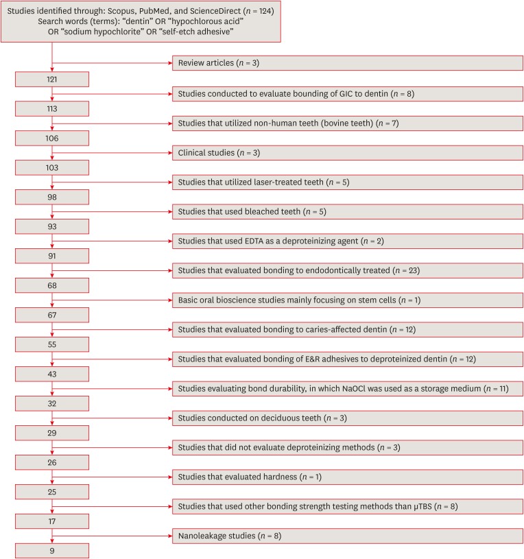
Materials and Methods An electronic search was performed using the following databases: Scopus, PubMed and ScienceDirect. The online search was performed using the following keywords: ‘dentin’ or ‘hypochlorous acid’ or ‘sodium hypochlorite’ and ‘self-etch adhesive.’ The following categories were excluded during the assessment process: non-English articles, randomized clinical trials, case reports, animal studies, and review articles. The reviewed studies were subjected to meta-analysis to quantify the effect of the application time and concentration of sodium hypochlorite (NaOCl) and hypochlorous acid (HOCl) deproteinizing agents on bonding to dentin.
-
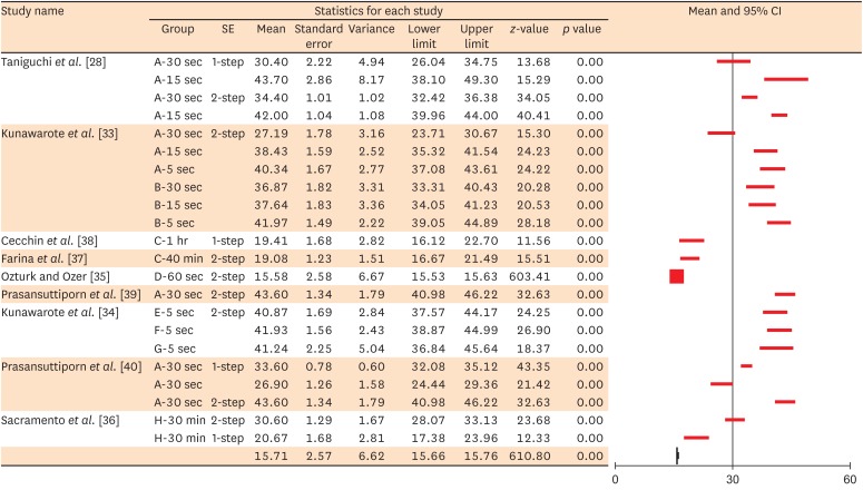
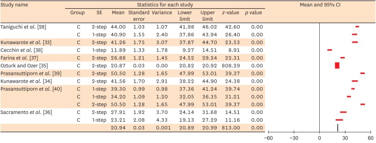
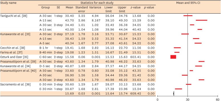
Results Only 9 laboratory studies fit the inclusion criteria of this systematic review. The results of the meta-analysis revealed that the pooled average microtensile bond strength values to dentin pre-treated with deproteinizing agents (15.71 MPa) was significantly lower than those of the non-treated control group (20.94 MPa).
-
Conclusions In light of the currently available scientific evidence, dentin surface pretreatment with deproteinizing agents does not enhance the bonding of SE adhesives to dentin. The HOCl deproteinizing agent exhibited minimal adverse effects on bonding to dentin in comparison with NaOCl solutions.
INTRODUCTION
MATERIALS AND METHODS
RESULTS
Summary of methodologies and results of the included studies
| Study | Sample size (molars) | Method, test machine, and speed | Adhesive system | Number, diameter, and shape of beam | Storage time | NaOCl concentration and time | Result |
|---|---|---|---|---|---|---|---|
| Taniguchi et al. [28] | 40 | - µTBS | 1-S SE and 2-S SE | Three hourglass-shaped specimens with a cross-sectional area of approximately 1 mm2 | 24 hr water storage | a. 6% NaOCl for 30 and 15 sec | Pretreatment of dentin with NaOCl for 30 sec adversely affected the bonding of SE adhesives to dentin |
| - Testing machine: EZ-test, Shimadzu Co., Kyoto, Japan | b. Control group: rinse with water | ||||||
| - Cross-head speed: 1.0 mm/min | |||||||
| Kunawarote et al. [33] | 39 | - µTBS | 2-S SE | Five hourglass-shaped specimens with a cross-sectional area of approximately 1 mm2 | 24 hr water storage | a. 6% NaOCl | The longer the dentin pretreatment time with NaOCl, the lower µTBS values were obtained |
| - Testing machine: EZ-test, Shimadzu Co., Kyoto, Japan | b. 50 ppm HOCl for 30, 15, and 5 sec | ||||||
| - Cross-head speed: 1 mm/min | c. Control group: rinse with water | ||||||
| Cecchin et al. [38] | 30 | - µTBS | 1-S SE | Four hourglass-shaped specimens with a cross-sectional area of approximately 1 mm2 | 24 hr water storage | a. 1% NaOCl applied to the dentin for 1 hr | The deproteinizing did not deteriorate the bonding of SE adhesive (XENO III, DENTSPLY, Tulsa, OK, USA) to dentin |
| - Universal testing machine (Emic DL 2000) at a cross-head speed of 0.5 mm/min | b. Control group: DI water | ||||||
| Farina et al. [37] | 60 | - µTBS | 2-S SE | Four hourglass-shaped specimens with a cross-sectional area of approximately 1 mm2 | 24 hr water storage | a. 1% NaOCl was applied to the dentin surface for 40 min | Dentin surface pretreatment with 1 % NaOCl reduced the bonding of SE to dentin |
| - Universal testing machine (Emic DL 2000) at a cross-head speed of 0.5 mm/min | b. Control group: DI water | ||||||
| Ozturk and Ozer [35] | 40 | - µTBS | 2-S SE | Three rectangular sticks (1.0 ± 0.03 mm2) | 24 hr water storage | a. 5% NaOCl for 1 min | Dentin surface pretreatment with NaOCl reduced the bonding of SE to dentin |
| - Testing apparatus (Bencor-Multi T, Danville Engineering Co., Danville, CA, USA) at a cross-head speed of 1 mm/min | b. Control group: DI water | ||||||
| Prasansuttiporn et al. [39] | 24 | - µTBS | 2-S SE | Four hourglass-shaped specimens with a cross-sectional area of approximately 1 mm2 | 24 hr water storage | a. 6% NaOCl for 30 sec | The NaOCl-treated group exhibited lower bond strength than the control group |
| - Universal testing machine (EZ-test, Shimadzu Crop., Kyoto, Japan) at a cross-head speed of 1 mm/min | b. Control group: DI water | ||||||
| Kunawarote et al. [34] | 40 | - µTBS | 2-S SE | Five hourglass-shaped specimens with a cross-sectional area of approximately 1 mm2 | 24 hr water storage | a. 806 mM NaOCl, | None of the pretreatments demonstrated a negative influence on the bonding of SE adhesives to normal dentin |
| - Testing machine (EZ-test, Shimadzu, Kyoto, Japan) at a cross-head speed of 1 mm/min | b. 0.95 or 1.91 mM HOCl for 5 sec | ||||||
| c. Control group: DI water | |||||||
| Prasansuttiporn et al. [40] | 36 | - µTBS | 1-S SE and 2-S SE | Five hourglass-shaped specimens with a cross-sectional area of approximately 1 mm2 | 24 hr water storage | a. 6% NaOCl for 30 sec | The recorded bond strength values of the deproteinized dentin group were significantly lower than those of the control group |
| - Universal testing machine (EZ-test, Shimadzu Crop., Kyoto, Japan) at a cross-head speed of 1 mm/min | b. Control group: DI water | ||||||
| Sacramento et al. [36] | 90 | - µTBS | 1-S SE and 2-S SE | Fourteen sticks with a surface area of about 1.0 mm2 | 24 hr water storage | a. 0.5% NaOCl for 30 min | The NaOCl-treated group exhibited lower bond strength than the control group |
| - Universal testing machine (Instron model 4411, Canton, MA, USA) at a cross-head speed of 0.5 mm/min. | b. Control group: DI water |

Overall analysis of μTBS and fracture modes reported in the reviewed studies
| Study | SE adhesive system | Deproteinizing agent | Time | Mean µTBS (MPa) | Mode of failure (%) | |||
|---|---|---|---|---|---|---|---|---|
| Cohesive in resin | Cohesive in dentin | Mixed | Adhesive | |||||
| Taniguchi et al. [28] | Bond Force (1-S) | 6% NaOCl | 30 sec | 30.4 | 4 | 4 | 83 | 4 |
| 15 sec | 43.7 | |||||||
| Clearfil SE Protect (2-S) | 6% NaOCl | 30 sec | 34.4 | 0 | 4 | 91.5 | 4 | |
| 15 sec | 42.0 | |||||||
| Kunawarote et al. [33] | Clearfil SE Bond (2-S) | 6% NaOCl | 30 sec | 27.19 | 0 | 0 | 90 | 10 |
| 15 sec | 38.43 | 0 | 20 | 65 | 15 | |||
| 5 sec | 40.34 | 0 | 35 | 58 | 7 | |||
| 50 ppm HOCl | 30 sec | 36.87 | 0 | 17 | 55 | 28 | ||
| 15 sec | 37.64 | 0 | 60 | 23 | 17 | |||
| 5 sec | 41.97 | 38 | 10 | 25 | 27 | |||
| Cecchin et al. [38] | XENO III (1-S) | 1% NaOCl | 1 hr | 19.41 | NA | NA | NA | NA |
| Farina et al. [37] | Clearfil SE Bond (2-S) | 1% NaOCl | 40 min | 19.08 | 0 | 0 | 27 | 73 |
| Ozturk and Ozer [35] | Clearfil SE Bond (2-S) | 5% NaOCl | 60 sec | 15.58 | 13.5 | 6.5 | 80 | |
| Prasansuttiporn et al. [39] | Clearfil Protect Bond (2-S) | 6% NaOCl | 30 sec | 43.6 | 7.5 | 7.5 | 85 | 0 |
| Kunawarote et al. [34] | Clearfil SE Bond (2-S) | 806.02 mM NaOCl | 5 sec | 40.87 | 0 | 40 | 50 | 10 |
| 0.95 mM HOCl | 5 sec | 41.93 | 35 | 15 | 35 | 15 | ||
| 1.91 mM HOCl | 5 sec | 41.24 | 27 | 7 | 38 | 28 | ||
| Prasansuttiporn et al. [40] | Clearfil s3 bond (1-S) | 6% NaOCl | 30 sec | 33.6 | 7 | 14.5 | 78.5 | 0 |
| Bond force (1-S) | 6% NaOCl | 30 sec | 26.9 | 22 | 0 | 64 | 14 | |
| Clearfil protect bond (2-S) | 6% NaOCl | 30 sec | 43.6 | 7.5 | 7.5 | 85 | 0 | |
| Sacramento et al. [36] | Clearfil protect bond (2-S) | 0.5% NaOCl | 30 min | 30.60 | 70 | 0 | 30 | 0 |
| Adper Prompt L-Pop (1-S) | 0.5% NaOCl | 30 min | 20.67 | 25 | 0 | 75 | 0 | |


Results of applying the medical statistical model of Borenstein et al. [70] to the meta-analysis outcomes


Discussion
Conclusions
-
Conflict of Interest: No potential conflict of interest was reported by the authors.
-
Author Contributions:
Conceptualization: Hamama HHH, Mahmoud SH.
Data curation: Alshaikh KH, Hamama HHH, Mahmoud SH.
Funding acquisition: Alshaikh KH, Hamama HHH, Mahmoud SH.
Investigation: Alshaikh KH, Hamama HHH, Mahmoud SH.
Methodology: Alshaikh KH, Hamama HHH, Mahmoud SH.
Project administration: Hamama HHH, Mahmoud SH.
Resources: Alshaikh KH, Hamama HHH, Mahmoud SH.
Supervision: Alshaikh KH, Hamama HHH, Mahmoud SH.
Validation: Hamama HHH, Mahmoud SH.
Visualization: Alshaikh KH.
Writing - original draft: Alshaikh KH.
Writing - review & editing: Hamama HHH, Mahmoud SH.
- 1. Jourdan M, Gagne S, Dubois-Laurent C, Maghraoui M, Huet S, Suel A, Hamama L, Briard M, Peltier D, Geoffriau E. Carotenoid content and root color of cultivated carrot: a candidate-gene association study using an original broad unstructured population. PLoS One 2015;10:e0116674.ArticlePubMedPMC
- 2. Miller SA, Forrest JL. Translating evidence-based decision making into practice: appraising and applying the evidence. J Evid Based Dent Pract 2009;9:164-182.ArticlePubMed
- 3. Fuentes V, Toledano M, Osorio R, Carvalho RM. Microhardness of superficial and deep sound human dentin. J Biomed Mater Res A 2003;66:850-853.ArticlePubMed
- 4. Ritter AV, Eidson RS, Donovan TE. Dental caries: etiology, clinical characteristics, risk assessment, and management. In: Heymann HO, Swift EJ, Ritter AV, editors. Sturdevant's art & science of operative dentistry. 6th ed. St. Louis (MO): Elsevier Mosby; 2013. p. 41-88.
- 5. Swift EJ, Perdigao J, Heymann HO. Bonding to enamel and dentin: a brief history and state of the art, 1995. Quintessence Int 1995;26:95-110.PubMed
- 6. Van Meerbeek B, De Munck J, Yoshida Y, Inoue S, Vargas M, Vijay P, Van Landuyt K, Lambrechts P, Vanherle G. Buonocore memorial lecture. Adhesion to enamel and dentin: current status and future challenges. Oper Dent 2003;28:215-235.PubMed
- 7. Hikita K, Van Meerbeek B, De Munck J, Ikeda T, Van Landuyt K, Maida T, Lambrechts P, Peumans M. Bonding effectiveness of adhesive luting agents to enamel and dentin. Dent Mater 2007;23:71-80.ArticlePubMed
- 8. Bowen RL. Adhesive bonding of various materials to hard tooth tissues. II. Bonding to dentin promoted by a surface-active comonomer. J Dent Res 1965;44:895-902.ArticlePubMedPDF
- 9. Xie J, Powers JM, McGuckin RS. In vitro bond strength of two adhesives to enamel and dentin under normal and contaminated conditions. Dent Mater 1993;9:295-299.ArticlePubMed
- 10. Reis AF, Giannini M, Kavaguchi A, Soares CJ, Line SR. Comparison of microtensile bond strength to enamel and dentin of human, bovine, and porcine teeth. J Adhes Dent 2004;6:117-121.PubMed
- 11. Hamama HH, Yiu CK, Burrow MF. Effect of chemomechanical caries removal on bonding of self-etching adhesives to caries-affected dentin. J Adhes Dent 2014;16:507-516.PubMed
- 12. Pashley DH. Smear layer: overview of structure and function. Proc Finn Dent Soc 1992;88:215-224.PubMed
- 13. Spencer P, Ye Q, Park J, Topp EM, Misra A, Marangos O, Wang Y, Bohaty BS, Singh V, Sene F, Eslick J, Camarda K, Katz JL. Adhesive/dentin interface: the weak link in the composite restoration. Ann Biomed Eng 2010;38:1989-2003.ArticlePubMedPMCPDF
- 14. Ishioka S, Caputo AA. Interaction between the dentinal smear layer and composite bond strength. J Prosthet Dent 1989;61:180-185.ArticlePubMed
- 15. Giachetti L, Bambi C, Scaminaci Russo D. SEM qualitative evaluation of four self-etching adhesive systems. Minerva Stomatol 2005;54:415-428.PubMed
- 16. Waidyasekera K, Nikaido T, Weerasinghe DS, Ichinose S, Tagami J. Reinforcement of dentin in self-etch adhesive technology: a new concept. J Dent 2009;37:604-609.ArticlePubMed
- 17. Ozer F, Blatz MB. Self-etch and etch-and-rinse adhesive systems in clinical dentistry. Compend Contin Educ Dent 2013;34:12-14.
- 18. Perdigão J, Reis A, Loguercio AD. Dentin adhesion and MMPs: a comprehensive review. J Esthet Restor Dent 2013;25:219-241.ArticlePubMedPDF
- 19. Peumans M, Kanumilli P, De Munck J, Van Landuyt K, Lambrechts P, Van Meerbeek B. Clinical effectiveness of contemporary adhesives: a systematic review of current clinical trials. Dent Mater 2005;21:864-881.ArticlePubMed
- 20. van Dijken JW, Sunnegårdh-Grönberg K, Lindberg A. Clinical long-term retention of etch-and-rinse and self-etch adhesive systems in non-carious cervical lesions. A 13 years evaluation. Dent Mater 2007;23:1101-1107.PubMed
- 21. Perdigão J, Geraldeli S, Hodges JS. Total-etch versus self-etch adhesive: effect on postoperative sensitivity. J Am Dent Assoc 2003;134:1621-1629.PubMed
- 22. Tay FR, King NM, Chan KM, Pashley DH. How can nanoleakage occur in self-etching adhesive systems that demineralize and infiltrate simultaneously? J Adhes Dent 2002;4:255-269.PubMed
- 23. Van Landuyt KL, Mine A, De Munck J, Countinho E, Peumans M, Jaecques S, Lambrechts P, Van Meerbeek B. Technique sensitivity of water-free one-step adhesives. Dent Mater 2008;24:1258-1267.ArticlePubMed
- 24. Tay FR, Pashley DH. Aggressiveness of contemporary self-etching systems. I: depth of penetration beyond dentin smear layers. Dent Mater 2001;17:296-308.PubMed
- 25. Yamauti M, Hashimoto M, Sano H, Ohno H, Carvalho RM, Kaga M, Tagami J, Oguchi H, Kubota M. Degradation of resin-dentin bonds using NaOCl storage. Dent Mater 2003;19:399-405.ArticlePubMed
- 26. Nikaido T, Takano Y, Sasafuchi Y, Burrow MF, Tagami J. Bond strengths to endodontically-treated teeth. Am J Dent 1999;12:177-180.PubMed
- 27. Zehnder M, Grawehr M, Hasselgren G, Waltimo T. Tissue-dissolution capacity and dentin-disinfecting potential of calcium hydroxide mixed with irrigating solutions. Oral Surg Oral Med Oral Pathol Oral Radiol Endod 2003;96:608-613.ArticlePubMed
- 28. Taniguchi G, Nakajima M, Hosaka K, Iwamoto N, Ikeda M, Foxton RM, Tagami J. Improving the effect of NaOCl pretreatment on bonding to caries-affected dentin using self-etch adhesives. J Dent 2009;37:769-775.ArticlePubMed
- 29. Montes MA, de Goes MF, Sinhoreti MA. The in vitro morphological effects of some current pre-treatments on dentin surface: a SEM evaluation. Oper Dent 2005;30:201-212.PubMed
- 30. Boyde A, Jones SJ. Backscattered electron imaging of dental tissues. Anat Embryol (Berl) 1983;168:211-226.ArticlePubMedPDF
- 31. Carvalho RM, Chersoni S, Frankenberger R, Pashley DH, Prati C, Tay FR. A challenge to the conventional wisdom that simultaneous etching and resin infiltration always occurs in self-etch adhesives. Biomaterials 2005;26:1035-1042.ArticlePubMed
- 32. Moher D, Liberati A, Tetzlaff J, Altman DG, Altman D, Antes G, Atkins D, Barbour V, Barrowman N, Berlin JA, Clark J, Clarke M, Cook D, D'Amico R, Deeks JJ, Devereaux PJ, Dickersin K, Egger M, Ernst E, Gøtzsche PC, Grimshaw J, Guyatt G, Higgins J, Ioannidis JP, Kleijnen J, Lang T, Liberati A, Magrini N, McNamee D, Moja L, Moher D, Mulrow C, Napoli M, Oxman A, Pham B, Rennie D, Sampson M, Schulz KF, Shekelle PG, Tetzlaff J, Tovey D, Tugwell P. Preferred reporting items for systematic reviews and meta-analyses: the PRISMA statement. Int J Surg 2010;8:336-341.ArticlePubMed
- 33. Kunawarote S, Nakajima M, Shida K, Kitasako Y, Foxton RM, Tagami J. Effect of dentin pretreatment with mild acidic HOCl solution on microtensile bond strength and surface pH. J Dent 2010;38:261-268.ArticlePubMed
- 34. Kunawarote S, Nakajima M, Foxton RM, Tagami J. Effect of pretreatment with mildly acidic hypochlorous acid on adhesion to caries-affected dentin using a self-etch adhesive. Eur J Oral Sci 2011;119:86-92.ArticlePubMed
- 35. Ozturk B, Ozer F. Effect of NaOCl on bond strengths of bonding agents to pulp chamber lateral walls. J Endod 2004;30:362-365.ArticlePubMed
- 36. Sacramento PA, Sampaio CS, de Carvalho FG, Pascon FM, Borges AF, Alves MC, Hosoya Y, Puppin-Rontani RM. Influence of NaOCl irrigation and water-storage on degradation and microstructure of resin-dentin interface. Int J Adhes Adhes 2013;47:117-124.Article
- 37. Farina AP, Cecchin D, Barbizam JV, Carlini-Júnior B. Influence of endodontic irrigants on bond strength of a self-etching adhesive. Aust Endod J 2011;37:26-30.ArticlePubMed
- 38. Cecchin D, Farina AP, Galafassi D, Barbizam JV, Corona SA, Carlini-Júnior B. Influence of sodium hypochlorite and edta on the microtensile bond strength of a self-etching adhesive system. J Appl Oral Sci 2010;18:385-389.ArticlePubMedPMC
- 39. Prasansuttiporn T, Nakajima M, Kunawarote S, Foxton RM, Tagami J. Effect of reducing agents on bond strength to NaOCl-treated dentin. Dent Mater 2011;27:229-234.ArticlePubMed
- 40. Prasansuttiporn T, Nakajima M, Foxton RM, Tagami J. Scrubbing effect of self-etching adhesives on bond strength to NaOCl-treated dentin. J Adhes Dent 2012;14:121-127.PubMed
- 41. Boyde A, Switsur VR, Stewart AD. An assessment of two new physical methods applied to the study of dental tissues. Arch Oral Biol 1962;7(Supplement):185-193.ArticlePubMed
- 42. Eick JD, Wilko RA, Anderson CH, Sorensen SE. Scanning electron microscopy of cut tooth surfaces and identification of debris by use of the electron microprobe. J Dent Res 1970;49:1359-1368.ArticlePDF
- 43. Perdigão J, Lopes M, Geraldeli S, Lopes GC, García-Godoy F. Effect of a sodium hypochlorite gel on dentin bonding. Dent Mater 2000;16:311-323.ArticlePubMed
- 44. Hashimoto M, Ohno H, Kaga M, Sano H, Endo K, Oguchi H. The extent to which resin can infiltrate dentin by acetone-based adhesives. J Dent Res 2002;81:74-78.ArticlePubMedPDF
- 45. Chan KM, Tay FR, King NM, Imazato S, Pashley DH. Bonding of mild self-etching primers/adhesives to dentin with thick smear layers. Am J Dent 2003;16:340-346.PubMed
- 46. Spencer P, Wang Y. Adhesive phase separation at the dentin interface under wet bonding conditions. J Biomed Mater Res 2002;62:447-456.ArticlePubMed
- 47. Wang Y, Spencer P. Hybridization efficiency of the adhesive/dentin interface with wet bonding. J Dent Res 2003;82:141-145.ArticlePubMedPDF
- 48. De Munck J, Vargas M, Iracki J, Van Landuyt K, Poitevin A, Lambrechts P, Van Meerbeek B. One-day bonding effectiveness of new self-etch adhesives to bur-cut enamel and dentin. Oper Dent 2005;30:39-49.PubMed
- 49. Boyde A. Methodology of calcified tissue specimen preparation for scanning electron microscopy. In: Dickson GR, editor. Methods of calcified tissue preparation. Amsterdam: Elsevier; 1984. p. 251-307.
- 50. Fawzy AS, Amer MA, El-Askary FS. Sodium hypochlorite as dentin pretreatment for etch-and-rinse single-bottle and two-step self-etching adhesives: atomic force microscope and tensile bond strength evaluation. J Adhes Dent 2008;10:135-144.PubMed
- 51. Wang L, Bassiri M, Najafi R, Najafi K, Yang J, Khosrovi B, Hwong W, Barati E, Belisle B, Celeri C, Robson MC. Hypochlorous acid as a potential wound care agent: part I. Stabilized hypochlorous acid: a component of the inorganic armamentarium of innate immunity. J Burns Wounds 2007;6:e5.PubMedPMC
- 52. Vargas MA, Cobb DS, Armstrong SR. Resin-dentin shear bond strength and interfacial ultrastructure with and without a hybrid layer. Oper Dent 1997;22:159-166.PubMed
- 53. Prati C, Chersoni S, Pashley DH. Effect of removal of surface collagen fibrils on resin-dentin bonding. Dent Mater 1999;15:323-331.ArticlePubMed
- 54. Saboia VP, Rodrigues AL, Pimenta LA. Effect of collagen removal on shear bond strength of two single-bottle adhesive systems. Oper Dent 2000;25:395-400.PubMed
- 55. Lai SC, Mak YF, Cheung GS, Osorio R, Toledano M, Carvalho RM, Tay FR, Pashley DH. Reversal of compromised bonding to oxidized etched dentin. J Dent Res 2001;80:1919-1924.ArticlePubMedPDF
- 56. Morris MD, Lee KW, Agee KA, Bouillaguet S, Pashley DH. Effects of sodium hypochlorite and RC-prep on bond strengths of resin cement to endodontic surfaces. J Endod 2001;27:753-757.ArticlePubMed
- 57. Hawkins CL, Davies MJ. Hypochlorite-induced oxidation of proteins in plasma: formation of chloramines and nitrogen-centred radicals and their role in protein fragmentation. Biochem J 1999;340:539-548.ArticlePubMedPMC
- 58. Rueggeberg FA, Margeson DH. The effect of oxygen inhibition on an unfilled/filled composite system. J Dent Res 1990;69:1652-1658.ArticlePubMedPDF
- 59. Mountouris G, Silikas N, Eliades G. Effect of sodium hypochlorite treatment on the molecular composition and morphology of human coronal dentin. J Adhes Dent 2004;6:175-182.PubMed
- 60. Vongphan N, Senawongse P, Somsiri W, Harnirattisai C. Effects of sodium ascorbate on microtensile bond strength of total-etching adhesive system to NaOCl treated dentine. J Dent 2005;33:689-695.ArticlePubMed
- 61. Hiraishi N, Kitasako Y, Nikaido T, Nomura S, Burrow MF, Tagami J. Effect of artificial saliva contamination on pH value change and dentin bond strength. Dent Mater 2003;19:429-434.ArticlePubMed
- 62. Haapasalo M, Qian W. Irrigants and intracanal medicaments. In: Ingle JI, Bakland L, Baumgartner J, editors. Ingle's endodontics. 6th ed. Hamilton: BC Decker; 2008. p. 992-1018.
- 63. Inoue S, Murata Y, Sano H, Kashiwada T. Effect of NaOCl treatment on bond strength between indirect resin core-buildup and dentin. Dent Mater J 2002;21:343-354.ArticlePubMed
- 64. Pioch T, Kobaslija S, Schagen B, Götz H. Interfacial micromorphology and tensile bond strength of dentin bonding systems after NaOCl treatment. J Adhes Dent 1999;1:135-142.PubMed
- 65. de Castro AK, Hara AT, Pimenta LA. Influence of collagen removal on shear bond strength of one-bottle adhesive systems in dentin. J Adhes Dent 2000;2:271-277.PubMed
- 66. Mainnemare A, Mégarbane B, Soueidan A, Daniel A, Chapple IL. Hypochlorous acid and taurine-N-monochloramine in periodontal diseases. J Dent Res 2004;83:823-831.ArticlePubMedPDF
- 67. Christensen CE, McNeal SF, Eleazer P. Effect of lowering the pH of sodium hypochlorite on dissolving tissue in vitro . J Endod 2008;34:449-452.ArticlePubMed
- 68. Guentzel JL, Liang Lam K, Callan MA, Emmons SA, Dunham VL. Reduction of bacteria on spinach, lettuce, and surfaces in food service areas using neutral electrolyzed oxidizing water. Food Microbiol 2008;25:36-41.ArticlePubMed
- 69. Mishra P, Palamara JE, Tyas MJ, Burrow MF. Effect of static loading of dentin beams at various pH levels. Calcif Tissue Int 2006;79:416-421.ArticlePubMedPDF
- 70. Borenstein M, Hedges LV, Higgins JP, Rothstein HR. Introduction to meta-analysis. Chichester: John Wiley % Sons; 2009.
REFERENCES
Tables & Figures
REFERENCES
Citations

- Is the Percentage of Collagen in Coronal Dentin Related to Microtensile Strength? An In Vitro Study
Taíssa Cássia de Souza Furtado, Gilberto Antonio Borges, Vinícius Rangel Geraldo-Martins, Bruno Henrique dos Reis Souza Oliveira, Renata Margarida Etchebehere, Sanívia Aparecida de Lima Pereira
Pesquisa Brasileira em Odontopediatria e Clínica Integrada.2026;[Epub] CrossRef -
Evaluating the remnants of Al
2
O
3
particles on different dentine substrate after sandblasting and various cleaning protocols
Faeze Hamze, Khotan Aflatoonian, Mahshid Mohammadibassir, Mohammad-Bagher Rezvani
Journal of Adhesion Science and Technology.2025; 39(6): 869. CrossRef - Preservation Strategies for Interfacial Integrity in Restorative Dentistry: A Non-Comprehensive Literature Review
Carmem S. Pfeifer, Fernanda S. Lucena, Fernanda M. Tsuzuki
Journal of Functional Biomaterials.2025; 16(2): 42. CrossRef - Outcome of Er, Cr:YSGG laser and antioxidant pretreatments on bonding quality to caries-induced dentin
Lamiaa M. Moharam, Haidy N. Salem, Ahmed Abdou, Rasha H. Afifi
BMC Oral Health.2025;[Epub] CrossRef - Advancing Adhesive Strategies for Endodontically Treated Teeth—Part II: Dentin Sealing Before Irrigation Increases Long‐Term Microtensile Bond Strength to Coronal Dentin
Joana A. Marques, Rui I. Falacho, Gabriela Almeida, Francisco Caramelo, João Miguel Santos, João Rocha, Markus B. Blatz, João Carlos Ramos, Paulo J. Palma
Journal of Esthetic and Restorative Dentistry.2025; 37(7): 1865. CrossRef - Effect of finishing protocols on dentin surface characteristics and bond strength after tooth preparation for indirect restorations
Paola Bernardes, Amanda das Graças Soares, Bárbara Inácio de Melo, Leandro Maruki Pereira, Regina Guenka Palma-Dibb, Rafael Rocha Pacheco, Marcel Santana Prudente, Luís Henrique Araújo Raposo
The Journal of Prosthetic Dentistry.2025;[Epub] CrossRef - A comparison of different cleaning approaches for blood contamination after curing universal adhesives on the dentine surface
Ting Liu, Haifeng Xie, Chen Chen
Dental Materials.2024; 40(11): 1786. CrossRef - Effect of fiber-reinforced direct restorative materials on the fracture resistance of endodontically treated mandibular molars restored with a conservative endodontic cavity design
Merve Nezir, Beyza Arslandaş Dinçtürk, Ceyda Sarı, Cemile Kedici Alp, Hanife Altınışık
Clinical Oral Investigations.2024;[Epub] CrossRef - Effect of the use of bromelain associated with bioactive glass-ceramic on dentin/adhesive interface
Rocio Geng Vivanco, Ana Beatriz Silva Sousa, Viviane de de Cássia Oliveira, Mário Alexandre Coelho Sinhoreti, Fernanda de Carvalho Panzeri Pires-de-Souza
Clinical Oral Investigations.2024;[Epub] CrossRef - Experimental and Chitosan-Infused Adhesive with Dentin Pretreated with Femtosecond Laser, Methylene Blue-Activated Low-Level Laser, and Phosphoric Acid
Fahad Alkhudhairy
Photobiomodulation, Photomedicine, and Laser Surgery.2024; 42(10): 634. CrossRef - Evaluation of Effective Bond Strength of Composite Resin to Etched Dentin after Dentin Pretreatment: An In-vitro Study
Muhammed Bilal, Shiraz Pasha, Arathi S. Nair
Journal of the Scientific Society.2024; 51(4): 545. CrossRef - Comparison of Different Dentin Deproteinizing Agents on Bond Strength and Microleakage of Universal Adhesive to Dentin
Fatih Bedir, Gül Yıldız Telatar
Journal of Advanced Oral Research.2023; 14(1): 44. CrossRef - Addition of metal chlorides to a HOCl conditioner can enhance bond strength to smear layer deproteinized dentin
Kittisak Sanon, Antonin Tichy, Takashi Hatayama, Ornnicha Thanatvarakorn, Taweesak Prasansuttiporn, Takahiro Wada, Yasushi Shimada, Keiichi Hosaka, Masatoshi Nakajima
Dental Materials.2022; 38(8): 1235. CrossRef - Internal and Marginal Adaptation of Adhesive Resin Cements Used for Luting Inlay Restorations: An In Vitro Micro-CT Study
Linah M. Ashy, Hanadi Marghalani
Materials.2022; 15(17): 6161. CrossRef - Collagen-depletion strategies in dentin as alternatives to the hybrid layer concept and their effect on bond strength: a systematic review
António H. S. Delgado, Madalena Belmar Da Costa, Mário Cruz Polido, Ana Mano Azul, Salvatore Sauro
Scientific Reports.2022;[Epub] CrossRef - NaOCl Application after Acid Etching and Retention of Cervical Restorations: A 3-Year Randomized Clinical Trial
M Favetti, T Schroeder, AF Montagner, RR Moraes, T Pereira-Cenci, MS Cenci
Operative Dentistry.2022; 47(3): 268. CrossRef - Resin infiltrant protects deproteinized dentin against erosive and abrasive wear
Ana Theresa Queiroz de Albuquerque, Bruna Oliveira Bezerra, Isabelly de Carvalho Leal, Maria Denise Rodrigues de Moraes, Mary Anne S. Melo, Vanara Florêncio Passos
Restorative Dentistry & Endodontics.2022;[Epub] CrossRef - Bis[2-(Methacryloyloxy) Ethyl] Phosphate as a Primer for Enamel and Dentine
R. Alkattan, G. Koller, S. Banerji, S. Deb
Journal of Dental Research.2021; 100(10): 1081. CrossRef - Influence of Dentine Pre-Treatment by Sandblasting with Aluminum Oxide in Adhesive Restorations. An In Vitro Study
Bruna Sinjari, Manlio Santilli, Gianmaria D’Addazio, Imena Rexhepi, Alessia Gigante, Sergio Caputi, Tonino Traini
Materials.2020; 13(13): 3026. CrossRef - A novel prime-&-rinse mode using MDP and MMPs inhibitors improves the dentin bond durability of self-etch adhesive
Jingqiu Xu, Mingxing Li, Wenting Wang, Zhifang Wu, Chaoyang Wang, Xiaoting Jin, Ling Zhang, Wenxiang Jiang, Baiping Fu
Journal of the Mechanical Behavior of Biomedical Materials.2020; 104: 103698. CrossRef - The effects of deproteinization and primer treatment on microtensile bond strength of self-adhesive resin cement to dentin
In-Hye Bae, Sung-Ae Son, Jeong-Kil Park
Korean Journal of Dental Materials.2019; 46(2): 99. CrossRef - Effect of Papain and Bromelain Enzymes on Shear Bond Strength of Composite to Superficial Dentin in Different Adhesive Systems
Farahnaz Sharafeddin, Mina Safari
The Journal of Contemporary Dental Practice.2019; 20(9): 1077. CrossRef
- Figure
- Related articles
-
- Evaluation of platelet concentrates in regenerative endodontics: a systematic review and meta-analysis
- Effect of surface treatment on glass ionomers in sandwich restorations: a systematic review and meta-analysis of laboratory studies
- Success rates comparison of endodontic microsurgery and single implants with comprehensive and explicit criteria: a systematic review and meta-analysis


Figure 1
Figure 2
Figure 3
Figure 4
Figure 5
Figure 6
Figure 7
Figure 8
Figure 9
Summary of methodologies and results of the included studies
| Study | Sample size (molars) | Method, test machine, and speed | Adhesive system | Number, diameter, and shape of beam | Storage time | NaOCl concentration and time | Result |
|---|---|---|---|---|---|---|---|
| Taniguchi et al. [ | 40 | - µTBS | 1-S SE and 2-S SE | Three hourglass-shaped specimens with a cross-sectional area of approximately 1 mm2 | 24 hr water storage | a. 6% NaOCl for 30 and 15 sec | Pretreatment of dentin with NaOCl for 30 sec adversely affected the bonding of SE adhesives to dentin |
| - Testing machine: EZ-test, Shimadzu Co., Kyoto, Japan | b. Control group: rinse with water | ||||||
| - Cross-head speed: 1.0 mm/min | |||||||
| Kunawarote et al. [ | 39 | - µTBS | 2-S SE | Five hourglass-shaped specimens with a cross-sectional area of approximately 1 mm2 | 24 hr water storage | a. 6% NaOCl | The longer the dentin pretreatment time with NaOCl, the lower µTBS values were obtained |
| - Testing machine: EZ-test, Shimadzu Co., Kyoto, Japan | b. 50 ppm HOCl for 30, 15, and 5 sec | ||||||
| - Cross-head speed: 1 mm/min | c. Control group: rinse with water | ||||||
| Cecchin et al. [ | 30 | - µTBS | 1-S SE | Four hourglass-shaped specimens with a cross-sectional area of approximately 1 mm2 | 24 hr water storage | a. 1% NaOCl applied to the dentin for 1 hr | The deproteinizing did not deteriorate the bonding of SE adhesive (XENO III, DENTSPLY, Tulsa, OK, USA) to dentin |
| - Universal testing machine (Emic DL 2000) at a cross-head speed of 0.5 mm/min | b. Control group: DI water | ||||||
| Farina et al. [ | 60 | - µTBS | 2-S SE | Four hourglass-shaped specimens with a cross-sectional area of approximately 1 mm2 | 24 hr water storage | a. 1% NaOCl was applied to the dentin surface for 40 min | Dentin surface pretreatment with 1 % NaOCl reduced the bonding of SE to dentin |
| - Universal testing machine (Emic DL 2000) at a cross-head speed of 0.5 mm/min | b. Control group: DI water | ||||||
| Ozturk and Ozer [ | 40 | - µTBS | 2-S SE | Three rectangular sticks (1.0 ± 0.03 mm2) | 24 hr water storage | a. 5% NaOCl for 1 min | Dentin surface pretreatment with NaOCl reduced the bonding of SE to dentin |
| - Testing apparatus (Bencor-Multi T, Danville Engineering Co., Danville, CA, USA) at a cross-head speed of 1 mm/min | b. Control group: DI water | ||||||
| Prasansuttiporn et al. [ | 24 | - µTBS | 2-S SE | Four hourglass-shaped specimens with a cross-sectional area of approximately 1 mm2 | 24 hr water storage | a. 6% NaOCl for 30 sec | The NaOCl-treated group exhibited lower bond strength than the control group |
| - Universal testing machine (EZ-test, Shimadzu Crop., Kyoto, Japan) at a cross-head speed of 1 mm/min | b. Control group: DI water | ||||||
| Kunawarote et al. [ | 40 | - µTBS | 2-S SE | Five hourglass-shaped specimens with a cross-sectional area of approximately 1 mm2 | 24 hr water storage | a. 806 mM NaOCl, | None of the pretreatments demonstrated a negative influence on the bonding of SE adhesives to normal dentin |
| - Testing machine (EZ-test, Shimadzu, Kyoto, Japan) at a cross-head speed of 1 mm/min | b. 0.95 or 1.91 mM HOCl for 5 sec | ||||||
| c. Control group: DI water | |||||||
| Prasansuttiporn et al. [ | 36 | - µTBS | 1-S SE and 2-S SE | Five hourglass-shaped specimens with a cross-sectional area of approximately 1 mm2 | 24 hr water storage | a. 6% NaOCl for 30 sec | The recorded bond strength values of the deproteinized dentin group were significantly lower than those of the control group |
| - Universal testing machine (EZ-test, Shimadzu Crop., Kyoto, Japan) at a cross-head speed of 1 mm/min | b. Control group: DI water | ||||||
| Sacramento et al. [ | 90 | - µTBS | 1-S SE and 2-S SE | Fourteen sticks with a surface area of about 1.0 mm2 | 24 hr water storage | a. 0.5% NaOCl for 30 min | The NaOCl-treated group exhibited lower bond strength than the control group |
| - Universal testing machine (Instron model 4411, Canton, MA, USA) at a cross-head speed of 0.5 mm/min. | b. Control group: DI water |
NaOCl, sodium hypochlorite; μTBS, microtensile bond strength; 1-S, one-step; 2-S, two-step; SE, self-etch; HOCl, hypochlorous acid; DI, distilled water.
Overall analysis of μTBS and fracture modes reported in the reviewed studies
| Study | SE adhesive system | Deproteinizing agent | Time | Mean µTBS (MPa) | Mode of failure (%) | |||
|---|---|---|---|---|---|---|---|---|
| Cohesive in resin | Cohesive in dentin | Mixed | Adhesive | |||||
| Taniguchi et al. [ | Bond Force (1-S) | 6% NaOCl | 30 sec | 30.4 | 4 | 4 | 83 | 4 |
| 15 sec | 43.7 | |||||||
| Clearfil SE Protect (2-S) | 6% NaOCl | 30 sec | 34.4 | 0 | 4 | 91.5 | 4 | |
| 15 sec | 42.0 | |||||||
| Kunawarote et al. [ | Clearfil SE Bond (2-S) | 6% NaOCl | 30 sec | 27.19 | 0 | 0 | 90 | 10 |
| 15 sec | 38.43 | 0 | 20 | 65 | 15 | |||
| 5 sec | 40.34 | 0 | 35 | 58 | 7 | |||
| 50 ppm HOCl | 30 sec | 36.87 | 0 | 17 | 55 | 28 | ||
| 15 sec | 37.64 | 0 | 60 | 23 | 17 | |||
| 5 sec | 41.97 | 38 | 10 | 25 | 27 | |||
| Cecchin et al. [ | XENO III (1-S) | 1% NaOCl | 1 hr | 19.41 | NA | NA | NA | NA |
| Farina et al. [ | Clearfil SE Bond (2-S) | 1% NaOCl | 40 min | 19.08 | 0 | 0 | 27 | 73 |
| Ozturk and Ozer [ | Clearfil SE Bond (2-S) | 5% NaOCl | 60 sec | 15.58 | 13.5 | 6.5 | 80 | |
| Prasansuttiporn et al. [ | Clearfil Protect Bond (2-S) | 6% NaOCl | 30 sec | 43.6 | 7.5 | 7.5 | 85 | 0 |
| Kunawarote et al. [ | Clearfil SE Bond (2-S) | 806.02 mM NaOCl | 5 sec | 40.87 | 0 | 40 | 50 | 10 |
| 0.95 mM HOCl | 5 sec | 41.93 | 35 | 15 | 35 | 15 | ||
| 1.91 mM HOCl | 5 sec | 41.24 | 27 | 7 | 38 | 28 | ||
| Prasansuttiporn et al. [ | Clearfil s3 bond (1-S) | 6% NaOCl | 30 sec | 33.6 | 7 | 14.5 | 78.5 | 0 |
| Bond force (1-S) | 6% NaOCl | 30 sec | 26.9 | 22 | 0 | 64 | 14 | |
| Clearfil protect bond (2-S) | 6% NaOCl | 30 sec | 43.6 | 7.5 | 7.5 | 85 | 0 | |
| Sacramento et al. [ | Clearfil protect bond (2-S) | 0.5% NaOCl | 30 min | 30.60 | 70 | 0 | 30 | 0 |
| Adper Prompt L-Pop (1-S) | 0.5% NaOCl | 30 min | 20.67 | 25 | 0 | 75 | 0 | |
μTBS, microtensile bond strength; SE, self-etch; 1-S, one-step; 2-S, two-step; NaOCl, sodium hypochlorite; HOCl, hypochlorous acid; NA, not available.
Results of applying the medical statistical model of Borenstein et al. [70] to the meta-analysis outcomes
| Factor | No. of study | µTBS (MPa) | |
|---|---|---|---|
| Deproteinizing agent | NaOCl | 8 | 16.21 ± 0.02b |
| HOCl | 2 | 40.17 ± 0.76a | |
| Application time (sec) | 5 | 2 | 41.33 ± 0.70a |
| 15 | 2 | 40.56 ± 0.70a | |
| 30 | 4 | 34.75 ± 0.40b | |
| SE adhesive | 1-S | 4 | 38.98 ± 0.49a |
| 2-S | 2 | 32.21 ± 0.62b | |
Data are shown as means ± standard deviations. Groups identified by different superscript letters within the rows for each factor were significantly different at p < 0.05.
μTBS, microtensile bond strength; NaOCl, sodium hypochlorite; HOCl, hypochlorous acid; SE, self-etch; 1-S, one-step; 2-S, two-step.
NaOCl, sodium hypochlorite; μTBS, microtensile bond strength; 1-S, one-step; 2-S, two-step; SE, self-etch; HOCl, hypochlorous acid; DI, distilled water.
μTBS, microtensile bond strength; SE, self-etch; 1-S, one-step; 2-S, two-step; NaOCl, sodium hypochlorite; HOCl, hypochlorous acid; NA, not available.
Data are shown as means ± standard deviations. Groups identified by different superscript letters within the rows for each factor were significantly different at
μTBS, microtensile bond strength; NaOCl, sodium hypochlorite; HOCl, hypochlorous acid; SE, self-etch; 1-S, one-step; 2-S, two-step.

 KACD
KACD
 ePub Link
 Cite
Cite

