Search
- Page Path
- HOME > Search
- Effect of moisture and pH on setting time and microhardness of three premixed calcium silicate-based root canal sealers: an in vitro experimental study
- Sooyoun Kim
- Restor Dent Endod 2025;50(4):e41. Published online November 28, 2025
- DOI: https://doi.org/10.5395/rde.2025.50.e41
-
 Abstract
 PDF
PDF PubReader
 ePub
ePub - Objectives
The study aimed to investigate how environmental conditions impact the setting time and microhardness of premixed calcium silicate-based sealers.
Methods
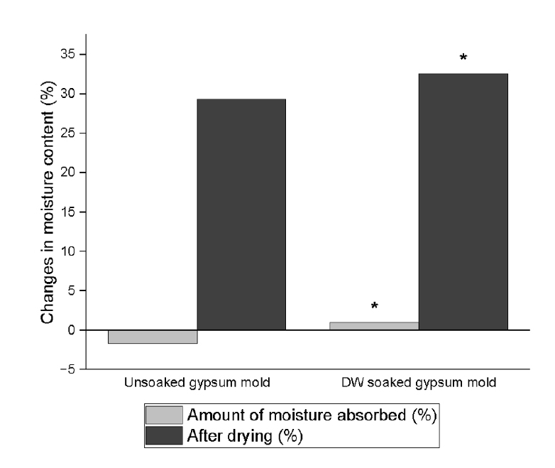
The setting time and microhardness of three sealers (Endoseal MTA [MARUCHI], One-Fil [MEDICLUS], and Well-Root ST [VERICOM]) were evaluated under four environmental conditions: unsoaked, distilled water-soaked, phosphate-buffered saline-soaked, and pH 5-soaked gypsum molds (n = 12/group/condition). The setting time was measured with Gilmore needles, and microhardness was assessed using a Vickers tester after 3 days. Welch’s analysis of variance and Games-Howell post hoc tests were used for statistical analysis.
Results
The sealer type and environmental conditions significantly influenced setting time and microhardness (p < 0.001). The initial and final setting times were the shortest in the unsoaked samples. For Endoseal MTA and One-Fil, the unsoaked condition exhibited significantly shorter setting times than the soaked conditions. Well-Root ST exhibited significantly longer setting times in acidic conditions. Surface microhardness was highest in the unsoaked group (p < 0.001). Among the soaked groups, the phosphate-buffered saline-soaked group had the lowest hardness for Endoseal MTA, whereas the pH 5-soaked group exhibited the lowest hardness for One-Fil and Well-Root ST. Endoseal MTA consistently demonstrated a lower microhardness than the other sealers (p < 0.001).
Conclusions
Moisture, pH, and solution chemistry influenced the setting time and microhardness of premixed calcium silicate sealers. Although acidic conditions generally prolong the setting time and reduce hardness, the effects vary based on the sealers used and the setting environment.
- 667 View
- 48 Download

- Difference in light transmittance and depth of cure of flowable composite depending on tooth thickness: an in vitro experimental study
- Seong-Pyo Bae, Myung-Jin Lee, Kyung-San Min, Mi-Kyung Yu, Kwang-Won Lee
- Restor Dent Endod 2025;50(4):e39. Published online November 28, 2025
- DOI: https://doi.org/10.5395/rde.2025.50.e39
-
 Abstract
 PDF
PDF PubReader
 ePub
ePub - Objectives
This study aimed to quantify light attenuation through varying tooth thicknesses and its impact on the depth of cure of composite resin.
Methods
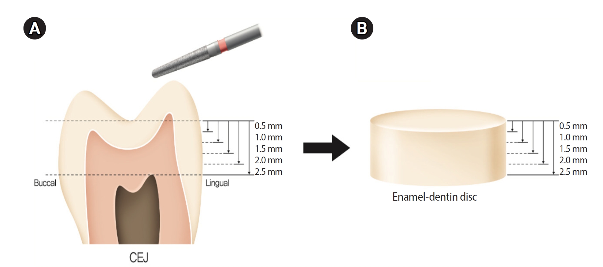
Twenty extracted premolars were used to create enamel-dentin discs that were sanded progressively in 0.5 mm increments from 2.5 mm to 0.5 mm. Light irradiance was measured with and without tooth specimens to evaluate light transmittance. Resin was cured beneath different thicknesses, and the depth of cure was assessed using the Vickers hardness test.
Results
The results demonstrated that light transmittance significantly decreased as tooth thickness increased (p < 0.01), leading to reduced resin polymerization. In the 2.0-mm and 2.5-mm tooth thickness groups, the depth of cure was significantly lower than in the control group without tooth specimens (p < 0.05).
Conclusions
Ultimately, for tooth structures exceeding 2 mm, self-cure or dual-cure resin polymerization is thought to be more efficient than light polymerization.
- 695 View
- 68 Download

- Effect of different storage media on elemental analysis and microhardness of cervical cavity margins restored with a bioactive material
- Hoda Saleh Ismail, Brian Ray Morrow, Ashraf Ibrahim Ali, Rabab Elsayed Elaraby Mehesen, Salah Hasab Mahmoud, Franklin Garcia-Godoy
- Restor Dent Endod 2024;49(1):e6. Published online January 17, 2024
- DOI: https://doi.org/10.5395/rde.2024.49.e6
-
 Abstract
 PDF
PDF PubReader
 ePub
ePub Objectives This study aimed to investigate the elemental analysis and microhardness of a bioactive material (Activa) and marginal tooth structure after storage in different media.
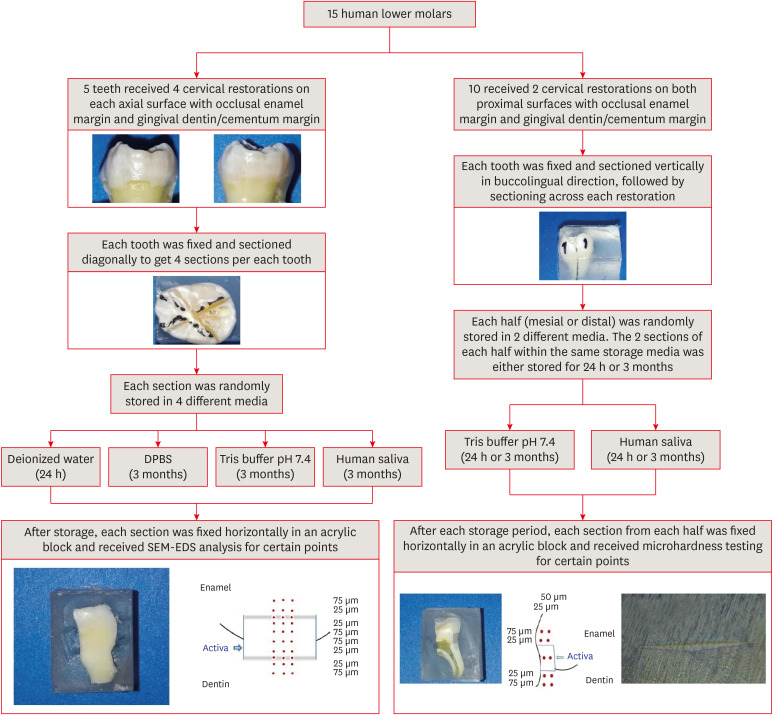
Materials and Methods Fifteen teeth received cervical restorations with occlusal enamel and gingival dentin margins using the tested material bonded with a universal adhesive, 5 of them on the 4 axial surfaces and the other 10 on only the 2 proximal surfaces. The first 5 teeth were sectioned into 4 restorations each, then stored in 4 different media; deionized water, Dulbecco's phosphate buffered saline (DPBS), Tris buffer, and saliva. The storage period for deionized water was 24 hours while it was 3 months for the other media. Each part was analyzed by scanning electron microscopy-energy dispersive spectroscopy (SEM-EDS) analysis for different substrates/distances and the wt% of calcium, phosphorus, silica, and fluoride were calculated. The other 10 teeth were sectioned across the restoration, stored in either Tris buffer or saliva for 24 hours or 3 months, and were evaluated for microhardness of different substrates/areas. Data were analyzed using analysis of variance and Tukey’s
post hoc test.Results Enamel and dentin interfaces in the DPBS group exhibited a significant increase in calcium and phosphorus wt%. Both silica and fluoride significantly increased in tooth structure up to a distance of 75 μm in the 3-month-media groups than the immediate group. Storage media did not affect the microhardness values.
Conclusions SEM-EDS analysis suggests an ion movement between Activa and tooth structure through a universal adhesive while stored in DPBS.
-
Citations
Citations to this article as recorded by- Elemental and micromorphological analysis of ion releasing restoration/carious dentin interface
Alaa Esmat Abdelsalam, Hoda Saleh Ismail, Hamdi Hosni Hamama
Scientific Reports.2025;[Epub] CrossRef - Influence of curing mode and aging on the bonding performance of universal adhesives in coronal and root dentin
Hoda Saleh Ismail, Ashraf Ibrahim Ali, Mohamed Elshirbeny Elawsya
BMC Oral Health.2024;[Epub] CrossRef
- Elemental and micromorphological analysis of ion releasing restoration/carious dentin interface
- 1,948 View
- 99 Download
- 2 Web of Science
- 2 Crossref

- Comparison between a bulk-fill resin-based composite and three luting materials on the cementation of fiberglass-reinforced posts
- Carlos Alberto Kenji Shimokawa, Paula Mendes Acatauassú Carneiro, Tamile Rocha da Silva Lobo, Roberto Ruggiero Braga, Míriam Lacalle Turbino, Adriana Bona Matos
- Restor Dent Endod 2023;48(3):e30. Published online August 8, 2023
- DOI: https://doi.org/10.5395/rde.2023.48.e30
-
 Abstract
 PDF
PDF PubReader
 ePub
ePub Objectives This study verified the possibility of cementing fiberglass-reinforced posts using a flowable bulk-fill composite (BF), comparing its push-out bond strength and microhardness with these properties of 3 luting materials.
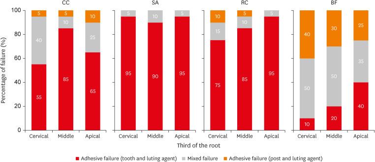
Materials and Methods Sixty endodontically treated bovine roots were used. Posts were cemented using conventional dual-cured cement (CC); self-adhesive cement (SA); dual-cured composite (RC); and BF. Push-out bond strength (
n = 10) and microhardness (n = 5) tests were performed after 1 week and 4 months of storage. Two-way repeated measures analysis of variance (ANOVA), 1-way ANOVA,t -test, and Tukeypost-hoc tests were applied for the push-out bond strength and microhardness results; and Pearson correlation test was applied to verify the correlation between push-out bond strength and microhardness results (α = 0.05).Results BF presented higher push-out bond strength than CC and SA in the cervical third before aging (
p < 0.01). No differences were found between push-out bond strength before and after aging for all the luting materials (p = 0.84). Regarding hardness, only SA presented higher values measured before than after aging (p < 0.01). RC and BF did not present 80% of the maximum hardness at the apical regions. A strong positive correlation was found between the luting materials' push-out bond strength and microhardness (p < 0.01, R2 = 0.7912).Conclusions The BF presented comparable or higher push-out bond strength and microhardness than the luting materials, which indicates that it could be used for cementing resin posts in situations where adequate light curing is possible.
-
Citations
Citations to this article as recorded by- Effects of a relined fiberglass post with conventional and self-adhesive resin cement
Wilton Lima dos Santos Junior, Marina Rodrigues Santi, Rodrigo Barros Esteves Lins, Luís Roberto Marcondes Martins
Restorative Dentistry & Endodontics.2024;[Epub] CrossRef
- Effects of a relined fiberglass post with conventional and self-adhesive resin cement
- 1,938 View
- 39 Download
- 1 Web of Science
- 1 Crossref

- Relationship between battery level and irradiance of light-curing units and their effects on the hardness of a bulk-fill composite resin
- Fernanda Harumi Oku Prochnow, Patricia Valéria Manozzo Kunz, Gisele Maria Correr, Marina da Rosa Kaizer, Carla Castiglia Gonzaga
- Restor Dent Endod 2022;47(4):e45. Published online November 3, 2022
- DOI: https://doi.org/10.5395/rde.2022.47.e45
-
 Abstract
 PDF
PDF PubReader
 ePub
ePub Objectives This study evaluated the relationship between the battery charge level and irradiance of light-emitting diode (LED) light-curing units (LCUs) and how these variables influence the Vickers hardness number (VHN) of a bulk-fill resin.
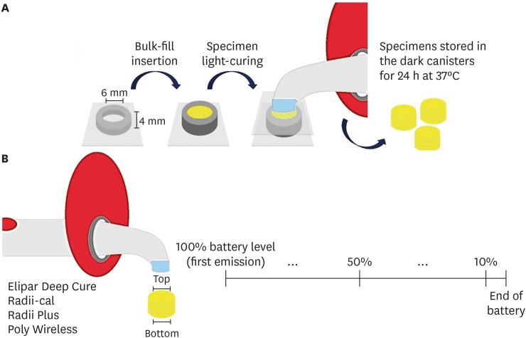
Materials and Methods Four LCUs were evaluated: Radii Plus (SDI), Radii-cal (SDI), Elipar Deep Cure (Filtek Bulk Fill, 3M Oral Care), and Poly Wireless (Kavo Kerr). Irradiance was measured using a radiometer every ten 20-second activations until the battery was discharged. Disks (4 mm thick) of a bulk-fill resin (Filtek Bulk Fill, 3M Oral Care) were prepared, and the VHN was determined on the top and bottom surfaces when light-cured with the LCUs with battery levels at 100%, 50% and 10%. Data were analyzed by 2-way analysis of variance, the Tukey’s test, and Pearson correlations (α = 5%).
Results Elipar Deep Cure and Poly Wireless showed significant differences between the irradiance when the battery was fully charged versus discharged (10% battery level). Significant differences in irradiance were detected among all LCUs, within each battery condition tested. Hardness ratios below 80% were obtained for Radii-cal (10% battery level) and for Poly Wireless (50% and 10% battery levels). The battery level showed moderate and strong, but non-significant, positive correlations with the VHN and irradiance.
Conclusions Although the irradiance was different among LCUs, it decreased in half of the devices along with a reduction in battery level. In addition, the composite resin effectiveness of curing, measured by the hardness ratio, was reduced when the LCUs’ battery was discharged.
-
Citations
Citations to this article as recorded by- Effect of erosive solutions and thermal cycling on the surface properties of universal injectable and regular consistency resin composites
Ahmed Abbas Rhaif, Hoda Saleh Ismail, Tawakol Ahmed Ahmed Enab, Nadia Mohamed Zaghloul
BMC Oral Health.2025;[Epub] CrossRef - Effect of Battery Level During Successive Charging Cycles on the Performance of Certified and Low-cost Uncertified Light-curing Units Available on E-commerce
TS Peres, G Oliveira, SP da Silva Sakamoto, M da Silva Faria, HL Carlo, CJ Soares
Operative Dentistry.2024; 49(6): 673. CrossRef - Influence of Exposure Distance on Light Irradiance of Dental Curing Lamps in Various Operating Modes
Anna Lehmann, Kacper Nijakowski, Marta Mroczyk, Filip Podgórski, Beata Czarnecka, Anna Surdacka
Applied Sciences.2024; 14(21): 9999. CrossRef - ESTADO DA INTENSIDADE LUMINOSA DAS LÂMPADAS DE FOTOPOLIMERIZAÇÃO DAS CLÍNICAS ODONTOLÓGICAS DOS CENTROS DE SAÚDE DA CIDADE DE CUENCA
Milton Alexis Quinchiguano Caraguay, David Ismael Bravo Achundia , Esteban Eduardo Amoroso Calle, Manuel Estuardo Bravo Calderon
RECISATEC - REVISTA CIENTÍFICA SAÚDE E TECNOLOGIA - ISSN 2763-8405.2023; 3(6): e36296. CrossRef
- Effect of erosive solutions and thermal cycling on the surface properties of universal injectable and regular consistency resin composites
- 1,780 View
- 27 Download
- 5 Web of Science
- 4 Crossref

- Effects of 3 different light-curing units on the physico-mechanical properties of bleach-shade resin composites
- Azin Farzad, Shahin Kasraei, Sahebeh Haghi, Mahboubeh Masoumbeigi, Hassan Torabzadeh, Narges Panahandeh
- Restor Dent Endod 2022;47(1):e9. Published online February 7, 2022
- DOI: https://doi.org/10.5395/rde.2022.47.e9
-
 Abstract
 PDF
PDF PubReader
 ePub
ePub Objectives This study investigated the microhardness, flexural strength, and color stability of bleach-shade resin composites cured with 3 different light-curing units.
Materials and Methods In this
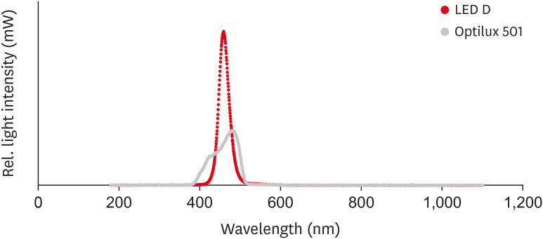
in vitro experimental study, 270 samples were fabricated of bleach and A2 shades of 3 commercial resin composites (Point 4, G-aenial Anterior, and Estelite Sigma Quick). Samples (n = 5 for each trial) were cured with Bluephase N, Woodpecker LED.D, and Optilux 501 units and underwent Vickers microhardness and flexural strength tests. The samples were tested after 24 hours of storage in distilled water. Color was assessed using a spectrophotometer immediately after preparation and 24 hours after curing. Data were analyzed using 3-way analysis of variance and the Tukey test (p ≤ 0.001).Results Samples cured with Optilux exhibited the highest and those cured with LED.D exhibited the lowest microhardness (
p = 0.023). The bleach shade of Point 4 composite cured with Optilux displayed the highest flexural strength, while the same composite and shade cured with Sigma Quick exhibited the lowest (p ≤ 0.001). The color change after 24 hours was greatest for the bleach shade of G-aenial cured with Bluephase N and least for the A2 shade of Sigma Quick cured with Optilux (p ≤ 0.001).Conclusions Light curing with polywave light-emitting diode (LED) yielded results between or statistically similar to those of quartz-tungsten-halogen and monowave LED in the microhardness and flexural strength of both A2 and bleach shades of resin composites. However, the brands of light-curing devices showed significant differences in color stability.
-
Citations
Citations to this article as recorded by- Mechanical Behaviour of Novel Nanohybrid Resin Composite Using Two Light Cure Systems
Ghada H. Naguib, Jumana Mazhar, Abeer Alnowaiser, Abdulghani Mira, Hisham Mously, Rabab Aljawi, Samar H. Abuzinadah, Mohamed T. Hamed
International Dental Journal.2025; 75(2): 1136. CrossRef - Repair Bond Strength of Aged Composite: Effect of Thermocycling and Surface Treatment
Sina Yarmoradian, Ladan Ranjbar Omrani, Elham Ahmadi, Niyousha Rafeie, Mahdi Abbasi, Nastaran Dabiri Shahabi
Journal of Research in Dental and Maxillofacial Sciences.2025; 10(3): 228. CrossRef - Evaluation of the Depth of Cure by Microhardness of Bulk-Fill Composites with Monowave and Polywave LED Light-Curing Units
Socratis Thomaidis, Dimitris Kampouropoulos, Maria Antoniadou, Afrodite Kakaboura
Applied Sciences.2024; 14(24): 11532. CrossRef - Effect of hard segment chemistry and structure on the self‐healing properties of UV‐curable coatings based on the urethane acrylates with built‐in Diels–Alder adduct
Paulina Bednarczyk, Karolina Mozelewska, Małgorzata Nowak, Joanna Klebeko, Joanna Rokicka, Paula Ossowicz‐Rupniewska
Journal of Applied Polymer Science.2023;[Epub] CrossRef - Effects of Dental Bleaching Agents on the Surface Roughness of Dental Restoration Materials
Alexandru Dan Popescu, Mihaela Jana Tuculina, Oana Andreea Diaconu, Lelia Mihaela Gheorghiță, Claudiu Nicolicescu, Cristian Niky Cumpătă, Cristiana Petcu, Jaqueline Abdul-Razzak, Ana Maria Rîcă, Ruxandra Voinea-Georgescu
Medicina.2023; 59(6): 1067. CrossRef - Effect of Polywave and Monowave Light Curing Units on Color Change of Composites Containing Trime-thylbenzoyl-Diphenyl-Phosphine Before and After Aging
Negar Madihi, Maryam Hoorizad ganjkar, Negin Nasoohi, Ali Kaboudanian Ardestani
Journal of Research in Dental and Maxillofacial Sciences.2023; 8(4): 249. CrossRef
- Mechanical Behaviour of Novel Nanohybrid Resin Composite Using Two Light Cure Systems
- 1,980 View
- 34 Download
- 4 Web of Science
- 6 Crossref

- Physicochemical characterization of two bulk fill composites at different depths
- Guillermo Grazioli, Carlos Enrique Cuevas-Suárez, Leina Nakanishi, Alejandro Francia, Rafael Ratto de Moraes
- Restor Dent Endod 2021;46(3):e39. Published online July 5, 2021
- DOI: https://doi.org/10.5395/rde.2021.46.e39
-
 Abstract
 PDF
PDF PubReader
 ePub
ePub Objectives This study analyzed the physical-chemical behavior of 2 bulk fill resin composites (BFCs; Filtek Bulk Fill [FBF], and Tetric-N-Ceram Bulk Fill [TBF]) used in 2- and 4-mm increments and compared them with a conventional resin composite (Filtek Z250).
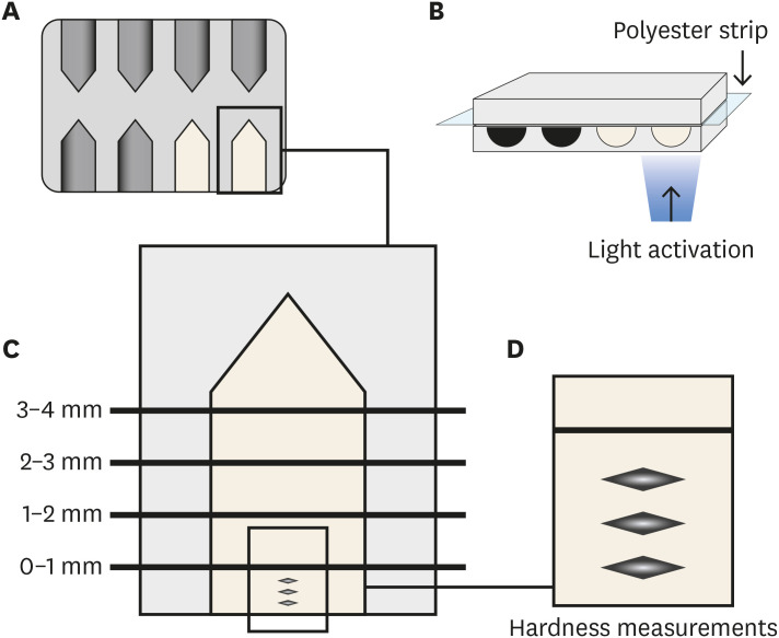
Materials and Methods Flexural strength and elastic modulus were evaluated by using a 3-point bending test. Knoop hardness was measured at depth areas 0–1, 1–2, 2–3, and 3–4 mm. The translucency parameter was measured using an optical spectrophotometer. Real-time polymerization kinetics was analyzed using Fourier transform infrared spectroscopy.
Results Flexural strength was similar among the materials, while TBF showed lower elastic modulus (Z250: 6.6 ± 1.3, FBF: 6.4 ± 0.9, TBF: 4.3 ± 1.3). The hardness of Z250 was similar only between 0–1 mm and 1–2 mm. Both BFCs had similar hardness until 2–3 mm, and showed significant decreases at 3–4 mm (FBF: 33.45 ± 1.95 at 0–1 mm to 23.19 ± 4.32 at 3–4 mm, TBF: 23.17 ± 2.51 at 0–1 mm to 15.11 ± 1.94 at 3–4 mm). The BFCs showed higher translucency than Z250. The polymerization kinetics of all the materials were similar at 2-mm increments. At 4-mm, only TBF had a similar degree of conversion compared with 2 mm.
Conclusions The BFCs tested had similar performance compared to the conventional composite when used in up to 2-mm increments. When the increment was thicker, the BFCs were properly polymerized only up to 3 mm.
-
Citations
Citations to this article as recorded by- Microhardness According to Surface, Distance and Time of Photopolymerization of a Bulk-Fill Resin: In Vitro Study
María José Loayza-Gallegos, Gino Hernan Vidalón-Romo, Julissa Amparo Dulanto-Vargas
Odovtos - International Journal of Dental Sciences.2026; 1(1): 384. CrossRef - Comparative In Vitro Analysis of Mechanical Properties in Three High-Viscosity Bulk-Fill Composite Resins
Carlos I. Santacruz, Jorge I. Fajardo, César A. Paltán, Ana del Carmen Armas-Vega, Eleonor Vélez León
Journal of Composites Science.2025; 9(11): 623. CrossRef - Translucency of bulk‐fill composite materials: A systematic review
Gaetano Paolone, Sofia Baldani, Niccolò De Masi, Mauro Mandurino, Giacomo Collivasone, Nicola Scotti, Enrico Gherlone, Giuseppe Cantatore
Journal of Esthetic and Restorative Dentistry.2024; 36(7): 995. CrossRef - Can composite packaging and selective enamel etching affect the clinical behavior of bulk-fill composite resin in posterior restorations? 24-month results of a randomized clinical trial
Marcos de Oliveira BARCELEIRO, Chane TARDEM, Elisa Gomes ALBUQUERQUE, Leticia de Souza LOPES, Stella Soares MARINS, Luiz Augusto POUBEL, Roberta BARCELOS, Romina ÑAUPARI-VILLASANTE, Alessandro Dourado LOGUERCIO, Fernanda Signorelli CALAZANS
Journal of Applied Oral Science.2023;[Epub] CrossRef - No-Cap Flowable Bulk-Fill Composite: Physico-Mechanical Assessment
Abdullah Alshehri, Feras Alhalabi, Ali Robaian, Mohammed A. S. Abuelqomsan, Abdulrahman Alshabib, Eman Ismail, Faisal Alzamil, Nawaf Alotaibi, Hamad Algamaiah
Polymers.2023; 15(8): 1847. CrossRef - The Microhardness and Surface Roughness Assessment of Bulk-Fill Resin Composites Treated with and without the Application of an Oxygen-Inhibited Layer and a Polishing System: An In Vitro Study
Ann Carrillo-Marcos, Giuliany Salazar-Correa, Leonor Castro-Ramirez, Marysela Ladera-Castañeda, Carlos López-Gurreonero, Hernán Cachay-Criado, Ana Aliaga-Mariñas, Alberto Cornejo-Pinto, Luis Cervantes-Ganoza, César Félix Cayo-Rojas
Polymers.2022; 14(15): 3053. CrossRef
- Microhardness According to Surface, Distance and Time of Photopolymerization of a Bulk-Fill Resin: In Vitro Study
- 1,979 View
- 19 Download
- 6 Web of Science
- 6 Crossref

- Influence of modeling agents on the surface properties of an esthetic nano-hybrid composite
- Zeynep Bilge Kutuk, Ecem Erden, Damla Lara Aksahin, Zeynep Elif Durak, Alp Can Dulda
- Restor Dent Endod 2020;45(2):e13. Published online January 29, 2020
- DOI: https://doi.org/10.5395/rde.2020.45.e13
-
 Abstract
 PDF
PDF PubReader
 ePub
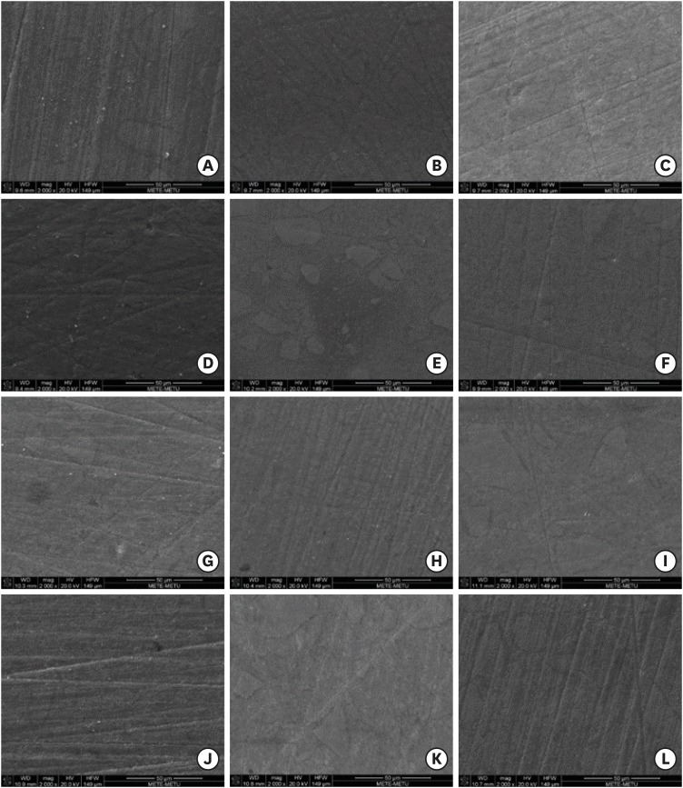
ePub Objective The aim of this study was to evaluate the influence of different modeling agents on the surface microhardness (Vickers hardness number; VHN), roughness (Ra), and color change (ΔE) of a nano-hybrid composite with or without exposure to discoloration by coffee.
Materials and Methods Sixty-four cylinder-shaped nano-hybrid composite specimens were prepared using a Teflon mold. The specimens' surfaces were prepared according to the following groups: group 1, no modeling agent; group 2, Modeling Liquid; group 3, a universal adhesive (G-Premio Bond); and group 4, the first step of a 2-step self-adhesive system (OptiBond XTR). Specimens were randomly allocated into 2 groups (
n = 8) according to the storage medium (distilled water or coffee). VHN, Ra, and ΔE were measured at 24 hours, 1 week, and 6 weeks. The Kruskal-Wallis test followed by the Bonferroni correction for pairwise comparisons was used for statistical analysis (α = 0.05).Results Storage time did not influence the VHN of the nano-hybrid composite in any group (
p > 0.05). OptiBond XTR Primer application affected the VHN negatively in all investigated storage medium and time conditions (p < 0.05). Modeling Liquid application yielded improved Ra values for the specimens stored in coffee at each time point (p < 0.05). Modeling Liquid application was associated with the lowest ΔE values in all investigated storage medium and time conditions (p < 0.05).Conclusion Different types of modeling agents could affect the surface properties and discoloration of nano-hybrid composites.
-
Citations
Citations to this article as recorded by- Do modeling liquid and glycerin gel compromise the color stability of one-shade composites
Ezgi Erden Kayalidere, Merve Sahin
Odontology.2026;[Epub] CrossRef - The Impact of Modeling Liquids on Surface Roughness and Color Properties of Bulkfill Resin Composites After Simulated Tooth Brushing: An in Vitro Study. Part I
Camila Falconí‐Páez, Claudia González‐Vaca, Juliana Guarneri, Newton Fahl, Paulina Aliaga‐Sancho, Maria Lujan Mendez‐Bauer, Cesar Augusto Galvão Arrais, Andrés Dávila‐Sánchez
Journal of Esthetic and Restorative Dentistry.2025; 37(2): 514. CrossRef - Coating Agents for Resin Composites: Effect on Color Stability, Roughness, and Surface Micromorphology Subjected to Brushing Wear
FR Hojo, TC Martins, WF Vieira-Junior, FMG França, CP Turssi, RT Basting
Operative Dentistry.2025; 50(1): 101. CrossRef - Effect of modeling liquid application on color stability and surface roughness of single-shade composites
Melek Güven Bekdaş, Ihsan Hubbezoglu
BMC Oral Health.2025;[Epub] CrossRef - Influence of modeling liquids on the color adaptation and optical properties of single and simply shade resin composites
Bengü Doğu Kaya, Mehmet Buldur, Burcu Gözetici-Çil
Odontology.2025;[Epub] CrossRef - Effect of modeling liquids on Vickers microhardness, flexural strength and color stability of resin-based composites
Ahmed Alshawi, Benin Dikmen, Sevda Ozel Yildiz, Ugur Erdemir
Materials Research Express.2025; 12(11): 115402. CrossRef - Does composite repair time affect repair protocol, immediate or delayed?
Murat Can Ersen, Nevin Cobanoglu
BMC Oral Health.2025;[Epub] CrossRef - Impact of combining dental composite brushes with modeling resins on the color stability and topographic features of composites
Abdulrahman A Balhaddad, Faisal Alharamlah, Alhanoof Aldossary, Wejdan Almutairi, Turki Alshehri, Mary Anne S Melo, Afnan O Al-Zain, Eman H Ismail
Journal of Applied Biomaterials & Functional Materials.2024;[Epub] CrossRef - Investigation of the Degree of Monomer Conversion in Dental Composites through Various Methods: An In Vitro Study
Musa Kazim Ucuncu, Ozge Celiksoz, Emine Sen, Yasemin Yucel Yucel, Bircan Dinc
Applied Sciences.2024; 14(11): 4406. CrossRef - EFEITO DOS LÍQUIDOS MODELADORES NA SUPERFÍCIE DA RESINA COMPOSTA – UMA REVISÃO DE LITERATURA
Samuel Silva Dias, Matheus Fernando Lopes, Jeffison Teles Dias, Caio Junji Tanaka, Jose Augusto Rodrigues
RECIMA21 - Revista Científica Multidisciplinar - ISSN 2675-6218.2024; 5(2): e524899. CrossRef - Effect of Instrument Lubricant on Mechanical Properties of Restorative Composite
G Pippin, D Tantbirojn, M Wolfgang, JS Nordin, A Versluis
Operative Dentistry.2024; 49(4): 475. CrossRef - Full analysis of the effects of modeler liquids on the properties of direct resin-based composites: a meta-analysis review of in vitro studies
Eduardo Trota Chaves, Lisia Lorea Valente, Eliseu Aldrighi Münchow
Clinical Oral Investigations.2023; 27(7): 3289. CrossRef - Influence of Modeling Liquids and Universal Adhesives Used as Lubricants on Color Stability and Translucency of Resin-Based Composites
Gaetano Paolone, Claudia Mazzitelli, Giacomo Zechini, Salvatore Scolavino, Cecilia Goracci, Nicola Scotti, Giuseppe Cantatore, Enrico Gherlone, Alessandro Vichi
Coatings.2023; 13(1): 143. CrossRef - Influence of Instrument Lubrication on Properties of Dental Composites
Juliusz Kosewski, Przemysław Kosewski, Agnieszka Mielczarek
European Journal of Dentistry.2022; 16(04): 719. CrossRef - Effect of Modelıng Liquid Use on Color and Whiteness Index Change of Composite Resins
Numan AYDIN, Serpil KARAOĞLANOĞLU, Bilge ERSÖZ
Cumhuriyet Dental Journal.2022; 25(Supplement): 119. CrossRef - Effects of Immediate Coating on Unset Composite with Different Bonding Agents to Surface Hardness
Nantawan Krajangta, Supissara Ninbanjong, Sunisa Khosook, Kanjana Chaitontuak, Awiruth Klaisiri
European Journal of Dentistry.2022; 16(04): 828. CrossRef - Modeling Liquids and Resin-Based Dental Composite Materials—A Scoping Review
Gaetano Paolone, Claudia Mazzitelli, Uros Josic, Nicola Scotti, Enrico Gherlone, Giuseppe Cantatore, Lorenzo Breschi
Materials.2022; 15(11): 3759. CrossRef - Shear Bond Strength of Composite Diluted with Composite-Handling Agents on Dentin and Enamel
Mijoo Kim, Deuk-Won Jo, Shahed Al Khalifah, Bo Yu, Marc Hayashi, Reuben H. Kim
Polymers.2022; 14(13): 2665. CrossRef - Effect of Modeling Resins on Microhardness of Resin Composites
Ezgi T. Bayraktar, Pinar Y. Atali, Bora Korkut, Ezgi G. Kesimli, Bilge Tarcin, Cafer Turkmen
European Journal of Dentistry.2021; 15(03): 481. CrossRef
- Do modeling liquid and glycerin gel compromise the color stability of one-shade composites
- 1,982 View
- 30 Download
- 19 Crossref

- Effect of dental bleaching on the microhardness and surface roughness of sealed composite resins
- Renan Aparecido Fernandes, Henrico Badaoui Strazzi-Sahyon, Thaís Yumi Umeda Suzuki, André Luiz Fraga Briso, Paulo Henrique dos Santos
- Restor Dent Endod 2020;45(1):e12. Published online January 10, 2020
- DOI: https://doi.org/10.5395/rde.2020.45.e12
-
 Abstract
 PDF
PDF PubReader
 ePub
ePub Objectives The aim of this
in vitro study was to evaluate the microhardness and surface roughness of composite resins before and after tooth bleaching procedures.Materials and Methods Sixty specimens were prepared of each composite resin (Filtek Supreme XT and Opallis), and BisCover LV surface sealant was applied to half of the specimens. Thirty enamel samples were obtained from the buccal and lingual surfaces of human molars for use as the control group. The surface roughness and microhardness were measured before and after bleaching procedures with 35% hydrogen peroxide or 16% carbamide (
n = 10). Data were analyzed using 1-way analysis of variance and the Fisher test (α = 0.05).Results Neither hydrogen peroxide nor carbamide peroxide treatment significantly altered the hardness of the composite resins, regardless of surface sealant application; however, both treatments significantly decreased the hardness of the tooth samples (
p < 0.05). The bleaching did not cause any change in surface roughness, with the exception of the unsealed Opallis composite resin and dental enamel, both of which displayed an increase in surface roughness after bleaching with carbamide peroxide (p < 0.05).Conclusions The microhardness and surface roughness of enamel and Opallis composite resin were influenced by bleaching procedures.
-
Citations
Citations to this article as recorded by- Effect of Bleaching on Surface Roughness and Color Parameters of Coffee-Stained Nanohybrid Dental Composites with Different Viscosities
Hetaf S. Redwan, Mohamed A. Hussein, Mohamed M. Abdul-Monem
European Journal of General Dentistry.2025; 14(01): 027. CrossRef - Effect of Staining and External Bleaching on the Color Stability and Surface Roughness of Universal-Shade Resin-Based Composite
AlHanouf AlHabdan, Amal Alsuhaibani, Lama Alomran, Lulwah Almutib
Clinical, Cosmetic and Investigational Dentistry.2025; Volume 17: 1. CrossRef - Comparative Analysis Between Strip and Gels Indicated for at Home Bleaching: Analysis of Color Alteration, Roughness and Microhardness of Dental Enamel
K. M. S. Aidar, L. T. A. Cintra, M. C. B. Ferreira, T. C. Fagundes, L. M. B. Esteves, J. Goto, A. Catelan, A. L. F. Briso
Journal of Esthetic and Restorative Dentistry.2025; 37(6): 1504. CrossRef - Surface properties and susceptibility to staining of a resin composite after brushing with different whitening toothpastes
Aline da Silva Barros, Carolina Meneghin Barbosa, Renata Siqueira Scatolin, Waldemir Francisco Vieira Junior, Laura Nobre Ferraz
Restorative Dentistry & Endodontics.2025; 50(1): e6. CrossRef - Degradation Resistance of Next-Generation Dental Composites Under Bleaching and Immersion: A Multiscale Investigation
Syed Zubairuddin Ahmed, Shahad Al-Qahtani, Naif H. Al-Qahtani, Hussah Al-Mulhim, Maha Al-Qahtani, Ali Albalushi, Sultan Akhtar
Prosthesis.2025; 7(3): 57. CrossRef - Effect of Over-the-Counter Whitening Dentifrices on the Color Stability and Microhardness of Composite Resins
Xinnuo Yu, Maria Pilar Melo, Sofia Folguera, Carmen Llena
Journal of Composites Science.2025; 9(7): 324. CrossRef - From Microstructure to Shade Shift: Confocal and Spectrophotometric Evaluation of Peroxide-Induced Dental Bleaching
Berivan Laura Rebeca Buzatu, Magda Mihaela Luca, Atena Galuscan, Adrian Ovidiu Vaduva, Aurora Doris Fratila, Ramona Dumitrescu, Ruxandra Sava-Rosianu, Octavia Balean, Roxana Buzatu, Daniela Jumanca
Journal of Clinical Medicine.2025; 14(13): 4642. CrossRef - In Vitro Evaluation of Chemical and Microhardness Alterations in Human Enamel Induced by Three Commercial In-Office Bleaching Agents
Berivan Laura Rebeca Buzatu, Atena Galuscan, Ramona Dumitrescu, Roxana Buzatu, Magda Mihaela Luca, Octavia Balean, Gabriela Vlase, Titus Vlase, Iasmina-Mădălina Anghel, Carmen Opris, Bianca Ioana Todor, Mihaela Adina Dumitrache, Daniela Jumanca
Dentistry Journal.2025; 13(8): 357. CrossRef - Effect of Hydrogen Peroxide Bleaching on Color Stability and Microhardness of Alkasite Restorative Materials: An In Vitro Study
Souad A Alfouzan, Rahaf A Alolayan, Asma Munir Khan
Cureus.2025;[Epub] CrossRef - Evaluation of Color Stability and Surface Roughness of Nanohybrid Resin Composites with Different Photoinitiator Systems After Staining and Home/Office Bleaching: An In Vitro Study
Fatma Yılmaz, Buse Kesgin
Meandros Medical And Dental Journal.2025; 26(3): 240. CrossRef - The Effect of Hydrogen Peroxide With Different Concentration on the Color and Surface Microhardness of the Resin Bracket
Song‐Yi Yang
Clinical and Experimental Dental Research.2025;[Epub] CrossRef - Comparative evaluation of different bleaching agents on the color stability, hardness and surface roughness of indirect esthetic restorative materials with different manufacturing methods
Ayse Atay, Defne Canpolat, Soner Sismanoglu, Aslihan Usumez
BMC Oral Health.2025;[Epub] CrossRef - Comparison of Microhardness and Surface Roughness of New Nanofiber Filled Flowable Composite
Rumeysa Hatice ENGINLER OZLEN, Zumrut Ceren OZDUMAN, Burcu OGLAKCI OZKOC, Evrim ELIGUZELOGLU DALKILIC
Bezmialem Science.2024; 12(4): 406. CrossRef - Effect of Bleaching Agents on Composite Resins with and without Bis-GMA: An In Vitro Study
María Melo, Bianca Dumitrache, James Ghilotti, José Luis Sanz, Carmen Llena
Journal of Functional Biomaterials.2024; 15(6): 144. CrossRef - Changes in physical properties of universal composites and CAD/CAM materials after bleaching and antioxidant applications: Scanning electron microscope and atomic force microscope evaluation
Oguz Kaan Tuysuz, Merve Gurses
Microscopy Research and Technique.2024; 87(5): 977. CrossRef - The Effects of Home and Over-The-Counter Whitening Agents on Surface Roughness and Microhardness of High Aesthetic Composites
Elif İpek KILIÇ DÖNMEZ, İhsan HUBBEZOĞLU
Cumhuriyet Dental Journal.2024; 27(1): 30. CrossRef - Effect of carbamide peroxide treatment on the ion release of different dental restorative materials
Merve Nur Yilmaz, Pinar Gul
BMC Oral Health.2024;[Epub] CrossRef - Inorganic Phosphate Effect in a Hydrogen Peroxide-based Bleaching Agent: Physicochemical, Mechanical, and Morphological Properties of Dental Enamel
KG Garcia, GP Nunes, ACB Delbem, PH dos Santos, GLP Fernandes, HF Robles, PBB Lemos, M Danelon
Operative Dentistry.2024; 49(4): 465. CrossRef - Effect of bleaching and repolishing on whiteness change and staining susceptibility of resin-based materials
Sultan Aktuğ Karademir, Samet Atasoy, Beyza Yılmaz
BMC Oral Health.2024;[Epub] CrossRef - Influence of Low pH on the Microhardness and Roughness Surface of Dental Composite—A Preliminary Study
Leszek Szalewski, Dorota Wójcik, Monika Sowa, Vladyslav Vivcharenko, Krzysztof Pałka
Materials.2024; 17(14): 3443. CrossRef - In Vitro Evaluation of the Effectiveness and pH Variation of Dental Bleaching Gels and Their Effect on Enamel Surface Roughness
Federica Veneri, Francesco Cavani, Giovanni Bolelli, Vittorio Checchi, Alessia Bizzi, Giacomo Setti, Luigi Generali
Dentistry Journal.2024; 12(12): 415. CrossRef - Does the combination of whitening toothpaste and hydrogen peroxide bleaching increase the surface roughness and change the morphology of a nanofilled composite?
Cecília Pereira da Silva Braga Tenório, Matheus Kury, Geyse Maria dos Santos Muniz Mota, Cecília Pedroso Turssi, Flávia Lucisano Botelho do Amaral, Vanessa Cavalli
Brazilian Journal of Oral Sciences.2024; 23: e241938. CrossRef - Effect of peroxide‐free and peroxide‐based in‐office bleaching on the surface and mechanical properties of CAD/CAM esthetic restorative materials
Majed M. Alsarani, Aftab Ahmed Khan, Leonel S. J. Bautista, Hanan Alsunbul, Jukka P. Matinlinna
European Journal of Oral Sciences.2024;[Epub] CrossRef - Effect of Repolishing on Color Stability, Translucency, and Surface Roughness of Aged Monochromatic Dental Composites
Mohamed M. Abdul-Monem, Mohamed A. Hussein, Mona G. Abdelrehim
European Journal of General Dentistry.2024; 13(03): 240. CrossRef - Color changes of nanofiller composite resin after glycerin application immersed in turmeric extract
Sukaton, Galih Sampoerno, Widyajeng Ayu Laksmi, Daradhasih Bestari Santiaji
Conservative Dentistry Journal.2023; 13(1): 37. CrossRef - Effects of Dental Bleaching Agents on the Surface Roughness of Dental Restoration Materials
Alexandru Dan Popescu, Mihaela Jana Tuculina, Oana Andreea Diaconu, Lelia Mihaela Gheorghiță, Claudiu Nicolicescu, Cristian Niky Cumpătă, Cristiana Petcu, Jaqueline Abdul-Razzak, Ana Maria Rîcă, Ruxandra Voinea-Georgescu
Medicina.2023; 59(6): 1067. CrossRef - Effect of Bleaching on the Microhardness and Modulus of Elasticity of ACTIVA BioACTIVE – RESTORATIVE: An In Vitro Study
Sushritha Sricharan, Swaroop Hegde, Narmada J., Indiresha H. Narayana, Chatura Mohan, Nithin K. Shetty
Journal of Advanced Oral Research.2023; 14(2): 190. CrossRef - The effect of bleaching on surface roughness and gloss of different CAD/CAM ceramic and hybrid ceramic materials
Ruwaida Z Alshali, Mohammed A AlQahtani, Dalea M Bukhary, Mlak A Alzahrani, Shatha S Alsoraihi, Majed A Alqahtani
Journal of Applied Biomaterials & Functional Materials.2023;[Epub] CrossRef - Effect of bleaching with 15% carbamide peroxide on color stability of microhybrid, nanohybrid, and nanofilled resin composites, each in 3 staining solutions (coffee, cola, red grape juice): A 3-phase study
Azadeh Ghaemi, Sanaz Sharifishoshtari, Mohsen Shahmoradi, Hossein Akbari, Parisa Boostanifard, Sepideh Bagheri, Mohammadreza Shokuhifar, Negin Ashoori, Vahid Rakhshan
Dental Research Journal.2023;[Epub] CrossRef - Micro-Hardness and Surface Roughness of Bulk-Fill Composite Resin: Effect of Surface Sealant Application and Two Bleaching Regimens
Reham Mohamad Attia, Eman Mohamed Sobhy, Mona El Said Abd El Hameed Essa
European Journal of General Dentistry.2023; 12(03): 169. CrossRef - Shear bond strength after using sealant before bonding: a systematic review and meta-analysis of in vitro studies
Jennifer Hoppe, Thomas Lehmann, Christoph-Ludwig Hennig, Ulrike Schulze-Späte, Collin Jacobs
Clinical Oral Investigations.2022; 26(1): 1. CrossRef - Effect of 16% Carbamide Peroxide and Activated-Charcoal-Based Whitening Toothpaste on Enamel Surface Roughness in Bovine Teeth: An In Vitro Study
Jorge Zamudio-Santiago, Marysela Ladera-Castañeda, Flor Santander-Rengifo, Carlos López-Gurreonero, Alberto Cornejo-Pinto, Ali Echavarría-Gálvez, Luis Cervantes-Ganoza, César Cayo-Rojas
Biomedicines.2022; 11(1): 22. CrossRef - Direct dentin bleaching: Would it be possible?
Camila Ferro Clemente, Sibele de Alcântara, Lívia Maria Alves Valentim da Silva, Lara Maria Bueno Esteves, Anderson Catelan, Karen Milaré Seiscento Aidar, Ticiane Cestari Fagundes, André Luiz Fraga Briso
Photodiagnosis and Photodynamic Therapy.2022; 40: 103121. CrossRef - EFFECT OF İN-OFFİCE BLEACHİNG ON THE SURFACE ROUGHNESS OF DİFFERENT COMPOSİTE RESİNS
Seher KAYA, Ozden OZEL BEKTAS
Cumhuriyet Dental Journal.2022; 25(Supplement): 78. CrossRef - Effect of Polishing on the Surface Microhardness of Nanohybrid Composite Resins Subjected to 35% Hydrogen Peroxide
Giovanna Gisella Ramírez-Vargas, Julia Elbia Medina y Mendoza, Ana Sixtina Aliaga-Mariñas, Marysela Irene Ladera-Castañeda, Luis Adolfo Cervantes-Ganoza, César Félix Cayo-Rojas
Journal of International Society of Preventive and Community Dentistry.2021; 11(2): 216. CrossRef - Intrapulpal Concentration of Hydrogen Peroxide of Teeth Restored With Bulk Fill and Conventional Bioactive Composites
DP Silva, BA Resende, M Kury, CB André, CPM Tabchoury, M Giannini, V Cavalli
Operative Dentistry.2021; 46(3): E158. CrossRef - An Environmental Scanning Electron Microscopy Evaluation on Comparison of Three Different Bleaching Agents using the Laser Activated in-Office Bleaching at Different Wavelengths
Shachi Goenka, Sushil Kumar Cirigiri, Kanika Poplai, Baig Mirza Aslam, Shalini Singh, Shweta Gangavane
Journal of Pharmacy and Bioallied Sciences.2021; 13(Suppl 2): S1478. CrossRef - Effects of Artificial Staining and Bleaching Protocols on the Surface Roughness, Color, and Whiteness Changes of an Aged Nanofilled Composite
Geyse Maria dos Santos Muniz Mota, Matheus Kury, Cecília Pereira da Silva Braga Tenório, Flávia Lucisano Botelho do Amaral, Cecília Pedroso Turssi, Vanessa Cavalli
Frontiers in Dental Medicine.2020;[Epub] CrossRef
- Effect of Bleaching on Surface Roughness and Color Parameters of Coffee-Stained Nanohybrid Dental Composites with Different Viscosities
- 2,628 View
- 40 Download
- 38 Crossref

- The effect of preheating resin composites on surface hardness: a systematic review and meta-analysis
- Ali A. Elkaffas, Radwa I. Eltoukhy, Salwa A. Elnegoly, Salah H. Mahmoud
- Restor Dent Endod 2019;44(4):e41. Published online October 29, 2019
- DOI: https://doi.org/10.5395/rde.2019.44.e41
-
 Abstract
 PDF
PDF PubReader
 ePub
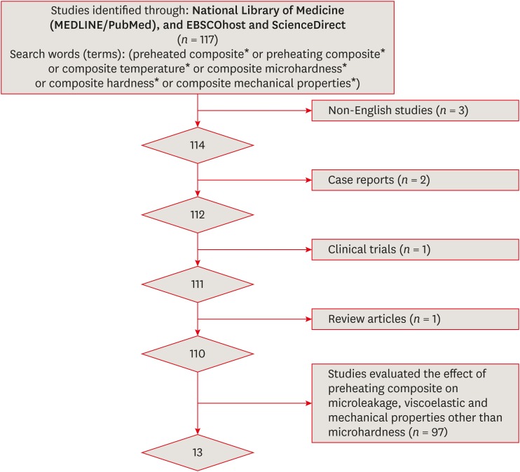
ePub Objectives This paper presents a systematic review and meta-analysis of the effect of preheating on the hardness of nanofilled, nanoceramic, nanohybrid, and microhybrid resin composites.
Materials and Methods An electronic search of papers on MEDLINE/PubMed, ScienceDirect, and EBSCOhost was performed. Only
in vitro studies were included. Non-English studies, case reports, clinical trials, and review articles were excluded. A meta-analysis of the reviewed studies was conducted to quantify differences in the microhardness of the Z250 microhybrid resin composite using the Comprehensive Meta-Analysis software.Results Only 13 studies met the inclusion criteria for this systematic review. The meta-analysis showed that there were significant differences between the non-preheated and preheated modes for both the top and bottom surfaces of the specimens (
p < 0.05). The microhardness of the Z250 resin composite on the top surface in the preheated mode (78.1 ± 2.9) was higher than in the non-preheated mode (67.4 ± 4.0;p < 0.001). Moreover, the microhardness of the Z250 resin composite on the bottom surface in the preheated mode (71.8 ± 3.8) was higher than in the non-preheated mode (57.5 ± 5.7,p < 0.001).Conclusions Although the results reported in the reviewed studies showed great variability, sufficient scientific evidence was found to support the hypothesis that preheating can improve the hardness of resin composites.
-
Citations
Citations to this article as recorded by- Influence of preheating and water storage on the color, whiteness, and translucency of modern resin‐based composites
Corina Mirela Prodan, Cristina Gasparik, Javier Ruiz‐López, Diana Dudea
Journal of Esthetic and Restorative Dentistry.2025; 37(2): 533. CrossRef - Effects of pre-heating on physical–mechanical–chemical properties of contemporary resin composites
Thamires Bueno, Nivien Masoud, Anna Akkus, Italo Silva, Karen McPherson, Adilson Yoshio Furuse, Fabio Rizzante
Odontology.2025; 113(1): 135. CrossRef - The effects of a carbonated beverage on the optical properties and microhardness of preheated bulk-fill composite resin restorations
Nancy Soliman Farghal, Ayya Abu Shamleh, Osamah Al Hurmuzi, Okba Mahmoud
Frontiers in Oral Health.2025;[Epub] CrossRef - Evaluation of Degree of Conversion, Flexural Strength, and Microhardness of a Novel Flowable Resin Composite
Bengü Doğu Kaya, Selinsu Öztürk, Nazlı Zeynep Kuzu, Ayşe Aslı Şenol, Erkut Kahramanoğlu, Pınar Yılmaz Atalı, Bilge Tarçın
Selcuk Dental Journal.2025; 12(2): 202. CrossRef - Clinical performance of different bulk‐fill composite resin systems in classIIcavities: A 2‐year randomized clinical trial
Badria Goda, Kareem Hamdi, Radwa I. Eltoukhy, Ashraf I. Ali, Salah Hasab Mahmoud
Journal of Esthetic and Restorative Dentistry.2024; 36(8): 1122. CrossRef - Comparative evaluation of microleakage in Class II cavities restored with snowplow technique using flowable or preheated packable bulk-fill composite resin as gingival increment by dye extraction method: An in vitro study
M. A. Ranjini, V. Geetha, B. Vedavathi, H. K. Ashok, Akshata J. Airsang, S. Swathi
Journal of Conservative Dentistry and Endodontics.2024; 27(11): 1158. CrossRef - Influence of Light‐Curing Time and Increment Thickness on the Properties of Bulk Fill Composite Resins With Distinct Application Systems
Carlos Rocha Gomes Torres, Taiana Paola Prado, Daniele Mara da Silva Ávila, Cesar Rogério Pucci, Alessandra Bühler Borges, Heng Bo Jiang
International Journal of Dentistry.2024;[Epub] CrossRef - Last Generation Bis-GMA Free Composite For Indirect Posterior Restorations: A Case Report
M. Delgado
Endodontics Today.2024; 21(4): 305. CrossRef - The clinical performance of dental resin composite repeatedly preheated: A randomized controlled clinical trial
Mahmoud Elkady, Safaa Abdelhakim, Mona Riad
Journal of Dentistry.2024; 144: 104940. CrossRef - Preheating effect on microhardness and depth of cure of three bulk-fill composite resins: An in vitro study
Aashna Sunil Sahetia, Divya Rupesh Jain, Padmaja Panditrao Sirsat, Meenal N. Gulve, Swapnil J. Kolhe, Surbhi P. Patel
Endodontology.2024;[Epub] CrossRef - Evaluation of Shear Bond Strength of Lithium Disilicate Veneers Using Pre-heated Resin Composite With Two Conventional Resin Cements: An In Vitro Study
Ghalia Akyle, Hassan Achour
Cureus.2024;[Epub] CrossRef - Effect of Glass Fiber Reinforcement on Marginal Microleakage in Class II Composite Restorations: An In Vitro Pilot Study
Csaba Dudás, Emánuel Kardos, Melinda Székely, Lea Ádám, Zsuzsanna Bardocz-Veres, Evelyn Szőllősi, Kinga Mária Jánosi, Bernadette Kerekes-Máthé
Dentistry Journal.2024; 12(12): 410. CrossRef - Effect of preheating on the physicochemical properties and bond strength of composite resins utilized as dental cements: An in vitro study
Carolina Carramilo Raposo, Luanna Marinho Sereno Nery, Edilausson Moreno Carvalho, Paulo Vitor Campos Ferreira, Diego Machado Ardenghi, José Bauer, Darlon Martins Lima
The Journal of Prosthetic Dentistry.2023; 129(1): 229.e1. CrossRef - Examining the Impact of Preheating on the Fracture Toughness and Microhardness of Composite Resin: A Systematic Review
Jay Bhopatkar, Anuja Ikhar, Manoj Chandak, Aditya Patel, Paridhi Agrawal
Cureus.2023;[Epub] CrossRef - In Vitro Effect of Mouthrinses on the Microhardness of Three Different Nanohybrid Composite Resins
Jhonn Luis Bernaldo-Faustino, Julissa Amparo Dulanto-Vargas, Kilder Maynor Carranza-Samanez, Carlos A. Munoz-Viveros
International Journal of Dentistry.2023; 2023: 1. CrossRef - Efecto del precalentamiento en la microdureza superficial de seis resinas compuestas
Gloria Cristina Moreno Abello, Kavhas Castro, Paula Alejandra Ovalle Barrera, Paula Bernal, Laura Catalina Lara Hernández
Universitas Odontologica.2023;[Epub] CrossRef - Wear and Color Stability of Preheated Bulk-fill and Conventional Resin Composites
AA Abdulmajeed, AA Suliman, BJ Selivany, A Altitinchi, TA Sulaiman
Operative Dentistry.2022; 47(5): 585. CrossRef - Comparison of Mechanical Properties of a Self-Adhesive Composite Cement and a Heated Composite Material
Anastazja Skapska, Zenon Komorek, Mariusz Cierech, Elzbieta Mierzwinska-Nastalska
Polymers.2022; 14(13): 2686. CrossRef - Effects of ionizing radiation on surface properties of current restorative dental materials
Débora Michelle Gonçalves de Amorim, Aretha Heitor Veríssimo, Anne Kaline Claudino Ribeiro, Rodrigo Othávio de Assunção e Souza, Isauremi Vieira de Assunção, Marilia Regalado Galvão Rabelo Caldas, Boniek Castillo Dutra Borges
Journal of Materials Science: Materials in Medicine.2021;[Epub] CrossRef - Quality assessment tools used in systematic reviews of in vitro studies: A systematic review
Linh Tran, Dao Ngoc Hien Tam, Abdelrahman Elshafay, Thao Dang, Kenji Hirayama, Nguyen Tien Huy
BMC Medical Research Methodology.2021;[Epub] CrossRef - Preheated composite: Innovative approach for aesthetic restoration
Reema N Asani, Vandana J Gade, Kalyani G Umale, Rachana Gawande, Rohit R Amburle, Raksha R Kusumbe, Purva P Kale, Priya R Kosare
Archives of Dental Research.2021; 11(2): 103. CrossRef
- Influence of preheating and water storage on the color, whiteness, and translucency of modern resin‐based composites
- 2,586 View
- 25 Download
- 21 Crossref

- Finishing and polishing effects of multiblade burs on the surface texture of 5 resin composites: microhardness and roughness testing
- Elodie Ehrmann, Etienne Medioni, Nathalie Brulat-Bouchard
- Restor Dent Endod 2019;44(1):e1. Published online November 26, 2018
- DOI: https://doi.org/10.5395/rde.2019.44.e1

-
 Abstract
 PDF
PDF PubReader
 ePub
ePub Objectives The aim of this
in vitro study was to test the effect of 2 finishing–polishing sequences (QB, combining a 12/15-fluted finishing bur and an EVO-Light polisher; QWB, adding a 30-fluted polishing bur after the 12/15-fluted finishing bur used in the QB sequence) on 5 nanotech-based resin composites (Filtek Z500, Ceram X Mono, Ceram X Duo, Tetric Evoceram, and Tetric Evoceram Bulk Fill) by comparing their final surface roughness and hardness values to those of a Mylar strip control group (MS).Materials and Methods Twelve specimens of each nanocomposite were prepared in Teflon moulds. The surface of each resin composite was finished with QB (5 samples), QWB (5 samples), or MS (2 samples), and then evaluated (60 samples). Roughness was analysed with an optical profilometer, microhardness was tested with a Vickers indenter, and the surfaces were examined by optical and scanning electron microscopy. Data were analysed using the Kruskal-Wallis test (
p < 0.05) followed by the Dunn test.Results For the hardness and roughness of nanocomposite resin, the QWB sequence was significantly more effective than QB (
p < 0.05). The Filtek Z500 showed significantly harder surfaces regardless of the finishing–polishing sequence (p < 0.05).Conclusions QWB yielded the best values of surface roughness and hardness. The hardness and roughness of the 5 nanocomposites presented less significant differences when QWB was used.
-
Citations
Citations to this article as recorded by- Effect of modeling liquid application on color stability and surface roughness of single-shade composites
Melek Güven Bekdaş, Ihsan Hubbezoglu
BMC Oral Health.2025;[Epub] CrossRef - Effects of different charcoal-containing whitening toothpastes on color and surface roughness of a supra-nanofilled composite resin
Meltem Nermin Polan, Sevil Gurgan
BMC Oral Health.2025;[Epub] CrossRef - Impact of different polishing techniques on surface roughness, gloss, and microhardness of zirconium oxide reinforced flowable bulk-fill resin composite: an in vitro study
Amr Elsayed Elnahas, Mohamed Elshirbeny Elawsya, Abeer ElSayed ElEmbaby
BMC Oral Health.2025;[Epub] CrossRef - Tek Renkli Monokromatik Kompozit Rezinlerle İlgili Bir Durum Değerlendirmesi
Kubra Nur Yeşilova, Sebnem Turkun
Selcuk Dental Journal.2025; 12(2): 331. CrossRef - Effect of different finishing and polishing systems on surface properties of universal single shade resin-based composites
Ghada Alharbi, Hend NA Al Nahedh, Loulwa M. Al-Saud, Nourah Shono, Ahmed Maawadh
BMC Oral Health.2024;[Epub] CrossRef - A comparative study of polishing systems on optical properties and surface roughness of additively manufactured and conventional resin based composites
Ayse Tugba Erturk-Avunduk, Sevim Atılan-Yavuz, Hande Filiz, Esra Cengiz-Yanardag
Scientific Reports.2024;[Epub] CrossRef - Effect of Instrument Lubricant on Mechanical Properties of Restorative Composite
G Pippin, D Tantbirojn, M Wolfgang, JS Nordin, A Versluis
Operative Dentistry.2024; 49(4): 475. CrossRef - An In Vitro Study regarding the Wear of Composite Materials Following the Use of Dental Bleaching Protocols
Alexandru Dan Popescu, Mihaela Jana Ţuculină, Lelia Mihaela Gheorghiță, Andrei Osman, Claudiu Nicolicescu, Smaranda Adelina Bugălă, Mihaela Ionescu, Jaqueline Abdul-Razzak, Oana Andreea Diaconu, Bogdan Dimitriu
Journal of Functional Biomaterials.2023; 14(10): 532. CrossRef - Akıllı Kromatik Teknolojili Kompozit Rezinlerin Farklı pH Değerlerindeki Sıvılarda Bekletilmesi Sonrası Oluşan Yüzey Pürüzlülüğü ve Renk Değişimlerinin Değerlendirilmesi
Fatih ÖZNURHAN, Aylin ÖZEL
Farabi Tıp Dergisi.2023; 2(4): 17. CrossRef - Enamel surface roughness evaluation after debonding and residual resin removal using four different burs
Rapeti Madhu Vanya, Anil Chirla, Uday Kumar Digumarthi, Tarakesh Karri, Bommareddy Radhika, Sanapala Manojna
Journal of Contemporary Orthodontics.2023; 7(3): 173. CrossRef - Finishing and Polishing of Composite Restoration: Assessment of Knowledge, Attitude and Practice Among Various Dental Professionals in India
Sankar Vishwanath, Sadasiva Kadandale, Senthil kumar Kumarappan, Anupama Ramachandran, Manu Unnikrishnan, Honap manjiri Nagesh
Cureus.2022;[Epub] CrossRef - Evaluation of different composite resin finishing and polishing protocols by confocal laser scan microscopy
Kayo Matheus Rodrigues de Souza, Roberto Victor de Melo Silva, Marlon Ferreira Dias, Paulo Cardoso Lins-Filho, Claudio Heliomar Vicente da Silva, Renata Pedrosa Guimarães
Brazilian Journal of Oral Sciences.2022; 21: e225334. CrossRef - Laboratory methods to simulate the mechanical degradation of resin composite restorations
Veronica P. Lima, Jaqueline B. Machado, Yu Zhang, Bas A.C. Loomans, Rafael R. Moraes
Dental Materials.2022; 38(1): 214. CrossRef - FARKLI POLİSAJ SİSTEMLERİNİN POSTERİOR BÖLGEDE KULLANILAN KOMPOZİT REZİNLERİN YÜZEY PÜRÜZLÜLÜĞÜ ÜZERİNE ETKİSİ
Meltem Nermin DURSUN, Cansu ATALAY
Atatürk Üniversitesi Diş Hekimliği Fakültesi Dergisi.2022; : 1. CrossRef - The Effect of Additional Finishing and Polishing Sequences on Hardness and Roughness of Two Different Dental Composites: An In Vitro Study
Kıvanç Dülger
Journal of Advanced Oral Research.2022; 13(2): 216. CrossRef - Effect of immediate and delayed finishing and polishing procedure on Streptococcal mutans adhesion and micro-hardness of composite resin surface: An in-vitro study
Tushar Kanti Majumdar, Moumita Khatua, Paromita Mazumdar, Sayantan Mukherjee
International Dental Journal of Student's Research.2022; 10(1): 5. CrossRef - Comparison of Polishing Systems on the Surface Roughness of Resin Based Composites Containing Different Monomers
Marina Gullo Augusto, Guilherme Schmitt de Andrade, Ingrid Fernandes Mathias-Santamaria, Amanda Maria de Oliveira Dal Piva, João Paulo Mendes Tribst
Journal of Composites Science.2022; 6(5): 146. CrossRef - THE EFFECT OF PH-CYCLING AND TOOTHBRUSHING SIMULATIONS ON SURFACE ROUGHNESS OF BULK-FILL COMPOSITES
Tuğba MİSİLLİ, Nihan GONULOL, Özge Gizem CABADAĞ, Lena ALMASIFAR, Derya DİNÇ
Clinical and Experimental Health Sciences.2021; 11(3): 487. CrossRef - A three-year randomized clinical trial evaluating direct posterior composite restorations placed with three self-etch adhesives
Joseph Sabbagh, Layal El Masri, Jean Claude Fahd, Paul Nahas
Biomaterial Investigations in Dentistry.2021; 8(1): 92. CrossRef - Press-On Force Effect on the Efficiency of Composite Restorations Final Polishing—Preliminary In Vitro Study
Anna Lehmann, Kacper Nijakowski, Natalia Potempa, Paweł Sieradzki, Mateusz Król, Olaf Czyż, Agnieszka Radziszewska, Anna Surdacka
Coatings.2021; 11(6): 705. CrossRef - Surface evaluations of a nanocomposite after different finishing and polishing systems for anterior and posterior restorations
Riccardo Monterubbianesi, Vincenzo Tosco, Giulia Orilisi, Simone Grandini, Giovanna Orsini, Angelo Putignano
Microscopy Research and Technique.2021; 84(12): 2922. CrossRef - Wear, roughness and microhardness analyses of single increment restorative materials submitted to different challenges in vitro
L. C. Oliveira, P. H. dos Santos, F. S. S. Ramos, M. D. Moda, A. L. F. Briso, T. C. Fagundes
European Archives of Paediatric Dentistry.2021; 22(2): 247. CrossRef - Neurotic personality trait as a predictor in the prognosis of composite restorations: A 24-month clinical follow up study
Sulthan Ibrahim Raja Khan, Dinesh Rao, Anupama Ramachandran, Bhaskaran Veni Ashok, Jagan Kumar Baskaradoss
Scientific Reports.2021;[Epub] CrossRef - The Effect of Finishing and Polishing Sequences on The Surface Roughness of Three Different Nanocomposites and Composite/Enamel and Composite/Cementum Interfaces
Ksenia Babina, Maria Polyakova, Inna Sokhova, Vladlena Doroshina, Marianna Arakelyan, Nina Novozhilova
Nanomaterials.2020; 10(7): 1339. CrossRef - Surface Geometry of Four Conventional Nanohybrid Resin‐Based Composites and Four Regular Viscosity Bulk Fill Resin‐Based Composites after Two‐Step Polishing Procedure
Mateusz Granat, Janusz Cieloszyk, Urszula Kowalska, Jadwiga Buczkowska-Radlińska, Ryta Łagocka, Ali Nokhodchi
BioMed Research International.2020;[Epub] CrossRef
- Effect of modeling liquid application on color stability and surface roughness of single-shade composites
- 2,229 View
- 23 Download
- 25 Crossref

- Light transmittance of CAD/CAM ceramics with different shades and thicknesses and microhardness of the underlying light-cured resin cement
- Zahra Jafari, Homayoon Alaghehmand, Yasaman Samani, Mina Mahdian, Soraya Khafri
- Restor Dent Endod 2018;43(3):e27. Published online June 4, 2018
- DOI: https://doi.org/10.5395/rde.2018.43.e27
-
 Abstract
 PDF
PDF PubReader
 ePub
ePub Objectives The aim of this
in vitro study was to evaluate the effects of the thickness and shade of 3 types of computer-aided design/computer-aided manufacturing (CAD/CAM) materials.Materials and Methods A total of 120 specimens of 2 shades (A1 and A3) and 2 thicknesses (1 and 2 mm) were fabricated using VITA Mark II (VM; VITA Zahnfabrik), IPS e.max CAD (IE; IvoclarVivadent), and VITA Suprinity (VS; VITA Zahnfabrik) (
n = 10 per subgroup). The amount of light transmission through the ceramic specimens was measured by a radiometer (Optilux, Kerr). Light-cured resin cement samples (Choice 2, Bisco) were fabricated in a Teflon mold and activated through the various ceramics with different shades and thicknesses using an LED unit (Bluephase, IvoclarVivadent). In the control group, the resin cement sample was directly light-cured without any ceramic. Vickers microhardness indentations were made on the resin surfaces (KoopaPazhoohesh) after 24 hours of dark storage in a 37°C incubator. Data were analyzed using analysis of variance followed by the Tukeypost hoc test (α = 0.05).Results Ceramic thickness and shade had significant effects on light transmission and the microhardness of all specimens (
p < 0.05). The mean values of light transmittance and microhardness of the resin cement in the VM group were significantly higher than those observed in the IE and VS groups. The lowest microhardness was observed in the VS group, due to the lowest level of light transmission (p < 0.05).Conclusion Greater thickness and darker shades of the 3 types of CAD/CAM ceramics significantly decreased the microhardness of the underlying resin cement.
-
Citations
Citations to this article as recorded by- Investigating the Ability to Mask Dental Discoloration by CAD/CAM Bleach Shade Ceramics in Different Thicknesses
Shervin Reybod, Fariba Ezoji, Ghazaleh Ahmadizenouz, Behnaz Esmaeili
Clinical and Experimental Dental Research.2025;[Epub] CrossRef - Impact of Ultrasonic Scaling on Microleakage in Lithium Disilicate Crowns Luted With Different Resin Cements
Waleed AL-Mutairi, Marwa Eltayeb I. Elagra, Hannah Wesley
International Journal of Dentistry.2025;[Epub] CrossRef - Light Polymerization through Glass-ceramics: Influence of Light-polymerizing Unit’s Emitted Power and Restoration Parameters (Shade, Translucency, and Thickness) on Transmitted Radiant Power
Ra’fat I. Farah, Ibrahim A. Alblihed, Alhareth A. Aljuoie, Bandar Alresheedi
Contemporary Clinical Dentistry.2024; 15(1): 35. CrossRef - Effect of computer-aided design/computer-aided manufacturing bleach shade ceramic thickness on its light transmittance and microhardness of light-cured resin cement
Pardis Sheibani, Ghazaleh Ahmadizenous, Behnaz Esmaeili, Ali Bijani
Dental Research Journal.2024;[Epub] CrossRef - Effects of shade and thickness on the translucency parameter of anatomic-contour zirconia, transmitted light intensity, and degree of conversion of the resin cement
Noppamath Supornpun, Molly Oster, Kamolphob Phasuk, Tien-Min G. Chu
The Journal of Prosthetic Dentistry.2023; 129(1): 213. CrossRef - The Effect of Different Surface Treatments on the Color Stabilities of Lithium Disilicate Material
Onur Doğan DAĞ, Göknil ALKAN DEMETOĞLU, Ayşegül KURT
Selcuk Dental Journal.2023; 10(2): 395. CrossRef - Effect of thickness of CAD/CAM materials on light transmission and resin cement polymerization using a blue light‐emitting diode light‐curing unit
Eduardo Fernandes de Castro, Bruna Marin Fronza, Jorge Soto‐Montero, Marcelo Giannini, Carlos Tadeu dos‐Santos‐Dias, Richard Bengt Price
Journal of Esthetic and Restorative Dentistry.2023; 35(2): 368. CrossRef - Effect of Optical Properties of Lithium Disilicate Glass Ceramics and Light-Curing Protocols on the Curing Performance of Resin Cement
Kejing Meng, Lu Wang, Jintao Wang, Zhuoqun Yan, Bin Zhao, Bing Li
Coatings.2022; 12(6): 715. CrossRef - Effect of the thickness of CAD‐CAM materials on the shear bond strength of light‐polymerized resin cement
Yener Okutan, Banucicek Kandemir, Mustafa Borga Donmez, Munir Tolga Yucel
European Journal of Oral Sciences.2022;[Epub] CrossRef - Influence of inhomogeneity of the polymerization light beam on the microhardness of resin cement under a CAD-CAM block
Yu-Ra Go, Kwang-Man Kim, Sung-Ho Park
The Journal of Prosthetic Dentistry.2022; 127(5): 802.e1. CrossRef - Evaluation of microhardness and water sorption/solubility of dual-cure resin cement through monolithic zirconia in different shades
Elham Ansarifard, Zahra Panbehzan, Rashin Giti
The Journal of Indian Prosthodontic Society.2021; 21(1): 50. CrossRef - Comparison between Different Shades of Monolithic Zirconia over Microhardness and Water Solubility and Sorption of Dual-cure Resin Cement
Sarika Sharma, Soni Kumari, Nikita Raman, Ashish K Srivastava, Gunja LNU, Arunendra S Chauhan
The Journal of Contemporary Dental Practice.2021; 22(9): 1019. CrossRef - Effect of light intensity, light-curing unit exposure time, and porcelain thickness of ips e.max press and vintage LD press on the hardness of resin cement
Silvia Naliani, Suzan Elias, Rosalina Tjandrawinata
Scientific Dental Journal.2020; 4(1): 21. CrossRef
- Investigating the Ability to Mask Dental Discoloration by CAD/CAM Bleach Shade Ceramics in Different Thicknesses
- 1,811 View
- 6 Download
- 13 Crossref

- The effect of thermocycling on the degree of conversion and mechanical properties of a microhybrid dental resin composite
- Mehrsima Ghavami-Lahiji, Melika Firouzmanesh, Hossein Bagheri, Tahereh S. Jafarzadeh Kashi, Fateme Razazpour, Marjan Behroozibakhsh
- Restor Dent Endod 2018;43(2):e26. Published online April 26, 2018
- DOI: https://doi.org/10.5395/rde.2018.43.e26
-
 Abstract
 PDF
PDF PubReader
 ePub
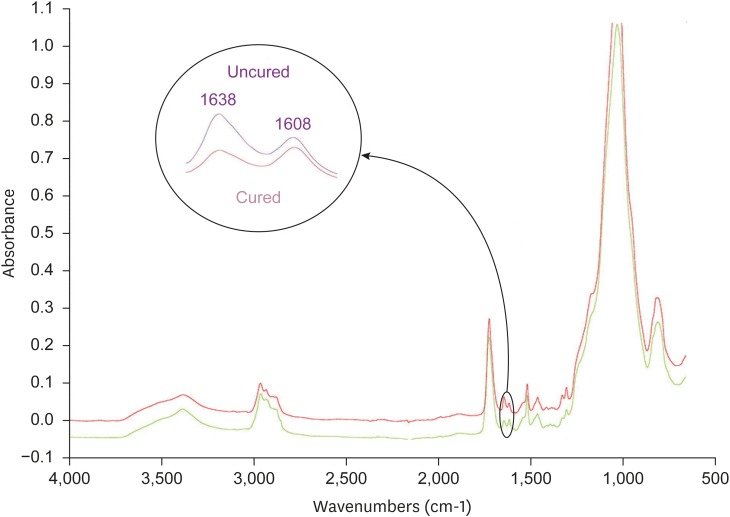
ePub Objective The purpose of this study was to investigate the degree of conversion (DC) and mechanical properties of a microhybrid Filtek Z250 (3M ESPE) resin composite after aging.
Method The specimens were fabricated using circular molds to investigate Vickers microhardness (Vickers hardness number [VHN]) and DC, and were prepared according to ISO 4049 for flexural strength testing. The initial DC (%) of discs was recorded using attenuated total reflectance-Fourier transforming infrared spectroscopy. The initial VHN of the specimens was measured using a microhardness tester under a load of 300 g for 15 seconds and the flexural strength test was carried out with a universal testing machine (crosshead speed, 0.5 mm/min). The specimens were then subjected to thermocycling in 5°C and 55°C water baths. Properties were assessed after 1,000–10,000 cycles of thermocycling. The surfaces were evaluated using scanning electron microscopy (SEM). Data were analyzed using 1-way analysis of variance followed by the Tukey honest significant difference
post hoc test.Results Statistical analysis showed that DC tended to increase up to 4,000 cycles, with no significant changes. VHN and flexural strength values significantly decreased upon thermal cycling when compared to baseline (
p < 0.05). However, there was no significant difference between initial and post-thermocycling VHN results at 1,000 cycles. SEM images after aging showed deteriorative changes in the resin composite surfaces.Conclusions The Z250 microhybrid resin composite showed reduced surface microhardness and flexural strength and increased DC after thermocycling.
-
Citations
Citations to this article as recorded by- Clinical Decision‐Making of Repair vs. Replacement of Defective Direct Dental Restorations: A Multinational Cross‐Sectional Study With Meta‐Analysis
Ömer Hatipoğlu, João Filipe Brochado Martins, Mohmed Isaqali Karobari, Nessrin Taha, Thiyezen Abdullah Aldhelai, Daoud M. Ayyad, Ahmed A. Madfa, Benjamin Martin‐Biedma, Rafael Fernández‐Grisales, Bakhyt A. Omarova, Wen Yi Lim, Suha Alfirjani, Kacper Nijak
Journal of Esthetic and Restorative Dentistry.2025; 37(4): 977. CrossRef - An In Vitro Evaluation of Novel Bioactive Liner's Effect on Marginal Adaptation of Class II Composite Restorations: A Scanning Electron Microscope Analysis
Girija S Sajjan, Naveena Ponnada, Praveen Dalavai, Madhu Varma Kanumuri, Venkata Karteek Varma Penmatsa, B V Sindhuja
World Journal of Dentistry.2025; 15(9): 749. CrossRef - Different contemporary resin cements for intracanal luting of glass fiber posts - Bonding and polymerization assessments
Anna Caroliny Detogni, Vitaliano Gomes de Araújo Neto, Caio Felipe de Almeida Nobre, Victor Pinheiro Feitosa, Mário Alexandre Coelho Sinhoreti
International Journal of Adhesion and Adhesives.2025; 138: 103951. CrossRef - Effect of food-simulating liquids and polishing times on the color stability of microhybrid and nanohybrid resin composites
Muhammet Fidan, Nevin Çankaya
Discover Nano.2025;[Epub] CrossRef - Effect of irrigation protocols for post space preparation on the bond of the resin luting agent and post to a hydraulic calcium silicate filled root: An in vitro study
Nuttanun Poeaim, Sirawut Hiran-us, Yanee Tantilertanant
The Journal of Prosthetic Dentistry.2025; 133(4): 1039.e1. CrossRef - Influence of Different Adhesives and Surface Treatments on Shear and Tensile Bond Strength and Microleakage with Micro-CT of Repaired Bulk-Fill Composites
Handan Yıldırım-Işık, Mediha Büyükgöze-Dindar
Polymers.2025; 17(12): 1680. CrossRef - Effect of thermal ageing on physico-mechanical properties and self-healing potential of experimental 3D-printed denture base resin composites
Mariam Raza Syed, Amr Fawzy
Journal of the Mechanical Behavior of Biomedical Materials.2025; 170: 107123. CrossRef - Effects of aging on the physicomechanical, antimicrobial, and cytotoxicity properties of flowable composite resin with strontium-modified phosphate-based glass
Seo-Hyun Kim, Hye-Bin Go, Myung-Jin Lee, Jae-Sung Kwon
Scientific Reports.2025;[Epub] CrossRef - Colour Stability and Optical Properties of Provisional Crowns Fabricated With Milling, 3D Printing, and Direct Technique
Tommaso Rinaldi, Carlos Serrano Granger, Andrea Santamaría Laorden, Jaime Orejas-Perez, Pablo Gómez Cogolludo
International Dental Journal.2025; 75(6): 103932. CrossRef - EVALUATE DEGREE OF CONVERSION OF NEW BIOACTIVE ORTHODONTIC ADHESIVE WITH COLOR CHANGE & FLUORESCENCE PROPERTY
Mohammed Younis, Neam Fakhri Neam Fakhri
BULLETIN OF STOMATOLOGY AND MAXILLOFACIAL SURGERY.2025; : 39. CrossRef - Antibacterial activity and physicochemical properties of light-curable fluoride varnishes containing strontium phosphate-based glass
Na-Yeon Kim, Mi-Sol Ryu, Ji-Min Lee, Soo-Yeon Jeong, Hye-Been Choi, Myung-Jin Lee, Song-Yi Yang
Clinical Oral Investigations.2025;[Epub] CrossRef - Systematic Review of Studies Comparing Microleakage After Restoration With Cention and Conventional Glass Ionomer Cement in Human Extracted Teeth
Rashmi Misra, Mansi Vandekar, Gayatri Pendse, Omkar Bhosale, Pauravi Hegde, Aashaka Vaishnav
Cureus.2025;[Epub] CrossRef - Evaluation of the radiopacity of different universal composite resins aged by thermocycling
Dilber Çölkesen, Alican Kuran, Neslihan Tekçe
Odontology.2025;[Epub] CrossRef - Exploring the sources and routes of micro- and nanoplastics from dental products and materials: their impact on human health - a systematic review
Vidhya Selvaraj, R. Saravanan, N. Raj Vikram, Uma revathi Gopalakrishnan, Ramsamy M
Next Research.2025; 2(4): 100925. CrossRef - Investigation of the mechanical response of MWCNTs infused carbon/glass fiber-based hybrid composites using digital image correlation
Somaiah Chowdary Mallampati, Ujendra Kumar Komal, Paladugu Rakesh, Parthapratim Barman
Construction and Building Materials.2025; 492: 143068. CrossRef - Mechanical, Surface and Physicochemical Properties of Nanozeolite‐Modified 3D Printed Hybrid Ceramics at Varying Concentrations: An In Vitro Study
Ahmed A. Holiel, Yomna M. Ibrahim, Noha Morsy
Journal of Esthetic and Restorative Dentistry.2025;[Epub] CrossRef - Impact of Graphitic Carbon Nitride on Dental Composite’s Mechanical and Antibacterial Properties
Zainab Rafaqat, Saad Liaqat, Ahmed Bari, Warda Khan Yousafzai, Umar Nishan, Sandleen Feroz, Nawshad Muhammad
Journal of Materials Engineering and Performance.2025;[Epub] CrossRef - Exploring the Biological and Chemical Properties of Emerging 3D-Printed Dental Resin Composites Compared to Conventional Light-Cured Materials
Nikola Živković, Stefan Vulović, Miloš Lazarević, Anja Baraba, Aleksandar Jakovljević, Mina Perić, Jelena Mitrić, Aleksandra Milić Lemić
Materials.2025; 18(22): 5170. CrossRef - Awareness of possible complications associated with direct composite restorations: A multinational survey among dentists from 13 countries with meta-analysis
Anna Lehmann, Kacper Nijakowski, Jakub Jankowski, David Donnermeyer, Paulo J. Palma, Milan Drobac, João Filipe Brochado Martins, Fatma Pertek Hatipoğlu, Indira Tulegenova, Muhammad Qasim Javed, Hamad Mohammad Alharkan, Olga Bekjanova, Sylvia Wyzga, Moataz
Journal of Dentistry.2024; 145: 105009. CrossRef - Comparative evaluation of bond strength and color stability of polyetheretherketone and zirconia layered with indirect composite before and after thermocycling: An in vitro study
Pooja Singh, Subhabrata Maiti, Amrutha Shenoy
The Journal of Indian Prosthodontic Society.2024; 24(3): 252. CrossRef - Biaxial flexural strength of hydrothermally aged resin-based materials
Rodrigo Ricci Vivan, Mariana Miranda de Toledo Piza, Bruna de Mello Silva, Thalya Fernanda Horsth Maltarollo, Gustavo Sivieri-Araujo, Murilo Priori Alcalde, Marco Antonio Hungaro Duarte, Estevam Augusto Bonfante, Henrico Badaoui Strazzi-Sahyon
Journal of the Mechanical Behavior of Biomedical Materials.2024; 155: 106568. CrossRef - Comparative Strength Study of Indirect Permanent Restorations: 3D-Printed, Milled, and Conventional Dental Composites
João Paulo Mendes Tribst, Adelheid Veerman, Gabriel Kalil Rocha Pereira, Cornelis Johannes Kleverlaan, Amanda Maria de Oliveira Dal Piva
Clinics and Practice.2024; 14(5): 1940. CrossRef - Influencia del termociclado sobre la estabilidad del color de dos resinas compuestas
//Influence of thermocycling on the color stability of two composite resins
Verónica Lucía Ventrera, María Eugenia Alejandra Barrionuevo
Revista de la Asociación Odontológica Argentina.2024; : 1. CrossRef - Efeito do protocolo de polimento e do armazenamento em meio úmido na variação de cor, massa e rugosidade superficial de resinas compostas
Leonardo Cruz Morais, Mateus Victória Gontijo, Gabriela Rodrigues Pires, Victor de Morais Gomes, Milton Carlos Kuga, Francisco Fernando Massola Filho, Amanda Gonçalves Franco, Alberto Nogueira da Gama Antunes
Cuadernos de Educación y Desarrollo.2024; 16(6): e4556. CrossRef - A comparison of the mechanical properties of 3D-printed, milled, and conventional denture base resin materials
Hyeong-Ju YU, You-Jung KANG, Yeseul PARK, Hoon KIM, Jee-Hwan KIM
Dental Materials Journal.2024; 43(6): 813. CrossRef - Effect of aging and fiber‐reinforcement on color stability, translucency, and microhardness of single‐shade resin composites versus multi‐shade resin composite
Muhammet Fidan, Özhan Yağci
Journal of Esthetic and Restorative Dentistry.2024; 36(4): 632. CrossRef - Impact of Artificial Aging on the Physical and Mechanical Characteristics of Denture Base Materials Fabricated via 3D Printing
Ahmed Altarazi, Julfikar Haider, Abdulaziz Alhotan, Nick Silikas, Hugh Devlin, Weihao Yuan
International Journal of Biomaterials.2024;[Epub] CrossRef - Synthesis, monomer conversion, and mechanical properties of polylysine based dental composites
Saadia Bano Lone, Rabia Zeeshan, Hina Khadim, Muhammad Adnan Khan, Abdul Samad Khan, Anila Asif
Journal of the Mechanical Behavior of Biomedical Materials.2024; 151: 106398. CrossRef - Bond strength and surface roughness assessment of novel antimicrobial polymeric coated dental cement
Ghada Naguib, Hisham Mously, Jumana Mazhar, Ibrahim Alkanfari, Abdulelah Binmahfooz, Mohammed Zahran, Mohamed T. Hamed
Discover Nano.2024;[Epub] CrossRef - Evaluation of microhardness, degree of conversion, and abrasion resistance of nanoglass and multiwalled carbon nanotubes reinforced three‐dimensionally printed denture base resins
Pansai Ashraf Mohamed, Yomna Mohamed Ibrahim, Kenda Ibrahim Hisham Hanno, Mohamed Mahmoud Abdul‐Monem
Journal of Prosthodontics.2024;[Epub] CrossRef - Effect of CAD-CAM block thickness and translucency on the polymerization of luting materials
Bengü Doğu Kaya, Selinsu Öztürk, Ayşe Aslı Şenol, Erkut Kahramanoğlu, Pınar Yılmaz Atalı, Bilge Tarçın
BMC Oral Health.2024;[Epub] CrossRef - Simulation of oral environmental conditions through artificial aging of teeth for the assessment of enamel discoloration in orthodontics
Celal Irgın
BMC Oral Health.2024;[Epub] CrossRef - Do universal adhesive systems affect color coordinates and color change of single-shade resin composites compared with a multi-shade composite?
Muhammet FİDAN, Özhan YAĞCI
Dental Materials Journal.2023; 42(6): 886. CrossRef - Fabrication, Evaluation, and Performance Ranking of Tri-calcium Phosphate and Silica Reinforced Dental Resin Composite Materials
Sonu Saini, Anoj Meena, Ramkumar Yadav, Amar Patnaik
Silicon.2023; 15(18): 8045. CrossRef - Can Modification with Urethane Derivatives or the Addition of an Anti-Hydrolysis Agent Influence the Hydrolytic Stability of Resin Dental Composite?
Agata Szczesio-Wlodarczyk, Izabela M. Barszczewska-Rybarek, Marta W. Chrószcz-Porębska, Karolina Kopacz, Jerzy Sokolowski, Kinga Bociong
International Journal of Molecular Sciences.2023; 24(5): 4336. CrossRef - Effect of veneering material type and thickness ratio on flexural strength of bi-layered PEEK restorations before and after thermal cycling
Ahmed Gouda, Ashraf Sherif, Mennatallah Wahba, Tarek Morsi
Clinical Oral Investigations.2023; 27(6): 2629. CrossRef - 3D printed denture base material: The effect of incorporating TiO2 nanoparticles and artificial ageing on the physical and mechanical properties
Ahmed Altarazi, Julfikar Haider, Abdulaziz Alhotan, Nick Silikas, Hugh Devlin
Dental Materials.2023; 39(12): 1122. CrossRef - Influence of silane coupling agent and aging on the repair bond strength of dental composites
Gustavo Jusué-Esparza, José Alejandro Rivera-Gonzaga, Guillermo Grazioli, Ana Josefina Monjarás-Ávila, J. Eliezer Zamarripa-Calderón, Carlos Enrique Cuevas-Suárez
Journal of Adhesion Science and Technology.2023; 37(5): 913. CrossRef - Degree of conversion of light‐polymerized composite resin in implant prosthesis screw access opening
Se‐Hyun Park, Yoon‐Hyuk Huh, Chan‐Jin Park, Lee‐Ra Cho, Kyung‐Ho Ko
Journal of Prosthodontics.2023; 32(9): 829. CrossRef - Investigation of the effect of matrix-interface formed with silane-based coupling agents on physico-chemical behavior and flow distance of dental composites
Zerin Yeşil Acar, Merve Tunç Koçyiğit, Meltem Asiltürk
Journal of Molecular Liquids.2023; 378: 121600. CrossRef - Evaluation of Water Sorption and Solubility of 3D-Printed, CAD/CAM Milled, and PMMA Denture Base Materials Subjected to Artificial Aging
Mariya Dimitrova, Angelina Vlahova, Ilian Hristov, Rada Kazakova, Bozhana Chuchulska, Stoyan Kazakov, Marta Forte, Vanja Granberg, Giuseppe Barile, Saverio Capodiferro, Massimo Corsalini
Journal of Composites Science.2023; 7(8): 339. CrossRef - Effect of thermocycling on internal microhardness of high and low viscosity bulk fill composite resins in class I cavities
Sâmara Luciana de Andrade LIMA, Lais Lemos CABRAL, Natália Russo CARLOS, Saulo André de Andrade LIMA, Kamila Rosamilia KANTOVITZ, Flávia Lucisano Botelho do AMARAL
RGO - Revista Gaúcha de Odontologia.2023;[Epub] CrossRef - Effects of an Acidic Environment on the Strength and Chemical Changes of Resin-based Composites
S Kang, B-H Cho
Operative Dentistry.2023; 48(4): E81. CrossRef - Influence of compressive forces and aging through thermocycling on the strength of mono incremental dental composite resins
Cristian Roberto Sigcho Romero, Henry Fabricio Mejía Mosquera, Sandra Marcela Quisiguiña Guevara, Yudy Jacqueline Alvarado Aguayo
Bionatura.2023; 8(4): 1. CrossRef - Push-out Bond Strength of Two Fiber Posts in Composite Resin Using Different Types of Silanization
RM Novis, BLT Leon, FMG França, CP Turssi, RT Basting, FLB Amaral
Operative Dentistry.2022; 47(2): 173. CrossRef - Penetration and Adaptation of the Highly Viscous Zinc-Reinforced Glass Ionomer Cement on Contaminated Fissures: An In Vitro Study with SEM Analysis
Galiah AlJefri, Sunil Kotha, Muhannad Murad, Reham Aljudaibi, Fatmah Almotawah, Sreekanth Mallineni
International Journal of Environmental Research and Public Health.2022; 19(10): 6291. CrossRef - Surface Characteristics and Color Stability of Dental PEEK Related to Water Saturation and Thermal Cycling
Liliana Porojan, Flavia Roxana Toma, Mihaela Ionela Bîrdeanu, Roxana Diana Vasiliu, Ion-Dragoș Uțu, Anamaria Matichescu
Polymers.2022; 14(11): 2144. CrossRef - Effects of aging and light-curing unit type on the volume and internal porosity of bulk-fill resin composite restoration
Afnan O. Al-Zain, Elaf A. Alboloshi, Walaa A. Amir, Maryam A. Alghilan, Eliseu A. Münchow
The Saudi Dental Journal.2022; 34(3): 243. CrossRef - An Evaluation of the Hydrolytic Stability of Selected Experimental Dental Matrices and Composites
Agata Szczesio-Wlodarczyk, Karolina Kopacz, Malgorzata Iwona Szynkowska-Jozwik, Jerzy Sokolowski, Kinga Bociong
Materials.2022; 15(14): 5055. CrossRef - Comparison of the Mechanical Properties and Push-out Bond Strength of Self-adhesive and Conventional Resin Cements on Fiber Post Cementation
MR Santi, RBE Lins, BO Sahadi, JR Soto-Montero, LRM Martins
Operative Dentistry.2022; 47(3): 346. CrossRef - Effect of Different Polymerization Times on Color Change, Translucency Parameter, and Surface Hardness of Bulk-Fill Resin Composites
HY Gonder, M Fidan
Nigerian Journal of Clinical Practice.2022; 25(10): 1751. CrossRef - Surface degradation and biofilm formation on hybrid and nanohybrid composites after immersion in different liquids
Gabriela Escamilla-Gómez, Octavio Sánchez-Vargas, Diana M. Escobar-García, Amaury Pozos-Guillén, Norma V. Zavala-Alonso, Mariana Gutiérrez-Sánchez, José E. Pérez-López, Gregorio Sánchez-Balderas, Gabriel F. Romo-Ramírez, Marine Ortiz-Magdaleno
Journal of Oral Science.2022; 64(4): 263. CrossRef - Effects of Different Adhesive Systems and Orthodontic Bracket Material on Enamel Surface Discoloration: An In Vitro Study
Ali Alqerban, Doaa R. M. Ahmed, Ali S. Aljhani, Dalal Almadhi, Amjad AlShahrani, Hussah AlAdwene, Abdulaziz Samran
Applied Sciences.2022; 12(24): 12885. CrossRef - Effects of Immediate Coating on Unset Composite with Different Bonding Agents to Surface Hardness
Nantawan Krajangta, Supissara Ninbanjong, Sunisa Khosook, Kanjana Chaitontuak, Awiruth Klaisiri
European Journal of Dentistry.2022; 16(04): 828. CrossRef - Rational durability of optical properties of chameleon effect of Omnichroma and Essentia composite thermocycled in black dark drinks (in vitro study)
Bassma Abdelhamed, Asmaa Abdel-Hakeem Metwally, Heba A. Shalaby
Bulletin of the National Research Centre.2022;[Epub] CrossRef - Comparative Evaluation of Shear Bond Strength of Nanohybrid Composite Restoration After the Placement of Flowable Compomer and Composite Using the Snowplow Technique
Meghna Dugar, Anuja Ikhar, Pradnya Nikhade, Manoj Chandak, Nidhi Motwani
Cureus.2022;[Epub] CrossRef - The First Step in Standardizing an Artificial Aging Protocol for Dental Composites—Evaluation of Basic Protocols
Agata Szczesio-Wlodarczyk, Magdalena Fronczek, Katarzyna Ranoszek-Soliwoda, Jarosław Grobelny, Jerzy Sokolowski, Kinga Bociong
Molecules.2022; 27(11): 3511. CrossRef - Effect of Different Surface Treatments on the Long‐Term Repair Bond Strength of Aged Methacrylate‐Based Resin Composite Restorations: A Systematic Review and Network Meta‐analysis
Mahdi Hadilou, Amirmohammad Dolatabadi, Morteza Ghojazadeh, Hossein Hosseinifard, Parnian Alizadeh Oskuee, Fatemeh Pournaghi Azar, Victor Feitosa
BioMed Research International.2022;[Epub] CrossRef - Edge chipping resistance of veneering composite resins
Parissa Nassary Zadeh, Bogna Stawarczyk, Rüdiger Hampe, Anja Liebermann, Felicitas Mayinger
Journal of the Mechanical Behavior of Biomedical Materials.2021; 116: 104349. CrossRef - The effect of radiation exposure and storage time on the degree of conversion and flexural strength of different resin composites
Ragia M. Taher, Lamiaa M. Moharam, Amin E. Amin, Mohamed H. Zaazou, Farid S. El-Askary, Mokhtar N. Ibrahim
Bulletin of the National Research Centre.2021;[Epub] CrossRef - Fracture Load of CAD/CAM Fabricated Cantilever Implant-Supported Zirconia Framework: An In Vitro Study
Ibraheem F. Alshiddi, Syed Rashid Habib, Muhammad Sohail Zafar, Salwa Bajunaid, Nawaf Labban, Mohammed Alsarhan
Molecules.2021; 26(8): 2259. CrossRef - A numerical, theoretical and experimental study of the effect of thermocycling on the matrix-filler interface of dental restorative materials
Yoan Boussès, Nathalie Brulat-Bouchard, Pierre-Olivier Bouchard, Yannick Tillier
Dental Materials.2021; 37(5): 772. CrossRef - Impact of polymerization and storage on the degree of conversion and mechanical properties of veneering resin composites
Felicitas MAYINGER, Marcel REYMUS, Anja LIEBERMANN, Marc RICHTER, Patrick KUBRYK, Henning GROẞEKAPPENBERG, Bogna STAWARCZYK
Dental Materials Journal.2021; 40(2): 487. CrossRef - Intrapulpal Concentration of Hydrogen Peroxide of Teeth Restored With Bulk Fill and Conventional Bioactive Composites
DP Silva, BA Resende, M Kury, CB André, CPM Tabchoury, M Giannini, V Cavalli
Operative Dentistry.2021; 46(3): E158. CrossRef - Silane content influences physicochemical properties in nanostructured model composites
Larissa Maria Cavalcante, Lucielle Guimarães Ferraz, Karinne Bueno Antunes, Isadora Martini Garcia, Luis Felipe Jochims Schneider, Fabrício Mezzomo Collares
Dental Materials.2021; 37(2): e85. CrossRef - AĞIZ GARGARALARI VE ANTİSEPTİKLERİNİN FARKLI KOMPOZİT REZİNLERİN RENK STABİLİTESİNE ETKİSİ
Turan Emre KUZU, Özcan KARATAŞ
Atatürk Üniversitesi Diş Hekimliği Fakültesi Dergisi.2021; : 1. CrossRef - Evaluation of Immediate and Delayed Microleakage of Class V Cavities Restored with Chitosan-incorporated Composite Resins: An In Vitro Study
Roopa R Nadig, Veena Pai, Arpita Deb
International Journal of Clinical Pediatric Dentistry.2021; 14(5): 621. CrossRef - Influence of Diode Laser for the Treatment of Dentin Hypersensitivity on Microleakage of Cervical Restorations
Doaa R. M. Ahmed, Diana G. Shaath, Jomana B. Alakeel, Abdulaziz A. Samran, Mona Abbassy
BioMed Research International.2021;[Epub] CrossRef - Ageing of Dental Composites Based on Methacrylate Resins—A Critical Review of the Causes and Method of Assessment
Agata Szczesio-Wlodarczyk, Jerzy Sokolowski, Joanna Kleczewska, Kinga Bociong
Polymers.2020; 12(4): 882. CrossRef - Flexural strength and surface microhardness of materials used for temporary dental disocclusion submitted to thermal cycling: An in vitro study
Tamires Borges de Lima, José Guilherme Neves, Ana Paula Terossi de Godoi, Ana Rosa Costa, Viviane Veroni Degan, Américo Bortolazzo Correr, Heloisa Cristina Valdrighi
International Orthodontics.2020; 18(3): 519. CrossRef - Evaluation of the repair capacities and color stabilities of a resin nanoceramic and hybrid CAD/CAM blocks
Hasibe Sevilay Bahadır, Yusuf Bayraktar
The Journal of Advanced Prosthodontics.2020; 12(3): 140. CrossRef - Effect of Different Surface Treatments of Resin Relined Fiber Posts Cemented With Self-adhesive Resin Cement on Push-out and Microtensile Bond Strength Tests
RV Machry, PE Fontana, TC Bohrer, LF Valandro, OB Kaizer
Operative Dentistry.2020; 45(4): E185. CrossRef - Influences of Successive Exposure to Bleaching and Fluoride Preparations on the Surface Hardness and Roughness of the Aged Resin Composite Restoratives
Khalid M. Abdelaziz, Shugufta Mir, Shafait Ullah Khateeb, Suheel M. Baba, Saud S. Alshahrani, Eman A. Alshahrani, Zahra A. Alsafi
Medicina.2020; 56(9): 476. CrossRef - Fracture Resistance of Lithıum Disilicate, Indirect Resin Composite and Zirconıa by Using Dual Cure Resin Cements
Mohammed BADWAN, Erkut KAHRAMANOĞLU
Clinical and Experimental Health Sciences.2020; 10(4): 435. CrossRef - Effect of Stress-decreasing Resin Thickness as an Intermediate Layer on Fracture Resistance of Class II Composite Restoration: An In Vitro Study
Dennis Dennis, Arwin Leonardy, Trimurni Abidin
World Journal of Dentistry.2020; 11(2): 91. CrossRef - Effect of Thermocycling on Biaxial Flexural Strength of CAD/CAM, Bulk Fill, and Conventional Resin Composite Materials
EB Benalcázar Jalkh, CM Machado, M Gianinni, I Beltramini, MMT Piza, PG Coelho, R Hirata, EA Bonfante
Operative Dentistry.2019; 44(5): E254. CrossRef - Mechanical properties of hybrid computer-aided design/computer-aided manufacturing (CAD/CAM) materials after aging treatments
Hae-Yong Jeong, Hae-Hyoung Lee, Yu-Sung Choi
Ceramics International.2018; 44(16): 19217. CrossRef
- Clinical Decision‐Making of Repair vs. Replacement of Defective Direct Dental Restorations: A Multinational Cross‐Sectional Study With Meta‐Analysis
- 6,416 View
- 47 Download
- 77 Crossref

- Analysis of the shelf life of chitosan stored in different types of packaging, using colorimetry and dentin microhardness
- Antonio Miranda da Cruz-Filho, Angelo Rafael de Vito Bordin, Luis Eduardo Souza-Flamini, Débora Fernandes da Costa Guedes, Paulo César Saquy, Ricardo Gariba Silva, Jesus Djalma Pécora
- Restor Dent Endod 2017;42(2):87-94. Published online March 27, 2017
- DOI: https://doi.org/10.5395/rde.2017.42.2.87
-
 Abstract
 PDF
PDF PubReader
 ePub
ePub Objectives Chitosan has been widely investigated and used. However, the literature does not refer to the shelf life of this solution. This study evaluated, through the colorimetric titration technique and an analysis of dentin micro-hardness, the shelf life of 0.2% chitosan solution.
Materials and Methods Thirty human canines were sectioned, and specimens were obtained from the second and third slices, from cemento-enamel junction to the apex. A 0.2% chitosan solution was prepared and distributed in 3 identical glass bottles (v1, v2, and v3) and 3 plastic bottles (p1, p2, and p3). At 0, 7, 15, 30, 45, 60, 90, 120, 150, and 180 days, the specimens were immersed in each solution for 5 minutes (
n = 3 each). The chelating effect of the solution was assessed by micro-hardness and colorimetric analysis of the dentin specimens. 17% EDTA and distilled water were used as controls. Data were analyzed statistically by two-way and Tukey-Kramer multiple comparison (α = 0.05).Results There was no statistically significant difference among the solutions with respect to the study time (
p = 0.113) and micro-hardness/time interaction (p = 0.329). Chitosan solutions and EDTA reduced the micro-hardness in a similar manner and differed significantly from the control group (p < 0.001). Chitosan solutions chelated calcium ions throughout the entire experiment.Conclusions Regardless of the storage form, chitosan demonstrates a chelating property for a minimum period of 6 months.
-
Citations
Citations to this article as recorded by- Chitosan’s Ability to Remove the Smear Layer—A Systematic Review of Ex Vivo Studies
Ana Ferreira-Reguera, Inês Ferreira, Irene Pina-Vaz, Benjamín Martín-Biedma, José Martín-Cruces
Medicina.2025; 61(1): 114. CrossRef - The Utilization of Chitosan and Arduino Interface in Making a Microplastic Filter
Kate Cyrene P. Pineda, Maeven Uriel A. Dela Cruz, Quirsten Daniel R. Repalda, Aldrin Jeynard A. Gonzales, DL Chaturika C. Douglas, Alina Siara D. Hajan, Julie Ann B. Real
International Journal of Innovative Science and Research Technology.2025; : 2742. CrossRef - Antimicrobial evaluation of root canal irrigants of natural sources with and without gamma radiation activation - An in vitro study
Hoda Raafat Yousri, Abeer Hashem Mahran, Ahmed Abdel Rahman Hashem, Amal A. El-Batouti
Endodontology.2024; 36(4): 383. CrossRef - Influence of Chitosan 0.2% in Various Final Cleaning Methods on the Bond Strength of Fiberglass Post to Intrarradicular Dentin
Naira Geovana Camilo, Alex da Rocha Gonçalves, Larissa Pinzan Flauzino, Cristiane Martins Rodrigues Bernardes, Andreza Maria Fábio Aranha, Priscilla Cardoso Lazari-Carvalho, Marco Aurélio de Carvalho, Helder Fernandes de Oliveira
Polymers.2023; 15(22): 4409. CrossRef - Evaluation and comparison of anti-inflammatory properties of ibuprofen using two drug delivery systems after third molar surgery: using chitosan microspheres as a carrier for local drug delivery in to the third molar socket and through the oral route
Karthik KP, Balamurugan R
British Journal of Oral and Maxillofacial Surgery.2021; 59(2): 191. CrossRef - Optimization of chitosan nanoparticle synthesis and its potential application as germination elicitor of Oryza sativa L.
K. Divya, Smitha Vijayan, Sreekumar Janardanan Nair, M.S. Jisha
International Journal of Biological Macromolecules.2019; 124: 1053. CrossRef - Crosstalk between chitosan and cell signaling pathways
Behrouz Farhadihosseinabadi, Amir Zarebkohan, Mohamad Eftekhary, Mohammad Heiat, Mehrdad Moosazadeh Moghaddam, Mazaher Gholipourmalekabadi
Cellular and Molecular Life Sciences.2019; 76(14): 2697. CrossRef
- Chitosan’s Ability to Remove the Smear Layer—A Systematic Review of Ex Vivo Studies
- 1,335 View
- 7 Download
- 7 Crossref

- Carbohydrate-electrolyte drinks exhibit risks for human enamel surface loss
- Mary Anne Sampaio de Melo, Vanara Florêncio Passos, Juliana Paiva Marques Lima, Sérgio Lima Santiago, Lidiany Karla Azevedo Rodrigues
- Restor Dent Endod 2016;41(4):246-254. Published online August 16, 2016
- DOI: https://doi.org/10.5395/rde.2016.41.4.246
-
 Abstract
 PDF
PDF PubReader
 ePub
ePub Objectives The aim of this investigation was to give insights into the impact of carbohydrate-electrolyte drinks on the likely capacity of enamel surface dissolution and the influence of human saliva exposure as a biological protective factor.
Materials and Methods The pH, titratable acidity (TA) to pH 7.0, and buffer capacity (β) of common beverages ingested by patients under physical activity were analyzed. Then, we randomly distributed 50 specimens of human enamel into 5 groups. Processed and natural coconut water served as controls for testing three carbohydrate-electrolyte drinks. In all specimens, we measured surface microhardness (Knoop hardness numbers) and enamel loss (profilometry, µm) for baseline and after simulated intake cycling exposure model. We also prepared areas of specimens to be exposed to human saliva overnight prior to the simulated intake cycling exposure. The cycles were performed by alternated immersions in beverages and artificial saliva. ANOVA two-way and Tukey HDS tests were used.
Results The range of pH, TA, and β were 2.85 - 4.81, 8.33 - 46.66 mM/L and 3.48 - 10.25 mM/L × pH, respectively. The highest capacity of enamel surface dissolution was found for commercially available sports drinks for all variables. Single time human saliva exposure failed to significantly promote protective effect for the acidic attack of beverages.
Conclusions In this study, carbohydrate-electrolyte drinks usually consumed during endurance training may have a greater capacity of dissolution of enamel surface depending on their physicochemical proprieties associated with pH and titratable acidity.
-
Citations
Citations to this article as recorded by- Evaluation of developmentally hypomineralised enamel after surface pretreatment with Papacarie Duo gel and different etching modes: an in vitro SEM and AFM study
Y.-L. Lee, K. C. Li, C. K. Y. Yiu, D. H. Boyd, M. Ekambaram
European Archives of Paediatric Dentistry.2022; 23(1): 117. CrossRef - Is the consumption of beverages and food associated to dental erosion? A cross-sectional study in Portuguese athletes
M.-R.G. Silva, M.-A. Chetti, H. Neves, M.-C. Manso
Science & Sports.2021; 36(6): 477.e1. CrossRef - Assessment of surface roughness changes on orthodontic acrylic resin by all-in-one spray disinfectant solutions
Kuei-ling Hsu, Abdulrahman A. Balhaddad, Isadora Martini Garcia, Fabricio Mezzomo Collares, Louis DePaola, Mary Anne Melo
Journal of Dental Research, Dental Clinics, Dental Prospects.2020; 14(2): 77. CrossRef - Nitrate-rich beetroot juice offsets salivary acidity following carbohydrate ingestion before and after endurance exercise in healthy male runners
Mia C. Burleigh, Nicholas Sculthorpe, Fiona L. Henriquez, Chris Easton, Yi-Hung Liao
PLOS ONE.2020; 15(12): e0243755. CrossRef - Dental erosion’ prevalence and its relation to isotonic drinks in athletes: a systematic review and meta-analysis
Pedro Henrique Pereira de Queiroz Gonçalves, Ludmila Silva Guimarães, Fellipe Navarro Azevedo de Azeredo, Letícia Maira Wambier, Lívia Azeredo A. Antunes, Leonardo Santos Antunes
Sport Sciences for Health.2020; 16(2): 207. CrossRef - Atomic force microscopy analysis of enamel nanotopography after interproximal reduction
Shadi Mohebi, Nazila Ameli
American Journal of Orthodontics and Dentofacial Orthopedics.2017; 152(3): 295. CrossRef
- Evaluation of developmentally hypomineralised enamel after surface pretreatment with Papacarie Duo gel and different etching modes: an in vitro SEM and AFM study
- 1,831 View
- 14 Download
- 6 Crossref

- Effect of three nanobiomaterials on microhardness of bleached enamel
- Maryam Khoroushi, Farinaz Shirban, Sara Kaveh, Samaneh Doustfateme
- Restor Dent Endod 2016;41(3):196-201. Published online July 14, 2016
- DOI: https://doi.org/10.5395/rde.2016.41.3.196
-
 Abstract
 PDF
PDF PubReader
 ePub
ePub Objectives The aim of this
in vitro study was to evaluate the effect of incorporating three different nanobiomaterials into bleaching material on microhardness of bleached enamel.Materials and Methods The crowns of 24 extracted sound human molars were sectioned. Sixty enamel specimens (2 × 3 × 4 mm) were selected and divided into five groups (
n = 12): Group 1 received no bleaching procedure (control); Group 2 underwent bleaching with a 40% hydrogen peroxide (HP) gel; Groups 3, 4, and 5 were bleached with a 40% HP gel modified by incorporation of bioactive glass (BAG), amorphous calcium phosphate (ACP) and hydroxyapatite (HA), respectively. The enamel microhardness was evaluated. The differences in Knoop microhardness data of each group were analyzed by one-way ANOVA, followed bypost hoc Tukey tests.Results Significant differences were observed between the study groups. The enamel microhardness changes in Groups 1, 3, 4, and 5 were significantly lower than that of Group 2 (
p < 0.001).Conclusions Within the limitations of this study, it can be concluded that incorporation of each one of the three tested biomaterials as remineralizing agents might be effective in decreasing enamel microhardness changes subsequent to in-office bleaching.
-
Citations
Citations to this article as recorded by- Protective role of calcium-based agents in dental bleaching gels: insights from a systematic review and meta-analysis of clinical and laboratory evidence
Gabriel Pereira Nunes, Renata de Oliveira Alves, Geórgia Rondó Peres, Matheus Henrique Faccioli Ragghianti, Priscila Toninatto Alves de Toledo, Alexandre Henrique dos Reis Prado, Carla Ferreira-Baptista, Alberto Carlos Botazzo Delbem
Clinical Oral Investigations.2025;[Epub] CrossRef - Analysis of Enamel Surface and Shear Bond Strength with Orthodontic Bracket at Different Time Intervals After Bleaching Treatment
Yu-Jin Lee, Ji-Yeon Hong, Hye-Min Ku, Song-Yi Yang
Journal of Dental Hygiene Science.2025; 25(2): 130. CrossRef - Effect of strontium fluorophosphate bioactive glass on color, microhardness and surface roughness of bleached enamel
Shiza Yezdani, Monisha Khatri, Sampath Vidhya, Sekar Mahalaxmi
Technology and Health Care.2024; 32(1): 285. CrossRef - In vitro evaluation of the effect of addition of biomaterials to carbamide peroxide on the bleaching efficacy and microhardness of enamel
Sowmya Kavoor, M. A. Ranjini, Naval Abdul Aziz, H. K. Ashok, Roopa R. Nadig
Journal of Conservative Dentistry and Endodontics.2024; 27(3): 310. CrossRef - Effect of hydrogen peroxide and its combination with nano-hydroxyapatite or nano-bioactive glass on the enamel demineralization and tooth color: An in vitro study
Elham Kheradmand, Alirea Daneshkazemi, Abdolrahim Davari, Maede Kave, Solmaz Ghanbarnejad
Dental Research Journal.2023;[Epub] CrossRef - Over‐the‐counter bleaching agents can help with tooth whitening maintenance
Olívia Santana Jorge, Carolina Noronha Ferraz de Arruda, Rafaella Tonani Torrieri, Rocio Geng Vivanco, Fernanda de Carvalho Panzeri Pires‐de‐Souza
Journal of Esthetic and Restorative Dentistry.2022; 34(2): 328. CrossRef - Effect of Indigenously Developed Nano-Hydroxyapatite Crystals from Chicken Egg Shell on the Surface Hardness of Bleached Human Enamel
Divya Kunam, Vidhya Sampath, Sujatha Manimaran, Mahalaxmi Sekar
Contemporary Clinical Dentistry.2019; 10(3): 489. CrossRef - Influence of Time Intervals between Bleaching Procedures on Enamel Microhardness and Surface Roughness
Roberta Pimentel de Oliveira, Juliana Costa Pereira Baia, Mara Eliane Soares Ribeiro, Mario Honorato da Silva e Souza Junior, Sandro Cordeiro Loretto
The Open Dentistry Journal.2018; 12(1): 555. CrossRef
- Protective role of calcium-based agents in dental bleaching gels: insights from a systematic review and meta-analysis of clinical and laboratory evidence
- 1,477 View
- 7 Download
- 8 Crossref

- The effect of different fluoride application methods on the remineralization of initial carious lesions
- Seon Mi Byeon, Min Ho Lee, Tae Sung Bae
- Restor Dent Endod 2016;41(2):121-129. Published online May 10, 2016
- DOI: https://doi.org/10.5395/rde.2016.41.2.121
-
 Abstract
 PDF
PDF PubReader
 ePub
ePub Objectives The purpose of this study was to assess the effect of single and combined applications of fluoride on the amount of fluoride release, and the remineralization and physical properties of enamel.
Materials and Methods Each of four fluoride varnish and gel products (Fluor Protector, FP, Ivoclar Vivadent; Tooth Mousse Plus, TM, GC; 60 Second Gel, A, Germiphene; CavityShield, CS, 3M ESPE) and two fluoride solutions (2% sodium fluoride, N; 8% tin(ii) fluoride, S) were applied on bovine teeth using single and combined methods (10 per group), and then the amount of fluoride release was measured for 4 wk. The electron probe microanalysis and the Vickers microhardness measurements were conducted to assess the effect of fluoride application on the surface properties of bovine teeth.
Results The amount of fluoride release was higher in combined applications than in single application (
p < 0.05). Microhardness values were higher after combined applications of N with FP, TM, and CS than single application of them, and these values were also higher after combined applications of S than single application of A (p < 0.05). Ca and P values were higher in combined applications of N with TM and CS than single application of them (p < 0.05). They were also increased after combined applications of the S with A than after single application (p < 0.05).Conclusions Combined applications of fluoride could be used as a basis to design more effective methods of fluoride application to provide enhanced remineralization.
-
Citations
Citations to this article as recorded by- Effect of Different Topical Fluorides on the Microhardness of Bleached Enamel: In Vitro Study
Soumyashri Das, Mansi Jain, HP Suma Sogi, Sonali Sukesh K, Apurva Gambhir, FNU Gagandeep
International Journal of Clinical Pediatric Dentistry.2025; 18(11): 1365. CrossRef - Therapeutic effect of ozone gel on the initial carious lesions
Maha A. Alsharqawy, Wedad M Etman, Mirvat M Salama, Reda G. Saleh
Tanta Dental Journal.2023; 20(3): 203. CrossRef - Evaluation of Remineralization Potential of Natural Substances on Artificially Induced Carious Lesions in Primary Teeth: An In Vitro Study
Kavitha Ramar, Pooja V Ravi, Rajakumar Sekar
International Journal of Clinical Pediatric Dentistry.2023; 16(2): 244. CrossRef - Upaya Preventif Kesehatan Gigi dan Mulut dengan Aplikasi Fluor pada Gigi Siswa SMPN 77 Jakarta
Agus Ardinansyah, Mochammad Atmaji Windrianto, Nur Hidayati Nosi Prastiyani
Info Abdi Cendekia.2023; 6(2): 74. CrossRef - Evaluation of the antibacterial activity of Enamelast® and Fluor defender® fluoride varnishes against Streptococcus mutans biofilm: an in vitro study in primary teeth
M. A. Matar, S. S. Darwish, R. S. Salma, W. A. Lotfy
European Archives of Paediatric Dentistry.2023; 24(5): 549. CrossRef - In-vitro evaluation of the anti-cariogenic effect of a hybrid coating associated with encapsulated sodium fluoride and stannous chloride in nanoclays on enamel
Sávio José Cardoso BEZERRA, Ítallo Emídio Lira VIANA, Idalina Vieira AOKI, Simone DUARTE, Anderson Takeo HARA, Taís SCARAMUCCI
Journal of Applied Oral Science.2022;[Epub] CrossRef - Comparative Evaluation of Salivary Fluoride Concentration after Topical Application of Silver Diamine Fluoride and Sodium Fluoride: A Randomized Controlled Trial
Nidhi Agarwal, V Vishnu Priya, Zohra Jabin, Iffat Nasim
International Journal of Clinical Pediatric Dentistry.2022; 15(3): 371. CrossRef - Release and Recharge of Fluoride Ions from Acrylic Resin Modified with Bioactive Glass
Zbigniew Raszewski, Danuta Nowakowska, Wlodzimierz Wieckiewicz, Agnieszka Nowakowska-Toporowska
Polymers.2021; 13(7): 1054. CrossRef - Enamel remineralisation-inducing materials for caries prevention
Sri Kunarti, Widya Saraswati, Dur Muhammad Lashari, Nadhifa Salma, Tasya Nafatila
Dental Journal.2021; 54(3): 165. CrossRef - Fluoride Concentration in Saliva following Professional Topical Application of 2% Sodium Fluoride Solution
Manjit Talwar, Amrit Tewari, H. S. Chawla, Vinod Sachdev, Suresh Sharma
Contemporary Clinical Dentistry.2019; 10(3): 423. CrossRef - Clinical and laboratory evaluation of the Elgydium Protection caries toothpaste effectiveness in patients with high intensity of dental caries
O. A. Zorina, N. B. Petruhina, A. Z. M, O. A. Boriskina, A. A. Tupicin, V. A. Prohodnaja
Stomatologiya.2019; 98(3): 21. CrossRef - Bleaching of simulated stained-remineralized caries lesions in vitro
Sarah S. Al-Angari, Frank Lippert, Jeffrey A. Platt, George J. Eckert, Carlos González-Cabezas, Yiming Li, Anderson T. Hara
Clinical Oral Investigations.2019; 23(4): 1785. CrossRef - Short-Time Antibacterial Effects of Dimethylaminododecyl Methacrylate on Oral Multispecies Biofilm In Vitro
Yujie Zhou, Suping Wang, Xuedong Zhou, Yiran Zou, Mingyun Li, Xian Peng, Biao Ren, Hockin H. K. Xu, Michael D. Weir, Lei Cheng, Yu Chen, Qi Han
BioMed Research International.2019; 2019: 1. CrossRef - Comparison of the Application of Different Fluoride Supplements on Enamel Demineralization Adjacent to Orthodontic Brackets: An In Vitro Study
Arman Mohammadi Shayan, Monireh Rassouli, Soodabeh Kimyai, Hadi Valizadeh, Mohammad Hossein Ahangar Atashi, Sahand Rikhtegaran
Iranian Journal of Orthodontics.2019;[Epub] CrossRef - Effects of nicomethanol hydrofluoride on dental enamel and synthetic apatites: a role for anti-caries protection
N. Sharkov
European Archives of Paediatric Dentistry.2017; 18(6): 411. CrossRef - Intérêt prophylactique et thérapeutique des chewing-gums sans sucre en orthodontie. Une étude menée auprès de professionnels de santé et de patients
Pauline Ferney, François Clauss, Damien Offner, Delphine Wagner
L'Orthodontie Française.2017; 88(3): 275. CrossRef - Silver Diamine Fluoride Has Efficacy in Controlling Caries Progression in Primary Teeth: A Systematic Review and Meta-Analysis
Ana Cláudia Chibinski, Letícia Maíra Wambier, Juliana Feltrin, Alessandro Dourado Loguercio, Denise Stadler Wambier, Alessandra Reis
Caries Research.2017; 51(5): 527. CrossRef - Dental Caries Management of a Patient with a High Caries Risk Based on the Caries Risk Assessment: a Case Peport
Dong-Hyun Lee, Sung-Ok Hong, Seok-Ryun Lee
Korean Journal of Dental Materials.2016; 43(3): 231. CrossRef
- Effect of Different Topical Fluorides on the Microhardness of Bleached Enamel: In Vitro Study
- 1,994 View
- 13 Download
- 18 Crossref

- Effect of acidic solutions on the microhardness of dentin and set OrthoMTA and their cytotoxicity on murine macrophage
- Soram Oh, Hiran Perinpanayagam, Yoon Lee, Jae-Won Kum, Yeon-Jee Yoo, Sang-Min Lim, Seok Woo Chang, Won-Jun Shon, Woocheol Lee, Seung-Ho Baek, Kee-Yeon Kum
- Restor Dent Endod 2016;41(1):12-21. Published online December 1, 2015
- DOI: https://doi.org/10.5395/rde.2016.41.1.12

-
 Abstract
 PDF
PDF PubReader
 ePub
ePub Objectives To evaluate the effects of three acids on the microhardness of set mineral trioxide aggregate (MTA) and root dentin, and cytotoxicity on murine macrophage.
Materials and Methods OrthoMTA (BioMTA) was mixed and packed into the human root dentin blocks of 1.5 mm diameter and 5 mm height. Four groups, each of ten roots, were exposed to 10% citric acid (CA), 5% glycolic acid (GA), 17% ethylenediaminetetraacetic acid (EDTA), and saline for five minutes after setting of the OrthoMTA. Vickers surface microhardness of set MTA and dentin was measured before and after exposure to solutions, and compared between groups using one-way ANOVA with Tukey test. The microhardness value of each group was analyzed using student
t test. Acid-treated OrthoMTA and dentin was examined by scanning electron microscope (SEM). Cell viability of tested solutions was assessed using WST-8 assay and murine macrophage.Results Three test solutions reduced microhardness of dentin. 17% EDTA demonstrated severe dentinal erosion, significantly reduced the dentinal microhardness compared to 10% CA (
p = 0.034) or 5% GA (p = 0.006). 10% CA or 5% GA significantly reduced the surface microhardness of set MTA compared to 17% EDTA and saline (p < 0.001). Acid-treated OrthoMTA demonstrated microporous structure with destruction of globular crystal. EDTA exhibited significantly more cellular toxicity than the other acidic solutions at diluted concentrations (0.2, 0.5, 1.0%).Conclusions Tested acidic solutions reduced microhardness of root dentin. Five minute's application of 10% CA and 5% GA significantly reduced the microhardness of set OrthoMTA with lower cellular cytotoxicity compared to 17% EDTA.
-
Citations
Citations to this article as recorded by- Impact of calcium hydroxide and 2-hydroxyisocaproic acid on the microhardness of root dentine: an in vitro study
Nandini T. Niranjan, Protim Ghosh Dastidar, Raghavendra Penukonda, Galvin Sim Siang Lin, Roopa Babannavar, Arun Jaysheel, Harshada Pattar
Odontology.2024; 112(3): 711. CrossRef - Evaluation of the Effect of Chitosan-Based Irrigation Solutions on the Bond Strength of Mineral Trioxide Aggregate to Bulk-Fill Composite
Arzu Şahin Mantı, Bağdagül Helvacıoğlu Kıvanç
Journal of Functional Biomaterials.2024; 15(12): 370. CrossRef - Effect of Various Acid Solutions as an Aid in Removing the OrthoMTA-Based Root Canal Filling
Naveen Chhabra, Abhishek Parolia
Materials.2023; 16(13): 4535. CrossRef - Effect of Glycolic Acid, Maleic Acid, and EDTA in the Removal of Smear Layer from Root Canal Dentin
Tarini Mullick, Nidambur Vasudev Ballal
Pesquisa Brasileira em Odontopediatria e Clínica Integrada.2023;[Epub] CrossRef - A comparative evaluation of the effect of various chelating agents on the microhardness of root canal dentin: An in vitro study
Mineet Kaul, Zinnie Nanda, Kranthikumar Reddy, Rahul Deore, Divya Mandlecha, Esha Jaiswal
Endodontology.2023; 35(3): 234. CrossRef - Calcium hydroxide and niobium pentoxide treatment effects before MTA placement
Kolli Sankeerthana, Kittappa Karthikeyan, Sekar Mahalaxmi
Australian Endodontic Journal.2023; 49(1): 48. CrossRef - Calcium silicate and calcium aluminate cements for dentistry reviewed
Carolyn Primus, James L. Gutmann, Franklin R. Tay, Anna B. Fuks
Journal of the American Ceramic Society.2022; 105(3): 1841. CrossRef - Influence of Acidic Environmental Conditions on Push‐Out Bonding Strength of Four Calcium Silicate‐Based Materials to Root Dentin
Beliz Özel, Raif Erişen, Boonlert Kukiattrakoon
International Journal of Dentistry.2022;[Epub] CrossRef - The effects of sodium hypochlorite and ethylenediaminetetraacetic acid on the microhardness of Mineral Trioxide Aggregate and TotalFill Bioceramic Putty
Jacklyn H.R. Chu, Kalie Y. Chia, Alexander L. Qui, Alex Moule, William N. Ha
Australian Endodontic Journal.2020; 46(1): 33. CrossRef - Pre-application of dentin bonding agent prevents discoloration caused by mineral trioxide aggregate
Yoo-Lim Choi, Young-Eun Jang, Bom Sahn Kim, Jin-Woo Kim, Yemi Kim
BMC Oral Health.2020;[Epub] CrossRef - Glycolic acid as the final irrigant in endodontics: Mechanical and cytotoxic effects
Yuri Dal Bello, Hisadora Fracaro Porsch, Ana Paula Farina, Matheus Albino Souza, Emmanuel João Nogueira Leal Silva, Ana Karina Bedran-Russo, Doglas Cecchin
Materials Science and Engineering: C.2019; 100: 323. CrossRef - Carbohydrate-electrolyte drinks exhibit risks for human enamel surface loss
Mary Anne Sampaio de Melo, Vanara Florêncio Passos, Juliana Paiva Marques Lima, Sérgio Lima Santiago, Lidiany Karla Azevedo Rodrigues
Restorative Dentistry & Endodontics.2016; 41(4): 246. CrossRef
- Impact of calcium hydroxide and 2-hydroxyisocaproic acid on the microhardness of root dentine: an in vitro study
- 2,390 View
- 13 Download
- 12 Crossref

- Effect of organic acids in dental biofilm on microhardness of a silorane-based composite
- Sedighe Sadat Hashemikamangar, Seyed Jalal Pourhashemi, Mohammad Talebi, Nazanin Kiomarsi, Mohammad Javad Kharazifard
- Restor Dent Endod 2015;40(3):188-194. Published online June 2, 2015
- DOI: https://doi.org/10.5395/rde.2015.40.3.188
-
 Abstract
 PDF
PDF PubReader
 ePub
ePub Objectives This study evaluated the effect of lactic acid and acetic acid on the microhardness of a silorane-based composite compared to two methacrylate-based composite resins.
Materials and Methods Thirty disc-shaped specimens each were fabricated of Filtek P90, Filtek Z250 and Filtek Z350XT. After measuring of Vickers microhardness, they were randomly divided into 3 subgroups (
n = 10) and immersed in lactic acid, acetic acid or distilled water. Microhardness was measured after 48 hr and 7 day of immersion. Data were analyzed using repeated measures ANOVA (p < 0.05). The surfaces of two additional specimens were evaluated using a scanning electron microscope (SEM) before and after immersion.Results All groups showed a reduction in microhardness after 7 day of immersion (
p < 0.001). At baseline and 7 day, the microhardness of Z250 was the greatest, followed by Z350 and P90 (p < 0.001). At 48 hr, the microhardness values of Z250 and Z350 were greater than P90 (p < 0.001 for both), but those of Z250 and Z350 were not significantly different (p = 0.095). Also, the effect of storage media on microhardness was not significant at baseline, but significant at 48 hr and after 7 day (p = 0.001 andp < 0.001, respectively). Lactic acid had the greatest effect.Conclusions The microhardness of composites decreased after 7 day of immersion. The microhardness of P90 was lower than that of other composites. Lactic acid caused a greater reduction in microhardness compared to other solutions.
-
Citations
Citations to this article as recorded by- Effect of hydroelectrolytic beverages on the roughness and microhardness of bulk fill resin composites
Renata Siqueira Scatolin, Caio Castro Grigoletto, Laura Nobre Ferraz, Rafael Pino Vitti
Brazilian Journal of Oral Sciences.2025; 24: e254003. CrossRef - Investigating the effect of three carbonated drinks on tooth enamel roughness and microhybrid composite
Sara Akbari Fard, Saeed Nemati Anaraki, Haleh Kazemi -Yazdi, Mahsa Qenaat
journal of research in dental sciences.2024; 21(3): 174. CrossRef - Evaluating the effect of natural, industrial juices and beverage on orthodontic bonding composite (in-vitro study)
Rusal S Ahmed, Alan I Saleem
Journal of Baghdad College of Dentistry.2023; 35(3): 10. CrossRef - Stoichiometric models of sucrose and glucose fermentation by oral streptococci: Implications for free acid formation and enamel demineralization
Marzieh Mansouri, Evan P. O'Brien, Karabi Mondal, Chien-Chia Chen, James L. Drummond, Luke Hanley, Karl J. Rockne
Dental Materials.2023; 39(4): 351. CrossRef - Effect of mouthwashes on the microhardness of aesthetic composite restorative materials
Noura Abdulaziz Alessa
Anales del Sistema Sanitario de Navarra.2023;[Epub] CrossRef - Evaluation of the Effect of Natural and Industrial Orange Juices and Beverage on Surface Roughness of Orthodontic Bonding Composite
Rusal Saad Ahmed, Alan Issa Saleem
Dental Hypotheses.2022; 13(3): 107. CrossRef - Effects of particle distribution and calculation method on results of nano-indentation technique in heterogeneous nanocomposites-experimental and numerical approaches
M. Heidari, A. Karimzadeh, M.R. Ayatollahi, M.Y. Yahya
International Journal of Solids and Structures.2021; 225: 111054. CrossRef - New Resin-Based Bulk-Fill Composites: in vitro Evaluation of Micro-Hardness and Depth of Cure as Infection Risk Indexes
Marco Colombo, Simone Gallo, Claudio Poggio, Vittorio Ricaldone, Carla Renata Arciola, Andrea Scribante
Materials.2020; 13(6): 1308. CrossRef - Tribological Behavior of Restorative Dental Microcomposites After Exposure to Mouth Acids
A. C. Branco, J. Brito, M. Codorniz, M. Steinhausen, F. Martins, J. Reis, P. Maurício, R. Colaço, A. P. Serro
Tribology Letters.2019;[Epub] CrossRef - Vickers Micro-Hardness of New Restorative CAD/CAM Dental Materials: Evaluation and Comparison after Exposure to Acidic Drink
Marco Colombo, Claudio Poggio, Alessandro Lasagna, Marco Chiesa, Andrea Scribante
Materials.2019; 12(8): 1246. CrossRef - 30 Months Clinical Evaluation of Posterior Composite Resin Restorations
Serdar Akarsu, Hüseyin Özgür Özdemir
The Journal of Dentists.2018; 6: 6. CrossRef - Survival and Associated Risk Factors of Selective Caries Removal Treatments in Primary Teeth: A Retrospective Study in a High Caries Risk Population
Ximena C. Melgar, Niek J.M. Opdam, Marcos Britto Correa, Renata Franzon, Flávio Fernando Demarco, Fernando B. Araujo, Luciano Casagrande
Caries Research.2017; 51(5): 466. CrossRef
- Effect of hydroelectrolytic beverages on the roughness and microhardness of bulk fill resin composites
- 1,433 View
- 6 Download
- 12 Crossref

- Effect of resin thickness on the microhardness and optical properties of bulk-fill resin composites
- Eun-Ha Kim, Kyoung-Hwa Jung, Sung-Ae Son, Bock Hur, Yong-Hoon Kwon, Jeong-Kil Park
- Restor Dent Endod 2015;40(2):128-135. Published online January 13, 2015
- DOI: https://doi.org/10.5395/rde.2015.40.2.128
-
 Abstract
 PDF
PDF PubReader
 ePub
ePub Objectives This study evaluated the effects of the resin thickness on the microhardness and optical properties of bulk-fill resin composites.
Methods Four bulk-fill (Venus Bulk Fill, Heraeus Kulzer; SDR, Dentsply Caulk; Tetric N-Ceram Bulk Fill, Ivoclar vivadent; SonicFill, Kerr) and two regular resin composites (Charisma flow, Heraeus Kulzer; Tetric N-Ceram, Ivoclar vivadent) were used. Sixty acrylic cylindrical molds were prepared for each thickness (2, 3 and 4 mm). The molds were divided into six groups for resin composites. The microhardness was measured on the top and bottom surfaces, and the colors were measured using Commission Internationale d'Eclairage (CIE)
L *a *b * system. Color differences according to the thickness and translucency parameters and the correlations between the microhardness and translucency parameter were analyzed. The microhardness and color differences were analyzed by ANOVA and Scheffe'spost hoc test, and a studentt -test, respectively. The level of significance was set to α = 0.05.Results The microhardness decreased with increasing resin thickness. The bulk-fill resin composites showed a bottom/top hardness ratio of almost 80% or more in 4 mm thick specimens. The highest translucency parameter was observed in Venus Bulk Fill. All resin composites used in this study except for Venus Bulk Fill showed linear correlations between the microhardness and translucency parameter according to the thickness.
Conclusions Within the limitations of this study, the bulk-fill resin composites used in this study can be placed and cured properly in the 4 mm bulk.
-
Citations
Citations to this article as recorded by- Experimental study of polishing systems on surface roughness and color stability of novel bulk-fill composite resins
Seda Nur Karakaş, Sevde Gül Batmaz, Volkan Çiftçi, Cihan Küden
BMC Oral Health.2025;[Epub] CrossRef - Color Change in Commercial Resin Composites with Different Photoinitiators
Feng Gao, David W. Berzins
Bioengineering.2025; 12(10): 1047. CrossRef - Fracture resistance of milled and 3D printed ultra-thin occlusal veneers made of CAD/CAM resin-based ceramics cemented by variable luting approaches
Ali A. Elkaffas, Abdullah Alshehri, Abdullah Ali Alqahtani, Abdulellah F. Almudahi, Khalid K. Alanazi, Feras Abdulqader Alhalabi, Mohammed Ali Abuelqomsan, Ali R. Alqahtani
BMC Oral Health.2025;[Epub] CrossRef - COLOUR STABILITY OF COMPOSITE RESINS IN THE PRESENCE OF HERBAL TEAS: A SPECTROPHOTOMETRIC STUDY
GOWRISH S., VANDANA SADANANDA, MURTAZA HATIM ZAKIYUDDIN
International Journal of Applied Pharmaceutics.2025; : 308. CrossRef - Comparison of Color Stability Between Single-Layer Bulk-Fill Composites and Bilayer Conventional and Bulk-Fill Composites
Hyunduk Kim, Hyuntae Kim, Ji-Soo Song, Mohammad AlQarni, Mohammad Alkeshan, Teo Jeon Shin, Young-Jae Kim, Jung-Wook Kim, Ki-Taeg Jang, Hong-Keun Hyun
THE JOURNAL OF THE KOREAN ACADEMY OF PEDTATRIC DENTISTRY.2025; 52(3): 266. CrossRef - Surface roughness of flowable bulk fill composite resin by different polishing protocols
Youn-Su Choi, Youngmin Kwon, Jin-Woo Kim, Se-Hee Park, Kyung-Mo Cho
Journal of Dental Rehabilitation and Applied Science.2025; 41(3): 188. CrossRef - The effect of polishing pastes on the surface roughness, microhardness, gloss, and color change of resin composites
Murat Büyükpolat, Numan Aydın, Bilge Eryılmaz, Serpil Karaoğlanoğlu, Bilge Ersöz
BMC Oral Health.2025;[Epub] CrossRef - Predictors of procedural errors in class II resin composite restorations using bitewing radiographs
Abdulrahman A. Balhaddad, Nawaf AlGhamdi, Mohammed Alqahtani, Osama A. Alsulaiman, Ali Alshammari, Malik J. Farraj, Ahmed A. Alsulaiman
The Saudi Dental Journal.2024; 36(4): 638. CrossRef - Evaluation of Surface Roughness and Microhardness of Bulk-fill and Nanohybrid Composite after Exposure to Different Beverages at Various Time Intervals – An In vitro Study
Sachin Bengal, Gautam P. Badole, Pratima R. Shenoi, Rajesh Kubde, Shriya Shahu
Annals of African Medicine.2024; 23(3): 466. CrossRef - Influence of Light‐Curing Time and Increment Thickness on the Properties of Bulk Fill Composite Resins With Distinct Application Systems
Carlos Rocha Gomes Torres, Taiana Paola Prado, Daniele Mara da Silva Ávila, Cesar Rogério Pucci, Alessandra Bühler Borges, Heng Bo Jiang
International Journal of Dentistry.2024;[Epub] CrossRef - Preheating effect on microhardness and depth of cure of three bulk-fill composite resins: An in vitro study
Aashna Sunil Sahetia, Divya Rupesh Jain, Padmaja Panditrao Sirsat, Meenal N. Gulve, Swapnil J. Kolhe, Surbhi P. Patel
Endodontology.2024;[Epub] CrossRef - Clinical Longevity of Sonicated and Unsonicated Composite Resin Restorations in Posterior Permanent Teeth: A Systematic Review and Meta-Analysis
Lorrane Salvador de Mello, Fabiola Fontes Galdino, Jayzon Stephan Brooks, Tatiana Kelly da Silva Fidalgo, Kátia Rodrigues Reis
Pesquisa Brasileira em Odontopediatria e Clínica Integrada.2024;[Epub] CrossRef - Translucency of bulk‐fill composite materials: A systematic review
Gaetano Paolone, Sofia Baldani, Niccolò De Masi, Mauro Mandurino, Giacomo Collivasone, Nicola Scotti, Enrico Gherlone, Giuseppe Cantatore
Journal of Esthetic and Restorative Dentistry.2024; 36(7): 995. CrossRef - Assessment of Microhardness of Bulk-Fill Class II Resin Composite Restorations Performed by Preclinical Students: An In Vitro Study
Ali Abdel-Halim Abdel-Azim Hassan, Abdulelah Sameer Sindi, Abeer Mohamed Atout, Mohamed SM Morsy, Khurshid A. Mattoo, Vishnu Teja Obulareddy, Ankita Mathur, Vini Mehta
European Journal of General Dentistry.2024; 13(02): 158. CrossRef - Effect of Indenter Load on Vickers Microhardness and Indentation Depth of One Resin Composite
Richard B. Price, Braden Sullivan
Materials.2024; 17(24): 6156. CrossRef - The Effect of Layer Thickness and Light Intensity on the Degree of Conversion, Microhardness and Cytotoxicity of Bulk Fill Composite Resins
Sevde Gül BATMAZ, Ayşe DÜNDAR, Çağatay BARUTÇUGİL
Clinical and Experimental Health Sciences.2023; 13(4): 795. CrossRef - Comparative Evaluation of the Color Stability and Clinical performance of bulk-filled composites: A Split-mouth Randomized Controlled Trial
Karuna YM, Srikant N, Kundabala M, Anupama Nayak P, Ashwin Rao, Maimoona TM
Research Journal of Pharmacy and Technology.2023; : 5091. CrossRef - Effect of Using Manufacturer-recommended Exposure Times to Photo-activate Bulk-fill and Conventional Resin-based Composites
LM Barcelos, SSL Braga, RAS Pereira, RB Price, CJ Soares
Operative Dentistry.2023; 48(3): 304. CrossRef - Effect of Different Polymerization Times on Color Change, Translucency Parameter, and Surface Hardness of Bulk-Fill Resin Composites
HY Gonder, M Fidan
Nigerian Journal of Clinical Practice.2022; 25(10): 1751. CrossRef - The effect of contemporary finishing and polishing systems on the surface roughness of bulk fill resin composite and nanocomposites
Seda Gömleksiz, Oğuzhan Gömleksiz
Journal of Esthetic and Restorative Dentistry.2022; 34(6): 915. CrossRef - Evaluation of the microleakage in class II cavities restored with composite resin by using different placement techniques and light cure units– An in vitro study
Swathi Miskin, Chandrasekhar Manduru, Nagalakshmi Reddy Sampathi, Upendranatha Reddy Nagireddy, Sujayeendranatha Reddy E, Sushma Chandra
International Dental Journal of Student's Research.2022; 10(2): 53. CrossRef - Comparison of mechanical and optical properties of a newly marketed universal composite resin with contemporary universal composite resins: An in vitro study
Sevil Gurgan, Uzay Koc Vural, Ivana Miletic
Microscopy Research and Technique.2022; 85(3): 1171. CrossRef - Effect of Additional Light Curing on Colour Stability of Composite Resins
Kubra Alan Unsal, Emel Karaman
International Dental Journal.2022; 72(3): 346. CrossRef - Evaluation of Glass-Ionomer versus Bulk-Fill Resin Composite: A Two-Year Randomized Clinical Study
İlhan Uzel, Arzu Aykut-Yetkiner, Nazan Ersin, Fahinur Ertuğrul, Elif Atila, Mutlu Özcan
Materials.2022; 15(20): 7271. CrossRef - Awareness and Utilization of Bulk-Fill Composites among Dental Practitioners in Saudi Arabia
Hani M. Nassar, Ensanya A. Abou Neel
The Open Dentistry Journal.2021; 15(1): 160. CrossRef - Dental Bulk-Fill Resin Composites Polymerization Efficiency: A Systematic Review and Meta-Analysis
Reem Ajaj, Nada Farsi, Lama Alzain, Nour Nuwaylati, Raneem Ghurab, Hani Nassar
Journal of Composites Science.2021; 5(6): 149. CrossRef - Handling and Mechanical Properties of Low-viscosity Bulk-fill Resin Composites
E Hirokane, T Takamizawa, T Tamura, S Shibasaki, A Tsujimoto, WW Barkmeier, MA Latta, M Miyazaki
Operative Dentistry.2021; 46(5): E185. CrossRef - Utilizing Light Cure Units: A Concise Narrative Review
Fatin A. Hasanain, Hani M. Nassar
Polymers.2021; 13(10): 1596. CrossRef - The Effect of Different Dietary and Therapeutic Solutions on the Color Stability of Resin-Matrix Composites Used in Dentistry: An In Vitro Study
Lígia Lopes-Rocha, José Manuel Mendes, Joana Garcez, Ana Góis Sá, Teresa Pinho, Júlio C. M. Souza, Orlanda Torres
Materials.2021; 14(21): 6267. CrossRef - Fracture resistance of endodontically treated premolars restored with bulk-fill composite resins
Fereshteh Shafiei, Paria Dehghanian, Nasibeh Ghaderi, Maryam Doozandeh
Dental Research Journal.2021; 18(1): 60. CrossRef - Mechanical properties of low and regular viscosity bulk fill composites in a 3D dentin cavity model
Rodolfo Xavier Sousa-Lima, Ana Margarida dos Santos Melo, Lílian Karine Cardoso Guimarães, Rodrigo Othávio de Assunção e Souza, Marília Regalado Galvão Rabelo Caldas, Isauremi Vieira de Assunção, Boniek Castillo Dutra Borges
Journal of Adhesion Science and Technology.2021; 35(3): 325. CrossRef - Comparative analysis of the strength characteristics of light polymerization polymers for dental restoration using the acoustic emission method. Fractographic studies of the surface and fractures of the samples: part two
V. Kukhta, V. Makeev, O. Kyrmanov, V. Skalsky, O. Stankevich
SUCHASNA STOMATOLOHIYA.2021; 109(5): 23. CrossRef - Impact of light-cure protocols on the porosity and shrinkage of commercial bulk fill dental resin composites with different flowability
Daina Dayana Arenas Buelvas, João Felipe Besegato, Bruno Luiz Santana Vicentin, Eduardo Inocente Jussiani, Márcio Grama Hoeppner, Avacir Casanova Andrello, Eduardo Di Mauro
Journal of Polymer Research.2020;[Epub] CrossRef - Effect of time on the post-irradiation curing of six resin-based composites
Christina Kaiser, Richard Bengt Price
Dental Materials.2020; 36(8): 1019. CrossRef The Effects of Irradiance on Translucency and Surface Gloss of Different Bulk-Fill Composite Resins: An in vitro Study
Abrar N Bin Nooh, Hend Al Nahedh, Mohammad AlRefeai, Fahad AlKhudhairy
Clinical, Cosmetic and Investigational Dentistry.2020; Volume 12: 571. CrossRef- Color Stability and Micro-Hardness of Bulk-Fill Composite Materials after Exposure to Common Beverages
Nora Bahbishi, Waad Mzain, Bayan Badeeb, Hani M. Nassar
Materials.2020; 13(3): 787. CrossRef - Polymerization Stress and Gap Formation of Self-adhesive, Bulk-fill and Flowable Composite Resins
EL Nakano, ASC de Souza, LCC Boaro, LH Catalani, RR Braga, F Gonçalves
Operative Dentistry.2020; 45(6): E308. CrossRef - Varying the Polishing Protocol Influences the Color Stability and Surface Roughness of Bulk-Fill Resin-Based Composites
Filipa Freitas, Teresa Pinheiro de Melo, António HS Delgado, Paulo Monteiro, João Rua, Luís Proença, Jorge Caldeira, Ana Mano Azul, José João Mendes
Journal of Functional Biomaterials.2020; 12(1): 1. CrossRef - Characterization and Comparative Analysis of Voids in Class II Composite Resin Restorations by Optical Coherence Tomography
CA Pardo Díaz, CAK Shimokawa, CS Sampaio, AZ Freitas, ML Turbino
Operative Dentistry.2020; 45(1): 71. CrossRef - Clinical performance and chemical-physical properties of bulk fill composites resin —a systematic review and meta-analysis
Leticia Cristina Cidreira Boaro, Diana Pereira Lopes, Andréia Santos Caetano de Souza, Ellea Lie Nakano, Mirko Dennys Ayala Perez, Carmem Silvia Pfeifer, Flávia Gonçalves
Dental Materials.2019; 35(10): e249. CrossRef - Evaluation of the Polymerization Depth of Bulk Fill Resin Composites Polymerized by Different Procedures: An In-Vitro Study
Esra ÖZYURT, Aysegul KURT, Handan YILDIRIM
Clinical and Experimental Health Sciences.2019; 9(4): 304. CrossRef - Internal adaptation of composite restorations with or without an intermediate layer: Effect of polymerization shrinkage parameters of the layer material
Seung-Hoon Han, Alireza Sadr, Yasushi Shimada, Junji Tagami, Sung-Ho Park
Journal of Dentistry.2019; 80: 41. CrossRef - Color Stability of Bulk-Fill Resin Composites after Immersion in Different Media
Sungkyoon Kang, Jihyun Song
THE JOURNAL OF THE KOREAN ACADEMY OF PEDTATRIC DENTISTRY.2019; 46(4): 353. CrossRef - Degree of Conversion and Polymerization Shrinkage of Low Shrinkage Bulk-Fill Resin Composites
Haidy N. Salem, Sherif M. Hefnawy, Shaymaa M. Nagi
Contemporary Clinical Dentistry.2019; 10(3): 465. CrossRef - Bulk-Fill Composites: Effectiveness of Cure With Poly- and Monowave Curing Lights and Modes
JK Gan, AU Yap, JW Cheong, N Arista, CBK Tan
Operative Dentistry.2018; 43(2): 136. CrossRef - Depth of cure of bulk fill resin composites: A systematic review
Renally Bezerra Wanderley Lima, Cristhian Camilo Madrid Troconis, Marina Barrêto Pereira Moreno, Fabián Murillo‐Gómez, Mario Fernando De Goes
Journal of Esthetic and Restorative Dentistry.2018; 30(6): 492. CrossRef - Influence of Bulk Thickness, Curing Time, and Curing Unit Type on the Microhardness of Different-Viscosity Bulk-Fill Composites
M. Saipullaev, U. Erdemir, E. Yildiz
Mechanics of Composite Materials.2018; 54(5): 675. CrossRef - Color of bulk‐fill composite resin restorative materials
Çağatay Barutcigil, Kubilay Barutcigil, Mehmet Mustafa Özarslan, Ayşe Dündar, Burak Yilmaz
Journal of Esthetic and Restorative Dentistry.2018;[Epub] CrossRef - Incremental and Bulk-fill Techniques With Bulk-fill Resin Composite in Different Cavity Configurations
S-H Han, S-H Park
Operative Dentistry.2018; 43(6): 631. CrossRef - Surface changes of various bulk‐fill resin‐based composites after exposure to different food‐simulating liquid and beverages
Saijai Tanthanuch, Boonlert Kukiattrakoon, Kasidit Eiam‐O‐Pas, Kan Pokawattana, Nicha Pamanee, Wichachon Thongkamkaew, Asok Kochatung
Journal of Esthetic and Restorative Dentistry.2018; 30(2): 126. CrossRef - Impact of 35% Hydrogen Peroxide on Color and Translucency Changes in Enamel and Dentin
Rebeca Pereira de Menezes, Paula Damasceno Silva, Pollyana Caldeira Leal, André Luis Faria-e-Silva
Brazilian Dental Journal.2018; 29(1): 88. CrossRef - A comparative study of bulk-fill composites: degree of conversion, post-gel shrinkage and cytotoxicity
Flávia Gonçalves, Luiza Mello de Paiva Campos, Ezequias Costa Rodrigues-Júnior, Fabrícia Viana Costa, Pamela Adeline Marques, Carlos Eduardo Francci, Roberto Ruggiero Braga, Letícia Cristina Cidreira Boaro
Brazilian Oral Research.2018;[Epub] CrossRef - Comparison of cytotoxicity test models for evaluating resin-based composites
SM Lim, AUJ Yap, CSL Loo, J Ng, CY Goh, CHL Hong, WS Toh
Human & Experimental Toxicology.2017; 36(4): 339. CrossRef - Comparison of Internal Adaptation in Class II Bulk-fill Composite Restorations Using Micro-CT
SH Han, SH Park
Operative Dentistry.2017; 42(2): 203. CrossRef - Color stability of bulk-fill and incremental-fill resin-based composites polished with aluminum-oxide impregnated disks
Uzay Koc-Vural, Ismail Baltacioglu, Pinar Altinci
Restorative Dentistry & Endodontics.2017; 42(2): 118. CrossRef - Evaluation of bulk-fill resin composite on the shear bond strength of metal brackets
Mi-Gyoung Park
Korean Journal of Dental Materials.2017; 44(2): 95. CrossRef - Effect of water storage on flexural strength of silorane and methacrylate-based composite resins
Narges Panahandeh, Hassan Torabzadeh, Hani Naderi, Seyedeh Mahsa Sheikh-Al-Eslamian
Restorative Dentistry & Endodontics.2017; 42(4): 309. CrossRef - Shear bond strength of different dentin substitute restorative materials to dentin of primary teeth
Faika ABDELMEGID, Fouad SALAMA, Nawaf ALBOGAMI, Muhannad ALBABTAIN, Abdulkareem ALQAHTANI
Dental Materials Journal.2016; 35(5): 782. CrossRef - Internal adaptation of resin composites at two configurations: Influence of polymerization shrinkage and stress
Seung-Hoon Han, Alireza Sadr, Junji Tagami, Sung-Ho Park
Dental Materials.2016; 32(9): 1085. CrossRef - Effect of a broad-spectrum LED curing light on the Knoop microhardness of four posterior resin based composites at 2, 4 and 6-mm depths
Maan M. ALShaafi, Thomas Haenel, Braden Sullivan, Daniel Labrie, Mohammed Q. Alqahtani, Richard B. Price
Journal of Dentistry.2016; 45: 14. CrossRef - Influence of increment thickness on light transmission, degree of conversion and micro hardness of bulk fill composites
Sufyan Garoushi, Pekka Vallittu, Akikazu Shinya, Lippo Lassila
Odontology.2016; 104(3): 291. CrossRef - Criteria for clinical translucency evaluation of direct esthetic restorative materials
Yong-Keun Lee
Restorative Dentistry & Endodontics.2016; 41(3): 159. CrossRef - The effect of different drinks on the color stability of different restorative materials after one month
Neslihan Tekçe, Safa Tuncer, Mustafa Demirci, Merve Efe Serim, Canan Baydemir
Restorative Dentistry & Endodontics.2015; 40(4): 255. CrossRef
- Experimental study of polishing systems on surface roughness and color stability of novel bulk-fill composite resins
- 2,052 View
- 17 Download
- 63 Crossref

- Effect of intracanal medicaments used in endodontic regeneration procedures on microhardness and chemical structure of dentin
- Ghaeth Hamdon Yassen, George Joseph Eckert, Jeffrey Allen Platt
- Restor Dent Endod 2015;40(2):104-112. Published online December 24, 2014
- DOI: https://doi.org/10.5395/rde.2015.40.2.104
-
 Abstract
 PDF
PDF PubReader
 ePub
ePub Objectives This study was performed to investigate the effects of different intracanal medicaments on chemical structure and microhardness of dentin.
Materials and Methods Fifty human dentin discs were obtained from intact third molars and randomly assigned into two control groups and three treatment groups. The first control group received no treatment. The second control group (no medicament group) was irrigated with sodium hypochlorite (NaOCl), stored in humid environment for four weeks and then irrigated with ethylenediaminetetraacetic acid (EDTA). The three treatment groups were irrigated with NaOCl, treated for four weeks with either 1 g/mL triple antibiotic paste (TAP), 1 mg/mL methylcellulose-based triple antibiotic paste (DTAP), or calcium hydroxide [Ca(OH)2] and finally irrigated with EDTA. After treatment, one half of each dentin disc was subjected to Vickers microhardness (
n = 10 per group) and the other half was used to evaluate the chemical structure (phosphate/amide I ratio) of treated dentin utilizing attenuated total reflection Fourier transform infrared spectroscopy (n = 5 per group). One-way ANOVA followed by Fisher's least significant difference were used for statistical analyses.Results Dentin discs treated with different intracanal medicaments and those treated with NaOCl + EDTA showed significant reduction in microhardness (
p < 0.0001) and phosphate/amide I ratio (p < 0.05) compared to no treatment control dentin. Furthermore, dentin discs treated with TAP had significantly lower microhardness (p < 0.0001) and phosphate/amide I ratio (p < 0.0001) compared to all other groups.Conclusions The use of DTAP or Ca(OH)2 medicaments during endodontic regeneration may cause significantly less microhardness reduction and superficial demineralization of dentin compared to the use of TAP.
-
Citations
Citations to this article as recorded by- Effect of allicin-incorporated graphene oxide hydrogel on dentin microhardness
Rathna Piriyanga, Manish Ranjan, Anand Sherwood, Mohammad Fareed, Mohmed Isaqali Karobari
BMC Oral Health.2025;[Epub] CrossRef - Recent Advances in Injectable Hydrogel Biotherapeutics for Regenerative Dental Medicine
Renan Dal‐Fabbro, Arwa Daghrery, Caroline Anselmi, Igor Paulino M. Soares, Alexandre Henrique dos Reis‐Prado, Pedro Henrique Chaves de Oliveira, Marco C. Bottino
Macromolecular Bioscience.2025;[Epub] CrossRef - Ex vivo analysis of clindamycin’s impact on dentin microhardness and surface chemistry
Mandana Naseri, Farshid Gholami, Kamyar Khosravi, Mohammadreza Vatankhah, Mohammad Atai, Omid Dianat
Journal of Conservative Dentistry and Endodontics.2025; 28(10): 982. CrossRef - Comparative evaluation of effect of modified triple antibiotic paste and calcium hydroxide as intracanal medicament on microhardness of root dentin: An in vitro study
Aparna Palekar, Piyush Mantri, Minal Awinashe, Basawaraj Biradar, Mukund Singh
Endodontology.2024;[Epub] CrossRef - The Use of Nanofibers in Regenerative Endodontic Therapy—A Systematic Review
Sebastian Candrea, Alexandrina Muntean, Anida-Maria Băbțan, Antonia Boca, Claudia Nicoleta Feurdean, Ioana Roxana Bordea, Adina Bianca Boșca, Aranka Ilea
Fibers.2024; 12(5): 42. CrossRef - Effect of sodium hypochlorite and ethylenediaminotetraacetic acid activated by laser and ultrasonic energy on surface morphology and chemical composition of intracanal dentin
Adriana Katunarić, Sandra Flinčec Grgac, Dragana Gabrić, Božidar Pavelić, Ivona Bago
Microscopy Research and Technique.2024; 87(4): 818. CrossRef - Development of a Thermoresponsive Core–Shell Hydrogel for Sequential Delivery of Antibiotics and Growth Factors in Regenerative Endodontics
Sayna Shamszadeh, Saeed Asgary, Mohammad Akrami, Fatemeh Mashhadiabbas, Alireza Akbarzadeh Baghban, Forough Shams
Frontiers in Bioscience-Elite.2024;[Epub] CrossRef - Intracanal medicaments and coronal sealing materials influence on root fracture resistance and coronal discoloration: An in vitro study
Rasoul Sahebalam, Marzie Boskabady, Maryam Naghavi, Samira Dehghanitafti
Saudi Endodontic Journal.2024; 14(2): 199. CrossRef - Final irrigation with bioglass solution in regenerative endodontic procedure induces tissue formation inside the root canals, collagen maturation, proliferation cell and presence of osteocalcin
Kiani dos Santos de Paula, Alexandre Henrique dos Reis‐Prado, Witalo Pereira de Jesus, Juliana Goto, Lara Cancella de Arantes, Marina Verçosa, Luciano Tavares Angelo Cintra, Edilson Ervolino, Raphael Escorsim Szawka, Murilo Camuri Crovace, Ricardo Alves d
International Endodontic Journal.2024; 57(5): 586. CrossRef - In vitro analysis of compressive strength of root dentin on application of intracanal medicaments for different time periods
Kushal Kumar Ghosh, Sayantan Mukherjee, Paromita Mazumdar, Sahil Ali, Lovely Das
Journal of Conservative Dentistry and Endodontics.2024; 27(12): 1289. CrossRef - Effects of endodontic root canal irrigants on tooth dentin revealed by infrared spectroscopy: a systematic literature review
Hamza Elfarraj, Franco Lizzi, Kerstin Bitter, Paul Zaslansky
Dental Materials.2024; 40(8): 1138. CrossRef - Comparative evaluation of antimicrobial efficacy of triple antibiotic paste and propolis extract using three different vehicles as intracanal medicament: An in vitro study
Gunde Veronica, B. V Thimma Reddy, Uday K. Chowdary Birapu, Raghavendra K. Jadadoddi, R Hemanth Kumar, Kanamarlapudi V. Saikiran
Journal of Dr. NTR University of Health Sciences.2024; 13(4): 323. CrossRef - Outcome of Regenerative Endodontic Procedures in Nonvital Immature Permanent Teeth Using 2 Intracanal Medications: A Prospective Randomized Clinical Study
Aladdin Al-Qudah, Mohammad Almomani, Layla Hassoneh, Lama Awawdeh
Journal of Endodontics.2023; 49(7): 776. CrossRef - The Effect of Calcium Hydroxide, Triple Antibiotic Paste and Chlorhexidine on Pain in Teeth with Symptomatic Apical Periodontitis: A Randomised Controlled Trial
Asma Munir Khan, Irfana Khursheed Ahmed Gangoo, Naila Amir Ali, Mansoor Khan, Muhammad Qasim Javed, Mustafa Hussein AlAttas, Ayman M. Abulhamael, Hammam Ahmed Bahammam, Loai Alsofi, Rayan Suliman Al Yahya
International Journal of Environmental Research and Public Health.2023; 20(4): 3091. CrossRef - Comparative evaluation of the depth of penetration and postoperative pain associated with the use of continuous chelation using HEBP and standard irrigation protocol in the endodontic treatment of adult permanent nonvital teeth: A randomized controlled tr
Janhvi Samir Parekh, Mrunalini J. Vaidya, Vibha R. Hegde
Endodontology.2023; 35(4): 344. CrossRef - Regenerative Endodontics and Minimally Invasive Dentistry: Intertwining Paths Crossing Over Into Clinical Translation
Hisham Elnawam, Menatallah Abdelmougod, Ahmed Mobarak, Mai Hussein, Hamdy Aboualmakarem, Michael Girgis, Rania El Backly
Frontiers in Bioengineering and Biotechnology.2022;[Epub] CrossRef - Effect of calcium hydroxide on fracture resistance and microhardness of dentin in human teeth
Simar Sethi, Alpa Gupta, Ansy Hanna Kurian, Dax Abraham, Parul Chauhan, Kritika Aneja, Sucheta Jala, Arundeep Singh
Endodontology.2022; 34(4): 223. CrossRef - Evaluation of Effects of Various Irrigating Solutions on Chemical Structure of Root Canal Dentin Using FTIR, SEM, and EDS: An In Vitro Study
Indu Padmakumar, Dharam Hinduja, Abdul Mujeeb, Raghu Kachenahalli Narasimhaiah, Ashwini Kumar Saraswathi, Mubashir Baig Mirza, Ali Robaian, Syed Nahid Basheer, Mohmed Isaqali Karobari, Giuseppe Alessandro Scardina
Journal of Functional Biomaterials.2022; 13(4): 197. CrossRef - The Effects of Double Antibiotic Paste and Amoxicillin-clavulanate Paste Used in Endodontic Regeneration on Microhardness of Radicular Dentine: An In vitro Study
Meenu Madhukumar, Praveena Geetha, K. Radhakrishnan Nair, Manu Unnikrishnan
Journal of Pharmacy and Bioallied Sciences.2021; 13(Suppl 1): S510. CrossRef - The effect of intracanal medication variations on microhardness of simulated immature root dentin
Pınar Serdar Eymirli, Ayhan Eymirli, Emel Uzunoğlu Özyürek
Australian Endodontic Journal.2021; 47(3): 616. CrossRef - Antibacterial Effect and Bioactivity of Innovative and Currently Used Intracanal Medicaments in Regenerative Endodontics
Sarah Alfadda, Theeb Alquria, Eda Karaismailoglu, Hacer Aksel, Adham A. Azim
Journal of Endodontics.2021; 47(8): 1294. CrossRef - Evaluation of the Effect of Nitrofurantoin Paste as an Intracanal Medicament on the Chemical Structure of Radicular Dentine
Mewan Abdulrahman, Bestoon Faraj, Kawa Dizaye
Sulaimani Dental Journal.2021; 8(2): 8. CrossRef - Effect of hydrogel-based antibiotic intracanal medicaments on crown discoloration
Rayan B. Yaghmoor, Jeffrey A. Platt, Kenneth J. Spolnik, Tien Min Gabriel Chu, Ghaeth H. Yassen
Restorative Dentistry & Endodontics.2021;[Epub] CrossRef - Microhardness and Fracture Resistance of Radicular Dentin Treated with Different Concentrations of Calcium Hydroxide in Endodontic Regeneration Procedures
Sara N. Hashem, Maha Adel Elhousiny
Open Access Macedonian Journal of Medical Sciences.2021; 9(D): 324. CrossRef - Effect of medicaments used in endodontic regeneration on the morphological characteristics of bovine radicular dentin: Experimental immature tooth model
Maira C. Conte, Cleonice da Silveira Teixeira, Eduardo A. Bortoluzzi, Wilson T. Felippe, Luciane G. P. dos Santos, Mariana T. Pandolfo, Patrícia da Agostim Cancelier, Lucas da Fonseca Roberti Garcia
Microscopy Research and Technique.2020; 83(4): 354. CrossRef - Effect of intracanal medicaments on radicular dentine: An attenuated total reflection-Fourier-transform infrared spectroscopy analysis
Promila Verma, Afsana Ansari, Aseem Prakash Tikku, Anil Chandra, Rakesh Kumar Yadav, Ramesh Bharti, Rhythm Bains
Asian Journal of Oral Health and Allied Sciences.2020; 10: 3. CrossRef - Effect of Hydrogel-Based Antibiotic Intracanal Medicaments on Push-Out Bond Strength
Rayan B. Yaghmoor, Jeffrey A. Platt, Kenneth J. Spolnik, Tien Min Gabriel Chu, Ghaeth H. Yassen
European Journal of Dentistry.2020; 14(04): 575. CrossRef - Antimicrobial Susceptibility of Enterococcus faecalis Isolated From Root Canal: An In Vitro Study
Nazanin Zargar, Mohammad J Nasiri, Hengameh Ashraf, Bahareh Hajikhani, Shirin Etminani Esfahani, Maryam Etminani Esfahani
Avicenna Journal of Pharmaceutical Research.2020; 1(2): 60. CrossRef - The Effect of Calcium Hydroxide and Nano–calcium Hydroxide on Microhardness and Superficial Chemical Structure of Root Canal Dentin: An Ex Vivo Study
Mandana Naseri, Leila Eftekhar, Farshid Gholami, Mohammad Atai, Omid Dianat
Journal of Endodontics.2019; 45(9): 1148. CrossRef - Antimicrobial efficacy of clindamycin and triple antibiotic paste as root canal medicaments on tubular infection: An in vitro study
Nazanin Zargar, Motahare Rayat Hosein Abadi, Mohammad Sabeti, Zahra Yadegari, Alireza Akbarzadeh Baghban, Omid Dianat
Australian Endodontic Journal.2019; 45(1): 86. CrossRef - Influence of silver nanoparticle solution on the mechanical properties of resin cements and intrarradicular dentin
Thaís Yumi Umeda Suzuki, Juno Gallego, Wirley Gonçalves Assunção, André Luiz Fraga Briso, Paulo Henrique dos Santos, Chun-Pin Lin
PLOS ONE.2019; 14(6): e0217750. CrossRef - Triple antibiotic paste: momentous roles and applications in endodontics: a review
Ardavan Parhizkar, Hanieh Nojehdehian, Saeed Asgary
Restorative Dentistry & Endodontics.2018;[Epub] CrossRef - Retention of BioAggregate and MTA as coronal plugs after intracanal medication for regenerative endodontic procedures: an ex vivo study
Suzan Abdul Wanees Amin, Shaimaa Ismail Gawdat
Restorative Dentistry & Endodontics.2018;[Epub] CrossRef - Fourier transform infrared spectroscopy (FTIR) application chemical characterization of enamel, dentin and bone
Camila de Carvalho Almança Lopes, Pedro Henrique Justino Oliveira Limirio, Veridiana Resende Novais, Paula Dechichi
Applied Spectroscopy Reviews.2018; 53(9): 747. CrossRef - Chlorhexidine Prevents Root Dentine Mineral Loss and Fracture Caused by Calcium Hydroxide over Time
Michael Ranniery Garcia Ribeiro, Érika Bárbara Abreu Fonseca Thomaz, Darlon Martins Lima, Tarcísio Jorge Leitão, José Bauer, Soraia De Fátima Carvalho Souza
International Journal of Dentistry.2017; 2017: 1. CrossRef - Attachment and proliferation of dental pulp stem cells on dentine treated with different regenerative endodontic protocols
M. A. Alghilan, L. J. Windsor, J. Palasuk, G. H. Yassen
International Endodontic Journal.2017; 50(7): 667. CrossRef - Second-generation Platelet Concentrate (Platelet-rich Fibrin) as a Scaffold in Regenerative Endodontics: A Case Series
Hengameh Bakhtiar, Shahram Esmaeili, Setareh Fakhr Tabatabayi, Mohammad Reza Ellini, Mohammad Hossein Nekoofar, Paul M.H. Dummer
Journal of Endodontics.2017; 43(3): 401. CrossRef - Antibacterial Effects of Antimicrobials Used in Regenerative Endodontics against Biofilm Bacteria Obtained from Mature and Immature Teeth with Necrotic Pulps
Jordon C. Jacobs, Alex Troxel, Ygal Ehrlich, Kenneth Spolnik, Josef S. Bringas, Richard L. Gregory, Ghaeth H. Yassen
Journal of Endodontics.2017; 43(4): 575. CrossRef - Triple Antibiotic Polymer Nanofibers for Intracanal Drug Delivery: Effects on Dual Species Biofilm and Cell Function
Divya Pankajakshan, Maria T.P. Albuquerque, Joshua D. Evans, Malgorzata M. Kamocka, Richard L. Gregory, Marco C. Bottino
Journal of Endodontics.2016; 42(10): 1490. CrossRef - Inhibitory effect of gels loaded with a low concentration of antibiotics against biofilm formation by Enterococcus faecalis and Porphyromonas gingivalis
Amnah A. Algarni, Ghaeth H. Yassen, Richard L. Gregory
Journal of Oral Science.2015; 57(3): 213. CrossRef
- Effect of allicin-incorporated graphene oxide hydrogel on dentin microhardness
- 2,169 View
- 24 Download
- 40 Crossref

- Surface microhardness of three thicknesses of mineral trioxide aggregate in different setting conditions
- Noushin Shokouhinejad, Leila Jafargholizadeh, Mehrfam Khoshkhounejad, Mohammad Hossein Nekoofar, Maryam Raoof
- Restor Dent Endod 2014;39(4):253-257. Published online August 20, 2014
- DOI: https://doi.org/10.5395/rde.2014.39.4.253
-
 Abstract
 PDF
PDF PubReader
 ePub
ePub Objectives This study aimed to compare the surface microhardness of mineral trioxide aggregate (MTA) samples having different thicknesses and exposed to human blood from one side and with or without a moist cotton pellet on the other side.
Materials and Methods Ninety cylindrical molds with three heights of 2, 4, and 6 mm were fabricated. In group 1 (dry condition), molds with heights of 2, 4, and 6 mm (10 molds of each) were filled with ProRoot MTA (Dentsply Tulsa Dental), and the upper surface of the material was not exposed to any additional moisture. In groups 2 and 3, a distilled water- or phosphate-buffered saline (PBS)-moistened cotton pellet was placed on the upper side of MTA, respectively. The lower side of the molds in all the groups was in contact with human blood-wetted foams. After 4 day, the Vickers microhardness of the upper surface of MTA was measured.
Results In the dry condition, the 4 and 6 mm-thick MTA samples showed significantly lower microhardness than the 2 mm-thick samples (
p = 0.003 andp = 0.001, respectively). However, when a distilled water- or PBS-moistened cotton pellet was placed over the MTA, no significant difference was found between the surface microhardness of samples having the abovementioned three thicknesses of the material (p = 0.210 andp = 0.112, respectively).Conclusions It could be concluded that a moist cotton pellet must be placed over the 4 to 6 mm-thick MTA for better hydration of the material. However, this might not be necessary when 2 mm-thick MTA is used.
-
Citations
Citations to this article as recorded by- Evaluation of the Effects of Different Irrigation Solutions on MTA and Dentin Microhardness
Gokay Buyukcolpan, İdil Özden, Hesna Sazak Öveçoğlu
Clinical and Experimental Health Sciences.2025; 15(3): 524. CrossRef - Retrograde Versus Orthograde Obturation in Relation to Root Resection: Evaluation of Microhardness of Mineral Trioxide Aggregate In Vitro
Lukas Stundžia, Rita Vėberienė, Indrė Graunaitė, Aurelijus Domeika, Neringa Skučaitė, Cesar Rogério Pucci
International Journal of Dentistry.2025;[Epub] CrossRef - MTA as modulator of periapical tissue healing in rat molar: A histological study
Christian Khoswanto, Ira Kusuma Dewi
Journal of Oral Biology and Craniofacial Research.2024; 14(2): 201. CrossRef - An Update on Endodontic Microsurgery of Mandibular Molars: A Focused Review
Sun Mi Jang, Euiseong Kim, Kyung-San Min
Medicina.2021; 57(3): 270. CrossRef - Assessment of Mineral Trioxide Aggregate Setting in Simulated Root Canal with Different Root Canal Wall Thickness: In Vitro Study
Radovan Žižka, Radim Čtvrtlík, Jan Tomáštík, Kamila Fačevicová, Ondřej Vencálek, Jiří Šedý, David Marinčák
Applied Sciences.2021; 11(4): 1727. CrossRef - Evaluation of the bioactivity of fluoride‐enriched mineral trioxide aggregate on osteoblasts
S. Proksch, J. Brossart, K. Vach, E. Hellwig, M. J. Altenburger, L. Karygianni
International Endodontic Journal.2018; 51(8): 912. CrossRef - Antimicrobial and antibiofilm activities of MTA supplemented with bismuth lipophilic nanoparticles
Rene HERNANDEZ-DELGADILLO, Casiano DEL ANGEL-MOSQUEDA, Juan Manuel SOLÍS-SOTO, Silvia MUNGUIA-MORENO, Nayely PINEDA-AGUILAR, Rosa Isela SÁNCHEZ-NÁJERA, Shankararaman CHELLAM, Claudio CABRAL-ROMERO
Dental Materials Journal.2017; 36(4): 503. CrossRef - Carbohydrate-electrolyte drinks exhibit risks for human enamel surface loss
Mary Anne Sampaio de Melo, Vanara Florêncio Passos, Juliana Paiva Marques Lima, Sérgio Lima Santiago, Lidiany Karla Azevedo Rodrigues
Restorative Dentistry & Endodontics.2016; 41(4): 246. CrossRef
- Evaluation of the Effects of Different Irrigation Solutions on MTA and Dentin Microhardness
- 1,307 View
- 7 Download
- 8 Crossref

-
Evaluation of softening ability of Xylene & Endosolv-R on three different epoxy resin based sealers within 1 to 2 minutes - an
in vitro study - Pratima Ramakrishna Shenoi, Gautam Pyarelal Badole, Rajiv Tarachand Khode
- Restor Dent Endod 2014;39(1):17-23. Published online January 20, 2014
- DOI: https://doi.org/10.5395/rde.2014.39.1.17
-
 Abstract
 PDF
PDF PubReader
 ePub
ePub Objectives This study evaluated the efficacy of Endosolv-R and Xylene in softening epoxy resin based sealer after 1 to 2 min exposure.
Materials and Methods Sixty Teflon molds (6 mm × 1.5 mm in inner diameter and depth) were equally divided into 3 groups of 20 each. AH 26 (Dentsply/De Trey), AH Plus (Dentsply/De Trey), Adseal (Meta-Biomed) were manipulated and placed in the molds allotted to each group and allowed to set at 37℃ in 100% humidity for 2 wk. Each group was further divided into 2 subgroups according to the solvents used, i.e. Xylene (Lobachemie) and Endosolv-R (Septodont). Specimens in each subgroup were exposed to respective solvents for 1 and 2 min and the corresponding Vicker's microhardness (HV) was assessed. Data was analysed by Mauchly's test and two-way analysis of variance (ANOVA) with repeated measures, and one-way ANOVA.
Results Initial hardness was significantly different among the three sealers with AH Plus having the greatest and Adseal having the least. After 2 min, Xylene softened AH Plus and Adseal sealer to 11% and 25% of their initial microhardness, respectively (
p < 0.001), whereas AH 26 was least affected, maintaining 89.4% of its initial microhardness. After 2 min, Endosolv-R softened AH 26, AH Plus and Adseal to 12.7, 5.6 and 8.1% of their initial microhardness, respectively (p < 0.001).Conclusions Endosolv-R was a significantly more effective short term softener for all the tested sealers after 2 min whereas Xylene was an effective short term softener against AH plus and Adseal but less effective against AH 26.
-
Citations
Citations to this article as recorded by- Comparative evaluation of retreatability of three different bioceramic sealers using xylene in root canal retreatment – An in vitro cone-beam computed tomography analysis
Neha Dhananjay Singh, Rashmi Nair, Neetu Maurya, Riya Jain, Arpit R. Kumbhare, Labdhi Maloo
Journal of Conservative Dentistry and Endodontics.2025; 28(7): 687. CrossRef - Comparative Evaluation of Pushout Bond Strength of AH Plus Sealer After Using Two Gutta–Percha Solvents with and Without Ultrasonic Activation Following Endodontic Retreatment: An In Vitro Study
Aiswarya Gopinath, Rethi Gopakumar, Faisal M. A. Gaffoor, M Muhammed Bilal, Sheena S. Raj, L J Deepika Nair
Journal of Pharmacy and Bioallied Sciences.2025; 17(Suppl 4): S3201. CrossRef - Influência do protocolo de remoção de resíduos de cimentos à base de resina epóxi sobre a interface de adesão com o adesivo universal, utilizado na estratégia condiciona-e-lava
Paulo Firmino Da Costa Neto, Mariana Bena Gelio, Elisângela Maria Pereira De Souza, Jardel Camilo do Carmo Monteiro, Adirson Jorge Júnior, Thais Piragine Leandrin, José Roberto Cury Saad, Milton Carlos Kuga
Cuadernos de Educación y Desarrollo.2023; 15(5): 4802. CrossRef - Comparative evaluation of the effectiveness of various solvents on dissolving efficacy of gutta percha- An in vitro- study
Syed Manzoor Ul Haq Bukhari, Sheeeban Rashid, Rahil Bhat
International Dental Journal of Student's Research.2023; 11(3): 121. CrossRef - Bonding effects of cleaning protocols and time-point of acid etching on dentin impregnated with endodontic sealer
Tatiane Miranda Manzoli, Joissi Ferrari Zaniboni, João Felipe Besegato, Flávia Angélica Guiotti, Andréa Abi Rached Dantas, Milton Carlos Kuga
Restorative Dentistry & Endodontics.2022;[Epub] CrossRef - Dissolving efficacy of xylene on epoxy resin-based and bioceramic-based root canal sealers
Cindy Willie
Scientific Dental Journal.2022; 6(1): 32. CrossRef - A comparative evaluation of removal of gutta percha using two retreatment file system: An in vitro study
Shruthi Mary Sunil, Balakrishnan Rajkumar, Vishesh Gupta, Akanksha Bhatt, Pragyan Paliwal
IP Indian Journal of Conservative and Endodontics.2020; 5(2): 53. CrossRef - Use of Resin Solvent as a Facilitator for Removal of Resin Composite Restoratives by Influencing their Mechanical Properties: Is this Possible?
Inas A Elghandour
Journal of Operative Dentistry & Endodontics.2019; 4(1): 1. CrossRef - Effect of the calcium silicate‐based sealer removal protocols and time‐point of acid etching on the dentin adhesive interface
Jéssika Mayhara Pereira Morais, Keli Regina Victorino, Wilfredo Gustavo Escalante‐Otárola, Keren Cristina Fagundes Jordão‐Basso, Regina Guenka Palma‐Dibb, Milton Carlos Kuga
Microscopy Research and Technique.2018; 81(8): 914. CrossRef - Persistence of Residues after Endodontic Retreatment related to the Obturation Technique and to the Solvent
Matheus Coelho Bandéca, Milton C Kuga, Keren CF Jordão-Basso, Mateus R Tonetto, Arturo J Aranda-Garcia, Fernando A Vázquez-Garcia, Suellen NL Lima, Jardel C do Carmo Monteiro
World Journal of Dentistry.2017; 8(1): 41. CrossRef - Retreatability of different endodontic sealers using chemical solvents
M.F. Obeid, M.M. Nagy
Tanta Dental Journal.2015; 12(4): 286. CrossRef - Do the Sealer Solvents Used Affect Apically Extruded Debris in Retreatment?
Burhan Can Çanakçi, Ozgur Er, Asiye Dincer
Journal of Endodontics.2015; 41(9): 1507. CrossRef
- Comparative evaluation of retreatability of three different bioceramic sealers using xylene in root canal retreatment – An in vitro cone-beam computed tomography analysis
- 1,381 View
- 11 Download
- 12 Crossref

-
Inhibitory effect on
Streptococcus mutans and mechanical properties of the chitosan containing composite resin - Ji-Sun Kim, Dong-Hoon Shin
- Restor Dent Endod 2013;38(1):36-42. Published online February 26, 2013
- DOI: https://doi.org/10.5395/rde.2013.38.1.36
-
 Abstract
 PDF
PDF PubReader
 ePub
ePub Objectives This study evaluated the antibacterial effect and mechanical properties of composite resins (LCR, MCR, HCR) incorporating chitosan with three different molecular weights (L, Low; M, Medium; H, High).
Materials and Methods Streptococcus (S). mutans 100 mL and each chitosan powder were inoculated in sterilized 10 mL Brain-Heart Infusion (BHI) solution, and was centrifuged for 12 hr. Absorbance of the supernatent was measured at OD660 to estimate the antibacterial activities of chitosan. AfterS. mutans was inoculated in the disc shaped chitosan-containing composite resins, the disc was cleansed with BHI and diluted with serial dilution method.S. mutans was spread onMitis-salivarius bacitracin agar. After then, colony forming unit (CFU) was measured to verify the inhibitory effect onS. mutans biofilm. To ascertain the effect on the mechanical properties of composite resin, 3-point bending and Vickers hardness tests were done after 1 and 3 wk water storage, respectively. Using 2-way analysis of variance (ANOVA) and Scheffe test, statistical analysis was done with 95% significance level.Results All chitosan powder showed inhibition effect against
S. mutans . CFU number in chitosan-containing composite resins was smaller than that of control resin without chitosan. The chitosan containing composite resins did not show any significant difference in flexural strength and Vickers hardness in comparison with the control resin. However, the composite resin, MCR showed a slightly decreased flexural strength and the maximum load than those of control and the other composite resins HCR and LCR.Conclusions LCR and HCR would be recommended as a feasible antibacterial restorative due to its antibacterial nature and mechanical properties.
-
Citations
Citations to this article as recorded by- Dental Resin Composites Modified with Chitosan: A Systematic Review
Wojciech Dobrzyński, Paweł J. Piszko, Jan Kiryk, Sylwia Kiryk, Mateusz Michalak, Agnieszka Kotela, Julia Kensy, Witold Świenc, Natalia Grychowska, Jacek Matys, Maciej Dobrzyński
Marine Drugs.2025; 23(5): 199. CrossRef - Bactericidal Effects of Ultraviolet-C Light-Emitting Diode Prototype Device Through Thin Optical Fiber
Mi-Jeong Jeon, Yu-Sung Choi, Deog-Gyu Seo
Applied Sciences.2025; 15(8): 4504. CrossRef - Antibacterial activity, cytotoxicity, and microshear bond strength of an experimental adhesive system containing chitosan-based silver oxide particles
Hamideh Sadat Mohammadipour, Alireza Boruziniat, Seyedeh Azam Hoseini, Hosein Bagheri, Navid Ramezanian, Abbas Tanhaieian, Solmaz Pourgonabadi, Arsalan Shahri
Odontology.2025;[Epub] CrossRef - Synthesis and evaluation of a novel antibacterial nanocomposite resin restorative material
Rasha Abd El Rahman El Naggar, Manal A. ElEbiary, ElRefaie Kenawy, Gehan A. Elolimy
Tanta Dental Journal.2025; 22(3): 439. CrossRef - The impact of chitosan in experimental resin with different photoinitiator systems
Isaías Donizeti Silva, Letícia Cristina Cidreira Boaro, Bruno Vilela Muniz, Karina Cogo-Muller, Flávia Gonçalves, William Cunha Brandt
Journal of the Mechanical Behavior of Biomedical Materials.2024; 150: 106323. CrossRef - The effect of adding chitosan nanoparticles on different properties of the adhesive and high-filled composite resin
Mahan Masoumi, Sara Valizadeh, Ricardo M. Carvalho, Alireza Akbari Moghaddam, Safoura Ghodsi
International Journal of Adhesion and Adhesives.2024; 134: 103766. CrossRef - Prospective and applications of bacterial nanocellulose in dentistry
Yasmin Alimardani, Esmaeel Mirzakhani, Fereshteh Ansari, Hadi Pourjafar, Nadia Sadeghi
Cellulose.2024; 31(13): 7819. CrossRef - BNN/TiO2 nanocomposite system–modified dental flow resins and the mechanism of the enhancement of mechanical and antibacterial properties
Xinzi Kong, Qize Han, Axue Jiang, Yurui Wang, Ruizhi Li, Yuting Wang, Shengjie Xiao, Rong Wei, Yu Ma
Biomaterials Science.2023; 11(8): 2775. CrossRef - Influence of the Loading with Newly Green Silver Nanoparticles Synthesized Using Equisetum sylvaticum on the Antibacterial Activity and Surface Hardness of a Composite Resin
Ionuț Tărăboanță, Ana Flavia Burlec, Simona Stoleriu, Andreia Corciovă, Adrian Fifere, Denisa Batir-Marin, Monica Hăncianu, Cornelia Mircea, Irina Nica, Andra Claudia Tărăboanță-Gamen, Sorin Andrian
Journal of Functional Biomaterials.2023; 14(8): 402. CrossRef - The Impact of Adding Chitosan Nanoparticles on Biofilm Formation, Cytotoxicity, and Certain Physical and Mechanical Aspects of Directly Printed Orthodontic Clear Aligners
Botan Barzan Taher, Tara Ali Rasheed
Nanomaterials.2023; 13(19): 2649. CrossRef - Synthesis of Submicrometric Chitosan Particles Loaded with Calcium Phosphate for Biomedical Applications
Diana Pereira Lopes, Selma Regina Muniz Freitas, Carina Baptiston Tanaka, Giovanne Delechiave, Lucia Nobuco Takamori Kikuchi, Roberto R. Braga, Jamie J. Kruzic, Maria Stella Moreira, Leticia Cristina Cidreira Boaro, Luiz Henrique Catalani, Flávia Gonçalve
AAPS PharmSciTech.2023;[Epub] CrossRef - Biodegradable Nonwoven Materials with Antipathogenic Layer
Longina Madej-Kiełbik, Karolina Gzyra-Jagieła, Jagoda Jóźwik-Pruska, Maria Wiśniewskia-Wrona, Marzena Dymel
Environments.2022; 9(7): 79. CrossRef - Comparative evaluation of eighth-generation bonding agent modified with 7% arginine and 0.12% chitosan for antibacterial property and microtensile bond strength
HimaliRajan Desai, SanjyotA Mulay, RonitR Shinde, PradeepK Shetty, SoumyaS Shetty
Journal of Conservative Dentistry.2022; 25(4): 440. CrossRef - Effects of the crosslinking of chitosan/DCPA particles in the antimicrobial and mechanical properties of dental restorative composites
Lucia Nobuco Takamori Kikuchi, Selma Regina Muniz Freitas, Aldo Ferreira Amorim, Giovanne Delechiave, Luiz Henrique Catalani, Roberto Ruggiero Braga, Maria Stella Moreira, Leticia Cristina Cidreira Boaro, Flávia Gonçalves
Dental Materials.2022; 38(9): 1482. CrossRef - Antimicrobial activity of lactoferrin-chitosan-gellan nanoparticles and their influence on strawberry preservation
Larissa G.R. Duarte, Carolina S.F. Picone
Food Research International.2022; 159: 111586. CrossRef - Polyphenol-Enriched Extract of Lacquer Sap Used as a Dentine Primer with Benefits of Improving Collagen Cross-Linking and Antibacterial Functions
Ying Zhao, Xi He, Han Wang, Huimin Wang, Zuosen Shi, Song Zhu, Zhanchen Cui
ACS Biomaterials Science & Engineering.2022; 8(9): 3741. CrossRef - Evaluation of the changes in physical properties and mineral content of enamel exposed to radiation after treating with remineralization agent
Merve Pelin Dur, Neslihan Celik, Nilgun Seven
Clinical Oral Investigations.2022; 26(9): 5673. CrossRef - Evaluation of Immediate and Delayed Microleakage of Class V Cavities Restored with Chitosan-incorporated Composite Resins: An In Vitro Study
Roopa R Nadig, Veena Pai, Arpita Deb
International Journal of Clinical Pediatric Dentistry.2021; 14(5): 621. CrossRef - Evaluation of Microleakage of Micro Hybrid Composite Resins versus Chitosan-Incorporated Composite Resins When Restored in Class V Cavities Using Total Etch and Self-Etch Adhesives
Arpita Deb, Veena Pai, Aesha Akhtar, Roopa R. Nadig
Contemporary Clinical Dentistry.2021; 12(4): 346. CrossRef - Nanomaterials Application in Orthodontics
Wojciech Zakrzewski, Maciej Dobrzynski, Wojciech Dobrzynski, Anna Zawadzka-Knefel, Mateusz Janecki, Karolina Kurek, Adam Lubojanski, Maria Szymonowicz, Zbigniew Rybak, Rafal J. Wiglusz
Nanomaterials.2021; 11(2): 337. CrossRef - Antibacterial Effect on Enterococcus Faecalis and Physical Properties of Chitosan Added Calcium Hydroxide Canal Filling Material
Sol Song, Yu-Jin Kim, Jung-Hwan Lee, Joonhaeng Lee, Jisun Shin, Jongbin Kim
THE JOURNAL OF THE KOREAN ACADEMY OF PEDTATRIC DENTISTRY.2021; 48(2): 198. CrossRef - Antibacterial and Bonding Properties of Universal Adhesive Dental Polymers Doped with Pyrogallol
Naji Kharouf, Ammar Eid, Louis Hardan, Rim Bourgi, Youri Arntz, Hamdi Jmal, Federico Foschi, Salvatore Sauro, Vincent Ball, Youssef Haikel, Davide Mancino
Polymers.2021; 13(10): 1538. CrossRef - Efficacy of chitosan-based chewing gum on reducing salivary S. mutans counts and salivary pH: a randomised clinical trial
Zahra Khamverdi, Fatemeh Farhadian, Salman Khazaei, Maryam Adabi
Acta Odontologica Scandinavica.2021; 79(4): 268. CrossRef - Effect of antiseptic gels in the microbiologic colonization of the suture threads after oral surgery
Samuel Rodríguez Zorrilla, Andrés Blanco Carrión, Abel García García, Pablo Galindo Moreno, Xabier Marichalar Mendía, Rafael Seoane Prado, Antonio J. Pérez Estévez, Mario Pérez-Sayáns
Scientific Reports.2020;[Epub] CrossRef - Evaluating antibacterial and surface mechanical properties of chitosan modified dental resin composites
Shahid Ali, Laila Sangi, Naresh Kumar, Bharat Kumar, Zohaib Khurshid, Muhammad S. Zafar
Technology and Health Care.2020; 28(2): 165. CrossRef - Comparison of antibacterial effects of orthodontic composites containing different nanoparticles on Streptococcus mutans at different times
Soghra Yassaei, Ali Nasr, Hengameh Zandi, Mohammad Nima Motallaei
Dental Press Journal of Orthodontics.2020; 25(2): 52. CrossRef - Development of novel dental restorative composites with dibasic calcium phosphate loaded chitosan fillers
Carina B. Tanaka, Diana P. Lopes, Lucia N.T. Kikuchi, Maria Stella Moreira, Luiz H. Catalani, Roberto R. Braga, Jamie J. Kruzic, Flávia Gonçalves
Dental Materials.2020; 36(4): 551. CrossRef - Effect of iodonium salt and chitosan on the physical and antibacterial properties of experimental infiltrants
Mariana Dias FLOR-RIBEIRO, Talita Signoreti GRAZIANO, Flávio Henrique Baggio AGUIAR, Rafael Nóbrega STIPP, Giselle Maria MARCHI
Brazilian Oral Research.2019;[Epub] CrossRef - Chitosan/Fluoride Nanoparticles for Preventing Dental Caries
Niousha Ebrahimi, Ali Asghar Soleimani, Jamal Rashidiani, Beheshteh Malekafzali, Fatemeh Abedini, Hossein Hosseinkhani
Current Dentistry.2019; 1(1): 61. CrossRef - Physical and chemical properties of model composites containing quaternary ammonium methacrylates
Marina Lermenn Vidal, Guilherme Ferreira Rego, Gil Mendes Viana, Lucio Mendes Cabral, Juliana Primo Basílio Souza, Nick Silikas, Luis Felipe Schneider, Larissa Maria Cavalcante
Dental Materials.2018; 34(1): 143. CrossRef - Chitosan—PRP nanosphere as a growth factors slow releasing device with superior antibacterial capability
Radyum Ikono, Etik Mardliyati, Iis Tentia Agustin, Muhammad Mufarrij Fuad Ulfi, Dimas Andrianto, Uswatun Hasanah, Boy Muchlis Bachtiar, Nofa Mardianingsih, Endang Winiati Bachtiar, Nurwenda Novan Maulana, Nurul Taufiqu Rochman, Li Xianqi, Hideaki Kagami,
Biomedical Physics & Engineering Express.2018; 4(4): 045026. CrossRef - The Antibacterial Effect of Two Cavity Disinfectants against One of Cariogenic Pathogen: An In vitro Comparative Study
Hanaa M. Elgamily, Hoda S. El-Sayed, Ali Abdelnabi
Contemporary Clinical Dentistry.2018; 9(3): 457. CrossRef - Chitosan-Properties and Applications in Dentistry
Kmiec M
Advances in Tissue Engineering & Regenerative Medicine: Open Access.2017;[Epub] CrossRef - Analysis of the shelf life of chitosan stored in different types of packaging, using colorimetry and dentin microhardness
Antonio Miranda da Cruz-Filho, Angelo Rafael de Vito Bordin, Luis Eduardo Souza-Flamini, Débora Fernandes da Costa Guedes, Paulo César Saquy, Ricardo Gariba Silva, Jesus Djalma Pécora
Restorative Dentistry & Endodontics.2017; 42(2): 87. CrossRef - Restorative materials containing antimicrobial agents: is there evidence for their antimicrobial and anticaries effects? A systematic review
GS do Amaral, T Negrini, M Maltz, RA Arthur
Australian Dental Journal.2016; 61(1): 6. CrossRef - Antibacterial capacity of cavity disinfectants against Streptococcus mutans and their effects on shear bond strength of a self-etch adhesive
Han-Sol CHA, Dong-Hoon SHIN
Dental Materials Journal.2016; 35(1): 147. CrossRef - Antibacterial Effect and Physical-Mechanical Properties of Temporary Restorative Material Containing Antibacterial Agents
Amanda Mahammad Mushashe, Carla Castiglia Gonzaga, Paulo Henrique Tomazinho, Leonardo Fernandes da Cunha, Denise Piotto Leonardi, Janes Francio Pissaia, Gisele Maria Correr
International Scholarly Research Notices.2015; 2015: 1. CrossRef - Antimicrobial properties of conventional restorative filling materials and advances in antimicrobial properties of composite resins and glass ionomer cements—A literature review
Cher Farrugia, Josette Camilleri
Dental Materials.2015; 31(4): e89. CrossRef - Antibacterial effect of self-etching adhesive systems onStreptococcus mutans
Seung-Ryong Kim, Dong-Hoon Shin
Restorative Dentistry & Endodontics.2014; 39(1): 32. CrossRef - Dental materials with antibiofilm properties
Zhejun Wang, Ya Shen, Markus Haapasalo
Dental Materials.2014; 30(2): e1. CrossRef - Antibacterial properties of composite resins incorporating silver and zinc oxide nanoparticles onStreptococcus mutansandLactobacillus
Shahin Kasraei, Lida Sami, Sareh Hendi, Mohammad-Yousef AliKhani, Loghman Rezaei-Soufi, Zahra Khamverdi
Restorative Dentistry & Endodontics.2014; 39(2): 109. CrossRef - Antibakterielle fyllinger - hvor står vi i dag?
Nils Jacobsen
Den norske tannlegeforenings Tidende.2014; 124(8): 616. CrossRef
- Dental Resin Composites Modified with Chitosan: A Systematic Review
- 1,699 View
- 11 Download
- 42 Crossref

- Color and hardness changes in artificial white spot lesions after resin infiltration
- Ji-Hoon Kim, Ho-Hyun Son, Juhea Chang
- Restor Dent Endod 2012;37(2):90-95. Published online May 18, 2012
- DOI: https://doi.org/10.5395/rde.2012.37.2.90
-
 Abstract
 PDF
PDF PubReader
 ePub
ePub Objectives The purpose of this study was to determine the effect of resin infiltration technique on color and surface hardness of white spot lesion (WSL) with various degrees of demineralization.
Materials and Methods Ten human upper premolars were cut and divided into quarters with a 3 × 4 mm window on the enamel surface. Each specimens were separated into four groups (
n = 10) and immersed in demineralization solution to create WSL: control, no treatment (baseline); 12 h, 12 hr demineralization; 24 h, 24 hr demineralization; 48 h, 48 hr demineralization. Resin infiltration was performed to the specimens using Icon (DMG). CIEL *a *b * color parameters of the enamel-dentin complex were determined using a spectroradiometer at baseline, after caries formation and after resin infiltration. Surface hardness was measured by Vickers Micro Hardness Tester (Shimadzu, HMV-2). The differences in color and hardness among the groups were analyzed with ANOVA followed by Tukey test.Results Resin infiltration induced color changes and increased the hardness of demineralized enamel. After resin infiltration, there was no difference in color change (Δ
E *) or microhardness among the groups (p < 0.05).Conclusion There was no difference in the effect of resin infiltration on color and hardness among groups with different extents of demineralization.
-
Citations
Citations to this article as recorded by- Resin infiltration revolution in minimal invasive treatment – A review
Akancha Kumari, Sonal Gupta, Ashima Varshney, Neha Lal
Journal of Global Oral Health.2025; 8: 124. CrossRef - Effect of CPP-ACPF, resin infiltration, and colloidal silica infiltration on surface microhardness of artificial white spot lesions in primary teeth: An in vitro study
ArantaAvinash Chindane, AnilT Patil, B Sandhyarani
Dental Research Journal.2022; 19(1): 52. CrossRef - IMMEDIATE RESULT OF ICON INFILTRATION IN WHITE SPOT LESIONS CAUSED BY FLUOROSIS: A CASE REPORT
Al-Sadi Abdulaziz Nasser Zain, P.M. Skrypnykov, V.I. Shynkevych, O.A. Pysarenko
Ukrainian Dental Almanac.2022; (2): 34. CrossRef - A comparative evaluation of penetration depth and surface microhardness of Resin Infiltrant, CPP-ACPF and Novamin on enamel demineralization after banding: an in vitro study
Nishita Rana, Namita Singh, Shaila, Abi. M. Thomas, Rajan Jairath
Biomaterial Investigations in Dentistry.2021; 8(1): 64. CrossRef - Spectrophotometric Evaluation of Color Change in Tooth Enamel Defects Using Resin Infiltrate: An In Vivo Study
Anil Gupta, Shikha Dogra, Sakshi Joshi, Vimanyu Kataria, Jyotika Saini, Monika Nagpal, Payal Narula
International Journal of Clinical Pediatric Dentistry.2020; 13(2): 150. CrossRef - Effect of Casein Phosphopeptide–amorphous Calcium Phosphate and Calcium Sodium Phosphosilicate on Artificial Carious Lesions: Anin vitroStudy
Iqra Chaudhary, Abhay M Tripathi
International Journal of Clinical Pediatric Dentistry.2017; 10(3): 261. CrossRef - Effect of Resin Infiltration on Artificial Caries: Anin vitroEvaluation of Resin Penetration and Microhardness
Deepesh Prajapati, Rashmi Nayak, Deepika Pai, Nagraj Upadhya, Vipin K Bhaskar, Pujan Kamath
International Journal of Clinical Pediatric Dentistry.2017; 10(3): 250. CrossRef - Application of quantitative light-induced fluorescence to determine the depth of demineralization of dental fluorosis in enamel microabrasion: a case report
Tae-Young Park, Han-Sol Choi, Hee-Won Ku, Hyun-Su Kim, Yoo-Jin Lee, Jeong-Bum Min
Restorative Dentistry & Endodontics.2016; 41(3): 225. CrossRef
- Resin infiltration revolution in minimal invasive treatment – A review
- 1,361 View
- 12 Download
- 8 Crossref

- Effect of glycerin on the surface hardness of composites after curing
- Hyun-Hee Park, In-Bog Lee
- J Korean Acad Conserv Dent 2011;36(6):483-489. Published online November 30, 2011
- DOI: https://doi.org/10.5395/JKACD.2011.36.6.483
-
 Abstract
 PDF
PDF PubReader
 ePub
ePub Objectives The purpose of this study was to examine the effect of glycerin topical application on the surface hardness of composite after curing.
Materials and Methods A composite (Z-250, 3M ESPE) was packed into a disc-shaped brass mold and light cured according to one of the following protocols. Group 1 (control) was exposed to air and light cured for 40 sec, group 2 was covered with a Mylar strip and light cured for 40 sec, group 3 was surface coated with glycerin and light cured for 40 sec, and group 4 was exposed to air and light cured for 20 sec and then surface coated with glycerin and cured for additional 20 sec. Twenty specimens were prepared for each group. The surface hardnesses of specimens were measured with or without polishing. Five days later, the surface hardness of each specimen was measured again. Data were analyzed by three-way ANOVA and Tukey's post hoc tests.
Results The surface hardnesses of the unpolished specimens immediately after curing decreased in the following order: group 2 > 3 > 4 > 1. For the polished specimens, there was no significant difference among the groups. Within the same group, the hardness measured after five days was increased compared to that immediately after curing, and the polished specimens showed greater hardness than did the unpolished specimens.
Conclusions The most effective way to increase the surface hardness of composite is polishing after curing. The uses of a Mylar strip or glycerin topical application before curing is recommended.
-
Citations
Citations to this article as recorded by- Do modeling liquid and glycerin gel compromise the color stability of one-shade composites
Ezgi Erden Kayalidere, Merve Sahin
Odontology.2026;[Epub] CrossRef - Effect of glycerin gel application for post-curing process on the flexural strength, elastic modulus, and microhardness of 3D printing resins
Luana de Melo Soares, Beatriz de Cássia Romano, Carolina Bosso André, Marcelo Giannini
Brazilian Dental Journal.2025;[Epub] CrossRef - Immediate Esthetic Build-Up (IEB) for Anterior and Premolar Teeth During Root Canal Therapy
Abdulkarim A Temsah
Cureus.2025;[Epub] CrossRef - Wear resistance of additive-manufactured denture teeth following mastication simulation
Marissa Gentle, Tariq Alsahafi, Rafiullah Bashiri, Amara Abreu-Serrano, Taiseer A. Sulaiman
The Journal of Prosthetic Dentistry.2025;[Epub] CrossRef - Effect of different finishing and polishing procedures on color stability of Ormocer- and methacrylate-based composites
Amira El-Najjar, Khaled Nour, Aya Samaha, Zainab Soliman, Farid El Askary
BMC Oral Health.2025;[Epub] CrossRef - Effects of different curing methods on the color stability of composite resins
Massimo Pisano, Alfredo Iandolo, Dina Abdellatif, Andrea Chiacchio, Marzio Galdi, Stefano Martina
Restorative Dentistry & Endodontics.2024;[Epub] CrossRef - Effect of post-curing conditions on surface characteristics, physico–mechanical properties, and cytotoxicity of a 3D-printed denture base polymer
Ke Luo, Qian Liu, Abdulaziz Alhotan, Jingtao Dai, An Li, Shulan Xu, Ping Li
Dental Materials.2024; 40(3): 500. CrossRef - Monomer Release from Dental Resins: The Current Status on Study Setup, Detection and Quantification for In Vitro Testing
Tristan Hampe, Andreas Wiessner, Holm Frauendorf, Mohammad Alhussein, Petr Karlovsky, Ralf Bürgers, Sebastian Krohn
Polymers.2022; 14(9): 1790. CrossRef - Clinical Tips to Improve the Restorative Management of Hypodontia: Part 1
Kajal B Patel, Julian Woolley, Martin Kelleher
Dental Update.2022; 49(3): 192. CrossRef - Evaluating oxygen shielding effect using glycerin or vacuum with varying temperature on 3D printed photopolymer in post-polymerization
Jung-Hwa Lim, Sang-Yub Lee, Hanna Gu, Gan Jin, Jong-Eun Kim
Journal of the Mechanical Behavior of Biomedical Materials.2022; 130: 105170. CrossRef - The Microhardness and Surface Roughness Assessment of Bulk-Fill Resin Composites Treated with and without the Application of an Oxygen-Inhibited Layer and a Polishing System: An In Vitro Study
Ann Carrillo-Marcos, Giuliany Salazar-Correa, Leonor Castro-Ramirez, Marysela Ladera-Castañeda, Carlos López-Gurreonero, Hernán Cachay-Criado, Ana Aliaga-Mariñas, Alberto Cornejo-Pinto, Luis Cervantes-Ganoza, César Félix Cayo-Rojas
Polymers.2022; 14(15): 3053. CrossRef - Direct anterior resin composite bridge – a case report
Cendranata Wibawa Ongkowijoyo, Sukaton Sukaton, Veronica Regina Rosselle
Indonesian Journal of Dental Medicine.2021; 4(1): 32. CrossRef - Evaluation of Residual Monomers Eluted from Pediatric Dental Restorative Materials
Tugba Bezgin, Ceren Cimen, Nurhan Ozalp, Iole Vozza
BioMed Research International.2021;[Epub] CrossRef - Benefit of Glycerine on Surface Hardness of Hybrid & Nanofill Resin Composite
Ferriza Tri Mardianti, Sukaton Sukaton, Galih Sampoerno
Conservative Dentistry Journal.2021; 11(1): 28. CrossRef - Improvement of aesthetics in a patient with tetracycline stains using the injectable composite resin technique: case report with 24-month follow-up
Jorge Cortés-Bretón Brinkmann, Maria Isabel Albanchez-González, Diana Marina Lobato Peña, Ignacio García Gil, Maria Jesús Suárez García, Jesus Peláez Rico
British Dental Journal.2020; 229(12): 774. CrossRef - Evaluation of Surface Roughness of Composite, Compomer and Carbomer After Curing Through Mylar Strip and Glycerin: A Comparative Study
Asli Topaloglu-Ak, Dilara Çayırgan, Melisa Uslu
Journal of Advanced Oral Research.2020; 11(1): 12. CrossRef - Wear resistance of 3D-printed denture tooth resin opposing zirconia and metal antagonists
Hyun-Suk Cha, Ji-Man Park, Tae-Hyung Kim, Joo-Hee Lee
The Journal of Prosthetic Dentistry.2020; 124(3): 387. CrossRef - The effect of glycerin on the surface hardness and roughness of nanofill composite
Diana Zakiyah, Ruslan Effendy, Edhie Arif Prasetyo
Conservative Dentistry Journal.2019; 8(2): 104. CrossRef - The repair bond strength to resin matrix in cured resin composites after water aging
Dhaifallah ALQARNI, Masatoshi NAKAJIMA, Keiichi HOSAKA, Kurumi IDE, Daiki NAGANO, Takehiro WADA, Masaomi IKEDA, Teerapong MAMANEE, Ornnicha THANATVARAKORN, Taweesak PRASANSUTTIPORN, Richard FOXTON, Junji TAGAMI
Dental Materials Journal.2019; 38(2): 233. CrossRef - Effects of glycerin application on the hardness of nanofilled composite immersed in tamarind soft drinks
Titis Mustikaningsih Handayani, Raditya Nugroho, Lusi Hidayati, Dwi Warna Aju Fatmawati, Agus Sumono
Dental Journal.2019; 52(2): 95. CrossRef - O uso do gel de glicerina melhora a estabilidade de cor de resinas compostas?
Marcus Vinicius Loureiro BERTOLO, Mário Alexandre Coelho SINHORETI, Julia Puppin RONTANI, Pedro Paulo Albuquerque Cavalcanti de ALBUQUERQUE, Luis Felipe Jochims SCHNEIDER
Revista de Odontologia da UNESP.2018; 47(4): 256. CrossRef - In vitro biofilm formation on resin-based composites cured under different surface conditions
Andrei C. Ionescu, Gloria Cazzaniga, Marco Ottobelli, Jack L. Ferracane, Gaetano Paolone, Eugenio Brambilla
Journal of Dentistry.2018; 77: 78. CrossRef - Inibição da polimerização de resinas compostas por materiais usados como matrizes oclusais
Inês Pampulha, João Pitta‐Lopes, Filipa Chasqueira, Jaime Portugal, Sofia Arantes‐Oliveira
Revista Portuguesa de Estomatologia, Medicina Dentária e Cirurgia Maxilofacial.2015; 56(1): 51. CrossRef
- Do modeling liquid and glycerin gel compromise the color stability of one-shade composites
- 4,515 View
- 40 Download
- 23 Crossref

- Effect of curing modes on micro-hardness of dual-cure resin cements
- Ki-Deok Lee, Se-Hee Park, Jin-Woo Kim, Kyung-Mo Cho
- J Korean Acad Conserv Dent 2011;36(2):132-138. Published online March 31, 2011
- DOI: https://doi.org/10.5395/JKACD.2011.36.2.132
-
 Abstract
 PDF
PDF PubReader
 ePub
ePub Objectives The purpose of this study was to evaluate curing degree of three dual-cure resin cements with the elapsed time in self-cure and dual-cure mode by means of the repeated measure of micro-hardness.
Materials and Methods Two dual-cure self-adhesive resin cements studied were Maxcem Elite (Kerr), Rely-X Unicem (3M ESPE) and one conventional dual-cure resin cement was Rely-X ARC resin cement (3M ESPE). Twenty specimens for each cements were made in Teflon mould and divided equally by self-cure and dual-cure mode and left in dark, 36℃, 100% relative humidity conditional-micro-hardness was measured at 10 min, 30 min, 1 hr, 3 hr, 6 hr, 12 hr and 24 hr after baseline. The results of micro-hardness value were statistically analyzed using independent samples
t -test and one-way ANOVA with multiple comparisons using Scheffe's test.Results The micro-hardness values were increased with time in every test groups. Dual-cure mode obtained higher micro-hardness value than self-cure mode except after one hour of Maxcem. Self-cured Rely-X Unicem showed lowest value and dual-cured Rely-X Unicem showed highest value in every measuring time.
Conclusions Sufficient light curing to dual-cure resin cements should provided for achieve maximum curing.
- 916 View
- 9 Download

- Effect of the difference in spectral outputs of the single and dual-peak LEDs on the microhardness and the color stability of resin composites
- Hye-Jung Park, Sung-Ae Son, Bock Hur, Hyeon-Cheol Kim, Yong-Hoon Kwon, Jeong-Kil Park
- J Korean Acad Conserv Dent 2011;36(2):108-113. Published online March 31, 2011
- DOI: https://doi.org/10.5395/JKACD.2011.36.2.108
-
 Abstract
 PDF
PDF PubReader
 ePub
ePub Objectives To determine the effect of the spectral output of single and dual-peak light emitting diode (LED) curing lights on the microhardness and color stability of commercial resin composites formulated with camphorquinone and alternative photoinitiators in combination.
Materials and Methods Three light-polymerized resin composites (Z100 (3M ESPE), Tetric Ceram (Ivoclar Vivadent) and Aelite LS Posterior (Bisco)) with different photoinitiator systems were used. The resin composites were packed into a Teflon mold (8 mm diameter and 2 mm thickness) on a cover glass. After packing the composites, they were light cured with single-peak and dual-peak LEDs. The Knoop microhardness (KHN) and color difference (ΔE) for 30 days were measured. The data was analyzed statistically using a student's
t -test (p < 0.05).Results All resin composites showed improved microhardness when a third-generation dual-peak LED light was used. The color stability was also higher for all resin composites with dual-peak LEDs. However, there was a significant difference only for Aelite LS Posterior.
Conclusions The dual-peak LEDs have a beneficial effect on the microhardness and color stability of resin composites formulated with a combination of camphorquinone and alternative photoinitiators.
-
Citations
Citations to this article as recorded by- Effect of irradiance from curing units on the microhardness of composite - a systematic review
Neenu Francis, Rakesh R. Rajan, Vijay Kumar, Anju Varughese, Vineetha Karuveetil, C. M. Sapna
Evidence-Based Dentistry.2022;[Epub] CrossRef - Micro-computed tomography evaluation of volumetric polymerization shrinkage and degree of conversion of composites cured by various light power outputs
Pablo J. ATRIA, Camila S. SAMPAIO, Eduardo CÁCERES, Jessica FERNÁNDEZ, Andre F. REIS, Marcelo GIANNINI, Paulo G. COELHO, Ronaldo HIRATA
Dental Materials Journal.2018; 37(1): 33. CrossRef - Effect of the irradiance distribution from light curing units on the local micro-hardness of the surface of dental resins
Thomas Haenel, Berenika Hausnerová, Johannes Steinhaus, Richard B.T. Price, Braden Sullivan, Bernhard Moeginger
Dental Materials.2015; 31(2): 93. CrossRef - 1,3-Butadiene as an Adhesion Promoter Between Composite Resin and Dental Ceramic in a Dielectric Barrier Discharge Jet
Geum-Jun Han, Sung-No Chung, Bae-Hyeock Chun, Chang-Keun Kim, Kyu Hwan Oh, Byeong-Hoon Cho
Plasma Chemistry and Plasma Processing.2013; 33(2): 539. CrossRef - Optimal combination of 3-component photoinitiation system to increase the degree of conversion of resin monomers
Chang-Gyu Kim, Ho-Jin Moon, Dong-Hoon Shin
Journal of Korean Academy of Conservative Dentistry.2011; 36(4): 313. CrossRef
- Effect of irradiance from curing units on the microhardness of composite - a systematic review
- 1,173 View
- 3 Download
- 5 Crossref

- Effect of pre-heating on some physical properties of composite resin
- Myoung Uk Jin, Sung Kyo Kim
- J Korean Acad Conserv Dent 2009;34(1):30-37. Published online January 31, 2009
- DOI: https://doi.org/10.5395/JKACD.2009.34.1.030
-
 Abstract
 PDF
PDF PubReader
 ePub
ePub The purpose of this study was to evaluate the effect of pre-heating on some physical properties of composite resin.
Eighty extracted, noncarious human molars were used in the present study. Four different temperatures of composite resin were used: 4℃, 17℃, 48℃, and 56℃. The 4℃ and 17℃ values represented the refrigerator storage temperature and room temperature respectively. For 48℃ and 56℃, composite resin was heated to the temperatures. As physical properties of composite resin, shear bond strength, microhardness, and degree of conversion were measured. The data for each group were subjected to one-way ANOVAs followed by the Tukey's HSD test at 95% confidence level.
Both in enamel and dentin, among composite resin of 4℃, 17℃, 48℃, and 56℃, the pre-heated composite resin up to 56℃ revealed the highest shear bond strength, and pre-heated composite resin to the higher temperature revealed higher shear bond strength.
Microhardness value was also higher with composite resin of higher temperature.
Degree of conversion was also higher with composite resin of the higher temperature.
In this study, it seems that pre-heating composite resin up to the higher temperature may show higher shear bond strength, higher microhardness value, and higher degree of conversion. Therefore, when using composite resin in the clinic, preheating the composite resin could be recommended to have enhanced physical properties of it.
-
Citations
Citations to this article as recorded by- Effect of Thermocycling on the Microhardness of Pre-Heated and Non-Heated Zirconium Composite Resin
P. Saloni, Kavitha Sankaran, S. Balaji Ganesh, S. Jayalakshmi, V. Vishnu Priya, R. Gayathri
Journal of International Oral Health.2025; 17(4): 304. CrossRef - Evaluation of Shear Bond Strength of Lithium Disilicate Veneers Using Pre-heated Resin Composite With Two Conventional Resin Cements: An In Vitro Study
Ghalia Akyle, Hassan Achour
Cureus.2024;[Epub] CrossRef - The different effects of preheating and heat treatment on the surface microhardness of nanohybrid resin composite
Brelian Elok Septyarini, Irfan Dwiandhono, Dian N. Agus Imam
Dental Journal.2020; 53(1): 6. CrossRef
- Effect of Thermocycling on the Microhardness of Pre-Heated and Non-Heated Zirconium Composite Resin
- 2,044 View
- 9 Download
- 3 Crossref

- Polymerization of dual cured composites by different thickness
- Yun Ju Kim, Myoung Uk Jin, Sung Kyo Kim, Tae-Yub Kwon, Young Kyung Kim
- J Korean Acad Conserv Dent 2008;33(3):169-176. Published online May 31, 2008
- DOI: https://doi.org/10.5395/JKACD.2008.33.3.169
-
 Abstract
 PDF
PDF PubReader
 ePub
ePub The purpose of this study was to evaluate the effect of thickness, filling methods and curing methods on the polymerization of dual cured core materials by means of microhardness test.
Two dual cured core materials, MultiCore Flow (Ivoclar Vivadent AG, Schaan, Liechtenstein) and Bis-Core (Bisco Inc., Schaumburg, IL, USA) were used in this study. 2 mm (bulky filled), 4 mm (bulky filled), 6 mm (bulky and incrementally filled) and 8 mm (bulky and incrementally filled)-thickness specimens were prepared with light cure or self cure mode. After storage at 37℃ for 24 hours, the Knoop hardness values (KHN) of top and bottom surfaces were measured and the microhardness ratio of top and bottom surfaces was calculated. The data were analyzed using one-way ANOVA and Scheffe multiple comparison test, with α = 0.05.
The effect of thickness on the polymerization of dual cured composites showed material specific results. In 2, 4 and 6 mm groups, the KHN of two materials were not affected by thickness. However, in 8 mm group of MultiCore Flow, the KHN of the bottom surface was lower than those of other groups (
p < 0.05). The effect of filling methods on the polymerization of dual cured composites was different by their thickness or materials. In 6 mm thickness, there was no significant difference between bulk and incremental filling groups. In 8 mm thickness, Bis-Core showed no significant difference between groups. However, in MultiCore Flow, the microhardness ratio of bulk filling group was lower than that of incremental filling group (p < 0.05). The effect of curing methods on the polymerization of dual cured composites showed material specific results. In Bis-Core, the KHN of dual cured group were higher than those of self cured group at both surfaces (p < 0.05). However, in MultiCore Flow, the results were not similar at both surfaces. At the top surface, dual cured group showed higher KHN than that of self cured group (p < 0.05). However, in the bottom surface, dual cured group showed lower value than that of self cured group (p < 0.05).-
Citations
Citations to this article as recorded by- Comparison of polymerization behaviors, microhardness and compressive strength between bulk-fill resin and dual-cured core resin
Hye Jeong Kim, Jiyoung Kwon, Hyun-Jung Kim, Reuben H. Kim, Duck-Su Kim, Ji-Hyun Jang
BMC Oral Health.2025;[Epub] CrossRef - Effect of curing modes on micro-hardness of dual-cure resin cements
Ki-Deok Lee, Se-Hee Park, Jin-Woo Kim, Kyung-Mo Cho
Journal of Korean Academy of Conservative Dentistry.2011; 36(2): 132. CrossRef
- Comparison of polymerization behaviors, microhardness and compressive strength between bulk-fill resin and dual-cured core resin
- 1,823 View
- 5 Download
- 2 Crossref

-
The effects of tooth bleaching agents on microhardness of enamel
in situ - Yoon-Woo Park, Se-Hee Park, Jin-Woo Kim, Kyung-Mo Cho
- J Korean Acad Conserv Dent 2006;31(6):470-476. Published online November 30, 2006
- DOI: https://doi.org/10.5395/JKACD.2006.31.6.470
-
 Abstract
 PDF
PDF PubReader
 ePub
ePub The objective of this
in situ study was to evaluate the effects of whitening strip (Claren, LG Household & Health Care Ltd, 2.6% hydrogen peroxide) and gel (Opalescence, Ultradent, 10% carbamide peroxide) on microhardness of enamel in comparison with untreated control. Extracted twenty human upper incisors were disinfected, cleaned, and labial side of each incisor sectioned into 3 fragments by 2 × 2 mm size. After sectioning, labial sides of fragments were flattened and fixed to orthodontic bracket using flowable composite resin. Specimens prepared from each tooth were attached to the labial side of upper incisors of twenty volunteers one by one and treated by three different methods: (1) untreated control (2) treated with whitening strip for 14 days (3) treated with whitening gel for 14 days.Microhardness (Microhardness tester, Zwick) of each specimen was measured at the baseline of pre-treatment, immediate after bleaching treatment, 14 days after bleaching treatment and Knoop Hardness Number was determined. Microhardness changes of experimental groups were compared.
The results show that tooth whitening strip and gel used in this study does not effect the micro-hardness of enamel during bleaching procedure.
-
Citations
Citations to this article as recorded by- Surface Damage and Bleaching Effect according to the Application Type of Home Tooth Bleaching Applicants
Na-Yeoun Tak, Do-Seon Lim, Hee-Jung Lim, Im-Hee Jung
Journal of Dental Hygiene Science.2020; 20(4): 252. CrossRef - Effect of the bleaching light on whitening efficacy
Jong-Hyun Park, Hye-Jin Shin, Deok-Young Park, Se-Hee Park, Jin-Woo Kim, Kyung-Mo Cho
Journal of Korean Academy of Conservative Dentistry.2009; 34(2): 95. CrossRef
- Surface Damage and Bleaching Effect according to the Application Type of Home Tooth Bleaching Applicants
- 2,698 View
- 10 Download
- 2 Crossref

- Influence of thickness on the degree of cure of composite resin core material
- Pyoung-Cheol Kwon, Jeong-Won Park
- J Korean Acad Conserv Dent 2006;31(5):352-358. Published online September 30, 2006
- DOI: https://doi.org/10.5395/JKACD.2006.31.5.352
-
 Abstract
 PDF
PDF PubReader
 ePub
ePub The purpose of this study was to investigate the influence of thickness on the degree of cure of dual-cured composite core.
2, 4, 6, 8 mm thickness Luxacore Dual and Luxacore Self (DMG Inc, Hamburg, Germany) core composites were cured by bulk or incremental filling with halogen curing unit or self-cure mode. The specimens were stored at 37℃ for 24 hours and the Knoop's hardness of top and bottom surfaces were measured.
The statistical analysis was performed using ANOVA and Tukey's test at p = 0.05 significance level.
In self cure mode, polymerization is not affected by the thickness. In Luxacore dual, polymerization of the bottom surface was effective in 2, 4 and 6 (incremental) mm specimens. However the 6 (bulk) and 8 (bulk, incremental) mm filling groups showed lower bottom/top hardness ratio (p < 0.05). Within the limitation of this experiment, incremental filling is better than bulk filling in case of over 4 mm depth, and bulk filling should be avoided.
-
Citations
Citations to this article as recorded by- Strategies for enhancing the shear bond strength using highly acidic universal adhesive and dual-cured composite resin
Sumin Choi, Jin-Woo Kim, Kyung-Mo Cho, Se-Hee Park, Yoon Lee
Journal of Dental Rehabilitation and Applied Science.2025; 41(1): 11. CrossRef - A Study of Composite Surface Hardness When Cured Using Special Accessories with a Curing Light
Samir Koheil
Dental Research and Management.2020; : 11. CrossRef - Effect of curing modes on micro-hardness of dual-cure resin cements
Ki-Deok Lee, Se-Hee Park, Jin-Woo Kim, Kyung-Mo Cho
Journal of Korean Academy of Conservative Dentistry.2011; 36(2): 132. CrossRef
- Strategies for enhancing the shear bond strength using highly acidic universal adhesive and dual-cured composite resin
- 1,184 View
- 3 Download
- 3 Crossref

- Surface hardness of the dental composite cured by light that penetrate tooth structure according to thickness of tooth structure, light intensity and curing time
- Soo-Kyung Cho, Dong-Jun Kim, Yun-Chan Hwang, Won-Mann Oh, In-Nam Hwang
- J Korean Acad Conserv Dent 2005;30(2):128-137. Published online March 31, 2005
- DOI: https://doi.org/10.5395/JKACD.2005.30.2.128
-
 Abstract
 PDF
PDF PubReader
 ePub
ePub In this study we measured the amount of light energy that was projected through the tooth material and analyzed the degree of polymerization by measuring the surface hardness of composites. For polymerization, Optilux 501 (Demetron, USA) with two types of light guide was used: a 12 mm diameter light guide with 840 mW/cm2 light intensity and a 7 mm diameter turbo light guide with 1100 mW/cm2.
Specimens were divided into three groups according to thickness of penetrating tooth (1 mm, 2 mm, 0 mm). Each group was further divided into four subgroups according to type of light guide and curing time (20 seconds, 40 seconds). Vickers'hardness was measured by using a microhardness tester. In 0 mm and 1 mm penetrating tooth group, which were polymerized by a turbo light guide for 40 seconds, showed the highest hardness values. The specimens from 2 mm penetrating tooth group, which were polymerized for 20 seconds, demonstrated the lowest hardness regardless of the types of light guides (p < 0.05).
The results of this study suggest that, when projecting tooth material over a specified thickness, the increase of polymerization will be limited even if light intensity or curing time is increased.
-
Citations
Citations to this article as recorded by- Comparison of Flexural Strength According to Post-Curing Treatment Time of Cast Resin Printed by 3D Printing Method
Jihyun Kim
International Journal of Clinical Preventive Dentistry.2024; 20(4): 170. CrossRef - Comparison of Surface Microhardness of the Flowable Bulk-Fill Resin and the Packable Bulk-Fill Resin according to Light Curing Time and Distance
Hyung-Min Kim, Moon-Jin Jeong, Hee-Jung Lim, Do-Seon Lim
Journal of Dental Hygiene Science.2023; 23(2): 123. CrossRef - Comparison of polymerization by time of light curing for dental 3D printing
Dong-Yeon Kim, Gwang-Young Lee
Journal of Korean Acedemy of Dental Technology.2022; 44(3): 76. CrossRef - Comparative analysis of the flexural strength of provisional restorative resins using a digital light processing printer according to the post-curing method
Young-Dae Park, Wol Kang
Journal of Korean Acedemy of Dental Technology.2020; 42(4): 341. CrossRef
- Comparison of Flexural Strength According to Post-Curing Treatment Time of Cast Resin Printed by 3D Printing Method
- 1,246 View
- 3 Download
- 4 Crossref

- The effect of environment on the physical properties of core materials
- Yoo-Sook Hwang, Kyoung-Kyu Choi, Sang-Jin Park
- J Korean Acad Conserv Dent 2005;30(2):86-94. Published online March 31, 2005
- DOI: https://doi.org/10.5395/JKACD.2005.30.2.086
-
 Abstract
 PDF
PDF PubReader
 ePub
ePub The purpose of this study was to measure the flexural strength and hardness of four core materials in 4 different medias and to evaluate the relationship between the physical properties.
For the flexural strength, the specimens were prepared from each of the following materials: Bisfil Core, Core Max, Fuji IX GP, Miracle Mix and randomly divided into four groups and stored at 37 degree C in the following medias: distilled water for 24 hours (DW/1), distilled water for 30 days (DW/30). 2% NaF for 30 days (NF/30), 0.02N lactic acid for 30 days (LA/30). After storage, the specimens were subjected to flexural strength testing and calculated to flexural modulus.
For hardness testing, specimens were prepared from four materials and storaged in the uniform way. After storage, the specimens were subjected to Vicker's hardness testing.
1. The flexural strength of Core Max were the highest, and the flexural strength of Miracle Mix were the lowest.
2. The hardness of Bisfil Core were the highest.
3. The hardness of Core Max were the highest.
4. The hardness of Miracle Mix were the lowest.
5. 2% NaF and 0.02N lactic acid negatively affected the flexural strength and hardness of four core materials.
- 752 View
- 0 Download

- Effect of each light curing units on the microhardness and microleakage of composite resin
- Eu-Jin Jung, Hee-Joo Lee, Bock Hur
- J Korean Acad Conserv Dent 2004;29(1):58-65. Published online January 31, 2004
- DOI: https://doi.org/10.5395/JKACD.2004.29.1.058
-
 Abstract
 PDF
PDF PubReader
 ePub
ePub The objectives of this study was to evaluate current visible light curing units regarding microhardness and microleakage. Fourty samples of composite resin(Z-250, 3M) were cured by different light curing units(Flipo, LOKKI; Credi II, 3M; XL 3000, 3M; Optilux 500,Demetron) in acrylic blocks. Microhardness was measured using a calibrated Vickers indenter on both top and bottom surfaces after 24 hours of storage in air at room temperature. Class V cavities were prepared on buccal and lingual surfaces of fourty extracted human molars. Each margin was on enamel and dentin/cementum. Composite resin(Z-250, 3M) was filled in cavities and cured by four different light curing units(Flipo, LOKKI; Credi II, 3M; XL 3000, 3M; Optilux 500, Demetron).
The results of this syudy were as follows:
Microhardness
1. Flipo showed low microhardness compared to Optilux 500, Credi II significantly in upper surface. Flipo didn't show a significant difference compared to XL 3000.
2. The microhardness resulting from curing with Flipo was lower than that of others on lower surfaces.
Microleakage
1. Dentin margin showed significantly high dye penetration rate than enamel margin in all groups(p<0.05).
2. No significant differences were found on both enamel and dentin margin regarding curing units.
-
Citations
Citations to this article as recorded by- Comparison of Surface Microhardness of the Flowable Bulk-Fill Resin and the Packable Bulk-Fill Resin according to Light Curing Time and Distance
Hyung-Min Kim, Moon-Jin Jeong, Hee-Jung Lim, Do-Seon Lim
Journal of Dental Hygiene Science.2023; 23(2): 123. CrossRef
- Comparison of Surface Microhardness of the Flowable Bulk-Fill Resin and the Packable Bulk-Fill Resin according to Light Curing Time and Distance
- 988 View
- 3 Download
- 1 Crossref

- Influence of microhardness and fluoride content of tooth structure by fluoride-containing restorative materials
- Su-Jong Lee, Young-Gon Cho, Jong-Uk Kim, Byung-Cheul Park
- J Korean Acad Conserv Dent 2004;29(1):36-43. Published online January 31, 2004
- DOI: https://doi.org/10.5395/JKACD.2004.29.1.036
-
 Abstract
 PDF
PDF PubReader
 ePub
ePub The purpose of this study was to compare the microhardness and the fluoride content of enamel and dentin around fluoride- or non fluoride-containing restorations. Forty extracted human teeth were used and prepared cervical cavities on proximal surface. Experimental teeth were divided into five groups. Group 1 : Prime & Bond NT and Z100, Group 2 : Prime & Bond NT and F2000, Group 3 : Scotchbond Multi-Purpose and Z100, Group 4 : Scothcbond Multi-purpose and F2000, Group 5 : Fuji II LC. The cavities were filled with dentin adhesives and restorative materials. After each tooth was bisected, one half was tested microhardness and the other half was analyzed the fluoride at the enamel and dentin by an EPMA-WDX device. The results were as follows:
1. There was no statistical difference among the microhardness of enamel surface in all group.
2. The microhardness at dentin of 100 µm point in Group 2 and 20 µm point in Group 4 was lower than that of normal dentin (p>0.05).
3. There was no statistical difference among the fluoride content of enamel surface in all group.
4. The fluoride content at the dentin of 30 µm point in Group 2 and 5 were higher than those at 100 µm and 200 µm point in Group 2 and normal dentin (p<0.05).
5. At the dentin of 30 µm point, Group 2 showed higher fluoride content than Group 1 and 3, and Group 5 showed higher fluoride content than other groups.
- 726 View
- 1 Download

- Polymerization ability of several light curing sources on composite resin
- Hye-Jin Shin, Jin-Woo Kim, Kyung-Mo Cho
- J Korean Acad Conserv Dent 2003;28(2):156-161. Published online March 31, 2003
- DOI: https://doi.org/10.5395/JKACD.2003.28.2.156
-
 Abstract
 PDF
PDF PubReader
 ePub
ePub The purpose of this study is to evaluate the polymerization ability of three different light sources by microhardness test. Stainless steel molds of 1, 2, 3, 4 and 5 mm in thickness of 7 mm in diameter were prepared. The hybrid composite Z100 was packed into the hole of the mold and curing light was activated for designated time. Three different light sources, conventional halogen, light emitting diode, and plasma arc, were used for curing of composite. Two different curing times applied; one is to follow the manufacturer's recommendation and the other is to extend the curing time of LED and plasma arc for balancing the light energy with halogen. Immediately after curing, the Vickers hardness was measured at the bottom of specimen.
The results were as follows.
The composite cured with LED showed equal to higher microhardnesss than halogen.
The composite was cured with plasma arc by manufacturer's recommendation showed lowest microhardness at all thickness. However, when curing time was extended, microhardness was higher than the others.
In conclusion, this study suggested that plasma arc needs properly extended curing time.
-
Citations
Citations to this article as recorded by- Power density of light curing units through resin inlays fabricated with direct and indirect composites
Hoon-Sang Chang, Young-Jun Lim, Jeong-Mi Kim, Sung-Ok Hong
Journal of Korean Academy of Conservative Dentistry.2010; 35(5): 353. CrossRef
- Power density of light curing units through resin inlays fabricated with direct and indirect composites
- 983 View
- 0 Download
- 1 Crossref

- Mechanical properties and microleakage of composite resin materials cured by variable light intensities
- Seung-Ryul Han, Kyung-San Min, Dong-Hoon Shin
- J Korean Acad Conserv Dent 2003;28(2):134-145. Published online March 31, 2003
- DOI: https://doi.org/10.5395/JKACD.2003.28.2.134
-
 Abstract
 PDF
PDF PubReader
 ePub
ePub Mechanical properties and microleakage of two composites [conventional hybrid type DenFil (VERICOM Co., Anyang, Korea) / micro matrix hybrid type Esthet X (Dentsply Caulk, Milford, DE, U.S.A.)] were evaluated to assess whether variable light intensity curing is better than conventional curing technique.
Curing was done for 40 seconds in two ways of 2 step soft-start technique and 5 step ramping technique. Three kinds of light intensities of 50, 100, 200 mW/cm2 were initially used for 10, 20, 30 seconds each and the maximum intensity of 600 mW/cm2 was used for the rest of curing time in a soft-start curing technique. In a ramping technique, curing was done with the same initial intensities and the light intensity was increased 5 times with the same rate to the maximum intensity of 600 mW/cm2.
After determining conditions that showed no different mechanical properties with conventional technique, Esthet X composite was filled in a class V cavity, which dimension was 4×3×1.5 mm and cured under those conditions.
Microleakage was evaluated in two ways of dye penetration and maximum gap estimation through SEM observation. ANOVA and Spearman's rho test were used to confirm any statistical significance among groups.
The results were as follows:
Several curing conditions of variable light intensities resulted in the similar mechanical properties with a conventional continuous curing technique, except conditions that start curing with an initial light intensity of 50 mW/cm2,
Conventional and ramping techniques were better than soft-start technique in mechanical properties of microhardness and compressive strength.
Soft-start group that started curing with an initial light intensity of 100 mW/cm2 for 10 seconds showed the least dye penetration. Soft-start group that started curing with an initial light intensity of 200 mW/cm2 for 10 seconds showed the smallest marginal gap, if there was no difference among groups.
Soft-start technique resulted in better dye-proof margin than conventional technique (p=0.014) and ramping technique(p=0.002).
There was a very low relationship(p=0.157) between the methods of dye penetration and marginal gap determination through SEM evaluation.
From the results of this study, it was revealed that ramping technique would be better than conventional technique in mechanical properties, however, soft-start technique might be better than conventional one in microleakage.
It was concluded that much endeavor should be made to find out the curing conditions, which have advantages of both aspects or to solve these kinds of problems through a novel idea of polymerization.
- 773 View
- 2 Download

- MICROHARDNESS AND MICROLEAKAGE OF COMPOSITE RESIN CURED BY VISIBLE LIGHT WITH VARIOUS BAND OF WAVELENGTH
- Soo-Man Park, Jae-Yong Lee, Seung-Ryul Han, Sang-Yoon Ha, Dong-Hoon Shin
- J Korean Acad Conserv Dent 2002;27(4):403-410. Published online January 14, 2002
- DOI: https://doi.org/10.5395/JKACD.2002.27.4.403
-
 Abstract
 PDF
PDF PubReader
 ePub
ePub ABSTRACT Several ways of curing are being tried to improve material’s properties and reduce marginal gap. However, all are considering about the pattern of light intensity. It was noted from the preliminary study the change of light wavelength from filter changing may give an impact on material’s property and microleakage.
The object of this study was to verify the effect of filters with various wavelength width on the microhardness and microleakage of composite resin; hybrid type of DenFil and submicron hybrid type of Esthet X. Composite resins were cured using 3 kinds of filter; narrow-banded(465-475 nm), mid-banded(430-470 nm), wide-banded(400-500 nm). After the estimation of microhardness, degree of dye penetration and the maximum gap from SEM evaluation were done between 4 groups that showed no difference in microhardness value of the lower surface.
The results were as follows:
Adequate microhardness could not be gained with a narrow-banded filter irrespective of curing time. At the upper surface, DenFil should be polymerized with middle or wide-banded filter for 20 seconds at least, while Esthet X be cured with middle or wide-banded filter for 30 seconds at least to get similar hardness value to control group.
There was little dye penetration in enamel margin, but all dentin margins showed much more dye penetration irrespective of curing conditions. Although there was no statistical difference, groups cured with mid-banded filter for 40 seconds and with wide-width filter for 20 seconds showed relatively less dye penetration.
It was revealed from the SEM examination that group cured with wide-banded filter had the smallest gap without statistical significance. Spearman’s rho test showed that the correlation between the results of dye penetration and SEM examination was very low.
From these results, it could be concluded that curing with wide-width filter would be better than the other techniques, even though the curing technique using mid-width filter seems to have its own unique advantage.
-
Citations
Citations to this article as recorded by- The Effect of Two Different Light-Curing Units and Curing Times on Bulk-Fill Restorative Materials
Gokcen Deniz Bayrak, Elif Yaman-Dosdogru, Senem Selvi-Kuvvetli
Polymers.2022; 14(9): 1885. CrossRef
- The Effect of Two Different Light-Curing Units and Curing Times on Bulk-Fill Restorative Materials
- 983 View
- 1 Download
- 1 Crossref


 KACD
KACD

 First
First Prev
Prev


