
Search
- Page Path
- HOME > Search
Research Articles
- Difference in light transmittance and depth of cure of flowable composite depending on tooth thickness: an in vitro experimental study
- Seong-Pyo Bae, Myung-Jin Lee, Kyung-San Min, Mi-Kyung Yu, Kwang-Won Lee
- Restor Dent Endod 2025;50(4):e39. Published online November 28, 2025
- DOI: https://doi.org/10.5395/rde.2025.50.e39
-
 Abstract
 PDF
PDF PubReader
 ePub
ePub - Objectives
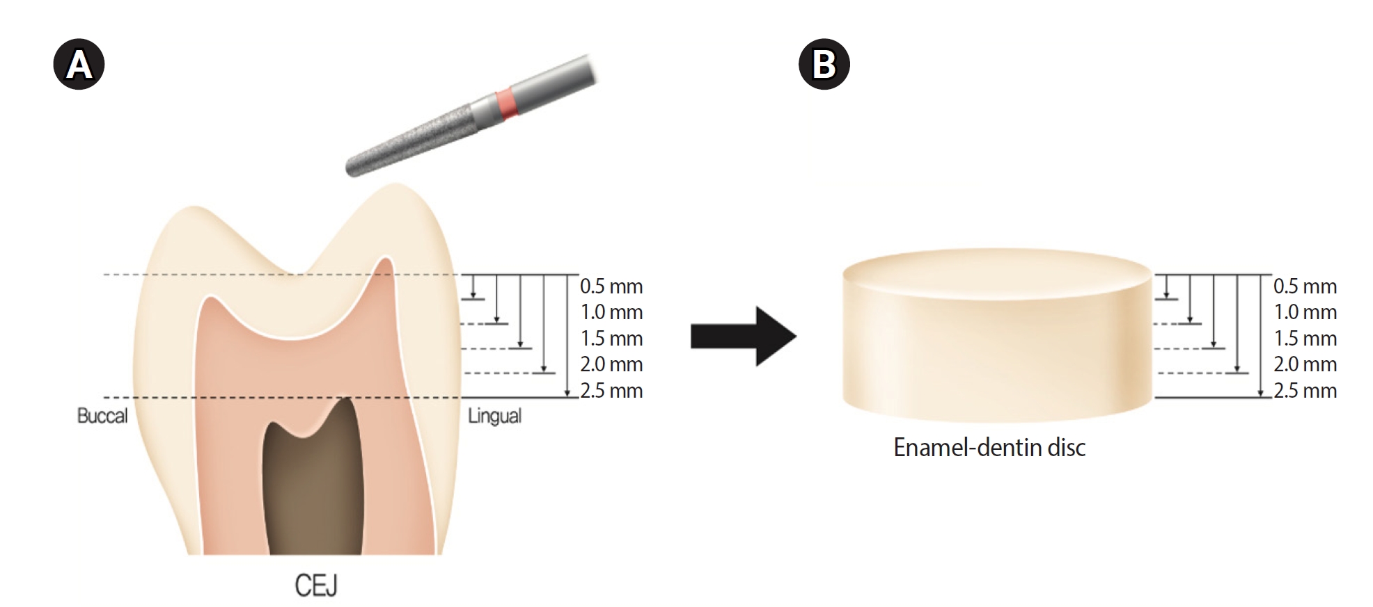
This study aimed to quantify light attenuation through varying tooth thicknesses and its impact on the depth of cure of composite resin.
Methods
Twenty extracted premolars were used to create enamel-dentin discs that were sanded progressively in 0.5 mm increments from 2.5 mm to 0.5 mm. Light irradiance was measured with and without tooth specimens to evaluate light transmittance. Resin was cured beneath different thicknesses, and the depth of cure was assessed using the Vickers hardness test.
Results
The results demonstrated that light transmittance significantly decreased as tooth thickness increased (p < 0.01), leading to reduced resin polymerization. In the 2.0-mm and 2.5-mm tooth thickness groups, the depth of cure was significantly lower than in the control group without tooth specimens (p < 0.05).
Conclusions
Ultimately, for tooth structures exceeding 2 mm, self-cure or dual-cure resin polymerization is thought to be more efficient than light polymerization.
- 2,238 View
- 153 Download

- Analysis of temperature change during polymerization according to resin thickness: an in vitro experimental study
- Kkot-Byeol Bae, Eun-Young Noh, Young-Tae Cho, Bin-Na Lee, Hoon-Sang Chang, Yun-Chan Hwang, Won-Mann Oh, In-Nam Hwang
- Restor Dent Endod 2025;50(4):e34. Published online November 12, 2025
- DOI: https://doi.org/10.5395/rde.2025.50.e34
-
Abstract
PDFPubReaderePub
- Objectives
This study aimed to analyze the temperature changes during the light curing of conventional flowable composite resin and bulk-fill composite resin of various thicknesses using an infrared thermographic camera.
Methods
Flowable composite resin (G-aenial Flo, GC Co.) and bulk-fill composite resin (SDR, Dentsply Caulk) were used. Specimens with thicknesses from 0.5 mm to 5.0 mm were prepared. The infrared thermographic camera measured the temperature changes at the maximum temperature rise point during light curing. The data were analyzed for maximum temperature, time to peak temperature, and temperature rise patterns.
Results
For G-aenial Flo, the maximum temperature tended to decrease with increasing thickness, whereas for SDR, the maximum temperature decreased up to 2.0 mm and then remained relatively consistent from 2.0 mm to 5.0 mm. At thicknesses of 1.5 mm or less, both resins showed a rapid temperature increase within the first 5 seconds, followed by a reduced rate of increase up to 80 seconds. At thicknesses of 2.0 mm or greater, the temperature peaked and then gradually decreased. Across all thicknesses, SDR was observed to reach peak temperature more rapidly than G-aenial Flo.
Conclusions
Observable differences in polymerization dynamics were identified between the two resin types, particularly at greater thicknesses. Although no statistical analysis was performed, these descriptive findings suggest that infrared thermographic cameras may be useful for indirectly assessing polymerization dynamics during resin polymerization. -
Citations
Citations to this article as recorded by
- Mapping the horizontal thermal distribution profile of composite resins during polymerization using real-time infrared thermography
Kkot-Byeol Bae, Young-Tae Cho, Bin-Na Lee, Hoon-Sang Chang, Yun-Chan Hwang, In-Nam Hwang
Journal of Dental Rehabilitation and Applied Science.2026; 42(2): 79. CrossRef
- Mapping the horizontal thermal distribution profile of composite resins during polymerization using real-time infrared thermography
- 2,391 View
- 107 Download
- 1 Crossref
- The effects of gingival blood flow on pulpal blood flow detection using ultrasound Doppler flowmetry: animal study
- Dohyun Kim, Hyoung-Seok Ko, Soo-Yeon Park, Seung-Yeon Ryu, Sung-ho Park
- Restor Dent Endod 2023;48(1):e9. Published online January 30, 2023
- DOI: https://doi.org/10.5395/rde.2023.48.e9
-
Abstract
PDFPubReaderePub
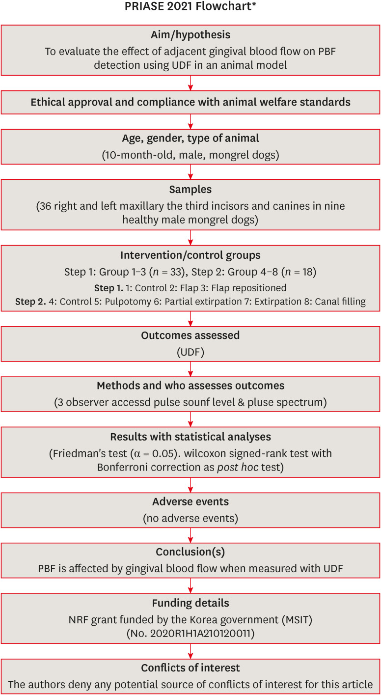
Objectives This study evaluated the effect of adjacent gingival blood flow on detection of pulpal blood flow (PBF) using ultrasound Doppler flowmetry (UDF) through animal study.
Materials and Methods The study included 36 right and left maxillary the third incisors and canines in 9 experimental dogs. The study included 2 main steps: In the first step, the pulse sound level (PSL) was recorded on the cervical part of each tooth without flap elevation (Group 1), with flap elevation (Group 2), and after it was repositioned in place (Group 3). In the second step, the PSL was recorded on the cervical part of each tooth (Group 4), after pulpotomy (Group 5), after partial pulp extirpation (Group 6), after complete extirpation (Group 7), and after canal filling (Group 8). In Groups 5–8, the study was performed with and without flap elevation in the left and right teeth, respectively. The PSL was graded as follows: 0, inaudible; 1, heard faintly; and 2, heard well. The difference between each group was analyzed using Friedman’s test with Wilcoxon signed-rank tests (α = 0.05).
Results In step 1, the PSL results were Group 1 > 2 and 3. In step 2, there was no significant difference between the groups when the flap was not elevated, while PSL results were Group 4 > 5 ≥ 6 and 7 ≥ 8 when the flap was elevated.
Conclusions PBF is affected by gingival blood flow when measured with UDF. UDF measurements require isolation of gingiva from the tooth.
-
Citations
Citations to this article as recorded by- Modern aspects of the use of hardware methods for diagnosing pulp vitality (Part 2. Non-traditional diagnostic methods)
K. V. Shadrina, L. Yu. Orekhova, V. D. Goncharov, V. Yu. Vashneva, E. S. Silina, E. V. Kosova, A. A. Petrov
Endodontics Today.2025; 23(3): 423. CrossRef - Exploring approaches to pulp vitality assessment: A scoping review of nontraditional methods
Farzaneh Afkhami, Patricia Paule Wright, Philip Yuan‐Ho Chien, Chun Xu, Laurence James Walsh, Ove Andreas Peters
International Endodontic Journal.2024; 57(8): 1065. CrossRef
- Modern aspects of the use of hardware methods for diagnosing pulp vitality (Part 2. Non-traditional diagnostic methods)
- 3,428 View
- 49 Download
- 1 Web of Science
- 2 Crossref
- Flow characteristics and alkalinity of novel bioceramic root canal sealers
- Anastasios Katakidis, Konstantinos Sidiropoulos, Elisabeth Koulaouzidou, Christos Gogos, Nikolaos Economides
- Restor Dent Endod 2020;45(4):e42. Published online August 18, 2020
- DOI: https://doi.org/10.5395/rde.2020.45.e42
-
Abstract
PDFPubReaderePub
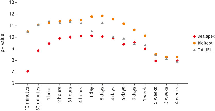
Objective This study aimed to examine the physical properties (pH and flow) of 2 novel bioceramic sealers.
Materials and Methods The tested sealers were a calcium hydroxide sealer (Sealapex) and 2 bioceramic sealers (BioRoot RCS and TotalFill BC Sealer). Flow measurements were conducted according to ISO 6876/2012, with a press method of 0.05 mL of sealer. The pH of fresh samples was tested immediately after manipulation, while set samples were stored for 3 times the recommended setting time. The predetermined time intervals ranged from 3 minutes to 24 hours for fresh samples and from 10 minutes to 7 days and 4 weeks for the set samples. Analysis of variance was performed, with
p = 0.05 considered indicating significance.Results The mean flow values were 26.99 mm for BioRoot, 28.19 for Sealapex, and 30.8 mm for TotalFill BC Sealer, satisfying the ISO standard. In the set samples, BioRoot RCS had higher pH values at 24 hours to 1 week after immersion in distilled water. At 2 weeks, both bioceramic sealers had similar pH values, greater than that of Sealapex. In the fresh samples, the bioceramic sealers had significantly higher initial pH values than Sealapex (
p < 0.05). At 24 hours post-immersion, all sealers showed an alkaline pH, with the highest pH observed for TotalFill.Conclusions The TotalFill BC Sealer demonstrated the highest flow. The bioceramic sealers initially presented higher alkaline activity than the polymeric calcium hydroxide sealer. However, at 3 and 4 weeks post-immersion, all sealers had similar pH values.
-
Citations
Citations to this article as recorded by- In vitro comparative evaluation of physicochemical and mechanical properties, cytocompatibility, and antimicrobial efficacy of various bioceramic root canal sealers
Fushi Wang, Jiaxing Li, Jingjing Wan, Siyuan Li, Shijia Tang, Li Wang, Liuyan Meng
Ceramics International.2026; 52(7): 9561. CrossRef - Comparative analysis between resin-based root canal sealer and recent bioceramic-based root canal sealers using MicroCT, film thickness, and solubility
Amira Galal Ismail, Manar M. Galal, Tamer M. Hamdy
Journal of Oral Biology and Craniofacial Research.2026; 16(2): 101400. CrossRef - Setting Characteristics, Solubility, Bioactivity and Interaction with Dentin of Four Calcium Silicate-Based Endodontic Sealers
Areti Dimitra Vrochari, Anastasia Agrafioti, Maria Dimitriadi, George Eliades
Journal of Functional Biomaterials.2026; 17(4): 192. CrossRef - Cryotherapy-Driven Modulation of Postoperative Pain in Single-Visit Endodontic Treatment Across Different Obturation Materials: A Retrospective Study
Kaan Ilıcalı, Ahter Şanal Çıkman, Özge Başar
Journal of Clinical Medicine.2026; 15(10): 3899. CrossRef - Adverse tissue reaction to a next-generation bioceramic sealer: a structured literature review and case report
Saverio Ceraulo, Antonio Barbarisi, Zhong Hao Hu, Ignazio Migliore, Dorina Lauritano, Gianluigi Caccianiga, Francesco Carinci
Frontiers in Dental Medicine.2026;[Epub] CrossRef - Effect of root canal moisture on intratubular penetration of bioceramic sealer using two obturation techniques: A confocal laser scanning microscopic study
Maryam Jasim Al-Qaralusi, Hikmet Abdul-Rahim Al-Gharrawi
Journal of Conservative Dentistry and Endodontics.2026; 29(7): 747. CrossRef - Functional and Bioactive Performance of Premixed Bioceramic Sealers with Warm Obturation: A Scoping Review
Patryk Wiśniewski, Stanisław Krokosz, Małgorzata Pietruska, Anna Zalewska
Gels.2025; 11(11): 932. CrossRef - Physicochemical properties of AH plus bioceramic sealer, Bio-C Sealer, and ADseal root canal sealer
Tamer M. Hamdy, Manar M. Galal, Amira Galal Ismail, Shehabeldin Saber
Head & Face Medicine.2024;[Epub] CrossRef - Characterization and Assessment of Physical Properties of 3 Single Syringe Hydraulic Cement–based Sealers
Veksina Raman, Josette Camilleri
Journal of Endodontics.2024; 50(3): 381. CrossRef - The Impact of Silver Nanoparticles on Dentinal Tubule Penetration of Endodontic Bioceramic Sealer
Sundus Bukhary, Sarah Alkahtany, Amal Almohaimede, Nourah Alkhayatt, Shahad Alsulaiman, Salma Alohali
Applied Sciences.2024; 14(24): 11639. CrossRef - Influence of root canal moisture on the penetration of TotalFill bioceramic sealer into the dentinal tubules: A confocal laser scanning microscopy study
Archika M Singh, Tarek M Elsewify, Walid S El-Sayed, Husam H Nuawafleh, Ranya F Elemam, Bassem M Eid
Saudi Endodontic Journal.2024; 14(2): 187. CrossRef - Unusual Canal Morphology in Mandibular Premolars With Two Distal and One Mesial Canal: A Case Series
Jinesh A, Sanjana Jayakumar Nair, Saurabh Gupta, Harsh Chansoria, Gaurav Rawat
Cureus.2024;[Epub] CrossRef - A scientometric, bibliometric, and thematic map analysis of hydraulic calcium silicate root canal sealers
Anastasios Katakidis, Konstantinos Kodonas, Anastasia Fardi, Christos Gogos
Restorative Dentistry & Endodontics.2023;[Epub] CrossRef - Thermal, chemical and physical analysis of VDW.1Seal, Fill Root ST, and ADseal root canal sealers
Shehabeldin Saber, Manar M. Galal, Amira Galal Ismail, Tamer M. Hamdy
Scientific Reports.2023;[Epub] CrossRef - α-tricalcium phosphate/fluorapatite-based cement - promising dental root canal filling material
Abdul Kazuz, Zeljko Radovanovic, Djordje Veljovic, Vesna Kojic, Dimitar Jakimov, Tamara Vlajic-Tovilovic, Vesna Miletic, Rada Petrovic, Djordje Janackovic
Processing and Application of Ceramics.2022; 16(1): 22. CrossRef
- In vitro comparative evaluation of physicochemical and mechanical properties, cytocompatibility, and antimicrobial efficacy of various bioceramic root canal sealers
- 3,926 View
- 49 Download
- 15 Crossref
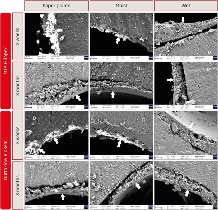
- Dentin moisture conditions strongly influence its interactions with bioactive root canal sealers
- Esin Ozlek, Hüseyin Gündüz, Elif Akkol, Prasanna Neelakantan
- Restor Dent Endod 2020;45(2):e24. Published online March 24, 2020
- DOI: https://doi.org/10.5395/rde.2020.45.e24
-
Abstract
PDFPubReaderePub
Objectives It is known that bioactive materials interact with the dentin to undergo biomineralization. The exact role of moisture in this interaction is unknown. Here, we investigate the effects of dentin moisture conditions on the dislocation resistance of two bioactive root canal sealers (MTA Fillapex [Angelus Solucoes Odontologicas] and GuttaFlow BioSeal [Colténe/Whaledent AG]) at 3 weeks and 3 months after obturation.
Materials and Methods Mandibular premolars (
n = 120) were prepared and randomly divided into 3 groups based on the dentin condition: group 1, dry dentin; group 2, moist dentin; group 3, wet dentin. Each group was divided into 2 subgroups for root canal filling: MTA Fillapex and GuttaFlow BioSeal. Dislocation resistance was evaluated by measuring the push-out bond strength at 3 weeks and 3 months. Failure modes were examined under a stereomicroscope. Data were statistically analyzed by Kruskal-Wallis test with a significance level of 5%.Results Moist dentin resulted in higher bond strength values for both materials at both time points. This was significantly higher than wet and dry dentin for both the sealers at the 3 months (
p < 0.05), while at 3 weeks it was significant only for GuttaFlow Bioseal. The different moisture conditions demonstrated similar trends in their effects on the dislocation resistance of the 2 root canal sealers.Conclusions The dentin moisture conditions had a significant impact on its interaction with the bioactive materials tested. Maintaining moist dentin, but not dry or wet dentin, may be advantageous before the filling root canals with bioactive sealers.
-
Citations
Citations to this article as recorded by- Do irrigation activation techniques and setting time affect the dentin bond strength of bioceramic-based root canal sealers?
Emine Şimşek, Selimcan Satar, Sude Gül Özdemir, Fatma Selenay Uçaş Yıldız, Özge Kurt, Makbule Bilge Akbulut
International Journal of Adhesion and Adhesives.2026; 149: 104367. CrossRef - Effect of root canal moisture on intratubular penetration of bioceramic sealer using two obturation techniques: A confocal laser scanning microscopic study
Maryam Jasim Al-Qaralusi, Hikmet Abdul-Rahim Al-Gharrawi
Journal of Conservative Dentistry and Endodontics.2026; 29(7): 747. CrossRef - The effect of moisture conditions and canal morphologies on the filling quality of iRoot SP with single-cone technique in root canals: an ex-vivo study
Jing Yang, Xiran Xu, Jian Zhang, Kehua Que
Frontiers in Dental Medicine.2025;[Epub] CrossRef - Bond Strength and Adhesive Interface Quality of New Pre‐Mixed Bioceramic Root Canal Sealer
Gustavo Creazzo, Bruna Monteiro de Barros Ciribelli Alves, Helena Cristina de Assis, Karen Gisselle Garay Villamayor, Manoel Damião de Sousa‐Neto, Jardel Francisco Mazzi‐Chaves, Fabiane Carneiro Lopes‐Olhê
Microscopy Research and Technique.2025; 88(7): 1989. CrossRef - Evaluation of apical seal and tubular penetration of a novel bioactive glass sealer, bioceramic sealer and resin–based sealer: an In-Vitro study
M. Bilal, S. Pasha, S. Kumar, S. Arif, S. Taj, A. Saleem
Endodontics Today.2025; 23(1): 39. CrossRef - Effect of Root Dentin Moisture on the Apical Sealing Ability of Root Canal Sealers: In vitro Study
Zahraa Khalil Alani, Manal Hussain Abd-alla
Al-Rafidain Journal of Medical Sciences ( ISSN 2789-3219 ).2025; 8(2): 122. CrossRef - The Effects of Different Post Space Conditioning Procedures and Different Endodontic Sealers on the Push-Out Bond Strengths of Fiber Posts
Leyla Ayranci, Ahmet Serkan Küçükekenci, Fatih Sarı, Ahmet Çetinkaya
Clinical and Experimental Health Sciences.2025; 15(3): 620. CrossRef - Evaluation of the retreatability of bioceramic root canal sealers with various formulations in simulated grooves
Meltem Sümbüllü, Afzal Ali, Abdulaziz Bakhsh, Hakan Arslan
PeerJ.2025; 13: e20398. CrossRef - Preparation and characterization of novel nano-tricalcium silicate-58s bioactive glass-based root canal sealer
Nawal Atiya Al-Sabawi, Sawsan Hameed Al-Jubori
Saudi Endodontic Journal.2024; 14(1): 90. CrossRef - The flow behavior and sealing ability of calcium silicate root canal cement containing dimethyl sulfoxide: An in vitro study
Bokyung Shin, Ji-Hwan Seo, Wonjung Kim, Yu Jin Ahn, Ho-Young Kim, Won-Jun Shon
Journal of the Mechanical Behavior of Biomedical Materials.2024; 150: 106156. CrossRef - Nanoleakage of apical sealing using a calcium silicate-based sealer according to canal drying methods
Yoon-Joo Lee, Kyung-Mo Cho, Se-Hee Park, Yoon Lee, Jin-Woo Kim
Restorative Dentistry & Endodontics.2024;[Epub] CrossRef - Effect of operators’ proficiency level and patients’ related factors on possible complications, using a high frequency polyamide sonic intracanal irrigation device: A prospective clinical cohort study
Tobias Hahn, David W. Christofzik, Karim Fawzy El-Sayed, Sandra Freitag-Wolf, Jonas Conrad, Christian Graetz, Birte Größner-Schreiber, Christof Dörfer, Artak Heboyan
PLOS ONE.2023; 18(5): e0285492. CrossRef - Physical-chemical properties and acellular bioactivity of newly prepared nano-tricalcium silicate-58s bioactive glass-based endodontic sealer
Nawal A. Al-Sabawi, Sawsan Hameed Al-Jubori
Journal of Oral Biosciences.2023; 65(4): 305. CrossRef - Biocompatibility analysis in subcutaneous tissue and physico-chemical analysis of pre-mixed calcium silicate–based sealers
Ana Cristina Padilha Janini, Lauter Eston Pelepenko, Juliana Minto Boldieri, Victor Augusto Benedicto dos Santos, Nilvan Alves da Silva, Ivo Milton Raimundo, Brenda P. F. A. Gomes, Marina Angélica Marciano
Clinical Oral Investigations.2023; 27(5): 2221. CrossRef - Canal Drying Protocols to Use with Calcium Silicate–based Sealer: Effect on Bond Strength and Adhesive Interface
Lais Lima Pelozo, Aline Evangelista Souza-Gabriel, Guilherme Nilson Alves dos Santos, Rafael Verardino Camargo, Fabiane Carneiro Lopes-Olhê, Manoel Damião Sousa-Neto, Jardel Francisco Mazzi-Chaves
Journal of Endodontics.2023; 49(9): 1154. CrossRef - Comparison of push‐out bond strength of endodontic sealers after root canal drying with different techniques
Ahmadreza Sarrafan, Ali Soleymani, Tasnim Bagheri Chenari, Seyedali Seyedmajidi
Clinical and Experimental Dental Research.2023; 9(2): 314. CrossRef - Designing Calcium Silicate Cements with On-Demand Properties for Precision Endodontics
A. Cahyanto, P. Rath, T.X. Teo, S.S. Tong, R. Malhotra, B.N. Cavalcanti, L.Z. Lim, K.S. Min, D. Ho, W.F. Lu, V. Rosa
Journal of Dental Research.2023; 102(13): 1425. CrossRef - Outcome of root canal treatment using warm vertical compaction with bioceramic and resin‐based sealers: A randomised clinical trial
Jinghao Hu, Yunjie Zhu, Shuli Deng, Zeji Wang, Fuming He
Australian Endodontic Journal.2023; 49(S1): 170. CrossRef - Evaluation of the Sealing Ability and Bond Strength of Two Endodontic Root Canal Sealers: An In Vitro Study
Manuel Marques Ferreira, José Pedro Martinho, Inês Duarte, Diogo Mendonça, Ana Catarina Craveiro, Maria Filomena Botelho, Eunice Carrilho, Carlos Miguel Marto, Ana Coelho, Anabela Paula, Siri Paulo, Nuno Chichorro, Ana Margarida Abrantes
Dentistry Journal.2022; 10(11): 201. CrossRef - How do imaging protocols affect the assessment of root-end fillings?
Fernanda Ferrari Esteves Torres, Reinhilde Jacobs, Mostafa EzEldeen, Karla de Faria-Vasconcelos, Juliane Maria Guerreiro-Tanomaru, Bernardo Camargo dos Santos, Mário Tanomaru-Filho
Restorative Dentistry & Endodontics.2022;[Epub] CrossRef - Effect of using calcium-silicate and silicone based root canal sealers in bulk or with main core material on bond strength
Gizem Kadı, Esin Özlek, Yousef Saed
Journal of Dental Research, Dental Clinics, Dental Prospects.2022; 16(4): 229. CrossRef - Physico-chemical properties of calcium silicate-based sealers in powder/liquid and ready-to-use forms
Ana C P Janini, Lauter E Pelepenko, Brenda P F A Gomes, Marina A Marciano
Brazilian Dental Journal.2022; 33(5): 18. CrossRef - Influence of dentin moisture conditions on the wetting action of different endodontic sealers using Rame-Hart goniometer: An in vitro study
Sivaji Kauravi, ShruthiH Attavar, GyanendraPratap Singh
Journal of International Oral Health.2022; 14(6): 624. CrossRef - Heating stability, physical and chemical analysis of calcium silicate‐based endodontic sealers
T. B. M. Antunes, A. C. P. Janini, L. E. Pelepenko, G. F. Abuna, E. M. Paiva, M. A. C. Sinhoreti, I. M. Raimundo, B. P. F. A. Gomes, A. de‐Jesus‐Soares, M. A. Marciano
International Endodontic Journal.2021; 54(7): 1175. CrossRef - Characterization, Antimicrobial Effects, and Cytocompatibility of a Root Canal Sealer Produced by Pozzolan Reaction between Calcium Hydroxide and Silica
Mi-Ah Kim, Vinicius Rosa, Prasanna Neelakantan, Yun-Chan Hwang, Kyung-San Min
Materials.2021; 14(11): 2863. CrossRef - Main and Accessory Canal Filling Quality of a Premixed Calcium Silicate Endodontic Sealer According to Different Obturation Techniques
Su-Yeon Ko, Hae Won Choi, E-Deun Jeong, Vinicius Rosa, Yun-Chan Hwang, Mi-Kyung Yu, Kyung-San Min
Materials.2020; 13(19): 4389. CrossRef
- Do irrigation activation techniques and setting time affect the dentin bond strength of bioceramic-based root canal sealers?
- 3,247 View
- 38 Download
- 26 Crossref
-
Comparative evaluation of the bond strength of self-adhering and bulk-fill flowable composites to MTA Plus, Dycal, Biodentine, and TheraCal: an
in vitro study - Aakrati Raina, Asheesh Sawhny, Saurav Paul, Sridevi Nandamuri
- Restor Dent Endod 2020;45(1):e10. Published online January 8, 2020
- DOI: https://doi.org/10.5395/rde.2020.45.e10
-
Abstract
PDFPubReaderePub
Objectives This study aimed to compare the shear bond strength (SBS) of a self-adhering flowable composite (Dyad Flow) and a bulk-fill flowable composite (Smart Dentin Replacement [SDR]) to several pulp-capping materials, including MTA Plus, Dycal, Biodentine, and TheraCal.
Materials and Methods Eighty acrylic blocks with 2-mm-deep central holes that were 4 mm in diameter were prepared and divided into 2 groups (
n = 40 each) according to the composite used (Dyad Flow or SDR). They were further divided into 4 sub-groups (n = 10 each) according to the pulp-capping agent used. SBS was tested using a universal testing machine at a crosshead speed of 1 mm/min. Data were analyzed using 2-way analysis of variance. Ap value of < 0.05 was considered to indicate statistical significance.Results A statistically significant difference (
p = 0.040) was found between Dyad Flow and SDR in terms of bond strength to MTA Plus, Dycal, Biodentine, and TheraCal.Conclusions Among the 8 sub-groups, the combination of TheraCal and SDR exhibited the highest SBS.
-
Citations
Citations to this article as recorded by- Vital pulp therapy: A three-year clinical case series on regenerative potential and adhesive restoration strategies
Vincenzo Tosco, Riccardo Monterubbianesi, Pietro Montagna, Giulia Orilisi, Flavia Vitiello, Hengyue Song, Angelo Putignano, Daniele De Santis, Giovanna Orsini
Regenesis Repair Rehabilitation.2026; 2(2): 99. CrossRef - Influence of Calcium Silicate‐Based Cement‐Repaired Root Canals on Push‐Out Bond Strength of Glass Fiber Posts: An In Vitro Study
Yasudthama Rattana‐aporn, Weeranuch Thong‐ngarm
Clinical and Experimental Dental Research.2026;[Epub] CrossRef - Shear Bond Strength of Liner Materials to Caries-Free and Caries-Affected Dentin
ZK Greene, NR Smith, T Gomes, NC Lawson
Operative Dentistry.2025; 50(3): 324. CrossRef - Shear Bond Strength of Biointeractive Restorative Materials to NeoMTA Plus and Biodentine
Zübeyde Uçar Gündoğar, Gül Keskin, Merve Yaman Küçükersen
Polymers.2025; 17(22): 3070. CrossRef - The Influence of an Adhesive Protocol on the Adhesion of Biodentine to Resin-Matrix Composites
Orlanda Torres, Nuno Caiado, Ana Sá, Rita Fidalgo-Pereira, Júlio C. M. Souza
Biomedical Materials & Devices.2025;[Epub] CrossRef - Hygroscopic bioactive light-cured composite promoting dentine bridge formation
Yunzi Long, Guibin Huang, Siyi Liu, Liju Xu, Ailing Li, Dong Qiu, Yanmei Dong
Regenerative Biomaterials.2024;[Epub] CrossRef - Comparative evaluation of shear bond strength and modes of failure of five different reinforced glass ionomer restorative cements to TheraCal LC: An in vitro study
Kalyani Gajanan Umale, Vandana Jaykumar Gade, Ambar W. Raut
Journal of Conservative Dentistry and Endodontics.2024; 27(2): 200. CrossRef - Evaluation of the Effect of Chitosan-Based Irrigation Solutions on the Bond Strength of Mineral Trioxide Aggregate to Bulk-Fill Composite
Arzu Şahin Mantı, Bağdagül Helvacıoğlu Kıvanç
Journal of Functional Biomaterials.2024; 15(12): 370. CrossRef - Radiopacity evaluations of the novel calcium-silicate and glass-Ionomer-based materials
Yeşim Şeşen Uslu, Elif Çelebi, Meriç Berkman
Journal of Health Sciences and Medicine.2024; 7(2): 192. CrossRef - Effect of Er Cr YSGG laser etching procedure on the bond strength of different calcium silicate cements
Yesim Sesen Uslu, Hakan Yasin Gönder, Pinar Sesen, Gizem Gunduz Bektaş
Lasers in Dental Science.2024;[Epub] CrossRef - The micro‐shear bond strength of new endodontic tricalcium silicate‐based putty: An in vitro study
Merve Yeniçeri Özata, Seda Falakaloğlu, Gianluca Plotino, Özkan Adıgüzel
Australian Endodontic Journal.2023; 49(1): 124. CrossRef - Analysis of the bond strength between conventional, putty or resin‐modified calcium silicate cement and bulk fill composites
İ Ipek, B Karaağaç Eskibağlar, Ş Yildiz, O Ataş, M Ünal
Australian Dental Journal.2023; 68(4): 265. CrossRef - Effect of Different Adhesive Strategies on the Microshear Bond Strength of Calcium-Silicate-Based Materials
Aliye Tuğçe Gürcan, Soner Şişmanoğlu, Görkem Sengez
Journal of Advanced Oral Research.2022; 13(2): 191. CrossRef - BULK FİLL KOMPOZİT REZİN RESTORATİF MATERYALLER
Merve NEZİR, Suat ÖZCAN
Atatürk Üniversitesi Diş Hekimliği Fakültesi Dergisi.2022; : 1. CrossRef - Effect of Bioinductive Cavity Liners on Shear Bond Strength of Dental Composite to Dentin
Saba Tohidkhah, Elham Ahmadi, Mahdi Abbasi, Reza Morvaridi Farimani, Ladan Ranjbar Omrani, Victor Feitosa
BioMed Research International.2022;[Epub] CrossRef - Bond Strength of Adhesive Systems to Calcium Silicate-Based Materials: A Systematic Review and Meta-Analysis of In Vitro Studies
Louis Hardan, Davide Mancino, Rim Bourgi, Alejandra Alvarado-Orozco, Laura Emma Rodríguez-Vilchis, Abigailt Flores-Ledesma, Carlos Enrique Cuevas-Suárez, Monika Lukomska-Szymanska, Ammar Eid, Maya-Line Danhache, Maryline Minoux, Youssef Haïkel, Naji Kharo
Gels.2022; 8(5): 311. CrossRef - How do imaging protocols affect the assessment of root-end fillings?
Fernanda Ferrari Esteves Torres, Reinhilde Jacobs, Mostafa EzEldeen, Karla de Faria-Vasconcelos, Juliane Maria Guerreiro-Tanomaru, Bernardo Camargo dos Santos, Mário Tanomaru-Filho
Restorative Dentistry & Endodontics.2022;[Epub] CrossRef - Evaluation of Shear Bond Strength of Resin‐Based Composites to Biodentine with Three Types of Seventh‐Generation Bonding Agents: An In Vitro Study
Huda Abbas Abdullah, Zahraa Abdulaali Al-Ibraheemi, Zanbaq Azeez Hanoon, Julfikar Haider, Boonlert Kukiattrakoon
International Journal of Dentistry.2022;[Epub] CrossRef - Evaluation of the Bond Strength of Different Pulp Capping Materials to Dental Adhesive Systems: An In Vitro Study
Sema Yazici Akbiyik, Elif Pınar Bakir, S¸eyhmus Bakir
Journal of Advanced Oral Research.2021; 12(2): 286. CrossRef - Differential Gene Expression Changes in Human Primary Dental Pulp Cells Treated with Biodentine and TheraCal LC Compared to MTA
Ok Hyung Nam, Ho Sun Lee, Jae-Hwan Kim, Yong Kwon Chae, Seoung-Jin Hong, Sang Wook Kang, Hyo-Seol Lee, Sung Chul Choi, Young Kim
Biomedicines.2020; 8(11): 445. CrossRef
- Vital pulp therapy: A three-year clinical case series on regenerative potential and adhesive restoration strategies
- 3,399 View
- 52 Download
- 20 Crossref
- Involvement of TRPA1 in the cinnamaldehyde-induced pulpal blood flow change in the feline dental pulp
- Dokyung Kim, Moon-Hwan Lee, Sung Kyo Kim
- Restor Dent Endod 2016;41(3):202-209. Published online July 29, 2016
- DOI: https://doi.org/10.5395/rde.2016.41.3.202
-
Abstract
PDFPubReaderePub
Objectives The purpose of this study was to investigate the involvement of TRPA1 in the cinnamaldehyde-induced pulpal blood flow (PBF) change in the feline dental pulp.
Materials and Methods Mandibles of eight cats were immobilized and PBF was monitored with a laser Doppler flowmetry at the mandibular canine tooth. To evaluate the effect of cinnamaldehyde on PBF, cinnamaldehyde was injected into the pulp through the lingual artery at a constant rate for 60 seconds. As a control, a mixture of 70% ethanol and 30% dimethyl sulfoxide (DMSO, vehicle) was used. To evaluate the involvement of transient receptor potential ankyrin 1 (TRPA1) in PBF change, AP18, a specific TRPA1 antagonist, was applied into the pulp through the Class V dentinal cavity followed by cinnamaldehyde-administration 3 minutes later. The paired variables of experimental data were statistically analyzed using paired
t -test. Ap value of less than 0.05 was considered as statistically significant.Results Administration of cinnamaldehyde (0.5 mg/kg, intra-arterial [i.a.]) induced significant increases in PBF (
p < 0.05). While administration of a TRPA1 antagonist, AP18 (2.5 - 3.0 mM, into the dentinal cavity [i.c.]) caused insignificant change of PBF (p > 0.05), administration of cinnamaldehyde (0.5 mg/kg, i.a.) following the application of AP18 (2.5 - 3.0 mM, i.c.) resulted in an attenuation of PBF increase from the control level (p < 0.05). As a result, a TRPA1 antagonist, AP18 effectively inhibited the vasodilative effect of cinnamaldehyde (p < 0.05).Conclusions The result of the present study provided a functional evidence that TRPA1 is involved in the mechanism of cinnamaldehyde-induced vasodilation in the feline dental pulp.
-
Citations
Citations to this article as recorded by- A simple model for the assessment of the agonistic activity of dibenzazepine derivatives by molecular moieties
Mohammad Hossein Keshavarz, Hossein Fakhraian, Norollah Saedi
Medicinal Chemistry Research.2021; 30(1): 215. CrossRef
- A simple model for the assessment of the agonistic activity of dibenzazepine derivatives by molecular moieties
- 3,019 View
- 6 Download
- 1 Crossref
Basic Researchs
- Microshear bond strength of a flowable resin to enamel according to the different adhesive systems
- Jeong-Ho Kim, Young-Gon Cho
- J Korean Acad Conserv Dent 2011;36(1):50-58. Published online January 31, 2011
- DOI: https://doi.org/10.5395/JKACD.2011.36.1.50
-
Abstract
PDFPubReaderePub
Objectives The purpose of this study was to compare the microshear bond strength (uSBS) of two total-etch and four self-etch adhesive systems and a flowable resin to enamel.
Materials and Methods Enamels of sixty human molars were used. They were divided into one of six equal groups (
n = 10) by adhesives used; OS group (One-Step Plus), SB group (Single Bond), CE group (Clearfil SE Bond), TY group (Tyrian SPE/One-Step Plus), AP group (Adper Prompt L-Pop) and GB group (G-Bond).After enamel surfaces were treated with six adhesive systems, a flowable composite resin (Filek Z 350) was bonded to enamel surface using Tygon tubes. the bonded specimens were subjected to uSBS testing and the failure modes of each group were observed under FE-SEM.
Results 1. The
u SBS of SB group was statistically higher than that of all other groups, and theu SBS of OS, SE and AP group was statistically higher than that of TY and GB group (p < 0.05).2. The
u SBS for TY group was statistically higher than that for GB group (p < 0.05).3. Adhesive failures in TY and GB group and mixed failures in SB group and SE group were often analysed. One cohesive failure was observed in OS, SB, SE and AP group, respectively.
Conclusions Although adhesives using the same step were applied the enamel surface, the uSBS of a flowable resin to enamel was different.
-
Citations
Citations to this article as recorded by- Enamel pretreatment with Er:YAG laser: effects on the microleakage of fissure sealant in fluorosed teeth
Mahtab Memarpour, Nasrin Kianimanesh, Bahareh Shayeghi
Restorative Dentistry & Endodontics.2014; 39(3): 180. CrossRef
- Enamel pretreatment with Er:YAG laser: effects on the microleakage of fissure sealant in fluorosed teeth
- 1,746 View
- 1 Download
- 1 Crossref
- Real-time measurement of dentinal fluid flow during desensitizing agent application
- Sun-Young Kim, Eun-Joo Kim, In-Bog Lee
- J Korean Acad Conserv Dent 2010;35(5):313-320. Published online September 30, 2010
- DOI: https://doi.org/10.5395/JKACD.2010.35.5.313
-
Abstract
PDFPubReaderePub
Objectives The aim of this study was to examine changes in the dentinal fluid flow (DFF) during desensitizing agent application and to compare permeability after application among the agents.
Materials and Methods A Class 5 cavity was prepared to exposure cervical dentin on an extracted human premolar which was connected to a sub-nanoliter fluid flow measuring device (NFMD) under 20 cm water pressure. DFF was measured from before application of desensitizing agent (Seal&Protect, SP; SuperSeal, SS; BisBlock, BB; Gluma desensitizer, GL; Bi-Fluoride 12, BF) through application procedure to 5 min after application.
Results DFF rate after each desensitizing agent application was significantly reduced when compared to initial DFF rate before application (
p < 0.05). SP showed a greater reduction in DFF rate than GL and BF did (p < 0.05). SS and BB showed a greater reduction in DFF rate than BF did (p < 0.05).Conclusions Characteristic DFF aspect of each desensitizing agent was shown in NFMD during the application procedure.
-
Citations
Citations to this article as recorded by- CPNE7 Induces Biological Dentin Sealing in a Dentin Hypersensitivity Model
S.H. Park, Y.S. Lee, D.S. Lee, J.C. Park, R. Kim, W.J. Shon
Journal of Dental Research.2019; 98(11): 1239. CrossRef
- CPNE7 Induces Biological Dentin Sealing in a Dentin Hypersensitivity Model
- 1,858 View
- 2 Download
- 1 Crossref
Original Articles
- Comparison of marginal microleakage between low and high flowable resins in class V cavity
- Sang-Bae Bae, Young-Gon Cho, Myeong-Seon Lee
- J Korean Acad Conserv Dent 2009;34(6):477-483. Published online November 30, 2009
- DOI: https://doi.org/10.5395/JKACD.2009.34.6.477
-
Abstract
PDFPubReaderePub
The purpose of this study was to compare the microleakage of low and high viscosity flowable resins in class V cavities applied with 1-step adhesives.
Forty class V cavities were prepared on the cervices of buccal and lingual surfaces of extracted molar teeth and divided into four groups (n=8). Cavities were restored with AQ Bond Plus/Metafil Flo α, G-Bond/UniFil LoFlo Plus (Low flow groups), AQ Bond Plus/Metafil Flo and G-Bond/UniFil Flow (High flow group), respectively.
Specimens were immersed in a 2% methylene blue solution for 24 hours, and bisected longitudinally. They were observed microleakages at the enamel and dentinal margins.
In conclusion, the low viscosity flowable resins showed lower marginal microleakage than do the high viscosity flowable resins in class V cavities.
- 1,335 View
- 7 Download
- Real-time measurement of dentinal tubular fluid flow during and after amalgam and composite restorations
- Sun-Young Kim, Byeong-Hoon Cho, Seung-Ho Baek, Bum-Sun Lim, In-Bog Lee
- J Korean Acad Conserv Dent 2009;34(6):467-476. Published online November 30, 2009
- DOI: https://doi.org/10.5395/JKACD.2009.34.6.467
-
Abstract
PDFPubReaderePub
The aim of this study was to measure the dentinal tubular fluid flow (DFF) during and after amalgam and composite restorations. A newly designed fluid flow measurement instrument was made. A third molar cut at 3 mm apical from the CEJ was connected to the flow measuring device under a hydrostatic pressure of 15 cmH2O. Class I cavity was prepared and restored with either amalgam (Copalite varnish and Bestaloy) or composite (Z-250 with ScotchBond MultiPurpose: MP, Single Bond 2: SB, Clearfil SE Bond: CE and Easy Bond: EB as bonding systems). The DFF was measured from the intact tooth state through restoration procedures to 30 minutes after restoration, and re-measured at 3 and 7days after restoration.
Inward fluid flow (IF) during cavity preparation was followed by outward flow (OF) after preparation. In amalgam restoration, the OF changed to IF during amalgam filling and slight OF followed after finishing.
In composite restoration, application CE and EB showed a continuous OF and air-dry increased rapidly the OF until light-curing, whereas in MP and SB, rinse and dry caused IF and OF, respectively. Application of hydrophobic bonding resin in MP and CE caused a decrease in flow rate or even slight IF. Light-curing of adhesive and composite showed an abrupt IF. There was no statistically significant difference in the reduction of DFF among the materials at 30 min, 3 and 7 days after restoration (P > 0.05).
-
Citations
Citations to this article as recorded by- Real-time measurement of dentinal fluid flow during desensitizing agent application
Sun-Young Kim, Eun-Joo Kim, In-Bog Lee
Journal of Korean Academy of Conservative Dentistry.2010; 35(5): 313. CrossRef
- Real-time measurement of dentinal fluid flow during desensitizing agent application
- 1,847 View
- 4 Download
- 1 Crossref
- Effect of an intermediate bonding resin and flowable resin on the compatibility of two-step total etching adhesives with a self-curing composite resin
- Sook-Kyung Choi, Ji-Wan Yum, Hyeon-Cheol Kim, Bock Hur, Jeong-Kil Park
- J Korean Acad Conserv Dent 2009;34(5):397-405. Published online September 30, 2009
- DOI: https://doi.org/10.5395/JKACD.2009.34.5.397
-
Abstract
PDFPubReaderePub
This study compared the effect of an activator, intermediate bonding resin and low-viscosity flowable resin on the microtensile bond strength of a self-curing composite resin used with two-step total etching adhesives. Twenty extracted permanent molars were used. The teeth were assigned randomly to nine groups (n=10) according to the adhesive system and application of additional methods (activator, intermediate adhesive, flowable resin). The bonding agents and additional applications of each group were applied to the dentin surfaces. Self-curing composite resin buildups were made for each tooth to form a core, 5mm in height. The restored teeth were then stored in distilled water at room temperature for 24h before sectioning. The microtensile bond strength of all specimens was examined. The data was analyzed statistically by one-way ANOVA and a Scheffe's test. The application of an intermediate bonding resin (Optibond FL adhesive) and low-viscosity flowable resin (Tetric N-flow) produced higher bond strength than that with the activator in all groups. Regardless of the method selected, Optibond solo plus produced the lowest µTBS to dentin. The failure modes of the tested dentin bonding agents were mostly adhesive failure but there were some cases showed cohesive failure in the resin.
- 1,971 View
- 11 Download
- Slumping tendency and rheological property of flowable composites
- In-Bog Lee, Sun-Hong Min, Sun-Young Kim, Byung-Hoon Cho, Seung-Ho Back
- J Korean Acad Conserv Dent 2009;34(2):130-136. Published online March 31, 2009
- DOI: https://doi.org/10.5395/JKACD.2009.34.2.130
-
Abstract
PDFPubReaderePub
The aim of this study was to develop a method for measuring the slumping resistance of flowable resin composites and to evaluate the efficacy using rheological methodology.
Five commercial flowable composites (Aelitefil flow:AF, Filtek flow:FF, DenFil flow:DF, Tetric flow:TF and Revolution:RV) were used. Same volume of composites in a syringe was extruded on a glass slide using a custom-made loading device. The resin composites were allowed to slump for 10 seconds at 25℃ and light cured. The aspect ratio (height/diameter) of cone or dome shaped specimen was measured for estimating the slumping tendency of composites. The complex viscosity of each composite was measured by a dynamic oscillatory shear test as a function of angular frequency using a rheometer. To compare the slumping tendency of composites, one way-ANOVA and Turkey's post hoc test was performed for the aspect ratio at 95% confidence level. Regression analysis was performed to investigate the relationship between the complex viscosity and the aspect ratio. The results were as follows.
1. Slumping tendency based on the aspect ratio varied among the five materials (AF < FF < DF < TF < RV).
2. Flowable composites exhibited pseudoplasticity in which the complex viscosity decreased with increasing frequency (shear rate). AF was the most significant, RV the least.
3. The slumping tendency was strongly related with the complex viscosity. Slumping resistance increased with increasing the complex viscosity.
The slumping tendency could be quantified by measuring the aspect ratio of slumped flowable composites. This method may be applicable to evaluate the clinical handling characteristics of flowable composites.
- 1,381 View
- 2 Download
- Is an oxygen inhibition layer essential for the interfacial bonding between resin composite layers?
- Sun-Young Kim, Byeong-Hoon Cho, Seung-Ho Baek, In-Bog Lee
- J Korean Acad Conserv Dent 2008;33(4):405-412. Published online July 31, 2008
- DOI: https://doi.org/10.5395/JKACD.2008.33.4.405
-
Abstract
PDFPubReaderePub
This study was aimed to investigate whether an oxygen inhibition layer (OIL) is essential for the interfacial bonding between resin composite layers or not.
A composite (Z-250, 3M ESPE) was filled in two layers using two aluminum plate molds with a hole of 3.7 mm diameter. The surface of first layer of cured composite was prepared by one of five methods as followings, thereafter second layer of composite was filled and cured: Group 1 - OIL is allowed to remain on the surface of cured composite; Group 2 - OIL was removed by rubbing with acetone-soaked cotton; Group 3 - formation of the OIL was inhibited using a Mylar strip; Group 4 - OIL was covered with glycerin and light-cured; Group 5 (control) - composite was bulk-filled in a layer. The interfacial shear bond strength between two layers was tested and the fracture modes were observed. To investigate the propagation of polymerization reaction from active area having a photo-initiator to inactive area without the initiator, a flowable composite (Aelite Flow) or an adhesive resin (Adhesive of ScotchBond Multipurpose) was placed over an experimental composite (Exp_Com) which does not include a photoinitiator and light-cured. After sectioning the specimen, the cured thickness of the Exp_Com was measured.
The bond strength of group 2, 3 and 4 did not show statistically significant difference with group 1. Groups 3 and 4 were not statistically significant different with control group 5. The cured thicknesses of Exp_Com under the flowable resin and adhesive resin were 20.95 (0.90) um and 42.13 (2.09), respectively.
-
Citations
Citations to this article as recorded by- Finishing and Polishing of Composite Restoration: Assessment of Knowledge, Attitude and Practice Among Various Dental Professionals in India
Sankar Vishwanath, Sadasiva Kadandale, Senthil kumar Kumarappan, Anupama Ramachandran, Manu Unnikrishnan, Honap manjiri Nagesh
Cureus.2022;[Epub] CrossRef - Evaluation of Surface Roughness of Composite, Compomer and Carbomer After Curing Through Mylar Strip and Glycerin: A Comparative Study
Asli Topaloglu-Ak, Dilara Çayırgan, Melisa Uslu
Journal of Advanced Oral Research.2020; 11(1): 12. CrossRef - Effect of glycerin on the surface hardness of composites after curing
Hyun-Hee Park, In-Bog Lee
Journal of Korean Academy of Conservative Dentistry.2011; 36(6): 483. CrossRef
- Finishing and Polishing of Composite Restoration: Assessment of Knowledge, Attitude and Practice Among Various Dental Professionals in India
- 2,917 View
- 21 Download
- 3 Crossref
- The influence of composite resin restoration on the stress distribution of notch shaped noncarious cervical lesion; A three dimensional finite element analysis study
- Chae-Kyung Lee, Jeong-Kil Park, Hyeon-Cheol Kim, Sung-Gwan Woo, Kwang-Hoon Kim, Kwon Son, Bock Hur
- J Korean Acad Conserv Dent 2007;32(1):69-79. Published online January 31, 2007
- DOI: https://doi.org/10.5395/JKACD.2007.32.1.069
-
Abstract
PDFPubReaderePub
The purpose of this study was to investigate the effects of composite resin restorations on the stress distribution of notch shaped noncarious cervical lesion using three-dimensional (3D) finite element analysis (FEA).
Extracted maxillary second premolar was scanned serially with Micro-CT (SkyScan1072; SkyScan, Aartselaar, Belgium). The 3D images were processed by 3D-DOCTOR (Able Software Co., Lexington, MA, USA). ANSYS (Swanson Analysis Systems, Inc., Houston, USA) was used to mesh and analyze 3D FE model. Notch shaped cavity was filled with hybrid or flowable resin and each restoration was simulated with adhesive layer thickness (40 µM). A static load of 500 N was applied on a point load condition at buccal cusp (loading A) and palatal cusp (loading B). The principal stresses in the lesion apex (internal line angle of cavity) and middle vertical wall were analyzed using ANSYS.
The results were as follows
1. Under loading A, compressive stress is created in the unrestored and restored cavity. Under loading B, tensile stress is created. And the peak stress concentration is seen at near mesial corner of the cavity under each load condition.
2. Compared to the unrestored cavity, the principal stresses at the cemeto-enamel junction (CEJ) and internal line angle of the cavity were more reduced in the restored cavity on both load conditions.
3. In teeth restored with hybrid composite, the principal stresses at the CEJ and internal line angle of the cavity were more reduced than flowable resin.
-
Citations
Citations to this article as recorded by- The influence of occlusal loads on stress distribution of cervical composite resin restorations: A three-dimensional finite element study
Chan-Seok Park, Bock Hur, Hyeon-Cheol Kim, Kwang-Hoon Kim, Kwon Son, Jeong-Kil Park
Journal of Korean Academy of Conservative Dentistry.2008; 33(3): 246. CrossRef
- The influence of occlusal loads on stress distribution of cervical composite resin restorations: A three-dimensional finite element study
- 2,133 View
- 3 Download
- 1 Crossref
- Effect of local anesthesia on pulpal blood flow in mechanically stimulated teeth
- Wan-Sik Chu, Seung-Ho Park, Dong-Kuk Ahn, Sung Kyo Kim
- J Korean Acad Conserv Dent 2006;31(4):257-262. Published online January 14, 2006
- DOI: https://doi.org/10.5395/JKACD.2006.31.4.257
-
Abstract
PDFPubReaderePub
Abstract The aims of the study were to evaluate the effect of epinephrine-containing local anesthetics on pulpal blood flow (PBF) and to investigate its effect on cavity preparation-induced PBF change. PBF was recorded using a laser Doppler flowmeter (Perimed Co., Sweden) from canines of nine cats under general anesthesia before and after injection of local anesthetics and after cavity preparation. 2% lidocaine hydrochloride with 1 : 100,000 epinephrine was administered by local infiltration given apical to the mandibular canine at the vestibular area and the same volume of isotonic saline was injected on the contralateral tooth as a control. A round carbide bur was operated at slow speed with isotonic saline flushing to grind spherical cavities with increasing depth through the enamel and into the dentin on both teeth. The obtained data was analyzed with paired
t -test.Cavity preparation caused significant increase of PBF (
n = 9,p < 0.05). Local infiltration of lidocaine with epinephrine resulted in decreases of PBF (n = 9,p < 0.05), whereas there was no significant change of PBF with the physiologic saline as a control. Cavity preparation on tooth anesthetized with lidocaine with epinephrine caused significantly less increase of PBF than in control tooth (p < 0.05).Therefore, the result of the present study demonstrates that local infiltration of 2% lidocaine with 1 : 100,000 epinephrine effectively reduces PBF increase caused by cavity preparation.
-
Citations
Citations to this article as recorded by- Effect of laser-induced pulpal anesthesia of single-rooted teeth with irreversible pulpitis treated by single-visit root canal therapy - A randomized clinical trial
Geeta Asthana, Dhwani Morakhia, Ravina Parmar, Rajashree Tamuli
Endodontology.2025; 37(3): 244. CrossRef - Systematic Injection Patterned-Technique of One-Per-Mil Tumescent Solution for Perforator-Based Skin Flap: Is it Better Than the Random Patterned-Technique?
Theddeus O. H. Prasetyono, Sweety Pribadi
International Surgery.2015; 100(9-10): 1308. CrossRef - Biologic response of local hemostatic agents used in endodontic microsurgery
Youngjune Jang, Hyeon Kim, Byoung-Duck Roh, Euiseong Kim
Restorative Dentistry & Endodontics.2014; 39(2): 79. CrossRef - Change in Pulpal Blood Flow of Heat-induced Neurogenic Inflammation in Feline Dental Plup
Min-Kyoung Park
Journal of the Korea Academia-Industrial cooperation Society.2013; 14(12): 6340. CrossRef - Cardiovascular effect of epinephrine in endodontic microsurgery: a review
Youngjune Jang, Euiseong Kim
Restorative Dentistry & Endodontics.2013; 38(4): 187. CrossRef
- Effect of laser-induced pulpal anesthesia of single-rooted teeth with irreversible pulpitis treated by single-visit root canal therapy - A randomized clinical trial
- 2,790 View
- 22 Download
- 5 Crossref
- The effect of marginal microleakge according to thickness of flowable resin
- Gi-Gang Song, Young-Gon Cho
- J Korean Acad Conserv Dent 2005;30(5):363-371. Published online September 30, 2005
- DOI: https://doi.org/10.5395/JKACD.2005.30.5.363
-
Abstract
PDFPubReaderePub
This study investigated the effect of thickness of flowable resin lining on marginal leakage in class II composite restorations. 80 experimental teeth were prepared with class II preparations with enamel margin or dentin margin. Each group was devided into four groups according to flowable resin lining thickness ; Control group - no flowable resin lining, Group 1 - 0.5 mm flowable resin lining, Group 2 - 1 mm flowable resin lining, Group 3 - 2 mm flowable resin lining. The cavities were restored using Scotchbond Multi-Purpose adhesive system, Filtek Flow and Filtek Z 250 composite resin.
Following one day storage in distilled water, the restored teeth were thermocycled for 500 cycles and immersed in 2% methylene blue for 24 hours.
The results of this study were as follows:
1. Ranking of mean microleakage scores at the enamel margins was Group 1 < Control = Group 2 < Group 3. The microleakage of Group 3 was significantly higher than that of Control, Group 1 and Group 2 (p < 0.05).
2. Ranking of mean microleakage scores at the dentin margins was Group 1 < Group 2 < Control < Group 3. The microleakage of Group 3 was significantly higher than that of Control, Group 1 (p < 0.05).
3. Compared with microleakage between the enamel and dentin margins, enamel margin group were significantly lower than dentin margin group.
- 1,073 View
- 0 Download
- REGULATION OF PULPAL MICROCIRCULATION BY CALCITONIN GENE-RELATED PEPTIDE
- Sung-Kyo Kim, Young-Kyung Kim, Myoung-Uk Jin
- J Korean Acad Conserv Dent 2005;30(6):470-476. Published online January 14, 2005
- DOI: https://doi.org/10.5395/JKACD.2005.30.6.470
-
Abstract
PDFPubReaderePub
Abstract The purpose of this study was to investigate the function of calcitonin gene-related peptide (CGRP) in regulatory mechanism of pulpal microcirculation with the aim of elucidating neurogenic inflammation.
Experiments were performed on twelve cats under general anesthesia. CGRP was administered through the femoral vein to see the systemic influence and through the external carotid artery to see the local effect. Sympathetic nerve to the dental pulp was stimulated electrically and pulpal blood flow (PBF) was measured with a laser Doppler flowmeter on the canine teeth to the drug administration. The paired variables of control and experimental data were compared by paired
t -test and differences withp < 0.05 were considered statistically significant.Systemic administration of CGRP (0.3 μg/kg) exerted decreases in systemic blood pressure and caused changes in PBF with an initial increase followed by decrease and a more marked second increase and decrease.
Close intra-arterial (i.a.) injection of CGRP (0.03 μ/kg) resulted in slight PBF increase. The effect of CGRP resulted in no significant increase in PBF in the presence of CGRP8-37.
The electrical stimulation of the sympathetic nerve alone resulted in PBF decreases. The i.a. administration of CGRP following the electrical stimulation of the sympathetic nerve compensated the decreased PBF. Therefore, CGRP effectively blocked the sympathetic nerve stimulation-induced PBF decrease.
Results of the present study have provided evidences that even though the local vasodilatory function of CGRP are weak, CGRP is effectively involved in blocking the vasoconstriction caused by sympathetic nerve stimulation in the feline dental pulp.
- 1,371 View
- 0 Download
- The polymerization rate and the degree of conversion of composite resins by different light sources
- Joo-Hee Ryoo, In-Bog Lee, Hyun-Mee Yoo, Mi-Ja Kim, Chang-In Seok, Hyuk-Choon Kwon
- J Korean Acad Conserv Dent 2004;29(4):386-398. Published online July 31, 2004
- DOI: https://doi.org/10.5395/JKACD.2004.29.4.386
-
Abstract
PDFPubReaderePub
Objectives The purpose of this study was to observe the reaction kinetics and the degree of polymerization of composite resins when cured by different light sources and to evaluate the effectiveness of the blue Light Emitting Diode Light Curing Units (LED LCUs) compared with conventional halogen LCUs.
Materials and Methods First, thermal analysis was performed by a differential scanning calorimeter (DSC). The LED LCU (Elipar Freelight, 320 mW/cm2) and the conventional halogen LCU (XL3000, 400 mW/cm2) were used in this study for curing three composite resins (SureFil, Z-250 and AEliteFLO). Second, the degree of conversion was obtained in the composite resins cured according to the above curing mode with a FTIR. Third, the measurements of depth of cure were carried out in accordance with ISO 4049 standards. Statistical analysis was performed by two-way ANOVA test at 95% levels of confidence and Duncan's procedure for multiple comparisons.
Results The heat of cure was not statistically different among the LCUs (p > 0.05). The composites cured by the LED (Exp) LCUs were statistically more slowly polymerized than by the halogen LCU and the LED (Std) LCU (p < 0.05). The composite resin groups cured by the LED (Exp) LCUs had significantly greater degree of conversion value than by the halogen LCU and the LED (Std) LCU (p = 0.0002). The composite resin groups cured by the LED (Std) LCUs showed significantly greater depth of cure value than by the halogen LCU and the LED (Exp) LCU (p < 0.05).
-
Citations
Citations to this article as recorded by- Features of polymerization kinetics and heat realize of epoxy resin modified with silicone, silane and siloxane additives
Sergey Savotchenko, Ekaterina Kovaleva
Polymer Bulletin.2024; 81(15): 13419. CrossRef - Kinetic features of polymerization of epoxy resin modified by silicon‐containing additives and mineral fillers
Ekaterina G. Kovaleva, Sergey E. Savotchenko
Polymer Engineering & Science.2022; 62(1): 75. CrossRef - Characterization of curing behavior of UV-curable LSR for LED embedded injection mold
Joon-Sung Tae, Kyung-Gyu Yim, Byung-Ohk Rhee, Jae B. Kwak
Korea-Australia Rheology Journal.2016; 28(4): 247. CrossRef
- Features of polymerization kinetics and heat realize of epoxy resin modified with silicone, silane and siloxane additives
- 2,324 View
- 5 Download
- 3 Crossref
- An electrochemical study of the sealing ability of three retrofilling materials
- Dong-Sung Park, Suh-Jin Sohn, Tae-Seok Oh, Hyun-Mi Yoo, Chan-Je Park, Soon-Ho Yim, Young-Kyoo Lee, Seung-Bum Kye
- J Korean Acad Conserv Dent 2004;29(4):365-369. Published online July 31, 2004
- DOI: https://doi.org/10.5395/JKACD.2004.29.4.365
-
Abstract
PDFPubReaderePub
The purpose of this study was to evaluate the apical sealing ability of Super-EBA, MTA and Dyract-flow as retrofilling materials. Forty-eight extracted human teeth with straight and single root canal were used in this study. The root canals were prepared to a #40 apical canal size and obturated with gutter-percha. Apicoectomies were performed and root end cavities were prepared to a depth of 3mm using an ultrasonic device. The root end cavities were filled with Super-EBA, MTA or Dyract-flow. Leakage was measured using an electrochemical technique for 4 weeks.
According to this study, the results were as follows.
1. Increasing leakage with time was observed in all groups.
2. No significant difference was noted among the 3 groups with time (p = 0.216).
3. No significant difference was noted among the 3 groups when measured within the same time interval (p = 0.814).
The results of this study suggest that the sealing ability of Dyract-flow is equal to that of Super-EBA and MTA, and Dyract-flow may be an alternative to other materials for root-end filling.
-
Citations
Citations to this article as recorded by- Evaluation of Sealing Effect and Working Time of Root Canal Filling MTA Materials
Hyojin Kim, Youngjin Kim, Soonhyeun Nam, Kwon Taeyub, Hyunjung Kim
THE JOURNAL OF THE KOREAN ACADEMY OF PEDTATRIC DENTISTRY.2016; 43(2): 129. CrossRef - Comparative analysis of physicochemical properties of root perforation sealer materials
Maura Cristiane Gonçales Orçati Dorileo, Fábio Luis Miranda Pedro, Matheus Coelho Bandeca, Orlando Aguirre Guedes, Ricardo Dalla Villa, Alvaro Henrique Borges
Restorative Dentistry & Endodontics.2014; 39(3): 201. CrossRef
- Evaluation of Sealing Effect and Working Time of Root Canal Filling MTA Materials
- 2,187 View
- 1 Download
- 2 Crossref
- Local application of NK1 receptor antagonists and pulpal blood flow in cat
- Young-Kyung Kim, Wan-Sik Chu, Ho-Jeong Lee, Dong-Kuk Ahn, Hyun-Mi Yoo, Sung-Kyo Kim
- J Korean Acad Conserv Dent 2004;29(3):239-248. Published online May 31, 2004
- DOI: https://doi.org/10.5395/JKACD.2004.29.3.239
-
Abstract
PDFPubReaderePub
The purpose of this study was to investigate the influence of NK1 receptor antagonists on the pulpal blood flow (PBF) when applied iontophoretically through the dentinal cavity of the teeth in order to understand whether iontophoretically applied NK1 receptor antagonists can control the pulpal inflammation.
Eleven cats were anesthetized with alpha-chloralose and urethane, and substance P (SP) was administered to the dental pulp through the catheterized lingual artery in doses that caused PBF change without the influence of systemic blood pressure. NK1 receptor antagonists were applied iontophoretically to the prepared dentinal cavity of ipsilateral canine teeth of the drug administration, and PBF was monitored. Data were analyzed statistically with paired t-test.
PBF increase after iontophoretic application of the NK1 receptor antagonists followed by the intra-arterial administration of SP was significantly less than PBF increase after iontophoretic application of the 0.9% saline followed by the intra-arterial administration of SP as a control (
p < 0.05).Iontophoretic application of the NK1 receptor antagonists (0.2~3.4 mM) following the intra-arterial administration of SP resulted in less increase of PBF than the iontophoretic application of the 0.9% saline following the intra-arterial administration of SP as a control (
p < 0.05).Therefore, the results of the present study provide evidences that the iontophoretic application is an effective method to deliver drugs to the dental pulp, and that iontophoretically applied NK1 receptor antagonists block SP-induced vasodilation effectively. The above results show the possibility that the iontophoretical application of NK1 receptor antagonists can control the neurogenic inflammation in the dental pulp.
- 1,514 View
- 2 Download
- The influence of epinephrine concentration in local anesthetics on pulpal and gingival blood flows
- Jae-Sang Lee, Sung-Kyo Kim
- J Korean Acad Conserv Dent 2003;28(6):475-484. Published online November 30, 2003
- DOI: https://doi.org/10.5395/JKACD.2003.28.6.475
-
PDFPubReaderePub
-
Citations
Citations to this article as recorded by- Biologic response of local hemostatic agents used in endodontic microsurgery
Youngjune Jang, Hyeon Kim, Byoung-Duck Roh, Euiseong Kim
Restorative Dentistry & Endodontics.2014; 39(2): 79. CrossRef - Cardiovascular effect of epinephrine in endodontic microsurgery: a review
Youngjune Jang, Euiseong Kim
Restorative Dentistry & Endodontics.2013; 38(4): 187. CrossRef - Effect of local anesthesia on pulpal blood flow in mechanically stimulated teeth
Wan-Sik Chu, Seung-Ho Park, Dong-Kuk Ahn, Sung Kyo Kim
Journal of Korean Academy of Conservative Dentistry.2006; 31(4): 257. CrossRef
- Biologic response of local hemostatic agents used in endodontic microsurgery
- 3,333 View
- 11 Download
- 3 Crossref
- The effect of viscosity, specimen geometry and adhesion on the linear polymerization shrinkage measurement of light cured composites
- In-Bog Lee, Ho-Hyun Son, Hyuk-Chun Kwon, Chung-Moon Um, Byeong-Hoon Cho
- J Korean Acad Conserv Dent 2003;28(6):457-466. Published online November 30, 2003
- DOI: https://doi.org/10.5395/JKACD.2003.28.6.457
-
Abstract
PDFPubReaderePub
Objectives The aim of study was to investigate the effect of flow, specimen geometry and adhesion on the measurement of linear polymerization shrinkage of light cured composite resins using linear shrinkage measuring device.
Methods Four commercially available composites - an anterior posterior hybrid composite Z100, a posterior packable composite P60 and two flowable composites, Filtek flow and Tetric flow - were studied. The linear polymerization shrinkage of composites was determined using 'bonded disc method' and 'non-bonded' free shrinkage method at varying C-factor in the range of 1~8 by changing specimen geometry. These measured linear shrinkage values were compared with free volumetric shrinkage values.
The viscosity and flow of composites were determined and compared by measuring the dropping speed of metal rod under constant load.
Results In non-bonded method, the linear shrinkage approximated one third of true volumetric shrinkage by isotropic contraction. However, in bonded disc method, as the bonded surface increased the linear shrinkage increased up to volumetric shrinkage value by anisotropic contraction. The linear shrinkage value increased with increasing C-factor and approximated true volumetric shrinkage and reached plateau at about C-factor 5~6. The more flow the composite was, reduced linear shrinkage was measured by compensation radial flow.
-
Citations
Citations to this article as recorded by- Influence of cavity size and restoration methods on the cusp deflection in composite restoration
Mi-Ra Lee, In-Bog Lee, Chang-In Seok, Sang-Tag Lee, Chung-Moon Um
Journal of Korean Academy of Conservative Dentistry.2004; 29(6): 532. CrossRef
- Influence of cavity size and restoration methods on the cusp deflection in composite restoration
- 1,864 View
- 2 Download
- 1 Crossref
- Microleakage of posterior packable composite resin at the gingival margins of class II cavities
- Su-Jin Choi, Mi-Ja Kim, Hyuk-Choon Kwon
- J Korean Acad Conserv Dent 2002;27(3):249-256. Published online May 31, 2002
- DOI: https://doi.org/10.5395/JKACD.2002.27.3.249
-
Abstract
PDFPubReaderePub
The use of flowable composite resins as liners in class II packable composite restoration has been suggested by some manufacturers. However, the contributions of this technique are unproven. The purpose of this study was to compare the gingival microleakage in class II packable composite restorations with or without the use of flowable composite resins as liners.
Slot cavities were prepared on both proximals of 80 extracted human molars and randomly assigned to 8 groups of 20 each. The gingival margins were located at 1mm above CEJ in 80 cavities (group1-4) and 1mm below CEJ in 80 cavities (group5-8). The prepared teeth were mounted in the customized tray with adjacent teeth to simulate clinical conditions and metallic matrix band (Sectional matrix) and wooden wedges were applied. After acid etching and application of Single Bond, each group was restored with the following materials using incremental placement technique: Group 1,5 (Filtek P60), group 2, 3, 4 and group 6, 7, 8 (AeliteFlo, TetricFlow, Revolution/Filtek P60). All specimens were thermocycled 500 times between 5℃ and 55℃ with 1 mimute dwell time, immersed 2% methylene blue dye for 24 hours and then rinsed with tab water. The specimens were embedded in clear resin and sectioned longitudinally through the center of restoration with a low speed diamond saw. Dye penetration at gingival margin was viewed at 20 magnification and analyzed on a scale of 0 to 4. Kruscal-Wallis One way analysis and Mann-Whitney Rank sum test were used to analyze the results.
The results of this study were as follows.
1. The leakage values seen at the enamel margin were significantly lower than those seen at the dentin margin(P<0.05).
2. On the enamel margin, packable composite resins with flowable liners showed lower leakage than those without flowable liners, but there were no significant differences among the four groups(P>0.05).
3. On the dentin margin, four groups demonstrated moderate to severe leakage, and there were no significant differences in leakage values(P>0.05).
-
Citations
Citations to this article as recorded by- Self-adhesion of low-viscosity composites to dentin surface
Tae-Hee Cho, Kyoung-Kyu Choi, Sang-Hyuk Park, Sang-Jin Park
Journal of Korean Academy of Conservative Dentistry.2003; 28(3): 209. CrossRef
- Self-adhesion of low-viscosity composites to dentin surface
- 1,847 View
- 8 Download
- 1 Crossref
- Microleakage of microfill and flowable composite resins in class V cavity after load cycling
- Suk-Ho Kang, Oh-young Kim, Myung-Hwan Oh, Byeong-Hoon Cho, Chung-Moon Um, Hyuk-Choon Kwon, Ho-Hyun Son
- J Korean Acad Conserv Dent 2002;27(2):142-149. Published online March 31, 2002
- DOI: https://doi.org/10.5395/JKACD.2002.27.2.142
-
Abstract
PDFPubReaderePub
Low-viscosity composite resins may produce better sealed margins than stiffer compositions (Kemp-Scholte and Davidson, 1988; Crim, 1989). Flowable composites have been recommended for use in Class V cavities but it is also controversial because of its high rates of shrinkage. On the other hand, in the study comparing elastic moduli and leakage, the microfill had the least leakage (Rundle et al. 1997). Furthermore, in the 1996 survey of the Reality Editorial Team, microfills were the clear choice for abfraction lesions.
The purpose of this study was to evaluate the microleakage of 6 compostite resins (2 hybrids, 2 microfills, and 2 flowable composites) with and without load cycling.
Notch-shaped Class V cavities were prepared on buccal surface of 180 extracted human upper premolars on cementum margin. The teeth were randomly divided into non-load cycling group (group 1) and load cycling group (group 2) of 90 teeth each. The experimental teeth of each group were randomly divided into 6 subgroups of 15 samples. All preparations were etched, and Single bond was applied. Preparations were restored with the following materials (n=15): hybrid composite resin [Z250(3M Dental Products Inc. St.Paul, USA), Denfil(Vericom, Ahnyang, Korea)], microfill [Heliomolar RO(Vivadent, Schaan, Liechtenstein), Micronew(Bisco Inc. Schaumburg, IL, USA)], and flowable composite [AeliteFlo(Bisco Inc. Schaumburg, IL, USA), Revolution(Kerr Corp. Orange, CA, USA)]. Teeth of group 2 were subjected to occlusal load (100N for 50,000 cycles) using chewing simulator(MTS 858 Mini Bionix II system, MTS Systems Corp., Minn. USA). All samples were coated with nail polish 1mm short of the restoration, placed in 2% methylene blue for 24 hours, and sectioned with a diamond wheel. Enamel and dentin/cementum margins were analyzed for microleakage on a sclale of 0 (no leakage) to 3 (3/3 of wall). Results were statistically analyzed by Kruscal-Wallis One way analysis, Mann-Whitney U-test, and Student-Newmann-Keuls method. (p=0.05)
Results 1. There was significantly less microleage in enamel margins than dentinal margins of all groups. (p<0.05)
2. There was no significant difference between six composite resin in enamel margin of group 1.
3. In dentin margin of group 1, flowable composite had more microleakage than others but not of significant differences.
4. There was no significant difference between six composite resin in enamel margin of group 2.
5. In dentin margin of group 2, the microleakage were R>A=H=M>D>Z. But there was no significant differences.
6. In enamel margins, load cycling did not affect the marginal microleakage in significant degree.
7. In dentin margins, load cycling did affect the marginal microleakage only in Revolution. (p<0.05)
-
Citations
Citations to this article as recorded by- Effect of a new resin monomer on the microleakage of composite resin restorations
JH Bae, YK Kim, PY Yoon, MA Lee, BH Cho
Journal of Korean Academy of Conservative Dentistry.2007; 32(5): 469. CrossRef
- Effect of a new resin monomer on the microleakage of composite resin restorations
- 1,768 View
- 2 Download
- 1 Crossref
- Influence of flowable composite lining on microleakage at the gingival dentin margin
- Jung Min Lee, Young Kyung Kim, Jeong-won Park
- J Korean Acad Conserv Dent 2002;27(4):394-402. Published online January 14, 2002
- DOI: https://doi.org/10.5395/JKACD.2002.27.4.394
- 1,886 View
- 0 Download
 First
First Prev
Prev

