Search
- Page Path
- HOME > Search
- The effect of using nanoparticles in bioactive glass on its antimicrobial properties
- Maram Farouk Obeid, Kareim Moustafa El-Batouty, Mohammed Aslam
- Restor Dent Endod 2021;46(4):e58. Published online October 29, 2021
- DOI: https://doi.org/10.5395/rde.2021.46.e58
-
 Abstract
 PDF
PDF PubReader
 ePub
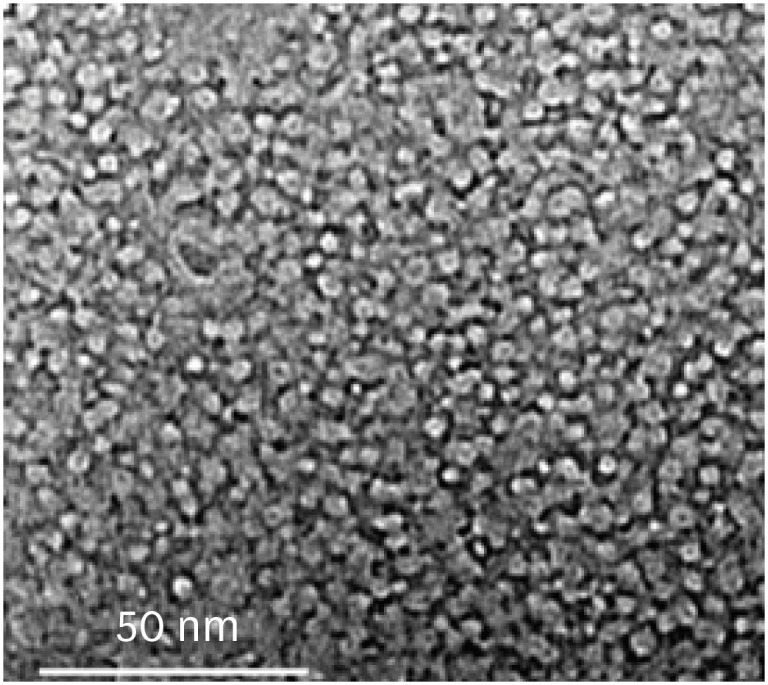
ePub Objectives This study addresses the effect of using nanoparticles (np) on the antimicrobial properties of bioactive glass (BAG) when used in intracanal medicaments against
Enterococcus faecalis (E. faecalis ) biofilms.Materials and Methods E. faecalis biofilms, grown inside 90 root canals for 21 days, were randomly divided into 4 groups according to the antimicrobial regimen followed (n = 20; BAG-np, BAG, calcium hydroxide [CaOH], and saline). After 1 week, residual live bacteria were quantified in terms of colony-forming units (CFU), while dead bacteria were assessed with a confocal laser scanning microscope.Results Although there was a statistically significant decrease in the mean CFU value among all groups, the nano-group performed the best. The highest percentage of dead bacteria was detected in the BAG-np group, with a significant difference from the BAG group.
Conclusions The reduction of particle size and use of a nano-form of BAG improved the antimicrobial properties of the intracanal treatment of
E. faecalis biofilms-
Citations
Citations to this article as recorded by- Size matters: Radiation shielding superiority of borate glasses with nano vs. micro ZnO
Aljawhara H. Almuqrin, M.I. Sayyed, M. Elsafi
Nuclear Engineering and Technology.2025; 57(9): 103614. CrossRef - Effect of Chitosan and bioactive glass nanomaterials as intracanal medicaments on TGF-β1 release from intraradicular dentin
Sarah Salah Hashem, Mohammed M. Khalefa, Mahmoud Hassan Mohamed, Hemat M. ELSheikh, Fatma Abd El-Rahman Taher
BMC Oral Health.2025;[Epub] CrossRef - Effect of Er: YAG laser, phthalocyanine activated photodynamic therapy, and bioactive glass nanoparticles on smear layer removal and push out bond strength of quartz fiber posts to canal dentin: a SEM assessment
Okba Mahmoud, Erum Zain
Frontiers in Dental Medicine.2025;[Epub] CrossRef - Advancements in Root Canal Therapy: Translational Innovations and the Role of Nanoparticles in Endodontic Treatment
Noha M. Badawi, Mohamed M. Kataia, Hadeel A. Mousa, Mozhgan Afshari
Journal of Nanotechnology.2025;[Epub] CrossRef - Propolis in Endodontics—Unveiling Its Therapeutic Potential: A Narrative Review
Poorani Durai, Santha Devy A, Mithila Mohan, Harish Ramalingam, Shasidharan P, Rahul Chaurasia M
World Journal of Dentistry.2025; 16(10): 959. CrossRef - Application of Nanomaterials in Endodontics
Farzaneh Afkhami, Yuan Chen, Laurence J. Walsh, Ove A. Peters, Chun Xu
BME Frontiers.2024;[Epub] CrossRef - Antimicrobial efficacy of newly prepared nano-tricalcium silicate-58s bioactive glass-based endodontic sealer
Nawal Atiya Al-Sabawi, Sawsan Hameed Al-Jubori
Endodontology.2024;[Epub] CrossRef - Antimicrobial Effects of Formulations of Various Nanoparticles and Calcium Hydroxide as Intra-canal Medications Against Enterococcus faecalis: A Systematic Review
Seema H Bukhari, Dax Abraham, Shakila Mahesh
Cureus.2024;[Epub] CrossRef - Effect of nanoparticles on antibacterial efficacy of intracanal medicament: A scoping review
Alpa Gupta, Arundeep Singh, Vivek Aggarwal
Endodontology.2023; 35(4): 283. CrossRef - Physical properties, marginal adaptation and bioactivity of an experimental mineral trioxide aggregate-like cement modified with bioactive materials
Abigailt Flores-Ledesma, Adriana Tejeda-Cruz, María A. Moyaho-Bernal, Ana Wintergerst, Yoshamin A. Moreno-Vargas, Jacqueline A. Rodríguez-Chávez, Carlos E. Cuevas-Suárez, Kenya Gutiérrez-Estrada, Jesús A. Arenas-Alatorre
Journal of Oral Science.2023; 65(2): 141. CrossRef - Nanopartículas antimicrobianas en endodoncia: Revisión narrativa
Gustavo Adolfo Tovar Rangel , Fanny Mildred González Sáenz , Ingrid Ximena Zamora Córdoba , Lina María García Zapata
Revista Estomatología.2023;[Epub] CrossRef
- Size matters: Radiation shielding superiority of borate glasses with nano vs. micro ZnO
- 1,504 View
- 25 Download
- 6 Web of Science
- 11 Crossref

- Silver nanoparticles in endodontics: recent developments and applications
- Aysenur Oncu, Yan Huang, Gulin Amasya, Fatma Semra Sevimay, Kaan Orhan, Berkan Celikten
- Restor Dent Endod 2021;46(3):e38. Published online July 1, 2021
- DOI: https://doi.org/10.5395/rde.2021.46.e38
-
 Abstract
 PDF
PDF PubReader
 ePub
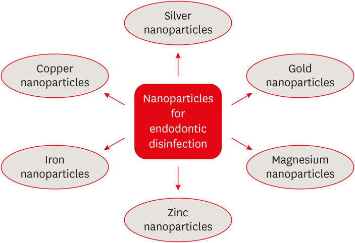
ePub The elimination of endodontic biofilms and the maintenance of a leak-proof canal filling are key aspects of successful root canal treatment. Several materials have been introduced to treat endodontic disease, although treatment success is limited by the features of the biomaterials used. Silver nanoparticles (AgNPs) have been increasingly considered in dental applications, especially endodontics, due to their high antimicrobial activity. For the present study, an electronic search was conducted using MEDLINE (PubMed), the Cochrane Central Register of Controlled Trials (CENTRAL), Google Scholar, and EMBASE. This review provides insights into the unique characteristics of AgNPs, including their chemical, physical, and antimicrobial properties; limitations; and potential uses. Various studies involving different application methods of AgNPs were carefully examined. Based on previous clinical studies, the synthesis, means of obtaining, usage conditions, and potential cytotoxicity of AgNPs were evaluated. The findings indicate that AgNPs are effective antimicrobial agents for the elimination of endodontic biofilms.
-
Citations
Citations to this article as recorded by- Endodontic Intracanal Medicaments and Agents
Anu Priya Guruswamy Pandian, Depti Bellani, Ritya Mary Jibu, Varsha Agnihotri
Dental Clinics of North America.2026; 70(1): 45. CrossRef - Advanced nanoparticle-based antibacterial delivery for endodontic disinfection: A systematic review and meta-analysis
Kanwalpreet Kaur, Seerat Kaura, Ravinder S Saini, Maurya Manjunath, Shashit Shetty Bavabeedu, Mario Alberto Alarcón-Sánchez, Javier Flores-Fraile, Artak Heboyan
Journal of Dentistry.2026; 166: 106347. CrossRef - Scoping review on the genotoxicity of silver nanoparticles in endodontics: therapeutic saviors or genetic saboteurs?
Galvin Sim Siang Lin, Widya Lestari, Mohd Haikal Muhamad Halil, Mohd Syafiq Abd Aziz
Odontology.2025; 113(2): 457. CrossRef - Bioceramics in Endodontics: Limitations and Future Innovations—A Review
Peramune Arachchilage Amila Saman Prasad Kumara, Paul Roy Cooper, Peter Cathro, Maree Gould, George Dias, Jithendra Ratnayake
Dentistry Journal.2025; 13(4): 157. CrossRef - Recent advances in antibacterial nanoformulations for endodontic applications
Tiago Dionísio, Pedro Brandão, Vanessa Machado, João Botelho, José João Mendes, Pedro Fonte
Expert Opinion on Drug Delivery.2025; 22(8): 1117. CrossRef - Systematic review of silver and vanadium-based antibiofilm agents: mechanisms and efficacy in oral biofilms
João Marcos Carvalho-Silva, Andréa Cândido dos Reis
Future Microbiology.2025; 20(10): 639. CrossRef - Nanomaterial-Enhanced Dentistry: A Clinical Perspective
Selvam Manoj, Radhakrishnan Sreena, Rajkumar Divya, Starlin Ebinesh, Shenbagaraman Akshaya, Srikumar Sugantha Angel, Arputharaj Joseph Nathanael
ACS Biomaterials Science & Engineering.2025; 11(8): 4671. CrossRef - Time-dependent Tooth Color Changes Following Conventional, Silver-based, and Photodynamic Root Canal Irrigants: An In Vitro Study
Laila Mohamed Mohamed Kenawi, Mohamed Fattouh, Khaled Abid Althaqafi, Abla Arafa
The Open Dentistry Journal.2025;[Epub] CrossRef - Antimicrobial Effects of Formulations of Various Nanoparticles and Calcium Hydroxide as Intra-canal Medications Against Enterococcus faecalis: A Systematic Review
Seema H Bukhari, Dax Abraham, Shakila Mahesh
Cureus.2024;[Epub] CrossRef - The Push-Out Bond Strength, Surface Roughness, and Antimicrobial Properties of Endodontic Bioceramic Sealers Supplemented with Silver Nanoparticles
Karla Navarrete-Olvera, Nereyda Niño-Martínez, Idania De Alba-Montero, Nuria Patiño-Marín, Facundo Ruiz, Horacio Bach, Gabriel-Alejandro Martínez-Castañón
Molecules.2024; 29(18): 4422. CrossRef - Synergistic bactericidal activity of chlorhexidine loaded on positively charged ionic liquid-protected silver nanoparticles as a root canal disinfectant against Enterococcus faecalis: An ex vivo study
Abbas Abbaszadegan, Elham Tayebikhorami, Ahmad Gholami, Nazanin Bonyanpour, Bahar Asheghi, Sara Nikmanesh
Journal of Ionic Liquids.2024; 4(2): 100117. CrossRef - Improving the Antimicrobial Potency of Berberine for Endodontic Canal Irrigation Using Polymeric Nanoparticles
Célia Marques, Liliana Grenho, Maria Helena Fernandes, Sofia A. Costa Lima
Pharmaceutics.2024; 16(6): 786. CrossRef - A narrative review on application of metal and metal oxide nanoparticles in endodontics
Roohollah Sharifi, Ahmad Vatani, Amir Sabzi, Mohsen Safaei
Heliyon.2024; 10(15): e34673. CrossRef - The Effectiveness of Silver Nanoparticles Mixed with Calcium Hydroxide against Candida albicans: An Ex Vivo Analysis
Maha Alghofaily, Jood Alfraih, Aljohara Alsaud, Norah Almazrua, Terrence S. Sumague, Sayed H. Auda, Fahd Alsalleeh
Microorganisms.2024; 12(2): 289. CrossRef - Evaluation of the efficacy of a novel disinfecting material on the surface topography of gutta-percha: An in vitro study
KHanisha Reddy, Lekshmi Chandran, TMurali Mohan, K Sudha, DL Malini, Bonney Dominic
Journal of Conservative Dentistry.2023; 26(1): 94. CrossRef - Silver Nanoparticles and Their Therapeutic Applications in Endodontics: A Narrative Review
Farzaneh Afkhami, Parisa Forghan, James L. Gutmann, Anil Kishen
Pharmaceutics.2023; 15(3): 715. CrossRef - Nanopartículas antimicrobianas en endodoncia: Revisión narrativa
Gustavo Adolfo Tovar Rangel , Fanny Mildred González Sáenz , Ingrid Ximena Zamora Córdoba , Lina María García Zapata
Revista Estomatología.2023;[Epub] CrossRef - Functionalized Nanoparticles: A Paradigm Shift in Regenerative Endodontic Procedures
Vinoo Subramaniam Ramachandran, Mensudar Radhakrishnan, Malathi Balaraman Ravindrran, Venkatesh Alagarsamy, Gowri Shankar Palanisamy
Cureus.2022;[Epub] CrossRef
- Endodontic Intracanal Medicaments and Agents
- 5,624 View
- 91 Download
- 13 Web of Science
- 18 Crossref

- The effectiveness of the supplementary use of the XP-endo Finisher on bacteria content reduction: a systematic review and meta-analysis
- Ludmila Smith de Jesus Oliveira, Rafaella Mariana Fontes de Bragança, Rafael Sarkis-Onofre, André Luis Faria-e-Silva
- Restor Dent Endod 2021;46(3):e37. Published online June 18, 2021
- DOI: https://doi.org/10.5395/rde.2021.46.e37
-
 Abstract
 PDF
PDF Supplementary Material
 PubReader
 ePub
ePub Objectives This systematic review evaluated the efficacy of the supplementary use of the XP-endo Finisher on bacteria content reduction in the root canal system.
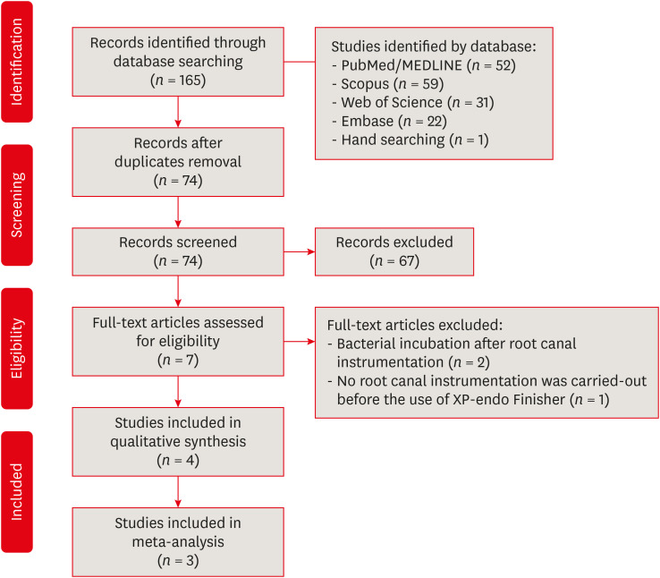
Materials and Methods In-vitro studies evaluating the use of the XP-endo Finisher on bacteria content were searched in four databases in July 2020. Two authors independently screened the studies for eligibility. Data were extracted, and risk of bias was assessed. Data were meta-analyzed by using random-effects model to compare the effect of the supplementary use (experimental) or not (control) of the XP-endo Finisher on bacteria counting reduction, and results from different endodontic protocols were combined. Four studies met the inclusion criteria while 1 study was excluded from the meta-analysis due to its high risk of bias and outlier data. The 3 studies that made it to the meta-analysis had an unclear risk of bias for at least one criterion.Results No heterogeneity was observed among the results of the studies included in the meta-analysis. The study excluded from the meta-analysis assessing the bacteria counting deep in the dentin demonstrated further bacteria reduction upon the use of the XP-endo Finisher.
Conclusions This systematic review found no evidence supporting the supplementary use of the XP-endo Finisher on further bacteria counting the reduction in the root canal.
-
Citations
Citations to this article as recorded by- Mapping risk of bias criteria in systematic reviews of in vitro endodontic studies: an umbrella review
Rafaella Rodrigues da Gama, Lucas Peixoto de Araújo, Evandro Piva, Leandro Perello Duro, Adriana Fernandes da Silva, Wellington Luiz de Oliveira da Rosa
Evidence-Based Dentistry.2025; 26(4): 179. CrossRef - Characteristics and Effectiveness of XP‐Endo Files and Systems: A Narrative Review
Sarah M. Alkahtany, Rana Alfadhel, Aseel AlOmair, Sarah Bin Durayhim, Kee Y. Kum
International Journal of Dentistry.2024;[Epub] CrossRef - Impact XP-endo finisher on the 1-year follow-up success of posterior root canal treatments: a randomized clinical trial
Ludmila Smith de Jesus Oliveira, Fabricio Eneas Diniz de Figueiredo, Janaina Araújo Dantas, Maria Amália Gonzaga Ribeiro, Carlos Estrela, Manoel Damião Sousa-Neto, André Luis Faria-e-Silva
Clinical Oral Investigations.2023; 27(12): 7595. CrossRef - Comparative analysis of the effectiveness of modern irrigants activation techniques in the process of mechanical root canal system treatment (Literature review)
Anatoliy Potapchuk, Vasyl Almashi, Arsenii Horzov, Victor Buleza
InterConf.2023; (34(159)): 200. CrossRef - Comparative analysis of the effectiveness of modern irrigants activation techniques in the protocol of chemomechanical root canal system treatment (literature review)
A. Potapchuk, V. Almashi, Y. Rak, Y. Melnyk, V. Buleza, A. Horzov
SUCHASNA STOMATOLOHIYA.2023; 114(3): 4. CrossRef - Methodological quality assessment criteria for the evaluation of laboratory‐based studies included in systematic reviews within the specialty of Endodontology: A development protocol
Venkateshbabu Nagendrababu, Paul V. Abbott, Christos Boutsioukis, Henry F. Duncan, Clovis M. Faggion, Anil Kishen, Peter E. Murray, Shaju Jacob Pulikkotil, Paul M. H. Dummer
International Endodontic Journal.2022; 55(4): 326. CrossRef
- Mapping risk of bias criteria in systematic reviews of in vitro endodontic studies: an umbrella review
- 2,368 View
- 15 Download
- 4 Web of Science
- 6 Crossref

- A novel antimicrobial-containing nanocellulose scaffold for regenerative endodontics
- Victoria Kichler, Lucas Soares Teixeira, Maick Meneguzzo Prado, Guilherme Colla, Daniela Peressoni Vieira Schuldt, Beatriz Serrato Coelho, Luismar Marques Porto, Josiane de Almeida
- Restor Dent Endod 2021;46(2):e20. Published online March 16, 2021
- DOI: https://doi.org/10.5395/rde.2021.46.e20
-
 Abstract
 PDF
PDF PubReader
 ePub
ePub Objectives The aim of this study was to evaluate bacterial nanocellulose (BNC) membranes incorporated with antimicrobial agents regarding cytotoxicity in fibroblasts of the periodontal ligament (PDLF), antimicrobial activity, and inhibition of multispecies biofilm formation.
Materials and Methods The tested BNC membranes were BNC + 1% clindamycin (BNC/CLI); BNC + 0.12% chlorhexidine (BNC/CHX); BNC + nitric oxide (BNC/NO); and conventional BNC (BNC; control). After PDLF culture, the BNC membranes were positioned in the wells and maintained for 24 hours. Cell viability was then evaluated using the MTS calorimetric test. Antimicrobial activity against
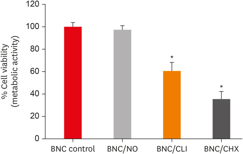
Enterococcus faecalis ,Actinomyces naeslundii , andStreptococcus sanguinis (S. sanguinis ) was evaluated using the agar diffusion test. To assess the antibiofilm activity, BNC membranes were exposed for 24 hours to the mixed culture. After sonicating the BNC membranes to remove the remaining biofilm and plating the suspension on agar, the number of colony-forming units (CFU)/mL was determined. Data were analyzed by 1-way analysis of variance and the Tukey, Kruskal-Wallis, and Dunn tests (α = 5%).Results PDLF metabolic activity after contact with BNC/CHX, BNC/CLI, and BNC/NO was 35%, 61% and 97%, respectively, compared to BNC. BNC/NO showed biocompatibility similar to that of BNC (
p = 0.78). BNC/CLI showed the largest inhibition halos, and was superior to the other BNC membranes againstS. sanguinis (p < 0.05). The experimental BNC membranes inhibited biofilm formation, with about a 3-fold log CFU reduction compared to BNC (p < 0.05).Conclusions BNC/NO showed excellent biocompatibility and inhibited multispecies biofilm formation, similarly to BNC/CLI and BNC/CHX.
-
Citations
Citations to this article as recorded by- Topic: Perspectives on Success and Failure of Endodontic Treatments
Ilma Robo, Manola Kelmendi, Eva Habazaj, Kleves Elezi, Rialda Xhizdari, Nevila Alliu
SN Comprehensive Clinical Medicine.2025;[Epub] CrossRef - Data about application of chlorhexidine as a periodontal irrigant –
Systematic Review.
Ilma Robo, Manola Kelmendi , Eva Habazaj , Kristi Sulanjaku , Nevila Alliu
Acta Stomatologica Marisiensis Journal.2025; 8(1): 6. CrossRef - Aqueous‐Phase Surface Amidation of TEMPO‐CNF Films for Improved Adsorption of Organic Pollutants in Water
Domenico Santandrea, Cécile Sillard, Valentina Beghetto, Julien Bras
ChemPlusChem.2025;[Epub] CrossRef - Materials design of gas-releasing nanoplatforms: strategies for precision delivery in oral healthcare
Haodong Zhong, Weiming Tan, Jian Zhang, Xiongwei Huang, Haizhan Chen, Jiyuan Zou, Yuxin Ye, Tao Wang, Xuechao Yang, Jiang Li, Li Yang, Lvhua Guo, Tao Luo
Materials & Design.2025; 258: 114704. CrossRef - Pushing the limits of bacterial cellulose for biomedicine: a review
Cristina Campano, Virginia Rivero-Buceta, Ana M. Hernandez-Arriaga, Maria T. Manoli, M. Auxiliadora Prieto
International Journal of Biological Macromolecules.2025; 323: 146701. CrossRef - Prospective and applications of bacterial nanocellulose in dentistry
Yasmin Alimardani, Esmaeel Mirzakhani, Fereshteh Ansari, Hadi Pourjafar, Nadia Sadeghi
Cellulose.2024; 31(13): 7819. CrossRef - Bacterial nanocelluloses as sustainable biomaterials for advanced wound healing and dressings
Atefeh Zarepour, Bahar Gok, Yasemin Budama-Kilinc, Arezoo Khosravi, Siavash Iravani, Ali Zarrabi
Journal of Materials Chemistry B.2024; 12(48): 12489. CrossRef - Sulfated endospermic nanocellulose crystals prevent the transmission of SARS-CoV-2 and HIV-1
Enrique Javier Carvajal-Barriga, Wendy Fitzgerald, Emilios K. Dimitriadis, Leonid Margolis, R. Douglas Fields
Scientific Reports.2023;[Epub] CrossRef - A Novel Approach for the Fabrication of 3D-Printed Dental Membrane Scaffolds including Antimicrobial Pomegranate Extract
Hatice Karabulut, Songul Ulag, Basak Dalbayrak, Elif Arisan, Turgut Taskin, Mehmet Guncu, Burak Aksu, Alireza Valanezhad, Oguzhan Gunduz
Pharmaceutics.2023; 15(3): 737. CrossRef - Current advances of nanocellulose application in biomedical field
M.Y. Leong, Y.L. Kong, M.Y. Harun, C.Y. Looi, W.F. Wong
Carbohydrate Research.2023; 532: 108899. CrossRef - Bacterial cellulose as a potential biopolymer in biomedical applications: a state-of-the-art review
Prachi Shrivastav, Sheersha Pramanik, Gayatri Vaidya, Mohamed A. Abdelgawad, Mohammed M. Ghoneim, Ajeet Singh, Bassam M. Abualsoud, Larissa Souza Amaral, Mohammed A. S. Abourehab
Journal of Materials Chemistry B.2022; 10(17): 3199. CrossRef - Nanocelluloses as new generation materials: natural resources, structure-related properties, engineering nanostructures, and technical challenges
Ahmed Barhoum, Vibhore K. Rastogi, Bhupender K. Mahur, Amit Rastogi, Fatehy M. Abdel-Haleem, Pieter Samyn
Materials Today Chemistry.2022; 26: 101247. CrossRef - The current natural/chemical materials and innovative technologies in periodontal diseases therapy and regeneration: A narrative review
Peyman Esmaeili Fard Barzegar, Reza Ranjbar, Mohsen Yazdanian, Elahe Tahmasebi, Mostafa Alam, Kamyar Abbasi, Hamid Tebyaniyan, Keyvan Esmaeili Fard Barzegar
Materials Today Communications.2022; 32: 104099. CrossRef
- Topic: Perspectives on Success and Failure of Endodontic Treatments
- 1,988 View
- 30 Download
- 14 Web of Science
- 13 Crossref

- Endodontic biofilms: contemporary and future treatment options
- Yeon-Jee Yoo, Hiran Perinpanayagam, Soram Oh, A-Reum Kim, Seung-Hyun Han, Kee-Yeon Kum
- Restor Dent Endod 2019;44(1):e7. Published online January 31, 2019
- DOI: https://doi.org/10.5395/rde.2019.44.e7
-
 Abstract
 PDF
PDF PubReader
 ePub
ePub Apical periodontitis is a biofilm-mediated infection. The biofilm protects bacteria from host defenses and increase their resistance to intracanal disinfecting protocols. Understanding the virulence of these endodontic microbiota within biofilm is essential for the development of novel therapeutic procedures for intracanal disinfection. Both the disruption of biofilms and the killing of their bacteria are necessary to effectively treat apical periodontitis. Accordingly, a review of endodontic biofilm types, antimicrobial resistance mechanisms, and current and future therapeutic procedures for endodontic biofilm is provided.
-
Citations
Citations to this article as recorded by- Evaluation of Anti-Biofilm Property of Zirconium Oxide Nanoparticles on Streptococcus mutans and Enterococcus faecalis: An In Vitro Study
Anu Priya Guruswamy Pandian, Anil Kumar Ramachandran, Priyanka Kodaganallur Pitchumani, Blessy Mathai, Davis C Thomas
Cureus.2025;[Epub] CrossRef - Recent advances in antibacterial nanoformulations for endodontic applications
Tiago Dionísio, Pedro Brandão, Vanessa Machado, João Botelho, José João Mendes, Pedro Fonte
Expert Opinion on Drug Delivery.2025; 22(8): 1117. CrossRef - Physical–Chemical Assessment and Antimicrobial Activity of Chlortetracycline-Loaded Collagen Sponges
Graţiela Teodora Tihan, Camelia Ungureanu, Ileana Rău, Roxana Gabriela Zgârian, Răzvan Constantin Barbaresso, Mădălina Georgiana Albu Kaya, Cristina-Elena Dinu-Pîrvu, Mihaela Violeta Ghica
Materials.2025; 18(17): 4029. CrossRef - A Review of Chemical Approaches Inherent to Endodontic Disinfection Protocols: Part 1
Fatima Peer, Yahya E. Choonara, Pradeep Kumar
South African Dental Journal.2025; 80(07): 352. CrossRef - Self-Sacrificial Antibacterial Coating with Photothermal Response for Inhibiting Implant Infection
Jinglin Zhang, Aijian Cao, Lizhen Chen, Dongliang Huo, Jingxian Zhang, Langhuan Huang, Shaozao Tan
ACS Applied Nano Materials.2024; 7(23): 26907. CrossRef - Biofilm in Endodontic Infection and its Advanced Therapeutic Options – An Updated Review
Srilekha Jayakumar, Dinesh Sridhar, Bindu M. John, Karthikeyan Arumugam, Prashanth Ponnusamy, Hema Pulidindi
Journal of Pharmacy and Bioallied Sciences.2024; 16(Suppl 2): S1104. CrossRef - Analysis of the chemical interaction of polyhexanide with endodontic irrigants
Z. S. Zurab, Yu. A. Generalova, A. A. Kulikova, A. Yu. Umarov, F. V. Badalov, A. Wehbe, E. M. Kakabadze
Endodontics Today.2024; 22(4): 319. CrossRef - In vitro evaluation of three engineered multispecies endodontic biofilms on a dentinal disk substrate
Wajih Hage, Dolla Karam Sarkis, Mireille Kallasy, May Mallah, Carla Zogheib
Biomaterial Investigations in Dentistry.2023;[Epub] CrossRef - In vitro evaluation of enterococcus faecalis growth in different conditions on dentinal substrate
Wajih Hage, Dolla Karam Sarkis, Mireille Kallassy, May Mallah, Carla Zogheib
Biomaterial Investigations in Dentistry.2023;[Epub] CrossRef - Bacteria associated with apical periodontitis promotes in vitro the differentiation of macrophages to osteoclasts
A. P. Torres-Monjarás, R. Sánchez-Gutiérrez, B. Hernández-Castro, L. González-Baranda, D. L. Alvarado-Hernández, A. Pozos-Guillén, A. Muñoz-Ruiz, V. Méndez-González, R. González-Amaro, M. Vitales-Noyola
Clinical Oral Investigations.2023; 27(6): 3139. CrossRef - Antimicrobial efficacy of Kerr pulp canal sealer (EWT) in combination with 10% amoxicillin on Enterococcus faecalis: A confocal laser scanning microscopic study
Madhureema De Sarkar, Kundabala Mala, Suchitra Shenoy Mala, Shama Prasada Kabekkodu, Srikant Natarajan, Neeta Shetty, Priyanka Madhav Kamath, Manuel Thomas
F1000Research.2023; 12: 725. CrossRef - Antimicrobial efficacy of Kerr pulp canal sealer (EWT) in combination with 10% amoxicillin on Enterococcus faecalis: A confocal laser scanning microscopic study
Madhureema De Sarkar, Kundabala Mala, Suchitra Shenoy Mala, Shama Prasada Kabekkodu, Srikant Natarajan, Neeta Shetty, Priyanka Madhav Kamath, Manuel Thomas
F1000Research.2023; 12: 725. CrossRef - Combined effect of electrical energy and graphene oxide on Enterococcus faecalis biofilms
Myung-Jin LEE, Mi-Ah KIM, Kyung-San MIN
Dental Materials Journal.2023; 42(6): 844. CrossRef - Innovative Curved-Tip Reactor for Non-Thermal Plasma and Plasma-Treated Water Generation: Synergistic Impact Comparison with Sodium Hypochlorite in Dental Root Canal Disinfection
Raúl Arguello-Sánchez, Régulo López-Callejas, Benjamín Gonzalo Rodríguez-Méndez, Rogelio Scougall-Vilchis, Ulises Velázquez-Enríquez, Antonio Mercado-Cabrera, Rosendo Peña-Eguiluz, Raúl Valencia-Alvarado, Carlo Eduardo Medina-Solís
Materials.2023; 16(22): 7204. CrossRef - Impact of antimicrobial photodynamic therapy on the bond-strength and penetration of endodontic sealers: A systematic review
Khalid H Almadi
Photodiagnosis and Photodynamic Therapy.2023; 41: 103249. CrossRef - Apical periodontitis in mesiobuccal roots of maxillary molars: influence of anatomy and quality of root canal treatment, a CBCT study
Samantha Jannone Carrion, Marcelo Santos Coelho, Adriana de Jesus Soares, Marcos Frozoni
Restorative Dentistry & Endodontics.2022;[Epub] CrossRef - Potential relationship between clinical symptoms and the root canal microbiomes of root filled teeth based on the next‐generation sequencing
Yajing Hou, Liu Wang, Lan Zhang, Xuelian Tan, Dingming Huang, Dongzhe Song
International Endodontic Journal.2022; 55(1): 18. CrossRef - Efficacy of 6% Sodium Hypochlorite on Infectious Content of Teeth with Symptomatic Irreversible Pulpitis
Rodrigo Arruda-Vasconcelos, Marlos Barbosa-Ribeiro, Lidiane M. Louzada, Beatriz I.N. Lemos, Adriana de-Jesus-Soares, Caio C.R. Ferraz, José F.A. Almeida, Marina A. Marciano, Brenda P.F. A. Gomes
Journal of Endodontics.2022; 48(2): 179. CrossRef - Specialized pro-resolving lipid mediators in endodontics: a narrative review
Davy Aubeux, Ove A. Peters, Sepanta Hosseinpour, Solène Tessier, Valérie Geoffroy, Fabienne Pérez, Alexis Gaudin
BMC Oral Health.2021;[Epub] CrossRef - Effects of curcumin-mediated antimicrobial photodynamic therapy associated to different chelators against Enterococcus faecalis biofilms
Daniela Alejandra Cusicanqui Méndez, Maricel Rosario Cardenas Cuéllar, Victor Feliz Pedrinha, Evelyn Giuliana Velásquez Espedilla, Flaviana Bombarda de Andrade, Patrícia de Almeida Rodrigues, Thiago Cruvinel
Photodiagnosis and Photodynamic Therapy.2021; 35: 102464. CrossRef - Effectiveness of D,L‐2‐hydroxyisocaproic acid (HICA) and alpha‐mangostin against endodontopathogenic microorganisms in a multispecies bacterial–fungal biofilm in anex vivotooth model
Warat Leelapornpisid, Lilyann Novak‐Frazer, Alison Qualtrough, Riina Rautemaa‐Richardson
International Endodontic Journal.2021; 54(12): 2243. CrossRef - In Vitro Evaluation of a New Combination of Three Antibiotic Paste Against Common Endodontic Pathogens
Prasanna Dahake, Nilima Thosar
Journal of Islamic Dental Association of IRAN.2021; 33(3): 58. CrossRef - Effect of using diode laser on Enterococcus faecalis and its lipoteichoic acid (LTA) in chronic apical periodontitis
Zhaohui Zou, Junu Bhandari, Baiyan Xiao, Xiaoyue Liang, Yu Zhang, Guohui Yan
Lasers in Medical Science.2021; 36(5): 1059. CrossRef - Prevalence of Bacteria of Genus Actinomyces in Persistent Extraradicular Lesions—Systematic Review
Mario Dioguardi, Vito Crincoli, Luigi Laino, Mario Alovisi, Diego Sovereto, Lorenzo Lo Muzio, Giuseppe Troiano
Journal of Clinical Medicine.2020; 9(2): 457. CrossRef - Evaluation of in vitro biofilm elimination of Enterococcus faecalis using a continuous ultrasonic irrigation device
Jennifer Galván-Pacheco, Marlen Vitales-Noyola, Ana M. González-Amaro, Heriberto Bujanda-Wong, Antonio Aragón-Piña, Verónica Méndez-González, Amaury Pozos-Guillén
Journal of Oral Science.2020; 62(4): 415. CrossRef - Comparison of the use of d-enantiomeric and l-enantiomeric antimicrobial peptides incorporated in a calcium-chelating irrigant against Enterococcus faecalis root canal wall biofilms
Wei-hu Ye, Lara Yeghiasarian, Christopher W. Cutler, Brian E. Bergeron, Stephanie Sidow, Hockin H.K. Xu, Li-na Niu, Jing-zhi Ma, Franklin R. Tay
Journal of Dentistry.2019; 91: 103231. CrossRef
- Evaluation of Anti-Biofilm Property of Zirconium Oxide Nanoparticles on Streptococcus mutans and Enterococcus faecalis: An In Vitro Study
- 4,214 View
- 117 Download
- 26 Crossref

-
Inhibition of nicotine-induced
Streptococcus mutans biofilm formation by salts solutions intended for mouthrinses - Abdulrahman A. Balhaddad, Mary Anne S. Melo, Richard L. Gregory
- Restor Dent Endod 2019;44(1):e4. Published online January 16, 2019
- DOI: https://doi.org/10.5395/rde.2019.44.e4
-
 Abstract
 PDF
PDF PubReader
 ePub
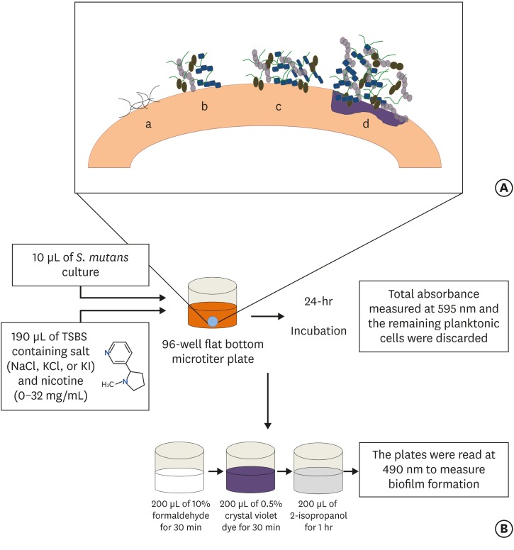
ePub Objectives Biofilm formation is critical to dental caries initiation and development. The aim of this study was to investigate the effects of nicotine exposure on
Streptococcus mutans (S. mutans ) biofilm formation concomitantly with the inhibitory effects of sodium chloride (NaCl), potassium chloride (KCl) and potassium iodide (KI) salts. This study examined bacterial growth with varying concentrations of NaCl, KCl, and KI salts and nicotine levels consistent with primary levels of nicotine exposure.Materials and Methods A preliminary screening experiment was performed to investigate the appropriate concentrations of NaCl, KCl, and KI to use with nicotine. With the data, a
S. mutans biofilm growth assay was conducted using nicotine (0–32 mg/mL) in Tryptic Soy broth supplemented with 1% sucrose with and without 0.45 M of NaCl, 0.23 M of KCl, and 0.113 M of KI. The biofilm was stained with crystal violet dye and the absorbance measured to determine biofilm formation.Results The presence of 0.45 M of NaCl, 0.23 M of KCl, and 0.113 M of KI significantly inhibited (
p < 0.05) nicotine-inducedS. mutans biofilm formation by 52%, 79.7%, and 64.1%, respectively.Conclusions The results provide additional evidence regarding the biofilm-enhancing effects of nicotine and demonstrate the inhibitory influence of these salts in reducing the nicotine-induced biofilm formation. A short-term exposure to these salts may inhibit
S. mutans biofilm formation.-
Citations
Citations to this article as recorded by- Biofilm forming and swarming activities of Bacillus cereus modulated by multiclass compounds
Abdul Rafay Rafiq, Mohsin Tariq, Syeda Tahseen Zahra, Temoor Ahmed
The Microbe.2026; 10: 100644. CrossRef - The Inhibition of Streptococcus mutans Biofilms following Exposure to Different Chocolate Ingredients
Hadi A. Almoabid, Leen Saleh Almutairi, Abdul Samad Khan, Mohammed A. Aljaffary, Rasha AlSheikh, Khalid S. Almulhim, Abdulrahman A. Balhaddad
European Journal of Dentistry.2025;[Epub] CrossRef - The impact of Caralluma munbyana extracts on Streptococcus mutans biofilm formation
Turki Alshehri, Israa Alkhalifah, Areeb Alotaibi, Alaa F. Alsulaiman, Abdullah Al Madani, Basil Almutairi, Abdulrahman A. Balhaddad
Frontiers in Dental Medicine.2025;[Epub] CrossRef - Tobacco‐enhanced biofilm formation by Porphyromonas gingivalis and other oral microbes
Jinlian Tan, Gwyneth J. Lamont, David A. Scott
Molecular Oral Microbiology.2024; 39(5): 270. CrossRef - Nicotine is a potent extracellular polysaccharide inducer in Fusobacterium nucleatum biofilms
Adaias Oliveira Matos, Valentim Adelino Ricardo Barão, Richard Lee Gregory
Brazilian Journal of Oral Sciences.2023;[Epub] CrossRef - Effect of eucalyptus oil on Streptococcus mutans and Enterococcus faecalis growth
Abdulrahman A. Balhaddad, Rasha N. AlSheikh
BDJ Open.2023;[Epub] CrossRef - Microorganisms: crucial players of smokeless tobacco for several health attributes
Akanksha Vishwakarma, Digvijay Verma
Applied Microbiology and Biotechnology.2021; 105(16-17): 6123. CrossRef - Microbiology of the American Smokeless Tobacco
A. J. Rivera, R. E. Tyx
Applied Microbiology and Biotechnology.2021; 105(12): 4843. CrossRef - The Impact of Photosensitizer Selection on Bactericidal Efficacy Of PDT against Cariogenic Biofilms: A Systematic Review and Meta-Analysis
Maurício Ítalo Silva Teófilo, Teresa Maria Amorim Zaranza de Carvalho Russi, Paulo Goberlanio de Barros Silva, Abdulrahman A. Balhaddad, Mary Anne S. Melo, Juliana P.M.L. Rolim
Photodiagnosis and Photodynamic Therapy.2021; 33: 102046. CrossRef - Antibacterial Activities of Methanol and Aqueous Extracts of Salvadora persica against Streptococcus mutans Biofilms: An In Vitro Study
Abdulrahman A. Balhaddad, Lamia Mokeem, Mary Anne S. Melo, Richard L. Gregory
Dentistry Journal.2021; 9(12): 143. CrossRef - The burden of root caries: Updated perspectives and advances on management strategies
Mohammed S. AlQranei, Abdulrahman A. Balhaddad, Mary A.S. Melo
Gerodontology.2021; 38(2): 136. CrossRef - Emerging Contact-Killing Antibacterial Strategies for Developing Anti-Biofilm Dental Polymeric Restorative Materials
Heba Mitwalli, Rashed Alsahafi, Abdulrahman A. Balhaddad, Michael D. Weir, Hockin H. K. Xu, Mary Anne S. Melo
Bioengineering.2020; 7(3): 83. CrossRef - In-Vitro Model of Scardovia wiggsiae Biofilm Formation and Effect of Nicotine
Abdulrahman A. Balhaddad, Hadeel M. Ayoub, Richard L. Gregory
Brazilian Dental Journal.2020; 31(5): 471. CrossRef - Antibacterial efficacy and remineralization capacity of glycyrrhizic acid added casein phosphopeptide‐amorphous calcium phosphate
Feride Sahin, Fatih Oznurhan
Microscopy Research and Technique.2020; 83(7): 744. CrossRef - Concentration dependence of quaternary ammonium monomer on the design of high-performance bioactive composite for root caries restorations
Abdulrahman A. Balhaddad, Maria S. Ibrahim, Michael D. Weir, Hockin H.K. Xu, Mary Anne S. Melo
Dental Materials.2020; 36(8): e266. CrossRef
- Biofilm forming and swarming activities of Bacillus cereus modulated by multiclass compounds
- 1,602 View
- 13 Download
- 15 Crossref

- Antifungal effects of synthetic human β-defensin 3-C15 peptide
- Sang-Min Lim, Ki-Bum Ahn, Christine Kim, Jong-Won Kum, Hiran Perinpanayagam, Yu Gu, Yeon-Jee Yoo, Seok Woo Chang, Seung Hyun Han, Won-Jun Shon, Woocheol Lee, Seung-Ho Baek, Qiang Zhu, Kee-Yeon Kum
- Restor Dent Endod 2016;41(2):91-97. Published online March 17, 2016
- DOI: https://doi.org/10.5395/rde.2016.41.2.91
-
 Abstract
 PDF
PDF PubReader
 ePub
ePub Objectives The purpose of this
ex vivo study was to compare the antifungal activity of a synthetic peptide consisting of 15 amino acids at the C-terminus of human β-defensin 3 (HBD3-C15) with calcium hydroxide (CH) and Nystatin (Nys) againstCandida albicans (C. albicans ) biofilm.Materials and Methods C. albicans were grown on cover glass bottom dishes or human dentin disks for 48 hr, and then treated with HBD3-C15 (0, 12.5, 25, 50, 100, 150, 200, and 300 µg/mL), CH (100 µg/mL), and Nys (20 µg/mL) for 7 days at 37℃. On cover glass, live and dead cells in the biomass were measured by the FilmTracer Biofilm viability assay, and observed by confocal laser scanning microscopy (CLSM). On dentin, normal, diminished and ruptured cells were observed by field-emission scanning electron microscopy (FE-SEM). The results were subjected to a two-tailedt -test, a one way analysis variance and apost hoc test at a significance level ofp = 0.05.Results C. albicans survival on dentin was inhibited by HBD3-C15 in a dose-dependent manner. There were fewer aggregations ofC. albicans in the groups of Nys and HBD3-C15 (≥ 100 µg/mL). CLSM showedC. albicans survival was reduced by HBD3-C15 in a dose dependent manner. Nys and HBD3-C15 (≥ 100 µg/mL) showed significant fungicidal activity compared to CH group (p < 0.05).Conclusions Synthetic HBD3-C15 peptide (≥ 100 µg/mL) and Nys exhibited significantly higher antifungal activity than CH against
C. albicans by inhibiting cell survival and biofilm.-
Citations
Citations to this article as recorded by- Anti-fungal peptides: an emerging category with enthralling therapeutic prospects in the treatment of candidiasis
Jyoti Sankar Prusty, Ashwini Kumar, Awanish Kumar
Critical Reviews in Microbiology.2025; 51(5): 755. CrossRef - Current status of antimicrobial peptides databases and computational tools for optimization
Madhulika Jha, Akash Nautiyal, Kumud Pant, Navin Kumar
Environment Conservation Journal.2025; 26(1): 281. CrossRef - Harnessing antimicrobial peptides in endodontics
Xinzi Kong, Vijetha Vishwanath, Prasanna Neelakantan, Zhou Ye
International Endodontic Journal.2024; 57(7): 815. CrossRef - Human β-defensins and their synthetic analogs: Natural defenders and prospective new drugs of oral health
Mumian Chen, Zihe Hu, Jue Shi, Zhijian Xie
Life Sciences.2024; 346: 122591. CrossRef - Candida albicans Virulence Factors and Pathogenicity for Endodontic Infections
Yeon-Jee Yoo, A Reum Kim, Hiran Perinpanayagam, Seung Hyun Han, Kee-Yeon Kum
Microorganisms.2020; 8(9): 1300. CrossRef - Innate Inspiration: Antifungal Peptides and Other Immunotherapeutics From the Host Immune Response
Derry K. Mercer, Deborah A. O'Neil
Frontiers in Immunology.2020;[Epub] CrossRef - Human salivary proteins and their peptidomimetics: Values of function, early diagnosis, and therapeutic potential in combating dental caries
Kun Wang, Xuedong Zhou, Wei Li, Linglin Zhang
Archives of Oral Biology.2019; 99: 31. CrossRef - Endodontic biofilms: contemporary and future treatment options
Yeon-Jee Yoo, Hiran Perinpanayagam, Soram Oh, A-Reum Kim, Seung-Hyun Han, Kee-Yeon Kum
Restorative Dentistry & Endodontics.2019;[Epub] CrossRef - Bioactive Peptides Against Fungal Biofilms
Karen G. N. Oshiro, Gisele Rodrigues, Bruna Estéfani D. Monges, Marlon Henrique Cardoso, Octávio Luiz Franco
Frontiers in Microbiology.2019;[Epub] CrossRef - Anticandidal Potential of Stem Bark Extract from Schima superba and the Identification of Its Major Anticandidal Compound
Chun Wu, Hong-Tan Wu, Qing Wang, Guey-Horng Wang, Xue Yi, Yu-Pei Chen, Guang-Xiong Zhou
Molecules.2019; 24(8): 1587. CrossRef - Synthetic Human β Defensin-3-C15 Peptide in Endodontics: Potential Therapeutic Agent in Streptococcus gordonii Lipoprotein-Stimulated Human Dental Pulp-Derived Cells
Yeon-Jee Yoo, Hiran Perinpanayagam, Jue-Yeon Lee, Soram Oh, Yu Gu, A-Reum Kim, Seok-Woo Chang, Seung-Ho Baek, Kee-Yeon Kum
International Journal of Molecular Sciences.2019; 21(1): 71. CrossRef - Candida Infections and Therapeutic Strategies: Mechanisms of Action for Traditional and Alternative Agents
Giselle C. de Oliveira Santos, Cleydlenne C. Vasconcelos, Alberto J. O. Lopes, Maria do S. de Sousa Cartágenes, Allan K. D. B. Filho, Flávia R. F. do Nascimento, Ricardo M. Ramos, Emygdia R. R. B. Pires, Marcelo S. de Andrade, Flaviane M. G. Rocha, Cristi
Frontiers in Microbiology.2018;[Epub] CrossRef - Perspectives for clinical use of engineered human host defense antimicrobial peptides
María Eugenia Pachón-Ibáñez, Younes Smani, Jerónimo Pachón, Javier Sánchez-Céspedes
FEMS Microbiology Reviews.2017; 41(3): 323. CrossRef - The synthetic human beta-defensin-3 C15 peptide exhibits antimicrobial activity against Streptococcus mutans, both alone and in combination with dental disinfectants
Ki Bum Ahn, A. Reum Kim, Kee-Yeon Kum, Cheol-Heui Yun, Seung Hyun Han
Journal of Microbiology.2017; 55(10): 830. CrossRef - Antibiofilm peptides against oral biofilms
Zhejun Wang, Ya Shen, Markus Haapasalo
Journal of Oral Microbiology.2017; 9(1): 1327308. CrossRef - Humanβ-Defensin 3 Reduces TNF-α-Induced Inflammation and Monocyte Adhesion in Human Umbilical Vein Endothelial Cells
Tianying Bian, Houxuan Li, Qian Zhou, Can Ni, Yangheng Zhang, Fuhua Yan
Mediators of Inflammation.2017; 2017: 1. CrossRef - Antifungal Effects of Synthetic Human Beta-defensin-3-C15 Peptide on Candida albicans –infected Root Dentin
Yeon-Jee Yoo, Ikyung Kwon, So-Ram Oh, Hiran Perinpanayagam, Sang-Min Lim, Ki-Bum Ahn, Yoon Lee, Seung-Hyun Han, Seok-Woo Chang, Seung-Ho Baek, Qiang Zhu, Kee-Yeon Kum
Journal of Endodontics.2017; 43(11): 1857. CrossRef - A 15-amino acid C-terminal peptide of beta-defensin-3 inhibits bone resorption by inhibiting the osteoclast differentiation and disrupting podosome belt formation
Ok-Jin Park, Jiseon Kim, Ki Bum Ahn, Jue Yeon Lee, Yoon-Jeong Park, Kee-Yeon Kum, Cheol-Heui Yun, Seung Hyun Han
Journal of Molecular Medicine.2017; 95(12): 1315. CrossRef
- Anti-fungal peptides: an emerging category with enthralling therapeutic prospects in the treatment of candidiasis
- 1,571 View
- 5 Download
- 18 Crossref

- Effect of organic acids in dental biofilm on microhardness of a silorane-based composite
- Sedighe Sadat Hashemikamangar, Seyed Jalal Pourhashemi, Mohammad Talebi, Nazanin Kiomarsi, Mohammad Javad Kharazifard
- Restor Dent Endod 2015;40(3):188-194. Published online June 2, 2015
- DOI: https://doi.org/10.5395/rde.2015.40.3.188
-
 Abstract
 PDF
PDF PubReader
 ePub
ePub Objectives This study evaluated the effect of lactic acid and acetic acid on the microhardness of a silorane-based composite compared to two methacrylate-based composite resins.
Materials and Methods Thirty disc-shaped specimens each were fabricated of Filtek P90, Filtek Z250 and Filtek Z350XT. After measuring of Vickers microhardness, they were randomly divided into 3 subgroups (
n = 10) and immersed in lactic acid, acetic acid or distilled water. Microhardness was measured after 48 hr and 7 day of immersion. Data were analyzed using repeated measures ANOVA (p < 0.05). The surfaces of two additional specimens were evaluated using a scanning electron microscope (SEM) before and after immersion.Results All groups showed a reduction in microhardness after 7 day of immersion (
p < 0.001). At baseline and 7 day, the microhardness of Z250 was the greatest, followed by Z350 and P90 (p < 0.001). At 48 hr, the microhardness values of Z250 and Z350 were greater than P90 (p < 0.001 for both), but those of Z250 and Z350 were not significantly different (p = 0.095). Also, the effect of storage media on microhardness was not significant at baseline, but significant at 48 hr and after 7 day (p = 0.001 andp < 0.001, respectively). Lactic acid had the greatest effect.Conclusions The microhardness of composites decreased after 7 day of immersion. The microhardness of P90 was lower than that of other composites. Lactic acid caused a greater reduction in microhardness compared to other solutions.
-
Citations
Citations to this article as recorded by- Effect of hydroelectrolytic beverages on the roughness and microhardness of bulk fill resin composites
Renata Siqueira Scatolin, Caio Castro Grigoletto, Laura Nobre Ferraz, Rafael Pino Vitti
Brazilian Journal of Oral Sciences.2025; 24: e254003. CrossRef - Investigating the effect of three carbonated drinks on tooth enamel roughness and microhybrid composite
Sara Akbari Fard, Saeed Nemati Anaraki, Haleh Kazemi -Yazdi, Mahsa Qenaat
journal of research in dental sciences.2024; 21(3): 174. CrossRef - Evaluating the effect of natural, industrial juices and beverage on orthodontic bonding composite (in-vitro study)
Rusal S Ahmed, Alan I Saleem
Journal of Baghdad College of Dentistry.2023; 35(3): 10. CrossRef - Stoichiometric models of sucrose and glucose fermentation by oral streptococci: Implications for free acid formation and enamel demineralization
Marzieh Mansouri, Evan P. O'Brien, Karabi Mondal, Chien-Chia Chen, James L. Drummond, Luke Hanley, Karl J. Rockne
Dental Materials.2023; 39(4): 351. CrossRef - Effect of mouthwashes on the microhardness of aesthetic composite restorative materials
Noura Abdulaziz Alessa
Anales del Sistema Sanitario de Navarra.2023;[Epub] CrossRef - Evaluation of the Effect of Natural and Industrial Orange Juices and Beverage on Surface Roughness of Orthodontic Bonding Composite
Rusal Saad Ahmed, Alan Issa Saleem
Dental Hypotheses.2022; 13(3): 107. CrossRef - Effects of particle distribution and calculation method on results of nano-indentation technique in heterogeneous nanocomposites-experimental and numerical approaches
M. Heidari, A. Karimzadeh, M.R. Ayatollahi, M.Y. Yahya
International Journal of Solids and Structures.2021; 225: 111054. CrossRef - New Resin-Based Bulk-Fill Composites: in vitro Evaluation of Micro-Hardness and Depth of Cure as Infection Risk Indexes
Marco Colombo, Simone Gallo, Claudio Poggio, Vittorio Ricaldone, Carla Renata Arciola, Andrea Scribante
Materials.2020; 13(6): 1308. CrossRef - Tribological Behavior of Restorative Dental Microcomposites After Exposure to Mouth Acids
A. C. Branco, J. Brito, M. Codorniz, M. Steinhausen, F. Martins, J. Reis, P. Maurício, R. Colaço, A. P. Serro
Tribology Letters.2019;[Epub] CrossRef - Vickers Micro-Hardness of New Restorative CAD/CAM Dental Materials: Evaluation and Comparison after Exposure to Acidic Drink
Marco Colombo, Claudio Poggio, Alessandro Lasagna, Marco Chiesa, Andrea Scribante
Materials.2019; 12(8): 1246. CrossRef - 30 Months Clinical Evaluation of Posterior Composite Resin Restorations
Serdar Akarsu, Hüseyin Özgür Özdemir
The Journal of Dentists.2018; 6: 6. CrossRef - Survival and Associated Risk Factors of Selective Caries Removal Treatments in Primary Teeth: A Retrospective Study in a High Caries Risk Population
Ximena C. Melgar, Niek J.M. Opdam, Marcos Britto Correa, Renata Franzon, Flávio Fernando Demarco, Fernando B. Araujo, Luciano Casagrande
Caries Research.2017; 51(5): 466. CrossRef
- Effect of hydroelectrolytic beverages on the roughness and microhardness of bulk fill resin composites
- 1,465 View
- 6 Download
- 12 Crossref

- Chelating and antibacterial properties of chitosan nanoparticles on dentin
- Aldo del Carpio-Perochena, Clovis Monteiro Bramante, Marco Antonio Hungaro Duarte, Marcia Regina de Moura, Fauze Ahmad Aouada, Anil Kishen
- Restor Dent Endod 2015;40(3):195-201. Published online March 30, 2015
- DOI: https://doi.org/10.5395/rde.2015.40.3.195
-
 Abstract
 PDF
PDF PubReader
 ePub
ePub Objectives The use of chitosan nanoparticles (CNPs) in endodontics is of interest due to their antibiofilm properties. This study was to investigate the ability of bioactive CNPs to remove the smear layer and inhibit bacterial recolonization on dentin.
Materials and Methods One hundred bovine dentin sections were divided into five groups (
n = 20 per group) according to the treatment. The irrigating solutions used were 2.5% sodium hypochlorite (NaOCl) for 20 min, 17% ethylenediaminetetraacetic acid (EDTA) for 3 min and 1.29 mg/mL CNPs for 3 min. The samples were irrigated with either distilled water (control), NaOCl, NaOCl-EDTA, NaOCl-EDTA-CNPs or NaOCl-CNPs. After the treatment, half of the samples (n = 50) were used to assess the chelating effect of the solutions using portable scanning electronic microscopy, while the other half (n = 50) were infected intra-orally to examine the post-treatment bacterial biofilm forming capacity. The biovolume and cellular viability of the biofilms were analysed under confocal laser scanning microscopy. The Kappa test was performed for examiner calibration, and the non-parametric Kruskal-Wallis and Dunn tests (p < 0.05) were used for comparisons among the groups.Results The smear layer was significantly reduced in all of the groups except the control and NaOCl groups (
p < 0.05). The CNPs-treated samples were able to resist biofilm formation significantly better than other treatment groups (p < 0.05).Conclusions CNPs could be used as a final irrigant during root canal treatment with the dual benefit of removing the smear layer and inhibiting bacterial recolonization on root dentin.
-
Citations
Citations to this article as recorded by- Effect of experimental dentifrices containing epigallocatechin-3-gallate–loaded chitosan nanoparticles on permeability, tubule occlusion, microhardness, and wear in eroded dentin
Karen Pintado-Palomino, Letícia de Sousa Franco, Renata Siqueira Scatolin, Luiza Araújo Gusmão, Antonio Claudio Tedesco, Mario Sadaiti Ogasawara, Raissa Manoel Garcia, Tais Scaramucci, Silmara Aparecida Corona
JADA Foundational Science.2026; 5: 100057. CrossRef - Advanced nanoparticle-based antibacterial delivery for endodontic disinfection: A systematic review and meta-analysis
Kanwalpreet Kaur, Seerat Kaura, Ravinder S Saini, Maurya Manjunath, Shashit Shetty Bavabeedu, Mario Alberto Alarcón-Sánchez, Javier Flores-Fraile, Artak Heboyan
Journal of Dentistry.2026; 166: 106347. CrossRef - Comparison of Various Irrigation Techniques for the Removal of Silicone Oil-Based Calcium Hydroxide Intracanal Medicament from the Apical Third: An SEM Study
Shalin Ann Saji, Chitharanjan Shetty, Gurmeen Kaur, Sunheri Bajpe, Chandraprabha Chandraprabha, Rashi Shroff, Shazeena Qaiser, Surabhi Gupta
Journal of Health and Allied Sciences NU.2025; 15(01): 103. CrossRef - Comparative evaluation of smear layer removal and dentin wettability using 1% phytic acid with and without 0.2% chitosan nanoparticles: An in vitro study
Rahul Halkai, Kiran R. Halkai, Syeda Uzma Mahveen
Saudi Endodontic Journal.2025; 15(1): 38. CrossRef - Chitosan’s Ability to Remove the Smear Layer—A Systematic Review of Ex Vivo Studies
Ana Ferreira-Reguera, Inês Ferreira, Irene Pina-Vaz, Benjamín Martín-Biedma, José Martín-Cruces
Medicina.2025; 61(1): 114. CrossRef - Nanoparticles modified bioceramic sealers on solubility, antimicrobial efficacy, pushout bond strength and marginal adaptation at apical-third of canal dentin
Basil Almutairi, Fahad Alkhudhairy
PeerJ.2025; 13: e18840. CrossRef - Optimization of chitosan nanoparticle dentin pretreatment with different concentrations and application times to improve bonding at resin-dentin interface
Rinki Meher, Rashmi Rekha Mallick, Priyanka Sarangi, Amit Jena, Shradha Suman, Gaurav Sharma
Journal of Conservative Dentistry and Endodontics.2025; 28(3): 248. CrossRef - Innovative strategy for chitosan nanoparticles biosynthesis using Gelidium amansii, statistical optimization, characterization, cytotoxicity and molecular docking against hepatocellular carcinoma
Noura El-Ahmady El-Naggar, Naglaa Elshafey, Hagar I. Alafifi, Manar A. Eltahy, Reem I. Haikl, Hagar A. ElShazly, Yasmin W. Ahmed, Hossam I. Hassan, Mohamed M. Safo, S.A. Haroun, Asmaa A. El-Sawah
International Journal of Biological Macromolecules.2025; 311: 143687. CrossRef - Enhancing root canal sealing: Exploring the sealing potential of epoxy and calcium silicate-based sealers with chitosan nanoparticle enhancement
S. Harishma, Srilekha Jayakumar, K Shibani Shetty, Barkavi Panchatcharam, Jwaalaa Rajkumar, S. Harshini
Endodontology.2025; 37(3): 306. CrossRef - An in vitro comparative evaluation of silver and chitosan nanoparticles on shear bond strength of nanohybrid composite using different adhesion protocols
Roopadevi Garlapati, Nagesh Bolla, Mayana Aameena Banu, Anila Bandlapally Sreenivasa Guptha, Niharika Halder, Ram Chowdary Basam
Journal of Conservative Dentistry and Endodontics.2025; 28(6): 522. CrossRef - Comparative evaluation of the effect of chitosan and titanium dioxide nanoparticles on the pushout bond strength of mineral trioxide aggregate: An in vitro comparative study
Garima Poddar, Suparna Ganguly Saha, Rolly S. Agarwal, Geetika Pable, Affrin Shaikh, Shakti Singh
Endodontology.2025; 37(3): 289. CrossRef - Antibacterial efficacy of chitosan nanoparticles against Enterococcus faecalis in planktonic and biofilm forms
Raras Ajeng Enggardipta, Minato Akizuki, Kazumitsu Sekine, Kenichi Hamada, Tomoko Sumitomo, Hiromichi Yumoto
Journal of Applied Microbiology.2025;[Epub] CrossRef - Corrosion Inhibition Properties of Chitosan Doped With Fe, Cu, Zn, and Co on the Fe(110) Surface: A Combined DFT and Monte Carlo Simulation Study
D. M. Mamand, Peshawa O. Hama, Rebaz Anwar Omer, Rebaz Obaid Kareem, Dana S. Muhammad, Sarkawt A. Hussen, Yousif Hussein Azeez
Surface and Interface Analysis.2025; 57(12): 936. CrossRef - Comparison of penetration depth of chitosan, zinc oxide, and silica-doped titanium novel nanoparticle irrigant solutions – A confocal laser scanning microscopic in vitro study
Sree Laksmi Bademela, T. B. V. G. Raju, Krishna Prasad Parvathaneni, Abitha Seshadri, Nadimpalli Mahendra Varma, Gowtam Dev Dondapati
Endodontology.2024; 36(3): 280. CrossRef - Combined use of XP-Endo Finisher and different chelating agents on the smear layer
Meenu Elizabeth Saju, Ramya Raghu, Ashish Shetty, Lekha Santhosh, Subhashini Rajasekhara, Priya C. Yadav
Endodontology.2024;[Epub] CrossRef - Therapeutic efficacy of chitosan-based hybrid nanomaterials to treat microbial biofilms and their infections – A review
Anisha Salim, Palanivel Sathishkumar
International Journal of Biological Macromolecules.2024; 283: 137850. CrossRef - Local and systemic adverse effects of nanoparticles incorporated in dental materials- a critical review
Harini Karunakaran, Jogikalmat Krithikadatta, Mukesh Doble
The Saudi Dental Journal.2024; 36(1): 158. CrossRef - Effect of final irrigation protocols with chitosan nanoparticle and genipin on dentine against collagenase degradation: An ex‐vivo study
S. N. Şengül, S. Ozturk, K. Ulubayram, N. Pekel Bayramgil, S. Kucukkaya Eren
International Endodontic Journal.2024; 57(4): 477. CrossRef - Application of Nanomaterials in Endodontics
Farzaneh Afkhami, Yuan Chen, Laurence J. Walsh, Ove A. Peters, Chun Xu
BME Frontiers.2024;[Epub] CrossRef - Evaluation of the Effect of Chitosan-Based Irrigation Solutions on the Bond Strength of Mineral Trioxide Aggregate to Bulk-Fill Composite
Arzu Şahin Mantı, Bağdagül Helvacıoğlu Kıvanç
Journal of Functional Biomaterials.2024; 15(12): 370. CrossRef - In vitro analysis of compressive strength of root dentin on application of intracanal medicaments for different time periods
Kushal Kumar Ghosh, Sayantan Mukherjee, Paromita Mazumdar, Sahil Ali, Lovely Das
Journal of Conservative Dentistry and Endodontics.2024; 27(12): 1289. CrossRef - The comparative of chitosan and chitosan nanoparticle versus ethylenediaminetetraacetic acid on the smear layer removal: A systematic review and meta‐analysis of in vitro study
Hasan İlhan, Elif Bahar Cakici, Fatih Cakici
Microscopy Research and Technique.2024; 87(2): 181. CrossRef - Final Irrigant Temoporfin, Femtosecond Laser, and Chitosan Nanoparticles on Extrusion Bond Strength of Glass Fiber Post, Microhardness, and Modulus of Elasticity of Canal Dentin
Lujain Ibrahim N. Aldosari
Journal of Biomaterials and Tissue Engineering.2024; 14(2): 78. CrossRef - Comparative analysis of an epoxy resin-based and a premixed calcium silicate-based sealer’s push-out bond strength with and without incorporation of chitosan nanoparticles: An in vitro investigation
S. Harishma, K. B. Jeyalakshmi, K. Shibani Shetty, S. Harshini
Journal of Conservative Dentistry and Endodontics.2024; 27(9): 970. CrossRef - Chitosan: A Versatile Biomaterial Revolutionizing Endodontic Therapy
Akash Thakare, Shweta Sedani, Simran Kriplani , Aditya Patel, Utkarsh Umre
Cureus.2024;[Epub] CrossRef - Evaluation of the Effect of Farnesol and/or Chitosan as a Final Irrigation on Enterococcus faecalis Biofilm; An In-vitro Study
Ardavan Moinafshar, Hanieh Paik, Rashid Ramazanzadeh, Amjad Ahmadi, Mohammad Rastegar Khosravi
Scientific Journal of Kurdistan University of Medical Sciences.2024; 29(1): 85. CrossRef - Bionanomaterials an emerging field of nanotechnology
A.R. Shelin, S. Meenakshi
Archives of Materials Science and Engineering.2023; 121(1): 33. CrossRef - Bonding of chitosan and nanochitosan modified universal adhesive to dentin
Yasmin Ezz El-Din, Ahmed El-Banna, Tarek Salah Hussein
International Journal of Adhesion and Adhesives.2023; 125: 103432. CrossRef - Nanoparticles and Their Antibacterial Application in Endodontics
Nicoletta Capuano, Alessandra Amato, Federica Dell’Annunziata, Francesco Giordano, Veronica Folliero, Federica Di Spirito, Pragati Rajendra More, Anna De Filippis, Stefano Martina, Massimo Amato, Massimiliano Galdiero, Alfredo Iandolo, Gianluigi Franci
Antibiotics.2023; 12(12): 1690. CrossRef - In vitro techniques for evaluating smear layer removal by root canal irrigants: a literature review
Luis Hernán Carrillo Varguez, Aracely Serrano-Medina, Eduardo Alberto López Maldonado, Eustolia Rodríguez Velázquez, José Manuel Cornejo-Bravo
Horizon Interdisciplinary Journal.2023; 1(2): 58. CrossRef - Applicability of a Natural Nano-derivative as a Mouth Rinse on Salivary pH and S. mutans Count: An Ex Vivo Study
Raja S Prathigudupu, Deepthi N Gavarraju, Sai S Kallam, Sai Sankar J Avula, Chaitanya M Sattenapalli, Amrutha Valli Audipudi
World Journal of Dentistry.2023; 14(3): 207. CrossRef - Nanopartículas antimicrobianas en endodoncia: Revisión narrativa
Gustavo Adolfo Tovar Rangel , Fanny Mildred González Sáenz , Ingrid Ximena Zamora Córdoba , Lina María García Zapata
Revista Estomatología.2023;[Epub] CrossRef - Quantification of Calcium Ions From the Irrigants Activated With Erbium-Doped Yttrium Aluminum Garnet (Er:YAG) Laser in the Root Dentin: An In Vitro Atomic Absorption Spectrophotometer Study
Dhanalakshmi P, Kiran Kumar N, K Rashmi, Biji Brigit, Shwetha R S, Sourabh T J
Cureus.2023;[Epub] CrossRef - Evaluation of chelating effect of chitosan as intracanal lubricant and an irrigant on smear layer removal – An in-vitro scanning electron microscope study
Thati Jyotsnanjali, M. A. Ranjini, G. R. Krishna Kumar, D. V. Swapna, S. N. Joshi, Roopa R. Nadig
Endodontology.2023; 35(3): 254. CrossRef - Assessment of the Effectiveness of Two Different Dentin Biomodifiers on Shear Bond Strength of Dentin and Resin Interface: A Comparative Study
Narendra V Penumatsa, AlWaleed Abushanan, Uthman S Uthman, Abdulhamid Al Ghwainem, Adel S Alqarni, Abdulfatah Alazmah
World Journal of Dentistry.2023; 14(1): 16. CrossRef - Scanning electron microscopy evaluation of smear layer removal using ethylenediaminetetraacetic acid, etidronic acid, and chitosan nanoparticle solution as root canal irrigants
Sunheri Bajpe, Chitharanjan Shetty, Aditya Shetty, Gurmeen Kaur, Shalin Ann Saji, Chandra Prabha
Endodontology.2023; 35(1): 48. CrossRef - Green fabrication of chitosan nanoparticles using Lavendula angustifolia, optimization, characterization and in‑vitro antibiofilm activity
Noura El-Ahmady El-Naggar, Marwa Eltarahony, Elsayed E. Hafez, Shimaa I. Bashir
Scientific Reports.2023;[Epub] CrossRef - Nanobiotechnology: Synthesis components and a few approaches for controlling plant diseases
Malavika Ram A K, Ramji Singh, Meenakshi Rana, S.A. Dwivedi, Kshitij Parmar, Abha Sharma, Chitranjan Kumar, Vineeta Pandey, Vikash Kumar, Shashank Mishra, Ajay Tomar
Plant Nano Biology.2023; 4: 100038. CrossRef - Physicochemical and biological properties of a biostimulating membrane (BBio) for pulp capping
Natalino Lourenço Neto, Luciana Lourenço Ribeiro Vitor, Silgia Aparecida da Costa, Sirlene Maria da Costa, Thiago Cruvinel, Thais Marchini Oliveira, Rodrigo Cardoso Oliveira, Maria Aparecida Andrade Moreira Machado
Materials Letters.2022; 308: 131186. CrossRef - In Vitro Study of Irrigation solution of Chitosan Nanoparticles to Inhibit the Adhesion and Biofilm Formation of Enterococcus faecalis in the Root Canal
Imelda Darmawi, Trimurni Abidin, Harry Agusnar, Basri A. Gani
Research Journal of Pharmacy and Technology.2022; : 2691. CrossRef - Nanoparticles in Endodontics Disinfection: State of the Art
Xavier Roig-Soriano, Eliana B. Souto, Firas Elmsmari, Maria Luisa Garcia, Marta Espina, Fernando Duran-Sindreu, Elena Sánchez-López, Jose Antonio González Sánchez
Pharmaceutics.2022; 14(7): 1519. CrossRef - An In Vitro Study Comparing the Antimicrobial Efficacy of 0.2% Chitosan, 3% Sodium Hypochlorite, 2% Chlorhexidine against Enterococcus faecalis, Alone and in Conjunction with Diode Laser
Sameer Makkar, Tamanpreet Kaur, Pallavi Goel, Virat Galhotra, Jatinder Mohan, Neetu Bala
International Journal of Clinical Pediatric Dentistry.2022; 15(1): 109. CrossRef - Chitosan-Based Carbon Dots with Applied Aspects: New Frontiers of International Interest in a Material of Marine Origin
Angel M. Villalba-Rodríguez, Reyna Berenice González-González, Manuel Martínez-Ruiz, Elda A. Flores-Contreras, María Fernanda Cárdenas-Alcaide, Hafiz M. N. Iqbal, Roberto Parra-Saldívar
Marine Drugs.2022; 20(12): 782. CrossRef - The Effect of Final Irrigation Protocols on the Apical Sealing Ability of Epoxy Resin-based and Bioceramic-based Root Canal Sealers
Anan Medhat, Angie Ghoneim, Nehal Nabil Roshdy
Open Access Macedonian Journal of Medical Sciences.2022; 10(D): 458. CrossRef - Molecular docking reveals Chitosan nanoparticle protection mechanism for dentin against Collagen-binding bacteria
Ziliang Zhou, Yanyan Yang, Lu He, Junmei Wang, Jie Xiong
Journal of Materials Science: Materials in Medicine.2022;[Epub] CrossRef - Evaluation of Free Available Chlorine of Sodium Hypochlorite When Admixed with 0.2% Chitosan: A Preliminary Study
Rupali Karale, Nithin K Shetty, Prashanth Bytarahosalli Rajachar, Mythreyee S Vidhya, Vinay Kumar Govindaraju
The Journal of Contemporary Dental Practice.2022; 22(10): 1171. CrossRef - Effect of chitosan irrigant solutions on the release of bioactive proteins from root dentin
Sara Quijano-Guauque, Lilia J. Bernal-Cepeda, Félix G. Delgado, Jaime E. Castellanos, Claudia García-Guerrero
Clinical Oral Investigations.2022; 27(2): 691. CrossRef - Chemical and morphological characterization of self-etch primers incorporated with nanochitosan
Pâmella Coelho Dias, Isabela Barbosa Quero, Juliana Jendiroba Faraoni, Regina Guenka Palma-Dibb
International Journal of Adhesion and Adhesives.2022; 118: 103215. CrossRef - The effects of different root canal irrigation protocols and artificial aging procedures on the bond strength between dentin and hybrid ceramic posts
Celalettin Topbaş, Şevki Çınar, Bike Altan, Dursun Ali Şirin, Mehmet Ali Fildişi
BMC Oral Health.2022;[Epub] CrossRef - Effect of two different concentrations of chitosan irrigation on smear layer removal during root canal treatment
Doaa M. Abd El-latif, Abeer M. Darrag, Dalia A. Sherif
Tanta Dental Journal.2022; 19(4): 204. CrossRef - Impact of Dentin Conditioning and Sealer Modification With Chitosan-Hydroxyapatite Nanocomplexes on the Antibacterial and Mechanical Characteristics of Root Dentin
Aldo del Carpio-Perochena, Eric Nicholson, Chandra Veer Singh, Josette Camilleri, Anil Kishen
Journal of Endodontics.2022; 48(10): 1319. CrossRef - Assessment of Antimicrobial Efficacy of Nano Chitosan, Chlorhexidine, Chlorhexidine/Nano Chitosan Combination versus Sodium Hypochlorite Irrigation in Patients with Necrotic Mandibular Premolars: A Randomized Clinical Trial
Maha Nasr, Alaa Diab, Nehal Roshdy, Amira Farouk
Open Access Macedonian Journal of Medical Sciences.2021; 9(D): 235. CrossRef - Enhanced visualization of the root canal morphology using a chitosan-based endo-radiopaque solution
Shashirekha Govind, Amit Jena, Satabdi Pattanaik, Mahaprasad Anarasi, Satyajit Mohapatra, Vinay Shivagange
Restorative Dentistry & Endodontics.2021;[Epub] CrossRef - Chitosan-Based Biomaterial, Calcium Hydroxide and Chlorhexidine for Potential Use as Intracanal Medication
Bruna de Siqueira Nunes, Rosana Araújo Rosendo, Abrahão Alves de Oliveira Filho, Marcus Vinícius Lia Fook, Wladymyr Jefferson Bacalhau de Sousa, Rossemberg Cardoso Barbosa, Hermano de Vasconcelos Pina, João Emídio da Silva Neto, Solomon Kweku Sagoe Amoah,
Materials.2021; 14(3): 488. CrossRef - Nanostructures as Targeted Therapeutics for Combating Oral Bacterial Diseases
Shima Afrasiabi, Nasim Chiniforush, Hamid Reza Barikani, Alireza Partoazar, Ramin Goudarzi
Biomedicines.2021; 9(10): 1435. CrossRef - Microbiological Aspects of Root Canal Infections and Disinfection Strategies: An Update Review on the Current Knowledge and Challenges
Jasmine Wong, Daniel Manoil, Peggy Näsman, Georgios N. Belibasakis, Prasanna Neelakantan
Frontiers in Oral Health.2021;[Epub] CrossRef - Nanomaterials Application in Endodontics
Wojciech Zakrzewski, Maciej Dobrzyński, Anna Zawadzka-Knefel, Adam Lubojański, Wojciech Dobrzyński, Mateusz Janecki, Karolina Kurek, Maria Szymonowicz, Rafał Jakub Wiglusz, Zbigniew Rybak
Materials.2021; 14(18): 5296. CrossRef - Preparation and application of chitosan biomaterials in dentistry
Chenxi Zhang, Didi Hui, Colin Du, Huan Sun, Wei Peng, Xiaobing Pu, Zhengyong Li, Jianxun Sun, Changchun Zhou
International Journal of Biological Macromolecules.2021; 167: 1198. CrossRef - The Potential Translational Applications of Nanoparticles in Endodontics
Jasmine Wong, Ting Zou, Angeline Hui Cheng Lee, Chengfei Zhang
International Journal of Nanomedicine.2021; Volume 16: 2087. CrossRef - Chitosan Enhances the Anti-Biofilm Activity of Biodentine against an Interkingdom Biofilm Model
Sumaya Abusrewil, Jason L. Brown, Christopher Delaney, Mark C. Butcher, Mohammed Tiba, J. Alun Scott, Gordon Ramage, William McLean
Antibiotics.2021; 10(11): 1317. CrossRef - Evaluation of Anti-Biofilm Activity of Mouthrinses Containing Tannic Acid or Chitosan on Dentin In Situ
Anton Schestakow, Moritz S. Guth, Tobias A. Eisenmenger, Matthias Hannig
Molecules.2021; 26(5): 1351. CrossRef - An All-inclusive Estimation of Antibacterial and Antifungal Efficiencies of Propolis and Cetrimide Root Canal Irrigants against Enterococcus faecalis and Candida albicans: An In vitro (Original Research) Study
Sumita Giri Nishad
Journal of Research and Advancement in Dentistry.2021; 12(5): 185. CrossRef - Carbohydrate-containing nanoparticles as vaccine adjuvants
Xinyuan Zhang, Zhigang Zhang, Ningshao Xia, Qinjian Zhao
Expert Review of Vaccines.2021; 20(7): 797. CrossRef - RANDOMIZED CLINICAL TRIAL OF ANTIMICROBIAL EFFICACY OF TWO HERBAL PRODUCTS AS ROOT CANAL IRRIGANTS IN PRIMARY ENDODONTIC INFECTIONS.
Sonam Dhall, Rakesh Mittal, Monika Tandan
Journal of Indian Dental Association.2021;[Epub] CrossRef - Preparation methods and applications of chitosan nanoparticles; with an outlook toward reinforcement of biodegradable packaging
Murat Yanat, Karin Schroën
Reactive and Functional Polymers.2021; 161: 104849. CrossRef -
Effect of the Incorporation of Chitosan and TiO
2
Nanoparticles on the Shear Bond Strength of an Orthodontic Adhesive: An In Vitro Study
Fahimeh Farzanegan, Hooman Shafaee, Majid Darroudi, Abdolrasoul Rangrazi
Journal of Advanced Oral Research.2021; 12(2): 261. CrossRef - Antibacterial effect of hyaluronan/chitosan nanofilm in the initial adhesion of Pseudomonas aeruginosa wild type, and IV pili and LPS mutant strains
Jacobo Hernandez-Montelongo, Gianlucca G. Nicastro, Thays de O. Pereira, Mariana Zavarize, Marisa M. Beppu, Waldemar A.A. Macedo, Regina L. Baldini, Monica A. Cotta
Surfaces and Interfaces.2021; 26: 101415. CrossRef - Randomized Clinical Trial of Antimicrobial Effi cacy of two Herbal Products as Root Canal Irrigants in Primary Endodontic Infections
Sonam Dhall, Rakesh Mittal, Monika Tandan
Journal of Indian Dental Association.2021;[Epub] CrossRef - Comparative Evaluation Of Fracture Resistance Of Root Dentin To Different Intracanal Medicaments: In-Vitro Study
Anita Sanap-Tandale, Nikhil Borse, Kunal Kunjir, Karan Bhargava
Annals of Dental Specialty.2021; 9(2): 86. CrossRef - Engineering Polymeric Nanosystems against Oral Diseases
Valeria Mercadante, Edoardo Scarpa, Valeria De Matteis, Loris Rizzello, Alessandro Poma
Molecules.2021; 26(8): 2229. CrossRef - Chelation capability of chitosan and chitosan derivatives: Recent developments in sustainable corrosion inhibition and metal decontamination applications
Chandrabhan Verma, M.A. Quraishi
Current Research in Green and Sustainable Chemistry.2021; 4: 100184. CrossRef - Comparative effects of final canal irrigation with chitosan and EDTA
Polliana Vilaça Silva Antunes, Luis Eduardo Souza Flamini, Jardel Francisco Mazzi Chaves, Ricardo Gariba Silva, Antonio Miranda da Cruz Filho
Journal of Applied Oral Science.2020;[Epub] CrossRef - Antibacterial property of chitosan against E. faecalis standard strain and clinical isolates
Apimon SUPOTNGARMKUL, Anchana PANICHUTTRA, Chootima RATISOONTORN, Mettachit NAWACHINDA, Oranart MATANGKASOMBUT
Dental Materials Journal.2020; 39(3): 456. CrossRef - Polymeric and inorganic nanoscopical antimicrobial fillers in dentistry
Pooyan Makvandi, Jun Ting Gu, Ehsan Nazarzadeh Zare, Behnaz Ashtari, Arash Moeini, Franklin R. Tay, Li-na Niu
Acta Biomaterialia.2020; 101: 69. CrossRef - A chitosan-based irrigant improves the dislocation resistance of a mineral trioxide aggregate-resin hybrid root canal sealer
Esin Ozlek, Priti Pragati Rath, Anil Kishen, Prasanna Neelakantan
Clinical Oral Investigations.2020; 24(1): 151. CrossRef - Detection, treatment and prevention of endodontic biofilm infections: what’s new in 2020?
Sumaya Abusrewil, Om Alkhir Alshanta, Khawlah Albashaireh, Saeed Alqahtani, Christopher J. Nile, James Alun Scott, William McLean
Critical Reviews in Microbiology.2020; 46(2): 194. CrossRef - Cytotoxicity of Chelating Agents Used In Endodontics and Their Influence on MMPs of Cell Membranes
Kellin Pivatto, Fabio Luis Miranda Pedro, Orlando Aguirre Guedes, Adriana Fernandes da Silva, Evandro Piva, Thiago Machado Pereira, Welligton Luiz de Oliveira da Rosa, Alvaro Henrique Borges
Brazilian Dental Journal.2020; 31(1): 32. CrossRef - The Effect of Chitosan Nanoparticle as A Final Irrigation Solution on The Smear Layer Removal, Micro-hardness and Surface Roughness of Root Canal Dentin
Diatri Nari Ratih, Raras Ajeng Enggardipta, Aqilla Tiara Kartikaningtyas
The Open Dentistry Journal.2020; 14(1): 19. CrossRef - Time-Dependent Effect of Chitosan Nanoparticles as Final Irrigation on the Apical Sealing Ability and Push-Out Bond Strength of Root Canal Obturation
Diatri Nari Ratih, Nikita Ika Sari, Pribadi Santosa, Nofa Mardia Ningsih Kaswati
International Journal of Dentistry.2020; 2020: 1. CrossRef - Targeting tuberculosis infection in macrophages using chitosan oligosaccharide nanoplexes
Uday Koli, Kayzad Nilgiriwala, Kalpana Sriraman, Ratnesh Jain, Prajakta Dandekar
Journal of Nanoparticle Research.2019;[Epub] CrossRef - Application of Antimicrobial Nanoparticles in Dentistry
Wenjing Song, Shaohua Ge
Molecules.2019; 24(6): 1033. CrossRef - Assessment of antibacterial activity of 2.5% NaOCl, chitosan nano-particles against Enterococcus faecalis contaminating root canals with and without diode laser irradiation: an in vitro study
Nehal Nabil Roshdy, Engy M. Kataia, Neveen A. Helmy
Acta Odontologica Scandinavica.2019; 77(1): 39. CrossRef - In Vitro Antimicrobial Effect of Bioadhesive Oral Membrane with Chlorhexidine Gel
Annelyze Podolan Kloster, Natalino Lourenço Neto, Silgia Aparecida da Costa, Thais Marchini Oliveira, Rodrigo Cardoso de Oliveira, Maria Aparecida Andrade Moreira Machado
Brazilian Dental Journal.2018; 29(4): 354. CrossRef - How to improve root canal filling in teeth subjected to radiation therapy for cancer
Fabiana de Góes Paiola, Fabiane Carneiro Lopes, Jardel Francisco Mazzi-Chaves, Rodrigo Dantas Pereira, Harley Francisco Oliveira, Alexandra Mussolino de Queiroz, Manoel Damião de Sousa-Neto
Brazilian Oral Research.2018;[Epub] CrossRef - Assessment of toxicity and oxidative DNA damage of sodium hypochlorite, chitosan and propolis on fibroblast cells
Zeliha Uğur Aydin, Kerem Engin Akpinar, Ceylan Hepokur, Demet Erdönmez
Brazilian Oral Research.2018;[Epub] CrossRef - Recent developments in the use of nanoparticles for treatment of biofilms
Chendong Han, Nicholas Romero, Stephen Fischer, Julia Dookran, Aaron Berger, Amber L. Doiron
Nanotechnology Reviews.2017; 6(5): 383. CrossRef - Assessment of the Amount of Calcium Ions Released after the use of Different Chelating Agents and Agitation Protocols
Fábio Luis Miranda Pedro, Laura Maria Amorim Santana Costa, Gilberto Siebert Filho, Orlando Aguirre Guedes, Thiago Machado Pereira, Alvaro Henrique Borges
The Open Dentistry Journal.2017; 11(1): 133. CrossRef - Wettability and surface morphology of eroded dentin treated with chitosan
Mirian Saavedra Ururahy, Fabiana Almeida Curylofo-Zotti, Rodrigo Galo, Lucas Fabricio Bahia Nogueira, Ana Paula Ramos, Silmara Aparecida Milori Corona
Archives of Oral Biology.2017; 75: 68. CrossRef - Biophysical and biological characterization of intraoral multilayer membranes as potential carriers: A new drug delivery system for dentistry
Mariana dos Santos Silva, Natalino Lourenço Neto, Silgia Aparecida da Costa, Sirlene Maria da Costa, Thais Marchini Oliveira, Rodrigo Cardoso de Oliveira, Maria Aparecida Andrade Moreira Machado
Materials Science and Engineering: C.2017; 71: 498. CrossRef - Antibacterial Properties of Chitosan Nanoparticles and Propolis Associated with Calcium Hydroxide against Single- and Multispecies Biofilms: An In Vitro and In Situ Study
Aldo del Carpio-Perochena, Anil Kishen, Rafael Felitti, Anjali Y. Bhagirath, Manoj R. Medapati, Christopher Lai, Rodrigo S. Cunha
Journal of Endodontics.2017; 43(8): 1332. CrossRef - Analysis of the shelf life of chitosan stored in different types of packaging, using colorimetry and dentin microhardness
Antonio Miranda da Cruz-Filho, Angelo Rafael de Vito Bordin, Luis Eduardo Souza-Flamini, Débora Fernandes da Costa Guedes, Paulo César Saquy, Ricardo Gariba Silva, Jesus Djalma Pécora
Restorative Dentistry & Endodontics.2017; 42(2): 87. CrossRef - Does nanobiotechnology create new tools to combat microorganisms?
Marlena K. Zielińska-Górska, Ewa Sawosz, Konrad Górski, André Chwalibog
Nanotechnology Reviews.2017; 6(2): 171. CrossRef - New frontiers for anti-biofilm drug development
Suzana M. Ribeiro, Mário R. Felício, Esther Vilas Boas, Sónia Gonçalves, Fabrício F. Costa, Ramar Perumal Samy, Nuno C. Santos, Octávio L. Franco
Pharmacology & Therapeutics.2016; 160: 133. CrossRef - The effect of combined use of chitosan and PIPS on push-out bond strength of root canal filling materials
Ugur Aydin, Fatih Aksoy, Samet Tosun, Abdul Semih Ozsevik
Journal of Adhesion Science and Technology.2016; 30(18): 2024. CrossRef - Organic Nanomaterials and Their Applications in the Treatment of Oral Diseases
Maria Virlan, Daniela Miricescu, Radu Radulescu, Cristina Sabliov, Alexandra Totan, Bogdan Calenic, Maria Greabu
Molecules.2016; 21(2): 207. CrossRef
- Effect of experimental dentifrices containing epigallocatechin-3-gallate–loaded chitosan nanoparticles on permeability, tubule occlusion, microhardness, and wear in eroded dentin
- 2,694 View
- 42 Download
- 95 Crossref

- Synergistic effect of xylitol and ursolic acid combination on oral biofilms
- Yunyun Zou, Yoon Lee, Jinyoung Huh, Jeong-Won Park
- Restor Dent Endod 2014;39(4):288-295. Published online August 27, 2014
- DOI: https://doi.org/10.5395/rde.2014.39.4.288
-
 Abstract
 PDF
PDF PubReader
 ePub
ePub Objectives This study was designed to evaluate the synergistic antibacterial effect of xylitol and ursolic acid (UA) against oral biofilms
in vitro .Materials and Methods S. mutans UA 159 (wild type),S. mutans KCOM 1207, KCOM 1128 andS. sobrinus ATCC 33478 were used. The susceptibility ofS. mutans to UA and xylitol was evaluated using a broth microdilution method. Based on the results, combined susceptibility was evaluated using optimal inhibitory combinations (OIC), optimal bactericidal combinations (OBC), and fractional inhibitory concentrations (FIC). The anti-biofilm activity of xylitol and UA onStreptococcus spp. was evaluated by growing cells in 24-well polystyrene microtiter plates for the biofilm assay. Significant mean differences among experimental groups were determined by Fisher's Least Significant Difference (p < 0.05).Results The synergistic interactions between xylitol and UA were observed against all tested strains, showing the FICs < 1. The combined treatment of xylitol and UA inhibited the biofilm formation significantly and also prevented pH decline to critical value of 5.5 effectively. The biofilm disassembly was substantially influenced by different age of biofilm when exposed to the combined treatment of xylitol and UA. Comparing to the single strain, relatively higher concentration of xylitol and UA was needed for inhibiting and disassembling biofilm formed by a mixed culture of
S. mutans 159 andS. sobrinus 33478.Conclusions This study demonstrated that xylitol and UA, synergistic inhibitors, can be a potential agent for enhancing the antimicrobial and anti-biofilm efficacy against
S. mutans andS. sobrinus in the oral environment.-
Citations
Citations to this article as recorded by- Therapeutic potential of ursolic acid (UA) and their derivatives with nanoformulations to combat nosocomial pathogens
Umesh Chand, Pramod Kumar Kushawaha
Future Journal of Pharmaceutical Sciences.2025;[Epub] CrossRef - Anti-cariogenic effect of experimental resin cement containing ursolic acid using dental microcosm biofilm
Jonghyun Jo, Mi-Jeong Jeon, Sun Kyu Park, Su-Jung Shin, Baek-il Kim, Jeong-Won Park
Journal of Dentistry.2024; 151: 105447. CrossRef - Alteration of oral microbial biofilms by sweeteners
Geum-Jae Jeong, Fazlurrahman Khan, Nazia Tabassum, Young-Mog Kim
Biofilm.2024; 7: 100171. CrossRef - Synergistic inhibitory activity of Glycyrrhizae Radix and Rubi Fructus extracts on biofilm formation of Streptococcus mutans
Youngseok Ham, Tae-Jong Kim
BMC Complementary Medicine and Therapies.2023;[Epub] CrossRef - Anti-Planktonic and Anti-Biofilm Properties of Pentacyclic Triterpenes—Asiatic Acid and Ursolic Acid as Promising Antibacterial Future Pharmaceuticals
Zuzanna Sycz, Dorota Tichaczek-Goska, Dorota Wojnicz
Biomolecules.2022; 12(1): 98. CrossRef - Does Secondary Plant Metabolite Ursolic Acid Exhibit Antibacterial Activity against Uropathogenic Escherichia coli Living in Single- and Multispecies Biofilms?
Zuzanna Sycz, Dorota Wojnicz, Dorota Tichaczek-Goska
Pharmaceutics.2022; 14(8): 1691. CrossRef - The physical properties and anticariogenic effect of experimental resin cement containing ursolic acid
Hyunkyung Yoo, So Youn Kim, Su-Jung Shin, Jeong-Won Park
Odontology.2021; 109(3): 641. CrossRef - Exploration of singular and synergistic effect of xylitol and erythritol on causative agents of dental caries
Siiri Kõljalg, Imbi Smidt, Anirikh Chakrabarti, Douwina Bosscher, Reet Mändar
Scientific Reports.2020;[Epub] CrossRef - The investigation of synergistic activity of protamine with conventional antimicrobial agents against oral bacteria
Masashi Fujiki, Michiyo Honda
Biochemical and Biophysical Research Communications.2020; 523(3): 561. CrossRef - Concentration in Saliva and Antibacterial Effect of Xylitol Chewing Gum: In Vivo and In Vitro Study
Fabio Cocco, Maria Grazia Cagetti, Osama Majdub, Guglielmo Campus
Applied Sciences.2020; 10(8): 2900. CrossRef - Enhanced synergistic effects of xylitol and isothiazolones for inhibition of initial biofilm formation by Pseudomonas aeruginosa ATCC 9027 and Staphylococcus aureus ATCC 6538
Gang Zhou, Hong Peng, Ying-si Wang, Xiao-mo Huang, Xiao-bao Xie, Qing-shan Shi
Journal of Oral Science.2019; 61(2): 255. CrossRef - Alkyl rhamnosides, a series of amphiphilic materials exerting broad-spectrum anti-biofilm activity against pathogenic bacteria via multiple mechanisms
Guanghua Peng, Xucheng Hou, Wenxi Zhang, Maoyuan Song, Mengya Yin, Jiaxing Wang, Jiajia Li, Yajie Liu, Yuanyuan Zhang, Wenkai Zhou, Xinru Li, Guiling Li
Artificial Cells, Nanomedicine, and Biotechnology.2018; 46(sup3): 217. CrossRef - Ursolic acid from apple pomace and traditional plants: A valuable triterpenoid with functional properties
Simone Tasca Cargnin, Simone Baggio Gnoatto
Food Chemistry.2017; 220: 477. CrossRef - Phytochemicals for human disease: An update on plant-derived compounds antibacterial activity
Ramona Barbieri, Erika Coppo, Anna Marchese, Maria Daglia, Eduardo Sobarzo-Sánchez, Seyed Fazel Nabavi, Seyed Mohammad Nabavi
Microbiological Research.2017; 196: 44. CrossRef - Ursolic acid (UA): A metabolite with promising therapeutic potential
Dharambir Kashyap, Hardeep Singh Tuli, Anil K. Sharma
Life Sciences.2016; 146: 201. CrossRef - Experimental Models of Oral Biofilms Developed on Inert Substrates: A Review of the Literature
Lopez-Nguyen Darrene, Badet Cecile
BioMed Research International.2016; 2016: 1. CrossRef - Multi-functional Liposomes Enhancing Target and Antibacterial Immunity for Antimicrobial and Anti-Biofilm Against Methicillin-Resistant Staphylococcus aureus
Yansha Meng, Xucheng Hou, Jiongxi Lei, Mengmeng Chen, Shuangchen Cong, Yuanyuan Zhang, Weiming Ding, Guiling Li, Xinru Li
Pharmaceutical Research.2016; 33(3): 763. CrossRef - Antibacterial activity of constituents from Salvia buchananii Hedge (Lamiaceae)
Angela Bisio, Anna Maria Schito, Anita Parricchi, Giacomo Mele, Giovanni Romussi, Nicola Malafronte, Patrizia Oliva, Nunziatina De Tommasi
Phytochemistry Letters.2015; 14: 170. CrossRef
- Therapeutic potential of ursolic acid (UA) and their derivatives with nanoformulations to combat nosocomial pathogens
- 2,023 View
- 6 Download
- 18 Crossref

-
Inhibition of
Streptococcus mutans biofilm formation on composite resins containing ursolic acid - Soohyeon Kim, Minju Song, Byoung-Duck Roh, Sung-Ho Park, Jeong-Won Park
- Restor Dent Endod 2013;38(2):65-72. Published online May 28, 2013
- DOI: https://doi.org/10.5395/rde.2013.38.2.65
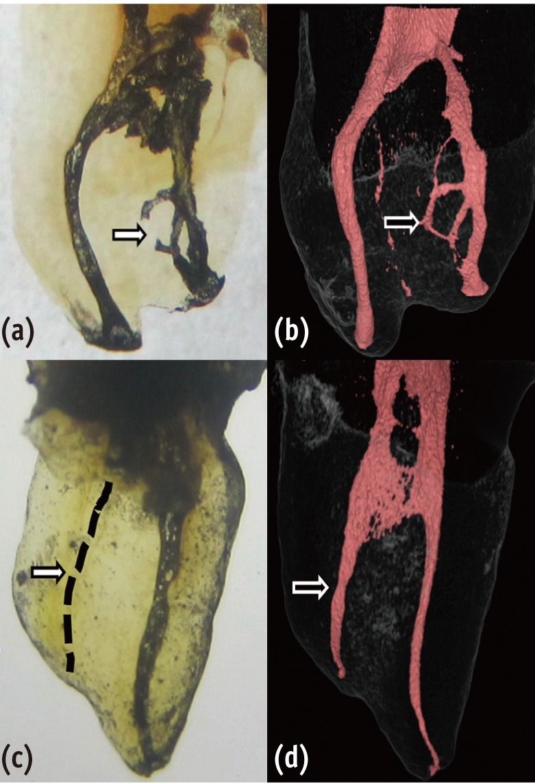
-
 Abstract
 PDF
PDF PubReader
 ePub
ePub Objectives To evaluate the inhibitory effect of ursolic acid (UA)-containing composites on
Streptococcus mutans (S. mutans ) biofilm.Materials and Methods Composite resins with five different concentrations (0.04, 0.1, 0.2, 0.5, and 1.0 wt%) of UA (U6753, Sigma Aldrich) were prepared, and their flexural strengths were measured according to ISO 4049. To evaluate the effect of carbohydrate source on biofilm formation, either glucose or sucrose was used as a nutrient source, and to investigate the effect of saliva treatment, the specimen were treated with either unstimulated whole saliva or phosphate-buffered saline (PBS). For biofilm assay, composite disks were transferred to
S. mutans suspension and incubated for 24 hr. Afterwards, the specimens were rinsed with PBS and sonicated. The colony forming units (CFU) of the disrupted biofilm cultures were enumerated. For growth inhibition test, the composites were placed on a polystyrene well cluster, andS. mutans suspension was inoculated. The optical density at 600 nm (OD600) was recorded by Infinite F200 pro apparatus (TECAN). One-way ANOVA and two-way ANOVA followed by Bonferroni correction were used for the data analyses.Results The flexural strength values did not show significant difference at any concentration (
p > 0.01). In biofilm assay, the CFU score decreased as the concentration of UA increased. The influence of saliva pretreatment was conflicting. The sucrose groups exhibited higher CFU score than glucose group (p < 0.05). In bacterial growth inhibition test, all experimental groups containing UA resulted in complete inhibition.Conclusions Within the limitations of the experiments, UA included in the composite showed inhibitory effect on
S. mutans biofilm formation and growth.-
Citations
Citations to this article as recorded by- Anti-cariogenic effect of experimental resin cement containing ursolic acid using dental microcosm biofilm
Jonghyun Jo, Mi-Jeong Jeon, Sun Kyu Park, Su-Jung Shin, Baek-il Kim, Jeong-Won Park
Journal of Dentistry.2024; 151: 105447. CrossRef - Rapid specific detection of oral bacteria using Cas13-based SHERLOCK
Jett Liu, Camden Carmichael, Hatice Hasturk, Wenyuan Shi, Batbileg Bor
Journal of Oral Microbiology.2023;[Epub] CrossRef - Novel Bioactive Nanocomposites Containing Calcium Fluoride and Calcium Phosphate with Antibacterial and Low-Shrinkage-Stress Capabilities to Inhibit Dental Caries
Abdullah Alhussein, Rashed Alsahafi, Abdulrahman A. Balhaddad, Lamia Mokeem, Abraham Schneider, Mary-Ann Jabra-Rizk, Radi Masri, Gary D. Hack, Thomas W. Oates, Jirun Sun, Michael D. Weir, Hockin H. K. Xu
Bioengineering.2023; 10(9): 991. CrossRef - Quorum sensing inhibition and antibiofilm action of triterpenoids: An updated insight
Sudipta Paul Bhattacharya, Snigdha Karmakar, Kusumita Acharya, Arijit Bhattacharya
Fitoterapia.2023; 167: 105508. CrossRef - The Application of Small Molecules to the Control of Typical Species Associated With Oral Infectious Diseases
Sirui Yang, Xiaoying Lyu, Jin Zhang, Yusen Shui, Ran Yang, Xin Xu
Frontiers in Cellular and Infection Microbiology.2022;[Epub] CrossRef - Anti-Planktonic and Anti-Biofilm Properties of Pentacyclic Triterpenes—Asiatic Acid and Ursolic Acid as Promising Antibacterial Future Pharmaceuticals
Zuzanna Sycz, Dorota Tichaczek-Goska, Dorota Wojnicz
Biomolecules.2022; 12(1): 98. CrossRef - Development and Physicochemical Characterization of Eugenia brejoensis Essential Oil-Doped Dental Adhesives with Antimicrobial Action towards Streptococcus mutans
Maury Luz Pereira, Danyelle Cristina Pereira Santos, Carlos Alberto Mendes Soares Júnior, Tamyris Alicely Xavier Nogueira Bazan, Clovis Macêdo Bezerra Filho, Márcia Vanusa da Silva, Maria Tereza dos Santos Correia, Andres Felipe Millan Cardenas, Fabiana S
Journal of Functional Biomaterials.2022; 13(3): 149. CrossRef - Does Secondary Plant Metabolite Ursolic Acid Exhibit Antibacterial Activity against Uropathogenic Escherichia coli Living in Single- and Multispecies Biofilms?
Zuzanna Sycz, Dorota Wojnicz, Dorota Tichaczek-Goska
Pharmaceutics.2022; 14(8): 1691. CrossRef - Prolonged Inhibition of Streptococcus mutans Growth and Biofilm Formation by Sustained Release of Chlorhexidine from Varnish Coated Dental Abutments: An in Vitro Study
Mark Feldman, Walid Shaaban Moustafa Elsayed, Michael Friedman, Irith Gati, Doron Steinberg, Hesham Marei, Paolo Francesco Manicone
International Journal of Dentistry.2022;[Epub] CrossRef - Interkingdom Signaling Interference: The Effect of Plant-Derived Small Molecules on Quorum Sensing in Plant-Pathogenic Bacteria
Janak Raj Joshi, Netaly Khazanov, Amy Charkowski, Adi Faigenboim, Hanoch Senderowitz, Iris Yedidia
Annual Review of Phytopathology.2021; 59(1): 153. CrossRef - Small Molecule Compounds, A Novel Strategy against Streptococcus mutans
Sirui Yang, Jin Zhang, Ran Yang, Xin Xu
Pathogens.2021; 10(12): 1540. CrossRef - Titanium dioxide nanotubes added to glass ionomer cements affect S. mutans viability and mechanisms of virulence
Isaac Jordão de Souza ARAÚJO, Mariana Gallante RICARDO, Orisson Ponce GOMES, Priscila Alves GIOVANI, Júlia PUPPIN-RONTANI, Vanessa Arias PECORARI, Elizabeth Ferreira MARTINEZ, Marcelo Henrique NAPIMOGA, Francisco Humberto NOCITI JUNIOR, Regina Maria PUPPI
Brazilian Oral Research.2021;[Epub] CrossRef - Effect of Ursolic and Oleanolic Acids on Lipid Membranes: Studies on MRSA and Models of Membranes
Sandrine Verstraeten, Lucy Catteau, Laila Boukricha, Joelle Quetin-Leclercq, Marie-Paule Mingeot-Leclercq
Antibiotics.2021; 10(11): 1381. CrossRef - Ursolic acid inhibits multi-species biofilms developed by Streptococcus mutans, Streptococcus sanguinis, and Streptococcus gordonii
Xiaoying Lyu, Liang Wang, Yusen Shui, Qingsong Jiang, Lan Chen, Wen Yang, Xiaoya He, Jumei Zeng, Yuqing Li
Archives of Oral Biology.2021; 125: 105107. CrossRef - The physical properties and anticariogenic effect of experimental resin cement containing ursolic acid
Hyunkyung Yoo, So Youn Kim, Su-Jung Shin, Jeong-Won Park
Odontology.2021; 109(3): 641. CrossRef - Ursolic acid: A systematic review of its pharmacology, toxicity and rethink on its pharmacokinetics based on PK-PD model
Qiang Sun, Man He, Meng Zhang, Sha Zeng, Li Chen, Lijuan Zhou, Haibo Xu
Fitoterapia.2020; 147: 104735. CrossRef - Effects of UVB and UVC irradiation on cariogenic bacteria in vitro
Shigeki Uchinuma, Yasushi Shimada, Khairul Matin, Keiichi Hosaka, Masahiro Yoshiyama, Yasunori Sumi, Junji Tagami
Lasers in Medical Science.2019; 34(5): 981. CrossRef - Ursolic acid (UA): A metabolite with promising therapeutic potential
Dharambir Kashyap, Hardeep Singh Tuli, Anil K. Sharma
Life Sciences.2016; 146: 201. CrossRef - Protective Effects on Gastric Lesion of Ursolic acid
Sun Whoe Kim, In Young Hwang, Sun Yi Lee, Choon Sik Jeong
Journal of Food Hygiene and Safety.2016; 31(4): 286. CrossRef - Ursolic Acid—A Pentacyclic Triterpenoid with a Wide Spectrum of Pharmacological Activities
Łukasz Woźniak, Sylwia Skąpska, Krystian Marszałek
Molecules.2015; 20(11): 20614. CrossRef - Antibacterial effect of self-etching adhesive systems onStreptococcus mutans
Seung-Ryong Kim, Dong-Hoon Shin
Restorative Dentistry & Endodontics.2014; 39(1): 32. CrossRef - Dental materials with antibiofilm properties
Zhejun Wang, Ya Shen, Markus Haapasalo
Dental Materials.2014; 30(2): e1. CrossRef - Antibacterial properties of composite resins incorporating silver and zinc oxide nanoparticles onStreptococcus mutansandLactobacillus
Shahin Kasraei, Lida Sami, Sareh Hendi, Mohammad-Yousef AliKhani, Loghman Rezaei-Soufi, Zahra Khamverdi
Restorative Dentistry & Endodontics.2014; 39(2): 109. CrossRef - Synergistic effect of xylitol and ursolic acid combination on oral biofilms
Yunyun Zou, Yoon Lee, Jinyoung Huh, Jeong-Won Park
Restorative Dentistry & Endodontics.2014; 39(4): 288. CrossRef - The virulence of Streptococcus mutans and the ability to form biofilms
W. Krzyściak, A. Jurczak, D. Kościelniak, B. Bystrowska, A. Skalniak
European Journal of Clinical Microbiology & Infectious Diseases.2014; 33(4): 499. CrossRef
- Anti-cariogenic effect of experimental resin cement containing ursolic acid using dental microcosm biofilm
- 1,680 View
- 5 Download
- 25 Crossref

-
Reconsideration of treatment protocol on the reduction of
Enterococcus faecalis associated with failed root canal treatment - Woo Cheol Lee, Seong-Tae Hong, WonJun Shon
- J Korean Acad Conserv Dent 2008;33(6):560-569. Published online November 30, 2008
- DOI: https://doi.org/10.5395/JKACD.2008.33.6.560
-
 Abstract
 PDF
PDF PubReader
 ePub
ePub Microorganism survived in the root canal after root canal cleaning and shaping procedure is a main cause of root canal treatment failure. There are several mechanisms for the bacteria to survive in the root canal after chemomechanical preparation and root canal irrigation. Bacteria organized as biofilm has been suggested as an etiology of persistent periapical lesion. Recent studies were focus on removal of
Enterococcus faecalis biofilm due to the report that the persistence of this bacteria after root canal treatment may be associated with its ability to form biofilm. Several investigations demonstrated that current root canal treatment protocol including use of NaOCl, EDTA and Chlorhexidine as irrigants is quite effective in eliminatingE. faecalis biofilm. However, this microorganism still can survive in inaccessible areas of root canal system and evade host immune response, suppress immune activity and produce biofilm. Up to date, there is no possible clinical method to completely get rid of bacteria from the root canal. Once the root canal treatment failure occurred, and conventional treatment incorporating current therapeutic protocol has failed, periapical surgery or extraction should be considered rather than prolong the ineffected retreatment procedure.
- 1,000 View
- 4 Download


 KACD
KACD

 First
First Prev
Prev


