Warning: mkdir(): Permission denied in /home/virtual/lib/view_data.php on line 81

Warning: fopen(upload/ip_log/ip_log_2024-12.txt): failed to open stream: No such file or directory in /home/virtual/lib/view_data.php on line 83

Warning: fwrite() expects parameter 1 to be resource, boolean given in /home/virtual/lib/view_data.php on line 84
A novel antimicrobial-containing nanocellulose scaffold for regenerative endodontics
Skip Navigation
Skip to contents

Restor Dent Endod : Restorative Dentistry & Endodontics

OPEN ACCESS

Articles

Page Path
HOME > Restor Dent Endod > Volume 46(2); 2021 > Article
Research Article A novel antimicrobial-containing nanocellulose scaffold for regenerative endodontics
Victoria Kichler1orcid, Lucas Soares Teixeira1orcid, Maick Meneguzzo Prado2orcid, Guilherme Colla2orcid, Daniela Peressoni Vieira Schuldt1orcid, Beatriz Serrato Coelho1orcid, Luismar Marques Porto2orcid, Josiane de Almeida1,2

DOI: https://doi.org/10.5395/rde.2021.46.e20
Published online: March 16, 2021

1Department of Endodontics, Faculty of Dentistry, University of Southern Santa Catarina, Palhoça, SC, Brazil.

2Department of Chemical Engineering, Federal University of Santa Catarina, Florianópolis, SC, Brazil.

Correspondence to Josiane de Almeida, PhD. Associate Professor, Department of Endodontics, Faculty of Dentistry, University of Southern Santa Catarina, Av. Pedra Branca, 25 Cidade Universitária, Palhoça, SC 88137-270, Brazil. dealmeidajosiane@hotmail.com
• Received: August 28, 2020   • Accepted: September 23, 2020

Copyright © 2021. The Korean Academy of Conservative Dentistry

This is an Open Access article distributed under the terms of the Creative Commons Attribution Non-Commercial License (https://creativecommons.org/licenses/by-nc/4.0/) which permits unrestricted non-commercial use, distribution, and reproduction in any medium, provided the original work is properly cited.

  • 21 Views
  • 1 Download
  • 11 Web of Science
prev next
  • Objectives
    The aim of this study was to evaluate bacterial nanocellulose (BNC) membranes incorporated with antimicrobial agents regarding cytotoxicity in fibroblasts of the periodontal ligament (PDLF), antimicrobial activity, and inhibition of multispecies biofilm formation.
  • Materials and Methods
    The tested BNC membranes were BNC + 1% clindamycin (BNC/CLI); BNC + 0.12% chlorhexidine (BNC/CHX); BNC + nitric oxide (BNC/NO); and conventional BNC (BNC; control). After PDLF culture, the BNC membranes were positioned in the wells and maintained for 24 hours. Cell viability was then evaluated using the MTS calorimetric test. Antimicrobial activity against Enterococcus faecalis, Actinomyces naeslundii, and Streptococcus sanguinis (S. sanguinis) was evaluated using the agar diffusion test. To assess the antibiofilm activity, BNC membranes were exposed for 24 hours to the mixed culture. After sonicating the BNC membranes to remove the remaining biofilm and plating the suspension on agar, the number of colony-forming units (CFU)/mL was determined. Data were analyzed by 1-way analysis of variance and the Tukey, Kruskal-Wallis, and Dunn tests (α = 5%).
  • Results
    PDLF metabolic activity after contact with BNC/CHX, BNC/CLI, and BNC/NO was 35%, 61% and 97%, respectively, compared to BNC. BNC/NO showed biocompatibility similar to that of BNC (p = 0.78). BNC/CLI showed the largest inhibition halos, and was superior to the other BNC membranes against S. sanguinis (p < 0.05). The experimental BNC membranes inhibited biofilm formation, with about a 3-fold log CFU reduction compared to BNC (p < 0.05).
  • Conclusions
    BNC/NO showed excellent biocompatibility and inhibited multispecies biofilm formation, similarly to BNC/CLI and BNC/CHX.
Teeth with incomplete root development that develop pulp necrosis have thin and fragile dentin walls, wide canals, and an open foramen, which makes it difficult to perform conventional filling procedures [1]. As a viable alternative to apexification, regenerative endodontic procedures (REPs) have proven successful in several cases [2]. However, to regenerate pulp tissue and restore physiological function, full resolution of the infectious process is necessary. The persistence of microorganisms in residual biofilms and bacterial antigens inside the root canals, even after conducting the disinfection protocol, prevents stem cells from populating the region and differentiating and allows full root formation [3,4].
Recent advances have focused on the functionalization of membranes and 3-dimensional (3D) nanofibers, improving their chemical and physical properties, and controlling their biological interactions [5,6,7]. Scaffolds can be chemically modified (e.g., through the incorporation of antimicrobial agents) as a disinfection strategy [5,8]. Bacterial nanocellulose (BNC) is a biomaterial that stands out as a structural support in tissue engineering and has high porosity and surface area, allowing the incorporation and release of drugs and substances [5,6,7,9,10]. BNC is a nanofibrillar hydrogel polymer biosynthesized by various bacterial species, such as Komagataeibacter hansenii (K. hansenii) [11]. Specific properties of BNC, such as biodegradability, flexibility, biocompatibility, hydrophilicity, and mechanical and tensile strength also make this 3D biomaterial promising, and may become a viable framework in REPs [5,6,10].
Recent research has shown that biopolymers containing nitric oxide (NO), and NO-releasing nanomatrix gel and NO-releasing biomaterials have significant antimicrobial effects against certain bacteria and are capable of promoting revascularization and root maturation [12,13,14,15]. Polymeric nanofibers incorporating minocycline, metronidazole and ciprofloxacin, or only clindamycin (CLI), have effective antimicrobial action against root canal bacteria and are biocompatible with dental pulp stem cells [16,17]. Despite the recognized broad-spectrum antimicrobial action of CHX and its capacity to facilitate the release of growth factors that stimulate dentin, it has shown direct and indirect negative effects on the survival of stem cells, being toxic in concentrations ranging from 0.001% to 2% [18,19,20]. However, CHX is usually used as a root canal irrigating solution; when incorporated into a nanofiber network, its antibiofilm and cytotoxic effects remain unknown.
Based on this premise, the immobilization of antimicrobial agents in the BNC nanofiber network could be an innovative disinfection strategy with considerable potential for use in REPs. Thus, the objective of this study was to evaluate BNC membranes incorporating different antimicrobial agents for cytotoxicity to periodontal ligament fibroblasts (PDLFs) and for inhibition of the formation of a multispecies biofilm (consisting of Enterococcus faecalis [E. faecalis], Actinomyces naeslundii [A. naeslundii], and Streptococcus sanguinis [S. sanguinis]). The null hypothesis was that there would be no difference in biocompatibility and antibacterial/antibiofilm activity between the conventional BNC and experimental BNC-based antimicrobial release systems.
Synthesis of bacterial nanocellulose
All BNC membranes were synthesized according to a previously published protocol [21]. Briefly, isolated colonies of K. hansenii (ATCC 23769) were resuspended in 1 mL of mannitol culture medium (25 g/L mannitol, 5.0 g/L yeast extract, 3.0 g/L bacteriological peptone). The solution was homogenized for 60 seconds in a vortex and allowed to decant for 10 seconds. After an optical density (OD) reading (DO660 = 0.150), a 10-fold dilution was performed in the mannitol medium and the inoculum was distributed on sterile plastic plates. The bacterial growth occurred in a static condition at 26°C for 7 days. The membranes formed at the liquid/air interface were then removed and transferred to a flask containing a 0.1 M sodium hydroxide solution and maintained for 24 hours at 50°C to remove bacteria and/or residues from the culture medium. The BNC membranes were then subjected to successive washes with distilled water, or until the pH of the rinse water was equivalent to that of the distilled water used in the wash. The membranes were autoclaved for 20 minutes at 121°C and kept refrigerated until use.
Incorporation of antimicrobial agents in BNC
Antimicrobial agents were incorporated into the BNC membranes as follows: G1) BNC + NO (BNC/NO); G2) BNC + 1% CLI (BNC/CLI); G3) BNC + 0.12% CHX (BNC/CHX); G4) conventional BNC (BNC; control).
The modification of BNC with NO followed the method described in the literature, for poly(vinyl alcohol), with modifications [22]. BNC membranes functionalized with sulfhydryl groups (BNC-SH) were prepared by esterifying 10% BNC with 3-mercaptopropionic acid (1.3 M) catalyzed with hydrochloric acid (HCl) (40 mM). The mixture was heated under stirring and reflux, for 3 hours at 100°C, in an inert atmosphere. BNC was subsequently removed, washed alternately with water and acetone 6 times, and freeze-dried. Dry BNC-SH was previously rehydrated. Each membrane was S-nitrosated (BNC-SNO) by immersion in a nitrous acid solution containing sodium nitrite (NaNO2) (54 mM) with HCl (final concentration, 24 mM). Each membrane was then transferred to another Falcon tube containing deionized water to remove excess reagents.
CHX and CLI were handled in a pharmacy (Dermus Farmácia Dermatológica e Cosmética, Florianópolis, SC, Brazil) and were incorporated by stirring the oxidized BNC with solutions for 24 hours at room temperature. Oxidized BNC was obtained following the methodology previously described, with some modifications [23]. First, the BNC was immersed in a 2:1 (v/v) solution of nitric acid and phosphoric acid. Then, 7% sodium nitrite (m/v) was added. The reaction time was 24 hours, in the absence of light, at 25°C with agitation. The membranes were then immersed in an aqueous solution of 0.2% (w/w) glycerol for 15 minutes to remove excess oxidant. Finally, the membranes were washed with acetone and allowed to dry at 25°C.
Cell culture
This study was approved by the Human Research Ethics Committee of the University of Southern Santa Catarina (protocol No. 3.163.671). Briefly, fragments of human PDLFs were grown in Petri dishes containing Dulbecco minimal essential medium (DMEM) (Sigma-Aldrich, St. Louis, MO, USA), supplemented with 10% fetal bovine serum (FBS) (Sigma-Aldrich) and 1% antibiotics (penicillin, streptomycin, and amphotericin), in an oven at 37°C with 5% CO2. When the cells reached a confluence of approximately 80%, they were washed with phosphate-buffered saline (PBS) (Sigma-Aldrich) to remove FBS and trypsinized with 3 mL of 0.05% trypsin. Then, 20 mL of DMEM was added for trypsin inactivation, and the cell count was calculated from the suspension obtained. Cell suspensions from the third pass were used for the cell cytotoxicity test.
MTS viability test
The viability of PDLFs after contact with the BNC was assessed using the MTS colorimetric assay (CellTiter 96 AQueous One Solution Cell Proliferation Assay, Promega, Madison, WI, USA). From the cell suspension obtained, 200 μL (104 cells/mL) were seeded in each well of a 96-well culture plate, in which the BNCs had been positioned (n = 3 per group). The plates were incubated for 24 hours in an oven at 37°C with 5% CO2. After this period, the membranes were removed from the wells, washed with 200 μL of PBS, and placed in the wells of a new plate. Wells containing conventional BNC comprised the control group. Next, 60 μL of the MTS reagent (Promega) diluted in DMEM (1:3) was added to the wells, and the absorbance was measured in a spectrophotometer at 490 nm after 2 hours. The values obtained in the experimental groups were calculated as the metabolic percentage compared to the control. Cell viability was expressed as a percentage. The experiment was carried out in triplicate.
Bacterial species
The facultative anaerobic bacterial species E. faecalis ATCC 29212, A. naeslundii ATCC 43146 and S. sanguinis ATCC 10556 were used. Fresh cultures were obtained by overnight incubation of 500 µL of each stock in 10 mL of brain-heart broth medium (BHI) (KASVI, Curitiba, PR, Brazil), pH 7.1, at 37°C under aerobic conditions.
Agar diffusion antimicrobial test
The overnight cultures were diluted in BHI broth (KASVI) to obtain a suspension of approximately 5 × 10⁸ colony-forming units (CFU)/mL (DO600 ≈ 0.5) for each cell line. Next, 100 µL of each suspension was individually plated on BHI agar (KASVI) and spread with a sterile swab in 3 directions. Membranes (G1 to G4, Ø = 6 mm) were placed at equidistant points. Filter paper discs (Ø = 6 mm) (Unifill, Curitiba, PR, Brazil) moistened with 10 µL of 0.12% CHX comprised the positive control group. The plates were incubated at 37°C for 48 hours, under aerobic conditions. The diameter of the bacterial growth inhibition zones formed around each material was measured in millimeters with the aid of a digital caliper and optical microscope. Three plates were used per bacterial strain in each replicate. The experiment was carried out in triplicate.
Formation of multispecies biofilm
For biofilm growth, 24-well culture plates were inoculated with 1.5 mL of growth medium per well and inoculated with the 1:100-diluted multispecies culture (OD ≈ 0.5 nm). The membranes of experimental groups 1 to 4 (Ø = 15 mm) were positioned in the wells and served as a substrate for the growth of the biofilm (n = 6). The apparatus was incubated for 24 hours under aerobic conditions, at 37°C.
Bacterial cell viability test
After 24 hours, the membranes were removed from the wells and washed 3 times with PBS (Sigma-Aldrich) to remove non-adherent biofilm cells. To determine the antibiofilm effect of BNCs, the membranes were transferred to plastic containers containing 2 mL of PBS (Sigma-Aldrich). The biofilms were removed from the substrates by sonication for 15 minutes at an amplitude of 40 W, and the resulting suspensions were serially diluted (1:100). Aliquots of 100 µL were plated on BHI agar (KASVI). The plates were incubated under aerobic conditions at 37°C, for 24 hours to 48 hours. The number of CFU/mL was then determined. The experiment was carried out in triplicate.
For the qualitative assessment of biofilm formation, 2 samples from each group were observed using a scanning electron microscope (SEM). For processing, the membranes were fixed in 2.5% glutaraldehyde, dehydrated in an alcohol ascending chain, mounted in stubs, and covered with gold. With the microscope operating at 10 kW, photomicrographs of significant areas were taken at magnifications ranging from ×1,000 to ×10,000.
Statistical analysis
The absorbance values obtained with the MTS viability test were subjected to statistical analysis using 1-way analysis of variance and the post hoc Tukey tests. The mean values of the inhibition zones (mm) and of CFUs, transformed into log10 CFU, were analyzed by the Kruskal-Wallis and post hoc Dunn tests. The level of significance was set at 5%. The analyses were performed with the aid of SPSS software version 21.0 (IBM, Armonk, NY, USA).
BNC cytotoxicity: MTS assay
Figure 1 presents the percentage of cell viability, assessed through metabolic activity, after contact for 24 hours with the different membranes. The metabolic activity of PDLFs after contact with BNC/NO, BNC/CLI and BNC/CHX was 97%, 61% and 35%, respectively, compared to the control (conventional BNC).
Figure 1

Percentage of periodontal ligament fibroblast (PDLF) metabolic activity after contact with the different membranes.
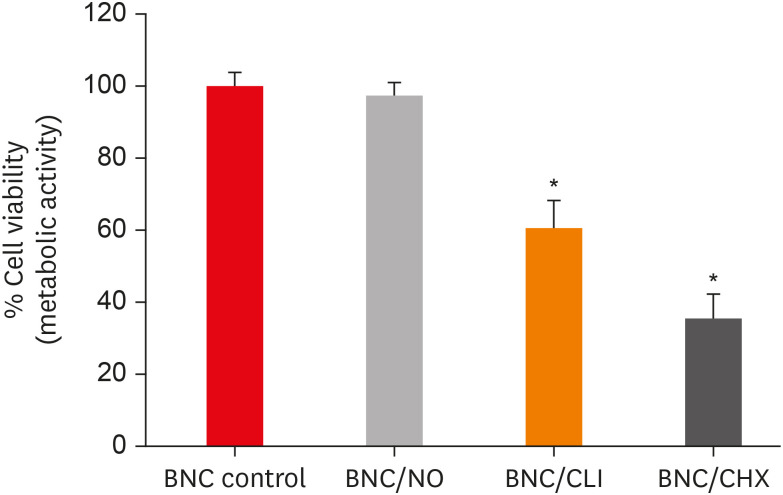
BNC, bacterial nanocellulose; NO, nitric oxide; CLI, clindamycin; CHX, chlorhexidine.
*Indicate significant differences compared to conventional BNC (control).
rde-46-e20-g001.jpg
BNC/NO was as biocompatible as conventional BNC (p = 0.78), with 97% viable periodontal ligament cells; and with a percentage of PDLF viability significantly higher than that obtained using the other experimental membranes (p < 0.001). BNC/CLI showed mild cytotoxicity, but had a significant effect compared to the control (p < 0.001). BNC/CHX showed a higher degree of cytotoxicity to PDLFs than all of the other membranes (p < 0.001).
Antimicrobial effects
Table 1 shows the mean values of inhibition halos resulting from the different BNC membranes against E. faecalis, S. sanguinis, and A. naeslundii. The negative control (BNC) had no antimicrobial effects against the 3 bacterial species, as represented by the absence of an inhibition zone. The filter discs incorporating 0.12% CHX showed inferior antimicrobial activity compared to the experimental groups, significantly different from BNC/CLI, for all tested bacterial species (p < 0.05).
Table 1

The inhibition halos value (mm) formed after the contact of the different bacterial nanocelluloses (BNCs) with Enterococcus faecalis (E. faecalis), Streptococcus sanguinis (S. sanguinis), and Actinomyces naeslundii (A. naeslundii)

Groups Bacterial species
E. faecalis S. sanguinis A. naeslundii
BNC/CHX 13.44 ± 2.74a,c 11.88 ± 1.45a 13.87 ± 2.85a,c
BNC/NO 11.00 ± 2.28b 12.16 ± 2.71a 9.33 ± 1.96c
BNC/CLI 21.77 ± 7.32a 19.66 ± 3.32b 20.66 ± 7.98a
BNC (negative control) 0b 0c 0b,c
CHX (positive control) 9.22 ± 2.16b,c 8.22 ± 2.16a,c 9.22 ± 1.71b,c
Data are shown as mean ± standard deviation. Different lower-case letters in the same column indicate a significant difference between groups (p < 0.05).
CHX, chlorhexidine; NO, nitric oxide; CLI, clindamycin.
The largest inhibition halos were observed for BNC/CLI, with its antimicrobial activity being superior to that of the controls, positive and negative, for the 3 bacterial species (p < 0.05). When tested against S. sanguinis, BNC/CLI also showed antimicrobial activity superior to BNC/CHX and BNC/NO (p < 0.05). Although the BNC/CHX and BNC/NO membranes showed inhibition halos between 9.33 mm and 13.87 mm, significant antimicrobial activity was only demonstrated for BNC/CHX compared to BNC for E. faecalis and S. sanguinis (p < 0.05).
Antibiofilm effect
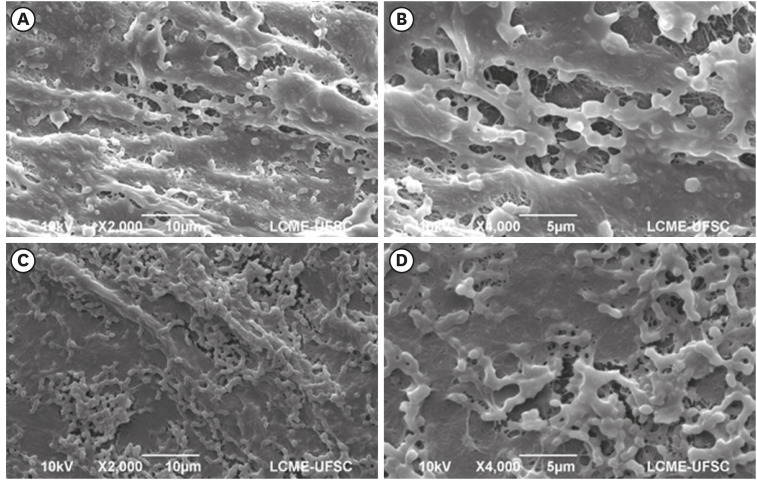
Figure 2 shows the mean CFU values present in the biofilm adhered to the different membranes after 24 hours of contact with the multispecies culture. An average of 2 × 10⁶ CFUs could adhere to conventional BNC and form a biofilm. The SEM photomicrographs showed a dense and homogeneous biofilm, with an extracellular matrix layer completely covering the bacteria and the membrane nanofiber network (Figure 3A-3D).
Figure 2

Mean colony-forming unit (CFU) values (log CFU/membrane) present in the multispecies biofilm that adhered to the different experimental bacterial nanocelluloses (BNCs).
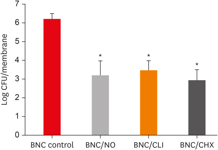
NO, nitric oxide; CLI, clindamycin; CHX, chlorhexidine.
*Indicate significant differences compared to control (conventional BNC).
rde-46-e20-g002.jpg
Figure 3

Dense and homogeneous bacterial biofilm covering the membrane surface in samples of the control group (conventional bacterial nanocellulose [BNC]) (magnification ×2,000 and ×4,000).
rde-46-e20-g003.jpg
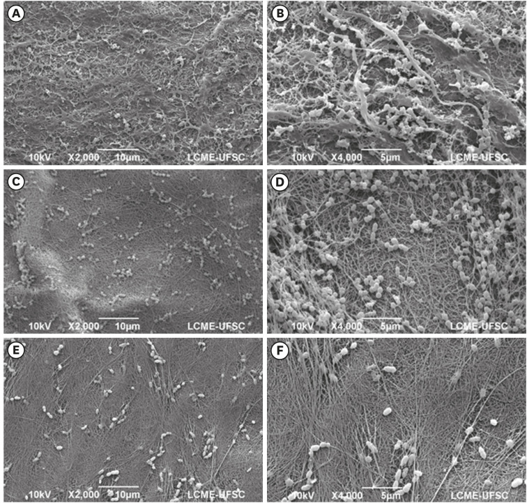
The biofilms formed on BNC/NO, BNC/CLI and BNC/CHX showed CFU values significantly lower than those of the control, BNC (p < 0.05), with an approximately 3-fold reduction in log CFU. In the SEM images, small bacterial clusters or isolated bacteria were irregularly distributed and interspersed in the BNC nanofiber network (Figure 4A-4F).
Figure 4

Isolated bacterial cells and small bacterial clusters, of varying sizes, irregularly distributed on the surface of the bacterial nanocellulose (BNC)/nitric oxide (NO) (A, B), BNC/clindamycin (CLI) (C, D), and BNC/chlorhexidine (CHX) (E, F) membranes (magnification ×2,000 and ×4,000).
rde-46-e20-g004.jpg
BNC is a biomaterial commonly used in the medical field as a dressing for wounds, skin substitutes, and artificial blood vessels [24]. Its potential for application as a 3D nanofibrous framework in tissue engineering is particularly remarkable [5]. The nanofibrillar structure of BNC simulates the extracellular matrix, allows cell regeneration, and contributes to tissue repair [5,25]. The incorporation of antimicrobial agents into the nanofiber network has gained prominence in the literature since antibiotic pastes and chemical irrigants have affected the survival and function of stem cells [8,20]. Thus, in addition to serving as cell anchorage, scaffolds could also contribute to the elimination of intraradicular biofilm [26].
In the present study, conventional BNC membranes served as a control group, since they do not promote an immune response, demonstrate excellent biocompatibility, and do not have antimicrobial action [5,6,25]. When BNC was incorporated with NO, 97% of PDLFs remained viable. This result corroborates a previous finding, in which NO-releasing dendrimers were used as antimicrobial agent in a scaffold and promoted minimal cytotoxicity in rat fibroblasts in concentrations able to eliminate microorganisms [27]. NO-releasing polymers could also increase the biocompatibility of materials implanted in subcutaneous and intravascular tissue, in addition to inhibiting bacterial adhesion [12,28]. In the present antibiofilm test, BNC/NO, BNC/CLI, and BNC/CHX showed similar results, significantly inhibiting biofilm formation compared to BNC. The ability of BNC to incorporate and release drugs and substances may have hindered bacterial adhesion to its surface [6]. Consequently, biofilm formation was partially inhibited. A marked reduction in Pseudomonas aeruginosa adhesion, with a decrease in biofilm formation sites, was demonstrated when NO was used in sol-gel microarrays [12]. As a free radical, NO can interact with oxygen and other reactive molecules, causing multifactorial damage to bacteria and leading to dysfunction and cell death [29].
Triple antibiotic paste, composed of ciprofloxacin, metronidazole, and minocycline, has been proposed as a medication for use in regenerative endodontics in an attempt to eliminate intracanal biofilms. However, minocycline is toxic and causes discoloration of the dental structure [30,31]. To overcome these disadvantages, it has been replaced by other antimicrobials, such as CLI and cefaclor [17,32]. In the present study, 61% of PDLFs remained viable after exposure to BNC with 1% CLI. According to an established classification, BNC/CLI can be considered slightly cytotoxic [30]. Findings have also demonstrated that nanofibers containing CLI associated with ciprofloxacin and metronidazole maintained more than 50% of viable cells, in addition to exerting antimicrobial actions [17]. The replacement of minocycline by CLI may prove to be a viable alternative for both antibiotic pastes and scaffolds [17,33]. In addition to satisfactory biocompatibility, BNC/CLI showed the best antimicrobial activity and inhibited multispecies biofilm formation. The antimicrobial efficacy of CLI has been proven, and it was found to be effective against several endodontic pathogens and capable of eradicating E. faecalis biofilm when used as an intracanal medication [17,33,34]. CLI inhibits bacterial protein synthesis by interfering with the formation of the peptide chain. Thus, the ability of bacteria to adhere to the substrate surface is reduced, decreasing its virulence [35].
PDLFs exposed to membranes incorporated with 0.12% CHX showed the lowest percentage of metabolic activity. Widbiller et al. [20] demonstrated that CHX solution exerted direct and indirect negative effects on dental pulp stem cells, being toxic in concentrations above 0.001%. With 5 minutes of exposure to 2% CHX, cell viability was 50%. Therefore, clinical protocols should limit both the exposure period and the concentration, in order to provide an environment where cells can survive [20]. It has also been reported that regardless of the duration of exposure to the CHX solution, concentrations between 0.06% and 2% promoted a cytotoxic effect on odontoblast-like cells, as they negatively affected mitochondrial activity and consequently impaired cell proliferation [36]. Despite the adverse cytotoxic effects, the incorporation of CHX into BNC promoted a satisfactory antimicrobial and antibiofilm action. In addition to the ability of BNC to incorporate antimicrobials into its structure, the substantivity of CHX and its antimicrobial properties may have favored its action [6,18].
Based on the present findings, the null hypothesis was partially rejected. The immobilization of antimicrobial agents, such as NO, CLI, and CHX, to the BNC nanofiber network is promising for regenerative endodontics, and may prove to be an innovative disinfection strategy with considerable potential for use in REPs. The low cost and sustainability of BNC also enhance its attractiveness from an economic perspective. Moreover, BNC is a biocompatible scaffold that does not influence or even favor cellular metabolic activity. Other in vitro and ex vivo study models are needed in order to explore the potential of BNC in regenerative endodontics. Tests that assess the migration, proliferation, and differentiation of stem cells in BNC and dentin are also needed.
Due to their antimicrobial properties and biocompatibility, BNC-based antimicrobial release systems are a potential scaffold option for REPs.
The authors are grateful to the Central Electronic Microscopy Laboratory (LCME) of the Federal University of Santa Catarina for collaborating with the SEM images.

Conflict of Interest: No potential conflict of interest relevant to this article was reported.

Author Contributions:

  • Conceptualization: de Almeida J, Kichler V, Teixeira LS.

  • Data curation: Colla G, de Almeida J.

  • Formal analysis: de Almeida J, Porto LM.

  • Funding acquisition: Porto LM, Colla G.

  • Investigation: Kichler V, Teixeira LS, Prado MM.

  • Methodology: Schuldt DPV, Coelho BS, Kichler V, Teixeira LS, Prado MM.

  • Project administration: de Almeida J.

  • Resources: Colla G, de Almeida J, Porto LM.

  • Software: Prado MM.

  • Supervision: de Almeida J, Porto LM, Colla G.

  • Validation: Schuldt DPV, Coelho BS.

  • Visualization: Schuldt DPV, Coelho BS.

  • Writing - original draft: Kichler V, Teixeira LS.

  • Writing - review & editing: de Almeida J, Porto LM, Colla G.

  • 1. Jeeruphan T, Jantarat J, Yanpiset K, Suwannapan L, Khewsawai P, Hargreaves KM. Mahidol study 1: comparison of radiographic and survival outcomes of immature teeth treated with either regenerative endodontic or apexification methods: a retrospective study. J Endod 2012;38:1330-1336. ArticlePubMed
  • 2. Diogenes A, Ruparel NB, Shiloah Y, Hargreaves KM. Regenerative endodontics: a way forward. J Am Dent Assoc 2016;147:372-380. PubMed
  • 3. Verma P, Nosrat A, Kim JR, Price JB, Wang P, Bair E, Xu HH, Fouad AF. Effect of residual bacteria on the outcome of pulp regeneration in vivo . J Dent Res 2017;96:100-106. ArticlePubMedPDF
  • 4. Vishwanat L, Duong R, Takimoto K, Phillips L, Espitia CO, Diogenes A, Ruparel SB, Kolodrubetz D, Ruparel NB. Effect of bacterial biofilm on the osteogenic differentiation of stem cells of apical papilla. J Endod 2017;43:916-922. PubMed
  • 5. Curvello R, Raghuwanshi VS, Garnier G. Engineering nanocellulose hydrogels for biomedical applications. Adv Colloid Interface Sci 2019;267:47-61. ArticlePubMed
  • 6. Vismara E, Bernardi A, Bongio C, Farè S, Pappalardo S, Serafini A, Pollegioni L, Rosini E, Torri G. Bacterial nanocellulose and its surface modification by glycidyl methacrylate and ethylene glycol dimethacrylate. Incorporation of vancomycin and ciprofloxacin. Nanomaterials (Basel) 2019;9:1668.ArticlePubMedPMC
  • 7. Osorio M, Ortiz I, Gañán P, Naranjo T, Zuluaga R, van Kooten TG, Castro C. Novel surface modification of three-dimensional bacterial nanocellulose with cell-derived adhesion proteins for soft tissue engineering. Mater Sci Eng C 2019;100:697-705.Article
  • 8. Bottino MC, Pankajakshan D, Nör JE. Advanced scaffolds for dental pulp and periodontal regeneration. Dent Clin North Am 2017;61:689-711. PubMedPMC
  • 9. Osorio M, Fernández-Morales P, Gañán P, Zuluaga R, Kerguelen H, Ortiz I, Castro C. Development of novel three-dimensional scaffolds based on bacterial nanocellulose for tissue engineering and regenerative medicine: effect of processing methods, pore size, and surface area. J Biomed Mater Res A 2019;107:348-359. ArticlePubMedPDF
  • 10. Reis EMD, Berti FV, Colla G, Porto LM. Bacterial nanocellulose-IKVAV hydrogel matrix modulates melanoma tumor cell adhesion and proliferation and induces vasculogenic mimicry in vitro . J Biomed Mater Res B Appl Biomater 2018;106:2741-2749. ArticlePubMedPDF
  • 11. Shoda M, Sugano Y. Recent advances in bacterial cellulose production. Biotechnol Bioprocess Eng 2005;10:1-8.ArticlePDF
  • 12. Dobmeier KP, Schoenfisch MH. Antibacterial properties of nitric oxide-releasing sol-gel microarrays. Biomacromolecules 2004;5:2493-2495. PubMed
  • 13. Seabra AB, Martins D, Simões MM, da Silva R, Brocchi M, de Oliveira MG. Antibacterial nitric oxide-releasing polyester for the coating of blood-contacting artificial materials. Artif Organs 2010;34:E204-E214. ArticlePubMed
  • 14. Moon CY, Nam OH, Kim M, Lee HS, Kaushik SN, Cruz Walma DA, Jun HW, Cheon K, Choi SC. Effects of the nitric oxide releasing biomimetic nanomatrix gel on pulp-dentin regeneration: pilot study. PLoS One 2018;13:e0205534. ArticlePubMedPMC
  • 15. Kim JO, Noh JK, Thapa RK, Hasan N, Choi M, Kim JH, Lee JH, Ku SK, Yoo JW. Nitric oxide-releasing chitosan film for enhanced antibacterial and in vivo wound-healing efficacy. Int J Biol Macromol 2015;79:217-225. ArticlePubMed
  • 16. Pankajakshan D, Albuquerque MT, Evans JD, Kamocka MM, Gregory RL, Bottino MC. Triple antibiotic polymer nanofibers for intracanal drug delivery: effects on dual species biofilm and cell function. J Endod 2016;42:1490-1495. PubMedPMC
  • 17. Karczewski A, Feitosa SA, Hamer EI, Pankajakshan D, Gregory RL, Spolnik KJ, Bottino MC. Clindamycin-modified triple antibiotic nanofibers: a stain-free antimicrobial intracanal drug delivery system. J Endod 2018;44:155-162. ArticlePubMed
  • 18. Gomes BP, Vianna ME, Zaia AA, Almeida JFA, Souza-Filho FJ, Ferraz CCR. Chlorhexidine in endodontics. Braz Dent J 2013;24:89-102. ArticlePubMed
  • 19. Galler KM, Buchalla W, Hiller KA, Federlin M, Eidt A, Schiefersteiner M, Schmalz G. Influence of root canal disinfectants on growth factor release from dentin. J Endod 2015;41:363-368. ArticlePubMed
  • 20. Widbiller M, Althumairy RI, Diogenes A. Direct and indirect effect of chlorhexidine on survival of stem cells from the apical papilla and its neutralization. J Endod 2019;45:156-160. PubMed
  • 21. De Souza SS, Berti FV, Oliveira KPV, Pittella C, Vasconcellos J, Pelissari C, Rambo CR, Porto LM. Nanocellulose biosynthesis by Komagataeibacter hansenii in a defined minimal culture medium. Cellulose 2018;26:1641-1655.ArticlePDF
  • 22. Lourenço SDM, de Oliveira MG. Topical photochemical nitric oxide release from porous poly(vinyl alcohol) membrane for visible light modulation of dermal vasodilation. J Photochem Photobiol Chem 2017;346:548-558.Article
  • 23. Kumar V, Yang T. HNO3/H3PO4-NANO2 mediated oxidation of cellulose - preparation and characterization of bioabsorbable oxidized celluloses in high yields and with different levels of oxidation. Carbohydr Polym 2002;48:403-412.Article
  • 24. Pittela CQP, Porto LM. Application of bacterial nanocellulose membranes for epithelial tissue repair. Revista de Enfermagem da UFJF 2015;1:223-232.
  • 25. Osorio M, Cañas A, Puerta J, Díaz L, Naranjo T, Ortiz I, Castro C. Ex vivo and in vivo biocompatibility assessment (blood and tissue) of three-dimensional bacterial nanocellulose biomaterials for soft tissue implants. Sci Rep 2019;9:10553. ArticlePubMedPMCPDF
  • 26. Albuquerque MTP, Nagata J, Bottino MC. Antimicrobial efficacy of triple antibiotic-eluting polymer nanofibers against multispecies biofilm. J Endod 2017;43:S51-S56. ArticlePubMedPMC
  • 27. Sun B, Slomberg DL, Chudasama SL, Lu Y, Schoenfisch MH. Nitric oxide-releasing dendrimers as antibacterial agents. Biomacromolecules 2012;13:3343-3354. ArticlePubMedPMC
  • 28. Frost MC, Batchelor MM, Lee YM, Zhang H, Kang Y, Oh B, Wilson GS, Gifford R, Rudich SM, Meyerhoff M. Preparation and characterization of implantable sensors with nitric oxide release coatings. Microchem J 2003;74:277-288.Article
  • 29. Sadrearhami Z, Nguyen TK, Namivandi-Zangeneh R, Jung K, Wong EHH, Boyer C. Recent advances in nitric oxide delivery for antimicrobial applications using polymer-based systems. J Mater Chem B 2018;6:2945-2959. ArticlePubMed
  • 30. Chuensombat S, Khemaleelakul S, Chattipakorn S, Srisuwan T. Cytotoxic effects and antibacterial efficacy of a 3-antibiotic combination: an in vitro study. J Endod 2013;39:813-819. ArticlePubMed
  • 31. Tanase S, Tsuchiya H, Yao J, Ohmoto S, Takagi N, Yoshida S. Reversed-phase ion-pair chromatographic analysis of tetracycline antibiotics. Application to discolored teeth. J Chromatogr B Biomed Sci Appl 1998;706:279-285. PubMed
  • 32. Rahhal JG, Rovai ED, Holzhausen M, Caldeira CL, Santos CF, Sipert CR. Root canal dressings for revascularization influence in vitro mineralization of apical papilla cells. J Appl Oral Sci 2019;27:e20180396. ArticlePubMedPMC
  • 33. Zargar N, Rayat Hosein Abadi M, Sabeti M, Yadegari Z, Akbarzadeh Baghban A, Dianat O. Antimicrobial efficacy of clindamycin and triple antibiotic paste as root canal medicaments on tubular infection: an in vitro study. Aust Endod J 2019;45:86-91. ArticlePubMedPDF
  • 34. Skucaite N, Peciuliene V, Vitkauskiene A, Machiulskiene V. Susceptibility of endodontic pathogens to antibiotics in patients with symptomatic apical periodontitis. J Endod 2010;36:1611-1616. ArticlePubMed
  • 35. Brook I, Lewis MA, Sándor GK, Jeffcoat M, Samaranayake LP, Vera Rojas J. Clindamycin in dentistry: more than just effective prophylaxis for endocarditis? Oral Surg Oral Med Oral Pathol Oral Radiol Endod 2005;100:550-558. ArticlePubMed
  • 36. Lessa FC, Aranha AM, Nogueira I, Giro EM, Hebling J, Costa CAS. Toxicity of chlorhexidine on odontoblast-like cells. J Appl Oral Sci 2010;18:50-58. PubMedPMC

Tables & Figures

REFERENCES

    Citations

    Citations to this article as recorded by  

      • ePub LinkePub Link
      • Cite
        CITE
        export Copy Download
        Close
        Download Citation
        Download a citation file in RIS format that can be imported by all major citation management software, including EndNote, ProCite, RefWorks, and Reference Manager.

        Format:
        • RIS — For EndNote, ProCite, RefWorks, and most other reference management software
        • BibTeX — For JabRef, BibDesk, and other BibTeX-specific software
        Include:
        • Citation for the content below
        A novel antimicrobial-containing nanocellulose scaffold for regenerative endodontics
        Restor Dent Endod. 2021;46(2):e20  Published online March 16, 2021
        Close
      • XML DownloadXML Download
      Figure
      • 0
      • 1
      • 2
      • 3
      A novel antimicrobial-containing nanocellulose scaffold for regenerative endodontics
      Image Image Image Image
      Figure 1 Percentage of periodontal ligament fibroblast (PDLF) metabolic activity after contact with the different membranes.BNC, bacterial nanocellulose; NO, nitric oxide; CLI, clindamycin; CHX, chlorhexidine.*Indicate significant differences compared to conventional BNC (control).
      Figure 2 Mean colony-forming unit (CFU) values (log CFU/membrane) present in the multispecies biofilm that adhered to the different experimental bacterial nanocelluloses (BNCs).NO, nitric oxide; CLI, clindamycin; CHX, chlorhexidine.*Indicate significant differences compared to control (conventional BNC).
      Figure 3 Dense and homogeneous bacterial biofilm covering the membrane surface in samples of the control group (conventional bacterial nanocellulose [BNC]) (magnification ×2,000 and ×4,000).
      Figure 4 Isolated bacterial cells and small bacterial clusters, of varying sizes, irregularly distributed on the surface of the bacterial nanocellulose (BNC)/nitric oxide (NO) (A, B), BNC/clindamycin (CLI) (C, D), and BNC/chlorhexidine (CHX) (E, F) membranes (magnification ×2,000 and ×4,000).
      A novel antimicrobial-containing nanocellulose scaffold for regenerative endodontics

      The inhibition halos value (mm) formed after the contact of the different bacterial nanocelluloses (BNCs) with Enterococcus faecalis (E. faecalis), Streptococcus sanguinis (S. sanguinis), and Actinomyces naeslundii (A. naeslundii)

      GroupsBacterial species
      E. faecalisS. sanguinisA. naeslundii
      BNC/CHX13.44 ± 2.74a,c11.88 ± 1.45a13.87 ± 2.85a,c
      BNC/NO11.00 ± 2.28b12.16 ± 2.71a9.33 ± 1.96c
      BNC/CLI21.77 ± 7.32a19.66 ± 3.32b20.66 ± 7.98a
      BNC (negative control)0b0c0b,c
      CHX (positive control)9.22 ± 2.16b,c8.22 ± 2.16a,c9.22 ± 1.71b,c

      Data are shown as mean ± standard deviation. Different lower-case letters in the same column indicate a significant difference between groups (p < 0.05).

      CHX, chlorhexidine; NO, nitric oxide; CLI, clindamycin.

      Table 1 The inhibition halos value (mm) formed after the contact of the different bacterial nanocelluloses (BNCs) with Enterococcus faecalis (E. faecalis), Streptococcus sanguinis (S. sanguinis), and Actinomyces naeslundii (A. naeslundii)

      Data are shown as mean ± standard deviation. Different lower-case letters in the same column indicate a significant difference between groups (p < 0.05).

      CHX, chlorhexidine; NO, nitric oxide; CLI, clindamycin.


      Restor Dent Endod : Restorative Dentistry & Endodontics
      Close layer
      TOP