
Search
- Page Path
- HOME > Search
Review Article
- Educational implications of a novel system for classifying root and canal anatomy in the human dentition: a narrative review
- Hany Mohamed Aly Ahmed, Paul Michael Howell Dummer
- J Korean Acad Conserv Dent ;Published online May 20, 2026
- DOI: https://doi.org/10.5395/rde.2026.51.e28 [Epub ahead of print]
-
 Abstract
 PDF
PDF PubReader
 ePub
ePub - A comprehensive understanding of both the external and internal anatomy of teeth is fundamental for the effective diagnosis and management of pulp and periapical pathoses. Recent progress in noninvasive, high-resolution imaging modalities, including cone-beam computed tomography and micro-computed tomography, has significantly enhanced the ability to examine the complex morphology of dental structures. These technological advancements have facilitated a level of anatomical detail that was previously unattainable, particularly in the assessment of crown, root, and canal systems. In response to this wealth of new anatomical data, a novel classification framework has been developed, enabling the systematic coding of root and canal configurations across all tooth types. This system offers a more nuanced and comprehensive representation of root canal anatomy compared to earlier classification models. This narrative review explores the implementation of this contemporary classification scheme in education, with a particular focus on its utility in recognizing anatomical variations and accessory canals for the benefit of undergraduate and postgraduate dental students as well as general dental practitioners.
- 1,068 View
- 44 Download

Case Report
- An unusual case of dens invaginatus on a mandibular second molar: a case report
- Davide Mancino, Dina Abdellatif, Alfredo Iandolo, Fabien Bornert, Youssef Haïkel
- Restor Dent Endod 2025;50(1):e2. Published online January 8, 2025
- DOI: https://doi.org/10.5395/rde.2025.50.e2

-
Abstract
PDFPubReaderePub
- The present case report describes the endodontic treatment of a type III B dens invaginatus (DI) in a three-rooted mandibular second molar since the invagination invades the root and extends apically. Clinical and cone-beam computed tomography examination of the mandibular second molar showed a broadened coronal morphology, DI, a third root, periapical radiolucency, and compression of a distal root canal by the invagination, which developed an atypical semilunar shape. The tooth was diagnosed with pulpal necrosis, symptomatic apical, and peri-invagination periodontitis. Consequently, three-dimensional virtual reconstruction was conducted to improve anatomical interpretation and case planning and accelerate the intraoperative phase by reducing operator stress and minimizing intraoperative variables. The present case report aims to raise awareness of the existence of DI on the mandibular second molar.
-
Citations
Citations to this article as recorded by
- Dens Invaginatus—Mandibular Second Molar—Case Report
Krystyna Pietrzycka, Natalia Lutomska, Cornelis H. Pameijer, Monika Lukomska-Szymanska
Dentistry Journal.2026; 14(1): 27. CrossRef - Type IIIb dens invaginatus in a maxillary second molar and its microscopic anatomical features: a case report
Mingming Li, Zhiwu Wu, Shaoying Duan, Yuling Zuo
BMC Oral Health.2025;[Epub] CrossRef
- Dens Invaginatus—Mandibular Second Molar—Case Report
- 4,370 View
- 291 Download
- 1 Web of Science
- 2 Crossref
Research Article
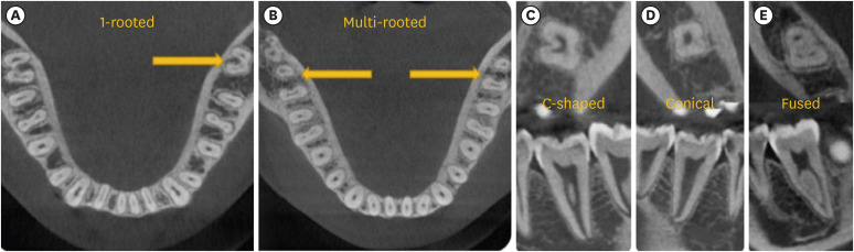
- Predictor factors of 1-rooted mandibular second molars on complicated root and canal anatomies of other mandibular teeth
- Hakan Aydın, Hatice Harorlı
- Restor Dent Endod 2024;49(1):e2. Published online January 3, 2024
- DOI: https://doi.org/10.5395/rde.2024.49.e2
-
Abstract
PDFPubReaderePub
Objectives This study aimed to determine the effects of 1-rooted mandibular second molar (MnSM) teeth on root canal anatomy complexities of the mandibular central incisor (MnCI), mandibular lateral incisor (MnLI), mandibular canine (MnCn), mandibular first premolar (MnFP), mandibular second premolar (MnSP), and mandibular first molar (MnFM) teeth.
Materials and Methods Cone-beam computed tomography images of 600 patients with full lower dentition were examined. Individuals with 1-rooted MnSMs were determined, and the complexity of root canal anatomy of other teeth was compared with individuals without 1-rooted MnSMs (Group-1; subjects with at least one 1-rooted MnSM, Group-2; subjects with more than a single root in both MnSMs). A second canal in MnCIs, MnLIs, MnCns, MnFPs, and MnSPs indicated a complicated root canal. The presence of a third root in MnFMs was recorded as complicated.
Results The prevalence of 1-rooted MnSMs was 12.2%, with the C-shaped root type being the most prevalent (9%). There were fewer complicated root canals in MnCIs (
p = 0.02), MnLIs (p < 0.001), and MnFPs (p < 0.001) in Group 1. The other teeth showed no difference between the groups (p > 0.05). According to logistic regression analysis, 1-rooted right MnSMs had a negative effect on having complex canal systems of MnLIs and MnFPs. Left MnSMs were explanatory variables on left MnLIs and both MnFPs.Conclusions In individuals with single-rooted MnSMs, a less complicated root canal system was observed in all teeth except the MnFMs.
-
Citations
Citations to this article as recorded by- Repair of furcal perforations using different calcium silicate cements: An in vitro study
Ariana Esperanza Apolo Aguilar, Maria Soledad Peñaherrera Manosalvas, Henry Paul Valverde Haro
Journal of Conservative Dentistry and Endodontics.2025; 28(10): 1007. CrossRef
- Repair of furcal perforations using different calcium silicate cements: An in vitro study
- 2,909 View
- 76 Download
- 1 Crossref
Case Report
- Successful nonsurgical treatment of type II dens invaginatus with 5 root canals using a self-adjusting file: a case report
- George Táccio de Miranda Candeiro, Antônio Sérgio Teixeira de Menezes, Ana Carolina Saldanha de Oliveira, Flávio Rodrigues Ferreira Alves
- Restor Dent Endod 2023;48(2):e17. Published online April 27, 2023
- DOI: https://doi.org/10.5395/rde.2023.48.e17
-
Abstract
PDFPubReaderePub
The present report describes the endodontic treatment of an Oehlers type II dens invaginatus in a maxillary lateral incisor with 5 root canals, an extremely rare condition. Apical periodontitis and related symptoms were noted. Cone-beam computed tomography was used to aid the diagnosis, reveal tooth morphology, and assist in canal location. The pulp chamber was carefully accessed, and the root canals were explored under magnification. All root canals were prepared with an R25 Reciproc Blue system and sodium hypochlorite (NaOCl) irrigation. After initial preparation, a self-adjusting file (SAF) with NaOCl and ethylenediaminetetraacetic acid was used to complement the disinfection. Additionally, calcium hydroxide medication was applied. Vertical compaction was used to fill the canals with a calcium silicate-based endodontic sealer and gutta-percha. After 12 months, the patient exhibited healing of the periapical region, absence of symptoms, and normal dental function. In conclusion, this nonsurgical treatment protocol was successful in promoting the cure of apical periodontitis. Both complementary disinfection with an SAF and use of calcium hydroxide medication should be considered when choosing the best treatment approach for dens invaginatus with very complex anatomy.
-
Citations
Citations to this article as recorded by- Non-surgical endodontic management of dens invaginatus type II in an immature maxillary lateral incisor using a bioceramic apical plug: A 3-year follow-up case report
Yahya Raja Alharbi, Qayed Saad Alharbi, Shaul Hameed Kolarkodi
Saudi Endodontic Journal.2026; 16(1): 115. CrossRef
- Non-surgical endodontic management of dens invaginatus type II in an immature maxillary lateral incisor using a bioceramic apical plug: A 3-year follow-up case report
- 2,999 View
- 89 Download
- 1 Crossref
Research Articles
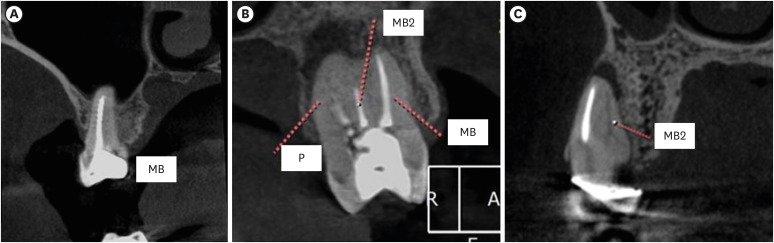
- Apical periodontitis in mesiobuccal roots of maxillary molars: influence of anatomy and quality of root canal treatment, a CBCT study
- Samantha Jannone Carrion, Marcelo Santos Coelho, Adriana de Jesus Soares, Marcos Frozoni
- Restor Dent Endod 2022;47(4):e37. Published online September 19, 2022
- DOI: https://doi.org/10.5395/rde.2022.47.e37
-
Abstract
PDFPubReaderePub
Objectives This study aimed to evaluate the prevalence of apical periodontitis (AP) in the mesiobuccal roots of root canal-treated maxillary molars.
Materials and Methods One thousand cone-beam computed tomography images of the teeth were examined by 2 dental specialists in oral radiology and endodontics. The internal anatomy of the roots, Vertucci’s classification, quality of root canal treatment, and presence of missed canals were evaluated; additionally, the correlation between these variables and AP was ascertained.
Results A total of 1,000 roots (692 first molars and 308 second molars) encompassing 1,549 canals were assessed, and the quality of the root canal filling in the majority (56.9%) of the canals was satisfactory. AP was observed in 54.4% of the teeth. A mesiolingual canal in the mesiobuccal root (MB2 canal) was observed in 54.9% of the images, and the majority (83.5%) of these canals were not filled. Significant associations were observed between the presence of an MB2 canal and the quality of the root canal filling and the presence of AP.
Conclusions AP was detected in more than half of the images. The MB2 canals were frequently missed or poorly filled.
-
Citations
Citations to this article as recorded by- Prevalence of Unfilled MB2 Canals and Their Association with Apical Periodontitis: A CBCT-Based Cross-Sectional Study in a German Population
Maythem Al Fartousi, Christian Ralf Gernhardt
Diagnostics.2026; 16(5): 796. CrossRef - When to perform cone beam computed tomography (CBCT) in primary root canal treatment? A CBCT-based cross-sectional study on the prevalence of MB2 canal in maxillary first molars
Bledar Lilaj, Elias Salzmann, Gernot Paul Hönigl, Rinet Dauti, Anton Dobsak, Sophie Pock, Barbara Cvikl
Dentomaxillofacial Radiology.2026; 55(4): 381. CrossRef - Deep learning–based detection of the second mesiobuccal canal in maxillary first molars using cone-beam computed tomography
Jiao Lin, Jialing Liu, Yuxin Jiang, Yang Liu, Bixin Wen, Shihao Li, Chenglong Li
BMC Oral Health.2026;[Epub] CrossRef - Anatomical Configuration of the MB2 Canal Using High-Resolution Cone-Beam Computed Tomography
Luciana Magrin Blank-Gonçalves, Emmanuel João Nogueira Leal da Silva, Monikelly do Carmo Chagas Nascimento, Ana Grasiela Limoeiro, Luiz Roberto Coutinho Manhães-Jr
Journal of Endodontics.2025; 51(5): 609. CrossRef - The Effect of Age and Gender on the Distance Between the Maxillary Sinus Cortical Bone and Maxillary Molars: A Cone-Beam Tomography Analysis
Thaysa Menezes Constantino, Marília Fagury Videira Marceliano-Alves, Vivian Ronquete, Ana Grasiela da Silva Limoeiro, Pablo Andres Amoroso-Silva, Mariano Simon Pedano, Tchilalo Boukpessi, Fábio Vidal, Thais Machado de Carvalho Coutinho
Sinusitis.2025; 9(1): 9. CrossRef - Retrospective study of the morphology of third maxillary molars among the population of Lower Silesia based on analysis of cone beam computed tomography
Anna Olczyk, Barbara Malicka, Katarzyna Skośkiewicz-Malinowska, Mohmed Isaqali Karobari
PLOS ONE.2024; 19(2): e0299123. CrossRef - Relationship between apical periodontitis and missed canals in mesio-buccal roots of maxillary molars: CBCT study
Badi B. Alotaibi, Kiran I. Khan, Muhammad Q. Javed, Smita D. Dutta, Safia S. Shaikh, Nawaf M. Almutairi
Journal of Taibah University Medical Sciences.2024; 19(1): 18. CrossRef - APICAL PERIODONTITIS IN MAXILLARY MOLARS WITH MISSED SECOND MESIO-BUCCAL ROOT CANAL: A CBCT STUDY
Cristina Coralia Nistor, Ioana Suciu , Ecaterina Ionescu , Anca Dragomirescu , Elena Coculescu , Andreea Baluta
Romanian Journal of Oral Rehabilitation.2024; 16(3): 100. CrossRef - Anatomic Comparison of Contralateral Maxillary Second Molars Using High-Resolution Micro-CT
Ghassan Dandache, Umut Aksoy, Mehmet Birol Ozel, Kaan Orhan
Symmetry.2023; 15(2): 420. CrossRef
- Prevalence of Unfilled MB2 Canals and Their Association with Apical Periodontitis: A CBCT-Based Cross-Sectional Study in a German Population
- 4,528 View
- 72 Download
- 8 Web of Science
- 9 Crossref
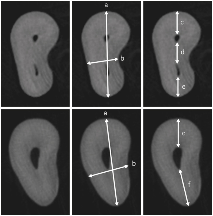
- Morphological characteristics of the mesiobuccal root in the presence of a second mesiobuccal canal: a micro-CT study
- Lucas P. Lopes Rosado, Matheus Lima Oliveira, Karla Rovaris, Deborah Queiroz Freitas, Frederico Sampaio Neves
- Restor Dent Endod 2022;47(1):e6. Published online January 18, 2022
- DOI: https://doi.org/10.5395/rde.2022.47.e6
-
Abstract
PDFPubReaderePub
Objectives This study investigated the internal morphology of mesiobuccal (MB) roots of maxillary molars with a second mesiobuccal (MB2) canal.
Materials and Methods Forty-seven maxillary first or second molars from Brazilians were scanned using micro-computed tomography. The following measurements were obtained from the MB roots: root thickness, root width, and dentin thickness of the buccal aspect of the first mesiobuccal (MB1) canal, between the MB1 and MB2 canals, and the palatal aspect of the MB2 and MB1 canals at 3 mm from the root apex and in the furcation region. For statistical analysis, the Student’s
t -test and analysis of variance with thepost-hoc Tukey test were used (α = 0.05).Results In maxillary molars with an MB2 canal, MB roots were significantly thicker (
p = 0.0014) and narrower (p = 0.0016) than in maxillary molars without an MB2 canal. The dentin thickness of the palatal aspect of the MB1 canal was also significantly greater than that of MB roots without an MB2 canal at 3 mm from the root apex (p = 0.0007) and in the furcation region (p < 0.0001). In the furcation region of maxillary molars with an MB2 canal, the dentin thickness between the MB1 and MB2 canals was significantly smaller than that in the buccal and palatal aspects (p < 0.0001).Conclusions The internal morphology of MB roots of maxillary molars with an MB2 canal revealed differences in dentin thickness, root diameter, and distance between the canals when compared with maxillary molars without an MB2 canal.
-
Citations
Citations to this article as recorded by- Association between lingual canal detection and buccolingual root width in mandibular anterior teeth: a retrospective CBCT Study
Önder Çam, Melis Oya Ateş, Ali Keleş
BMC Oral Health.2026;[Epub] CrossRef - Effectiveness and safety of three NiTi systems in endodontic retreatment of MB1 and MB2 root canals: a micro-CT and CBCT combined analysis
Airton Oliveira Santos-Junior, Rocharles Cavalcante Fontenele, Karina Ines Medina Carita Tavares, Fernanda Ferrari Esteves Torres, Jáder Camilo Pinto, Pedro Luis Busto Rosim, Andréa Gonçalves, Marco Antonio Hungaro Duarte, Juliane Maria Guerreiro-Tanomaru
Clinical Oral Investigations.2025;[Epub] CrossRef - Cone-beam computed tomography evaluation of root and canal morphology of maxillary molars in a Chinese kazakh population
Shuchun Yang, Chenye Li, Hui Shi, Ming Liu, Xu Wang
BMC Oral Health.2025;[Epub] CrossRef - Can maxillary molar dimensions predict the presence of the second mesiobuccal canal?
Lucas P. Lopes Rosado, Deborah Queiroz Freitas, Karla Rovaris, Matheus L. Oliveira, Frederico Sampaio Neves
Oral Radiology.2023; 39(3): 482. CrossRef - Can the detection of second mesiobuccal canals be enhanced based on the volume of adjacent canals?
Lucas P. Lopes Rosado, Deborah Q. Freitas, Karla Rovaris, Matheus L. Oliveira, Frederico S. Neves
Archives of Oral Biology.2023; 146: 105604. CrossRef - Assessment of the coronal root canal morphology of permanent maxillary first molars using digital 3D-reconstruction technology based on micro-computed tomography data
Mudan Wang, Yuxuan Gao, Qi Deng, Yuan Gao, Dongzhe Song, Dingming Huang
Journal of Dental Sciences.2023; 18(2): 586. CrossRef
- Association between lingual canal detection and buccolingual root width in mandibular anterior teeth: a retrospective CBCT Study
- 2,606 View
- 48 Download
- 7 Web of Science
- 6 Crossref
- Shape and anatomical relationship of the mental foramen to the mandibular premolars in an Indian sub-population: a retrospective CBCT analysis
- Komal Sheth, Kulvinder Singh Banga, Ajinkya M. Pawar, James L. Gutmann, Hyeon-Cheol Kim
- Restor Dent Endod 2022;47(1):e1. Published online December 13, 2021
- DOI: https://doi.org/10.5395/rde.2022.47.e1
-
Abstract
PDFPubReaderePub
Objectives This study assessed the shape and anatomical relationship of the mental foramen (MF) to mandibular posterior teeth in an Indian sub-population.
Materials and Methods In total, 475 existing cone-beam computed tomography records exhibiting 950 MFs and including the bilateral presence of mandibular premolars and first molars were assessed. Images were evaluated 3-dimensionally to ascertain the position, shape, and anatomical proximity of MFs to mandibular teeth. The position and shape of MFs were measured and calculated. The Pythagorean theorem was used to calculate the distance between the root apex of the mandibular teeth and the MF.
Results MFs exhibited a predominantly round shape (left: 67% and right: 65%) followed by oval (left: 30% and right: 31%) in both males and females and in different age groups. The root apices of mandibular second premolars (left: 71% and right: 62%) were closest to the MF, followed by distal to the first premolars and mesial to the second premolars. The mean vertical distance between the MF and the nearest tooth apex calculated on sagittal sections was 2.20 mm on the right side and 2.32 mm on the left side; no significant difference was found according to sex or age. The distance between the apices of the teeth and the MF was ≥ 4 mm (left; 4.09 ± 1.27 mm and right; 4.01 ± 1.15 mm).
Conclusions These findings highlight the need for clinicians to be aware of the location of the MF in treatment planning and while performing non-surgical and surgical endodontic procedures.
-
Citations
Citations to this article as recorded by- Morphometric analysis of mental foramen in retained cadaveric specimens of mandibles of Sri Lankan population
Dadallage Tharanga De Silva, Usliyanage Clifford Priyantha Perera
Anatomical Science International.2026; 101(3): 340. CrossRef - Anatomical and radiographic assessment of variations of the mental foramen and their impact on success of local anaesthesia administration
Isratul Jannat, M. Ummay Salma, Nipu Rani Chowdhury, Kulsum Nahar, Dilruba Binte Mostafa, Khandokar Emanuzzaman Emon, Shahela Sarmin
International Journal of Research in Medical Sciences.2026; 14(3): 823. CrossRef - Clinical Implications of the Localization and Morphological Variability of the Mental Foramen—A Systematic Review
Mariola Krzykawska-Krupska, Janusz Pach, Piotr Regulski, Jacek Tomczyk, Izabela Strużycka, Kazimierz Szopiński, Katarzyna Osipowicz, Anna Pogorzelska
Diagnostics.2026; 16(5): 779. CrossRef - Optimising Treatment Strategies: Labial versus Labio-inferior Plating Using Three-dimensional Miniplates for Mandibular Symphysis and Parasymphysis Fractures
Akash P Muralidharan, Kalyani Bhate, K Mithun Nilgiri, Sumithra S Nair, Lakshmi Shetty, Rose Johnson
Advances in Human Biology.2025; 15(2): 242. CrossRef - A Cross-Sectional CBCT Study of Anterior Loop, Accessory Mental Foramen, and Lingual Foramina in Patients’ Mandibles: Implications for Safer Implant Planning
Abbas Shokri, Mohammad Mahdi Maleki, Leili Tapak
Journal of Maxillofacial and Oral Surgery.2025;[Epub] CrossRef - Radiographic Recognition of Mental Nerve for Secured Dental Implant Placement by Cone-Beam Computed Tomography in Mosul City Population
Asmaa B. Al-Saffar, Mekdad H. Alrigbo, Rawaa Y. Al-Rawee
Journal of Craniofacial Surgery.2024; 35(7): 2049. CrossRef - Accuracy of Implant Size Prediction Based on Edentulous Ridge Dimension on Cone-beam Computed Tomography - A Retrospective Study
Hunter R. Jolicoeur, Gerard A. Camargo, Tamara G. Stephenson, Wenjian Zhang
Annals of Maxillofacial Surgery.2024; 14(2): 187. CrossRef - Mental Foramenin Panoramik Radyografi ve Konik Işınlı Bilgisayarlı Tomografi Görüntüleri Üzerinde Morfolojik Analizi
Ezgi UZUN, Burak Kerem APAYDIN, Ayşen TİL
Selcuk Dental Journal.2023; 10(3): 540. CrossRef - Evaluation of the Possible Relationship between the Curvature and
Horizontal Course of the Inferior Alveolar Canal
Cansu G. Koca, M. Fatih Çiçek, Sanaz Sadry, Ozan Yenidünya, Fatma Akkoca Kaplan, Aras Erdil
Current Medical Imaging Formerly Current Medical Imaging Reviews.2023;[Epub] CrossRef
- Morphometric analysis of mental foramen in retained cadaveric specimens of mandibles of Sri Lankan population
- 3,961 View
- 70 Download
- 10 Web of Science
- 9 Crossref
- Enhanced visualization of the root canal morphology using a chitosan-based endo-radiopaque solution
- Shashirekha Govind, Amit Jena, Satabdi Pattanaik, Mahaprasad Anarasi, Satyajit Mohapatra, Vinay Shivagange
- Restor Dent Endod 2021;46(3):e33. Published online June 4, 2021
- DOI: https://doi.org/10.5395/rde.2021.46.e33
-
Abstract
PDFPubReaderePub
Objectives This study aimed to investigate the efficacy of ionic and non-ionic-based contrast media (
in vitro study) and the combinatorial effect of chitosan-based endo-radiopaque solution (CERS) (in vivo study) for visualization of the root canal anatomy.Materials and Methods In vitr o study (120 teeth): The root canal of maxillary premolars and molars (in vitro group 1 and 2 respectively,n = 60 each) were analyzed using 4 different contrast media (subgroups: Omnipaque 350, Iopamidol, Xenetix 350, and Urografin 76;n = 15 each) in combination with 5.25% sodium hypochlorite (NaOCl). Based on the results of thein vitro study,in vivo study (80 teeth) was done to compare Xenetix 350 + 5.25% NaOCl with CERS (in vivo group 1 and 2 respectively,n = 40 each) on maxillary and mandibular premolars and molars. Two endodontists used radiovisiography to assess the depth of ingress and identify the aberrant root anatomy after access cavity preparation, and after initial cleaning and shaping of canals. Kruskal-Wallis test was used forin vitro comparison (p < 0.05), and Wilcoxon signed-rank test and Mann-WhitneyU test forin vivo analysis (p < 0.01).Results In vitro study, Xenetix 350 + 5.25% NaOCl facilitated a significant higher visualization (p < 0.05). Forin vivo study, CERS had a statistically significant depth of ingress (p < 0.01), and was efficient in identifying the aberrant root canal anatomy of premolars and molars.Conclusions CERS facilitates better visualization of the root canal anatomy of human premolars and molars.
-
Citations
Citations to this article as recorded by- Influence of irrigating solutions on the hydration of calcium silicate-based dental biomaterials: An in vitro study
Pradeep M. Divya, Amit Jena, Saumyakanta Mohanty, Govind Shashirekha, Rashmi Rekha Mallick, Priyanka Sarangi
Journal of Conservative Dentistry and Endodontics.2025; 28(8): 758. CrossRef - Improving Endodontic Radiograph Interpretation with TV-CLAHE for Enhanced Root Canal Detection
Barbara Obuchowicz, Joanna Zarzecka, Michał Strzelecki, Marzena Jakubowska, Rafał Obuchowicz, Adam Piórkowski, Elżbieta Zarzecka-Francica, Julia Lasek
Journal of Clinical Medicine.2025; 14(15): 5554. CrossRef - Efficacy of sonic and ultrasonic activation on irrigant penetration in different tapered preparations: An in vitro study
M. Rama Sowmya, Kavalipurapu Venkata Teja, Pradeep Solete, Sahil Choudhari, S Delphine Priscilla Antony, Mohammed Mustafa
Endodontology.2024; 36(4): 370. CrossRef - Analysis of the value of visualized root canal technique in the clinical treatment of endodontics
Nana SUN, Nannan WANG, Xin QIAN
Panminerva Medica.2023;[Epub] CrossRef
- Influence of irrigating solutions on the hydration of calcium silicate-based dental biomaterials: An in vitro study
- 2,568 View
- 26 Download
- 2 Web of Science
- 4 Crossref
-
Isthmuses, accessory canals, and the direction of root curvature in permanent mandibular first molars: an
in vivo computed tomography study - Aria Chuppani Dastgerdi, Manizheh Navabi, Vahid Rakhshan
- Restor Dent Endod 2020;45(1):e7. Published online December 12, 2019
- DOI: https://doi.org/10.5395/rde.2020.45.e7
-
Abstract
PDFPubReaderePub
Objectives This study was performed to assess the anatomy of mandibular first molars.
Materials and Methods In this
in vivo study, cone-beam computed tomography (CBCT) volumes of 312 bilateral intact first mandibular molars from 156 patients (79 men and 77 women; average age, 35.6 ± 11.2 years) were investigated in terms of the direction of each canal's curvature in the buccolingual and mesiodistal dimensions (direction of the position of the apex in relation to the longitudinal axis of the root), the presence of an isthmus (a narrow, ribbon-shaped communication between 2 root canals) in 3 segments (0–2, 2–4, and 4–6 mm) from the apex), and the presence and number of accessory canals (smaller canals besides the main root canals, connecting the pulp to the periodontium). Data were analyzed statistically (α = 0.05).Results Mesiolingual canals were mostly buccally and distally inclined, while mesiobuccal and distolingual canals were mostly distally curved. Isthmuses were more common in younger patients (χ2 test,
p < 0.05). The average numbers of accessory canals in the apical, middle, and coronal segments were 9.9 ± 4.2, 6.9 ± 2.9, and 9.3 ± 3.0 canals per segment, respectively (analysis of variance,p < 0.001). Age and sex were not associated with the number of accessory canals (p > 0.05).Conclusions The complex anatomy of these teeth deserves attention during non-surgical or surgical endodontic treatment. Around the apex, isthmuses might be more prevalent in younger and female individuals.
-
Citations
Citations to this article as recorded by- Micro-CT and histological examination of accessory canals in 34 equine cheek teeth
Szabolcs A. Korsós, Carsten Staszyk, Matthieu Boone, Iván Josipovic, Jörg Vogelsberg, Lieven Vlaminck
Frontiers in Veterinary Science.2024;[Epub] CrossRef - Diagnostic value of cone beam computed tomography for root canal morphology assessment – a micro-CT based comparison
Mariana Pires, Jorge N.R. Martins, Mário Rito Pereira, Isabel Vasconcelos, Rui Pereira da Costa, Isabel Duarte, António Ginjeira
Clinical Oral Investigations.2024;[Epub] CrossRef - Exploring age and gender variations in root canal morphology of maxillary premolars in Saudi sub population: a cross-sectional CBCT study
Mohmed Isaqali Karobari, Azhar Iqbal, Rumesa Batul, Abdul Habeeb Adil, Jamaluddin Syed, Hmoud Ali Algarni, Meshal Aber Alonazi, Tahir Yusuf Noorani
BMC Oral Health.2024;[Epub] CrossRef - Comprehensive analysis of root canal morphology in maxillary premolars among the Pakistani subpopulation: a CBCT-based study
Hmoud Ali Algarni, Meshal Aber Alonazi, Hamza Arshad, Fatima Zahra, Fahad Umer, Irfan Maqbool, Azhar Iqbal, Mohmed Isaqali Karobari
European Journal of Medical Research.2024;[Epub] CrossRef - The efficacy of a novel SWEEPS laser-activated irrigation compared to ultrasonic activation in the removal of pulp tissue from an isthmus area in the apical third of the root canal
Ivona Bago, Adriana Đurin, Debora Kanižaj, Lovorka Batelja Vuletić, Ivana Vidović Zdrilić, Ivica Anić
Lasers in Medical Science.2023;[Epub] CrossRef - Effect of passive ultrasonic irrigation on hard tissue debris removal: a systematic review and meta-analysis
Ana Flávia Almeida Barbosa, Carolina Oliveira de Lima, Luciana Moura Sassone, Raissa Dias Fares, Tatiana Kelly da Silva Fidalgo, Emmanuel João Nogueira Leal Silva
Brazilian Oral Research.2021;[Epub] CrossRef
- Micro-CT and histological examination of accessory canals in 34 equine cheek teeth
- 3,669 View
- 30 Download
- 6 Crossref
- Fused roots of maxillary molars: characterization and prevalence in a Latin American sub-population: a cone beam computed tomography study
- Maytté Marcano-Caldera, Jose Luis Mejia-Cardona, María del Pilar Blanco-Uribe, Elena Carolina Chaverra-Mesa, Didier Rodríguez-Lezama, Jose Hernán Parra-Sánchez
- Restor Dent Endod 2019;44(2):e16. Published online April 22, 2019
- DOI: https://doi.org/10.5395/rde.2019.44.e16
-
Abstract
PDFPubReaderePub
Objectives The upper molars generally have three roots; therefore, different combinations of fusion can occur, increasing the possibility of finding more complex root canal systems. The purpose of this study was to evaluate the prevalence and characterization of fused roots in first and second maxillary molars using cone-beam computed tomography (CBCT) in a Colombian population.
Materials and Methods A total of 1274 teeth were evaluated, of which 534 were maxillary first molars and 740 were maxillary second molars. Axial sections were made at the cervical, middle, and apical levels to determine the prevalence of root fusion and the types of fusion.
Results Overall, 43% of the molars (
n = 551) presented some type of fused root. Root fusion was present in 23.4% of the maxillary first molars. The most frequent type of fused root was type 3 (distobuccal-palatal; DB-P) (58.9%). Root fusion was observed in 57.6% of the maxillary second molars, and the most prevalent type of fused root was type 6 (cone-shaped) (45.2%). Of the maxillary molars, 12.5% were classified as C-shaped.Conclusion Within the limitations of this study, there was a high prevalence of fused roots in maxillary molars in the Colombian population, mainly in the maxillary second molars. In first molars, the most common type of fused root was type 3 (DB-P) and in second molars, the most common type was type 6 (cone-shaped). Additionally, molars with root fusion presented variation at different levels of the radicular portion, with implications for treatment quality.
-
Citations
Citations to this article as recorded by- Intentional Tooth Replantation: Current Evidence and Future Research Directions for Case Selection, Extraction Approaches, and Post-Operative Management
Rahul Minesh Shah, Thomas Manders, Georgios Romanos
Dentistry Journal.2026; 14(1): 59. CrossRef - Management of a rare bilateral maxillary first molar with six canals using a cone-beam computed tomography: Report of two cases
Aishwarya D. Jain, Nimisha Chinmay Shah, Abhya Jain, Shreya S. Volety
Saudi Endodontic Journal.2025; 15(2): 186. CrossRef - Assessment of root and root canal morphology in maxillary molars with fused roots using Cone Beam Computer Tomography (CBCT) in a Sri Lankan population
Ruvienath Daham Weerasinghe Rajapaksa, Manil Christopher Nishan Fonseka, Ruwan Duminda Jayasinghe, Rasika Manori Jayasinghe
Journal of Oral Biology and Craniofacial Research.2025; 15(6): 1297. CrossRef - Cone-beam computed tomography evaluation of root and canal morphology of maxillary molars in a Chinese kazakh population
Shuchun Yang, Chenye Li, Hui Shi, Ming Liu, Xu Wang
BMC Oral Health.2025;[Epub] CrossRef - A Twisted Tale of Dilacerated Fused Roots Mimicking Radicular Dens Invaginatus
Rashmi D Sathe, Preeti P. Nair, Richa Bajpai, Bidushi Mishra
Journal of the International Clinical Dental Research Organization.2025; 17(2): 214. CrossRef - Exploring the sex-associated differences in molars fused roots
Maria Eduarda Nunis Locks, Erika Calvano Küchler, Leonardo Santos Antunes, Alice Corrêa Silva-Sousa, Natanael Henrique Ribeiro Mattos, Camila Paiva Perin, Paulo Henrique Condeixa França, Peter Proff, Christian Kirschneck, Flares Baratto-Filho
Annals of Anatomy - Anatomischer Anzeiger.2024; 254: 152245. CrossRef - Cone beam computed tomography analysis of the root and canal morphology of the maxillary second molars in a Syrian subpopulation
Safaa Allawi, Mouhammad Al-Tayyan, Hassan Achour, Eyad Al-Toutangy, Yasser Alsayed Tolibah
BMC Oral Health.2024;[Epub] CrossRef - Prevalence of root fusion in canine maxillary second molar teeth using cone-beam computed tomography
Kristin Linder, Scott MacGee, Loren Schultz
Frontiers in Veterinary Science.2023;[Epub] CrossRef - Dentine thickness in maxillary fused molars depends on the fusion type: An ex vivo micro‐computed tomography study
Cangül Keskin, Defne Toplu, Ali Keleş
International Endodontic Journal.2023; 56(5): 637. CrossRef - Root and canal-specific features of maxillary first molars with fused roots
Katarina Beljic-Ivanovic, Branislav Karadzic
Vojnosanitetski pregled.2022; 79(11): 1092. CrossRef - Micro-CT Analysis of the Root Canal Configuration of Maxillary Second Molars with Fusion
Cangül KESKİN, Özgür ÖZDEMİR, Ali KELEŞ
European Annals of Dental Sciences.2022; 49(Suppl 1): 25. CrossRef - Assessment of C-Shaped Canal Morphology in Mandibular and Maxillary Second Molars in an Iraqi Subpopulation Using Cone-Beam Computed Tomography
Kazhan Abdalrahman, Ranjdar Talabani, Sara Kazzaz, Dlsoz Babarasul, Berndt Koslowski
Scanning.2022; 2022: 1. CrossRef - Analysis of Root and Canal Morphology of Fused and Separate Rooted Maxillary Molar Teeth in Turkish Population
H Aydin
Nigerian Journal of Clinical Practice.2021; 24(3): 435. CrossRef - Investigating prevalence of dental anomalies in Eastern Province of Saudi Arabia through digital orthopantomogram
Jehan ALHumaid, Maryam Buholayka, Arishiya Thapasum, Muhanad Alhareky, Maha Abdelsalam, Amr Bughsan
Saudi Journal of Biological Sciences.2021; 28(5): 2900. CrossRef - Preferred Reporting Items for Epidemiologic Cross-sectional Studies on Root and Root Canal Anatomy Using Cone-beam Computed Tomographic Technology: A Systematized Assessment
Jorge N.R. Martins, Anil Kishen, Duarte Marques, Emmanuel João Nogueira Leal Silva, João Caramês, António Mata, Marco A. Versiani
Journal of Endodontics.2020; 46(7): 915. CrossRef - Second mesiobuccal root canal in maxillary molars—A systematic review and meta-analysis of prevalence studies using cone beam computed tomography
Jorge N.R. Martins, Duarte Marques, Emmanuel João Nogueira Leal Silva, João Caramês, António Mata, Marco A. Versiani
Archives of Oral Biology.2020; 113: 104589. CrossRef
- Intentional Tooth Replantation: Current Evidence and Future Research Directions for Case Selection, Extraction Approaches, and Post-Operative Management
- 2,812 View
- 15 Download
- 16 Crossref
Case Report
- Maxillary first molar with 7 root canals diagnosed using cone-beam computed tomography
- Evaldo Rodrigues, Antônio Henrique Braitt, Bruno Ferraz Galvão, Emmanuel João Nogueira Leal da Silva
- Restor Dent Endod 2017;42(1):60-64. Published online August 29, 2016
- DOI: https://doi.org/10.5395/rde.2017.42.1.60
-
Abstract
PDFPubReaderePub
Root canal anatomy is complex, and the recognition of anatomic variations could be a challenge for clinicians. This case report describes the importance of cone beam computed tomographyic (CBCT) imaging during endodontic treatment. A 23 year old woman was referred by her general dental practitioner with the chief complaint of spontaneous pain in her right posterior maxilla. From the clinical and radiographic findings, a diagnosis of symptomatic irreversible pulpitis was made and endodontic treatment was suggested to the patient. The patient underwent CBCT examination, and CBCT scan slices revealed seven canals: three mesiobuccal (MB1, MB2, and MB3), two distobuccal (DB1 and DB2), and two palatal (P1 and P2). Canals were successfully treated with reciprocating files and filled using single-cone filling technique. Precise knowledge of root canal morphology and its variation is important during root canal treatment. CBCT examination is an excellent tool for identifying and managing these complex root canal systems.
-
Citations
Citations to this article as recorded by- KONİK IŞINLI BİLGİSAYARLI TOMOGRAFİ İLE DOĞRULANMIŞ OLAĞANDIŞI ÜST BİRİNCİ BÜYÜK AZI DİŞİN ENDODONTİK TEDAVİSİ
Didem Seda Gültekin, Funda Kont Çobankara
Journal of International Dental Sciences.2025; 11(1): 46. CrossRef - Clinical Significance of Mesiobuccal and Distobuccal Canal Variations in Maxillary Molars: A Case Series and a Mini Review
Mohsen Aminsobhani, Somayeh Majidi, Vlaho Brailo
Case Reports in Dentistry.2025;[Epub] CrossRef - An Unusual Case of Maxillary First Molar: A Case Report
Reetu Shrestha
International Journal of Innovative Science and Research Technology (IJISRT).2024; : 1330. CrossRef - Root canal therapy of maxillary first molar with seven canals diagnosed using cone beam computed tomography – a case report
Saini Rashmi, Saini V. Kumar
Tanta Dental Journal.2022; 19(3): 169. CrossRef - Four-Rooted Maxillary First Molars: A Systematic Review and Meta-Analysis
Gabriel Magnucki, Sven V. K. Mietling, Sreekanth Kumar Mallineni
International Journal of Dentistry.2021; 2021: 1. CrossRef - Endodontic treatment of various palatal roots in maxillary molars
Chengshi Wei, Keyi Li, Lili Shen, Guangliang Bai, Xiufen Tian
The Journal of the American Dental Association.2021; 152(12): 1044. CrossRef - Diversity of root canal morphology of maxillary first molars
Juhász Kincső-Réka, Kovács Mónika, Pop Mihai, Pop Silvia, Kerekes-Máthé Bernadette
Bulletin of Medical Sciences.2021; 94(1): 63. CrossRef - Endodontic Management of Maxillary First Molar with Seven Root Canals Diagnosed Using Cone-beam Computed Tomography: A Case Report
Ravindranath Megha, Venkatachalam Prakash
World Journal of Dentistry.2021; 12(1): 89. CrossRef - Endodontic management of the maxillary first molar with special root canals: A case report and review of the literature
Zhi-Hui Zhang, Hai-Lin Yao, Yan Zhang, Xiao Wang
World Journal of Clinical Cases.2020; 8(12): 2590. CrossRef - Management of a permanent maxillary first molar with unusual crown and root anatomy: a case report
Prateeksha Chowdhry, Pallavi Reddy, Mamta Kaushik
Restorative Dentistry & Endodontics.2018;[Epub] CrossRef - Usefulness of cone beam computed tomography in perplexing endodontic cases
Amandeep Kaur, Ajay Logani
Endodontology.2018; 30(2): 187. CrossRef - Endodontic management of a maxillary first molar with seven root canal systems evaluated using cone-beam computed tomography scanning
VijayReddy Venumuddala, Sridhar Moturi, SV Satish, BKalyan Chakravarthy, Sudhakar Malapati
Journal of International Society of Preventive and Community Dentistry.2017; 7(5): 297. CrossRef
- KONİK IŞINLI BİLGİSAYARLI TOMOGRAFİ İLE DOĞRULANMIŞ OLAĞANDIŞI ÜST BİRİNCİ BÜYÜK AZI DİŞİN ENDODONTİK TEDAVİSİ
- 5,344 View
- 17 Download
- 12 Crossref
Letter to Editor
- Working width, a deserted aspect of Endodontics
- Denzil Albuquerque, Jojo Kottoor
- Restor Dent Endod 2015;40(4):334-335. Published online September 23, 2015
- DOI: https://doi.org/10.5395/rde.2015.40.4.334
-
PDFPubReaderePub
-
Citations
Citations to this article as recorded by- Effect of Coronal Flaring on Initial Apical File Size Estimation in Curved Canals Using Three Distinct Rotary Instruments: A Comparative In Vitro Study
Vinodhini Varatharajan, Muhammed Abdul Rahman Thazhathveedan, Mohammed Salman Kuttikkodan, Ismail Puzhangaraillath Mundanatayil, Amrutha Ravindran Thazhe Mangool, Ashraf Karumbil
Cureus.2024;[Epub] CrossRef - How much to enlarge? A letter to the editor
Krishnamachari Janani, Kavalipurapu Venkata Teja, Kumar Chandan Srivatsava
Saudi Endodontic Journal.2023; 13(3): 288. CrossRef
- Effect of Coronal Flaring on Initial Apical File Size Estimation in Curved Canals Using Three Distinct Rotary Instruments: A Comparative In Vitro Study
- 2,479 View
- 67 Download
- 2 Crossref
Case Reports
- Endodontic treatment of a mandibular first molar with 8 canals: a case report
- Ankit Arora, Shashi Rashmi Acharya, Padmaja Sharma
- Restor Dent Endod 2015;40(1):75-78. Published online October 13, 2014
- DOI: https://doi.org/10.5395/rde.2015.40.1.75
-
Abstract
PDFPubReaderePub
Presented here is a case where 8 canals were located in a mandibular first molar. A patient with continuing pain in mandibular left first molar even after completion of biomechanical preparation was referred by a dentist. Following basic laws of the pulp chamber floor anatomy, 8 canals were located in three steps with 4 canals in each root. In both of the roots, 4 separate canals commenced which joined into two canals and exited as two separate foramina. At 6 mon follow-up visit, the tooth was found to be asymptomatic and revealed normal radiographic periapical area. The case stresses on the fact that understanding the laws of pulp chamber anatomy and complying with them while attempting to locate additional canals can prevent missing canals.
-
Citations
Citations to this article as recorded by- Birooted Mandibular Canine: A Systematic Review and Meta-Analysis
Amelia Hoppe, Kamila Chęcińska, Maciej Chęciński, Natalia Turosz, Maciej Sikora
Journal of Clinical Medicine.2026; 15(9): 3381. CrossRef - How Do Different Image Modules Impact the Accuracy of Working Length Measurements in Digital Periapical Radiography? An In Vitro Study
Vahide Hazal Abat, Rabia Figen Kaptan
Diagnostics.2025; 15(3): 305. CrossRef - Determinants of the Number of Main Canals in a Tooth: Deciphering Potential Mechanisms
Andrea Alejandra Moreno Robalino, José Luis Álvarez Vásquez
Universitas Odontologica.2023;[Epub] CrossRef - Application Of Cone-Beam Computed Tomography In Diagnosis And Treatment Of Multiple Canals– A Case Report
Gyanendra Pratap Singh, Shruthi H Attavar, Sivaji Kavuri
Annals of Dental Specialty.2022; 10(2): 15. CrossRef - Four distal root canals in a two-rooted permanent mandibular first molar
Urvashi M. Ujariya, Foram H. Patel, Rajendra P. Bharatiya, Anjali K. Kothari
Endodontology.2022; 34(3): 212. CrossRef - Utilizing Cone-Beam Computed Tomography for Identifying and Managing Multiple Canals: A Case Report
Gyanendra Pratap Singh, Shruthi H Attavar, Sivaji Kavuri
Journal of Current Research in Oral Surgery.2022; 2(1): 37. CrossRef - Morphology and prevalence of middle canals in the mandibular molars: A systematic review
Rashmi Bansal, Sapna Hegde, Madhusudan Astekar
Journal of Oral and Maxillofacial Pathology.2018; 22(2): 216. CrossRef - Endodontic management of a permanent mandibular first molar with unusual root canal configurations: Two case reports
Mohammad Ahmad Alenezi, Mustafa Aldajani, Hind O. Al-Qathami, Seraj Al-Shommrani
Saudi Endodontic Journal.2017; 7(3): 181. CrossRef
- Birooted Mandibular Canine: A Systematic Review and Meta-Analysis
- 6,546 View
- 36 Download
- 8 Crossref
- Surgical endodontic management of infected lateral canals of maxillary incisors
- Ji-Hyun Jang, Jung-Min Lee, Jin-Kyu Yi, Sung-Baik Choi, Sang-Hyuk Park
- Restor Dent Endod 2015;40(1):79-84. Published online October 10, 2014
- DOI: https://doi.org/10.5395/rde.2015.40.1.79
-
Abstract
PDFPubReaderePub
This case report presents surgical endodontic management outcomes of maxillary incisors that were infected via the lateral canals. Two cases are presented in which endodontically-treated maxillary central incisors had sustained lateral canal infections. A surgical endodontic treatment was performed on both teeth. Flap elevation revealed vertical bone destruction along the root surface and infected lateral canals, and microscopy revealed that the lateral canals were the origin of the lesions. After the infected lateral canals were surgically managed, both teeth were asymptomatic and labial fistulas were resolved. There were no clinical or radiographic signs of surgical endodontic management failure at follow-up visits. This case report highlights the clinical significance and surgical endodontic management of infected lateral canal of maxillary incisor. It is important to be aware of root canal anatomy variability in maxillary incisors. Maxillary central incisors infected via the lateral canal can be successfully managed by surgical endodontic treatment.
-
Citations
Citations to this article as recorded by- Surgical Management of Radicular Cyst with Platelet-rich Fibrin Placement followed by Nonvital Bleaching of a Discolored Maxillary Left Central Incisor (21)
Sagarika Sortey, Gautam Badole, Pratima Shenoi, Rajesh Kubde, Shriya Shahu, Ankita Ramteke, Varsha Uttarwar
Bharati Vidyapeeth Journal of Dentistry and Allied Sciences.2025; 2(1): 31. CrossRef - Apical Surgery of a Maxillary Left Central Tooth Using NeoPutty After Retreatment Failure: A Case Report
Sajedeh Namaei Ghasemi, Zakieh Kheradmand, Siavash Moushekhian, Zeinab Ghasemi
Clinical Case Reports.2025;[Epub] CrossRef - Cone Beam Computed Tomography as a Diagnostic Tool in the Diagnosis of an Iatrogenic Root Defect of a Root Canal Treated Maxillary Central Incisor with Periapical Lesion and Its Management by Re-apicectomy
Swathi Aravelli, Uday Kumar, Gunnam Sai Nishitha, K. Mallika Yadav, P. Sivaram, Nimeshika Ramachandruni
Bharati Vidyapeeth Journal of Dentistry and Allied Sciences.2025; 2(4): 155. CrossRef - On the Causes of Persistent Apical Periodontitis. Findings From Endodontic Microsurgery: A Case Report
Mateo José Pesántez-Ibarra, Carolina Berruecos-Orozco, Jeimmy Katherine Molina-Barrera, Néstor Ríos-Osorio, Rafael Fernández-Grisales
Journal of Endodontic Microsurgery.2025;[Epub] CrossRef - Expert consensus on difficulty assessment of endodontic therapy
Dingming Huang, Xiaoyan Wang, Jingping Liang, Junqi Ling, Zhuan Bian, Qing Yu, Benxiang Hou, Xinmei Chen, Jiyao Li, Ling Ye, Lei Cheng, Xin Xu, Tao Hu, Hongkun Wu, Bin Guo, Qin Su, Zhi Chen, Lihong Qiu, Wenxia Chen, Xi Wei, Zhengwei Huang, Jinhua Yu, Zhen
International Journal of Oral Science.2024;[Epub] CrossRef - Surgical endodontic treatment of maxillary incisors: Case report
Moazzy I. Almansour
Clinical Case Reports.2023;[Epub] CrossRef - Resective and Regenerative Approach for an Unresolved Periapical Lesion: A Surgical Case Report With 24-Month Follow-Up
Anchu R Thomas, Melwin Mathew, Sunil K Nettemu, Anoop Mayya
Cureus.2023;[Epub] CrossRef - An in vitro endodontic model to quantify the accessory canal filling potential of the vertical and lateral condensation techniques
Thomas Gerhard Wolf, Louisa Willems, Benjamín Briseño‐Marroquín
Australian Endodontic Journal.2021; 47(2): 245. CrossRef - Application of a new system for classifying root and canal anatomy in studies involving micro‐computed tomography and cone beam computed tomography: Explanation and elaboration
H. M. A. Ahmed, N. Ibrahim, N. S. Mohamad, P. Nambiar, R. F. Muhammad, M. Yusoff, P. M. H. Dummer
International Endodontic Journal.2021; 54(7): 1056. CrossRef - German Dentists’ Preferences for the Treatment of Apical Periodontitis: A Cross-Sectional Survey
Jonas Conrad, Jan Retelsdorf, Sameh Attia, Christof Dörfer, Mohamed Mekhemar
International Journal of Environmental Research and Public Health.2020; 17(20): 7447. CrossRef - Surgical management of an accessory canal in a maxillary premolar: a case report
Hee-Jin Kim, Mi-Kyung Yu, Kwang-Won Lee, Kyung-San Min
Restorative Dentistry & Endodontics.2019;[Epub] CrossRef - A new system for classifying accessory canal morphology
H. M. A. Ahmed, P. Neelakantan, P. M. H. Dummer
International Endodontic Journal.2018; 51(2): 164. CrossRef - Effects of antimicrobial photodynamic therapy and surgical endodontic treatment on the bacterial load reduction and periapical lesion healing. Three years follow up
Aguinaldo S. Garcez, Julio G. Arantes-Neto, Debora P. Sellera, Eduardo Rodrigues Fregnani
Photodiagnosis and Photodynamic Therapy.2015; 12(4): 575. CrossRef
- Surgical Management of Radicular Cyst with Platelet-rich Fibrin Placement followed by Nonvital Bleaching of a Discolored Maxillary Left Central Incisor (21)
- 2,805 View
- 17 Download
- 13 Crossref
- Root canal treatment of a mandibular second premolar with three separate root canals
- Seok-Ryun Lee, Seol-Hee Shin, Sung-Ok Hong, Chang-Kyu Song, Hoon-Sang Chang, Kyung-San Min
- J Korean Acad Conserv Dent 2010;35(4):302-305. Published online July 31, 2010
- DOI: https://doi.org/10.5395/JKACD.2010.35.4.302
-
Abstract
PDFPubReaderePub
Mandibular premolars show a wide variety of root canal anatomy. Especially, the occurrence of three canals with three separate foramina in mandibular second premolars is very rare. This case report describes the root canal treatment of an unusual morphological configuration of the root canal system and supplements previous reports of the existence of such configuration in mandibular second premolar.
-
Citations
Citations to this article as recorded by- Effective management of mandibular second premolar with root anomalies
Ashwaq Faia Asiri
Saudi Endodontic Journal.2023; 13(1): 28. CrossRef
- Effective management of mandibular second premolar with root anomalies
- 2,777 View
- 11 Download
- 1 Crossref
 First
First Prev
Prev

