Search
- Page Path
- HOME > Search
- Effect of irrigation protocols on smear layer removal, bond strength and nanoleakage of fiber posts using a self-adhesive resin cement
- Rodrigo Stadler Alessi, Renata Terumi Jitumori, Bruna Fortes Bittencourt, Giovana Mongruel Gomes, João Carlos Gomes
- Restor Dent Endod 2023;48(3):e28. Published online July 27, 2023
- DOI: https://doi.org/10.5395/rde.2023.48.e28

-
 Abstract
 PDF
PDF PubReader
 ePub
ePub Objectives This study aimed to investigate the effect of the application method of 2% chlorhexidine (CHX) and its influence on the adhesion of fiberglass posts cemented with a self-adhesive resin cement.
Materials and Methods Sixty human mandibular premolars were endodontically treated and divided into 5 groups (
n = 12), according to the canal irrigant and its application method: 2 groups with conventional syringe irrigation (CSI)—2.5% sodium hypochlorite (NaOCl) (control) and 2% CHX— and 3 groups with 2% CHX irrigation/activation—by passive ultrasonic irrigation (PUI), Easy Clean file, and XP-Endo Finisher file. Two roots per group were evaluated for smear layer (SL) removal by scanning electron microscopy. For other roots, fiber posts were luted using a self-adhesive resin cement. The roots were sectioned into 6 slices for push-out bond strength (BS) (7/group) and nanoleakage (NL) (3/group). Data from SL removal were submitted to Kruskal-Wallis and Student-Newman-Keuls tests (α = 0.05). Data from BS and NL were evaluated by 2-way analysis of variance and Tukey’s test (α = 0.05).Results For SL removal and BS, the CHX irrigation/activation promoted better values than CSI with CHX (
p < 0.05), but it was not significantly different from CSI with NaOCl (p > 0.05). For NL, the lowest values were obtained by the chlorhexidine irrigation/activation groups (p < 0.05).Conclusions Active 2% CHX irrigation can be used to improve the post space cleaning and adhesion before fiber post cementation with self-adhesive resin cements.
-
Citations
Citations to this article as recorded by- Effects of radiotherapy dose and endodontic irrigants on universal resin cement bonding to root dentin: mechanical and interfacial analyses
Lívia Ribeiro, Luíz Carlos de Lima Dias-Júnior, Paulo Henrique dos Santos, Mariana Comparotto Minamisako, Paulo Marcelo Rodrigues, Vicente Ribeiro Netto, Bruno Alexandre Pacheco de Castro Henriques, Renata Gondo Machado, Cleonice da Silveira Teixeira, Luc
International Journal of Adhesion and Adhesives.2026; 146: 104252. CrossRef - Laser‐Activated Irrigation via Photon‐Induced Photoacoustic Streaming and Shock Wave Enhanced Emission on Smear Layer Removal Efficacy, Pushout Bond Strength, and Sealer Adaptation: A SEM Assessment
Basil Almutairi, Fahad Alkhudhairy
Microscopy Research and Technique.2025; 88(6): 1806. CrossRef - The impact of passive ultrasonic irrigation on the bond strength of two different self-etch adhesives to human pulp chamber dentine: a laboratory investigation
Mohammed Turky, Jukka Matinlinna, Monika Lukomska-Szymanska, Venkateshbabu Nagendrababu, Paul M. H. Dummer, Ahmad Abdel Hamid Elheeny, Nermin Alsayed Mahmoud
BMC Oral Health.2025;[Epub] CrossRef - The effect of nanoparticles incorporation titanium dioxide and zirconium oxide within self-adhesive resin cement on the push-out bond strength of the fiber post to the radicular dentin: An in vitro study
Sawsan Hameed Al-Jubori, Maha Anwer AL-Murad
Saudi Endodontic Journal.2025; 15(2): 162. CrossRef - The Effects of Different Post Space Conditioning Procedures and Different Endodontic Sealers on the Push-Out Bond Strengths of Fiber Posts
Leyla Ayranci, Ahmet Serkan Küçükekenci, Fatih Sarı, Ahmet Çetinkaya
Clinical and Experimental Health Sciences.2025; 15(3): 620. CrossRef - Evaluation of Microleakage Using Different Luting Cements in Kedo Zirconia Crowns: An In Vitro Assessment
Guru Vishnu, Ganesh Jeevanandan
Cureus.2024;[Epub] CrossRef
- Effects of radiotherapy dose and endodontic irrigants on universal resin cement bonding to root dentin: mechanical and interfacial analyses
- 3,189 View
- 68 Download
- 6 Web of Science
- 6 Crossref

- Effect of an aluminum chloride hemostatic agent on the dentin shear bond strength of a universal adhesive
- Sujin Kim, Yoorina Choi, Sujung Park
- Restor Dent Endod 2023;48(2):e14. Published online March 22, 2023
- DOI: https://doi.org/10.5395/rde.2023.48.e14
-
 Abstract
 PDF
PDF PubReader
 ePub
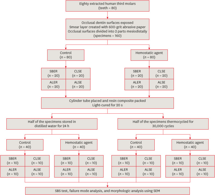
ePub Objectives This study investigated the effect of an aluminum chloride hemostatic agent on the shear bond strength (SBS) of a universal adhesive to dentin.
Materials and Methods Eighty extracted human molars were trimmed at the occlusal dentin surfaces and divided mesiodistally. According to hemostatic agent application, specimens were randomly allocated into control (C) and hemostatic agent (Traxodent; H) groups. Each group was divided into 4 subgroups according to the adhesive system (
n = 20): Scotchbond Multi-Purpose (SBER), Clearfil SE Bond (CLSE), All-Bond Universal etch-and-rinse mode (ALER), and All-Bond Universal self-etch mode (ALSE). SBS was measured for half of the specimens at 24 hours, and the other half were thermocycled in water baths (group T). Fracture surfaces were examined to determine the failure mode. The SBS was measured, and data were analyzed using 1-way analysis of variance, the Student’st -test, and the Tukey honestly significant difference test (p = 0.05).Results No significant differences in SBS were found between groups C and H for any adhesive system at 24 hours. After thermocycling, a statistically significant difference was observed between CT+ALSE and HT+ALSE (
p < 0.05). When All-Bond Universal was applied to hemostatic agent-contaminated dentin, the SBS of H+ALSE was significantly lower than that of H+ALER (p < 0.05). The SBER subgroups showed no significant differences in SBS regardless of treatment and thermocycling.Conclusions When exposed dentin was contaminated by an aluminum chloride hemostatic agent before dentin adhesive treatment, application of All-Bond Universal in etch-and-rinse mode was superior to self-etch mode.
-
Citations
Citations to this article as recorded by- Nature-driven blue-emissive N, S-CDs: Harnessing sequential "switch-off-on" fluorescence signals for detection of chrysin and Al³⁺ along with cellular imaging versatility
Maha Mohammad Abdel-Monem, Mohamed I. Walash, Asmaa Kamal El-Deen
Talanta Open.2025; : 100466. CrossRef - Comparative Evaluation of the Shear Bond Strength of Self-Adhesive and Glass Ionomer Cement to Dentin After Removal of Hemostatic Agents Using Different Cleansing Protocols: An In Vitro Study
Hemashree Namburajan, Mathew Chalakuzhiyil Abraham, Vidhyasankari N, Rajkumar K, Abhinayaa Suthagar, Vishnupriya Venkatasubramanian, Sindhuja Nagarajan
Cureus.2025;[Epub] CrossRef - Emalje- og dentinadhesiver: Avgjørende faser i klinisk behandling
Torgils Lægreid, Tom Paulseth, Arne Lund
Den norske tannlegeforenings Tidende.2024; 134(8): 604. CrossRef
- Nature-driven blue-emissive N, S-CDs: Harnessing sequential "switch-off-on" fluorescence signals for detection of chrysin and Al³⁺ along with cellular imaging versatility
- 2,950 View
- 68 Download
- 2 Web of Science
- 3 Crossref

- Chitosan-induced biomodification on demineralized dentin to improve the adhesive interface
- Isabella Rodrigues Ziotti, Vitória Leite Paschoini, Silmara Aparecida Milori Corona, Aline Evangelista Souza-Gabriel
- Restor Dent Endod 2022;47(3):e28. Published online June 15, 2022
- DOI: https://doi.org/10.5395/rde.2022.47.e28
-
 Abstract
 PDF
PDF PubReader
 ePub
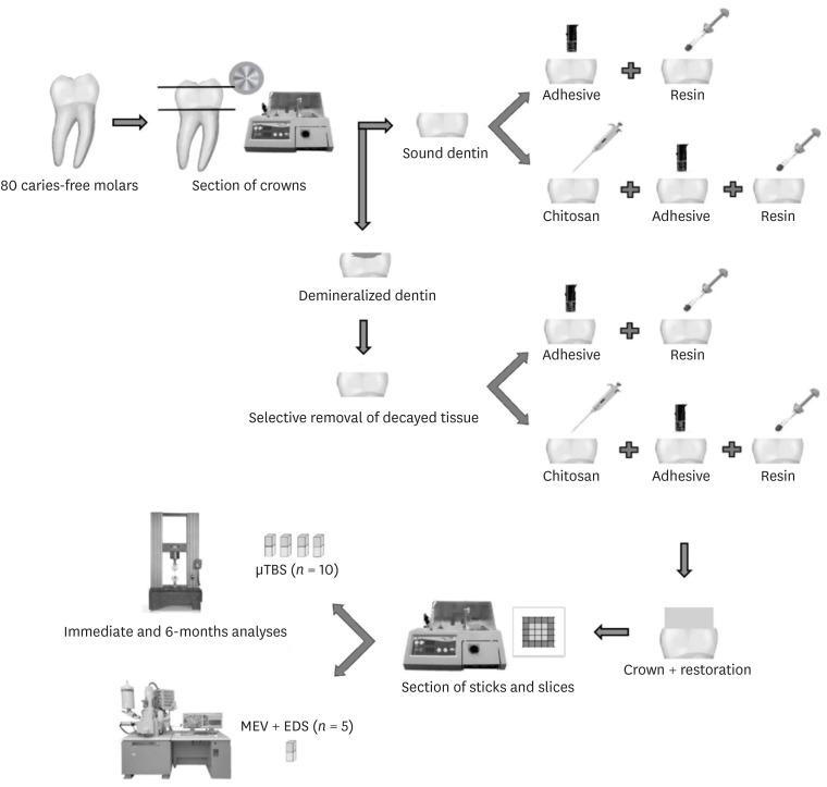
ePub Objectives Metalloproteinase-inhibiting agents, such as chitosan, can prevent collagen degradation in demineralized dental substrates, thereby improving the adhesive interface. This study evaluated the bond strength (BS) and chemical and morphological characterization of the adhesive interface after applying chitosan solution to demineralized dentin.
Materials and Methods The 80 third molars were selected. Forty teeth underwent caries induction using the pH cycling method. The teeth were divided according to the treatment: distilled water (control) and 2.5% chitosan solution. The surfaces were restored using adhesive and composite resins. Half of the specimens in each group were aged, and the other half underwent immediate analyses. The teeth were sectioned and underwent the microtensile bond strength test (µTBS), and chemical and morphological analyses using energy-dispersive spectroscopy and scanning electron microscopy, respectively. Data analysis was performed using 3-way analysis of variance.
Results For µTBS, sound dentin was superior to demineralized dentin (
p < 0.001), chitosan-treated specimens had higher bond strength than the untreated ones (p < 0.001), and those that underwent immediate analysis had higher values than the aged specimens (p = 0.019). No significant differences were observed in the chemical or morphological compositions.Conclusions Chitosan treatment improved bond strength both immediately and after aging, even in demineralized dentin.
-
Citations
Citations to this article as recorded by- Recent advances in medical applications of chitosan-based biomaterials
Dinesh Kumar Sharma
International Journal of Polymeric Materials and Polymeric Biomaterials.2025; 74(11): 1027. CrossRef - Push-Out Bond Strength of Different Luting Cements Following Post Space Irrigation with 2% Chitosan: An In Vitro Study
Shimaa Rifaat, Ahmed Rahoma, Hind Muneer Alharbi, Sawsan Jamal Kazim, Shrouq Ali Aljuaid, Basmah Omar Alakloby, Faraz A. Farooqi, Noha Taymour
Prosthesis.2025; 7(1): 18. CrossRef - Bioinspired Dentin Biomodification: Current Evidence and Emerging Approaches
Priyanka S R, Sharath Pare
International Journal of Innovative Science and Research Technology.2025; : 219. CrossRef - A synergistic approach to tooth remineralization using nano-chitosan, fluoride, and pulsed magnetic field
Alaa M. Khalil, Samar A. Abbassy, Mona Mohy ElDin, Sherif Kandil, Ahmed M. El-Khatib
Scientific Reports.2025;[Epub] CrossRef - Chitosan-based Nano/Biomaterials in Bone Tissue Engineering and Regenerative Medicine: Recent Progress and Advances
Taha Jafari, Seyed Morteza Naghib, M. R. Mozafari
Current Organic Synthesis.2025; 22(4): 457. CrossRef - Influence of Non-Staining Chitosan-Based Nano-Silver Fluoride on Shear Bond Strengths of Dental Restorations
Bennett T. Amaechi, Sima Abdollahi, Tejal Gohil, Amos C. Obiefuna, Temitayo Omoniyi, Temitope O. Omosebi, Thais S. Phillips, Noha Elhabashi
Journal of Composites Science.2025; 9(10): 518. CrossRef - Does dentin pretreatment with chitosan improve the bond strength of restorative material? A systematic review and meta-analysis of in vitro studies
Luísa Valente Gotardo Lara Alves, Nathália Mancioppi Cerqueira, Amanda Pelegrin Candemil, André Luis Faria-e-Silva, Manoel Damião Sousa-Neto, Aline Evangelista Souza-Gabriel
International Journal of Adhesion and Adhesives.2024; 128: 103553. CrossRef - Comparative Evaluation of Apical Leakage in Root Canal Obturation Using AH Plus Sealer, Bioceramic Sealer, and Bioceramic Sealer Incorporated With Chitosan Nanoparticles: An In Vitro Study
Sushmita Rane, Varsha Pandit, Sanpreet S Sachdev, Shivani Chauhan, Rishabh Mistry, Barun Kumar
Cureus.2024;[Epub] CrossRef - Aesthetic impact of resin infiltration and its mechanical effect on ceramic bonding for white spot lesions
Jiaen Shu, Yijia Huang, Xueying Ma, Zhonghua Duan, Pei Wu, Sijing Chu, Yuqiong Wu, Yuhua Wang
BMC Oral Health.2024;[Epub] CrossRef - Effect of Incorporating Chitosan to Resin Modified Glass Ionomer Cement on Shear Bond Strength to Dentin (An In vitro Comparative Study)
Aya Tahseen Khudhair, Muna Saleem Khalaf
Journal of International Society of Preventive and Community Dentistry.2024; 14(3): 225. CrossRef - Biomodification of eroded and abraded dentin with epigallocatechin-3-gallate (EGCG)
Bruna Dantas Abreu, Renata Siqueira Scatolin, Silmara Aparecida Milori Corona, Fabiana Almeida Curylofo Zotti
Journal of the Mechanical Behavior of Biomedical Materials.2023; 147: 106158. CrossRef - Chitosan-Based Biomaterials for Tissue Regeneration
Yevgeniy Kim, Zharylkasyn Zharkinbekov, Kamila Raziyeva, Laura Tabyldiyeva, Kamila Berikova, Dias Zhumagul, Kamila Temirkhanova, Arman Saparov
Pharmaceutics.2023; 15(3): 807. CrossRef - Er:YAG laser in selective caries removal and dentin treatment with chitosan: a randomized clinical trial in primary molars
Rai Matheus Carvalho Santos, Renata Siqueira Scatolin, Sérgio Luiz de Souza Salvador, Aline Evangelista Souza-Gabriel, Silmara Aparecida Milori Corona
Lasers in Medical Science.2023;[Epub] CrossRef - Effect of Dentin Surface Pretreatment With Chitosan Nanoparticles on Immediate and Prolonged Shear Bond Strength of Resin Composite: An in Vitro Study
Shaymaa Ali Abdul-Razzaq, Muna Saleem Khalaf
Dental Hypotheses.2023; 14(3): 84. CrossRef - MODERN TRENDS AND PERSPECTIVES OF THE DEVELOPMENT OF ADHESIVE DENTISTRY. INNOVATIVE TECHNIQUES FOR THE APPLICATION OF ADHESIVE SYSTEMS
Oleksandr O. Pompii, Viktor A. Tkachenko, Tetiana M. Kerimova, Elina S. Pompii
Wiadomości Lekarskie.2023; 76(12): 2721. CrossRef
- Recent advances in medical applications of chitosan-based biomaterials
- 2,192 View
- 68 Download
- 14 Web of Science
- 15 Crossref

- Is dentin biomodification with collagen cross-linking agents effective for improving dentin adhesion? A systematic review and meta-analysis
- Julianne Coelho Silva, Edson Luiz Cetira Filho, Paulo Goberlânio de Barros Silva, Fábio Wildson Gurgel Costa, Vicente de Paulo Aragão Saboia
- Restor Dent Endod 2022;47(2):e23. Published online May 6, 2022
- DOI: https://doi.org/10.5395/rde.2022.47.e23
-
 Abstract
 PDF
PDF Supplementary Material
 PubReader
 ePub
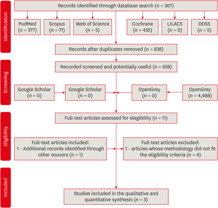
ePub Objectives The aim of this investigation was to evaluate the effectiveness of collagen cross-linking agents (CCLAs) used in combination with the adhesive technique in restorative procedures.
Materials and Methods In this systematic review, the authors followed the Preferred Reporting Items for Systematic Reviews and Meta-Analyses checklist. An electronic search was performed using PubMed, Scopus, Web of Science, Cochrane Library, LILACS, and DOSS, up to October 2020. The gray literature was also researched. Only randomized clinical trials were selected.
Results The selection process yielded 3 studies from the 838 retrieved. The addition of CCLAs in the retention of restorations increased the number of events. The postoperative sensitivity scores and marginal adaptation scores showed no significant difference between the CCLA and control groups, and the marginal pigmentation scores showed a significant increase in the CCLA group. There were no caries events in any group throughout the evaluation period.
Conclusions This systematic review showed that there is no clinical efficacy to justify the use of CCLAs in the protocols performed.
-
Citations
Citations to this article as recorded by- Riboflavin-ultraviolet-A collagen crosslinking treatments in improving dentin bonding and resistance to enzymatic digestion
Yung-Show Chiang, Ping-Ju Chen, Chun-Chan Ting, Yuh-Ling Chen, Shu-Fen Chuang
Journal of Dental Sciences.2025; 20(1): 109. CrossRef - Effect of dentin bio modifications and matrix metalloproteinase activity on bond strength – A systematic review and meta-analysis
D. Agarwal, S. R. Srinidhi, S. D. Aggarwal, P. Ingle, S. Tandon
Endodontics Today.2025; 23(1): 71. CrossRef - O USO DE ADESIVO AUTOCONDICIONANTE E RESINA FLOW COMO INTERFACE ADESIVA PROTETORA DA DENTINA FRENTE À IRRIGAÇÃO COM NaClO NO TRATAMENTO ENDODÔNTICO: ESTUDO IN-VITRO
Luís Daniel Ramos de Oliveira, Leandro Botelho Hanna, José Augusto Rodrigues
RECIMA21 - Revista Científica Multidisciplinar - ISSN 2675-6218.2025; 6(12): e6127063. CrossRef - Stability of dentin matrix treated with caffeic acid phenethyl ester at different concentrations
Aline Honorato Damázio, Rosanna Tarkany Basting, Enrico Coser Bridi, Fabiana Mantovani Gomes França, Flávia Lucisano Botelho do Amaral, Cecilia Pedroso Turssi, Waldemir Francisco Vieira Junior, Roberta Tarkany Basting
Brazilian Journal of Oral Sciences.2024; 23: e244006. CrossRef - Effect of Collagen Crosslinkers on Dentin Bond Strength of Adhesive Systems: A Systematic Review and Meta-Analysis
Louis Hardan, Umer Daood, Rim Bourgi, Carlos Enrique Cuevas-Suárez, Walter Devoto, Maciej Zarow, Natalia Jakubowicz, Juan Eliezer Zamarripa-Calderón, Mateusz Radwanski, Giovana Orsini, Monika Lukomska-Szymanska
Cells.2022; 11(15): 2417. CrossRef
- Riboflavin-ultraviolet-A collagen crosslinking treatments in improving dentin bonding and resistance to enzymatic digestion
- 1,975 View
- 45 Download
- 3 Web of Science
- 5 Crossref

- Bonding effects of cleaning protocols and time-point of acid etching on dentin impregnated with endodontic sealer
- Tatiane Miranda Manzoli, Joissi Ferrari Zaniboni, João Felipe Besegato, Flávia Angélica Guiotti, Andréa Abi Rached Dantas, Milton Carlos Kuga
- Restor Dent Endod 2022;47(2):e21. Published online April 6, 2022
- DOI: https://doi.org/10.5395/rde.2022.47.e21
-
 Abstract
 PDF
PDF PubReader
 ePub
ePub Objectives This study aimed to investigate the bonding effects of cleaning protocols on dentin impregnated with endodontic sealer residues using ethanol (E) or xylol (X). The effects of dentin acid etching immediately (I) or 7 days (P) after cleaning were also evaluated. For bonding to dentin, universal adhesive (Scotchbond Universal; 3M ESPE) was used. The persistence of sealer residues, hybrid layer formation and microshear bond strength were the performed analysis.
Materials and Methods One hundred and twenty bovine dentin specimens were allocated into 4 groups (
n = 10): G1 (E+I); G2 (X+I); G3 (E+P); and G4 (X+P). The persistence of sealer residues was evaluated by SEM. Confocal laser scanning microscopy images were taken to measure the formed hybrid layer using the Image J program. For microshear bond strength, 4 resin composite cylinders were placed over the dentin after the cleaning protocols. ANOVA followed by Tukey test and Kruskal-Wallis followed by Dunn test were used for parametric and non-parametric data, respectively (α = 5%).Results G2 and G4 groups showed a lower persistence of residues (
p < 0.05) and thicker hybrid layer than the other groups (p < 0.05). No bond strength differences among all groups were observed (p > 0.05).Conclusions Dentin cleaning using xylol, regardless of the time-point of acid etching, provided lower persistence of residues over the surface and thicker hybrid layer. However, the bond strength of the universal adhesive system in etch-and-rinse strategy was not influenced by the cleaning protocols or time-point of acid etching.
-
Citations
Citations to this article as recorded by- Efficacy of Post-Endodontic Access Cavity Cleaning Techniques: A Randomized Clinical Study
Ayse Karadayi, Elif Irem Altintas, Ezgi Tüter Bayraktar, Bora Korkut
Journal of Endodontics.2025;[Epub] CrossRef - Does cleaning of post space before cementation of fiber reinforced post affect the push-out bond strength to resin cement?
Maher S. Hajjaj, Khalid A. Alghamdi, Abdulrahman A. Alshehri, Hassan A. Almusallam, Nabeel M. Munshi, Osamah A. Alsulimani, Naseeba H. Khouja, Yousef A. Alnowailaty, Saeed J. Alzahrani
BMC Oral Health.2025;[Epub] CrossRef - Influence of the Use of a Mixed Solution of Equal Amounts of Amyl Acetate, Acetone, and Ethanol on the Cleaning of Endodontic Sealer Residues on the Bond Strength of the Fiber Post Cementation System: A Laboratory Investigation
Antonia Patricia Oliveira Barros, Ana Paula Aparecida Raimundo Alves Freitas, Frederico Guilherme Otto Kokol, Elizangela Maria Pereira de Souza, Adirson Jorge Junior, Cristiane de Melo Alencar, Marcelo Ferrarezi de Andrade, Milton Carlos Kuga
The Open Dentistry Journal.2024;[Epub] CrossRef - Effects of the application protocol and bonding strategy of the universal adhesive on dentin previously impregnated with bioceramic sealer
Antonia Patricia Oliveira Barros, Joatan Lucas de Sousa Gomes Costa, Jardel Camilo do Carmo Monteiro, Lucas David Galvani, Marcelo Ferrarezi de Andrade, José Roberto Cury Saad, Milton Carlos Kuga
International Journal of Adhesion and Adhesives.2024; 134: 103765. CrossRef - Influência do protocolo de remoção de resíduos de cimentos à base de resina epóxi sobre a interface de adesão com o adesivo universal, utilizado na estratégia condiciona-e-lava
Paulo Firmino Da Costa Neto, Mariana Bena Gelio, Elisângela Maria Pereira De Souza, Jardel Camilo do Carmo Monteiro, Adirson Jorge Júnior, Thais Piragine Leandrin, José Roberto Cury Saad, Milton Carlos Kuga
Cuadernos de Educación y Desarrollo.2023; 15(5): 4802. CrossRef
- Efficacy of Post-Endodontic Access Cavity Cleaning Techniques: A Randomized Clinical Study
- 2,068 View
- 38 Download
- 3 Web of Science
- 5 Crossref

- Effects of dentin surface preparations on bonding of self-etching adhesives under simulated pulpal pressure
- Chantima Siriporananon, Pisol Senawongse, Vanthana Sattabanasuk, Natchalee Srimaneekarn, Hidehiko Sano, Pipop Saikaew
- Restor Dent Endod 2022;47(1):e4. Published online December 28, 2021
- DOI: https://doi.org/10.5395/rde.2022.47.e4
-
 Abstract
 PDF
PDF PubReader
 ePub
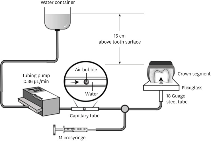
ePub Objectives This study evaluated the effects of different smear layer preparations on the dentin permeability and microtensile bond strength (µTBS) of 2 self-etching adhesives (Clearfil SE Bond [CSE] and Clearfil Tri-S Bond Universal [CTS]) under dynamic pulpal pressure.
Materials and Methods Human third molars were cut into crown segments. The dentin surfaces were prepared using 4 armamentaria: 600-grit SiC paper, coarse diamond burs, superfine diamond burs, and carbide burs. The pulp chamber of each crown segment was connected to a dynamic intra-pulpal pressure simulation apparatus, and the permeability test was done under a pressure of 15 cmH2O. The relative permeability (%P) was evaluated on the smear layer-covered and bonded dentin surfaces. The teeth were bonded to either of the adhesives under pulpal pressure simulation, and cut into sticks after 24 hours water storage for the µTBS test. The resin-dentin interface and nanoleakage observations were performed using a scanning electron microscope. Statistical comparisons were done using analysis of variance and
post hoc tests.Results Only the method of surface preparation had a significant effect on permeability (
p < 0.05). The smear layers created by the carbide and superfine diamond burs yielded the lowest permeability. CSE demonstrated a higher µTBS, with these values in the superfine diamond and carbide bur groups being the highest. Microscopic evaluation of the resin-dentin interface revealed nanoleakage in the coarse diamond bur and SiC paper groups for both adhesives.Conclusions Superfine diamond and carbide burs can be recommended for dentin preparation with the use of 2-step CSE.
-
Citations
Citations to this article as recorded by- The effect of different adhesive strategies and diamond burs on dentin bond strength of universal resin cements
Chavakorn Atsavathavornset, Pipop Saikaew, Choltacha Harnirattisai, Hidehiko Sano
Clinical Oral Investigations.2025;[Epub] CrossRef - Universal adhesive systems in dentistry: A narrative review
Svetlana N. Razumova, Anzhela S. Brago, Oxana R. Ruda, Zoya A. Guryeva, Elvira V. Adzhieva
Russian Journal of Dentistry.2024; 28(5): 512. CrossRef - Delayed light activation of resin composite affects the bond strength of adhesives under dynamic simulated pulpal pressure
Nattaporn Sukprasert, Choltacha Harnirattisai, Pisol Senawongse, Hidehiko Sano, Pipop Saikaew
Clinical Oral Investigations.2022; 26(11): 6743. CrossRef
- The effect of different adhesive strategies and diamond burs on dentin bond strength of universal resin cements
- 2,872 View
- 42 Download
- 2 Web of Science
- 3 Crossref

- Adhesive systems applied to dentin substrate under electric current: systematic review
- Carolina Menezes Maciel, Tatiane Cristina Vieira Souto, Bárbara de Almeida Pinto, Laís Regiane Silva-Concilio, Kusai Baroudi, Rafael Pino Vitti
- Restor Dent Endod 2021;46(4):e55. Published online November 5, 2021
- DOI: https://doi.org/10.5395/rde.2021.46.e55
-
 Abstract
 PDF
PDF PubReader
 ePub
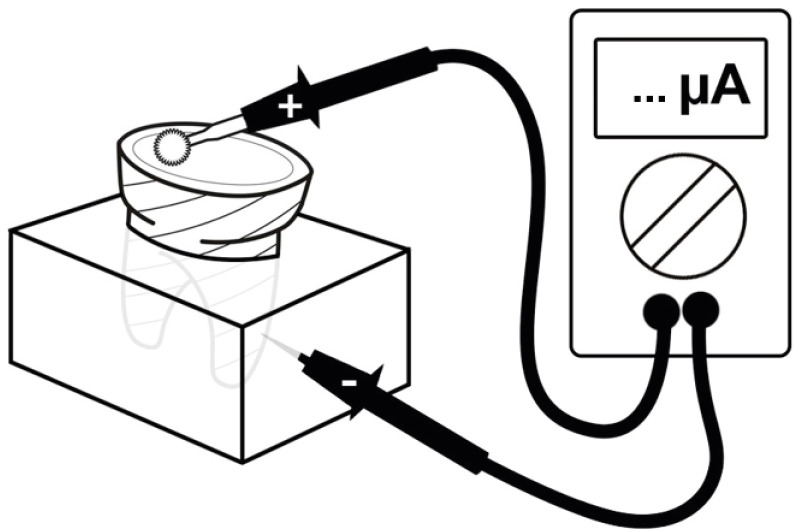
ePub Objectives The purpose of this systematic review was to collect and discuss the technique of adhesive systems application on dentin substrate under electric current.
Materials and Methods The first search strategy was based on data available at PubMed, LILACS, Scielo, Scopus, and Cochrane Library, using a combination of descriptors such as “dentin bond agents OR adhesive system AND electric current OR electrobond” or “dentin bonding agents OR dentin bonding agent application OR adhesive system AND electric current OR electrobond”, with no limit regarding the publication year. The second search strategy was based on the articles' references found previously. An additional search strategy was applied that concerned the proposed theme in the SBU-UNICAMP (Unicamp's Library System Institutional Repository).
Results Twelve studies published between 2006 and 2020 were found. The analyses of the selected studies showed that the use of electric current during adhesive systems application on dentin, whether conventional or self-conditioning, increases resinous monomer infiltration in the dentin substrate, which improves the hybridization processes and the bond strength of the restorative material to dentin.
Conclusions Despite the favorable results related to the use of this technique, there is still no specific protocol for the application of adhesive systems under electric current.
-
Citations
Citations to this article as recorded by- Advances in Resin-Dentin Bonding: Evaluating Pre-Treatment Techniques for Improved Adhesion
Rim Bourgi
Journal of Dental Health and Oral Research.2025; : 1. CrossRef - Iontophoresis effects of two-step self-etch and total-etch systems on dentin permeability and sealing of composite restoration under simulated pulpal pressure
Orapin Ajcharanukul, Peeraya Santikulluk, Palat Sasingha, Sirithorn Sabpawat, Kanokporn Sukyanan
BMC Oral Health.2022;[Epub] CrossRef
- Advances in Resin-Dentin Bonding: Evaluating Pre-Treatment Techniques for Improved Adhesion
- 1,849 View
- 14 Download
- 1 Web of Science
- 2 Crossref

- Effect of adhesive application method on repair bond strength of composite
- Hee Kyeong Oh, Dong Hoon Shin
- Restor Dent Endod 2021;46(3):e32. Published online June 4, 2021
- DOI: https://doi.org/10.5395/rde.2021.46.e32
-
 Abstract
 PDF
PDF PubReader
 ePub
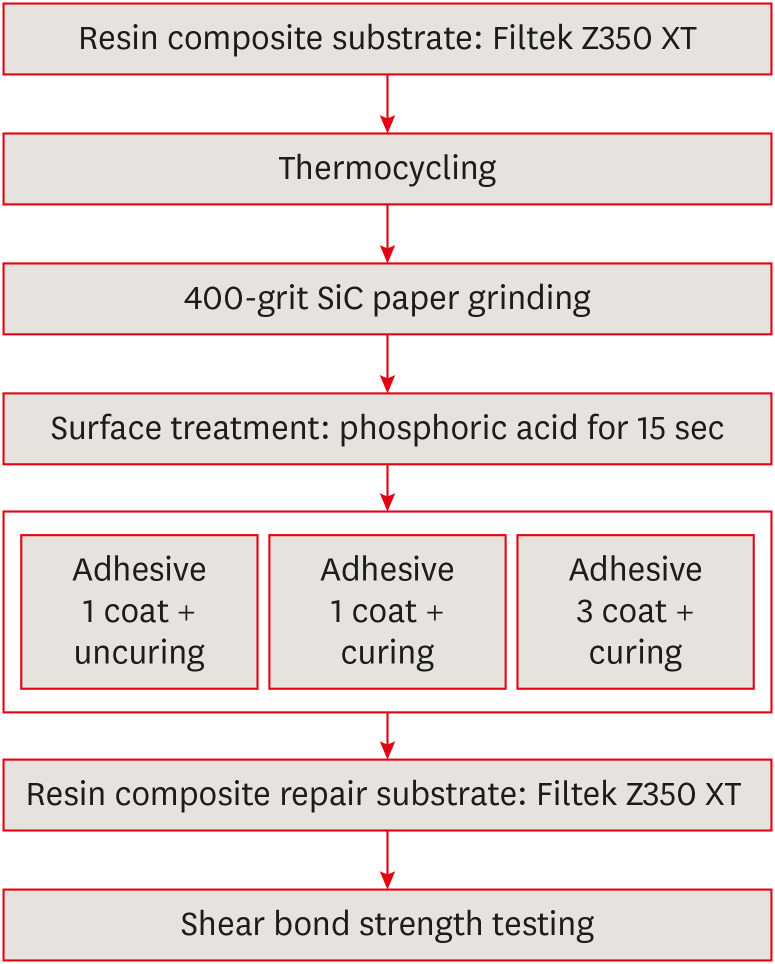
ePub Objectives This study aimed to evaluate the effect of the application method of universal adhesives on the shear bond strength (SBS) of repaired composites, applied with different thicknesses.
Materials and Methods The 84 specimens (Filtek Z350 XT) were prepared, stored in distilled water for a week and thermocycled (5,000 cycles, 5°C to 55°C). They were roughened using 400-grit sandpapers and etched with phosphoric acid. Then, specimens were equally divided into 2 groups; Single Bond Universal (SU) and Prime&Bond Universal (PB). Each group was subdivided into 3 subgroups according to application methods (
n = 14); UC: 1 coat + uncuring, 1C: 1 coat + curing, 3C: 3 coats + curing. After storage of the repaired composite for 24 hours, specimens were subjected to the SBS test and the data were statistically analyzed by 2-way analysis of variance and independentt -tests. Specimens were examined with a stereomicroscope to analyze fracture mode and a scanning electron microscope to observe the interface.Results Adhesive material was a significant factor (
p = 0.001). Bond strengths with SU were higher than PB. The highest strength was obtained from the 1C group with SU. Bonding in multiple layers increased adhesive thicknesses, but there was no significant difference in SBS values (p = 0.255). Failure mode was predominantly cohesive in old composites.Conclusions The application of an adequate bonding system plays an important role in repairing composite resin. SU showed higher SBS than PB and the additional layers increased the adhesive thickness without affecting SBS.
-
Citations
Citations to this article as recorded by- The effect of different surface treatments and adhesive systems on shear bond strength in universal nanohybrid composite resin repair
Merve Kütük Ömeroğlu, Melek Çam, Işıl Doğruer, Zeynep Buket Kaynar
BMC Oral Health.2025;[Epub] CrossRef - Effect of Universal Adhesive Etching Mode on Shear Bond Strength of Pulp Capping Materials to Deep Dentin
Shahram Amirifar, Saba Tohidkhah, Seyedeh Mahsa Sheikh-Al-Eslamian, Mahdi Abbasi, Fatemeh Farshad, Elham Ahmadi, Carlos M. Ardila
BioMed Research International.2025;[Epub] CrossRef - Shear Bond Strength and Finite Element Stress Analysis of Composite Repair Using Various Adhesive Strategies With and Without Silane Application
Elif Ercan Devrimci, Hande Kemaloglu, Cem Peskersoy, Tijen Pamir, Murat Turkun
Applied Sciences.2025; 15(15): 8159. CrossRef
- The effect of different surface treatments and adhesive systems on shear bond strength in universal nanohybrid composite resin repair
- 3,604 View
- 24 Download
- 3 Web of Science
- 3 Crossref

- Microleakage and characteristics of resin-tooth tissues interface of a self-etch and an etch-and-rinse adhesive systems
- Xuan Vinh Tran, Khanh Quang Tran
- Restor Dent Endod 2021;46(2):e30. Published online May 18, 2021
- DOI: https://doi.org/10.5395/rde.2021.46.e30
-
 Abstract
 PDF
PDF PubReader
 ePub
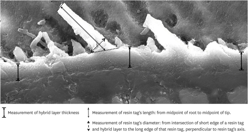
ePub Objectives This study was conducted to compare the microleakage and characteristics of the resin-tooth tissue interface between self-etch and etch-and-rinse adhesive systems after 48 hours and 3 months.
Materials and Methods 40 extracted premolar teeth were randomly divided into 2 groups: 1-step self-etch adhesive system – Optibond™ All-In-One, and 2-step etch-and-rinse adhesive system - Adper™ Single Bond 2. Both groups were subjected to 500 thermocycles (5°C–55°C) before scanning electron microscope (SEM) analysis or microleakage trial at 48-hour and 3-month time periods.
Results SEM images showed the hybrid layer thickness, diameter, and length of resin tags of the self-etch adhesive (0.42 ± 0.14 µm; 1.49 ± 0.45 µm; 16.35 ± 14.26 µm) were smaller than those of the etch-and-rinse adhesive (4.39 ± 1.52 µm; 3.49 ± 1 µm; 52.81 ± 35.81 µm). In dentin, the microleakage scores of the 2 adhesives were not different in both time periods (48 hours/3 months). However, the microleakage score of etch-and-rinse adhesive increased significantly after 3 months (0.8 ± 0.63 and 1.9 ± 0.88,
p < 0.05).Conclusions The self-etch adhesive exhibited better long-term sealing ability in dentin when compared to that of the etch-and-rinse adhesive. The greater hybrid layer thickness and dimensions of resin tags did not guarantee reliable, long-lasting sealing in the bonding area.
-
Citations
Citations to this article as recorded by- Efficacy of different adhesive systems in bonding direct resin composite restorations: a systematic review and meta-analysis
Ravinder S. Saini, Rajesh Vyas, Sunil Kumar Vaddamanu, Syed Altafuddin Quadri, Seyed Ali Mosaddad, Artak Heboyan
Evidence-Based Dentistry.2025; 26(2): 115. CrossRef - Characterisation of universal adhesive bonded resin-dentin interface after focused ultrasound smear layer conditioning
Cheryl Fu, Peta L. Clode, Amr S. Fawzy
International Journal of Adhesion and Adhesives.2025; 142: 104115. CrossRef - Effect of Dentin Pretreatment With Dimethyl Sulfoxide Solution on Interfacial Fracture Toughness of Composite Resin to Wet and Dry Dentin
Fatemeh Molaei, Mehrsima Ghavami-Lahiji, Seyedeh Maryam Tavangar, Hannah Wesley
International Journal of Dentistry.2025;[Epub] CrossRef - Resin tags formation by modified Renewal MI formulations in a carious dentine model
Nabih Alkhouri, Wendy Xia, Paul Ashley, Anne Young
Frontiers in Oral Health.2024;[Epub] CrossRef - Effect of propolis added to single‐bottle adhesives on water permeation through the hybrid layer
Lucineide Silva da Rocha, Daniela Ferreira de Oliveira, Cinthya Luna Veloso de Lima, Ticiano Gomes do Nascimento, Johnnatan Duarte de Freitas, Jeniffer Mclaine Duarte de Freitas, Isabel Cristina Celerino de Moraes Porto
European Journal of Oral Sciences.2024;[Epub] CrossRef - Exploration and preliminary clinical investigation of an adhesive approach for primary tooth restoration
Xiangqin Xu, Jiansheng Zhu, May Lei Mei, Huaying Wu, Kaipeng Xie, Shoulin Wang, Yaming Chen
The Journal of Biomedical Research.2023; 37(2): 138. CrossRef - Adhesion to enamel and dentine: an update
Rana Alkattan
Primary Dental Journal.2023; 12(3): 33. CrossRef - Effects of carbodiimide combined with ethanol–wet bonding pretreatment on dentin bonding properties: an in vitro study
Xiaoxiao You, Long Chen, Jie Xu, Sihui Li, Zhenghao Zhang, Ling Guo
PeerJ.2022; 10: e14238. CrossRef - The effects of amalgam contamination and different surface modifications on microleakage of dentin bonded to bulk fill composite when using different adhesive protocols
Nojoud Alshehri, Abdullah Aljamhan, Mohammed Bin-Shuwaish
BMC Oral Health.2022;[Epub] CrossRef - Development of low-shrinkage dental adhesives via blending with spiroorthocarbonate expanding monomer and unsaturated epoxy resin monomer
Zonghua Wang, Xiaoran Zhang, Shuo Yao, Jiaxin Zhao, Chuanjian Zhou, Junling Wu
Journal of the Mechanical Behavior of Biomedical Materials.2022; 133: 105308. CrossRef - Influence of silver nanoparticles on the resin-dentin bond strength and antibacterial activity of a self-etch adhesive system
Jia Wang, Wei Jiang, Jingping Liang, Shujun Ran
The Journal of Prosthetic Dentistry.2022; 128(6): 1363.e1. CrossRef
- Efficacy of different adhesive systems in bonding direct resin composite restorations: a systematic review and meta-analysis
- 2,363 View
- 36 Download
- 10 Web of Science
- 11 Crossref

- Interface between calcium silicate cement and adhesive systems according to adhesive families and cement maturation
- Nelly Pradelle-Plasse, Caroline Mocquot, Katherine Semennikova, Pierre Colon, Brigitte Grosgogeat
- Restor Dent Endod 2021;46(1):e3. Published online December 9, 2020
- DOI: https://doi.org/10.5395/rde.2021.46.e3
-
 Abstract
 PDF
PDF PubReader
 ePub
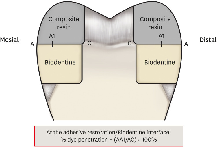
ePub Objectives This study aimed to evaluate the interface between a calcium silicate cement (CSC), Biodentine and dental adhesives in terms of sealing ability.
Materials and Methods Microleakage test: 160 standardized class II cavities were prepared on 80 extracted human molars. The cavities were filled with Biodentine and then divided into 2 experimental groups according to the time of restoration: composite resin obturation 15 minutes after Biodentine handling (D0); restoration after 7 days (D7). Each group was then divided into 8 subgroups (
n = 5) according to the adhesive system used: etch-and-rinse adhesive (Prime & Bond); self-etch adhesive 2 steps (Optibond XTR and Clearfil SE Bond); self-etch adhesive 1 step (Xeno III, G-aenial Bond, and Clearfil Tri-S Bond); and universal used as etch-and-rinse or self-etch (ScotchBond Universal ER or SE). After thermocycling, the teeth were immersed in a silver nitrate solution, stained, longitudinally sectioned, and the Biodentine/adhesive percolation was quantified. Scanning electron microscopic observations: Biodentine/adhesive interfaces were observed.Results A tendency towards less microleakage was observed when Biodentine was etched (2.47%) and when restorations were done without delay (D0: 4.31%, D7: 6.78%), but this was not significant. The adhesives containing 10-methacryloyloxydecyl dihydrogen phosphate monomer showed the most stable results at both times studied. All Biodentine/adhesive interfaces were homogeneous and regular.
Conclusions The good sealing of the CSC/adhesive interface is not a function of the system adhesive family used or the cement maturation before restoration. Biodentine can be used as a dentine substitute.
-
Citations
Citations to this article as recorded by- Comparison of compressive strength, surface microhardness, and surface microstructure of different types of bioceramics following varying surface treatments
Zeynep Hale Keleş, Vasfiye Işık, Soner Sismanoglu
Journal of the Australian Ceramic Society.2025;[Epub] CrossRef - Effect of Er Cr YSGG laser etching procedure on the bond strength of different calcium silicate cements
Yesim Sesen Uslu, Hakan Yasin Gönder, Pinar Sesen, Gizem Gunduz Bektaş
Lasers in Dental Science.2024;[Epub] CrossRef - Managing Cracked Teeth with Root Extension: A Prospective Preliminary Study Using Biodentine™ Material
Kênia Maria Soares de Toubes, Isabella Sousa Corrêa, Regina Célia Lopes Valadares, Stephanie Quadros Tonelli, Fábio Fernandes Borém Bruzinga, Frank Ferreira Silveira, Dr Karthikeyan Ramalingam
International Journal of Dentistry.2024;[Epub] CrossRef - In Vitro Resistance of Natural Molars vs. Additive-Manufactured Simulators Treated with Pulpotomy and Endocrown
Marie-Laure Munoz-Sanchez, Alexis Gravier, Olivier Francois, Emmanuel Nicolas, Martine Hennequin, Nicolas Decerle
Journal of Functional Biomaterials.2023; 14(9): 444. CrossRef - Characterisation of the calcium silicate‐based cement–composite interface and the bonding strength with total‐etch or single/two‐stage self‐etch adhesive systems
Abidin Talha Mutluay, Merve Mutluay
Australian Endodontic Journal.2022; 48(3): 501. CrossRef - Bond Strength of Adhesive Systems to Calcium Silicate-Based Materials: A Systematic Review and Meta-Analysis of In Vitro Studies
Louis Hardan, Davide Mancino, Rim Bourgi, Alejandra Alvarado-Orozco, Laura Emma Rodríguez-Vilchis, Abigailt Flores-Ledesma, Carlos Enrique Cuevas-Suárez, Monika Lukomska-Szymanska, Ammar Eid, Maya-Line Danhache, Maryline Minoux, Youssef Haïkel, Naji Kharo
Gels.2022; 8(5): 311. CrossRef
- Comparison of compressive strength, surface microhardness, and surface microstructure of different types of bioceramics following varying surface treatments
- 2,512 View
- 43 Download
- 6 Web of Science
- 6 Crossref

- Bonding of a resin-modified glass ionomer cement to dentin using universal adhesives
- Muhittin Ugurlu
- Restor Dent Endod 2020;45(3):e36. Published online June 15, 2020
- DOI: https://doi.org/10.5395/rde.2020.45.e36
-
 Abstract
 PDF
PDF PubReader
 ePub
ePub Objectives This study aims to assess the effect of universal adhesives pretreatment on the bond strength of resin-modified glass ionomer cement to dentin.
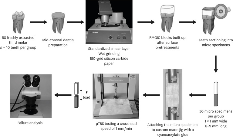
Materials and Methods Fifty caries-free human third molars were employed. The teeth were randomly assigned into five groups (
n = 10) based on dentin surface pretreatments: Single Bond Universal (3M Oral Care), Gluma Bond Universal (Heraeus Kulzer), Prime&Bond Elect (Dentsply), Cavity Conditioner (GC) and control (no surface treatment). After Fuji II LC (GC) was bonded to the dentin surfaces, the specimens were stored for 7 days at 37°C. The specimens were segmented into microspecimens, and the microspecimens were subjugated to microtensile bond strength testing (1.0 mm/min). The modes of failure analyzed using a stereomicroscope and scanning electron microscopy. Data were statistically analyzed with one-way analysis of variance and Duncan tests (p = 0.05).Results The surface pretreatments with the universal adhesives and conditioner increased the bond strength of Fuji II LC to dentin (
p < 0.05). Single Bond Universal and Gluma Bond Universal provided higher bond strength to Fuji II LC than Cavity Conditioner (p < 0.05). The bond strengths obtained from Prime&Bond Elect and Cavity Conditioner were not statistically different (p > 0.05).Conclusions The universal adhesives and polyacrylic acid conditioner could increase the bond strength of resin-modified glass ionomer cement (RMGIC) to dentin. The use of universal adhesives before the application of RMGIC may be more beneficial in improving bond strength.
-
Citations
Citations to this article as recorded by- Impact of nanochitosan incorporation on the performance of resin-modified glass ionomer luting cement: a comprehensive in vitro study
Mostafa A. Abdelshafi, Nesma Elgohary, Ahmed Shams
BMC Oral Health.2026;[Epub] CrossRef - Clinical evaluation of giomer-based injectable resin composite versus resin-modified glass ionomer in class V carious lesions over 18 months: A randomized clinical trial
Reham Hendam, Rania Mosallam, Dina Kamal
Journal of Conservative Dentistry and Endodontics.2025; 28(1): 50. CrossRef - Push-Out Bond Strength of Different Luting Cements Following Post Space Irrigation with 2% Chitosan: An In Vitro Study
Shimaa Rifaat, Ahmed Rahoma, Hind Muneer Alharbi, Sawsan Jamal Kazim, Shrouq Ali Aljuaid, Basmah Omar Alakloby, Faraz A. Farooqi, Noha Taymour
Prosthesis.2025; 7(1): 18. CrossRef - Bioactive restorative materials in dentistry: a comprehensive review of mechanisms, clinical applications, and future directions
Dina Abozaid, Amr Azab, Mohammad A. Bahnsawy, Mohamed Eldebawy, Abdullah Ayad, Romesa soomro, Enas Elwakeel, Maged Ahmed Mohamed
Odontology.2025;[Epub] CrossRef - A Comparative Evaluation of Marginal Leakage and Shear Bond Strength of Cention N, Resin-Modified Glass Ionomer Cement (RMGIC), and Conventional Glass Ionomer Cement (GIC): An In Vitro Study
Khushboo Singh, Debapriya Pradhan, Saurabh Tiwari, Raksha Thakur, Priyamvada Sharma, Devika Agrawal, Mahima Singh, Devshree Jawalikar, Delphina Michael Kapoor, Jyoti Priiya Kodimela
Cureus.2025;[Epub] CrossRef - Assessment of Nanosilver Fluoride Application on the Microtensile Bond Strength of Glass Ionomer Cement and Resin-modified Glass Ionomer Cement on Primary Carious Dentin: An In Vitro Study
Ila Srinivasan, Yuthi Milit, Anushka Das, Neeraja Ramamurthy
International Journal of Clinical Pediatric Dentistry.2024; 17(5): 565. CrossRef - Effect of Surface Treatments on Shear-bond Strength of Glass Ionomer Cements to Silver Diamine Fluoride-treated Simulated Carious Dentin
WT Koh, OT Yeoh, NA Yahya, AU Yap
Operative Dentistry.2024; 49(6): 714. CrossRef - Desensitizing agents’ post-bleaching effect on orthodontic bracket bond strength
Gufa Bagus Pamungkas, Dyah Karunia, Sri Suparwitri
Dental Journal.2024; 57(1): 45. CrossRef - Successful Rehabilitation of Traumatized Immature Teeth by Different Vital Pulp Therapies in Pediatric Patients
Mohammad Kamran Khan
Journal of the Scientific Society.2023; 50(1): 111. CrossRef - Do bioactive materials show greater retention rates in restoring permanent teeth than non-bioactive materials? A systematic review and network meta-analysis of randomized controlled trials
Juliana Benace Fernandes, Sheila Mondragón Contreras, Manuela da Silva Spinola, Graziela Ribeiro Batista, Eduardo Bresciani, Taciana Marco Ferraz Caneppele
Clinical Oral Investigations.2023;[Epub] CrossRef - Effects of tooth preparation on the microleakage of fissure sealant
Gesti Kartiko Sari, Sri Kuswandari, Putri Kusuma Wardani Mahendra
Dental Journal (Majalah Kedokteran Gigi).2022; 55(2): 67. CrossRef - Rheological Properties, Surface Microhardness, and Dentin Shear Bond Strength of Resin-Modified Glass Ionomer Cements Containing Methacrylate-Functionalized Polyacids and Spherical Pre-Reacted Glass Fillers
Whithipa Thepveera, Wisitsin Potiprapanpong, Arnit Toneluck, Somruethai Channasanon, Chutikarn Khamsuk, Naruporn Monmaturapoj, Siriporn Tanodekaew, Piyaphong Panpisut
Journal of Functional Biomaterials.2021; 12(3): 42. CrossRef
- Impact of nanochitosan incorporation on the performance of resin-modified glass ionomer luting cement: a comprehensive in vitro study
- 4,040 View
- 36 Download
- 12 Crossref

- The influence of nanofillers on the properties of ethanol-solvated and non-solvated dental adhesives
- Leonardo Bairrada Tavares da Cruz, Marcelo Tavares Oliveira, Cintia Helena Coury Saraceni, Adriano Fonseca Lima
- Restor Dent Endod 2019;44(3):e28. Published online July 24, 2019
- DOI: https://doi.org/10.5395/rde.2019.44.e28
-
 Abstract
 PDF
PDF PubReader
 ePub
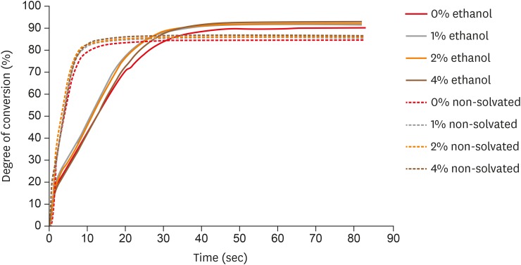
ePub Objectives The aim of this study was to evaluate the influence of different concentrations of nanofillers on the chemical and physical properties of ethanol-solvated and non-solvated dental adhesives.
Materials and Methods Eight experimental adhesives were prepared with different nanofiller concentrations (0, 1, 2, and 4 wt%) and 2 solvent concentrations (0% and 10% ethanol). Several properties of the experimental adhesives were evaluated, such as water sorption and solubility (
n = 5, 20 seconds light activation), real-time degree of conversion (DC;n = 3, 20 and 40 seconds light activation), and stability of cohesive strength at 6 months (CS;n = 20, 20 seconds light activation) using the microtensile test. A light-emitting diode (Bluephase 20i, Ivoclar Vivadent) with an average light emittance of 1,200 mW/cm2 was used.Results The presence of solvent reduced the DC after 20 seconds of curing, but increased the final DC, water sorption, and solubility of the adhesives. Storage in water reduced the strength of the adhesives. The addition of 1 wt% and 2 wt% nanofillers increased the polymerization rate of the adhesives.
Conclusions The presence of nanofillers and ethanol improved the final DC, although the DC of the solvated adhesives at 20 seconds was lower than that of the non-solvated adhesives. The presence of ethanol reduced the strength of the adhesives and increased their water sorption and solubility. However, nanofillers did not affect the water sorption and strength of the tested adhesives.
-
Citations
Citations to this article as recorded by- Effect of boric acid on the color stability and mechanical properties of 3D-printed permanent resins
Dalndushe Abdulai, Rafat Sasany, Raghib Suradi, Mehran Moghbel, Seyed Ali Mosaddad
Scientific Reports.2025;[Epub] CrossRef - Development of a Boron Nitride-Filled Dental Adhesive System
Senthilguru Kulanthaivel, Jeremiah Poppen, Sandra Ribeiro Cunha, Benjamin Furman, Kyumin Whang, Erica C. Teixeira
Polymers.2023; 15(17): 3512. CrossRef - Analyses of Experimental Dental Adhesives Based on Zirconia/Silver Phosphate Nanoparticles
Abdul Khan, Yasmin Alhamdan, Hala Alibrahim, Khalid Almulhim, Muhammad Nawaz, Syed Ahmed, Khalid Aljuaid, Ijlal Ateeq, Sultan Akhtar, Mohammad Ansari, Intisar Siddiqui
Polymers.2023; 15(12): 2614. CrossRef - Mechanical characterization and adhesive properties of a dental adhesive modified with a polymer antibiotic conjugate
Camila Sabatini, Russell J. Aguilar, Ziwen Zhang, Steven Makowka, Abhishek Kumar, Megan M. Jones, Michelle B. Visser, Mark Swihart, Chong Cheng
Journal of the Mechanical Behavior of Biomedical Materials.2022; 129: 105153. CrossRef
- Effect of boric acid on the color stability and mechanical properties of 3D-printed permanent resins
- 1,315 View
- 8 Download
- 4 Crossref

- Influence of different universal adhesives on the repair performance of hybrid CAD-CAM materials
- Gülbike Demirel, İsmail Hakkı Baltacıoğlu
- Restor Dent Endod 2019;44(3):e23. Published online May 20, 2019
- DOI: https://doi.org/10.5395/rde.2019.44.e23
-
 Abstract
 PDF
PDF PubReader
 ePub
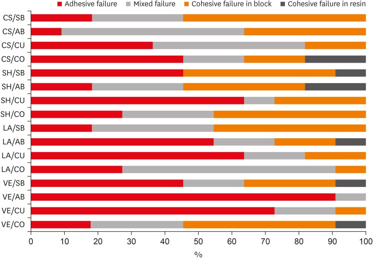
ePub Objectives The aim of this study was to investigate the microshear bond strength (μSBS) of different universal adhesive systems applied to hybrid computer-aided design/computer-aided manufacturing (CAD-CAM) restorative materials repaired with a composite resin.
Materials and Methods Four types of CAD-CAM hybrid block materials—Lava Ultimate (LA), Vita Enamic (VE), CeraSmart (CS), and Shofu Block HC (SH)—were used in this study, in combination with the following four adhesive protocols: 1) control: porcelain primer + total etch adhesive (CO), 2) Single Bond Universal (SB), 3) All Bond Universal (AB), and 4) Clearfil Universal Bond (CU). The μSBS of the composite resin (Clearfil Majesty Esthetic) was measured and the data were analyzed using two-way analysis of variance and the Tukey test, with the level of significance set at
p < 0.05.Results The CAD-CAM block type and block-adhesive combination had significant effects on the bond strength values (
p < 0.05). Significant differences were found between the following pairs of groups: VE/CO and VE/AB, CS/CO and CS/AB, VE/CU and CS/CU, and VE/AB and CS/AB (p < 0.05).Conclusions The μSBS values were affected by hybrid block type. All tested universal adhesive treatments can be used as an alternative to the control treatment for repair, except the AB system on VE blocks (the VE/AB group). The μSBS values showed variation across different adhesive treatments on different hybrid CAD-CAM block types.
-
Citations
Citations to this article as recorded by- Effect of surface treatments on the bond strength of resin-repaired resin matrix CAD-CAM ceramic: A scoping review
Ana Beatriz de Souza Albergardi, João Pedro Justino de Oliveira Limírio, Jéssica Marcela de Luna Gomes, Aldiéris Alves Pesqueira, Eduardo Piza Pellizzer
Journal of Dentistry.2025; 154: 105594. CrossRef - Bond strength to aged CAD/CAM composites and polymer-infiltrated ceramic network using a universal adhesive with or without previous application of a universal primer
Clemens Lechte, Lisa Sophia Faesser, Jana Biermann, Alexandra Schmidt, Philipp Kanzow, Annette Wiegand
International Journal of Adhesion and Adhesives.2025; 140: 104017. CrossRef - The Influence of Thermocycling and Ultraviolet Aging on Surface Characteristics and the Repair Bond Strength of CAD/CAM Resin Nanoceramics
Beyza Unalan Degirmenci, Alperen Degirmenci, Zelal Seyfioglu Polat
Journal of Functional Biomaterials.2025; 16(5): 156. CrossRef - Impact of in vitro findings on clinical protocols for the adhesion of CAD-CAM blocks: A systematic integrative review and meta-analysis
Maria João Calheiros-Lobo, Ricardo Carbas, Lucas F.M. da Silva, Teresa Pinho
The Journal of Prosthetic Dentistry.2024; 131(6): 1051. CrossRef - Repair protocols for indirect monolithic restorations: a literature review
Lucas Saldanha da Rosa, Rafaela Oliveira Pilecco, Pablo Machado Soares, Marília Pivetta Rippe, Gabriel Kalil Rocha Pereira, Luiz Felipe Valandro, Cornelis Johannes Kleverlaan, Albert J. Feilzer, João Paulo Mendes Tribst
PeerJ.2024; 12: e16942. CrossRef - Bonding performance of universal adhesives with concomitant use of silanes to CAD/CAM blocks
Marina AMARAL, Jaqueline Maria Brandão RIZZATO, Victoria Caroline Souza de ALMEIDA, Priscila Christiane Suzy LIPORONI, Rayssa Ferreira ZANATTA
RGO - Revista Gaúcha de Odontologia.2023;[Epub] CrossRef - Resistencia a la fractura de una nanocerámica CAD/CAM reparada con dos tratamientos de superficie: estudio in vitro
Marcelo Geovanny Cascante-Calderón, Kevin Alejandro Reascos Flores, Inés María Villacís-Altamirano, Anggely Maite Bayas Salinas, Jessica Elizabeth Taraguay Galindo
Universitas Odontologica.2023;[Epub] CrossRef - Influence of surface treatments and adhesive protocols on repair bond strength of glass‐matrix and resin‐matrix CAD/CAM ceramics
Rana Turunç‐Oğuzman, Soner Şişmanoğlu
Journal of Esthetic and Restorative Dentistry.2023; 35(8): 1322. CrossRef - Effect of Anti-COVID-19 Mouthwashes on Shear Bond Strength of Resin-Matrix Ceramics Repaired with Resin Composite Using Universal Adhesive: An In Vitro Study
Wichuda Limsiriwong, Awiruth Klaisiri, Nantawan Krajangta
Journal of Functional Biomaterials.2023; 14(3): 158. CrossRef - Effect of ceramic primers with different chemical contents on the shear bond strength of CAD/CAM ceramics with resin cement after thermal ageing
Mehmet Uğur, İdris Kavut, Özgür Ozan Tanrıkut, Önder Cengiz
BMC Oral Health.2023;[Epub] CrossRef - Effect of microstructure of reinforced CAD/CAM hybrid composite resin block on shear bond strength of composite resin
Sung-Ho Um, Minjeong Shin, Shin-hye Chung, Young-Seok Park, Bum-Soon Lim
Korean Journal of Dental Materials.2023; 50(1): 29. CrossRef - Dentin contamination during repair procedures: A threat to universal adhesives?
Anne‐Katrin Lührs, Cosima Brachmann, Silke Jacker‐Guhr
Clinical and Experimental Dental Research.2022; 8(3): 771. CrossRef - Influence of mechanical and chemical pre-treatments on the repair of a hybrid ceramic
Sascha Niklas Jung, Stefan Rüttermann
Dental Materials.2022; 38(7): 1140. CrossRef - Influence of different repair protocols and artificial aging on bond strength of composite to a CAD/CAM polymer-infiltrated ceramic
Ece İrem OĞUZ, Gökhan ÇİÇEKCİ
Cumhuriyet Dental Journal.2021; 24(1): 37. CrossRef - REZİN MATRİKS SERAMİKLER-DERLEME
Elif Melike AKARCA, Dilara ŞAHİN, Ragibe Şenay CANAY
Atatürk Üniversitesi Diş Hekimliği Fakültesi Dergisi.2021; : 1. CrossRef - REZİN MATRİKS SERAMİKLER-DERLEME
Elif Melike AKARCA, Dilara ŞAHİN, Ragibe Şenay CANAY
Atatürk Üniversitesi Diş Hekimliği Fakültesi Dergisi.2021; : 1. CrossRef - Microshear bond strength of contemporary self-adhesive resin cements to CAD/CAM restorative materials: effect of surface treatment and aging
Soner Şişmanoğlu, Rana Turunç-Oğuzman
Journal of Adhesion Science and Technology.2020; 34(22): 2484. CrossRef - Influence of different surface treatments and universal adhesives on the repair of CAD-CAM composite resins: An in vitro study
Soner Sismanoglu, Zuhal Yildirim-Bilmez, Aysegul Erten-Taysi, Pınar Ercal
The Journal of Prosthetic Dentistry.2020; 124(2): 238.e1. CrossRef
- Effect of surface treatments on the bond strength of resin-repaired resin matrix CAD-CAM ceramic: A scoping review
- 2,136 View
- 12 Download
- 18 Crossref

- Influence of silver nanoparticles on resin-dentin bond strength durability in a self-etch and an etch-and-rinse adhesive system
- Zahra Jowkar, Fereshteh Shafiei, Elham Asadmanesh, Fatemeh Koohpeima
- Restor Dent Endod 2019;44(2):e13. Published online March 29, 2019
- DOI: https://doi.org/10.5395/rde.2019.44.e13
-
 Abstract
 PDF
PDF PubReader
 ePub
ePub Objectives This study evaluated the effect of dentin pretreatment with silver nanoparticles (SNPs) and chlorhexidine (CHX) on the microshear bond strength (µSBS) durability of different adhesives to dentin.
Materials and Methods Occlusal surfaces of 120 human molars were ground to expose flat dentin surfaces. The specimens were randomly assigned to six groups (
n = 20). Three groups (A, B, and C) were bonded with Adper Single Bond 2 (SB) and the other groups (D, E, and F) were bonded with Clearfil SE Bond (SEB). Dentin was pretreated with CHX in groups B and E, and with SNPs in groups C and F. The specimens were restored with Z250 composite. Half of the bonded surfaces in each group underwent µSBS testing after 24 hours and the other half was tested after 6 months of water storage.Results SNP application was associated with a higher µSBS than was observed in the CHX and control groups for SEB after 24 hours (
p < 0.05). A significantly lower µSBS was observed when no dentin pretreatment was applied compared to dentin pretreatment with CHX and SNPs for SB after 24 hours (p < 0.05). The µSBS values of the 6-month specimens were significantly lower than those obtained from the 24-hour specimens for all groups (p < 0.05). This decrease was much more pronounced when both adhesives were used without any dentin pretreatment (p < 0.05).Conclusions SNPs and CHX reduced the degradation of resin-dentin bonds over a 6-month period for both adhesive systems.
-
Citations
Citations to this article as recorded by- An in vitro comparative evaluation of silver and chitosan nanoparticles on shear bond strength of nanohybrid composite using different adhesion protocols
Roopadevi Garlapati, Nagesh Bolla, Mayana Aameena Banu, Anila Bandlapally Sreenivasa Guptha, Niharika Halder, Ram Chowdary Basam
Journal of Conservative Dentistry and Endodontics.2025; 28(6): 522. CrossRef - Nanoparticle-enhanced dental adhesives: improving dentin bond strength through multifunctional nanotechnology
Suleiman Ibrahim Mohammad, Asokan Vasudevan, Lashin Saad Ali, Wenchang Chen
The Journal of Adhesion.2025; : 1. CrossRef - The Effect of Silver Nanoparticles on Bond Strength of Calcium Silicate-Based Sealer: An In Vitro Study
Sundus Bukhary, Sarah Alkahtany, Dalal AlDabeeb
Applied Sciences.2024; 14(21): 9817. CrossRef - Performance of self-etching adhesives on caries-affected primary dentin treated with glutaraldehyde or silver diamine fluoride
Marcelly Tupan Christoffoli Wolowski, Andressa Mioto Stabile Grenier, Victória Alícia de Oliveira, Caroline Anselmi, Mariana Sversut Gibin, Lidiane Vizioli de Castro-Hoshino, Francielle Sato, Cristina Perez, Régis Henke Scheffel, Josimeri Hebling, Mauro L
Journal of the Mechanical Behavior of Biomedical Materials.2024; 150: 106293. CrossRef - The Impact of Silver Nanoparticles on Dentinal Tubule Penetration of Endodontic Bioceramic Sealer
Sundus Bukhary, Sarah Alkahtany, Amal Almohaimede, Nourah Alkhayatt, Shahad Alsulaiman, Salma Alohali
Applied Sciences.2024; 14(24): 11639. CrossRef - Effect of silver diamine fluoride on the longevity of the bonding properties to caries-affected dentine
LP Muniz, M Wendlinger, GD Cochinski, PHA Moreira, AFM Cardenas, TS Carvalho, AD Loguercio, A Reis, FSF Siqueira
Journal of Dentistry.2024; 143: 104897. CrossRef - Evaluation of Chitosan-Oleuropein Nanoparticles on the Durability of Dentin Bonding
Shuya Zhao, Yunyang Zhang, Yun Chen, Xianghui Xing, Yu Wang, Guofeng Wu
Drug Design, Development and Therapy.2023; Volume 17: 167. CrossRef - Influence of silver nanoparticles on the resin-dentin bond strength and antibacterial activity of a self-etch adhesive system
Jia Wang, Wei Jiang, Jingping Liang, Shujun Ran
The Journal of Prosthetic Dentistry.2022; 128(6): 1363.e1. CrossRef - Marginal Integrity of Composite Restoration with and without Surface Pretreatment by Gold and Silver Nanoparticles vs Chlorhexidine: A Randomized Controlled Trial
Aya AEM Nemt-Allah, Shereen H Ibrahim, Amira F El-Zoghby
The Journal of Contemporary Dental Practice.2022; 22(10): 1087. CrossRef - Effect of Cavity Disinfectants on Dentin Bond Strength and Clinical Success of Composite Restorations—A Systematic Review of In Vitro, In Situ and Clinical Studies
Ana Coelho, Inês Amaro, Beatriz Rascão, Inês Marcelino, Anabela Paula, José Saraiva, Gianrico Spagnuolo, Manuel Marques Ferreira, Carlos Miguel Marto, Eunice Carrilho
International Journal of Molecular Sciences.2020; 22(1): 353. CrossRef
- An in vitro comparative evaluation of silver and chitosan nanoparticles on shear bond strength of nanohybrid composite using different adhesion protocols
- 1,367 View
- 12 Download
- 10 Crossref

- Do universal adhesives promote bonding to dentin? A systematic review and meta-analysis
- Ali A. Elkaffas, Hamdi H. H. Hamama, Salah H. Mahmoud
- Restor Dent Endod 2018;43(3):e29. Published online June 18, 2018
- DOI: https://doi.org/10.5395/rde.2018.43.e29
-
 Abstract
 PDF
PDF PubReader
 ePub
ePub Objectives The aims of this study were to conduct a systematic review of the microtensile bond strength (µTBS) of multi-mode adhesives to dentin and to perform a meta-analysis to assess the significance of differences in the µTBS of one of the most commonly used universal adhesives (Scotchbond Universal, 3M ESPE) depending on whether the etch-and-rinse or self-etch mode was used.
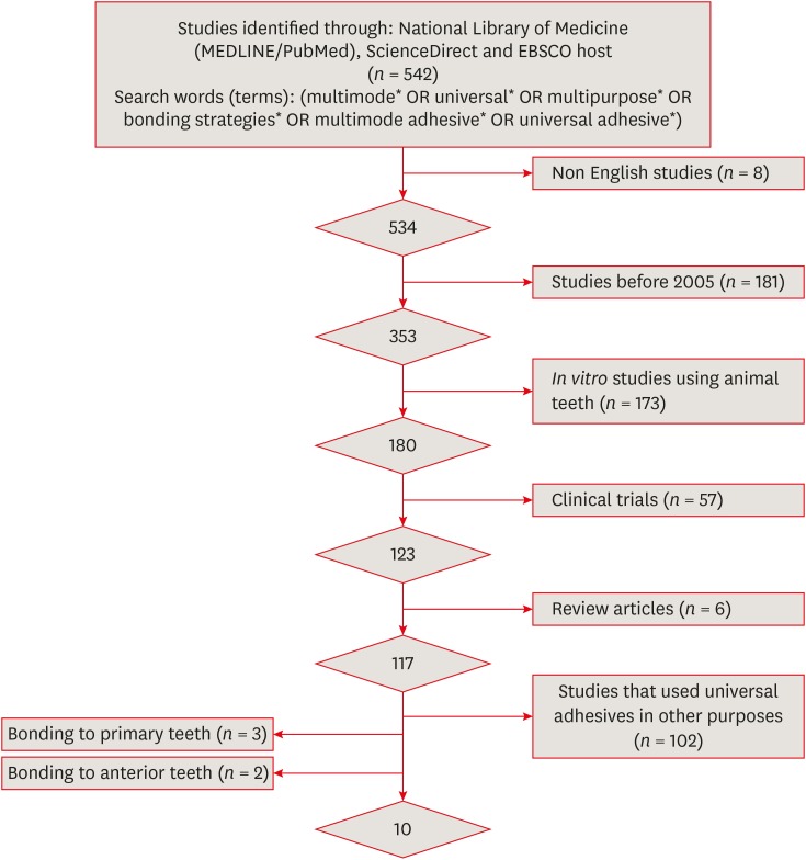
Materials and Methods An electronic search was performed of MEDLINE/PubMed, ScienceDirect, and EBSCOhost. Laboratory studies that evaluated the µTBS of multi-mode adhesives to dentin using either the etch-and-rinse or self-etch mode were selected. A meta-analysis was conducted of the reviewed studies to quantify the differences in the µTBS of Scotchbond Universal adhesive.
Results Only 10 studies fulfilled the inclusion criteria for the systematic review. Extensive variation was found in the restorative materials, testing methodologies, and failure mode in the reviewed articles. Furthermore, variation was also observed in the dimensions of the microtensile testing beams. The meta-analysis showed no statistically significant difference between the etch-and-rinse and self-etch modes for Scotchbond Universal adhesive (
p > 0.05).Conclusions Multi-mode ‘universal’ adhesives can achieve substantial bonding to dentin, regardless of the used modes (either etch-and-rinse or self-etch).
-
Citations
Citations to this article as recorded by- Influence of Proximal-Cervical Undermined Enamel Areas on Marginal Quality and Enamel Integrity of Laboratory and CAD/CAM Ceramic Inlays and Partial Crowns
Roland Frankenberger, Katharina Friedrich, Marie-Christine Dudek, Julia Winter, Norbert Krämer, Matthias J. Roggendorf
Journal of Functional Biomaterials.2025; 16(3): 82. CrossRef - Improving Bonding Protocols: The Effect of Selective Dentin Etching with Two Different Universal Adhesives—An In Vitro Study
Sandro Ferreira, Tiago Rodrigues, Mariana Nunes, Ana Mano Azul, José João Mendes, Ana Filipa Chasqueira, Joana Costa
Polymers.2025; 17(9): 1215. CrossRef - Effect of surface treatment on glass ionomers in sandwich restorations: a systematic review and meta-analysis of laboratory studies
Hoda S. Ismail, Ashraf Ibrahim Ali, Franklin Garcia-Godoy
Restorative Dentistry & Endodontics.2025; 50(2): e13. CrossRef - Wet vs. Dry Dentin Bonding: A Systematic Review and Meta-Analysis of Adhesive Performance and Hybrid Layer Integrity
Mircea Popescu, Mădălina Malița, Andrei Vorovenci, Andreea Angela Ștețiu, Viorel Ștefan Perieanu, Radu Cătălin Costea, Mihai David, Raluca Mariana Costea, Maria Antonia Ștețiu, Andi Ciprian Drăguș, Cristina Maria Șerbănescu, Andrei Burlibașa, Oana Eftene,
Oral.2025; 5(3): 63. CrossRef - The Effect of Different Multimode Adhesives On Microleakage of Class V Composite Restorations in Three Etching Modes
Fatma Yılmaz, Sevgi Kurşun, Zeliha Öztürk
ADO Klinik Bilimler Dergisi.2025; 14(3): 177. CrossRef - Controversies about refrigeration of dental adhesives: a review
Omar Abd El-Maksoud, Hamdi Hosni Hamdan Hamama, Ramy Ahmed Wafaie, Salah Hasab Mahmoud
BDJ Open.2025;[Epub] CrossRef - Tooth-composite bond failure with a universal and an etch-and-rinse adhesive depending on mode and frequency of application
Ellen Schulz-Kornas, Mathilde Tittel, Hartmut Schneider, Maximilian Bemmann, Marco Pellino, Tobias Meissner, Florian Fuchs, Christian Hannig, Florian Tetschke, Kyung-Jin Park, Michaela Strumpski, Rainer Haak
Dental Materials.2024; 40(2): 359. CrossRef - Comparison of postoperative hypersensitivity between Total-etch and Universal adhesive system: a randomized clinical trial
Kiran Javed, Nouman Noor, Muhammad Zubair Nasir, Manzoor Ahmed Manzoor
Scientific Reports.2024;[Epub] CrossRef - Adhesion and sealing of different universal adhesive systems associated with bulk‐fill resins after using endodontic irrigation solutions: An in vitro study
Érika Mayumi Omoto, Anderson Catelan, Paulo Henrique dos Santos, Luciano Tavares Angelo Cintra, Fernanda de Souza e Silva Ramos, Caio César Pavani, André Luiz Fraga Briso, Ticiane Cestari Fagundes
Australian Endodontic Journal.2024; 50(2): 309. CrossRef - Evaluation of the effects of combined application of dimethylaminohexadecyl methacrylate and MDP on dentin bonding and antimicrobial properties
Jiadi Shen, Ming Ma, Yun Huang, Haochen Miao, Xin Wei
Journal of Materials Science.2023; 58(31): 12685. CrossRef - Efficacy of adhesive strategies for restorative dentistry: A systematic review and network meta-analysis of double-blind randomized controlled trials over 12 months of follow-up
Kevin Sheng-Kai Ma, Li-Tzu Wang, Markus B. Blatz
Journal of Prosthodontic Research.2023; 67(1): 35. CrossRef - Impact of Preceded Tumor Therapeutic Irradiation on the Microtensile Bond Strength of Universal Adhesives Applied in Self-Etch Mode to Human Dentin In Vitro
Sina Broscheit, Dirk Vordermark, Reinhard Gerlach, Christian Ralf Gernhardt
Applied Sciences.2023; 13(13): 7873. CrossRef - Effect of the Adhesive Strategy on Clinical Performance and Marginal Integrity of a Universal Adhesive in Non-Carious Cervical Lesions in a Randomized 36-Month Study
Rainer Haak, Gesa Stache, Hartmut Schneider, Matthias Häfer, Gerhard Schmalz, Ellen Schulz-Kornas
Journal of Clinical Medicine.2023; 12(18): 5776. CrossRef - Universal Adhesives in Clinical Dentistry
Fusun Ozer, Shilpa Patnaikuni
Science, Art and Religion.2023; 2(1--2): 6. CrossRef - Deep proximal margin rebuilding with direct esthetic restorations: a systematic review of marginal adaptation and bond strength
Hoda S. Ismail, Ashraf I. Ali, Rabab El. Mehesen, Jelena Juloski, Franklin Garcia-Godoy, Salah H. Mahmoud
Restorative Dentistry & Endodontics.2022;[Epub] CrossRef - Improving Properties of an Experimental Universal Adhesive by Adding a Multifunctional Dendrimer (G-IEMA): Bond Strength and Nanoleakage Evaluation
Joana Vasconcelos e Cruz, António H. S. Delgado, Samuel Félix, José Brito, Luísa Gonçalves, Mário Polido
Polymers.2022; 14(7): 1462. CrossRef - Scoping review of trials evaluating adhesive strategies in pediatric dentistry: where do simplified strategies lie?
António H. S. Delgado, Hasan Jamal, Anne Young, Paul Ashley
BMC Oral Health.2021;[Epub] CrossRef - Does acid etching prior to applying universal adhesives affect the bond strength of glass fiber post to root dentin?
Helder Callegaro Velho, Eduardo Trindade Dalence, Pablo Soares Machado, Marília Pivetta Rippe, Jovito Adiel Skupien, Vinícius Felipe Wandscher
International Journal of Adhesion and Adhesives.2021; 105: 102795. CrossRef - Does Adhesive Layer Thickness and Tag Length Influence Short/Long-Term Bond Strength of Universal Adhesive Systems? An In-Vitro Study
Naji Kharouf, Tarek Ashi, Ammar Eid, Levi Maguina, Jihed Zghal, Nairy Sekayan, Rim Bourgi, Louis Hardan, Salvatore Sauro, Youssef Haikel, Davide Mancino
Applied Sciences.2021; 11(6): 2635. CrossRef - Chronological history and current advancements of dental adhesive systems development: a narrative review
Maicon Sebold, Carolina Bosso André, Beatriz Ometto Sahadi, Lorenzo Breschi, Marcelo Giannini
Journal of Adhesion Science and Technology.2021; 35(18): 1941. CrossRef - Laboratory methods for measuring adhesive bond strength between restoration materials and hard tooth tissues
I.Ya. Poyurovskaya, A.P. Polikarpova, F.S. Rusanov
Stomatologiya.2021; 100(5): 88. CrossRef - Effect of Curcumin Suspension and Vitamin C on Dentin Shear Bond Strength and Durability. A Pilot Study
Dalia A. Abuelenain, Ensanya A. Abou Neel, Tariq S. Abuhaimed, Amal M. Alamri, Hanan S. Ammar, Sahar M. N. Bukhary
The Open Dentistry Journal.2021; 15(1): 540. CrossRef - Effect of 9.3 μm CO2 and 2.94 μm Er:YAG Laser vs. Bur Preparations on Marginal Adaptation in Enamel and Dentin of Mixed Class V Cavities Restored With Different Restorative Systems
Clara Isabel Anton y Otero, Enrico Di Bella, Ivo Krejci, Tissiana Bortolotto
Frontiers in Dental Medicine.2021;[Epub] CrossRef - Adhesion strategy and curing mode of a universal adhesive influence the bonding of dual-cured core build-up resin composite to dentin
Ahmed Eid Elsayed, Mohamed Amr Kamel, Farid Sabry El-Askary
Journal of Adhesion Science and Technology.2021; 35(1): 52. CrossRef - Influence of etching mode and composite resin type on bond strength to dentin using universal adhesive system
Stefan Dačić, Milan Miljković, Aleksandar Mitić, Goran Radenković, Marija Anđelković‐Apostolović, Milica Jovanović
Microscopy Research and Technique.2021; 84(6): 1212. CrossRef - Universal adhesives - a new direction in the development of adhesive systems
A. Tichý, K. Hosaka, J. Tagami
Česká stomatologie a praktické zubní lékařství.2020; 120(1): 4. CrossRef - Effect of Over-Etching and Prolonged Application Time of a Universal Adhesive on Dentin Bond Strength
Phoebe Burrer, Hoang Dang, Matej Par, Thomas Attin, Tobias T. Tauböck
Polymers.2020; 12(12): 2902. CrossRef - Profile of a 10-MDP-based universal adhesive system associated with chlorhexidine: Dentin bond strength and in situ zymography performance
Marina Ciccone Giacomini, Polliana Mendes Candia Scaffa, Rafael Simões Gonçalves, Giovanna Speranza Zabeu, Cristina de Mattos Pimenta Vidal, Marcela Rocha de Oliveira Carrilho, Heitor Marques Honório, Linda Wang
Journal of the Mechanical Behavior of Biomedical Materials.2020; 110: 103925. CrossRef - Universal dental adhesives: Current status, laboratory testing, and clinical performance
Sanket Nagarkar, Nicole Theis‐Mahon, Jorge Perdigão
Journal of Biomedical Materials Research Part B: Applied Biomaterials.2019; 107(6): 2121. CrossRef - Modifying Adhesive Materials to Improve the Longevity of Resinous Restorations
Wen Zhou, Shiyu Liu, Xuedong Zhou, Matthias Hannig, Stefan Rupf, Jin Feng, Xian Peng, Lei Cheng
International Journal of Molecular Sciences.2019; 20(3): 723. CrossRef
- Influence of Proximal-Cervical Undermined Enamel Areas on Marginal Quality and Enamel Integrity of Laboratory and CAD/CAM Ceramic Inlays and Partial Crowns
- 5,174 View
- 45 Download
- 30 Crossref

- Effect of various bleaching treatments on shear bond strength of different universal adhesives and application modes
- Fatma Dilsad Oz, Zeynep Bilge Kutuk
- Restor Dent Endod 2018;43(2):e20. Published online April 16, 2018
- DOI: https://doi.org/10.5395/rde.2018.43.e20
-
 Abstract
 PDF
PDF PubReader
 ePub
ePub Objectives The aim of this
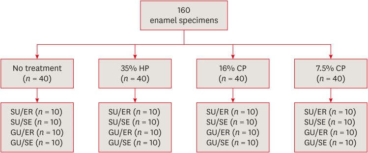
in vitro study was to evaluate the bond strength of 2 universal adhesives used in different application modes to bleached enamel.Materials and Methods Extracted 160 sound human incisors were used for the study. Teeth were divided into 4 treatment groups: No treatment, 35% hydrogen peroxide, 16% carbamid peroxide, 7.5% carbamid peroxide. After bleaching treatments, groups were divided into subgroups according to the adhesive systems used and application modes (
n = 10): 1) Single Bond Universal, etch and rinse mode; 2) Single Bond Universal, self-etch mode; 3) Gluma Universal, etch and rinse mode; 4) Gluma Universal, self-etch mode. After adhesive procedures nanohybrid composite resin cylinders were bonded to the enamel surfaces. All specimens were subjected to shear bond strength (SBS) test after thermocycling. Data were analyzed using a 3-way analysis of variance (ANOVA) and Tukeypost hoc test.Results No significant difference were found among bleaching groups (35% hydrogen peroxide, 16% carbamid peroxide, 7.5% carbamid peroxide, and no treatment groups) in the mean SBS values. There was also no difference in SBS values between Single Bond Universal and Gluma Universal at same application modes, whereas self-etch mode showed significantly lower SBS values than etch and rinse mode (
p < 0.05).Conclusions The bonding performance of the universal adhesives was enhanced with the etch and rinse mode application to bleached enamel and non-bleached enamel.
-
Citations
Citations to this article as recorded by- Antioxidant effect on shear bond strength of resin composite to in-office versus home bleached enamel surface
Maha Mosaad Mohamed, Magda E. -A. Shalaby, Eman A. E. -G. Shebl
Tanta Dental Journal.2025; 22(3): 409. CrossRef - Effects of Time-Elapsed Bleaching on the Surface and Mechanical Properties of Dentin Substrate Using Hydrogen Peroxide-Free Nanohydroxyapatite Gel
Aftab Khan, Abdulaziz AlKhureif, Manal Almutairi, Abrar Nooh, Saeed Hassan, Yasser Alqahtani
International Journal of Nanomedicine.2024; Volume 19: 10307. CrossRef - Effect of sodium ascorbate on the shear bond strength of orthodontic brackets to bleached enamel using universal dental adhesive
Saeid Sadeghian, Kamyar Fathpour, Mahshid Biglari
Dental Research Journal.2023;[Epub] CrossRef - Quantitative Measurements of the Depth of Enamel Demineralization before and after Bleach: An In Vitro Study
Sara Naim, Gianrico Spagnuolo, Essam Osman, Syed Sarosh Mahdi, Gopi Battineni, Syed Saad B. Qasim, Mariangela Cernera, Hasna Rifai, Nada Jaafar, Elie Maalouf, Carina Mehanna Zogheib, Konstantinos Michalakis
BioMed Research International.2022;[Epub] CrossRef - DİŞ BEYAZLATMA İŞLEMİNİN LİTYUM DİSİLİKAT SERAMİĞİN BAĞLANMA DAYANIMINA ETKİSİ
Merve YILDIRAK, Rıfat GÖZNELİ
Atatürk Üniversitesi Diş Hekimliği Fakültesi Dergisi.2020; : 1. CrossRef - The Effect of Different Bleaching Protocols, Used with and without Sodium Ascorbate, on Bond Strength between Composite and Enamel
Maroun Ghaleb, Giovanna Orsini, Angelo Putignano, Sarah Dabbagh, Georges Haber, Louis Hardan
Materials.2020; 13(12): 2710. CrossRef - Influence of phototherapy on adhesive strength and microleakage of bleached enamel bonded to orthodontic brackets: An in-vitro study
Erum Khan, Ibrahim Alshahrani, Muhammad Abdullah Kamran, Abdulaziz Samran, Ali Alqerban, Saad Abdul Rehman
Photodiagnosis and Photodynamic Therapy.2019; 25: 344. CrossRef - Effect of Er: YAG Laser on Microtensile Bond Strength of Bleached Dentin to Composite
Mohsen Rezaei, Elham Aliasghar, Mohammad Bagher Rezvani, Nasim Chiniforush, Zohreh Moradi
Journal of Lasers in Medical Sciences.2019; 10(2): 117. CrossRef
- Antioxidant effect on shear bond strength of resin composite to in-office versus home bleached enamel surface
- 2,115 View
- 21 Download
- 8 Crossref

- Effect of smear layer deproteinization on bonding of self-etch adhesives to dentin: a systematic review and meta-analysis
- Khaldoan H. Alshaikh, Hamdi H. H. Hamama, Salah H. Mahmoud
- Restor Dent Endod 2018;43(2):e14. Published online March 6, 2018
- DOI: https://doi.org/10.5395/rde.2018.43.e14
-
 Abstract
 PDF
PDF PubReader
 ePub
ePub Objectives The aim of this systematic review was to critically analyze previously published studies of the effects of dentin surface pretreatment with deproteinizing agents on the bonding of self-etch (SE) adhesives to dentin. Additionally, a meta-analysis was conducted to quantify the effects of the above-mentioned surface pretreatment methods on the bonding of SE adhesives to dentin.
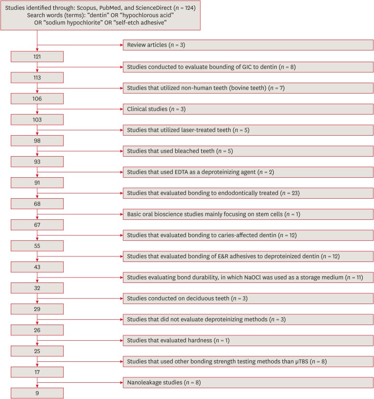
Materials and Methods An electronic search was performed using the following databases: Scopus, PubMed and ScienceDirect. The online search was performed using the following keywords: ‘dentin’ or ‘hypochlorous acid’ or ‘sodium hypochlorite’ and ‘self-etch adhesive.’ The following categories were excluded during the assessment process: non-English articles, randomized clinical trials, case reports, animal studies, and review articles. The reviewed studies were subjected to meta-analysis to quantify the effect of the application time and concentration of sodium hypochlorite (NaOCl) and hypochlorous acid (HOCl) deproteinizing agents on bonding to dentin.
Results Only 9 laboratory studies fit the inclusion criteria of this systematic review. The results of the meta-analysis revealed that the pooled average microtensile bond strength values to dentin pre-treated with deproteinizing agents (15.71 MPa) was significantly lower than those of the non-treated control group (20.94 MPa).
Conclusions In light of the currently available scientific evidence, dentin surface pretreatment with deproteinizing agents does not enhance the bonding of SE adhesives to dentin. The HOCl deproteinizing agent exhibited minimal adverse effects on bonding to dentin in comparison with NaOCl solutions.
-
Citations
Citations to this article as recorded by- Is the Percentage of Collagen in Coronal Dentin Related to Microtensile Strength? An In Vitro Study
Taíssa Cássia de Souza Furtado, Gilberto Antonio Borges, Vinícius Rangel Geraldo-Martins, Bruno Henrique dos Reis Souza Oliveira, Renata Margarida Etchebehere, Sanívia Aparecida de Lima Pereira
Pesquisa Brasileira em Odontopediatria e Clínica Integrada.2026;[Epub] CrossRef -
Evaluating the remnants of Al
2
O
3
particles on different dentine substrate after sandblasting and various cleaning protocols
Faeze Hamze, Khotan Aflatoonian, Mahshid Mohammadibassir, Mohammad-Bagher Rezvani
Journal of Adhesion Science and Technology.2025; 39(6): 869. CrossRef - Preservation Strategies for Interfacial Integrity in Restorative Dentistry: A Non-Comprehensive Literature Review
Carmem S. Pfeifer, Fernanda S. Lucena, Fernanda M. Tsuzuki
Journal of Functional Biomaterials.2025; 16(2): 42. CrossRef - Outcome of Er, Cr:YSGG laser and antioxidant pretreatments on bonding quality to caries-induced dentin
Lamiaa M. Moharam, Haidy N. Salem, Ahmed Abdou, Rasha H. Afifi
BMC Oral Health.2025;[Epub] CrossRef - Advancing Adhesive Strategies for Endodontically Treated Teeth—Part II: Dentin Sealing Before Irrigation Increases Long‐Term Microtensile Bond Strength to Coronal Dentin
Joana A. Marques, Rui I. Falacho, Gabriela Almeida, Francisco Caramelo, João Miguel Santos, João Rocha, Markus B. Blatz, João Carlos Ramos, Paulo J. Palma
Journal of Esthetic and Restorative Dentistry.2025; 37(7): 1865. CrossRef - Effect of finishing protocols on dentin surface characteristics and bond strength after tooth preparation for indirect restorations
Paola Bernardes, Amanda das Graças Soares, Bárbara Inácio de Melo, Leandro Maruki Pereira, Regina Guenka Palma-Dibb, Rafael Rocha Pacheco, Marcel Santana Prudente, Luís Henrique Araújo Raposo
The Journal of Prosthetic Dentistry.2025;[Epub] CrossRef - A comparison of different cleaning approaches for blood contamination after curing universal adhesives on the dentine surface
Ting Liu, Haifeng Xie, Chen Chen
Dental Materials.2024; 40(11): 1786. CrossRef - Effect of fiber-reinforced direct restorative materials on the fracture resistance of endodontically treated mandibular molars restored with a conservative endodontic cavity design
Merve Nezir, Beyza Arslandaş Dinçtürk, Ceyda Sarı, Cemile Kedici Alp, Hanife Altınışık
Clinical Oral Investigations.2024;[Epub] CrossRef - Effect of the use of bromelain associated with bioactive glass-ceramic on dentin/adhesive interface
Rocio Geng Vivanco, Ana Beatriz Silva Sousa, Viviane de de Cássia Oliveira, Mário Alexandre Coelho Sinhoreti, Fernanda de Carvalho Panzeri Pires-de-Souza
Clinical Oral Investigations.2024;[Epub] CrossRef - Experimental and Chitosan-Infused Adhesive with Dentin Pretreated with Femtosecond Laser, Methylene Blue-Activated Low-Level Laser, and Phosphoric Acid
Fahad Alkhudhairy
Photobiomodulation, Photomedicine, and Laser Surgery.2024; 42(10): 634. CrossRef - Evaluation of Effective Bond Strength of Composite Resin to Etched Dentin after Dentin Pretreatment: An In-vitro Study
Muhammed Bilal, Shiraz Pasha, Arathi S. Nair
Journal of the Scientific Society.2024; 51(4): 545. CrossRef - Comparison of Different Dentin Deproteinizing Agents on Bond Strength and Microleakage of Universal Adhesive to Dentin
Fatih Bedir, Gül Yıldız Telatar
Journal of Advanced Oral Research.2023; 14(1): 44. CrossRef - Addition of metal chlorides to a HOCl conditioner can enhance bond strength to smear layer deproteinized dentin
Kittisak Sanon, Antonin Tichy, Takashi Hatayama, Ornnicha Thanatvarakorn, Taweesak Prasansuttiporn, Takahiro Wada, Yasushi Shimada, Keiichi Hosaka, Masatoshi Nakajima
Dental Materials.2022; 38(8): 1235. CrossRef - Internal and Marginal Adaptation of Adhesive Resin Cements Used for Luting Inlay Restorations: An In Vitro Micro-CT Study
Linah M. Ashy, Hanadi Marghalani
Materials.2022; 15(17): 6161. CrossRef - Collagen-depletion strategies in dentin as alternatives to the hybrid layer concept and their effect on bond strength: a systematic review
António H. S. Delgado, Madalena Belmar Da Costa, Mário Cruz Polido, Ana Mano Azul, Salvatore Sauro
Scientific Reports.2022;[Epub] CrossRef - NaOCl Application after Acid Etching and Retention of Cervical Restorations: A 3-Year Randomized Clinical Trial
M Favetti, T Schroeder, AF Montagner, RR Moraes, T Pereira-Cenci, MS Cenci
Operative Dentistry.2022; 47(3): 268. CrossRef - Resin infiltrant protects deproteinized dentin against erosive and abrasive wear
Ana Theresa Queiroz de Albuquerque, Bruna Oliveira Bezerra, Isabelly de Carvalho Leal, Maria Denise Rodrigues de Moraes, Mary Anne S. Melo, Vanara Florêncio Passos
Restorative Dentistry & Endodontics.2022;[Epub] CrossRef - Bis[2-(Methacryloyloxy) Ethyl] Phosphate as a Primer for Enamel and Dentine
R. Alkattan, G. Koller, S. Banerji, S. Deb
Journal of Dental Research.2021; 100(10): 1081. CrossRef - Influence of Dentine Pre-Treatment by Sandblasting with Aluminum Oxide in Adhesive Restorations. An In Vitro Study
Bruna Sinjari, Manlio Santilli, Gianmaria D’Addazio, Imena Rexhepi, Alessia Gigante, Sergio Caputi, Tonino Traini
Materials.2020; 13(13): 3026. CrossRef - A novel prime-&-rinse mode using MDP and MMPs inhibitors improves the dentin bond durability of self-etch adhesive
Jingqiu Xu, Mingxing Li, Wenting Wang, Zhifang Wu, Chaoyang Wang, Xiaoting Jin, Ling Zhang, Wenxiang Jiang, Baiping Fu
Journal of the Mechanical Behavior of Biomedical Materials.2020; 104: 103698. CrossRef - The effects of deproteinization and primer treatment on microtensile bond strength of self-adhesive resin cement to dentin
In-Hye Bae, Sung-Ae Son, Jeong-Kil Park
Korean Journal of Dental Materials.2019; 46(2): 99. CrossRef - Effect of Papain and Bromelain Enzymes on Shear Bond Strength of Composite to Superficial Dentin in Different Adhesive Systems
Farahnaz Sharafeddin, Mina Safari
The Journal of Contemporary Dental Practice.2019; 20(9): 1077. CrossRef
- Is the Percentage of Collagen in Coronal Dentin Related to Microtensile Strength? An In Vitro Study
- 2,494 View
- 23 Download
- 22 Crossref

- Comparison of bond strengths of ceramic brackets bonded to zirconia surfaces using different zirconia primers and a universal adhesive
- Ji-Yeon Lee, Jaechan Ahn, Sang In An, Jeong-won Park
- Restor Dent Endod 2018;43(1):e7. Published online January 22, 2018
- DOI: https://doi.org/10.5395/rde.2018.43.e7
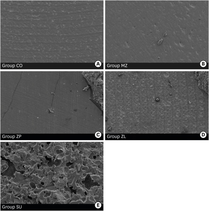
-
 Abstract
 PDF
PDF PubReader
 ePub
ePub Objectives The aim of this study is to compare the shear bond strengths of ceramic brackets bonded to zirconia surfaces using different zirconia primers and universal adhesive.
Materials and Methods Fifty zirconia blocks (15 × 15 × 10 mm, Zpex, Tosoh Corporation) were polished with 1,000 grit sand paper and air-abraded with 50 µm Al2O3 for 10 seconds (40 psi). They were divided into 5 groups: control (CO), Metal/Zirconia primer (MZ, Ivoclar Vivadent), Z-PRIME Plus (ZP, Bisco), Zirconia Liner (ZL, Sun Medical), and Scotchbond Universal adhesive (SU, 3M ESPE). Transbond XT Primer (used for CO, MZ, ZP, and ZL) and Transbond XT Paste was used for bracket bonding (Gemini clear ceramic brackets, 3M Unitek). After 24 hours at 37°C storage, specimens underwent 2,000 thermocycles, and then, shear bond strengths were measured (1 mm/min). An adhesive remnant index (ARI) score was calculated. The data were analyzed using one-way analysis of variance and the Bonferroni test (
p = 0.05).Results Surface treatment with primers resulted in increased shear bond strength. The SU group showed the highest shear bond strength followed by the ZP, ZL, MZ, and CO groups, in that order. The median ARI scores were as follows: CO = 0, MZ = 0, ZP = 0, ZL = 0, and SU = 3 (
p < 0.05).Conclusions Within this experiment, zirconia primer can increase the shear bond strength of bracket bonding. The highest shear bond strength is observed in SU group, even when no primer is used.
-
Citations
Citations to this article as recorded by- Effectiveness of universal adhesives for orthodontic bonding to enamel and restorative materials: A systematic review
Claire-Adeline Dantagnan, Maureen Boudrot, Julia Bosco, Gauthier Dot, Ali Nassif, Philippe François, Jean-Pierre Attal
International Orthodontics.2026; 24(2): 101089. CrossRef - State-of-the-Art Zirconia and Glass–Ceramic Materials in Restorative Dentistry: Properties, Clinical Applications, Challenges, and Future Perspectives
Sorin Gheorghe Mihali, Adela Hiller
Applied Sciences.2025; 15(23): 12841. CrossRef - Shear bond strength and ARI scores of metal brackets to glazed glass ceramics and zirconia: an in vitro study investigating surface treatment protocols
Claire Pédemay, Philippe François, Vincent Fouquet, Sarah Abdel-Gawad, Jean-Pierre Attal, Claire-Adeline Dantagnan
BMC Oral Health.2024;[Epub] CrossRef - Impact of different pretreatments and attachment materials on shear bond strength of indirectly bonded brackets using CAD/CAM transfer trays to monolithic zirconia
Rebecca Jungbauer, Christian M. Hammer, Daniel Edelhoff, Peter Proff, Bogna Stawarczyk
Dental Materials.2023; 39(2): 170. CrossRef - Mechanical and chemical surface treatment enhances bond strength between zirconia and orthodontic brackets: an in vitro study
Nareudee Limpuangthip, Atikom Surintanasarn, Ploylada Vitavaspan
BDJ Open.2023;[Epub] CrossRef - Effect of Different Surface Treatments and Orthodontic Bracket Type on Shear Bond Strength of High‐Translucent Zirconia: An In Vitro Study
Yasamin Babaee Hemmati, Hamid Neshandar Asli, Mehran Falahchai, Sina Safary, Sandrine Bittencourt Berger
International Journal of Dentistry.2022;[Epub] CrossRef - Does Surface Treatment With Different Primers Increase The Shear Bond Strength Between Metallic Bracket and Monolithic Zirconia?
Emine Begüm BÜYÜKERKMEN, Ayşe Selenge AKBULUT, Murat KEÇECİ
Selcuk Dental Journal.2022; 9(2): 451. CrossRef - Effect of Different Surface Treatments on the Surface Roughness and Orthodontic Bond Strength of Partially-stabilized Zirconia
Mustafa Borga Dönmez, Betül Ballı Demirel, Münir Demirel, Yasemin Gündoğdu, Hamdi Şükür Kılıç
Meandros Medical and Dental Journal.2022; 23(3): 335. CrossRef - Shear Bond Strength of Polypropylene Fiber in Orthodontic Adhesive on Glazed Monolithic Zirconia
Dhanabhol Riowruangsanggoon, Apiwat Riddhabhaya, Nattisa Niyomtham, Irin Sirisoontorn
Polymers.2022; 14(21): 4627. CrossRef - Effects of Three Novel Bracket Luting Agents Containing Zirconia Primer on Shear Bond Strength of Metal Orthodontic Brackets Attached to Monolithic Zirconia Crowns: A Preliminary In Vitro Study
Milad Shamohammadi Heidari, Mehrnaz Moradinejad, Hamed Tabatabaei, Vahid Rakhshan, Dinesh Rokaya
International Journal of Dentistry.2022;[Epub] CrossRef - Impact of different pretreatments and attachment materials on shear bond strength between monolithic zirconia restorations and metal brackets
Rebecca Jungbauer, Peter Proff, Daniel Edelhoff, Bogna Stawarczyk
Scientific Reports.2022;[Epub] CrossRef - Bracket Bonding to All-Ceramic Materials with Universal Adhesives
Cecilia Goracci, Giuseppe Di Bello, Lorenzo Franchi, Chris Louca, Jelena Juloski, Jovana Juloski, Alessandro Vichi
Materials.2022; 15(3): 1245. CrossRef - Effect of enamel-surface modifications on shear bond strength using different adhesive materials
Bo-wen Zheng, Shan Cao, Majedh Abdo Ali Al-Somairi, Jia He, Yi Liu
BMC Oral Health.2022;[Epub] CrossRef - The effect of various mechanical and chemical surface conditioning on the bonding of orthodontic brackets to all ceramic materials
Dalia A. Abuelenain, Amal I. Linjawi, Ahmed S. Alghamdi, Fahad M. Alsadi
Journal of Dental Sciences.2021; 16(1): 370. CrossRef - The Performance of Universal Adhesives on Orthodontic Bracket Bonding
Muhittin Ugurlu, Muhammed Hilmi Buyukcavus
European Journal of General Dentistry.2021; 10(01): 019. CrossRef - A comparison of shear bond strength of brackets bonded to zirconia
Hannah Knott, Xiaoming Xu, Edwin Kee, Qingzhao Yu, Paul Armbruster, Richard Ballard
Australasian Orthodontic Journal.2021; 37(1): 62. CrossRef - Influence of Surface Treatment and Resin Cements on the Bond Strength between the Y-TZP Zirconia and Composite Resin Interface
Lucas Campagnaro Maciel, Amanda Pádua Proeza, Hélyda Coelho Guimarães Balbino, Marcela Moráo Corteletti, Ricardo Huver De Jesus, Laís Regiane da Silva Concílio
Journal of Health Sciences.2019; 21(5): 477. CrossRef - Effect of Simplified Bonding on Shear Bond Strength between Ceramic Brackets and Dental Zirconia
Ga-Youn Ju, Soram Oh, Bum-Soon Lim, Hyun-Seung Lee, Shin Hye Chung
Materials.2019; 12(10): 1640. CrossRef
- Effectiveness of universal adhesives for orthodontic bonding to enamel and restorative materials: A systematic review
- 2,232 View
- 16 Download
- 18 Crossref

- Comparing the effect of a desensitizing material and a self-etch adhesive on dentin sensitivity after periodontal surgery: a randomized clinical trial
- Hila Hajizadeh, Atefeh Nemati-Karimooy, Sara Majidinia, Amir Moeintaghavi, Marjaneh Ghavamnasiri
- Restor Dent Endod 2017;42(3):168-175. Published online July 21, 2017
- DOI: https://doi.org/10.5395/rde.2017.42.3.168
-
 Abstract
 PDF
PDF PubReader
 ePub
ePub Objectives This double-blind randomized placebo-controlled clinical trial evaluated the ability of a desensitizing agent and a self-etch adhesive on cervical dentin sensitivity (CDS) after periodontal surgery.
Materials and Methods Ninety hypersensitive teeth of 13 subjects were included in the study. After periodontal surgery, the teeth of each posterior sextant treated with one of the following materials: G1: Clearfil S3 Bond (Kuraray Dental), G2: Gluma Desensitizer (Heraeus Kulzer), and G3: placebo (water). The sensitivity was assessed using evaporative stimuli before treatment (baseline, T0), 1 day after treatment (T1), after 1 week (T2), and after 1 month (T3) according to visual analog scale (VAS).
Results Following the treatment, all the 3 groups showed significant reduction of CDS in T1 compared to T0. Reduction of CDS between T1 and T2 was observed only in G1 but there was no significant difference between T2 and T3 in this group. Although we observed a significant difference in T3 compared to T1 and T2 in G2 and G3, comparison of treatment groups in each assessment time showed a significant difference only in T3. According to paired comparison, this was due to the difference between G2 and G3.
Conclusions Dentin sensitivity following periodontal surgery will decrease spontaneously over time, but treating the sensitive teeth with Gluma Desensitizer and Clearfil S3 Bond can have some benefits.
-
Citations
Citations to this article as recorded by- Biomineralization reaction from nanosized calcium silicate: A new method for reducing dentin hypersensitivity
Mi-Jeong Jeon, Yu-Sung Choi, Jeong-Kil Park, Jin-Soo Ahn, Yu-Chih Chiang, Deog-Gyu Seo
Journal of Dental Sciences.2025; 20(1): 428. CrossRef - Effectiveness of Self-etching Adhesive Only Versus in Combination with Gluma Desensitizer for Preventing Post-composite Sensitivity - A Prospective Study
Hemamalini Rath, Shilpa Mahapatra, Sri Priya Narayanan
Indian Journal of Dental Research.2025; 36(1): 32. CrossRef - Efficacy of seventh generation bonding agents as desensitizers in patients with dentin hypersensitivity: a randomized clinical trial
Sumaiya Shabbir, Shahbaz Ahmed, Syed Jaffar Abbas Zaidi, Sania Riaz, Huma Sarwar, Muhammad Taqi, Zia ur Rahman Khan
BMC Oral Health.2024;[Epub] CrossRef - Investigation of the crystal formation from calcium silicate in human dentinal tubules and the effect of phosphate buffer saline concentration
Mi-Jeong Jeon, Jin-Soo Ahn, Jeong-Kil Park, Deog-Gyu Seo
Journal of Dental Sciences.2024; 19(4): 2278. CrossRef - The effect of fluoride iontophoresis on seal ability of self-etch adhesive in human dentin in vitro
Kanittha Kijsamanmith, Parintorn Wallanon, Chanya Pitchayasatit, Poonnapha Kittiratanaviwat
BMC Oral Health.2022;[Epub] CrossRef - The study of toothpaste desensitizing properties
S. B. Ulitovskiy, O. V. Kalinina, A. A. Leontev, O. V. Khabarova, L. I. Pankrateva, E. S. Soloveva, N. K. Fok
Parodontologiya.2022; 27(1): 81. CrossRef - Effectiveness and cytotoxicity of two desensitizing agents: a dentin permeability measurement and dentin barrier testing in vitro study
Ruodan Jiang, Yongxiang Xu, Feilong Wang, Hong Lin
BMC Oral Health.2022;[Epub] CrossRef - A randomized clinical trial of dentin hypersensitivity reduction over one month after a single topical application of comparable materials
Samar Hatem Abuzinadah, Abdulrahman Jafar Alhaddad
Scientific Reports.2021;[Epub] CrossRef - Comparison between effectiveness of dentine desensitizer and one bottle self-etch adhesive on dentine hypersensitivity
Muhammad Zohaib Younus, Muhammad Adeel Ahmed, Azeem Ul Yaqin Syed, Jiand Malik Baloch, Muhammad Ali, Abubakar Sheikh
Technology and Health Care.2021; 29(6): 1153. CrossRef - A long-term evaluation of experimental potassium oxalate concentrations on dentin hypersensitivity reduction: A triple-blind randomized clinical trial
Alexia da Mata Galvão, Livia Fávaro Zeola, Guilherme Faria Moura, Daniela Navarro Ribeiro Teixeira, Ramon Corrêa de Queiroz Gonzaga, Gisele Rodrigues da Silva, Paulo Vinícius Soares
Journal of Dentistry.2019; 89: 103180. CrossRef
- Biomineralization reaction from nanosized calcium silicate: A new method for reducing dentin hypersensitivity
- 2,164 View
- 10 Download
- 10 Crossref

- Bonding of the silane containing multi-mode universal adhesive for lithium disilicate ceramics
- Hyun-Young Lee, Geum-Jun Han, Juhea Chang, Ho-Hyun Son
- Restor Dent Endod 2017;42(2):95-104. Published online January 25, 2017
- DOI: https://doi.org/10.5395/rde.2017.42.2.95
-
 Abstract
 PDF
PDF PubReader
 ePub
ePub Objectives This study evaluated the influence of a multi-mode universal adhesive (MUA) containing silane (Single Bond Universal, 3M EPSE) on the bonding of resin cement to lithium disilicate.
Materials and Methods Thirty IPS e.max CAD specimens (Ivoclar Vivadent) were fabricated. The surfaces were treated as follows: Group A, adhesive that did not contain silane (ANS, Porcelain Bonding Resin, Bisco); Group B, silane (S) and ANS; Group C, hydrofluoric acid (HF), S, and ANS; Group D, MUA; Group E, HF and MUA. Dual-cure resin cement (NX3, Kerr) was applied and composite resin cylinders of 0.8 mm in diameter were placed on it before light polymerization. Bonded specimens were stored in water for 24 hours or underwent a 10,000 thermocycling process prior to microshear bond strength testing. The data were analyzed using multivariate analysis of variance (
p < 0.05).Results Bond strength varied significantly among the groups (
p < 0.05), except for Groups A and D. Group C showed the highest initial bond strength (27.1 ± 6.9 MPa), followed by Group E, Group B, Group D, and Group A. Thermocycling significantly reduced bond strength in Groups B, C, and E (p < 0.05). Bond strength in Group C was the highest regardless of the storage conditions (p < 0.05).Conclusions Surface treatment of lithium disilicate using HF and silane increased the bond strength of resin cement. However, after thermocycling, the silane in MUA did not help achieve durable bond strength between lithium disilicate and resin cement, even when HF was applied.
-
Citations
Citations to this article as recorded by- The influence of different factors on the bond strength of lithium disilicate-reinforced glass–ceramics to Resin: a machine learning analysis
Jiawen Liu, Suqing Tu, Mingjuan Wang, Du Chen, Chen Chen, Haifeng Xie
BMC Oral Health.2025;[Epub] CrossRef - Influence of different primers and adhesive system combinations on the durability of resin bonding to lithium disilicate
Christine Yazigi, Shila Alawi, Sebastian Wille, Matthias Kern
The Journal of Prosthetic Dentistry.2025; 134(3): 749. CrossRef - Shear Bond Strength and Finite Element Stress Analysis of Composite Repair Using Various Adhesive Strategies With and Without Silane Application
Elif Ercan Devrimci, Hande Kemaloglu, Cem Peskersoy, Tijen Pamir, Murat Turkun
Applied Sciences.2025; 15(15): 8159. CrossRef - Effect of multiple firings on mechanical and optical properties of CAD/CAM lithium disilicate-based glass ceramics
Chawal Padunglappisit, Pitsucha Charoensakthanakul, Sintwo Wongthongdee, Kan Wongkamhaeng
BMC Oral Health.2025;[Epub] CrossRef - Effect of universal adhesives and self-etch ceramic primers on bond strength to glass-ceramics: A systematic review and meta-analysis of in vitro studies
Renally Bezerra Wanderley Lima, Isis de Araújo Ferreira Muniz, Débora e Silva Campos, Fabián Murillo-Gómez, Ana Karina Maciel de Andrade, Rosângela Marques Duarte, Grace Mendonça de Souza
The Journal of Prosthetic Dentistry.2024; 131(3): 392. CrossRef - Effect of the difference water amounts and hydrolysis times of silane coupling agent on the shear bond strength between lithium disilicate glass ceramic and composite resin
Pimchanok OSOTPRASIT, Sasipin LAUVAHUTANON, Yosnarong SIRIMETHAWONG, Patcharanun CHAIAMORNSUP, Pornpot JIANGKONGKHO
Dental Materials Journal.2024; 43(3): 375. CrossRef - Is additional silane application necessary for a new silane‐containing universal adhesive to bond to glass ceramics?
Priscila Luciane da Silva, Hélio Radke Bittencourt, Luiz Henrique Burnett, Ana Maria Spohr
Journal of Esthetic and Restorative Dentistry.2024; 36(10): 1452. CrossRef - The Effect of Various Lasers on the Bond Strength Between Orthodontic Brackets and Dental Ceramics: A Systematic Review and Meta-Analysis
Seyed Ali Mosaddad, Jaafar Abduo, Mehrnaz Zakizade, Hamid Tebyaniyan, Ahmed Hussain
Photobiomodulation, Photomedicine, and Laser Surgery.2024; 42(1): 20. CrossRef - Long-Term Bonding Performance of One-Bottle vs. Two-Bottle Bonding Agents to Lithium Disilicate Ceramics
Masao Irie, Masahiro Okada, Yukinori Maruo, Goro Nishigawa, Takuya Matsumoto
Polymers.2024; 16(16): 2266. CrossRef - Bond strength to different CAD/CAM lithium disilicate reinforced ceramics
Mona Alhomuod, Jin‐Ho Phark, Sillas Duarte
Journal of Esthetic and Restorative Dentistry.2023; 35(1): 129. CrossRef - Surface Treatment Effect on Shear Bond Strength between Lithium Disilicate Glass-Ceramic and Resin Cement
Siripan Simasetha, Awiruth Klaisiri, Tool Sriamporn, Kraisorn Sappayatosok, Niyom Thamrongananskul
European Journal of Dentistry.2022; 16(02): 373. CrossRef - Bonding of Clear Aligner Composite Attachments to Ceramic Materials: An In Vitro Study
Bashair A. Alsaud, Maher S. Hajjaj, Ahmad I. Masoud, Ensanya A. Abou Neel, Dalia A. Abuelenain, Amal I. Linjawi
Materials.2022; 15(12): 4145. CrossRef - Bonding of different resin luting materials to composite, polymer-infiltrated and feldspathic ceramic CAD/CAM blocks
Burcu Dikici, Esra Can Say
Journal of Adhesion Science and Technology.2022; 36(14): 1572. CrossRef - Influence of mechanical and chemical pre-treatments on the repair of a hybrid ceramic
Sascha Niklas Jung, Stefan Rüttermann
Dental Materials.2022; 38(7): 1140. CrossRef - Effect of Silane-Containing Universal Adhesives on the Bonding Strength of Lithium Disilicate
Yu-Ri Kim, Jae-Hoon Kim, Sung-Ae Son, Jeong-Kil Park
Materials.2021; 14(14): 3976. CrossRef - Ceramics in dentistry: which material is appropriate for the anterior or posterior Dentition? Part 1: materials science
Loo Chien Win, Peter Sands, Stephen J Bonsor, FJ Trevor Burke
Dental Update.2021; 48(8): 680. CrossRef - The effect of different ceramic surface treatments on the repair bond strength of resin composite to lithium disilicate ceramic
Nanako UEDA, Tomohiro TAKAGAKI, Toru NIKAIDO, Rena TAKAHASHI, Masaomi IKEDA, Junji TAGAMI
Dental Materials Journal.2021; 40(5): 1073. CrossRef - Bonding Strength of Universal Adhesives to Indirect Substrates: A Meta‐Analysis of in Vitro Studies
Carlos Enrique Cuevas‐Suárez, Wellington Luiz de Oliveira da Rosa, Rafael Pino Vitti, Adriana Fernandes da Silva, Evandro Piva
Journal of Prosthodontics.2020; 29(4): 298. CrossRef - Effect of different surface treatments and multimode adhesive application on the Weibull characteristics, wettability, surface topography and adhesion to CAD/CAM lithium disilicate ceramic
Karina Barbosa Souza, Dayanne Monielle Duarte Moura, Sarah Emille Gomes da Silva, Gabriela Monteiro de Araújo, Rafael de Almeida Spinelli Pinto, Fabíola Pessôa Pereira Leite, Mutlu Özcan, Rodrigo Othávio de Assunção e Souza
Journal of Applied Oral Science.2020;[Epub] CrossRef - Effects of the ratio of silane to 10-methacryloyloxydecyl dihydrogenphosphate (MDP) in primer on bonding performance of silica-based and zirconia ceramics
Minkhant Koko, Tomohiro Takagaki, Ahmed Abdou, Masanao Inokoshi, Masaomi Ikeda, Takahiro Wada, Motohiro Uo, Toru Nikaido, Junji Tagami
Journal of the Mechanical Behavior of Biomedical Materials.2020; 112: 104026. CrossRef - Influence of surface treatments and repair materials on the shear bond strength of CAD/CAM provisional restorations
Ki-Won Jeong, Sung-Hun Kim
The Journal of Advanced Prosthodontics.2019; 11(2): 95. CrossRef - Microtensile bond strengths of adhesively bonded polymer-based CAD/CAM materials to dentin
Nuray CAPA, Esra CAN SAY, Cansin CELEBI, Ayca CASUR
Dental Materials Journal.2019; 38(1): 75. CrossRef - Simplified Surface Treatments for Ceramic Cementation: Use of Universal Adhesive and Self-Etching Ceramic Primer
Heloísa A. B. Guimarães, Paula C. Cardoso, Rafael A. Decurcio, Lúcio J. E. Monteiro, Letícia N. de Almeida, Wellington F. Martins, Ana Paula R. Magalhães
International Journal of Biomaterials.2018; 2018: 1. CrossRef - Effects of surface treatments on repair bond strength of a new CAD/CAM ZLS glass ceramic and two different types of CAD/CAM ceramics
Ayse Seda Ataol, Gulfem Ergun
Journal of Oral Science.2018; 60(2): 201. CrossRef - An in vitro evaluation of fracture load of implant‐supported zirconia‐based prostheses fabricated with different veneer materials
Hiroki Takata, Futoshi Komine, Junichi Honda, Markus B. Blatz, Hideo Matsumura
Clinical Oral Implants Research.2018; 29(4): 396. CrossRef - Effects of multiple firings on mechanical properties and resin bonding of lithium disilicate glass-ceramic
Hongliang Meng, Haifeng Xie, Lu Yang, Bingzhuo Chen, Ying Chen, Huaiqin Zhang, Chen Chen
Journal of the Mechanical Behavior of Biomedical Materials.2018; 88: 362. CrossRef
- The influence of different factors on the bond strength of lithium disilicate-reinforced glass–ceramics to Resin: a machine learning analysis
- 3,781 View
- 17 Download
- 26 Crossref

- Effect of adhesive luting on the fracture resistance of zirconia compared to that of composite resin and lithium disilicate glass ceramic
- Myung-Jin Lim, Kwang-Won Lee
- Restor Dent Endod 2017;42(1):1-8. Published online October 14, 2016
- DOI: https://doi.org/10.5395/rde.2017.42.1.1

-
 Abstract
 PDF
PDF PubReader
 ePub
ePub Objectives The purpose of this study was to evaluate the effect of adhesive luting on the fracture resistance of zirconia compared to that of a composite resin and a lithium disilicate glass ceramic.
Materials and Methods The specimens (dimension: 2 mm × 2 mm × 25 mm) of the composite resin, lithium disilicate glass ceramic, and yttria-stabilized tetragonal zirconia polycrystal (Y-TZP) were prepared. These were then divided into nine groups: three non-luting groups, three non-adhesive luting groups, and three adhesive luting groups, for each restorative material. In the non-luting groups, specimens were placed on the bovine tooth without any luting agents. In the non-adhesive luting groups, only zinc phosphate cement was used for luting the specimen to the bovine tooth. In the adhesive luting groups, specimens were pretreated, and the adhesive luting procedure was performed using a self-adhesive resin cement. For all the groups, a flexural test was performed using universal testing machine, in which the fracture resistance was measured by recording the force at which the specimen was fractured.
Results The fracture resistance after adhesive luting increased by approximately 29% in the case of the composite resin, 26% in the case of the lithium disilicate glass ceramic, and only 2% in the case of Y-TZP as compared to non-adhesive luting.
Conclusions The fracture resistance of Y-TZP did not increased significantly after adhesive luting as compared to that of the composite resin and the lithium disilicate glass ceramic.
-
Citations
Citations to this article as recorded by- The influence of endodontic access preparation on the mechanical strength of zirconia crowns: A literature review
Abdulrahman Almalki
Saudi Endodontic Journal.2026; 16(1): 20. CrossRef - Cyclic fatigue of a repaired 4 YSZ ceramic: Effect of the repair protocol on the adhesive and mechanical behavior
Pablo Machado Soares, Lucas Saldanha da Rosa, Rafaela Oliveira Pilecco, Gabriel Kalil Rocha Pereira, Amanda Maria de Oliveira Dal Piva, João Paulo Mendes Tribst, Luiz Felipe Valandro, Cornelis Johannes Kleverlaan, Marilia Pivetta Rippe
Heliyon.2024; 10(1): e23709. CrossRef - Effect of wall thickness on shape accuracy of hollow zirconia artificial teeth fabricated by a 3D printer
Hiro Kobayashi, Franz Sebastian Schwindling, Akinori Tasaka, Peter Rammelsberg, Shuichiro Yamashita, Stefan Rues
Journal of Prosthodontic Research.2024; 69(2): 233. CrossRef - Effect of powder air polishing and ultrasonic scaling on the marginal and internal interface (tooth-veneer) of ceramic veneers: an in vitro study
Florian Fuchs, Laura Antonia Mayer, Lena Unterschütz, Dirk Ziebolz, Nadia Oberueck, Ellen Schulz‑Kornas, Sebastian Hahnel, Andreas Koenig
Clinical Oral Investigations.2024;[Epub] CrossRef - Clinical performance of two onlay designs for molars after root canal treatment
Shujiang Chen, Meng Lu, Zhimin Zhu, Wenchuan Chen
Journal of Oral Science.2023; 65(3): 171. CrossRef - Tooth‐cusp preservation with lithium disilicate onlay restorations: A fatigue resistance study
Elizabeth Griffis, Islam Abd Alraheam, Lee Boushell, Terrence Donovan, Dennis Fasbinder, Taiseer A. Sulaiman
Journal of Esthetic and Restorative Dentistry.2022; 34(3): 512. CrossRef - Cyclic fatigue vs static loading for shear bond strength test of lithium disilicate and dentin substrates: A comparison of resin cement viscosities
Kiara Serafini Dapieve, Renan Vaz Machry, Ana Carolina Cadore-Rodrigues, Jessica Klöckner Knorst, Gabriel Kalil Rocha Pereira, Niek De Jager, Luiz Felipe Valandro, Cornelis Johannes Kleverlaan
Dental Materials.2022; 38(12): 1910. CrossRef - Fracture resistance and 3D finite element analysis of machined ceramic crowns bonded to endodontically treated molars with two planes versus flat occlusal preparation designs: an in vitro study
Omnia Nabil, Carl Hany Halim, Ashraf Hassan Mokhtar
F1000Research.2021; 8: 1020. CrossRef - Establishment of optimal variable elastic modulus distribution in the design of full-crown restorations by finite element analysis
Jianghai CHEN, Yutao JIAN, Shumin CHEN, Xiaodong WANG, Li DAO, Ke ZHAO
Dental Materials Journal.2021; 40(6): 1403. CrossRef - Load-bearing capacity of CAD/CAM 3D-printed zirconia, CAD/CAM milled zirconia, and heat-pressed lithium disilicate ultra-thin occlusal veneers on molars
A. Ioannidis, D. Bomze, C.H.F. Hämmerle, J. Hüsler, O. Birrer, S. Mühlemann
Dental Materials.2020; 36(4): e109. CrossRef - Fracture resistance and 3D finite element analysis of machined ceramic crowns bonded to endodontically treated molars with two planes versus flat occlusal preparation designs: an in vitro study
Omnia Nabil, Carl Hany Halim, Ashraf Hassan Mokhtar
F1000Research.2019; 8: 1020. CrossRef - The effect of adhesive failure and defects on the stress distribution in all-ceramic crowns
Yonggang Liu, Yuanzhi Xu, Bo Su, Dwayne Arola, Dongsheng Zhang
Journal of Dentistry.2018; 75: 74. CrossRef
- The influence of endodontic access preparation on the mechanical strength of zirconia crowns: A literature review
- 1,423 View
- 8 Download
- 12 Crossref

- Marginal microleakage of cervical composite resin restorations bonded using etch-and-rinse and self-etch adhesives: two dimensional vs. three dimensional methods
- Maryam Khoroushi, Ailin Ehteshami
- Restor Dent Endod 2016;41(2):83-90. Published online April 18, 2016
- DOI: https://doi.org/10.5395/rde.2016.41.2.83
-
 Abstract
 PDF
PDF PubReader
 ePub
ePub Objectives This study was evaluated the marginal microleakage of two different adhesive systems before and after aging with two different dye penetration techniques.
Materials and Methods Class V cavities were prepared on the buccal and lingual surfaces of 48 human molars. Clearfil SE Bond and Single Bond (self-etching and etch-and-rinse systems, respectively) were applied, each to half of the prepared cavities, which were restored with composite resin. Half of the specimens in each group underwent 10,000 cycles of thermocycling. Microleakage was evaluated using two dimensional (2D) and three dimensional (3D) dye penetration techniques separately for each half of each specimen. Data were analyzed with SPSS 11.5 (SPSS Inc.), using the Kruskal-Wallis and Mann-Whitney U tests (α = 0.05).
Results The difference between the 2D and 3D microleakage evaluation techniques was significant at the occlusal margins of Single bond groups (
p = 0.002). The differences between 2D and 3D microleakage evaluation techniques were significant at both the occlusal and cervical margins of Clearfil SE Bond groups (p = 0.017 andp = 0.002, respectively). The difference between the 2D and 3D techniques was significant at the occlusal margins of non-aged groups (p = 0.003). The difference between these two techniques was significant at the occlusal margins of the aged groups (p = 0.001). The Mann-Whitney test showed significant differences between the two techniques only at the occlusal margins in all specimens.Conclusions Under the limitations of the present study, it can be concluded that the 3D technique has the capacity to detect occlusal microleakage more precisely than the 2D technique.
-
Citations
Citations to this article as recorded by- The current advancements in chitosan nanoparticles in the management of non-surgical periodontitis treatment
Mehrnaz Sadighi Shamami, Mohammad Ekhlaspour, Jameel M. A. Sulaiman, Radhwan Abdul Kareem, Nahed Mahmood Ahmed Alsultany, Kamyar Nasiri, Naghmeh Shenasa
Nanotoxicology.2025; 19(3): 290. CrossRef - Post‐Gel Polymerization Shrinkage Strain and Marginal Integrity of Repeatedly Preheated Thermo‐Viscous and Matrix‐Modified Bulk‐Fill Resin Composite (Pre‐Clinical Study)
Ahmed Amir, Rasha Zaghlool, Mona Riad
Journal of Esthetic and Restorative Dentistry.2025;[Epub] CrossRef - Effect of different types of adhesive systems on the bond strength and marginal integrity of composite restorations in cavities prepared with the erbium laser—a systematic review
Deepti Dua, Ankur Dua, Eugenia Anagnostaki, Riccardo Poli, Steven Parker
Lasers in Medical Science.2022; 37(1): 19. CrossRef - Comparing the Ability of Various Resin-Based Composites and Techniques to Seal Margins in Class-II Cavities
Abdullah Saleh Aljamhan, Sultan Ali Alhazzaa, Abdulrahman Hamoud Albakr, Syed Rashid Habib, Muhammad Sohail Zafar
Polymers.2021; 13(17): 2921. CrossRef - Comparison of the Ability of Two Brands of CBCT with That of SEM to Detect the Marginal Leakage of Class V Composite Resin Restorations
Mitra Karbasi Kheir, Leili Khayam, Mehrbakhsh Nilashi
The Scientific World Journal.2021; 2021: 1. CrossRef - Analysis of microleakage and marginal gap presented by new polymeric systems in class V restorations: An in vitro study
Jefferson Ricardo Pereira, Hugo Alberto Vidotti, Lindomar Corrêa Júnior, Alef Vermudt, Mauro de Souza Almeida, Saulo Pamato
The Saudi Dental Journal.2021; 33(3): 156. CrossRef - Hydrolysis-resistant and stress-buffering bifunctional polyurethane adhesive for durable dental composite restoration
Jiahui Zhang, Xiaowei Guo, Xiaomeng Zhang, Huimin Wang, Jiufu Zhu, Zuosen Shi, Song Zhu, Zhanchen Cui
Royal Society Open Science.2020; 7(7): 200457. CrossRef - A comparison of the marginal and internal fit of porcelain laminate veneers fabricated by pressing and CAD-CAM milling and cemented with 2 different resin cements
Ziad N. Al-Dwairi, Rana M. Alkhatatbeh, Nadim Z. Baba, Charles J. Goodacre
The Journal of Prosthetic Dentistry.2019; 121(3): 470. CrossRef - Microleakage in class V cavities prepared using conventional method versus Er:YAG laser restored with glass ionomer cement or resin composite
Sertac Peker, Figen Eren Giray, Basak Durmus, Nural Bekiroglu, Betül Kargül, Mutlu Özcan
Journal of Adhesion Science and Technology.2017; 31(5): 509. CrossRef
- The current advancements in chitosan nanoparticles in the management of non-surgical periodontitis treatment
- 1,371 View
- 8 Download
- 9 Crossref

- Orthodontic bracket bonding to glazed full-contour zirconia
- Ji-Young Kwak, Hyo-Kyung Jung, Il-Kyung Choi, Tae-Yub Kwon
- Restor Dent Endod 2016;41(2):106-113. Published online April 14, 2016
- DOI: https://doi.org/10.5395/rde.2016.41.2.106
-
 Abstract
 PDF
PDF PubReader
 ePub
ePub Objectives This study evaluated the effects of different surface conditioning methods on the bond strength of orthodontic brackets to glazed full-zirconia surfaces.
Materials and Methods Glazed zirconia (except for the control, Zirkonzahn Prettau) disc surfaces were pre-treated: PO (control), polishing; BR, bur roughening; PP, cleaning with a prophy cup and pumice; HF, hydrofluoric acid etching; AA, air abrasion with aluminum oxide; CJ, CoJet-Sand. The surfaces were examined using profilometry, scanning electron microscopy, and electron dispersive spectroscopy. A zirconia primer (Z-Prime Plus, Z) or a silane primer (Monobond-S, S) was then applied to the surfaces, yielding 7 groups (PO-Z, BR-Z, PP-S, HF-S, AA-S, AA-Z, and CJ-S). Metal bracket-bonded specimens were stored in water for 24 hr at 37℃, and thermocycled for 1,000 cycles. Their bond strengths were measured using the wire loop method (
n = 10).Results Except for BR, the surface pre-treatments failed to expose the zirconia substructure. A significant difference in bond strengths was found between AA-Z (4.60 ± 1.08 MPa) and all other groups (13.38 ± 2.57 - 15.78 ± 2.39 MPa,
p < 0.05). For AA-Z, most of the adhesive remained on the bracket.Conclusions For bracket bonding to glazed zirconia, a simple application of silane to the cleaned surface is recommended. A zirconia primer should be used only when the zirconia substructure is definitely exposed.
-
Citations
Citations to this article as recorded by- Evaluation of Different Surface Roughening Techniques on Clear Aligner Attachments Bonded to Monolithic Zirconia: In Vitro Study
Nehal F Albelasy, Ahmad M Hafez, Abdullah S Alhunayni
The Journal of Contemporary Dental Practice.2025; 25(12): 1104. CrossRef - An Innovative Method of Permanent Retention on Veneered Crowns
Yugandhar Garlapati, Sampath Krishna Veni, Jashva Vamsi Kogila, Polisetty Siva Krishna, K. N. Anand Kumar
Journal of Indian Orthodontic Society.2025; 59(3): 279. CrossRef - Effect of Different Primers on the Shear Bond Strength of Orthodontic Brackets Bonded to Reinforced Polyetheretherketone (PEEK) Substrate
Ahmed Akram EL-Awady, Khaled Samy ElHabbak, Hussein Ramadan Mohamed, Ahmed Elsayed Elwan, Karim Sherif Adly, Moamen Ahmed Abdalla, Ehab Mohamed Kamal, Ahmed Leithy Alameldin
Dentistry Journal.2024; 12(6): 188. CrossRef - The Effect of Various Lasers on the Bond Strength Between Orthodontic Brackets and Dental Ceramics: A Systematic Review and Meta-Analysis
Seyed Ali Mosaddad, Jaafar Abduo, Mehrnaz Zakizade, Hamid Tebyaniyan, Ahmed Hussain
Photobiomodulation, Photomedicine, and Laser Surgery.2024; 42(1): 20. CrossRef - Shear Bond Strength of Clear Aligner Attachment Using 4-META/MMA-TBB Resin Cement on Glazed Monolithic Zirconia
Kasidit Nitasnoraset, Apiwat Riddhabhaya, Chidchanok Sessirisombat, Hitoshi Hotokezaka, Noriaki Yoshida, Irin Sirisoontorn
Polymers.2024; 16(14): 1988. CrossRef - Orthodontic bonding in special circumstances
Angus Burns, Annie Hughes, Michael O’Sullivan
British Dental Journal.2024; 237(5): 400. CrossRef - Bonding Effectiveness of Saliva-Contaminated Monolithic Zirconia Ceramics Using Different Decontamination Protocols
Necla Demir, Ozge Genc, Ipek Balevi Akkese, Meral Arslan Malkoc, Mutlu Ozcan, Konstantinos Michalakis
BioMed Research International.2024; 2024: 1. CrossRef - Comparison of shear bond strength of metallic orthodontic brackets bonded to zirconia models underwent different surface conditioning methods and different primer systems
Amena Raafat Khaled, Enas Talb Al-Jwary
APOS Trends in Orthodontics.2024; 15: 251. CrossRef - Shear bond strength and ARI scores of metal brackets to glazed glass ceramics and zirconia: an in vitro study investigating surface treatment protocols
Claire Pédemay, Philippe François, Vincent Fouquet, Sarah Abdel-Gawad, Jean-Pierre Attal, Claire-Adeline Dantagnan
BMC Oral Health.2024;[Epub] CrossRef - Enhanced Bracket Retention on Reinforced Polyetheretherketone: Role of Specialized Primers in Shear Bond Strength
Mehmet Yılmaz, Ayşe Demir
International Journal of Dental Research and Allied Sciences.2024; 4(2): 64. CrossRef - Mechanical and chemical surface treatment enhances bond strength between zirconia and orthodontic brackets: an in vitro study
Nareudee Limpuangthip, Atikom Surintanasarn, Ploylada Vitavaspan
BDJ Open.2023;[Epub] CrossRef - Shear bond strength of orthodontic brackets bonded to a new version of zirconium all ceramic restoration: An in vitro comparative study
Assem Abd EL-wahab, Marwa Shamaa, Ahmed Hafez, Noha El-Wassefy, Shaza Hammad
Heliyon.2023; 9(5): e16249. CrossRef - Evaluation of the effects of different composite materials and surface roughening techniques in bonding attachments of clear aligner on monolithic zirconia
Semiha Arslan, Hamiyet Kilinc
Orthodontics & Craniofacial Research.2023; 26(4): 546. CrossRef - Effect of Different Types of Adhesive Agents on Orthodontic Bracket Shear Bond Strength: A Cyclic Loading Study
Irfan Eser, Orhan Cicek, Nurhat Ozkalayci, Mehmet Yetmez, Hande Erener
Materials.2023; 16(2): 724. CrossRef - Bracket Bonding to All-Ceramic Materials with Universal Adhesives
Cecilia Goracci, Giuseppe Di Bello, Lorenzo Franchi, Chris Louca, Jelena Juloski, Jovana Juloski, Alessandro Vichi
Materials.2022; 15(3): 1245. CrossRef - Effects of Three Novel Bracket Luting Agents Containing Zirconia Primer on Shear Bond Strength of Metal Orthodontic Brackets Attached to Monolithic Zirconia Crowns: A Preliminary In Vitro Study
Milad Shamohammadi Heidari, Mehrnaz Moradinejad, Hamed Tabatabaei, Vahid Rakhshan, Dinesh Rokaya
International Journal of Dentistry.2022;[Epub] CrossRef - Does Surface Treatment With Different Primers Increase The Shear Bond Strength Between Metallic Bracket and Monolithic Zirconia?
Emine Begüm BÜYÜKERKMEN, Ayşe Selenge AKBULUT, Murat KEÇECİ
Selcuk Dental Journal.2022; 9(2): 451. CrossRef - Effect of different primer agents on shear bond strength of ceramic orthodontic brackets bonded to zirconia ceramics
Ebru Kucukkaraca, Canan Akay
Journal of the Australian Ceramic Society.2022; 58(2): 645. CrossRef - Shear Bond Strength of Polypropylene Fiber in Orthodontic Adhesive on Glazed Monolithic Zirconia
Dhanabhol Riowruangsanggoon, Apiwat Riddhabhaya, Nattisa Niyomtham, Irin Sirisoontorn
Polymers.2022; 14(21): 4627. CrossRef - Bond Integrity and Surface Topography of Orthodontic Metal Brackets to Ceramic and Polymer-Based Restorations. An In-Vitro Study Design
Ali Alqerban
Science of Advanced Materials.2021; 13(4): 650. CrossRef - The effect of surface treatment and thermocycling on the shear bond strength of orthodontic brackets to the Y-TZP zirconia ceramics: A systematic review
Tamzid AHMED, Nashid FAREEN, Mohammad Khursheed ALAM
Dental Press Journal of Orthodontics.2021;[Epub] CrossRef - Orthodontic Bonding: Review of the Literature
Ali H. Alzainal, Ahmed Shehab Majud, Abdulfatah M. Al-Ani, Adil O. Mageet
International Journal of Dentistry.2020; 2020: 1. CrossRef - Shear bond strength between orthodontic metal brackets and Y-TZP according to the various ceramic surface treatments before and after thermocycling
Ji-Bong Choi, Seon-Mi Byeon
Korean Journal of Dental Materials.2020; 47(2): 83. CrossRef - Bond Strength and Failure Pattern of Orthodontic Tubes Adhered to a Zirconia Surface Submitted to Different Modes of Application of a Ceramic Primer
Francisco da Silva Araújo Milagres, Dauro Douglas Oliveira, Giordani Santos Silveira, Emanuelle de Fátima Ferreira Oliveira, Alberto Nogueira da Gama Antunes
Materials.2019; 12(23): 3922. CrossRef - Shear bond strength of orthodontic brackets bonded to a new all-ceramic crown composed of lithium silicate infused with zirconia: An in vitro comparative study
Ryan Gardiner, Richard Ballard, Qingzhao Yu, Edwin Kee, Xiaoming Xu, Paul Armbruster
International Orthodontics.2019; 17(4): 726. CrossRef - Comparison of bond strengths of ceramic brackets bonded to zirconia surfaces using different zirconia primers and a universal adhesive
Ji-Yeon Lee, Jaechan Ahn, Sang In An, Jeong-won Park
Restorative Dentistry & Endodontics.2018;[Epub] CrossRef
- Evaluation of Different Surface Roughening Techniques on Clear Aligner Attachments Bonded to Monolithic Zirconia: In Vitro Study
- 1,987 View
- 12 Download
- 26 Crossref

- The effect of saliva decontamination procedures on dentin bond strength after universal adhesive curing
- Jayang Kim, Sungok Hong, Yoorina Choi, Sujung Park
- Restor Dent Endod 2015;40(4):299-305. Published online October 2, 2015
- DOI: https://doi.org/10.5395/rde.2015.40.4.299
-
 Abstract
 PDF
PDF PubReader
 ePub
ePub Objectives The purpose of this study was to investigate the effectiveness of multiple decontamination procedures for salivary contamination after curing of a universal adhesive on dentin bond strength according to its etch modes.
Materials and Methods Forty-two extracted bovine incisors were trimmed by exposing the labial dentin surfaces and embedded in cylindrical molds. A universal adhesive (All-Bond Universal, Bisco) was used. The teeth were randomly divided into groups according to etch mode and decontamination procedure. The adhesive was applied according to the manufacturer's instructions for a given etch mode. With the exception of the control groups, the cured adhesive was contaminated with saliva for 20 sec. In the self-etch group, the teeth were divided into three groups: control, decontamination with rinsing and drying, and decontamination with rinsing, drying, and adhesive. In the etch-and-rinse group, the teeth were divided into four groups: control, decontamination with rinsing and drying, decontamination with rinsing, drying, and adhesive, and decontamination with rinsing, drying, re-etching, and reapplication of adhesive. A composite resin (Filtek Z350XT, 3M ESPE) was used for filling and was cured on the treated surfaces. Shear bond strength was measured, and failure modes were evaluated. The data were subjected to one-way analysis of variation and Tukey's HSD test.
Results The etch-and-rinse subgroup that was decontaminated by rinse, drying, re-etching, and reapplication of adhesive showed a significantly higher bond strength.
Conclusions When salivary contamination occurs after curing of the universal adhesive, additional etching improves the bond strength to dentin.
-
Citations
Citations to this article as recorded by- Comparative evaluation of different methods of saliva decontamination on microshear bond strength of composite to composite: An in vitro study
Sara Ordooei Javan, Reza Movahedian, Somayeh Hosseini Tabatabaei
Dental Research Journal.2025;[Epub] CrossRef - Advances in Resin-Dentin Bonding: Evaluating Pre-Treatment Techniques for Improved Adhesion
Rim Bourgi
Journal of Dental Health and Oral Research.2025; : 1. CrossRef - Effect of contamination and decontamination methods on the bond strength of adhesive systems to dentin: A systematic review
Rim Bourgi, Carlos Enrique Cuevas‐Suarez, Walter Devoto, Ana Josefina Monjarás‐Ávila, Paulo Monteiro, Khalil Kharma, Monika Lukomska‐Szymanska, Louis Hardan
Journal of Esthetic and Restorative Dentistry.2023; 35(8): 1218. CrossRef - Universal adhesive application to contaminated/non-contaminated dentin with three different protocols: An in vitro shear bond strength and SEM analysis
Tuğçe BALOGLU GONCU, Nasibe Aycan YILMAZ
Dental Materials Journal.2022; 41(4): 633. CrossRef - Tükürük kontaminasyon/dekontaminasyonunun üniversal adezivlerin dentine bağlanma dayanımına etkisi
Cansu ATALAY, Aybüke USLU, Ece MERAL, Ayşe YAZICI, A. Atila ERTAN
Selcuk Dental Journal.2021; 8(3): 611. CrossRef - Bioactive glass ceramic can improve the bond strength of sealant/enamel?
R. E. Silveira, R. G. Vivanco, R. C. de Morais, G. Da Col dos Santos Pinto, F. de C. P. Pires-de-Souza
European Archives of Paediatric Dentistry.2019; 20(4): 325. CrossRef - Universal dental adhesives: Current status, laboratory testing, and clinical performance
Sanket Nagarkar, Nicole Theis‐Mahon, Jorge Perdigão
Journal of Biomedical Materials Research Part B: Applied Biomaterials.2019; 107(6): 2121. CrossRef - Effect of Saliva Decontamination on Bond Strength of 1-step Self-etching Adhesives to Dentin of Primary Posterior Teeth
Junhee Lee, Shin Kim, Taesung Jeong, Jonghyun Shin, Eungyung Lee, Jiyeon Kim
THE JOURNAL OF THE KOREAN ACADEMY OF PEDTATRIC DENTISTRY.2019; 46(3): 274. CrossRef - Polymeric materials and films in dentistry: An overview
Dinesh Rokaya, Viritpon Srimaneepong, Janak Sapkota, Jiaqian Qin, Krisana Siraleartmukul, Vilailuck Siriwongrungson
Journal of Advanced Research.2018; 14: 25. CrossRef - Cytotoxicity of Light-Cured Dental Materials according to Different Sample Preparation Methods
Myung-Jin Lee, Mi-Joo Kim, Jae-Sung Kwon, Sang-Bae Lee, Kwang-Mahn Kim
Materials.2017; 10(3): 288. CrossRef
- Comparative evaluation of different methods of saliva decontamination on microshear bond strength of composite to composite: An in vitro study
- 2,455 View
- 16 Download
- 10 Crossref

- Effects of endodontic tri-antibiotic paste on bond strengths of dentin adhesives to coronal dentin
- Parvin Mirzakoucheki, Ricardo Walter, Navid Khalighinejad, Maryam Zare Jahromi, Sanaz Mirsattari, Navid Akbarzadeh
- Restor Dent Endod 2015;40(2):136-142. Published online February 12, 2015
- DOI: https://doi.org/10.5395/rde.2015.40.2.136
-
 Abstract
 PDF
PDF PubReader
 ePub
ePub Objectives The aim of this study was to evaluate the effects of tri-antibiotic paste (TAP) on microtensile bond strengths (MTBS) of dental adhesives to dentin.
Materials and Methods Sixty extracted molars had their occlusal surfaces flattened to expose dentin. They were divided into two groups, i.e., control group with no dentin treatment and experimental group with dentin treatment with TAP. After 10 days, specimens were bonded using self-etch (Filtek P90 adhesive) or etch-and-rinse (Adper Single Bond Plus) adhesives and restored with composite resin. Teeth were sectioned into beams, and the specimens were subjected to MTBS test. Data were analyzed using two-way ANOVA and post hoc Tukey tests.
Results There was a statistically significant interaction between dentin treatment and adhesive on MTBS to coronal dentin (
p = 0.003). Despite a trend towards worse MTBS being noticed in the experimental groups, TAP application showed no significant effect on MTBS (p = 0.064).Conclusions The etch-and-rinse adhesive Adper Single Bond Plus presented higher mean bond strengths than the self-etch adhesive Filtek P90, irrespective of the group. The superior bond performance for Adper Single Bond when compared to Filtek P90 adhesive was confirmed by a fewer number of adhesive failures. The influence of TAP in bond strength is insignificant.
-
Citations
Citations to this article as recorded by- Efecto antimicrobiano como medicación intraconducto de la pasta triantibiótica.
Paúl Sebastián Ulloa Amores, Diana Álvarez Álvarez, María Elizabeth Moscoso Abad, Magda Zulay Bastidas Calva
Revista de la Asociación Dental Mexicana.2024; 81(4): 211. CrossRef - Effect of Intracanal Medicaments on Push-out Bond Strength of Calcium Silicate-based Materials
Hyuntae Jeong, Sunmi Yang, Seonmi Kim, Namki Choi, Jaehwan Kim
THE JOURNAL OF THE KOREAN ACADEMY OF PEDTATRIC DENTISTRY.2018; 45(4): 455. CrossRef
- Efecto antimicrobiano como medicación intraconducto de la pasta triantibiótica.
- 1,160 View
- 4 Download
- 2 Crossref

- Effect of additional etching and ethanol-wet bonding on the dentin bond strength of one-step self-etch adhesives
- Joonghee Ahn, Kyoung-Hwa Jung, Sung-Ae Son, Bock Hur, Yong-Hoon Kwon, Jeong-Kil Park
- Restor Dent Endod 2015;40(1):68-74. Published online November 18, 2014
- DOI: https://doi.org/10.5395/rde.2015.40.1.68
-
 Abstract
 PDF
PDF PubReader
 ePub
ePub Objectives This study examined the effects of additional acid etching on the dentin bond strength of one-step self-etch adhesives with different compositions and pH. The effect of ethanol wetting on etched dentin bond strength of self-etch adhesives was also evaluated.
Materials and Methods Forty-two human permanent molars were classified into 21 groups according to the adhesive types (Clearfil SE Bond [SE, control]; G-aenial Bond [GB]; Xeno V [XV]; Beauti Bond [BB]; Adper Easy Bond [AE]; Single Bond Universal [SU]; All Bond Universal [AU]), and the dentin conditioning methods. Composite resins were placed on the dentin surfaces, and the teeth were sectioned. The microtensile bond strength was measured, and the failure mode of the fractured specimens was examined. The data were analyzed statistically using two-way ANOVA and Duncan's
post hoc test.Results In GB, XV and SE (pH ≤ 2), the bond strength was decreased significantly when the dentin was etched (
p < 0.05). In BB, AE and SU (pH 2.4 - 2.7), additional etching did not affect the bond strength (p > 0.05). In AU (pH = 3.2), additional etching increased the bond strength significantly (p < 0.05). When adhesives were applied to the acid etched dentin with ethanol-wet bonding, the bond strength was significantly higher than that of the no ethanol-wet bonding groups, and the incidence of cohesive failure was increased.Conclusions The effect of additional acid etching on the dentin bond strength was influenced by the pH of one-step self-etch adhesives. Ethanol wetting on etched dentin could create a stronger bonding performance of one-step self-etch adhesives for acid etched dentin.
-
Citations
Citations to this article as recorded by- Influence of Different Application Modes of a Universal Adhesive System on the Bond Strength of Bulk‐Fill Composite Resin to Enamel and Dentin in Primary Teeth
Ali Nozari, Maryam Pakniyat Jahromi, Farnaz Haji Abbas Oghli, Zahra Jowkar, Seyed Ahmadreza Hamidi
Clinical and Experimental Dental Research.2024;[Epub] CrossRef - Effect of a novel pretreatment on the microtensile bond strength of universal adhesives with dentin
Yixiang Pan, Jiajia Xu, Xue Cai, Xiaodong Li, Xiaoyan Wang
Journal of Dental Sciences.2023; 18(3): 1148. CrossRef - Microfluidic Organ-on-A-chip: A Guide to Biomaterial Choice and Fabrication
Uyen M. N. Cao, Yuli Zhang, Julie Chen, Darren Sayson, Sangeeth Pillai, Simon D. Tran
International Journal of Molecular Sciences.2023; 24(4): 3232. CrossRef - Effect of phytic acid on bond strength and interfacial integrity of universal adhesive to deep dentin
Ahmed Mostafa Attia, Ahmed Fawzy Abo-Elezz, Rehab Khalil Safy
Brazilian Dental Journal.2022; 33(5): 116. CrossRef - Microtensile Bond Strength of Total-Etch and Self-Etch Universal Adhesives Containing 10-MDP: A Systematic Review
I. Hisham Ismail, N.A. Abdul Razak, N.D. Mohd Ramzi, M.Y.P. Mohd Yusof
The Journal of Dentists.2022; 10: 12. CrossRef - Biomodification of dentin collagen by primers with crosslinking reagents using ethanol wet bonding technique
Talita Arrais Daniel Mendes, Samuel Chillavert Dias Pascoal, Marcelo Victor Sidou Lemos, Sérgio Lima Santiago, Juliano Sartori Mendonça
International Journal of Adhesion and Adhesives.2022; 119: 103254. CrossRef - Is the presence of 10-MDP associated to higher bonding performance for self-etching adhesive systems? A meta-analysis of in vitro studies
Julia Fehrenbach, Cristina Pereira Isolan, Eliseu Aldrighi Münchow
Dental Materials.2021; 37(10): 1463. CrossRef - The effect of additional chlorhexidine and/or ethanol on the bond strength of universal adhesives
Zeynep Buket Kaynar, Magrur Kazak, Nazmiye Donmez, Evrim Eliguzeloglu Dalkilic
Journal of Adhesion Science and Technology.2021; 35(4): 375. CrossRef - Evaluation of the Effect of Cold Plasma Treatment on the Microshear Bond Strength of Composite Resin Restorations to Dentin using Different Adhesive Systems and the Effect of Thermocycling
Sara Valizadeh, Elham Farhadi, Aida Moradi, Sedighe S. Hashemikamangar
The Open Dentistry Journal.2021; 15(1): 734. CrossRef - Bond Strength of Universal Adhesives to Dentin: A Systematic Review and Meta-Analysis
Louis Hardan, Rim Bourgi, Naji Kharouf, Davide Mancino, Maciej Zarow, Natalia Jakubowicz, Youssef Haikel, Carlos Enrique Cuevas-Suárez
Polymers.2021; 13(5): 814. CrossRef - Effects of simplified ethanol–wet bonding and hydrophobic coating on resin–dentin bonding properties
Xia Wang, He Li, Liang Chen, Yue Wang, Jianfei Bai, Defei Wang, Hong Liu
Journal of Adhesion Science and Technology.2021; 35(9): 913. CrossRef - Effect of dentin biomodification techniques on the stability of the bonded interface
Nida Mehmood, Rajni Nagpal, UdaiPratap Singh, Meenal Agarwal
Journal of Conservative Dentistry.2021; 24(3): 265. CrossRef - Assessment of nanohardness, elastic modulus, and nanoleakage of the adhesive interface using the ethanol-wet-bonding technique
Mauricio Yugo Souza, Jéssica Lopes Andrade, Taciana Marco Ferraz Caneppele, Eduardo Bresciani
International Journal of Adhesion and Adhesives.2020; 99: 102572. CrossRef - The improvement of biocompatibility of adhesives
Cigdem Atalayin, Huseyin Tezel, Zeynep Ergucu, Nimet Unlu, Guliz Armagan, Taner Dagci, Timur Kose
Clinical Oral Investigations.2019; 23(8): 3213. CrossRef - Comparison of the micro-tensile bond strengths of four different universal adhesives to caries-affected dentin after ER:YAG laser irradiation
Nazmiye DÖNMEZ, Ayça Sarıalioğlu GÜNGÖR, Barış KARABULUT, Şeyda Hergüner SİSO
Dental Materials Journal.2019; 38(2): 218. CrossRef - Six-month performance of restorations produced with the ethanol-wet-bonding technique: a randomized trial
Maurício Yugo de SOUZA, Ana Luiza Barbosa JUREMA, Taciana Marco Ferraz CANEPPELE, Eduardo BRESCIANI
Brazilian Oral Research.2019;[Epub] CrossRef - Influence of ethanol-wet dentin, adhesive mode of application, and aging on bond strength of universal adhesive
Mauricio Yugo de SOUZA, Rebeca DI NICOLÓ, Eduardo BRESCIANI
Brazilian Oral Research.2018;[Epub] CrossRef - Effects of light curing modes and ethanol-wet bonding on dentin bonding properties
Mu-zi Li, Jin-rui Wang, Hong Liu, Xia Wang, Kang Gan, Xiu-ju Liu, De-li Niu, Xiao-qing Song
Journal of Zhejiang University-SCIENCE B.2016; 17(9): 703. CrossRef - Effect of an Er,Cr:YSGG laser preparation on dentin bond strength of a universal adhesive
A. Rüya Yazici, Emel Karaman, Duygu Tuncer, Gizem Berk, Atilla Ertan
Journal of Adhesion Science and Technology.2016; 30(22): 2477. CrossRef - The effect of saliva decontamination procedures on dentin bond strength after universal adhesive curing
Jayang Kim, Sungok Hong, Yoorina Choi, Sujung Park
Restorative Dentistry & Endodontics.2015; 40(4): 299. CrossRef
- Influence of Different Application Modes of a Universal Adhesive System on the Bond Strength of Bulk‐Fill Composite Resin to Enamel and Dentin in Primary Teeth
- 1,757 View
- 6 Download
- 20 Crossref

- A study on the compatibility between one-bottle dentin adhesives and composite resins using micro-shear bond strength
- Minju Song, Yooseok Shin, Jeong-Won Park, Byoung-Duck Roh
- Restor Dent Endod 2015;40(1):30-36. Published online September 26, 2014
- DOI: https://doi.org/10.5395/rde.2015.40.1.30
-
 Abstract
 PDF
PDF PubReader
 ePub
ePub Objectives This study was performed to determine whether the combined use of one-bottle self-etch adhesives and composite resins from same manufacturers have better bond strengths than combinations of adhesive and resins from different manufacturers.
Materials and Methods 25 experimental micro-shear bond test groups were made from combinations of five dentin adhesives and five composite resins with extracted human molars stored in saline for 24 hr. Testing was performed using the wire-loop method and a universal testing machine. Bond strength data was statistically analyzed using two way analysis of variance (ANOVA) and Tukey's
post hoc test.Results Two way ANOVA revealed significant differences for the factors of dentin adhesives and composite resins, and significant interaction effect (
p < 0.001). All combinations with Xeno V (Dentsply De Trey) and Clearfil S3 Bond (Kuraray Dental) adhesives showed no significant differences in micro-shear bond strength, but other adhesives showed significant differences depending on the composite resin (p < 0.05). Contrary to the other adhesives, Xeno V and BondForce (Tokuyama Dental) had higher bond strengths with the same manufacturer's composite resin than other manufacturer's composite resin.Conclusions Not all combinations of adhesive and composite resin by same manufacturers failed to show significantly higher bond strengths than mixed manufacturer combinations.
-
Citations
Citations to this article as recorded by- Influence of etching mode and composite resin type on bond strength to dentin using universal adhesive system
Stefan Dačić, Milan Miljković, Aleksandar Mitić, Goran Radenković, Marija Anđelković‐Apostolović, Milica Jovanović
Microscopy Research and Technique.2021; 84(6): 1212. CrossRef - Is the presence of 10-MDP associated to higher bonding performance for self-etching adhesive systems? A meta-analysis of in vitro studies
Julia Fehrenbach, Cristina Pereira Isolan, Eliseu Aldrighi Münchow
Dental Materials.2021; 37(10): 1463. CrossRef - Dentin bond strengths of all-in-one adhesives combined with different manufacturers’ flowable resin composites
Koichi SHINKAI, Daiki YOSHII, Akira KOIDE, Masaya SUZUKI, Shiro SUZUKI
Dental Materials Journal.2021; 40(5): 1094. CrossRef - DİŞ HEKİMLİĞİNDE ADEZİV SİSTEMLER
Elmas TÜRKER, Buket AYNA
Atatürk Üniversitesi Diş Hekimliği Fakültesi Dergisi.2018;[Epub] CrossRef - Influence of EDC on Dentin-Resin Shear Bond Strength and Demineralized Dentin Thermal Properties
Lin Tang, Yi Zhang, Yuhua Liu, Yongsheng Zhou
Materials.2016; 9(11): 920. CrossRef
- Influence of etching mode and composite resin type on bond strength to dentin using universal adhesive system
- 1,287 View
- 7 Download
- 5 Crossref

- Microtensile bond strength of silorane-based composite specific adhesive system using different bonding strategies
- Laura Alves Bastos, Ana Beatriz Silva Sousa, Brahim Drubi-Filho, Fernanda de Carvalho Panzeri Pires-de-Souza, Lucas da Fonseca Roberti Garcia
- Restor Dent Endod 2015;40(1):23-29. Published online August 25, 2014
- DOI: https://doi.org/10.5395/rde.2015.40.1.23
-
 Abstract
 PDF
PDF PubReader
 ePub
ePub Objectives The aim of this study was to evaluate the effect of pre-etching on the bond strength of silorane-based composite specific adhesive system to dentin.
Materials and Methods Thirty human molars were randomly divided into 5 groups according to the different bonding strategies. For teeth restored with silorane-based composite (Filtek Silorane, 3M ESPE), the specific self-etching adhesive system (Adhesive System P90, 3M ESPE) was used with and without pre-etching (Pre-etching/Silorane and Silorane groups). Teeth restored with methacrylate based-composite (Filtek Z250, 3M ESPE) were hybridized with the two-step self-etching system (Clearfil SE Bond, Kuraray), with and without pre-etching (Pre-etching/Methacrylate and Methacrylate groups), or three-step adhesive system (Adper Scotchbond Multi-Purpose, 3M ESPE) (Three-step/Methacrylate group) (
n = 6). The restored teeth were sectioned into stick-shaped test specimens (1.0 × 1.0 mm), and coupled to a universal test machine (0.5 mm/min) to perform microtensile testing.Results Pre-etching/Methacrylate group presented the highest bond strength values, with significant difference from Silorane and Three-step/Methacrylate groups (
p < 0.05). However, it was not significantly different from Preetching/Silorane and Methacrylate groups.Conclusions Pre-etching increased bond strength of silorane-based composite specific adhesive system to dentin.
-
Citations
Citations to this article as recorded by- Is the presence of 10-MDP associated to higher bonding performance for self-etching adhesive systems? A meta-analysis of in vitro studies
Julia Fehrenbach, Cristina Pereira Isolan, Eliseu Aldrighi Münchow
Dental Materials.2021; 37(10): 1463. CrossRef
- Is the presence of 10-MDP associated to higher bonding performance for self-etching adhesive systems? A meta-analysis of in vitro studies
- 1,254 View
- 4 Download
- 1 Crossref

- Enamel pretreatment with Er:YAG laser: effects on the microleakage of fissure sealant in fluorosed teeth
- Mahtab Memarpour, Nasrin Kianimanesh, Bahareh Shayeghi
- Restor Dent Endod 2014;39(3):180-186. Published online May 22, 2014
- DOI: https://doi.org/10.5395/rde.2014.39.3.180
-
 Abstract
 PDF
PDF PubReader
 ePub
ePub Objectives The purpose of this
in vitro study was to evaluate the microleakage and penetration of fissure sealant in permanent molar teeth with fluorosis after pretreatment of the occlusal surface.Materials and Methods A total of 120 third molars with mild dental fluorosis were randomly divided into 6 groups (
n = 20). The tooth surfaces were sealed with an unfilled resin fissure sealant (FS) material. The experimental groups included: 1) phosphoric acid etching (AE) + FS (control); 2) AE + One-Step Plus (OS, Bisco) + FS; 3) bur + AE + FS; 4) bur + AE + OS + FS; 5) Er:YAG laser + AE + FS; and 6) Er:YAG laser + AE + OS + FS. After thermocycling, the teeth were immersed in 0.5% fuchsin and sectioned. Proportions of mircoleakage (PM) and unfilled area (PUA) were measured by digital microscope.Results Overall, there were significant differences among all groups in the PM (
p = 0.00). Group 3 showed the greatest PM, and was significantly different from groups 2 to 6 (p < 0.05). Group 6 showed the lowest PM. Pretreatment with Er:YAG with or without adhesive led to less PM than bur pretreatment. There were no significant differences among groups in PUA.Conclusions Conventional acid etching provided a similar degree of occlusal seal in teeth with fluorosis compared to those pretreated with a bur or Er:YAG laser. Pretreatment of pits and fissures with Er:YAG in teeth with fluorosis may be an alternative method before fissure sealant application.
-
Citations
Citations to this article as recorded by- Effects of Er: YAG laser and acid etching on bond strength of clear aligner attachments to fluorotic enamel
Rui Xia, Jie Lei, Maoxuan Luo, Yao Xiao, Rawaa A. Faris
PLOS One.2025; 20(8): e0328937. CrossRef - Comparative Evaluation of Penetrative and Adaptive Properties of Unfilled and Filled Resin-Based Sealants When Placed using Conventional acid Etching, Lasing, and Fissurotomy Bur Technique of Enamel Preparation
Poonam Ramrao Shingare, Vishwas Chaugule, Neha Pankey, Pallavi Kakade
Contemporary Clinical Dentistry.2022; 13(4): 349. CrossRef - Laser Tooth Preparation for Pit and Fissure Sealing
Yair Schwimmer, Nurit Beyth, Diana Ram, Eitan Mijiritsky, Esti Davidovich
International Journal of Environmental Research and Public Health.2020; 17(21): 7813. CrossRef - The clinical effects of laser preparation of tooth surfaces for fissure sealants placement: a systematic review and meta-analysis
Yunhan Zhang, Yan Wang, Yandi Chen, Yang Chen, Qiong Zhang, Jing Zou
BMC Oral Health.2019;[Epub] CrossRef - Effect of laser preparation on adhesion of a self‐adhesive flowable composite resin to primary teeth
Mahtab Memarpour, Fereshteh Shafiei, Faranak Razmjoei, Nasrin Kianimanesh
Microscopy Research and Technique.2016; 79(4): 334. CrossRef
- Effects of Er: YAG laser and acid etching on bond strength of clear aligner attachments to fluorotic enamel
- 1,470 View
- 11 Download
- 5 Crossref

-
Antibacterial effect of self-etching adhesive systems on
Streptococcus mutans - Seung-Ryong Kim, Dong-Hoon Shin
- Restor Dent Endod 2014;39(1):32-38. Published online January 20, 2014
- DOI: https://doi.org/10.5395/rde.2014.39.1.32
-
 Abstract
 PDF
PDF PubReader
 ePub
ePub Objectives In this study, we evaluated the antibacterial activity of self-etching adhesive systems against
Streptococcus mutans using the agar diffusion method.Materials and Methods Three 2-step systems, Clearfil SE Bond (SE, Kuraray), Contax (CT, DMG), and Unifil Bond (UnB, GC), and three 1-step systems, Easy Bond (EB, 3M ESPE), U-Bond (UB, Vericom), and All Bond SE (AB, BISCO) were used. 0.12% chlorhexidine (CHX, Bukwang) and 37% phosphoric acid gel (PA, Vericom) were used as positive controls.
Results The antibacterial activity of CHX and PA was stronger than that of the other groups, except SE. After light activation, the inhibition zone was reduced in the case of all 2-step systems except CT. However, all 1-step systems did not exhibit any inhibition zone upon the light activation.
Conclusions SE may be better than CT or UnB among the 2-step systems with respect to antibacterial activity, however, 1-step systems do not exhibit any antibacterial activity after light curing.
-
Citations
Citations to this article as recorded by- Incorporation of chlorhexidine in self-adhesive resin cements
Idris M. MEHDAWI, Ranna KITAGAWA, Haruaki KITAGAWA, Satoshi YAMAGUCHI, Nanako HIROSE, Tomoki KOHNO, Satoshi IMAZATO
Dental Materials Journal.2022; 41(5): 675. CrossRef - Antibacterial and Bonding Properties of Universal Adhesive Dental Polymers Doped with Pyrogallol
Naji Kharouf, Ammar Eid, Louis Hardan, Rim Bourgi, Youri Arntz, Hamdi Jmal, Federico Foschi, Salvatore Sauro, Vincent Ball, Youssef Haikel, Davide Mancino
Polymers.2021; 13(10): 1538. CrossRef - Epigallocatechin-3-gallate and Epigallocatechin-3-O-(3-O-methyl)-gallate Enhance the Bonding Stability of an Etch-and-Rinse Adhesive to Dentin
Hao-Han Yu, Ling Zhang, Fan Yu, Fang Li, Zheng-Ya Liu, Ji-Hua Chen
Materials.2017; 10(2): 183. CrossRef - An In vitro Assessment of Antibacterial Activity of Three Self-etching Primers Against Oral Microflora
Sneha Dipak Shinde, Vikram Pai, R. Vijay Naik
APOS Trends in Orthodontics.2017; 7: 181. CrossRef - Functional Dental Restorative Materials That Hinder Oral Biofilm
Hércules Bezerra Dias, Victor Trassi Fernandes da Silva Souza, Rafael Amorim Martins, Ana Carolina Bosco Mendes, Monica Irma Aparecida Valdeci de Souza, Ângela Cristina Cilense Zuanon, Alessandra Nara de Souza Rastelli
Current Oral Health Reports.2017; 4(1): 22. CrossRef - In vitroantibacterial activity of various adhesive materials against oral streptococci
Emre Ozel, Fetiye Kolayli, Elif Bahar Tuna, Doganhan Er
Biotechnology & Biotechnological Equipment.2016; 30(1): 121. CrossRef - A systematic review about antibacterial monomers used in dental adhesive systems: Current status and further prospects
Alexandra Rubin Cocco, Wellington Luiz de Oliveira da Rosa, Adriana Fernandes da Silva, Rafael Guerra Lund, Evandro Piva
Dental Materials.2015; 31(11): 1345. CrossRef
- Incorporation of chlorhexidine in self-adhesive resin cements
- 1,309 View
- 4 Download
- 7 Crossref

- Micro-CT evaluation of internal adaptation in resin fillings with different dentin adhesives
- Seung-Hoon Han, Sung-Ho Park
- Restor Dent Endod 2014;39(1):24-31. Published online January 20, 2014
- DOI: https://doi.org/10.5395/rde.2014.39.1.24
-
 Abstract
 PDF
PDF PubReader
 ePub
ePub Objectives The purpose of present study was to evaluate the internal adaptation of composite restorations using different adhesive systems.
Materials and Methods Typical class I cavities were prepared in 32 human third molars. The teeth were divided into the following four groups: 3-step etch-and-rinse, 2-step etch-and-rinse, 2-step self-etch and 1-step self-etch system were used. After the dentin adhesives were applied, composite resins were filled and light-cured in two layers. Then, silver nitrate solution was infiltrated, and all of the samples were scanned by micro-CT before and after thermo-mechanical load cycling. For each image, the length to which silver nitrate infiltrated, as a percentage of the whole pulpal floor length, was calculated (%SP). To evaluate the internal adaptation using conventional method, the samples were cut into 3 pieces by two sectioning at an interval of 1 mm in the middle of the cavity and they were dyed with Rhodamine-B. The cross sections of the specimens were examined by stereomicroscope. The lengths of the parts where actual leakage was shown were measured and calculated as a percentage of real leakage (%RP). The values for %SP and %RP were compared.
Results After thermo-mechanical loading, all specimens showed significantly increased %SP compared to before thermo-mechanical loading and 1-step self-etch system had the highest %SP (
p < 0.05). There was a tendency for %SP and %RP to show similar microleakage percentage depending on its sectioning.Conclusions After thermo-mechanical load cycling, there were differences in internal adaptation among the groups using different adhesive systems.
-
Citations
Citations to this article as recorded by- Effect of different factors on microleakage and fracture strength of CAD‐CAM produced inlays
Meryem Gülce Subaşı, Gürel Pekkan, Meral Arslan Malkoç, Hilal Ekşi Özsoy
Journal of Prosthodontics.2025;[Epub] CrossRef - Non-Destructive In Vitro Evaluation of an Internal Adaptation of Recent Pulp-Capping Materials in Permanent Teeth Using OCT and Micro-CT
Ahmed Y. Alzahrani, Amani A. Al Tuwirqi, Nada O. Bamashmous, Turki A. Bakhsh, Eman A. El Ashiry
Children.2023; 10(8): 1318. CrossRef - Internal Adaptation of Cusp-weakened Class I Preparations Restored with Bulk-fill, Bi-layered, and Incremental Restorative Techniques: A Micro-CT Analysis
DH Floriani, RN Rached, SA Ignácio, EM Souza
Operative Dentistry.2022; 47(5): 527. CrossRef - An in vitro micro-CT assessment of bioactive restorative materials interfacial adaptation to dentin
Priyanka Angadala, Jyothi Mandava, Ravichandra Ravi, KoteswarRao Hanumanthu, Prasanthi Penmatsa, Hema Pulidindi
Dental Research Journal.2022; 19(1): 56. CrossRef - Tomographic Evaluation of the Internal Adaptation for Recent Calcium Silicate‐Based Pulp Capping Materials in Primary Teeth
A. A. Al Tuwirqi, E. A. El Ashiry, A. Y. Alzahrani, N. Bamashmous, T. A. Bakhsh, Iole Vozza
BioMed Research International.2021;[Epub] CrossRef - Micro-computed tomography in preventive and restorative dental research: A review
Mehrsima Ghavami-Lahiji, Reza Tayefeh Davalloo, Gelareh Tajziehchi, Paria Shams
Imaging Science in Dentistry.2021; 51(4): 341. CrossRef - Validation of a method of quantifying 3D leakage in dental restorations
Fabio A.P. Rizzante, Rana A.F. Sedky, Adilson Y. Furuse, Sorin Teich, Sérgio K. Ishikiriama, Gustavo Mendonça
The Journal of Prosthetic Dentistry.2020; 123(6): 839. CrossRef - Comparison of micro-CT and conventional dye penetration for microleakage assessment after different aging conditions
Rayssa Ferreira Zanatta, Annette Wiegand, Christian Dullin, Alessandra Bühler Borges, Carlos Rocha Gomes Torres, Marta Rizk
International Journal of Adhesion and Adhesives.2019; 89: 161. CrossRef - Comparison of Internal Adaptation of Bulk-fill and Increment-fill Resin Composite Materials
FS Alqudaihi, NB Cook, KE Diefenderfer, MC Bottino, JA Platt
Operative Dentistry.2019; 44(1): E32. CrossRef - Effects of occlusal cavity configuration on 3D shrinkage vectors in a flowable composite
Dalia Kaisarly, Moataz El Gezawi, Guangyun Lai, Jian Jin, Peter Rösch, Karl-Heinz Kunzelmann
Clinical Oral Investigations.2018; 22(5): 2047. CrossRef - Bonding Strategies of Resin Cement to Er, Cr:YSGG Lased Dentin: Micro-CT Evaluation and Microshear Bond Strength Testing
Gökçe Meriç, Simge Taşar, Kaan Orhan
The International Journal of Artificial Organs.2016; 39(2): 72. CrossRef - Calcium hypochlorite as a dentin deproteinization agent: Microleakage, scanning electron microscopy and elemental analysis
Michele Bortoluzzi de Conto Ferreira, Bruno Carlini Júnior, Daniel Galafassi, Delton Luiz Gobbi
Microscopy Research and Technique.2015; 78(8): 676. CrossRef
- Effect of different factors on microleakage and fracture strength of CAD‐CAM produced inlays
- 1,310 View
- 3 Download
- 12 Crossref

- Effects of dentin moisture on the push-out bond strength of a fiber post luted with different self-adhesive resin cements
- Sevinç Aktemur Türker, Emel Uzunoğlu, Zeliha Yılmaz
- Restor Dent Endod 2013;38(4):234-240. Published online November 12, 2013
- DOI: https://doi.org/10.5395/rde.2013.38.4.234
-
 Abstract
 PDF
PDF PubReader
 ePub
ePub Objectives This study evaluated the effects of intraradicular moisture on the pushout bond strength of a fibre post luted with several self-adhesive resin cements.
Materials and Methods Endodontically treated root canals were treated with one of three luting cements: (1) RelyX U100, (2) Clearfil SA, and (3) G-Cem. Roots were then divided into four subgroups according to the moisture condition tested: (I) dry: excess water removed with paper points followed by dehydration with 95% ethanol, (II) normal moisture: canals blot-dried with paper points until appearing dry, (III) moist: canals dried by low vacuum using a Luer adapter, and (IV) wet: canals remained totally flooded. Two 1-mm-thick slices were obtained from each root sample and bond strength was measured using a push-out test setup. The data were analysed using a two-way analysis of variance and the Bonferroni
post hoc test withp = 0.05.Results Statistical analysis demonstrated that moisture levels had a significant effect on the bond strength of luting cements (
p < 0.05), with the exception of G-Cem. RelyX U100 displayed the highest bond strength under moist conditions (III). Clearfil SA had the highest bond strength under normal moisture conditions (II). Statistical ranking of bond strength values was as follows: RelyX U100 > Clearfil SA > G-Cem.Conclusions The degree of residual moisture significantly affected the adhesion of luting cements to radicular dentine.
-
Citations
Citations to this article as recorded by- Push-Out Bond Strength of Different Luting Cements Following Post Space Irrigation with 2% Chitosan: An In Vitro Study
Shimaa Rifaat, Ahmed Rahoma, Hind Muneer Alharbi, Sawsan Jamal Kazim, Shrouq Ali Aljuaid, Basmah Omar Alakloby, Faraz A. Farooqi, Noha Taymour
Prosthesis.2025; 7(1): 18. CrossRef - Dentin bond strength of resin luting agents under a simulated intra-oral environment
Takashi Washino, Hanemi Tsuruta, Masaomi Ikeda, Michael F. Burrow, Toru Nikaido
Asian Pacific Journal of Dentistry.2024; 24(2): 13. CrossRef - Effects of a relined fiberglass post with conventional and self-adhesive resin cement
Wilton Lima dos Santos Junior, Marina Rodrigues Santi, Rodrigo Barros Esteves Lins, Luís Roberto Marcondes Martins
Restorative Dentistry & Endodontics.2024;[Epub] CrossRef - Effect of dentin moisture on the adhesive properties of luting fiber posts using adhesive strategies
Renata Terumi JITUMORI, Rafaela Caroline RODRIGUES, Alessandra REIS, João Carlos GOMES, Giovana Mongruel GOMES
Brazilian Oral Research.2023;[Epub] CrossRef - Influence of different intraradicular chemical pretreatments on the bond strength of adhesive interface between dentine and fiber post cements: A systematic review and network meta‐analysis
Ana Luiza Barbosa Jurema, Ayla Macyelle de Oliveira Correia, Manuela da Silva Spinola, Eduardo Bresciani, Taciana Marco Ferraz Caneppele
European Journal of Oral Sciences.2022;[Epub] CrossRef - SELF ADEZİV REZİN SİMANLAR / SELF ADHESIVE RESIN CEMENTS
Kübra AMAÇ, Engin ESENTÜRK, Bilge TURHAN BAL
Atatürk Üniversitesi Diş Hekimliği Fakültesi Dergisi.2022; : 1. CrossRef - Postspace pretreatment with 17% ethylenediamine tetraacetic acid, 7% maleic acid, and 1% phytic acid on bond strength of fiber posts luted with a self-adhesive resin cement
PriyaC Yadav, Ramya Raghu, Ashish Shetty, Subhashini Rajasekhara
Journal of Conservative Dentistry.2021; 24(6): 558. CrossRef - Development and characterization of biological bovine dentin posts
Alice Gonçalves Penelas, Eduardo Moreira da Silva, Laiza Tatiana Poskus, Amanda Cypriano Alves, Isis Ingrid Nogueira Simões, Viviane Hass, José Guilherme Antunes Guimarães
Journal of the Mechanical Behavior of Biomedical Materials.2019; 92: 197. CrossRef - Evaluation of the influence of time and concentration of sodium hypochlorite on the bond strength of glass fibre post
Beau Knight, Robert M. Love, Roy George
Australian Endodontic Journal.2018; 44(3): 267. CrossRef - Test methods for bond strength of glass fiber posts to dentin: A review
F. C. Dos Santos, M. D. Banea, H. L. Carlo, S. De Barros
The Journal of Adhesion.2017; 93(1-2): 159. CrossRef - Is the bonding of self-adhesive cement sensitive to root region and curing mode?
Thaynara Faelly BOING, Giovana Mongruel GOMES, João Carlos GOMES, Alessandra REIS, Osnara Maria Mongruel GOMES
Journal of Applied Oral Science.2017; 25(1): 2. CrossRef - A Twofold Comparison between Dual Cure Resin Modified Cement and Glass Ionomer Cement for Orthodontic Band Cementation
Hanaa El Attar, Omnia Elhiny, Ghada Salem, Ahmed Abdelrahman, Mazen Attia
Open Access Macedonian Journal of Medical Sciences.2016; 4(4): 695. CrossRef - Shear bond strengths of various self-adhesive resin cements between bovine dentin and 4 types of adherends
Ah-Jin Kim, Da-Ryeong Park, Seunghan Oh, Ji-Myung Bae
Korean Journal of Dental Materials.2015; 42(4): 365. CrossRef
- Push-Out Bond Strength of Different Luting Cements Following Post Space Irrigation with 2% Chitosan: An In Vitro Study
- 1,569 View
- 4 Download
- 13 Crossref

- Effect of different air-drying time on the microleakage of single-step self-etch adhesives
- Horieh Moosavi, Maryam Forghani, Esmatsadat Managhebi
- Restor Dent Endod 2013;38(2):73-78. Published online May 28, 2013
- DOI: https://doi.org/10.5395/rde.2013.38.2.73
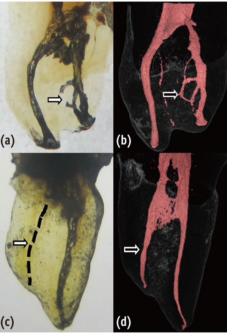
-
 Abstract
 PDF
PDF PubReader
 ePub
ePub Objectives This study evaluated the effect of three different air-drying times on microleakage of three self-etch adhesive systems.
Materials and Methods Class I cavities were prepared for 108 extracted sound human premolars. The teeth were divided into three main groups based on three different adhesives: Opti Bond All in One (OBAO), Clearfil S3 Bond (CSB), Bond Force (BF). Each main group divided into three subgroups regarding the air-drying time: without application of air stream, following the manufacturer's instruction, for 10 sec more than manufacturer's instruction. After completion of restorations, specimens were thermocycled and then connected to a fluid filtration system to evaluate microleakage. The data were statistically analyzed using two-way ANOVA and Tukey-test (α = 0.05).
Results The microleakage of all adhesives decreased when the air-drying time increased from 0 sec to manufacturer's instruction (
p < 0.001). The microleakage of BF reached its lowest values after increasing the drying time to 10 sec more than the manufacturer's instruction (p < 0.001). Microleakage of OBAO and CSB was significantly lower compared to BF in all three drying time (p < 0.001).Conclusions Increasing in air-drying time of adhesive layer in one-step self-etch adhesives caused reduction of microleakage, but the amount of this reduction may be dependent on the adhesive components of self-etch adhesives.
-
Citations
Citations to this article as recorded by- Species profile of volatile organic compounds emission and health risk assessment from typical indoor events in daycare centers
Hailin Zheng, Júlia Csemezová, Marcel Loomans, Shalika Walker, Florent Gauvin, Wim Zeiler
Science of The Total Environment.2024; 918: 170734. CrossRef - Development of Drying Process for Removal of Residual Moisture from Biomass Pretreated with Ethanol and Its Kinetic and Thermodynamic Analysis
Seo-Young Park, Jin-Hyun Kim
Biotechnology and Bioprocess Engineering.2021; 26(5): 814. CrossRef - Effect of 9.3 μm CO2 and 2.94 μm Er:YAG Laser vs. Bur Preparations on Marginal Adaptation in Enamel and Dentin of Mixed Class V Cavities Restored With Different Restorative Systems
Clara Isabel Anton y Otero, Enrico Di Bella, Ivo Krejci, Tissiana Bortolotto
Frontiers in Dental Medicine.2021;[Epub] CrossRef - Development of Drying Process for Removal of Residual Solvent from Crystalline Vancomycin and Kinetic and Thermodynamic Analysis Thereof
Tae-Hun Yoon, Jin-Hyun Kim
Biotechnology and Bioprocess Engineering.2020; 25(5): 777. CrossRef - Effect of adhesive air-drying time on bond strength to dentin: A systematic review and meta-analysis
Mohamed M. Awad, Ali Alrahlah, Jukka P. Matinlinna, Hamdi Hosni Hamama
International Journal of Adhesion and Adhesives.2019; 90: 154. CrossRef - Optical Evaluation of Enamel Microleakage with One-Step Self-Etch Adhesives
Alaa Turkistani, Maha Almutairi, Nouf Banakhar, Reem Rubehan, Sulafa Mugharbil, Ahmed Jamleh, Adnan Nasir, Turki Bakhsh
Photomedicine and Laser Surgery.2018; 36(11): 589. CrossRef - Improved drying method for removal of residual solvents from paclitaxel by pre-treatment with ethanol and water
Chung-Gi Lee, Jin-Hyun Kim
Process Biochemistry.2015; 50(6): 1031. CrossRef
- Species profile of volatile organic compounds emission and health risk assessment from typical indoor events in daycare centers
- 1,747 View
- 3 Download
- 7 Crossref

- Effect of chlorhexidine application on the bond strength of resin core to axial dentin in endodontic cavity
- Yun-Hee Kim, Dong-Hoon Shin
- Restor Dent Endod 2012;37(4):207-214. Published online November 21, 2012
- DOI: https://doi.org/10.5395/rde.2012.37.4.207
-
 Abstract
 PDF
PDF PubReader
 ePub
ePub Objectives This study evaluated the influence of chlorhexidine (CHX) on the microtensile bonds strength (µTBS) of resin core with two adhesive systems to dentin in endodontic cavities.
Materials and Methods Flat dentinal surfaces in 40 molar endodontic cavities were treated with self-etch adhesive system, Contax (DMG) and total-etch adhesive system, Adper Single Bond 2 (3M ESPE) after the following surface treatments: (1) Priming only (Contax), (2) CHX for 15 sec + rinsing + priming (Contax), (3) Etching with priming (Adper Single Bond 2), (4) Etching + CHX for 15 sec + rinsing + priming (Adper Single Bond 2). Resin composite build-ups were made with LuxaCore (DMG) using a bulk method and polymerized for 40 sec. For each condition, half of specimens were submitted to µTBS after 24 hr storage and half of them were submitted to thermocycling of 10,000 cycles between 5℃ and 55℃ before testing. The data were analyzed using ANOVA and independent
t -test at a significance level of 95%.Results CHX pre-treatment did not affect the bond strength of specimens tested at the immediate testing period, regardless of dentin surface treatments. However, after 10,000 thermocycling, all groups showed reduced bond strength. The amount of reduction was greater in groups without CHX treatments than groups with CHX treatment. These characteristics were the same in both self-etch adhesive system and total-etch adhesive system.
Conclusions 2% CHX application for 15 sec proved to alleviate the decrease of bond strength of dentin bonding systems. No significant difference was shown in µTBS between total-etching system and self-etching system.
-
Citations
Citations to this article as recorded by- Micro Tensile bond strength and microleakage assessment of total-etch and self-etch adhesive bonded to carious affected dentin disinfected with Chlorhexidine, Curcumin, and Malachite green
Zeeshan Qamar, Nishath Sayed Abdul, R Naveen Reddy, Mahesh Shenoy, Saleh Alghufaili, Yousef Alqublan, Ali Barakat
Photodiagnosis and Photodynamic Therapy.2023; 43: 103636. CrossRef - The Classification and Selection of Adhesive Agents; an Overview for the General Dentist
Naji Ziad Arandi
Clinical, Cosmetic and Investigational Dentistry.2023; Volume 15: 165. CrossRef - Influence of chlorhexidine 2% and sodium hypochlorite 5.25% on micro-tensile bond strength of universal adhesive system (G-Premio Bond)
Nafiseh Fazelian, Abbas Rahimi Dashtaki, MohammadAmin Eftekharian, Batool Amiri
Brazilian Journal of Oral Sciences.2022;[Epub] CrossRef - Comparative evaluation of the effects of different methods of post space preparation in primary anterior teeth on the fracture resistance of tooth restorations
Bahman Seraj, Sara Ghadimi, Ebrahim Najafpoor, Fatemeh Abdolalian, razieh khanmohammadi
Journal of Dental Research, Dental Clinics, Dental Prospects.2019; 13(2): 141. CrossRef - Chemical, microbial, and host‐related factors: effects on the integrity of dentin and the dentin–biomaterial interface
Marcela T. Carrilho, Fabiana Piveta, Leo Tjäderhane
Endodontic Topics.2015; 33(1): 50. CrossRef - MMP Inhibitors on Dentin Stability
A.F. Montagner, R. Sarkis-Onofre, T. Pereira-Cenci, M.S. Cenci
Journal of Dental Research.2014; 93(8): 733. CrossRef - Thermal cycling for restorative materials: Does a standardized protocol exist in laboratory testing? A literature review
Anna Lucia Morresi, Maurizio D'Amario, Mario Capogreco, Roberto Gatto, Giuseppe Marzo, Camillo D'Arcangelo, Annalisa Monaco
Journal of the Mechanical Behavior of Biomedical Materials.2014; 29: 295. CrossRef
- Micro Tensile bond strength and microleakage assessment of total-etch and self-etch adhesive bonded to carious affected dentin disinfected with Chlorhexidine, Curcumin, and Malachite green
- 1,244 View
- 3 Download
- 7 Crossref

- Bonding efficacy of cured or uncured dentin adhesives in indirect resin
- Ji-Hyun Jang, Bin-Na Lee, Hoon-Sang Chang, Yun-Chan Hwang, Won-Mann Oh, In-Nam Hwang
- J Korean Acad Conserv Dent 2011;36(6):490-497. Published online November 30, 2011
- DOI: https://doi.org/10.5395/JKACD.2011.36.6.490
-
 Abstract
 PDF
PDF PubReader
 ePub
ePub Objectives This study examined the effect of the uncured dentin adhesives on the bond interface between the resin inlay and dentin.
Materials and Methods Dentin surface was exposed in 24 extracted human molars and the teeth were assigned to indirect and direct resin restoration group. For indirect resin groups, exposed dentin surfaces were temporized with provisional resin. The provisional restoration was removed after 1 wk and the teeth were divided further into 4 groups which used dentin adhesives (OptiBond FL, Kerr; One-Step, Bisco) with or without light-curing, respectively (Group OB-C, OB-NC, OS-C and OS-NC). Pre-fabricated resin blocks were cemented on the entire surfaces with resin cement. For the direct resin restoration groups, the dentin surfaces were treated with dentin adhesives (Group OB-D and OS-D), followed by restoring composite resin. After 24 hr, the teeth were assigned to microtensile bond strength (µTBS) and confocal laser scanning microscopy (CLSM), respectively.
Results The indirect resin restoration groups showed a lower µTBS than the direct resin restoration groups. The µTBS values of the light cured dentin adhesive groups were higher than those of the uncured dentin adhesive groups (
p < 0.05). CLSM analysis of the light cured dentin adhesive groups revealed definite and homogenous hybrid layers. However, the uncured dentin adhesive groups showed uncertain or even no hybrid layer.Conclusions Light-curing of the dentin adhesive prior to the application of the cementing material in luting a resin inlay to dentin resulted in definite, homogenous hybrid layer formation, which may improve the bond strength.
- 1,425 View
- 9 Download

- Microshear bond strength of a self-etching primer adhesive to enamel according to the type of bur
- Jin-Ho Jeong, Young-Gon Cho, Myung-Seon Lee
- J Korean Acad Conserv Dent 2011;36(6):477-482. Published online November 30, 2011
- DOI: https://doi.org/10.5395/JKACD.2011.36.6.477
-
 Abstract
 PDF
PDF PubReader
 ePub
ePub Objectives The purpose of this study was to compare the microshear bond strength (uSBS) to enamel prepared with different burs and to determine what type of bur were chosen when a self-etching primer adhesive was used.
Materials and Methods Enamel of forty-two human molars were used. They were divided into one of six groups (n = 7), Group 1, coarse (125 - 150 µm) diamond bur; Group 2, standard (106 - 125 µm) diamond bur; Group 3, fine (53 - 63 µm) diamond bur; Group 4, extrafine (20 - 30 µm) diamond bur; Group 5, plain-cut carbide bur (no. 245); Group 6, cross-cut carbide bur (no. 557). Clearfil SE Bond and Clearfil AP-X (Kuraray Medical Inc.) was bonded to enamel surface. The bonded specimens were subjected to uSBS testing.
Results The uSBS of Group 4 was the highest among groups and it was significantly higher than that of Groups 1, 2, 3, and 6 (
p < 0.05), but it was not significantly different from that of Group 5.Conclusions Different burs used on enamel surface affected the microshear bond strengths of a self-etching primer adhesive to the enamel surface. In the case of Clearfil SE Bond, extrafine diamond and plain-cut carbide bur are recommended for bonding to enamel.
-
Citations
Citations to this article as recorded by- Sixty-month follow up of three different universal adhesives used with a highly-filled flowable resin composite in the restoration of non-carious cervical lesion
Fatma Dilsad Oz, Canan Ozturk, Reza Soleimani, Sevil Gurgan
Clinical Oral Investigations.2022; 26(8): 5377. CrossRef
- Sixty-month follow up of three different universal adhesives used with a highly-filled flowable resin composite in the restoration of non-carious cervical lesion
- 897 View
- 3 Download
- 1 Crossref

- Effect of Er:YAG lasing on the dentin bonding strength of two-step adhesives
- Byeong-Choon Song, Young-Gon Cho, Myung-Seon Lee
- J Korean Acad Conserv Dent 2011;36(5):409-418. Published online September 30, 2011
- DOI: https://doi.org/10.5395/JKACD.2011.36.5.409
-
 Abstract
 PDF
PDF PubReader
 ePub
ePub Objectives The purpose of this study was to compare the microshear bond strength (µSBS) and bonding interfaces of two-step total-etching and self-etching adhesive systems to three etch types of dentin either the acid etched, laser etched or laser and acid etched.
Materials and Methods The occlusal dentinal surfaces of thirty human molars were used. They were divided into six groups: group 1, 37% H3PO4 + Single Bond 2 (3M ESPE); group 2, Er:YAG laser (KEY Laser 3, KaVo) + Single Bond 2; group 3, Er:YAG laser + 37% H3PO4 + Single Bond 2; group 4, Clearfil SE Primer + Bond (Kuraray); group 5, Er:YAG laser + Clearfil SE Bond; group 6, Er:YAG laser + Clearfil SE Primer + Bond. The samples were subjected to µSBS testing 24 hr after bonding. Also scanning microscopic evaluations were made on the resin-dentin interfaces of six specimens.
Results The µSBS of group 2 was significantly lower than that of groups 1 and 3 in Single Bond 2 (
p < 0.05). There were significant differences among the uSBS of groups 4, 5, and 6 in Clearfil SE Bond (p < 0.05). Very short and slender resin tags were observed in groups 2 and 5. Long and slender resin tags and lateral branches of tags were observed in groups 3 and 6.Conclusions Treatment of dentin surface using phosphoric acid or self-etching primer improved the adhesion of Er:YAG lased dentin.
-
Citations
Citations to this article as recorded by- Effect of Acid or Laser Treatment on Degradation of Dentin Matrix
Aslihan Usumez, Tugrul Sari, Roda Seseogullari Dirihan, Mehmet Esad Guven, Serra Oguz Ahmet, Norbert Gutknecht, Arzu Tezvergil Mutluay
Lasers in Dental Science.2022; 6(2): 99. CrossRef - Ablation of carious dental tissue using an ultrashort pulsed laser (USPL) system
Christoph Engelbach, Claudia Dehn, Christoph Bourauel, Jörg Meister, Matthias Frentzen
Lasers in Medical Science.2015; 30(5): 1427. CrossRef
- Effect of Acid or Laser Treatment on Degradation of Dentin Matrix
- 826 View
- 1 Download
- 2 Crossref

- The effects of total-etch, wet-bonding, and light-curing of adhesive on the apical seal of a resin-based root canal filling system
- Won-Il Ryu, Won-Jun Shon, Seung-Ho Baek, In-Han Lee, Byeong-Hoon Cho
- J Korean Acad Conserv Dent 2011;36(5):385-396. Published online September 30, 2011
- DOI: https://doi.org/10.5395/JKACD.2011.36.5.385
-
 Abstract
 PDF
PDF PubReader
 ePub
ePub Objectives This study evaluated the effects of adhesion variables such as the priming concepts of canal wall and the curing modes of adhesives on the sealing ability of a resin-based root canal filling system.
Materials and Methods Apical microleakage of the Resilon-RealSeal systems filled with 3 different combinations of adhesion variables was compared with the conventional gutta-percha filling using a dye penetration method. Experimental groups were SEDC, Resilon (Resilon Research LLC) filling with self-etch RealSeal (SybronEndo) primer and dual-cure RealSeal sealer; NELC, Resilon filling with no etching, Scotchbond Multi-Purpose (3M ESPE) primer application and light-curing adhesive; and TELC, Resilon filling with Scotchbond Multi-Purpose primer and adhesive used under total etch / wet bonding and light-cure protocols. GPCS, gutta-percha filling with conventional AH26 plus sealer, was the control group.
Results The median longitudinal dye penetration length of TELC was significantly shorter than those of GPCS and SEDC (Kruskal-Wallis test,
p < 0.05). In the cross-sectional microleakage scores, TELC showed significant differences from other groups at 2 to 5 mm from the apical foramen (Kruskal-Wallis test,p < 0.05).Conclusions When a resin-based root canal filling material was used, compared to the self-etching primer and the dual-cure sealer, the total etch/wet-bonding with primer and light-curing of adhesive showed improved apical sealing and was highly recommended.
- 1,154 View
- 1 Download

- Effect of adhesive hydrophobicity on microtensile bond strength of low-shrinkage silorane resin to dentin
- So-Yeun Cho, Hyun-Young Kang, Kyoung-A Kim, Mi-Kyung Yu, Kwang-Won Lee
- J Korean Acad Conserv Dent 2011;36(4):280-289. Published online July 31, 2011
- DOI: https://doi.org/10.5395/JKACD.2011.36.4.280
-
 Abstract
 PDF
PDF PubReader
 ePub
ePub Objectives The purpose of this study was to evaluate µTBS (microtensile bond strength) of current dentin bonding adhesives which have different hydrophobicity with low-shrinkage silorane resin.
Materials and Methods Thirty-six human third molars were used. Middle dentin was exposed. The teeth were randomly assigned to nine experimental groups: Silorane self-etch adhesives (SS), SS + phosphoric acid etching (SS + pa), Adper easy bond (AE), AE + Silorane system bonding (AE + SSb), Clearfil SE bond (CSE), CSE + SSb, All-Bond 2 (AB2), AB2 + SSb, All-Bond 3 (AB3). After adhesive's were applied, the clinical crowns were restored with Filtek LS (3M ESPE). The 0.8 mm × 0.8 mm sticks were submitted to a tensile load using a Micro Tensile Tester (Bisco Inc.). Water sorption was measured to estimate hydrophobicity adhesives.
Results µTBS of silorane resin to 5 adhesives: SS, 23.2 MPa; CSE, 19.4 MPa; AB3, 30.3 MPa; AB2 and AE, no bond. Additional layering of SSb: CSE + SSb, 26.2 MPa; AB2 + SSb, 33.9 MPa; AE + SSb, no bond. High value of µTBS was related to cohesive failure. SS showed the lowest water sorption. AE showed the highest solubility.
Conclusions The hydrophobicity of adhesive increased, and silorane resin bond-strength was also increased. Additional hydrophobic adhesive layer did not increase the bond-strength to silorane resin except AB2 + SSb. All-Bond 3 showed similar µTBS & water sorption with SS. By these facts, we could reach a conclusion that All-Bond 3 is a competitive adhesive which can replace the Silorane adhesive system.
-
Citations
Citations to this article as recorded by- Microtensile bond strength of silorane-based composite specific adhesive system using different bonding strategies
Laura Alves Bastos, Ana Beatriz Silva Sousa, Brahim Drubi-Filho, Fernanda de Carvalho Panzeri Pires-de-Souza, Lucas da Fonseca Roberti Garcia
Restorative Dentistry & Endodontics.2015; 40(1): 23. CrossRef
- Microtensile bond strength of silorane-based composite specific adhesive system using different bonding strategies
- 1,268 View
- 1 Download
- 1 Crossref

- Influence of application methods of one-step self-etching adhesives on microtensile bond strength
- Chul-Kyu Choi, Sung-Ae Son, Jin-Hee Ha, Bock Hur, Hyeon-Cheol Kim, Yong-Hun Kwon, Jeong-Kil Park
- J Korean Acad Conserv Dent 2011;36(3):203-210. Published online May 31, 2011
- DOI: https://doi.org/10.5395/JKACD.2011.36.3.203
-
 Abstract
 PDF
PDF PubReader
 ePub
ePub Objectives The purpose of this study was to evaluate the effect of various application methods of one-step self-etch adhesives to microtensile resin-dentin bond strength.
Materials and Methods Thirty-six extracted human molars were used. The teeth were assigned randomly to twelve groups (
n = 15), according to the three different adhesive systems (Clearfil Tri-S Bond, Adper Prompt L-Pop, G-Bond) and application methods. The adhesive systems were applied on the dentin as follows: 1) The single coating, 2) The double coating, 3) Manual agitation, 4) Ultrasonic agitation. Following the adhesive application, light-cure composite resin was constructed. The restored teeth were stored in distilled water at room temperature for 24 hours, and prepared 15 specimens per groups. Then microtensile bond strength was measured and the failure mode was examined.Results Manual agitation and ultrasonic agitation of adhesive significantly increased the microtensile bond strength than single coating and double coating did. Double coating of adhesive significantly increased the microtensile bond strength than single coating did and there was no significant difference between the manual agitation and ultrasonic agitation group. There was significant difference in microtensile bonding strength among all adhesives and Clearfil Tri-S Bond showed the highest bond strength.
Conclusions In one-step self-etching adhesives, there was significant difference according to application methods and type of adhesives. No matter of the material, the manual or ultrasonic agitation of the adhesive showed significantly higher microtensile bond strength.
-
Citations
Citations to this article as recorded by- Effect of Baicalein on Bond Strength of Indirect Ceramic Restoration
Nuray Zulkadir Ergin, Aslı Seçilmiş
Süleyman Demirel Üniversitesi Sağlık Bilimleri Dergisi.2025; 16(3): 356. CrossRef - The Classification and Selection of Adhesive Agents; an Overview for the General Dentist
Naji Ziad Arandi
Clinical, Cosmetic and Investigational Dentistry.2023; Volume 15: 165. CrossRef
- Effect of Baicalein on Bond Strength of Indirect Ceramic Restoration
- 1,812 View
- 11 Download
- 2 Crossref

- The effect of the strength and wetting characteristics of Bis-GMA/TEGDMA-based adhesives on the bond strength to dentin
- Eun-Sook Park, Chang-Keun Kim, Ji-Hyun Bae, Byeong-Hoon Cho
- J Korean Acad Conserv Dent 2011;36(2):139-148. Published online March 31, 2011
- DOI: https://doi.org/10.5395/JKACD.2011.36.2.139
-
 Abstract
 PDF
PDF PubReader
 ePub
ePub Objectives This study investigated the effect of the strength and wetting characteristics of adhesives on the bond strength to dentin. The experimental adhesives containing various ratios of hydrophobic, low-viscosity Bis-M-GMA, with Bis-GMA and TEGDMA, were made and evaluated on the mechanical properties and bond strength to dentin.
Materials and Methods Five experimental adhesives formulated with various Bis-GMA/Bis-M-GMA/TEGDMA ratios were evaluated on their viscosity, degree of conversion (DC), flexural strength (FS), and microtensile bond strength (MTBS). The bonded interfaces were evaluated with SEM and the solubility parameter was calculated to understand the wetting characteristics of the adhesives.
Results Although there were no significant differences in the DC between the experimental adhesives at 48 hr after curing (
p > 0.05), the experimental adhesives that did not contain Bis-GMA exhibited a lower FS than did those containing Bis-GMA (p < 0.05). The experimental adhesives that had very little to no TEGDMA showed significantly lower MTBS than did those containing a higher content of TEGDMA (p < 0.05). The formers exhibited gaps at the interface between the adhesive layer and the hybrid layer. The solubility parameter of TEGDMA approximated those of the components of the primed dentin, rather than Bis-GMA and Bis-M-GMA.Conclusions To achieve a good dentin bond, a strong base monomer, such as Bis-GMA, cannot be completely replaced by Bis-M-GMA for maintaining mechanical strength. For compatible copolymerization between the adhesive and the primed dentin as well as dense cross-linking of the adhesive layer, at least 30% fraction of TEGDMA is also needed.
-
Citations
Citations to this article as recorded by- Equivalence study of the resin-dentine interface of internal tunnel restorations when using an enamel infiltrant resin with ethanol-wet dentine bonding
Andrej M. Kielbassa, Sabrina Summer, Wilhelm Frank, Edward Lynch, Julia-Susanne Batzer
Scientific Reports.2024;[Epub] CrossRef - Physical properties and cytotoxicity of antimicrobial dental resin adhesives containing dimethacrylate oligomers of Ciprofloxacin and Metronidazole
Yasaman Delaviz, Timothy W. Liu, Ashley R. Deonarain, Yoav Finer, Babak Shokati, J. Paul Santerre
Dental Materials.2019; 35(2): 229. CrossRef
- Equivalence study of the resin-dentine interface of internal tunnel restorations when using an enamel infiltrant resin with ethanol-wet dentine bonding
- 1,939 View
- 7 Download
- 2 Crossref

- Effect of curing modes on micro-hardness of dual-cure resin cements
- Ki-Deok Lee, Se-Hee Park, Jin-Woo Kim, Kyung-Mo Cho
- J Korean Acad Conserv Dent 2011;36(2):132-138. Published online March 31, 2011
- DOI: https://doi.org/10.5395/JKACD.2011.36.2.132
-
 Abstract
 PDF
PDF PubReader
 ePub
ePub Objectives The purpose of this study was to evaluate curing degree of three dual-cure resin cements with the elapsed time in self-cure and dual-cure mode by means of the repeated measure of micro-hardness.
Materials and Methods Two dual-cure self-adhesive resin cements studied were Maxcem Elite (Kerr), Rely-X Unicem (3M ESPE) and one conventional dual-cure resin cement was Rely-X ARC resin cement (3M ESPE). Twenty specimens for each cements were made in Teflon mould and divided equally by self-cure and dual-cure mode and left in dark, 36℃, 100% relative humidity conditional-micro-hardness was measured at 10 min, 30 min, 1 hr, 3 hr, 6 hr, 12 hr and 24 hr after baseline. The results of micro-hardness value were statistically analyzed using independent samples
t -test and one-way ANOVA with multiple comparisons using Scheffe's test.Results The micro-hardness values were increased with time in every test groups. Dual-cure mode obtained higher micro-hardness value than self-cure mode except after one hour of Maxcem. Self-cured Rely-X Unicem showed lowest value and dual-cured Rely-X Unicem showed highest value in every measuring time.
Conclusions Sufficient light curing to dual-cure resin cements should provided for achieve maximum curing.
- 916 View
- 9 Download

- Microshear bond strength of a flowable resin to enamel according to the different adhesive systems
- Jeong-Ho Kim, Young-Gon Cho
- J Korean Acad Conserv Dent 2011;36(1):50-58. Published online January 31, 2011
- DOI: https://doi.org/10.5395/JKACD.2011.36.1.50
-
 Abstract
 PDF
PDF PubReader
 ePub
ePub Objectives The purpose of this study was to compare the microshear bond strength (uSBS) of two total-etch and four self-etch adhesive systems and a flowable resin to enamel.
Materials and Methods Enamels of sixty human molars were used. They were divided into one of six equal groups (
n = 10) by adhesives used; OS group (One-Step Plus), SB group (Single Bond), CE group (Clearfil SE Bond), TY group (Tyrian SPE/One-Step Plus), AP group (Adper Prompt L-Pop) and GB group (G-Bond).After enamel surfaces were treated with six adhesive systems, a flowable composite resin (Filek Z 350) was bonded to enamel surface using Tygon tubes. the bonded specimens were subjected to uSBS testing and the failure modes of each group were observed under FE-SEM.
Results 1. The
u SBS of SB group was statistically higher than that of all other groups, and theu SBS of OS, SE and AP group was statistically higher than that of TY and GB group (p < 0.05).2. The
u SBS for TY group was statistically higher than that for GB group (p < 0.05).3. Adhesive failures in TY and GB group and mixed failures in SB group and SE group were often analysed. One cohesive failure was observed in OS, SB, SE and AP group, respectively.
Conclusions Although adhesives using the same step were applied the enamel surface, the uSBS of a flowable resin to enamel was different.
-
Citations
Citations to this article as recorded by- Enamel pretreatment with Er:YAG laser: effects on the microleakage of fissure sealant in fluorosed teeth
Mahtab Memarpour, Nasrin Kianimanesh, Bahareh Shayeghi
Restorative Dentistry & Endodontics.2014; 39(3): 180. CrossRef
- Enamel pretreatment with Er:YAG laser: effects on the microleakage of fissure sealant in fluorosed teeth
- 975 View
- 1 Download
- 1 Crossref

- Effect of 2% chlorhexidine application on microtensile bond strength of resin composite to dentin using one-step self-etch adhesives
- Soon-Ham Jang, Bock Hur, Hyeon-Cheol Kim, Yong-Hun Kwon, Jeong-Kil Park
- J Korean Acad Conserv Dent 2010;35(6):486-491. Published online November 30, 2010
- DOI: https://doi.org/10.5395/JKACD.2010.35.6.486
-
 Abstract
 PDF
PDF PubReader
 ePub
ePub Objectives This study examined the effect of 2% chlorhexidine on the µTBS of a direct composite restoration using one-step self-etch adhesives on human dentin.
Materials and Methods Twenty-four extracted permanent molars were used. The teeth were assigned randomly to six groups (
n = 10), according to the adhesive system and application of chlorhexidine. With or without the application of chlorhexidine, each adhesive system was applied to the dentin surface. After the bonding procedure, light-cure composite resin buildups were produced. The restored teeth were stored in distilled water at room temperature for 24 hours, and then cut and glued to the jig of the microtensile testing machine. A tensile load was applied until the specimen failed. The failure mode was examined using an operating microscope. The data was analyzed statistically using one-way ANOVA, Student'st -test (p < 0.05) and Scheffé's test.Results Regardless of the application of chlorhexidine, the Clearfil S3 Bond showed the highest µTBS, followed by G-Bond and Xeno V. Adhesive failure was the main failure mode of the dentin bonding agents tested with some samples showing cohesive failure.
Conclusions The application of 2% chlorhexidine did not affect the µTBS of the resin composite to the dentin using a one-step self-etch adhesive.
- 1,232 View
- 2 Download

- The effect of solvent evaporation of dentin adhesive on bonding efficacy
- Min-Woo Cho, Ji-Yeon Kim, Duck-Su Kim, Kyoung-Kyu Choi
- J Korean Acad Conserv Dent 2010;35(5):321-334. Published online September 30, 2010
- DOI: https://doi.org/10.5395/JKACD.2010.35.5.321
-
 Abstract
 PDF
PDF PubReader
 ePub
ePub Objectives The purpose of this study is to evaluate bonding efficacy by means of measuring the effect of remained solvent on Degree of conversion(DC) and µTBS and FE-SEM examination.
Materials and Methods Two 2-step total etching adhesives and two single-step self etching adhesives were used in this study. First, volume weight loss of 4 dentin adhesives were measured using weighting machine in process of time in normal conditions and calculate degree of evaporation (DE). Reaction/reference intensity ratio were measured using micro-Raman spectroscopy and calculate DC according to DE. Then 2 experimental groups were prepared according to air-drying methods (under, over) and control group was prepared to manufacturer's instruction. Total 12 groups were evaluated by means of micro tensile bond strength and FE-SEM examination.
Results Degree of evaporation (DE) was increased as time elapsed but different features were observed according to the kind of solvents. Acetone based adhesive showed higher DE than ethanol and butanol based adhesive. Degree of conversion (DC) was increased according to DE except for S3 bond. In µTBS evaluation, bond strength was increased by additional air-drying. Large gaps and droplets were observed in acetone based adhesives by FE-SEM pictures.
Conclusions Additional air-drying is recommended for single-step self etching adhesive but careful consideration is required for 2-step total etching adhesive because of oxygen inhibition layer. Evaporation method is carefully chose and applied according to the solvent type.
-
Citations
Citations to this article as recorded by- Experimental study on the formability of aluminum pouch for lithium polymer battery by manufacturing processes
Minsook Yu, Munyong Song, Minha Kim, Dongsoo Kim
Journal of Mechanical Science and Technology.2019; 33(9): 4353. CrossRef
- Experimental study on the formability of aluminum pouch for lithium polymer battery by manufacturing processes
- 1,371 View
- 1 Download
- 1 Crossref

- The effect of the removal of chondroitin sulfate on bond strength of dentin adhesives and collagen architecture
- Jong-Ryul Kim, Sang-Jin Park, Gi-Woon Choi, Kyoung-Kyu Choi
- J Korean Acad Conserv Dent 2010;35(3):211-221. Published online May 31, 2010
- DOI: https://doi.org/10.5395/JKACD.2010.35.3.211
-
 Abstract
 PDF
PDF PubReader
 ePub
ePub Proteoglycan is highly hydrophilic and negatively charged which enable them attract the water. The objective of study was to investigate the effects of Proteoglycan on microtensile bond strength of dentin adhesives and on architecture of dentin collagen matrix of acid etched dentin by removing the chondroitin sulphate attached on Proteoglycan. A flat dentin surface in mid-coronal portion of tooth was prepared. After acid etching, half of the specimens were immersed in 0.1 U/mL chondroitinase ABC (C-ABC) for 48 h at 37℃, while the other half were stored in distilled water. Specimens were bonded with the dentin adhesive using three different bonding techniques (wet, dry and re-wet) followed by microtensile bond strength test. SEM examination was done with debonded specimen, resin-dentin interface and acid-etched dentin surface with/without C-ABC treatment.
For the subgroups using wet-bonding or dry-bonding technique, microtensile bond strength showed no significant difference after C-ABC treatment (p > 0.05). Nevertheless, the subgroup using rewetting technique after air dry in the Single Bond 2 group demonstrated a significant decrease of microtensile bond strength after C-ABC treatment. Collagen architecture is loosely packed and some fibrils are aggregated together and relatively collapsed compared with normal acid-etched wet dentin after C-ABC treatment. Further studies are necessary for the contribution to the collagen architecture of noncollagenous protein under the various clinical situations and several dentin conditioners and are also needed about long-term effect on bond strength of dentin adhesive.
- 923 View
- 1 Download

- Effect of the exponential curing of composite resin on the microtensile dentin bond strength of adhesives
- So-Rae Seong, Duck-kyu Seo, In-Bog Lee, Ho-Hyun Son, Byeong-Hoon Cho
- J Korean Acad Conserv Dent 2010;35(2):125-133. Published online March 31, 2010
- DOI: https://doi.org/10.5395/JKACD.2010.35.2.125
-
 Abstract
 PDF
PDF PubReader
 ePub
ePub Objectives Rapid polymerization of overlying composite resin causes high polymerization shrinkage stress at the adhesive layer. In order to alleviate the shrinkage stress, increasing the light intensity over the first 5 seconds was suggested as an exponential curing mode by an LED light curing unit (Elipar FreeLight2, 3M ESPE). In this study, the effectiveness of the exponential curing mode on reducing stress was evaluated with measuring microtensile bond strength of three adhesives after the overlying composite resin was polymerized with either continuous or exponential curing mode.
Methods Scotchbond Multipurpose Plus (MP, 3M ESPE), Single Bond 2 (SB, 3M ESPE), and Adper Prompt (AP, 3M ESPE) were applied onto the flat occlusal dentin of extracted human molar. The overlying hybrid composite (Denfil, Vericom, Korea) was cured under one of two exposing modes of the curing unit. At 48h from bonding, microtensile bond strength was measured at a crosshead speed of 1.0 mm/min. The fractured surfaces were observed under FE-SEM.
Results There was no statistically significant difference in the microtensile bond strengths of each adhesive between curing methods (Two-way ANOVA, p > 0.05). The microtensile bond strengths of MP and SB were significantly higher than that of AP (p < 0.05). Mixed failures were observed in most of the fractured surfaces, and differences in the failure mode were not observed among groups.
Conclusion The exponential curing method had no beneficial effect on the microtensile dentin bond strengths of three adhesives compared to continuous curing method.
-
Citations
Citations to this article as recorded by- The effect of the strength and wetting characteristics of Bis-GMA/TEGDMA-based adhesives on the bond strength to dentin
Eun-Sook Park, Chang-Keun Kim, Ji-Hyun Bae, Byeong-Hoon Cho
Journal of Korean Academy of Conservative Dentistry.2011; 36(2): 139. CrossRef
- The effect of the strength and wetting characteristics of Bis-GMA/TEGDMA-based adhesives on the bond strength to dentin
- 1,015 View
- 1 Download
- 1 Crossref

- Microtensile bond strength of self-etching and self-adhesive resin cements to dentin and indirect composite resin
- Jae-Gu Park, Young-Gon Cho, Il-Sin Kim
- J Korean Acad Conserv Dent 2010;35(2):106-115. Published online March 31, 2010
- DOI: https://doi.org/10.5395/JKACD.2010.35.2.106
-
 Abstract
 PDF
PDF PubReader
 ePub
ePub The purpose of this study was to evaluate the microtensile bond strength (µTBS), failure modes and bonding interfaces of self-etching and three self-adhesive resin cements to dentin and indirect composite resin.
Cylindrical composite blocks (Tescera, Bisco Inc.) were luted with resin cements (PA: Panavia F 2.0, Kuraray Medical Inc., RE: RelyX Unicem Clicker, 3M ESPE., MA: Maxem, Kerr Co., BI: BisCem, Bisco Inc.) on the prepared occlusal dentin surfaces of 20 extracted molars. After storage in distilled water for 24 h, 1.0 mm × 1.0 mm composite-dentin beams were prepared. µTBS was tested at a cross-head speed of 0.5 mm/min. Data were analyzed with one-way ANOVA and Tukey's HSD test. Dentin sides of all fractured specimens and interfaces of resin cements-dentin or resin cements-composite were examined at FE-SEM (Field Emission-Scanning Electron Microscope).
In conclusion, PA and RE showed higher bond strength and closer adaptation than MA and BI when indirect composite blocks were luted to dentin using a self-etching and three self-adhesive resin cements.
- 895 View
- 2 Download

- Effect of cutting instruments on the dentin bond strength of a self-etch adhesive
- Young-Gon Lee, So-Ra Moon, Young-Gon Cho
- J Korean Acad Conserv Dent 2010;35(1):13-19. Published online January 31, 2010
- DOI: https://doi.org/10.5395/JKACD.2010.35.1.013
-
 Abstract
 PDF
PDF PubReader
 ePub
ePub The purpose of this study was to compare the microshear bond strength of a self-etching primer adhesive to dentin prepared with different diamond points, carbide burs and SiC papers, and also to determine which SiC paper yield similar strength to that of dentinal surface prepared with points or burs.
Fifty-six human molar were sectioned to expose the occlusal dentinal surfaces of crowns and slabs of 1.2 mm thick were made. Dentinal surfaces were removed with three diamond points, two carbide burs, and three SiC papers. They were divided into one of eight equal groups (n = 7); Group 1: standard diamond point(TF-12), Group 2: fine diamond point (TF-12F), Group 3: extrafine diamond point (TF-12EF), Group 4: plain-cut carbide bur (no. 245), Group 5: cross-cut carbide bur (no. 557), Group 6 : P 120-grade SiC paper, Group 7: P 220-grade SiC paper, Group 8: P 800-grade SiC paper.
Clearfil SE Bond was applied on dentinal surface and Clearfil AP-X was placed on dentinal surface using Tygon tubes. After the bonded specimens were subjected to uSBS testing, the mean uSBS (n = 20 for each group) was statistically compared using one-way ANOVA and Tukey HSD test.
In conclusion, the use of extrafine diamond point is recommended for improved bonding of Clearfil SE Bond to dentin. Also the use of P 220-grade SiC paper in vitro will be yield the results closer to dentinal surface prepared with fine diamond point or carbide burs
in vivo .-
Citations
Citations to this article as recorded by- Evaluation of the flexural and repair bond strengths of 3D-printed temporary restorations
Nazmi Dinçer, Şafak Külünk, Seniha Kısakürek, Ibrahim Duran
BMC Oral Health.2025;[Epub] CrossRef - Comparison of shear bond strength between various temporary prostheses resin blocks fabricated by subtractive and additive manufacturing methods bonded to self-curing reline resin
Hyo-Min Ryu, Jin-Han Lee
The Journal of Korean Academy of Prosthodontics.2023; 61(3): 189. CrossRef - The Effect of Aging and Different Surface Treatments on Temporary Cement Bonding of Temporaray Crown Materials
Sebahat FINDIK AYDINER, Nuran YANIKOĞLU, Zeynep YEŞİL DUYMUŞ
Cumhuriyet Dental Journal.2023; 26(2): 144. CrossRef - Influence of surface treatments and repair materials on the shear bond strength of CAD/CAM provisional restorations
Ki-Won Jeong, Sung-Hun Kim
The Journal of Advanced Prosthodontics.2019; 11(2): 95. CrossRef - Shear bond strength of dental CAD-CAM hybrid restorative materials repaired with composite resin
Yun-Hee Moon, Jonghyuk Lee, Myung-Gu Lee
The Journal of Korean Academy of Prosthodontics.2016; 54(3): 193. CrossRef - Microshear bond strength of a self-etching primer adhesive to enamel according to the type of bur
Jin-Ho Jeong, Young-Gon Cho, Myung-Seon Lee
Journal of Korean Academy of Conservative Dentistry.2011; 36(6): 477. CrossRef
- Evaluation of the flexural and repair bond strengths of 3D-printed temporary restorations
- 1,019 View
- 12 Download
- 6 Crossref

- Comparison of marginal microleakage between low and high flowable resins in class V cavity
- Sang-Bae Bae, Young-Gon Cho, Myeong-Seon Lee
- J Korean Acad Conserv Dent 2009;34(6):477-483. Published online November 30, 2009
- DOI: https://doi.org/10.5395/JKACD.2009.34.6.477
-
 Abstract
 PDF
PDF PubReader
 ePub
ePub The purpose of this study was to compare the microleakage of low and high viscosity flowable resins in class V cavities applied with 1-step adhesives.
Forty class V cavities were prepared on the cervices of buccal and lingual surfaces of extracted molar teeth and divided into four groups (n=8). Cavities were restored with AQ Bond Plus/Metafil Flo α, G-Bond/UniFil LoFlo Plus (Low flow groups), AQ Bond Plus/Metafil Flo and G-Bond/UniFil Flow (High flow group), respectively.
Specimens were immersed in a 2% methylene blue solution for 24 hours, and bisected longitudinally. They were observed microleakages at the enamel and dentinal margins.
In conclusion, the low viscosity flowable resins showed lower marginal microleakage than do the high viscosity flowable resins in class V cavities.
- 747 View
- 5 Download


 KACD
KACD

 First
First Prev
Prev


