
Previous issues
- Page Path
- HOME > Browse articles > Previous issues
- Volume 46 (3); August 2021
-


Review Articles
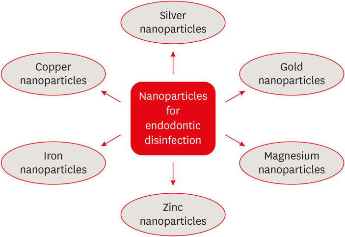
- Silver nanoparticles in endodontics: recent developments and applications
- Aysenur Oncu, Yan Huang, Gulin Amasya, Fatma Semra Sevimay, Kaan Orhan, Berkan Celikten
- Restor Dent Endod 2021;46(3):e38. Published online July 1, 2021
- DOI: https://doi.org/10.5395/rde.2021.46.e38
-
 Abstract
 PDF
PDF PubReader
 ePub
ePub The elimination of endodontic biofilms and the maintenance of a leak-proof canal filling are key aspects of successful root canal treatment. Several materials have been introduced to treat endodontic disease, although treatment success is limited by the features of the biomaterials used. Silver nanoparticles (AgNPs) have been increasingly considered in dental applications, especially endodontics, due to their high antimicrobial activity. For the present study, an electronic search was conducted using MEDLINE (PubMed), the Cochrane Central Register of Controlled Trials (CENTRAL), Google Scholar, and EMBASE. This review provides insights into the unique characteristics of AgNPs, including their chemical, physical, and antimicrobial properties; limitations; and potential uses. Various studies involving different application methods of AgNPs were carefully examined. Based on previous clinical studies, the synthesis, means of obtaining, usage conditions, and potential cytotoxicity of AgNPs were evaluated. The findings indicate that AgNPs are effective antimicrobial agents for the elimination of endodontic biofilms.
-
Citations
Citations to this article as recorded by
- Endodontic Intracanal Medicaments and Agents
Anu Priya Guruswamy Pandian, Depti Bellani, Ritya Mary Jibu, Varsha Agnihotri
Dental Clinics of North America.2026; 70(1): 45. CrossRef - Time-dependent Tooth Color Changes Following Conventional, Silver-based, and Photodynamic Root Canal Irrigants: An In Vitro Study
Laila Mohamed Mohamed Kenawi, Mohamed Fattouh, Khaled Abid Althaqafi, Abla Arafa
The Open Dentistry Journal.2026;[Epub] CrossRef - Advanced nanoparticle-based antibacterial delivery for endodontic disinfection: A systematic review and meta-analysis
Kanwalpreet Kaur, Seerat Kaura, Ravinder S Saini, Maurya Manjunath, Shashit Shetty Bavabeedu, Mario Alberto Alarcón-Sánchez, Javier Flores-Fraile, Artak Heboyan
Journal of Dentistry.2026; 166: 106347. CrossRef - Research Trends, Visualization, and Dynamics of Scientific Publications on Silver Nanoparticles in Dentistry: A Scientometric Approach
Fran Espinoza-Carhuancho, Berly Delgado-Cumpa, Lucia Quispe-Tasayco, Cesar Mauricio-Vilchez, Sylvia A Chein-Villacampa, Frank Mayta-Tovalino
The Journal of Contemporary Dental Practice.2026; 27(2): 223. CrossRef - Nanoparticle-Assisted Endodontic Irrigation: A Systematic Review of Preclinical Studies
Hashim Mueen Hussein, Ahmed Ali
Clinical, Cosmetic and Investigational Dentistry.2026; Volume 18: 1. CrossRef - Nanomaterial-enhanced regenerative therapies in endodontics: Current evidence, clinical applications, and future directions
Muskan Jain, Rhythm Bains, Promila Verma, Rakesh Kumar Yadav
Journal of Healthcare Research and Education.2026; 2: 8. CrossRef - Scoping review on the genotoxicity of silver nanoparticles in endodontics: therapeutic saviors or genetic saboteurs?
Galvin Sim Siang Lin, Widya Lestari, Mohd Haikal Muhamad Halil, Mohd Syafiq Abd Aziz
Odontology.2025; 113(2): 457. CrossRef - Bioceramics in Endodontics: Limitations and Future Innovations—A Review
Peramune Arachchilage Amila Saman Prasad Kumara, Paul Roy Cooper, Peter Cathro, Maree Gould, George Dias, Jithendra Ratnayake
Dentistry Journal.2025; 13(4): 157. CrossRef - Recent advances in antibacterial nanoformulations for endodontic applications
Tiago Dionísio, Pedro Brandão, Vanessa Machado, João Botelho, José João Mendes, Pedro Fonte
Expert Opinion on Drug Delivery.2025; 22(8): 1117. CrossRef - Systematic review of silver and vanadium-based antibiofilm agents: mechanisms and efficacy in oral biofilms
João Marcos Carvalho-Silva, Andréa Cândido dos Reis
Future Microbiology.2025; 20(10): 639. CrossRef - Nanomaterial-Enhanced Dentistry: A Clinical Perspective
Selvam Manoj, Radhakrishnan Sreena, Rajkumar Divya, Starlin Ebinesh, Shenbagaraman Akshaya, Srikumar Sugantha Angel, Arputharaj Joseph Nathanael
ACS Biomaterials Science & Engineering.2025; 11(8): 4671. CrossRef - Antimicrobial Effects of Formulations of Various Nanoparticles and Calcium Hydroxide as Intra-canal Medications Against Enterococcus faecalis: A Systematic Review
Seema H Bukhari, Dax Abraham, Shakila Mahesh
Cureus.2024;[Epub] CrossRef - The Push-Out Bond Strength, Surface Roughness, and Antimicrobial Properties of Endodontic Bioceramic Sealers Supplemented with Silver Nanoparticles
Karla Navarrete-Olvera, Nereyda Niño-Martínez, Idania De Alba-Montero, Nuria Patiño-Marín, Facundo Ruiz, Horacio Bach, Gabriel-Alejandro Martínez-Castañón
Molecules.2024; 29(18): 4422. CrossRef - Synergistic bactericidal activity of chlorhexidine loaded on positively charged ionic liquid-protected silver nanoparticles as a root canal disinfectant against Enterococcus faecalis: An ex vivo study
Abbas Abbaszadegan, Elham Tayebikhorami, Ahmad Gholami, Nazanin Bonyanpour, Bahar Asheghi, Sara Nikmanesh
Journal of Ionic Liquids.2024; 4(2): 100117. CrossRef - Improving the Antimicrobial Potency of Berberine for Endodontic Canal Irrigation Using Polymeric Nanoparticles
Célia Marques, Liliana Grenho, Maria Helena Fernandes, Sofia A. Costa Lima
Pharmaceutics.2024; 16(6): 786. CrossRef - A narrative review on application of metal and metal oxide nanoparticles in endodontics
Roohollah Sharifi, Ahmad Vatani, Amir Sabzi, Mohsen Safaei
Heliyon.2024; 10(15): e34673. CrossRef - The Effectiveness of Silver Nanoparticles Mixed with Calcium Hydroxide against Candida albicans: An Ex Vivo Analysis
Maha Alghofaily, Jood Alfraih, Aljohara Alsaud, Norah Almazrua, Terrence S. Sumague, Sayed H. Auda, Fahd Alsalleeh
Microorganisms.2024; 12(2): 289. CrossRef - Evaluation of the efficacy of a novel disinfecting material on the surface topography of gutta-percha: An in vitro study
KHanisha Reddy, Lekshmi Chandran, TMurali Mohan, K Sudha, DL Malini, Bonney Dominic
Journal of Conservative Dentistry.2023; 26(1): 94. CrossRef - Silver Nanoparticles and Their Therapeutic Applications in Endodontics: A Narrative Review
Farzaneh Afkhami, Parisa Forghan, James L. Gutmann, Anil Kishen
Pharmaceutics.2023; 15(3): 715. CrossRef - Nanopartículas antimicrobianas en endodoncia: Revisión narrativa
Gustavo Adolfo Tovar Rangel , Fanny Mildred González Sáenz , Ingrid Ximena Zamora Córdoba , Lina María García Zapata
Revista Estomatología.2023;[Epub] CrossRef - Functionalized Nanoparticles: A Paradigm Shift in Regenerative Endodontic Procedures
Vinoo Subramaniam Ramachandran, Mensudar Radhakrishnan, Malathi Balaraman Ravindrran, Venkatesh Alagarsamy, Gowri Shankar Palanisamy
Cureus.2022;[Epub] CrossRef
- Endodontic Intracanal Medicaments and Agents
- 7,306 View
- 134 Download
- 16 Web of Science
- 21 Crossref

- Traditional and minimally invasive access cavities in endodontics: a literature review
- Ioanna Kapetanaki, Fotis Dimopoulos, Christos Gogos
- Restor Dent Endod 2021;46(3):e46. Published online August 13, 2021
- DOI: https://doi.org/10.5395/rde.2021.46.e46
-
Abstract
PDFPubReaderePub
The aim of this review was to evaluate the effects of different access cavity designs on endodontic treatment and tooth prognosis. Two independent reviewers conducted an unrestricted search of the relevant literature contained in the following electronic databases: PubMed, Science Direct, Scopus, Web of Science, and OpenGrey. The electronic search was supplemented by a manual search during the same time period. The reference lists of the articles that advanced to second-round screening were hand-searched to identify additional potential articles. Experts were also contacted in an effort to learn about possible unpublished or ongoing studies. The benefits of minimally invasive access (MIA) cavities are not yet fully supported by research data. There is no evidence that this approach can replace the traditional approach of straight-line access cavities. Guided endodontics is a new method for teeth with pulp canal calcification and apical infection, but there have been no cost-benefit investigations or time studies to verify these personal opinions. Although the purpose of MIA cavities is to reflect clinicians' interest in retaining a greater amount of the dental substance, traditional cavities are the safer method for effective instrument operation and the prevention of iatrogenic complications.
-
Citations
Citations to this article as recorded by- Benefits of Using Magnification in Access Cavity Preparation by Undergraduate Dental Students: A Micro‐Computed Tomography Study
Manal Almaslamani, Okba Mahmoud, Aya Ali, Mawada Abdelmagied
European Journal of Dental Education.2026; 30(1): 77. CrossRef - Impact of conservative and traditional endodontic accesses on the strength of maxillary zirconia crowns
Carlos A. Jurado, Gustavo Morrice, Mark Antal, Silvia Rojas‐Rueda, Francisco X. Azpiazu‐Flores, Brian R. Morrow, Franklin Garcia‐Godoy, Damian J. Lee
Journal of Prosthodontics.2026; 35(3): 323. CrossRef - Effect of access cavity design on canal instrumentation efficiency and fracture resistance in mandibular molars: A cone-beam computed tomography study
Dalia Al-Harith, Rawan Meshal AlOtaibi
Saudi Journal of Oral Sciences.2025; 12(1): 72. CrossRef - Full-coverage Porcelain-fused-to-metal Crown with Guided Access for Future Endodontic Treatment: A Comparative Pilot In Vitro Study
Mohammed Mashyakhy, Hemant Chourasia, Hafiz Adawi, Abdulaziz Abu-Melha, Elham Khudhayr, Rafif Bakri, Taif Kameli, Khalid Moashy, Hitesh Chohan
The Journal of Contemporary Dental Practice.2025; 26(3): 234. CrossRef - Comparative evaluation of root canal morphology in mandibular first premolars with deep radicular grooves using direct vision, dental operating microscope, 2D radiographic visualisation and micro-computed tomography
Mohmed Isaqali Karobari, Hany Mohamed Aly Ahmed, Mohd Fadhli Bin Khamis, Norliza Ibrahim, Tahir Yusuf Noorani, Miriam Fatima Zaccaro Scelza
PLOS One.2025; 20(7): e0329439. CrossRef - A Finite Element Method Study of Stress Distribution in Dental Hard Tissues: Impact of Access Cavity Design and Restoration Material
Mihaela-Roxana Boțilă, Dragos Laurențiu Popa, Răzvan Mercuț, Monica Mihaela Iacov-Crăițoiu, Monica Scrieciu, Sanda Mihaela Popescu, Veronica Mercuț
Bioengineering.2024; 11(9): 878. CrossRef - Impact of Access Cavity Design on Fracture Resistance of Endodontically Treated Maxillary First Premolar: In Vitro
Anju Daniel, Abdul Rahman Saleh, Anas Al-Jadaa, Waad Kheder
Brazilian Dental Journal.2024;[Epub] CrossRef - Management of Traumatized Teeth With Severely Calcified Canals and Minimally Invasive Access Cavity Using the AReneto® System: A Case Report
Pucha Sai Manaswini, Varun Prabhuji, Champa C, Srirekha A, Veena S Pai
Cureus.2024;[Epub] CrossRef - Exploring the Impact of Access Cavity Designs on Canal Orifice Localization and Debris Presence: A Scoping Review
Mario Dioguardi, Davide La Notte, Diego Sovereto, Cristian Quarta, Andrea Ballini, Vito Crincoli, Riccardo Aiuto, Mario Alovisi, Angelo Martella, Lorenzo Lo Muzio
Clinical and Experimental Dental Research.2024;[Epub] CrossRef - The effect of computer aided navigation techniques on the precision of endodontic access cavities: A systematic review and meta-analysis
P. R. Kesharani, S. D. Aggarwal, N. K. Patel, J. A. Patel, D. A. Patil, S. H. Modi
Endodontics Today.2024; 22(3): 244. CrossRef - Minimally Invasive Access Cavity Designs: A Review
Sushmita Rane, Varsha Pandit, Ashwini Gaikwad, Shivani Chavan, Rajlaxmi Patil, Mrunal Shinde
Journal of Pharmacy and Bioallied Sciences.2024; 16(Suppl 3): S1971. CrossRef - Influence of Cavity Designs on Fracture Resistance: Analysis of the Role of Different Access Techniques to the Endodontic Cavity in the Onset of Fractures: Narrative Review
Mario Dioguardi, Davide La Notte, Diego Sovereto, Cristian Quarta, Angelo Martella, Andrea Ballini, Cornelis H. Pameijer
The Scientific World Journal.2024;[Epub] CrossRef - Digital precision meets dentin preservation: PriciGuide™ system for guided access opening
Varun Prabhuji, A. Srirekha, Veena Pai, Archana Srinivasan, S. M. Laxmikanth, Shwetha Shanbhag
Journal of Conservative Dentistry and Endodontics.2024; 27(8): 884. CrossRef - Minimal Invasive Endodontics: A Comprehensive Narrative Review
Jaydip Marvaniya, Kishan Agarwal, Dhaval N Mehta, Nirav Parmar, Ritwik Shyamal , Jenee Patel
Cureus.2022;[Epub] CrossRef
- Benefits of Using Magnification in Access Cavity Preparation by Undergraduate Dental Students: A Micro‐Computed Tomography Study
- 8,585 View
- 245 Download
- 10 Web of Science
- 14 Crossref
Research Articles
- Postoperative pain after endodontic treatment of necrotic teeth with large intentional foraminal enlargement
- Ricardo Machado, Daniel Comparin, Sérgio Aparecido Ignácio, Ulisses Xavier da Silva Neto
- Restor Dent Endod 2021;46(3):e31. Published online May 31, 2021
- DOI: https://doi.org/10.5395/rde.2021.46.e31
-
Abstract
PDFPubReaderePub
Objectives To evaluate postoperative pain after endodontic treatment of necrotic teeth using large intentional foraminal enlargement (LIFE).
Materials and Methods The sample included 60 asymptomatic necrotic teeth (with or without chronic apical periodontitis), and a periodontal probing depth of 3 mm, previously accessed and referred to perform endodontic treatment. After previous procedures, the position and approximate size of the apical foramen (AF) were determined by using an apex locator and K flexo-files, respectively. The chemomechanical preparation was performed with Profile 04 files 2 mm beyond the AF to achieve the LIFE, using 2.5 mL of 2.5% NaOCl at each file change. The filling was performed by Tagger's hybrid technique and EndoFill sealer. Phone calls were made to all the patients at 24, 48 and 72 hours after treatment, to classify postoperative pain. Statistical analysis was performed by different tests with a significance level of 5%.
Results Age, gender, periradicular status and tooth type did not influence postoperative pain (
p > 0.05). Only 1 patient (1.66%) reported severe pain after 72 hours. Moderate pain was reported by 7, 4 and 3 patients after 24, 48 and 72 hours, respectively (p = 0.0001). However, paired analyses showed a statistically significant difference only between 24 and 72 hours (p = 0.04). Sealer extrusion did not influence the postoperative pain (p > 0.05).Conclusions Acute or moderate postoperative pain was uncommon after endodontic treatment of necrotic teeth with LIFE.
Trial Registration The Brazilian Clinical Trials Registry Identifier:
RBR-3r967t -
Citations
Citations to this article as recorded by- Postoperative Pain After Endodontic Treatment in HIV‐Positive Patients Under HAART: A Prospective Observational Cohort Study
Marcos Felipe Iparraguirre Nuñovero, Marco Antonio Hungaro Duarte, Luciana Reis Azevedo Alanis, Bruno Cavalini Cavenago, Ulisses Xavier da Silva Neto, Everdan Carneiro
International Endodontic Journal.2026; 59(5): 788. CrossRef - Does maintaining apical patency reduce early postoperative pain after root canal treatment? A randomized controlled trial in asymptomatic vital single-rooted teeth
Ozan Arda Deger, Sehnaz Yilmaz, Kübra Gürler
Clinical Oral Investigations.2026;[Epub] CrossRef - Evaluation of Postoperative Pain Frequency in Single‐Session Endodontic Treatments With Patency and Foraminal Enlargement
Viviane Barbosa Godoy, Ana Grasiela Limoeiro, Vanessa Sandini, Vini Mehta, Wayne Martins Nascimento, Marilia Fagury Videira Marceliano‐Alves, Marcos Frozoni
Clinical and Experimental Dental Research.2026;[Epub] CrossRef - Assessment of apical extrusion in regenerative endodontics: a comparative study of different irrigation methods using three-dimensional immature tooth models
Vahide Hazal Abat, Gökçen Deniz Bayrak, Mustafa Gündoğar
Odontology.2025; 113(1): 213. CrossRef - Clinical Advances in Calcium Phosphate for Maxillomandibular Bone Regeneration: From Bench to Bedside
Seyed Ali Mostafavi Moghaddam, Hamid Mojtahedi, Amirhossein Bahador, Lotfollah Kamali Hakim, Hamid Tebyaniyan
Ceramics.2025; 8(4): 129. CrossRef - Postoperative pain after single-visit root canal treatments in necrotic teeth comparing instruments’ kinematics and apical instrumentation limits – a prospective randomized multicenter clinical trial
Ricardo Machado, Guilherme Moreira, Daniel Comparin, Arthur Pimentel Barroso, Jaqueline Nascimento, Caio Cézar Randi Ferraz, Sérgio Aparecido Ignácio, Lucas da Fonseca Roberti Garcia, Rodrigo Rodrigues Amaral, David Shadid, Ulisses Xavier da Silva Neto
BMC Oral Health.2024;[Epub] CrossRef - Assessment of mechanical allodynia in healthy teeth adjacent and contralateral to endodontically diseased teeth: a clinical study
Vaishnavi Ratnakar Patankar, Ashish K Jain, Rahul D Rao, Prajakta R Rao
Restorative Dentistry & Endodontics.2024;[Epub] CrossRef - A systematic review and meta-analysis on the effects of phototherapy on postoperative pain in conventional endodontic reintervention
Larissa Pereira Nunes, Gabriel Pereira Nunes, Túlio Morandin Ferrisse, Henrico Badaoui Strazzi-Sahyon, Eloi Dezan-Júnior, Luciano Tavares Angelo Cintra, Gustavo Sivieri-Araujo
Clinical Oral Investigations.2024;[Epub] CrossRef - The effect of intracanal cryotherapy with and without foraminal enlargement on pain prevention after endodontic treatment: a randomized clinical trial
Marcos Felipe Iparraguirre Nuñovero, Marco Antonio Hungaro Duarte, André Vinícius Kaled Segato, Ulisses Xavier da Silva Neto, Vania Portela Ditzel Westphalen, Everdan Carneiro
Scientific Reports.2024;[Epub] CrossRef - Clinical determination of anatomical diameter in different dental groups correlating them with gender, age, tooth/canal and pulpoperiradicular diagnosis: an observational clinical study
Ricardo Machado, Gabriel Filipe Pamplona, Claudemir de Souza Júnior, Jaqueline Nascimento, Eduardo Donato Eing Elgelke Back, Daniel Comparin, Sérgio Aparecido Ignácio, Stella Maria Glaci Reinke, Ana Cristina Kovalik, Ulisses Xavier da Silva Neto
Scientific Reports.2023;[Epub] CrossRef - How much to enlarge? A letter to the editor
Krishnamachari Janani, Kavalipurapu Venkata Teja, Kumar Chandan Srivatsava
Saudi Endodontic Journal.2023; 13(3): 288. CrossRef - Efficiency of diode laser in control of post-endodontic pain: a randomized controlled trial
Hend H. Ismail, Maram Obeid, Ehab Hassanien
Clinical Oral Investigations.2023; 27(6): 2797. CrossRef - Periapical Healing following Root Canal Treatment Using Different Endodontic Sealers: A Systematic Review
Akshay Khandelwal, Krishnamachari Janani, KavalipurapuVenkata Teja, Jerry Jose, Gopi Battineni, Francesco Riccitiello, Alessandra Valletta, Ajitha Palanivelu, Gianrico Spagnuolo, Vincenzo Grassia
BioMed Research International.2022;[Epub] CrossRef
- Postoperative Pain After Endodontic Treatment in HIV‐Positive Patients Under HAART: A Prospective Observational Cohort Study
- 6,468 View
- 72 Download
- 11 Web of Science
- 13 Crossref
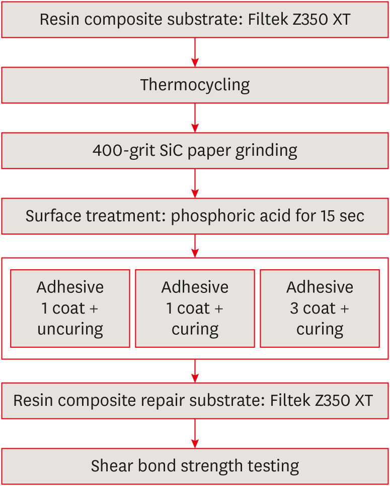
- Effect of adhesive application method on repair bond strength of composite
- Hee Kyeong Oh, Dong Hoon Shin
- Restor Dent Endod 2021;46(3):e32. Published online June 4, 2021
- DOI: https://doi.org/10.5395/rde.2021.46.e32
-
Abstract
PDFPubReaderePub
Objectives This study aimed to evaluate the effect of the application method of universal adhesives on the shear bond strength (SBS) of repaired composites, applied with different thicknesses.
Materials and Methods The 84 specimens (Filtek Z350 XT) were prepared, stored in distilled water for a week and thermocycled (5,000 cycles, 5°C to 55°C). They were roughened using 400-grit sandpapers and etched with phosphoric acid. Then, specimens were equally divided into 2 groups; Single Bond Universal (SU) and Prime&Bond Universal (PB). Each group was subdivided into 3 subgroups according to application methods (
n = 14); UC: 1 coat + uncuring, 1C: 1 coat + curing, 3C: 3 coats + curing. After storage of the repaired composite for 24 hours, specimens were subjected to the SBS test and the data were statistically analyzed by 2-way analysis of variance and independentt -tests. Specimens were examined with a stereomicroscope to analyze fracture mode and a scanning electron microscope to observe the interface.Results Adhesive material was a significant factor (
p = 0.001). Bond strengths with SU were higher than PB. The highest strength was obtained from the 1C group with SU. Bonding in multiple layers increased adhesive thicknesses, but there was no significant difference in SBS values (p = 0.255). Failure mode was predominantly cohesive in old composites.Conclusions The application of an adequate bonding system plays an important role in repairing composite resin. SU showed higher SBS than PB and the additional layers increased the adhesive thickness without affecting SBS.
-
Citations
Citations to this article as recorded by- The Effect of Adhesive Procedures on the Repair Bond Strength of Aged Universal Composites
Mehmet Semih Velioğlu, Merve Gürses, Fatma Sağ Güngör, Sinem Özdemir, Atiye Tuğba Kuzgun Türkmen, Nimet Ünlü
Selcuk Dental Journal.2026; 13(1): 129. CrossRef - The effect of different surface treatments and adhesive systems on shear bond strength in universal nanohybrid composite resin repair
Merve Kütük Ömeroğlu, Melek Çam, Işıl Doğruer, Zeynep Buket Kaynar
BMC Oral Health.2025;[Epub] CrossRef - Effect of Universal Adhesive Etching Mode on Shear Bond Strength of Pulp Capping Materials to Deep Dentin
Shahram Amirifar, Saba Tohidkhah, Seyedeh Mahsa Sheikh-Al-Eslamian, Mahdi Abbasi, Fatemeh Farshad, Elham Ahmadi, Carlos M. Ardila
BioMed Research International.2025;[Epub] CrossRef - Shear Bond Strength and Finite Element Stress Analysis of Composite Repair Using Various Adhesive Strategies With and Without Silane Application
Elif Ercan Devrimci, Hande Kemaloglu, Cem Peskersoy, Tijen Pamir, Murat Turkun
Applied Sciences.2025; 15(15): 8159. CrossRef
- The Effect of Adhesive Procedures on the Repair Bond Strength of Aged Universal Composites
- 5,045 View
- 35 Download
- 3 Web of Science
- 4 Crossref
- Enhanced visualization of the root canal morphology using a chitosan-based endo-radiopaque solution
- Shashirekha Govind, Amit Jena, Satabdi Pattanaik, Mahaprasad Anarasi, Satyajit Mohapatra, Vinay Shivagange
- Restor Dent Endod 2021;46(3):e33. Published online June 4, 2021
- DOI: https://doi.org/10.5395/rde.2021.46.e33
-
Abstract
PDFPubReaderePub
Objectives This study aimed to investigate the efficacy of ionic and non-ionic-based contrast media (
in vitro study) and the combinatorial effect of chitosan-based endo-radiopaque solution (CERS) (in vivo study) for visualization of the root canal anatomy.Materials and Methods In vitr o study (120 teeth): The root canal of maxillary premolars and molars (in vitro group 1 and 2 respectively,n = 60 each) were analyzed using 4 different contrast media (subgroups: Omnipaque 350, Iopamidol, Xenetix 350, and Urografin 76;n = 15 each) in combination with 5.25% sodium hypochlorite (NaOCl). Based on the results of thein vitro study,in vivo study (80 teeth) was done to compare Xenetix 350 + 5.25% NaOCl with CERS (in vivo group 1 and 2 respectively,n = 40 each) on maxillary and mandibular premolars and molars. Two endodontists used radiovisiography to assess the depth of ingress and identify the aberrant root anatomy after access cavity preparation, and after initial cleaning and shaping of canals. Kruskal-Wallis test was used forin vitro comparison (p < 0.05), and Wilcoxon signed-rank test and Mann-WhitneyU test forin vivo analysis (p < 0.01).Results In vitro study, Xenetix 350 + 5.25% NaOCl facilitated a significant higher visualization (p < 0.05). Forin vivo study, CERS had a statistically significant depth of ingress (p < 0.01), and was efficient in identifying the aberrant root canal anatomy of premolars and molars.Conclusions CERS facilitates better visualization of the root canal anatomy of human premolars and molars.
-
Citations
Citations to this article as recorded by- Influence of irrigating solutions on the hydration of calcium silicate-based dental biomaterials: An in vitro study
Pradeep M. Divya, Amit Jena, Saumyakanta Mohanty, Govind Shashirekha, Rashmi Rekha Mallick, Priyanka Sarangi
Journal of Conservative Dentistry and Endodontics.2025; 28(8): 758. CrossRef - Improving Endodontic Radiograph Interpretation with TV-CLAHE for Enhanced Root Canal Detection
Barbara Obuchowicz, Joanna Zarzecka, Michał Strzelecki, Marzena Jakubowska, Rafał Obuchowicz, Adam Piórkowski, Elżbieta Zarzecka-Francica, Julia Lasek
Journal of Clinical Medicine.2025; 14(15): 5554. CrossRef - Efficacy of sonic and ultrasonic activation on irrigant penetration in different tapered preparations: An in vitro study
M. Rama Sowmya, Kavalipurapu Venkata Teja, Pradeep Solete, Sahil Choudhari, S Delphine Priscilla Antony, Mohammed Mustafa
Endodontology.2024; 36(4): 370. CrossRef - Analysis of the value of visualized root canal technique in the clinical treatment of endodontics
Nana SUN, Nannan WANG, Xin QIAN
Panminerva Medica.2023;[Epub] CrossRef
- Influence of irrigating solutions on the hydration of calcium silicate-based dental biomaterials: An in vitro study
- 2,568 View
- 26 Download
- 2 Web of Science
- 4 Crossref
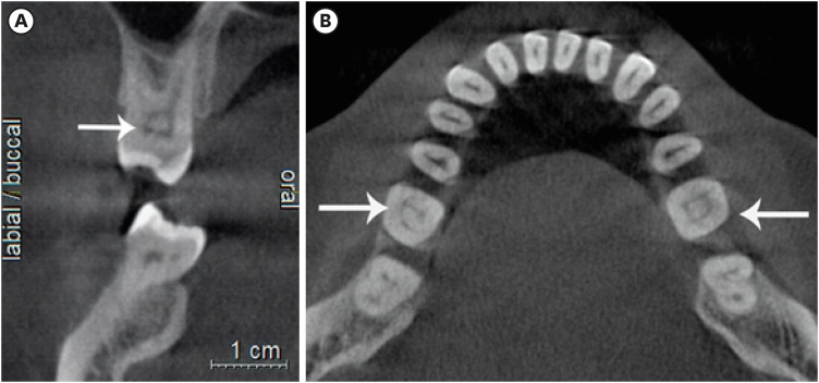
- Evaluation of the relation between the pulp stones and direct restorations using cone beam computed tomography in a Turkish subpopulation
- Güzide Pelin Sezgin, Sema Sönmez Kaplan, Tuna Kaplan
- Restor Dent Endod 2021;46(3):e34. Published online June 8, 2021
- DOI: https://doi.org/10.5395/rde.2021.46.e34
-
Abstract
PDFPubReaderePub
Objectives This study aimed to assess the presence of pulp stones through an examination of cone beam computed tomography images and correlate their prevalence with age, sex, dental arch and side, tooth type, and restoration type and depth.
Materials and Methods Cone beam computed tomography images obtained from 673 patients and archival data on 11,494 teeth were evaluated. The associations of pulp stones with age, sex, dental arch and side, tooth type, and restoration type and depth were noted. All the measurements were subjected to a χ2 test and one sample χ2 test (
p < 0.05).Results In the study group, 163 (24.2%) patients and 379 (3.3%) teeth had at least one pulp stone. The pulp stone frequency in those aged 30–39 years was significantly greater than in those aged 18–29 and ≥ 60 years, and the frequency was higher in females than in males (
p < 0.05). The highest prevalence of pulp stones was found in maxillary dental arches and molar teeth (p < 0.05). Pulp stones were significantly more common in medium-depth restorations (p < 0.05).Conclusions Maxillary molar teeth, medium-depth restorations, individuals aged 30–39 years and females had a greater percentage of pulp stones.
-
Citations
Citations to this article as recorded by- Comparative Detection and Inter-Modality Agreement of Pulp Stones Using Digital Periapical Radiography and CBCT at Two Voxel Sizes: An Ex Vivo Study
Hassan Hamed Kaabi, Sarah Saeed Binhassan, Sultan Hamad Alrumaih, Mohammed Jamal Alotaibi, Abdullah Khalid Bakarman, Nawaf Abdulaziz Alghamdi, Hamad Abdullah Almuhaythif, Qamar Mohammadziad Hashem, Abdulfatah Samih Alazmah
Diagnostics.2026; 16(7): 961. CrossRef - Cone-Beam Computed Tomography Assessment of the Prevalence and Association of Pulp Calcification with Dental and Periodontal Pathology: A Descriptive Study
José Luis Sanz, Lucía Callado, Stefana Mantale, Jenifer Nicolás, James Ghilotti, Carmen Llena
Journal of Clinical Medicine.2025; 14(4): 1373. CrossRef - Prevalence of mineralization in the pulp chamber in patients according to CBCT data
V. A. Molokova, I. N. Antonova, V. A. Osipova
Endodontics Today.2025; 23(2): 188. CrossRef - Could carotid artery calcifications and pulp stones be an alarm sign for diabetes mellitus? A retrospective observational study
Motahare Baghestani, Mohadese Faregh, Seyed Hossein Razavi, Fatemeh Owlia
BMC Endocrine Disorders.2025;[Epub] CrossRef - Distribution and influencing factors of pulp stones based on CBCT: a retrospective observational study from southwest China
Wantong Zhang, Yao Wang, Lin Ye, Yan Zhou
BMC Oral Health.2024;[Epub] CrossRef - Prevalence and Association of Calcified Pulp Stones with Periodontitis: A Cone-Beam Computed Tomography Study in Saudi Arabian Population
Abdullah Saad Alqahtani
Journal of Pharmacy and Bioallied Sciences.2024; 16(Suppl 1): S644. CrossRef - The Prevalence And Distribution Of Pulp Stones: A Cone-Beam Computed Tomography Study İn A Group Of Turkish Patients
Mujgan Firincioglulari, Seçil Aksoy, Melis Gülbeş, Umut Aksoy, Kaan Orhan
ADO Klinik Bilimler Dergisi.2024; 13(3): 496. CrossRef - Radiographical examination of pulp stone distribution by cone beam computed tomography
Fatma Tunç, Emre Çulha, Muazzez Naz Baştürk
Journal of Health Sciences and Medicine.2024; 7(4): 472. CrossRef - Cone-Beam Computed Tomography-Based Investigation of the Prevalence and Distribution of Pulp Stones and Their Relation to Local and Systemic Factors in the Makkah Population: A Cross-Sectional Study
Laila M Kenawi, Haytham S Jaha, Mashael M Alzahrani, Jihan I Alharbi, Shahad F Alharbi, Taif A Almuqati, Rehab A Alsubhi, Wahdan M Elkwatehy
Cureus.2024;[Epub] CrossRef - Cone beam computed tomography assessment of the prevalence and association of pulp calcification with periodontitis
Lingling Xiang, Botao Wang, Yuan Zhang, Jintao Wang, Peipei Wu, Jian Zhang, Liangjun Zhong, Rui He
Odontology.2023; 111(1): 248. CrossRef - Three-dimensional analysis for detection of pulp stones in a Saudi population using cone beam computed tomography
Hassan H. Kaabi, Abdullah M. Riyahi, Nassr S. Al-Maflehi, Saleh F. Alrumayyan, Abdullah K. Bakrman, Yazeed A. Almutaw
Journal of Oral Science.2023; 65(4): 257. CrossRef
- Comparative Detection and Inter-Modality Agreement of Pulp Stones Using Digital Periapical Radiography and CBCT at Two Voxel Sizes: An Ex Vivo Study
- 3,074 View
- 36 Download
- 12 Web of Science
- 11 Crossref
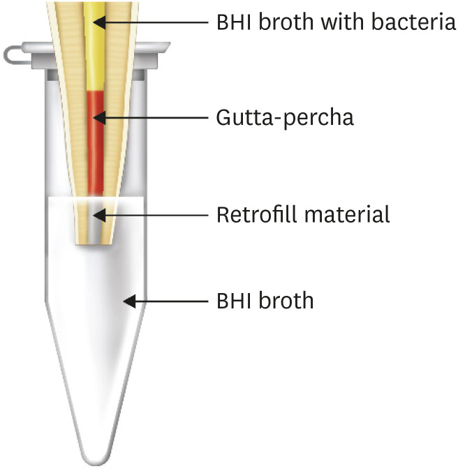
- Comparison of the sealing ability of various bioceramic materials for endodontic surgery
- Benjamin Rencher, Ana M. Chang, Hanson Fong, James D. Johnson, Avina Paranjpe
- Restor Dent Endod 2021;46(3):e35. Published online June 8, 2021
- DOI: https://doi.org/10.5395/rde.2021.46.e35
-
Abstract
PDFPubReaderePub
Objectives Endosequence Bioceramic Root Repair Material (BC-RRM) is used in endodontic microsurgery. It is available as a paste and a putty. However, no studies to date have examined the sealing ability of these forms alone or in combination as root-end filling materials. Hence, this study aimed to compare the sealing properties of these 2 forms of BC-RRM.
Materials and Methods Forty-two extracted upper anterior teeth were divided into 3 experimental groups, a positive and negative control. After the root canal treatment, the root ends were resected, retroprepared and retrofilled with either putty, paste + putty or mineral trioxide aggregate (MTA). The teeth were mounted in tubes so the apical 3 mm was submerged in Brain Heart Infusion (BHI) broth. The coronal portions of the canals were inoculated with
Enterococcus faecalis and BHI broth and incubated for 30 days. The broth in the tubes was analyzed for colony forming units to check for leakage of bacteria from the canal. The teeth from the groups were sectioned and analyzed using scanning electron microscopy (SEM). The Kruskal-Wallis test and analysis of variance were used to analyze the data with a significance levelp < 0.05.Results The BC-RRM and MTA groups showed similar sealing ability. The positive control showed leakage in all samples. The SEM imaging showed the presence of bacteria in all experimental groups at the material-tooth interface.
Conclusions No significant differences were noted in the experimental groups, providing sufficient evidence that any combination could be effectively used during endodontic microsurgery.
-
Citations
Citations to this article as recorded by- Sealing ability and marginal adaptation of premixed versus manually mixed bioceramic root-end filling materials: a systematic review and meta-analysis
Ayham Hattab, Mouhammad Al-Tayyan, Osama Hajeer
BMC Oral Health.2026;[Epub] CrossRef - Long-term outcome of combined orthograde and surgical management of perforating internal root resorption using a bioceramic repair material: an eight-year CBCT-documented case report
Mohammed Howait, Loai Alsofi
BMC Oral Health.2026;[Epub] CrossRef - An Ex-vivo Evaluation of Sealability of Three Bioceramic Physical Variants in Coronal and Apical Thirds of Root Canals
Murali H Rao, Rajkumar Krishnan, Pavithra Gopal, Elizabeth Thomas
The Journal of Contemporary Dental Practice.2025; 25(11): 1022. CrossRef - Clinical applications and classification of calcium silicate-based cements based on their history and evolution: a narrative review
Kenta Tsuchiya, Salvatore Sauro, Hidehiko Sano, Jukka P. Matinlinna, Monica Yamauti, Shuhei Hoshika, Yu Toida, Rafiqul Islam, Atsushi Tomokiyo
Clinical Oral Investigations.2025;[Epub] CrossRef - Strontium- and bioactive glass-enriched dentin repair cement: Mechanical performance and physicochemical characteristics
Nathalia Cristina Tavella-Silva, Larissa Moreira Spinola Castro Raucci, Victor Miguel Polizeli, Carlos Eduardo Saraiva Miranda, Ivone Regina de Oliveira, Walter Raucci Neto
Ceramics International.2025; 51(22): 35947. CrossRef - Conventional vs. Ready‐To‐Use Bioceramic Cements: In Vitro Bond Strength Performance in Blood‐Contaminated Dentine
Gabriela Kato Bego, Graziela Bianchi Leoni, Elias Daniel Covas Rodrigues, Larissa Moreira Spinola de Castro Raucci, Walter Raucci Neto
Australian Endodontic Journal.2025; 51(2): 466. CrossRef - Effect of calcium silicate-based repair sealers on bone healing in rat skull defects: histological and histomorphometric study
J. M. Sauer, C. E. S. Bueno, R. A. Pelegrine, C. E. Fontana, E. F. Martinez, P. G. Montagner, W. M. Nascimento, A. G. S. Limoeiro, D. G. P. Rocha, M. F. V. Marceliano-Alves, M. P. W. Galhardi, M. Klymus, A. S. Martin
Endodontics Today.2025; 23(3): 433. CrossRef - Randomized Trial of Bioceramic Apical Barrier Methods in Necrotic Immature Incisors: Effects on Pain, Extrusion, and Procedure Duration
Yasser Alsayed Tolibah, Nada Bshara, Osama Aljabban, Mohammad Tamer Abbara, Marwan Alhaji, Imad-Addin Almasri, Ziad D. Baghdadi
Children.2025; 12(10): 1423. CrossRef - Sealing ability of mineral trioxide aggregate: A scoping review of laboratory assessment methods
Kenta Tsuchiya, Salvatore Sauro, Jukka P. Matinlinna, Hidehiko Sano, Monica Yamauti, Deepak Mehta, Kyung‐San Min, Atsushi Tomokiyo
European Journal of Oral Sciences.2025;[Epub] CrossRef - Root Development Following Bioceramic Material Application in Immature Permanent Teeth: A Case Series With 24‐Month Follow‐Up
Yasser Alsayed Tolibah, Nada Bshara, Mohammad Tamer Abbara, Marwan Alhaji, Osama Aljabban, Ibrahim Ali Ahmad, Ziad D. Baghdadi, Hannah Wesley
Case Reports in Dentistry.2025;[Epub] CrossRef - Laboratory study of the sealing ability of materials used in retrograde root filling
L.A. Nadzharyan, A.V. Vasilyev, V.A. Badalyan, A.S. Galkin, A.V. Mironov, F.F. Losev
Stomatology.2025; 104(6): 5. CrossRef - Evaluation of the Marginal Adaptation of Two Hydraulic Calcium Silicate Cements Used in Apical Plugs: An In Vitro Study
Sara Filipe, José Pedro Martinho, Siri Paulo, Catarina Carvalho, Ana Coelho, Inês Amaro, Eunice Carrilho, Anabela Paula, Carlos Miguel Marto, Henrique Girão, Mónica Zuzarte, Ana S. Pires, Manuel Marques Ferreira
Applied Sciences.2024; 14(2): 480. CrossRef - A Study on Nanoleakage of Apical Retrograde Filling of Premixed Calcium Silicate-Based Cement Using a Lid Technique
Nyamsuren Enkhbileg, Jin Woo Kim, Seok Woo Chang, Se-Hee Park, Kyung Mo Cho, Yoon Lee
Materials.2024; 17(10): 2366. CrossRef - The outcome of combined use of iRoot BP Plus and iRoot SP for root-end filling in endodontic microsurgery: a randomized controlled trial
Xu Dong, Qin Su, Wen Li, Jinbo Yang, Dongzhe Song, Jing Yang, Xin Xu
Clinical Oral Investigations.2024;[Epub] CrossRef - Bacterial sealing ability of calcium silicate-based sealer for endodontic surgery: an in-vitro study
Mai M. Mansour, Sybel M. Moussa, Marwa A. Meheissen, Mahmoud R. Aboelseoud
BMC Oral Health.2024;[Epub] CrossRef - Evaluation of Marginal Adaptation of Three Biomaterials as Apical Barrier in Experimental Apexification Model
Nagehan Aktaş, Didem Sakaryalı Uyar, Didem Atabek
ADO Klinik Bilimler Dergisi.2024; 13(3): 409. CrossRef - In vitro evaluation of the sealing ability of combined use of iRoot BP Plus and iRoot SP for root-end filling
Xu Dong, Qian Xie, Xin Xu
Clinical Oral Investigations.2023; 27(6): 2969. CrossRef - Outcomes of endodontic microsurgery using different calcium silicate–based retrograde filling materials: a cohort retrospective cone-beam computed tomographic analysis
Rawan F. Eskandar, Mey A. Al-Habib, Mohammed A. Barayan, Hadeel Y. Edrees
BMC Oral Health.2023;[Epub] CrossRef - Bioceramics in Endodontics: Updates and Future Perspectives
Xu Dong, Xin Xu
Bioengineering.2023; 10(3): 354. CrossRef - Biological properties of Ceraputty as a retrograde filling material: an in vitro study on hPDLSCs
Sergio López-García, Francisco J. Rodríguez-Lozano, José Luis Sanz, Leopoldo Forner, María Pilar Pecci-Lloret, Adrián Lozano, Laura Murcia, Sonia Sánchez-Bautista, Ricardo E. Oñate-Sánchez
Clinical Oral Investigations.2023; 27(8): 4233. CrossRef - Bone Window Technique in Endodontic Microsurgery – Report of Two Cases
Spyros Floratos, Vasileios Molonis, Apostolos Tsolakis, Stylianos Kykalos, Konstantinos Kontzoglou
Journal of Endodontic Microsurgery.2022; 2: 24. CrossRef
- Sealing ability and marginal adaptation of premixed versus manually mixed bioceramic root-end filling materials: a systematic review and meta-analysis
- 7,168 View
- 137 Download
- 17 Web of Science
- 21 Crossref
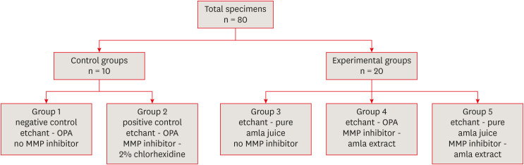
-
Comparative evaluation of
Emblica officinalis as an etchant and an MMP inhibitor with orthophosphoric acid and chlorhexidine on the microshear bond strength of composite resin: anex vivo study - Divya Sangeetha Rajkumar, Annapoorna Ballagere Mariswamy
- Restor Dent Endod 2021;46(3):e36. Published online June 8, 2021
- DOI: https://doi.org/10.5395/rde.2021.46.e36
-
Abstract
PDFPubReaderePub
Objectives This study aimed to evaluate
Emblica officinalis (Indian gooseberry or amla) as an acid etchant and matrix metalloproteinase (MMP) inhibitor, and to compare its effect on the microshear bond strength of composite resin with orthophosphoric acid (OPA) and 2% chlorhexidine (CHX) as an acid etchant and MMP inhibitor, respectively.Materials and Methods The etching effect and MMP-inhibiting action of amla on dentin samples were confirmed by scanning electron microscopy (SEM) and gelatin zymography, respectively. Dentinal slabs (3 mm thick) from 80 extracted human molars were divided into 10 and 20 samples to form 2 control groups and 3 experimental groups. Groups 1, 2, and 4 were etched with OPA and groups 3 and 5 with amla juice. An MMP inhibitor was then applied: CHX for group 2 and amla extract for groups 4 and 5. Groups 1 and 3 received no MMP inhibitor. All specimens received a standardized bonding protocol and composite resin build-up, and were subjected to microshear bond strength testing. The force at which the fracture occurred was recorded and statistically analyzed.
Results Amla juice had a similar etching effect as a self-etch adhesive in SEM and 100% amla extract was found to inhibit MMP-9 by gelatin zymography. The microshear bond strength values of amla were lower than those obtained for OPA and CHX, but the difference was not statistically significant.
Conclusions Amla has a promising role as an acid etchant and MMP inhibitor, but further studies are necessary to substantiate its efficacy.
-
Citations
Citations to this article as recorded by- Quality Control Analysis of Modified Pippalyadi Rasayana: A Herbo - Mineral Formulation for Pediatric Bronchial Asthma
Vrushti Adhyaru, V. K. Kori, C R Harisha
Journal of Ayurveda and Integrated Medical Sciences.2026; 11(5): 47. CrossRef - In vitro assessment of anti-glioblastoma potential of Emblica officinalis methanolic fruit extract and green nanoparticles in U87-MG cells
Kokkonda Jackson Sugunakara Chary, Anuradha Sharma, Amrita Singh
Medical Oncology.2025;[Epub] CrossRef - Eco-conscious synthesis of novel 1,2,4-triazolo[1,5-a]pyrimidine derivatives as potent Anti-microbial agent and comparative study of cell viability and cytotoxicity in HEK-293 cell line utilizing Indian gooseberry (Phyllanthus emblica) fruit extract
Bhaktiben R. Bhatt, Kamalkishor Pandey, Tarosh Patel, Anupama Modi, Chandani Halpani, Vaibhav D. Bhatt, Bharat C. Dixit
Bioorganic Chemistry.2024; 153: 107936. CrossRef - Cell mediated ECM-degradation as an emerging tool for anti-fibrotic strategy
Peng Zhao, Tian Sun, Cheng Lyu, Kaini Liang, Yanan Du
Cell Regeneration.2023;[Epub] CrossRef - Insight into the development of versatile dentin bonding agents to increase the durability of the bonding interface
Isabel Cristina Celerino de Moraes Porto, Teresa de Lisieux Guedes Ferreira Lôbo, Raphaela Farias Rodrigues, Rodrigo Barros Esteves Lins, Marcos Aurélio Bomfim da Silva
Frontiers in Dental Medicine.2023;[Epub] CrossRef
- Quality Control Analysis of Modified Pippalyadi Rasayana: A Herbo - Mineral Formulation for Pediatric Bronchial Asthma
- 2,617 View
- 23 Download
- 4 Web of Science
- 5 Crossref
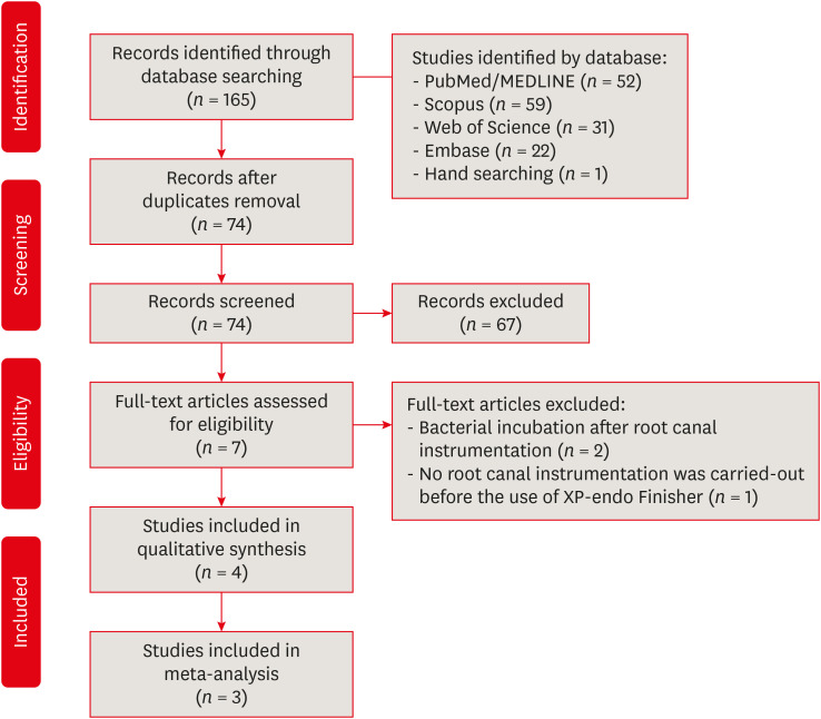
- The effectiveness of the supplementary use of the XP-endo Finisher on bacteria content reduction: a systematic review and meta-analysis
- Ludmila Smith de Jesus Oliveira, Rafaella Mariana Fontes de Bragança, Rafael Sarkis-Onofre, André Luis Faria-e-Silva
- Restor Dent Endod 2021;46(3):e37. Published online June 18, 2021
- DOI: https://doi.org/10.5395/rde.2021.46.e37
-
Abstract
PDF
 Supplementary MaterialPubReaderePub
 Objectives This systematic review evaluated the efficacy of the supplementary use of the XP-endo Finisher on bacteria content reduction in the root canal system.
Materials and Methods In-vitro studies evaluating the use of the XP-endo Finisher on bacteria content were searched in four databases in July 2020. Two authors independently screened the studies for eligibility. Data were extracted, and risk of bias was assessed. Data were meta-analyzed by using random-effects model to compare the effect of the supplementary use (experimental) or not (control) of the XP-endo Finisher on bacteria counting reduction, and results from different endodontic protocols were combined. Four studies met the inclusion criteria while 1 study was excluded from the meta-analysis due to its high risk of bias and outlier data. The 3 studies that made it to the meta-analysis had an unclear risk of bias for at least one criterion.Results No heterogeneity was observed among the results of the studies included in the meta-analysis. The study excluded from the meta-analysis assessing the bacteria counting deep in the dentin demonstrated further bacteria reduction upon the use of the XP-endo Finisher.
Conclusions This systematic review found no evidence supporting the supplementary use of the XP-endo Finisher on further bacteria counting the reduction in the root canal.
-
Citations
Citations to this article as recorded by- Mapping risk of bias criteria in systematic reviews of in vitro endodontic studies: an umbrella review
Rafaella Rodrigues da Gama, Lucas Peixoto de Araújo, Evandro Piva, Leandro Perello Duro, Adriana Fernandes da Silva, Wellington Luiz de Oliveira da Rosa
Evidence-Based Dentistry.2025; 26(4): 179. CrossRef - Characteristics and Effectiveness of XP‐Endo Files and Systems: A Narrative Review
Sarah M. Alkahtany, Rana Alfadhel, Aseel AlOmair, Sarah Bin Durayhim, Kee Y. Kum
International Journal of Dentistry.2024;[Epub] CrossRef - Impact XP-endo finisher on the 1-year follow-up success of posterior root canal treatments: a randomized clinical trial
Ludmila Smith de Jesus Oliveira, Fabricio Eneas Diniz de Figueiredo, Janaina Araújo Dantas, Maria Amália Gonzaga Ribeiro, Carlos Estrela, Manoel Damião Sousa-Neto, André Luis Faria-e-Silva
Clinical Oral Investigations.2023; 27(12): 7595. CrossRef - Comparative analysis of the effectiveness of modern irrigants activation techniques in the process of mechanical root canal system treatment (Literature review)
Anatoliy Potapchuk, Vasyl Almashi, Arsenii Horzov, Victor Buleza
InterConf.2023; (34(159)): 200. CrossRef - Comparative analysis of the effectiveness of modern irrigants activation techniques in the protocol of chemomechanical root canal system treatment (literature review)
A. Potapchuk, V. Almashi, Y. Rak, Y. Melnyk, V. Buleza, A. Horzov
SUCHASNA STOMATOLOHIYA.2023; 114(3): 4. CrossRef - Methodological quality assessment criteria for the evaluation of laboratory‐based studies included in systematic reviews within the specialty of Endodontology: A development protocol
Venkateshbabu Nagendrababu, Paul V. Abbott, Christos Boutsioukis, Henry F. Duncan, Clovis M. Faggion, Anil Kishen, Peter E. Murray, Shaju Jacob Pulikkotil, Paul M. H. Dummer
International Endodontic Journal.2022; 55(4): 326. CrossRef
- Mapping risk of bias criteria in systematic reviews of in vitro endodontic studies: an umbrella review
- 3,473 View
- 26 Download
- 4 Web of Science
- 6 Crossref
- Physicochemical characterization of two bulk fill composites at different depths
- Guillermo Grazioli, Carlos Enrique Cuevas-Suárez, Leina Nakanishi, Alejandro Francia, Rafael Ratto de Moraes
- Restor Dent Endod 2021;46(3):e39. Published online July 5, 2021
- DOI: https://doi.org/10.5395/rde.2021.46.e39
-
Abstract
PDFPubReaderePub
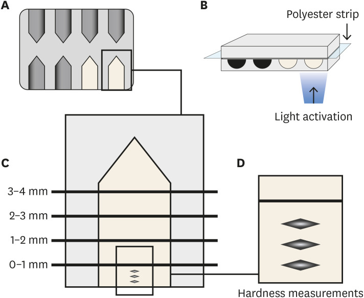
Objectives This study analyzed the physical-chemical behavior of 2 bulk fill resin composites (BFCs; Filtek Bulk Fill [FBF], and Tetric-N-Ceram Bulk Fill [TBF]) used in 2- and 4-mm increments and compared them with a conventional resin composite (Filtek Z250).
Materials and Methods Flexural strength and elastic modulus were evaluated by using a 3-point bending test. Knoop hardness was measured at depth areas 0–1, 1–2, 2–3, and 3–4 mm. The translucency parameter was measured using an optical spectrophotometer. Real-time polymerization kinetics was analyzed using Fourier transform infrared spectroscopy.
Results Flexural strength was similar among the materials, while TBF showed lower elastic modulus (Z250: 6.6 ± 1.3, FBF: 6.4 ± 0.9, TBF: 4.3 ± 1.3). The hardness of Z250 was similar only between 0–1 mm and 1–2 mm. Both BFCs had similar hardness until 2–3 mm, and showed significant decreases at 3–4 mm (FBF: 33.45 ± 1.95 at 0–1 mm to 23.19 ± 4.32 at 3–4 mm, TBF: 23.17 ± 2.51 at 0–1 mm to 15.11 ± 1.94 at 3–4 mm). The BFCs showed higher translucency than Z250. The polymerization kinetics of all the materials were similar at 2-mm increments. At 4-mm, only TBF had a similar degree of conversion compared with 2 mm.
Conclusions The BFCs tested had similar performance compared to the conventional composite when used in up to 2-mm increments. When the increment was thicker, the BFCs were properly polymerized only up to 3 mm.
-
Citations
Citations to this article as recorded by- Microhardness According to Surface, Distance and Time of Photopolymerization of a Bulk-Fill Resin: In Vitro Study
María José Loayza-Gallegos, Gino Hernan Vidalón-Romo, Julissa Amparo Dulanto-Vargas
Odovtos - International Journal of Dental Sciences.2026; 1(1): 384. CrossRef - Analysis of the effect of thickness and irradiance on polymerization delay and initial rate in light-cured resin filling materials
Brian W. Darvell, Alex Gareau, Daniel Labrie, Richard B. Price, Jeffrey W. Stansbury
Dental Materials.2026; 42(7): 1218. CrossRef - Comparative In Vitro Analysis of Mechanical Properties in Three High-Viscosity Bulk-Fill Composite Resins
Carlos I. Santacruz, Jorge I. Fajardo, César A. Paltán, Ana del Carmen Armas-Vega, Eleonor Vélez León
Journal of Composites Science.2025; 9(11): 623. CrossRef - Translucency of bulk‐fill composite materials: A systematic review
Gaetano Paolone, Sofia Baldani, Niccolò De Masi, Mauro Mandurino, Giacomo Collivasone, Nicola Scotti, Enrico Gherlone, Giuseppe Cantatore
Journal of Esthetic and Restorative Dentistry.2024; 36(7): 995. CrossRef - Can composite packaging and selective enamel etching affect the clinical behavior of bulk-fill composite resin in posterior restorations? 24-month results of a randomized clinical trial
Marcos de Oliveira BARCELEIRO, Chane TARDEM, Elisa Gomes ALBUQUERQUE, Leticia de Souza LOPES, Stella Soares MARINS, Luiz Augusto POUBEL, Roberta BARCELOS, Romina ÑAUPARI-VILLASANTE, Alessandro Dourado LOGUERCIO, Fernanda Signorelli CALAZANS
Journal of Applied Oral Science.2023;[Epub] CrossRef - No-Cap Flowable Bulk-Fill Composite: Physico-Mechanical Assessment
Abdullah Alshehri, Feras Alhalabi, Ali Robaian, Mohammed A. S. Abuelqomsan, Abdulrahman Alshabib, Eman Ismail, Faisal Alzamil, Nawaf Alotaibi, Hamad Algamaiah
Polymers.2023; 15(8): 1847. CrossRef - The Microhardness and Surface Roughness Assessment of Bulk-Fill Resin Composites Treated with and without the Application of an Oxygen-Inhibited Layer and a Polishing System: An In Vitro Study
Ann Carrillo-Marcos, Giuliany Salazar-Correa, Leonor Castro-Ramirez, Marysela Ladera-Castañeda, Carlos López-Gurreonero, Hernán Cachay-Criado, Ana Aliaga-Mariñas, Alberto Cornejo-Pinto, Luis Cervantes-Ganoza, César Félix Cayo-Rojas
Polymers.2022; 14(15): 3053. CrossRef
- Microhardness According to Surface, Distance and Time of Photopolymerization of a Bulk-Fill Resin: In Vitro Study
- 3,237 View
- 24 Download
- 7 Web of Science
- 7 Crossref
- YouTube as a source of information about pulpotomy and pulp capping: a cross sectional reliability analysis
- Konstantinos Kodonas, Anastasia Fardi
- Restor Dent Endod 2021;46(3):e40. Published online July 6, 2021
- DOI: https://doi.org/10.5395/rde.2021.46.e40
-
Abstract
PDFPubReaderePub
Objectives The purpose of this study was to critically evaluate the quality, reliability and educational content of the information of vital pulp treatment videos available on YouTube.
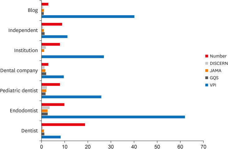
Materials and Methods The keywords “pulpotomy” and “pulp capping” were searched on YouTube on 5th July 2020, until 60 English language videos of each search term with a duration shorter than 15 minutes were acquired. Video characteristics were recorded and Video Power Index (VPI) was calculated. Reliability and educational quality of videos were evaluated using the Modified DISCERN score, the
Journal of American Medical Association (JAMA) benchmark criteria and Global Quality Scores (GQS). Videos were categorized by uploading source.Results Regarding pulpotomy, 31.7% of the videos were uploaded by specialists and 68.3% were directed by non-specialists. In the case of pulp capping, the corresponding percentages were 45% and 55%, respectively. Videos uploaded by specialists had significantly higher modified DISCERN, JAMA and GQS scores compared to those uploaded by non-specialists. Endodontists tended to have the highest reliability and VPI scores.
Conclusions YouTube videos on vital pulp treatment contain low educational quality or incomplete information. Low popularity of dental pulp capping and pulpotomy videos may be attributed to the specialized nature of these procedures. As YouTube represents an important source for patient information about different health topics, reliable informative videos should be uploaded by specialized dental professionals.
-
Citations
Citations to this article as recorded by- Evaluation of YouTubeTM Videos Regarding ICON as an Information Resource: A Cross-Sectional Study
Sevim Atılan Yavuz, Zeyneb Merve Ozdemır, Derya Gursel Surmelioglu
Mersin Üniversitesi Tıp Fakültesi Lokman Hekim Tıp Tarihi ve Folklorik Tıp Dergisi.2026; 16(1): 282. CrossRef - Quality and Accuracy Assessment of YouTube Videos on Vital Pulp Therapy
Divya Nangia, Vasudev Ballal, Prashant Bhasin, Esha Sukhala, Ashima Garg, Meenu G. Singla, Hemanshi Kumar
Australian Endodontic Journal.2026;[Epub] CrossRef - Assessing the Quality of YouTube® Videos on Nitrous Oxide/Oxygen Inhalation: A Multi-Dimensional Approach for Pediatric Dentists
Sanaa N. Al-Haj Ali, Nehal AlHarbi, Hessah H. Almutairi
Pesquisa Brasileira em Odontopediatria e Clínica Integrada.2025;[Epub] CrossRef - Is YouTube™ a useful resource of information about bichectomy? A cross-sectional study
H.ɪ. Durmuş, B. Ege, S. Bayazıt, M. Koparal
Annales de Chirurgie Plastique Esthétique.2025;[Epub] CrossRef - Assessing the reliability and educational value of YouTube videos on computer-controlled local anesthesia in dentistry
Hulya Cerci Akcay, Erdal Cem Kargu, Nefise Seker, Tanay Chaubal
PLOS One.2025; 20(8): e0329291. CrossRef - A content analysis of YouTube videos on interproximal enamel reduction
Weng Yan Tam, Jack Shen Tham, Smita Nimbalkar, Shilpa Gunjal, Kirti Saxena
APOS Trends in Orthodontics.2025; 0: 1. CrossRef - Comparison of YouTube, TikTok, and Instagram as digital sources for obtaining information about pulp therapy in primary and permanent teeth
Hüseyin Gürkan Güneç, Emine Kaya, Dila Nur Okumuş, Merve Gül Erence
Restorative Dentistry & Endodontics.2025; 50(3): e26. CrossRef - Evaluation of Endodontic Retreatment Videos on The Youtube Platform: Quality and Content Analysis
Birgül Özaşır, Tufan Özaşır, Derin Buğu Yüzer, Deniz İmamoğlu, Kamran Gülşahı
European Annals of Dental Sciences.2025; 52(2): 103. CrossRef - Is YouTube a reliable source for learning pre-endodontic build-up? A cross-sectional study
Merve Gökyar, İdil Özden, Hesna Sazak Öveçoğlu
Restorative Dentistry & Endodontics.2025; 50(3): e27. CrossRef - Quality of Patient-Centered eHealth Information on Erosive Tooth Wear: Systematic Search and Evaluation of Websites and YouTube Videos
Lena Holland, Amelie Friederike Kanzow, Annette Wiegand, Philipp Kanzow
Journal of Medical Internet Research.2024; 26: e49514. CrossRef - Is it safe to learn about vital pulp capping from YouTube™ videos? A content and quality analysis
Celalettin Topbaş, Tuğçe Paksoy, Ayşe Gülnihal İslamoğlu, Kemal Çağlar, Abdurrahman Kerim Kul
International Journal of Medical Informatics.2024; 185: 105409. CrossRef - Assessment of the quality of oral biopsy procedure videos shared on YouTube
A. Díaz‐Rodríguez, J. Limeres‐Posse, R. Albuquerque, V. Brailo, R. Cook, J. C. Fricain, G. Lodi, L. Monteiro, L. Silva, B. Carey, M. Diniz‐Freitas
Oral Diseases.2024; 30(5): 3081. CrossRef - İmplant üstü protezler hakkında bilgi veren internet sitelerinin okunabilirliklerinin değerlendirilmesi
Tugba TEMİZCİ
Selcuk Dental Journal.2023; 10(4): 156. CrossRef - Online Audio-Visual Information on the Treatment of OSA with Mandibular Advancement Devices: Analysis of Quality, Reliability and Contents
Serena Incerti-Parenti, Maria Lavinia Bartolucci, Elena Biondi, Andrea Fiordelli, Corrado Paganelli, Giulio Alessandri-Bonetti
Applied Sciences.2023; 13(9): 5727. CrossRef - Evaluating YouTube as a Patient Information Source for the Risks of Root Canal Treatment
Stewart McLean, Neil Cook, Alexander Rovira-Wilde, Shanon Patel, Shalini Kanagasingam
Journal of Endodontics.2023; 49(2): 155. CrossRef - Assessment of reliability and information quality of YouTube videos about root canal treatment after 2016
Myoung-jun Jung, Min-Seock Seo
BMC Oral Health.2022;[Epub] CrossRef - Is the YouTube™ a useful resource of information about orthognathic surgery?: A cross-sectional study
Seyma Bayazıt, Bilal Ege, Mahmut Koparal
Journal of Stomatology, Oral and Maxillofacial Surgery.2022; 123(6): e981. CrossRef - YoutubeTM Content Analysis as a Means of Information in Oral Medicine: A Systematic Review of the Literature
Antonio Romano, Fausto Fiori, Massimo Petruzzi, Fedora Della Vella, Rosario Serpico
International Journal of Environmental Research and Public Health.2022; 19(9): 5451. CrossRef
- Evaluation of YouTubeTM Videos Regarding ICON as an Information Resource: A Cross-Sectional Study
- 2,884 View
- 29 Download
- 15 Web of Science
- 18 Crossref
- Anesthetic efficacy in vital asymptomatic teeth using different local anesthetics: a systematic review with network meta-analysis
- Amy Kia Cheen Liew, Yi-Chun Yeh, Dalia Abdullah, Yu-Kang Tu
- Restor Dent Endod 2021;46(3):e41. Published online July 21, 2021
- DOI: https://doi.org/10.5395/rde.2021.46.e41
-
Abstract
PDFSupplementary MaterialPubReaderePub
Objectives This study aimed to evaluate the efficacy of various local anesthesia (LA) in vital asymptomatic teeth.
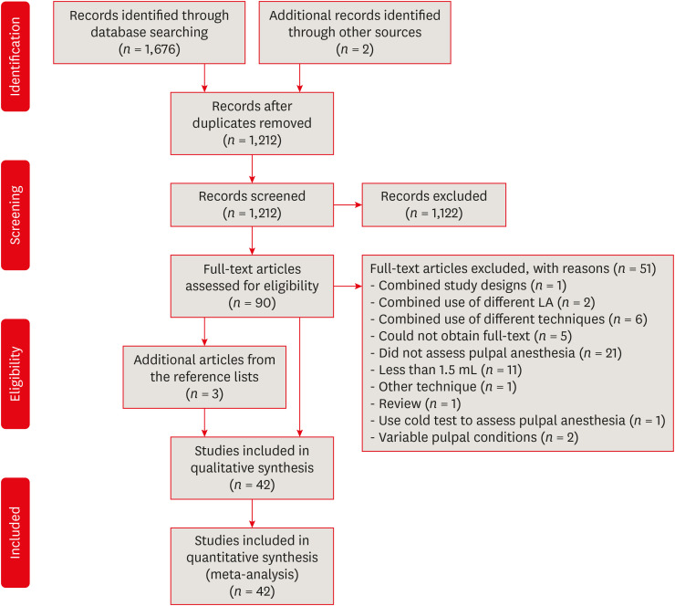
Materials and Methods Randomized controlled trials comparing pulpal anesthesia of various LA on vital asymptomatic teeth were included in this review. Searches were conducted in the Cochrane CENTRAL, MEDLINE (via PubMed), EMBASE, ClinicalTrials.gov, Google Scholar and 3 field-specific journals from inception to May 3, 2019. Study selection, data extraction, and risk of bias assessment using Cochrane Risk of Bias Tool were done by 2 independent reviewers in duplicate. Network meta-analysis (NMA) was performed within the frequentist setting using STATA 15.0. The LA was ranked, and the surface under the cumulative ranking (SUCRA) line was plotted. The confidence of the NMA estimates was assessed using the CINeMA web application.
Results The literature search yielded 1,678 potentially eligible reports, but only 42 were included in this review. For maxillary buccal infiltration, articaine 4% with epinephrine 1:100,000 was more efficacious than lidocaine 2% with epinephrine 1:100,000 (odds ratio, 2.11; 95% confidence interval, 1.14–3.89). For mandibular buccal infiltration, articaine 4% with epinephrine 1:100,000 was more efficacious than various lidocaine solutions. The SUCRA ranking was highest for articaine 4% with epinephrine when used as maxillary and mandibular buccal infiltrations, and lidocaine 2% with epinephrine 1:80,000 when used as inferior alveolar nerve block. Inconsistency and imprecision were detected in some of the NMA estimates.
Conclusions Articaine 4% with epinephrine is superior when maxillary or mandibular infiltration is required in vital asymptomatic teeth.
-
Citations
Citations to this article as recorded by- Anatomical Basis of the Palatal Injection Technique for Pulpal Anesthesia of Maxillary Teeth
Sergey Kabak, Joe Iwanaga, Yuliya Melnichenko, Ruslan Mekhtiev, Nina Savrasova
Clinical Anatomy.2026; 39(2): 134. CrossRef - Articaine in Oral and Maxillofacial Anesthesia: What Does High-Level Evidence Reveal About Nerve Block and Infiltration Strategies?
Rafael Dascanio, Francisco Groppo, Mutlu Özcan, Camila Batista da Silva Candido
Journal of Oral and Maxillofacial Surgery.2026;[Epub] CrossRef - Adrenaline in pulp capping treatment of reversible pulpitis
Si-Yun Yang, Jin-Zhu Wang, Hao Fan, Min Chen
World Journal of Clinical Cases.2024; 12(22): 5024. CrossRef - Effect of 810 nm Diode Laser Irradiation on the Time of Initiation and Depth of Anesthesia for Endodontic Treatment of Mandibular First Molars with Symptomatic Irreversible Pulpitis: A Clinical Trial
Elham Khoshbin, Leila Ghasemi, Rooholah Behroozi, Zahra Khosravi, Afsaneh Rahmati, Loghman Rezaeisoufi, Hamed Karkehabadi
Photobiomodulation, Photomedicine, and Laser Surgery.2023; 41(9): 475. CrossRef - The potential of articaine as new generation of local anesthesia in dental clinics: A review
Wen Luo, Kaiyue Zheng, Huifang Kuang, Zhixin Li, Jinrong Wang, Jie Mei
Medicine.2022; 101(48): e32089. CrossRef
- Anatomical Basis of the Palatal Injection Technique for Pulpal Anesthesia of Maxillary Teeth
- 5,431 View
- 47 Download
- 3 Web of Science
- 5 Crossref
- Ten years of minimally invasive access cavities in Endodontics: a bibliometric analysis of the 25 most-cited studies
- Emmanuel João Nogueira Leal Silva, Karem Paula Pinto, Natasha C. Ajuz, Luciana Moura Sassone
- Restor Dent Endod 2021;46(3):e42. Published online July 21, 2021
- DOI: https://doi.org/10.5395/rde.2021.46.e42
-
Abstract
PDFPubReaderePub
Objectives This study aimed to analyze the main features of the 25 most-cited articles in minimally invasive access cavities.
Materials and Methods An electronic search was conducted on the Clarivate Analytics' Web of Science ‘All Databases’ to identify the most-cited articles related to this topic. Citation counts were cross-matched with data from Elsevier's Scopus and Google Scholar. Information about authors, contributing institutions and countries, year and journal of publication, study design and topic, access cavity, and keywords were analyzed.
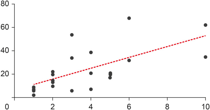
Results The top 25 most-cited articles received a total of 572 (Web of Science), 1,160 (Google Scholar) and 631 (Scopus) citations. It was observed a positive significant association between the number of citations and age of publication (
r = 0.6907,p < 0.0001); however, there was no significant association regarding citation density and age of publication (r = −0.2631,p = 0.2038). TheJournal of Endodontics made the highest contribution (n = 15, 60%). The United States had the largest number of publications (n = 7) followed by Brazil (n = 4), with the most contributions from the University of Tennessee and Grande Rio University (n = 3), respectively. The highest number of most-cited articles wereex vivo studies (n = 16), and ‘fracture resistance’ was the major topic studied (n = 10).Conclusions This study revealed a growing interest for researchers in the field of minimally invasive access cavities. Future trends are focused on the expansion of collaborative networks and the conduction of laboratory studies on under-investigated parameters.
-
Citations
Citations to this article as recorded by- Sixty Years of Ethylenediaminetetraacetic Acid Use in Endodontics: A Comprehensive Bibliometric Study
Camila Segatto Hartmann, Luiz Fernando Monteiro Czornobay, Julia Menezes Savaris, Aurélio de Oliveira Rocha, Lucas Menezes dos Anjos, Bruno Alexandre Pacheco de Castro Henriques, Mariane Cardoso, Lucas da Fonseca Roberti Garcia, Cleonice da Silveira Teixe
Journal of Endodontics.2026; 52(4): 512. CrossRef - Factors Associated With the Citation Impact of Evidence Synthesis Reviews in Endodontics: A Bibliometric Analysis
Rafaella Rodrigues da Gama, Lucas Peixoto de Araújo, Melissa Feres Damian, Wellington Luiz de Oliveira da Rosa
International Endodontic Journal.2026; 59(7): 1384. CrossRef - A descriptive bibliometric and thematic analysis of postgraduate theses in endodontics in Turkiye
Güliz Rana Tellioğlu Avcı, Merve Yeniçeri Özata
Anatolian Current Medical Journal.2026; 8(4): 613. CrossRef - Research Trends in Internal Root Resorption from 1947 to 2022: A Bibliometric Analysis of the 50 Most-cited Articles
Laise Pena Braga Monteiro, Larissa Pillar Gomes Martel, Roberta Fonseca de Castro, Emmanuel João Nogueira Leal da Silva, Juliana Melo da Silva Brandão
Journal of Advanced Oral Research.2025; 16(2): 196. CrossRef - Bibliometric analysis of the publications that list the most-cited articles in endodontics
Oscar Alejandro Gutiérrez-Alvarez, Luis Alberto Pantoja-Villa, Benigno Miguel Calderón-Rojas
Endodontology.2025; 37(2): 128. CrossRef - Evaluation of the forces applied by rubber dam clamps on mandibular first molar teeth with different endodontic access cavities: a 3D FEA study
Mehmet Eskibağlar, Serkan Erdem, Büşra Karaağaç Eskibağlar, Mete Onur Kaman
PeerJ.2024; 12: e17921. CrossRef - A Global Overview of Guided Endodontics: A Bibliometric Analysis
Thaine Oliveira Lima, Aurélio de Oliveira Rocha, Lucas Menezes dos Anjos, Nailson Silva Meneses Júnior, Marco Antonio Hungaro Duarte, Murilo Priori Alcalde, Mariane Cardoso, Rodrigo Ricci Vivan
Journal of Endodontics.2024; 50(1): 10. CrossRef - Novel method for augmented reality guided endodontics: An in vitro study
Marco Farronato, Andres Torres, Mariano S. Pedano, Reinhilde Jacobs
Journal of Dentistry.2023; 132: 104476. CrossRef - Contribution of Türkiye to the Field of Endodontology: A Visualized Bibliometric Analysis Based on Web of Science
Olcay ÖZDEMİR, Yağız ÖZBAY, Neslihan YILMAZ ÇIRAKOĞLU
Medical Records.2023; 5(1): 91. CrossRef - Effect of access cavities on the biomechanics of mandibular molars: a finite element analysis
Xiao Wang, Dan Wang, Yi-rong Wang, Xiao-gang Cheng, Long-xing Ni, Wei Wang, Yu Tian
BMC Oral Health.2023;[Epub] CrossRef - Contemporary research trends on nanoparticles in endodontics: a bibliometric and scientometric analysis of the top 100 most-cited articles
Sıla Nur Usta, Zeliha Uğur-Aydın, Kadriye Demirkaya, Cumhur Aydın
Restorative Dentistry & Endodontics.2023;[Epub] CrossRef - Evolving trend of systematic reviews and meta-analyses in endodontics: A bibliometric study
GalvinSim Siang Lin, JiaZheng Leong, WenXin Chong, MikoChong Kha Chee, ChinSheng Lee, Manahil Maqbool, TahirYusuf Noorani
Saudi Endodontic Journal.2022; 12(3): 236. CrossRef - Global research trends on photodynamic therapy in endodontics: A bibliometric analysis
Lucas Peixoto de Araújo, Wellington Luiz de Oliveira da Rosa, Leandro Bueno Gobbo, Tamares Andrade da Silva, José Flávio Affonso de Almeida, Caio Cezar Randi Ferraz
Photodiagnosis and Photodynamic Therapy.2022; 40: 103039. CrossRef - Minimal Invasive Endodontics: A Comprehensive Narrative Review
Jaydip Marvaniya, Kishan Agarwal, Dhaval N Mehta, Nirav Parmar, Ritwik Shyamal , Jenee Patel
Cureus.2022;[Epub] CrossRef
- Sixty Years of Ethylenediaminetetraacetic Acid Use in Endodontics: A Comprehensive Bibliometric Study
- 4,275 View
- 39 Download
- 10 Web of Science
- 14 Crossref
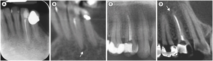
- Which factors related to apical radiolucency may influence its radiographic detection? A study using CBCT as reference standard
- Rocharles Cavalcante Fontenele, Eduarda Helena Leandro Nascimento, Hugo Gaêta-Araujo, Laís Oliveira de Araujo Cardelli, Deborah Queiroz Freitas
- Restor Dent Endod 2021;46(3):e43. Published online July 21, 2021
- DOI: https://doi.org/10.5395/rde.2021.46.e43
-
Abstract
PDFPubReaderePub
Objectives This study aimed to evaluate the detection rate of apical radiolucencies in 2-dimensional images using cone-beam computed tomography (CBCT) as the reference standard, and to determine which factors related to the apical radiolucencies and the teeth could influence its detection.
Materials and Methods The sample consisted of exams of patients who had panoramic (PAN) and/or periapical (PERI) radiography and CBCT. The exams were assessed by 2 oral radiologists and divided into PAN+CBCT (227 teeth–285 roots) and PERI+CBCT (94 teeth–115 roots). Radiographic images were evaluated for the presence of apical radiolucency, while CBCT images were assessed for presence, size, location, and involvement of the cortical bone (thinning, expansion, and destruction). Diagnostic values were obtained for PERI and PAN.
Results PERI and PAN presented high accuracy (0.83 and 0.77, respectively) and specificity (0.89 and 0.91, respectively), but low sensitivity, especially for PAN (0.40 vs. 0.65 of PERI). The size of the apical radiolucency was positively correlated with its detection in PERI and PAN (
p < 0.001). For PAN, apical radiolucencies were 3.93 times more frequently detected when related to single-rooted teeth (p = 0.038). The other factors did not influence apical radiolucency detection (p > 0.05).Conclusions PERI presents slightly better accuracy than PAN for the detection of apical radiolucency. The size is the only factor related to radiolucency that influences its detection, for both radiographic exams. For PAN, apical radiolucency is most often detected in single-rooted teeth.
-
Citations
Citations to this article as recorded by- Radiomics-based classification of pediatric dental trauma in periapical radiographs: a preliminary study
Mengtian Peng, Bin Yu, Juan Hu, Xiaoxin Xie, Jihong He
BMC Medical Imaging.2025;[Epub] CrossRef - Increasing Diagnostic Acumen in Endodontics
Shilpa Thakkar, Dana Mominkhan
Dental Clinics of North America.2025; 69(4): 479. CrossRef - Three-dimensional clinical assessment for MRONJ risk in oncologic patients following tooth extractions
Catalina Moreno Rabie, Rocharles Cavalcante Fontenele, Nicolly Oliveira Santos, Fernanda Nogueira Reis, Tim Van den Wyngaert, Reinhilde Jacobs
Dentomaxillofacial Radiology.2023;[Epub] CrossRef - Quality of techniques used to assess clinical outcomes of regenerative endodontic treatment in necrotic mature teeth
Roy George
Evidence-Based Dentistry.2022; 23(3): 98. CrossRef
- Radiomics-based classification of pediatric dental trauma in periapical radiographs: a preliminary study
- 4,619 View
- 28 Download
- 3 Web of Science
- 4 Crossref
Letters to the Editor
- Case report on combining PRF with alloplastic bone substitute in Endo-Perio lesion
- Mansi Bansal, Manish Khatri, Komal Puri
- Restor Dent Endod 2021;46(3):e44. Published online August 11, 2021
- DOI: https://doi.org/10.5395/rde.2021.46.e44
- 1,444 View
- 30 Download
- Author's Reply to Case report on combining PRF with alloplastic bone substitute in Endo-Perio lesion
- Lata Goyal
- Restor Dent Endod 2021;46(3):e45. Published online August 11, 2021
- DOI: https://doi.org/10.5395/rde.2021.46.e45
- 1,218 View
- 18 Download
 First
First Prev
Prev

