Search
- Page Path
- HOME > Search
- Comparison of remineralization in caries-affected dentin using calcium silicate, glass ionomer cement, and resin-modified glass ionomer cement: an in vitro study
- Kwanchanok Youcharoen, Onwara Akkaratham, Papichaya Intajak, Pipop Saikaew, Sirichan Chiaraputt
- Restor Dent Endod 2025;50(4):e37. Published online November 14, 2025
- DOI: https://doi.org/10.5395/rde.2025.50.e37
-
 Abstract
 PDF
PDF PubReader
 ePub
ePub - Objectives
This study evaluated the ability of calcium silicate cement (CSC) as a remineralizing agent compared with conventional glass ionomer cement (GIC) and resin-modified GIC (RMGIC) to remineralize artificial caries-affected dentin.
Methods
Twenty-five class V cavities were prepared on extracted human third molars. Twenty teeth underwent artificial caries induction. The remaining five teeth with sound dentin serve as the positive control. The twenty demineralized teeth were subdivided into four groups (n = 5): carious dentin without restoration (negative control [NC]), carious dentin restored with CSC (Biodentine, Septodont), carious dentin restored with GI (Fuji IX, GC Corporation), and carious dentin restored with RMGIC (Fuji II LC, GC Corporation). Following restoration, the specimens were stored in artificial saliva for 7 days. The elastic modulus was evaluated by a nanoindentation test. The mineral composition was analyzed by scanning electron microscopy-energy-dispersive X-ray spectroscopy (SEM-EDX), and the mineral composition at the dentin-material interface.
Results
CSC had a higher modulus of elasticity compared to GI, RMGI, and NC groups (p < 0.05). Higher calcium and phosphorus content was observed under CSC restorations, as indicated by SEM-EDX examination, which may lead to better remineralization.
Conclusions
Compared to GI and RMGI, CSC showed the best remineralization and mechanical reinforcement in caries-affected dentin, indicating CSC for use in minimally invasive restorative dentistry.
- 1,163 View
- 152 Download

- The influence of bioactive glass (BGS-7) on enamel remineralization: an in vitro study
- Chaeyoung Lee, Eunseon Jeong, Kun-Hwa Sung, Su-Jung Park, Yoorina Choi
- Restor Dent Endod 2025;50(4):e33. Published online October 15, 2025
- DOI: https://doi.org/10.5395/rde.2025.50.e33
-
 Abstract
 PDF
PDF PubReader
 ePub
ePub - Objectives
The aim of this study was to compare the remineralizing capacity of bioactive glass (BGS-7, CGBIO) with other agents.
Methods
Twenty caries-free third molars were sectioned and demineralized. Specimens were divided into four groups: (1) control, (2) Clinpro XT varnish (Solventum), (3) 1.23% acidulated phosphate fluoride gel, and (4) a new type of CaO-SiO2-P2O5-B2O3 system of bioactive glass ceramics (BGS-7). Agents were applied and stored in simulated body fluid at 37℃ for 2 weeks. Microhardness was measured using the Vickers hardness testing method. Five specimens per group were analyzed using quantitative light-induced fluorescence (QLF) to assess mineral loss. Field-emission scanning electron microscopy (FE-SEM) and energy-dispersive X-ray spectroscopy (EDS) were used to examine the surface morphology and elemental composition. Data were analyzed using paired t-test and one-way analysis of variance (p < 0.05).
Results
BGS-7 showed the highest microhardness values and the greatest recovery in QLF analysis (p < 0.05). FE-SEM revealed granular precipitates on demineralized enamel in the BGS-7 group. EDS confirmed the presence of newly formed silicon and fluoride layers.
Conclusions
BGS-7 demonstrated superior remineralization capacity compared to other agents, suggesting its potential as an effective remineralizing material.
- 1,053 View
- 143 Download

- Effects of CTHRC1 on odontogenic differentiation and angiogenesis in human dental pulp stem cells
- Jong-soon Kim, Bin-Na Lee, Hoon-Sang Chang, In-Nam Hwang, Won-Mann Oh, Yun-Chan Hwang
- Restor Dent Endod 2023;48(2):e18. Published online April 28, 2023
- DOI: https://doi.org/10.5395/rde.2023.48.e18
-
 Abstract
 PDF
PDF PubReader
 ePub
ePub Objectives This study aimed to determine whether collagen triple helix repeat containing-1 (CTHRC1), which is involved in vascular remodeling and bone formation, can stimulate odontogenic differentiation and angiogenesis when administered to human dental pulp stem cells (hDPSCs).
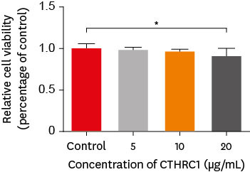
Materials and Methods The viability of hDPSCs upon exposure to CTHRC1 was assessed with the WST-1 assay. CTHRC1 doses of 5, 10, and 20 µg/mL were administered to hDPSCs. Reverse-transcription polymerase reaction was used to detect dentin sialophosphoprotein, dentin matrix protein 1, vascular endothelial growth factor, and fibroblast growth factor 2. The formation of mineralization nodules was evaluated using Alizarin red. A scratch wound assay was conducted to evaluate the effect of CTHRC1 on cell migration. Data were analyzed using 1-way analysis of variance followed by the Tukey
post hoc test. The threshold for statistical significance was set atp < 0.05.Results CTHRC1 doses of 5, 10, and 20 µg/mL had no significant effect on the viability of hDPSCs. Mineralized nodules were formed and odontogenic markers were upregulated, indicating that CTHRC1 promoted odontogenic differentiation. Scratch wound assays demonstrated that CTHRC1 significantly enhanced the migration of hDPSCs.
Conclusions CTHRC1 promoted odontogenic differentiation and mineralization in hDPSCs.
- 1,568 View
- 34 Download

- Push-out bond strength and intratubular biomineralization of a hydraulic root-end filling material premixed with dimethyl sulfoxide as a vehicle
- Ju-Ha Park, Hee-Jin Kim, Kwang-Won Lee, Mi-Kyung Yu, Kyung-San Min
- Restor Dent Endod 2023;48(1):e8. Published online January 20, 2023
- DOI: https://doi.org/10.5395/rde.2023.48.e8
-
 Abstract
 PDF
PDF PubReader
 ePub
ePub Objectives This study was designed to evaluate the parameters of bonding performance to root dentin, including push-out bond strength and dentinal tubular biomineralization, of a hydraulic bioceramic root-end filling material premixed with dimethyl sulfoxide (Endocem MTA Premixed) in comparison to a conventional powder-liquid–type cement (ProRoot MTA).
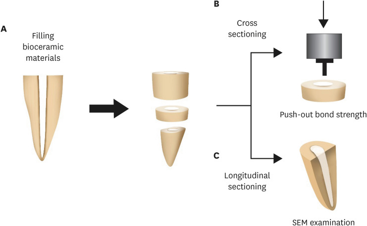
Materials and Methods The root canal of a single-rooted premolar was filled with either ProRoot MTA or Endocem MTA Premixed (
n = 15). A slice of dentin was obtained from each root. Using the sliced specimen, the push-out bond strength was measured, and the failure pattern was observed under a stereomicroscope. The apical segment was divided into halves; the split surface was observed under a scanning electron microscope, and intratubular biomineralization was examined by observing the precipitates formed in the dentinal tubule. Then, the chemical characteristics of the precipitates were evaluated with energy-dispersive X-ray spectroscopic (EDS) analysis. The data were analyzed using the Student’st -test followed by the Mann-WhitneyU test (p < 0.05).Results No significant difference was found between the 2 tested groups in push-out bond strength, and cohesive failure was the predominant failure type. In both groups, flake-shaped precipitates were observed along dentinal tubules. The EDS analysis indicated that the mass percentage of calcium and phosphorus in the precipitate was similar to that found in hydroxyapatite.
Conclusions Regarding bonding to root dentin, Endocem MTA Premixed may have potential for use as an acceptable root-end filling material.
-
Citations
Citations to this article as recorded by- Effectiveness of Sectioning Method and Filling Materials on Roughness and Cell Attachments in Root Resection Procedure
Tarek Ashi, Naji Kharouf, Olivier Etienne, Bérangère Cournault, Pierre Klienkoff, Varvara Gribova, Youssef Haikel
European Journal of Dentistry.2025; 19(01): 240. CrossRef - Bond Strength and Adhesive Interface Quality of New Pre‐Mixed Bioceramic Root Canal Sealer
Gustavo Creazzo, Bruna Monteiro de Barros Ciribelli Alves, Helena Cristina de Assis, Karen Gisselle Garay Villamayor, Manoel Damião de Sousa‐Neto, Jardel Francisco Mazzi‐Chaves, Fabiane Carneiro Lopes‐Olhê
Microscopy Research and Technique.2025; 88(7): 1989. CrossRef - Evaluation of clinical and radiographic outcome of premixed injectable mineral trioxide aggregate and conventional mineral trioxide aggregate as pulpotomy medicaments in primary molars – A split-mouth randomized control trial
U. S. Aiswarya, Sharan S. Sargod, Sundeep K. Hegde, H. T. Ajay Rao, Nanditha Hegde
Journal of Indian Society of Pedodontics and Preventive Dentistry.2025; 43(4): 559. CrossRef - Evaluation of the root dentin bond strength and intratubular biomineralization of a premixed calcium aluminate-based hydraulic bioceramic endodontic sealer
Yu-Na Lee, Min-Kyeong Kim, Hee-Jin Kim, Mi-Kyung Yu, Kwang-Won Lee, Kyung-San Min
Journal of Oral Science.2024; 66(2): 96. CrossRef - Removal efficiency of a fast setting pozzalan-based bioactive cement: a micro CT study
Feyza Çetinkaya, Ahter Şanal Çıkman, Ali Keleş, Banu Arıcıoğlu
BMC Oral Health.2024;[Epub] CrossRef - Antibacterial Activity and Sustained Effectiveness of Calcium Silicate-Based Cement as a Root-End Filling Material against Enterococcus faecalis
Seong-Hee Moon, Seong-Jin Shin, Seunghan Oh, Ji-Myung Bae
Materials.2023; 16(18): 6124. CrossRef
- Effectiveness of Sectioning Method and Filling Materials on Roughness and Cell Attachments in Root Resection Procedure
- 3,141 View
- 85 Download
- 6 Web of Science
- 6 Crossref

-
Hard tissue formation after direct pulp capping with osteostatin and MTA
in vivo - Ji-Hye Yoon, Sung-Hyeon Choi, Jeong-Tae Koh, Bin-Na Lee, Hoon-Sang Chang, In-Nam Hwang, Won-Mann Oh, Yun-Chan Hwang
- Restor Dent Endod 2021;46(2):e17. Published online February 25, 2021
- DOI: https://doi.org/10.5395/rde.2021.46.e17
-
 Abstract
 PDF
PDF PubReader
 ePub
ePub Objectives In recent
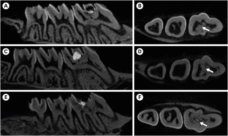
in vitro study, it was reported that osteostatin (OST) has an odontogenic effect and synergistic effect with mineral trioxide aggregate (MTA) in human dental pulp cells. Therefore, the aim of this study was to evaluate whether OST has a synergistic effect with MTA on hard tissue formationin vivo .Materials and Methods Thirty-two maxillary molars of Spraque-Dawley rats were used in this study. An occlusal cavity was prepared and the exposed pulps were randomly divided into 3 groups: group 1 (control; ProRoot MTA), group 2 (OST 100 μM + ProRoot MTA), group 3 (OST 10 mM + ProRoot MTA). Exposed pulps were capped with each material and cavities were restored with resin modified glass ionomer. The animals were sacrificed after 4 weeks. All harvested teeth were scanned with micro-computed tomography (CT). The samples were prepared and hard tissue formation was evaluated histologically. For immunohistochemical analysis, the specimens were sectioned and incubated with primary antibodies against dentin sialoprotein (DSP).
Results In the micro-CT analysis, it is revealed that OST with ProRoot MTA groups showed more mineralized bridge than the control (
p < 0.05). In the H&E staining, it is showed that more quantity of the mineralized dentin bridge was formed in the OST with ProRoot MTA group compared to the control (p < 0.05). In all groups, DSP was expressed in newly formed reparative dentin area.Conclusions OST can be a supplementary pulp capping material when used with MTA to make synergistic effect in hard tissue formation.
-
Citations
Citations to this article as recorded by- Pulpal responses to mineral trioxide aggregate with and without zinc oxide addition in mature canine teeth after full pulpotomy
Behnam Bolhari, Neda Kardouni Khouzestani, Hadi Assadian, Saeed Farzad-Mohajeri, Mohammad Mehdi Dehghan, Soheil Niavarzi, Behnam Dorost, Venkateshbabu Nagendrababu, Henry F. Duncan, Artak Heboyan, Antonio Signore, Stefano Benedicenti
Scientific Reports.2025;[Epub] CrossRef - Research Advancements in Peptides for Promoting Reparative Dentin Regeneration in Direct Pulp Capping: A Narrative Review
Jiawen Wang, Shuwei Qiao, Tianjia Huang, Junjie Lian, Song Zhu
International Journal of Peptide Research and Therapeutics.2025;[Epub] CrossRef - Biocompatibility and pro-mineralization effects of premixed calcium silicate-based materials on human dental pulp stem cells: An in vitro and in vivo study
Nyein Chan KO, Sonoko NODA, Yamato OKADA, Kento TAZAWA, Nobuyuki KAWASHIMA, Takashi OKIJI
Dental Materials Journal.2024; 43(5): 729. CrossRef - Osteostatin, a peptide for the future treatment of musculoskeletal diseases
Daniel Lozano, Arancha R. Gortazar, Sergio Portal-Núñez
Biochemical Pharmacology.2024; 223: 116177. CrossRef - Comparison of bioactive material failure rates in vital pulp treatment of permanent matured teeth – a systematic review and network meta-analysis
Péter Komora, Orsolya Vámos, Noémi Gede, Péter Hegyi, Kata Kelemen, Adél Galvács, Gábor Varga, Beáta Kerémi, János Vág
Scientific Reports.2024;[Epub] CrossRef - Hard tissue formation in pulpotomized primary teeth in dogs with nanomaterials MCM-48 and MCM-48/hydroxyapatite: an in vivo animal study
Sahar Talebi, Nosrat Nourbakhsh, Ardeshir Talebi, Amir Abbas Nourbakhsh, Abbas Haghighat, Maziar Manshayi, Hamid Reza Bakhsheshi, Razieh Karimi, Rahman Nazeri, Kenneth J.D. Mackenzie
BMC Oral Health.2024;[Epub] CrossRef - Reparative Mineralized Tissue Characterization by Different Bioactive Direct Pulp-capping Agents
Mrunal Shinde, Varsha Pandit, Sarita Singh, Aniket Jadhav, Sarah Marium, Smita Patil
Journal of the International Clinical Dental Research Organization.2024; 16(1): 8. CrossRef - Effects of mineral trioxide aggregate and methyl sulfonyl methane on pulp exposure via RUNX2 and RANKL pathways
Altar Ateş, Ayca Kurt, Tolga Mercantepe
Odontology.2024; 112(3): 895. CrossRef - Effects of barium titanate on the dielectric constant, radiopacity, and biological properties of tricalcium silicate-based bioceramics
Yoorina CHOI, Yun-Chan HWANG, Mi-Kyung YU, Kwang-Won LEE, Kyung-San MIN
Dental Materials Journal.2023; 42(1): 55. CrossRef - Bioactive potential of Bio‐C Pulpo is evidenced by presence of birefringent calcite and osteocalcin immunoexpression in the rat subcutaneous tissue
Marcela Borsatto Queiroz, Rafaela Nanami Handa Inada, Camila Soares Lopes, Juliane Maria Guerreiro‐Tanomaru, Estela Sasso‐Cerri, Mário Tanomaru‐Filho, Paulo Sérgio Cerri
Journal of Biomedical Materials Research Part B: Applied Biomaterials.2022; 110(10): 2369. CrossRef - The Influence of New Bioactive Materials on Pulp–Dentin Complex Regeneration in the Assessment of Cone Bone Computed Tomography (CBCT) and Computed Micro-Tomography (Micro-CT) from a Present and Future Perspective—A Systematic Review
Mirona Paula Palczewska-Komsa, Bartosz Gapiński, Alicja Nowicka
Journal of Clinical Medicine.2022; 11(11): 3091. CrossRef - A Breakthrough in the Era of Calcium Silicate-Based Cements: A Critical Review
Payal S Chaudhari, Manoj G Chandak, Akshay A Jaiswal, Nikhil P Mankar, Priyanka Paul
Cureus.2022;[Epub] CrossRef - Effectiveness of Direct Pulp Capping Bioactive Materials in Dentin Regeneration: A Systematic Review
Ermin Nie, Jiali Yu, Rui Jiang, Xiangzhen Liu, Xiang Li, Rafiqul Islam, Mohammad Khursheed Alam
Materials.2021; 14(22): 6811. CrossRef
- Pulpal responses to mineral trioxide aggregate with and without zinc oxide addition in mature canine teeth after full pulpotomy
- 2,672 View
- 36 Download
- 12 Web of Science
- 13 Crossref

- Biomineralization of three calcium silicate-based cements after implantation in rat subcutaneous tissue
- Ranjdar Mahmood Talabani, Balkees Taha Garib, Reza Masaeli, Kavosh Zandsalimi, Farinaz Ketabat
- Restor Dent Endod 2021;46(1):e1. Published online December 2, 2020
- DOI: https://doi.org/10.5395/rde.2021.46.e1
-
 Abstract
 PDF
PDF PubReader
 ePub
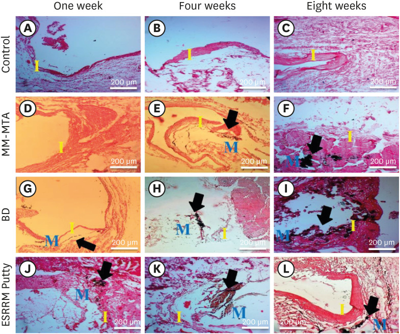
ePub Objectives The aim of this study was to evaluate the dystrophic mineralization deposits from 3 calcium silicate-based cements (Micro-Mega mineral trioxide aggregate [MM-MTA], Biodentine [BD], and EndoSequence Root Repair Material [ESRRM] putty) over time after subcutaneous implantation into rats.
Materials and Methods Forty-five silicon tubes containing the tested materials and 15 empty tubes (serving as a control group) were subcutaneously implanted into the backs of 15 Wistar rats. At 1, 4, and 8 weeks after implantation, the animals were euthanized (
n = 5 animals/group), and the silicon tubes were removed with the surrounding tissues. Histopathological tissue sections were stained with von Kossa stain to assess mineralization. Scanning electron microscopy and energy-dispersive X-ray spectroscopy (SEM/EDX) were also used to assess the chemical components of the surface precipitates deposited on the implant and the pattern of calcium and phosphorus distribution at the material-tissue interface. The calcium-to-phosphorus ratios were compared using the non-parametric Kruskal-Wallis test at a significance level of 5%.Results The von Kossa staining showed that both BD and ESRRM putty induced mineralization starting at week 1; this mineralization increased further until the end of the study. In contrast, MM-MTA induced dystrophic calcification later, from 4 weeks onward. SEM/EDX showed no statistically significant differences in the calcium- and phosphorus-rich areas among the 3 materials at any time point (
p > 0.05).Conclusions After subcutaneous implantation, biomineralization of the 3-calcium silicate-based cements started early and increased over time, and all 3 tested cements generated calcium- and phosphorus-containing surface precipitates.
-
Citations
Citations to this article as recorded by- Evaluating Retrieval-Augmented Large Language Models on External Cervical Resorption: A Comparative Study of Gemini and NotebookLM
Marc Garcia-Font, Nicolás Dufey-Portilla, Fernando Durán-Sindreu, José Antonio González Sánchez, Gustavo Rodríguez Millán, Venkateshbabu Nagendrababu, Paul M.H. Dummer, Francesc Abella Sans
Journal of Endodontics.2025;[Epub] CrossRef - Antibacterial, biocompatible, and mineralization‐inducing properties of calcium silicate‐based cements
Taimy Cruz Hondares, Xiaoxiao Hao, Yanfang Zhao, Yuyin Lin, Dobrawa Napierala, Janice G. Jackson, Ping Zhang
International Journal of Paediatric Dentistry.2024; 34(6): 843. CrossRef - Bioactive potential of Bio‐C Pulpo is evidenced by presence of birefringent calcite and osteocalcin immunoexpression in the rat subcutaneous tissue
Marcela Borsatto Queiroz, Rafaela Nanami Handa Inada, Camila Soares Lopes, Juliane Maria Guerreiro‐Tanomaru, Estela Sasso‐Cerri, Mário Tanomaru‐Filho, Paulo Sérgio Cerri
Journal of Biomedical Materials Research Part B: Applied Biomaterials.2022; 110(10): 2369. CrossRef
- Evaluating Retrieval-Augmented Large Language Models on External Cervical Resorption: A Comparative Study of Gemini and NotebookLM
- 2,258 View
- 19 Download
- 3 Web of Science
- 3 Crossref

- The effect of different fluoride application methods on the remineralization of initial carious lesions
- Seon Mi Byeon, Min Ho Lee, Tae Sung Bae
- Restor Dent Endod 2016;41(2):121-129. Published online May 10, 2016
- DOI: https://doi.org/10.5395/rde.2016.41.2.121
-
 Abstract
 PDF
PDF PubReader
 ePub
ePub Objectives The purpose of this study was to assess the effect of single and combined applications of fluoride on the amount of fluoride release, and the remineralization and physical properties of enamel.
Materials and Methods Each of four fluoride varnish and gel products (Fluor Protector, FP, Ivoclar Vivadent; Tooth Mousse Plus, TM, GC; 60 Second Gel, A, Germiphene; CavityShield, CS, 3M ESPE) and two fluoride solutions (2% sodium fluoride, N; 8% tin(ii) fluoride, S) were applied on bovine teeth using single and combined methods (10 per group), and then the amount of fluoride release was measured for 4 wk. The electron probe microanalysis and the Vickers microhardness measurements were conducted to assess the effect of fluoride application on the surface properties of bovine teeth.
Results The amount of fluoride release was higher in combined applications than in single application (
p < 0.05). Microhardness values were higher after combined applications of N with FP, TM, and CS than single application of them, and these values were also higher after combined applications of S than single application of A (p < 0.05). Ca and P values were higher in combined applications of N with TM and CS than single application of them (p < 0.05). They were also increased after combined applications of the S with A than after single application (p < 0.05).Conclusions Combined applications of fluoride could be used as a basis to design more effective methods of fluoride application to provide enhanced remineralization.
-
Citations
Citations to this article as recorded by- Effect of Different Topical Fluorides on the Microhardness of Bleached Enamel: In Vitro Study
Soumyashri Das, Mansi Jain, HP Suma Sogi, Sonali Sukesh K, Apurva Gambhir, FNU Gagandeep
International Journal of Clinical Pediatric Dentistry.2025; 18(11): 1365. CrossRef - Therapeutic effect of ozone gel on the initial carious lesions
Maha A. Alsharqawy, Wedad M Etman, Mirvat M Salama, Reda G. Saleh
Tanta Dental Journal.2023; 20(3): 203. CrossRef - Evaluation of Remineralization Potential of Natural Substances on Artificially Induced Carious Lesions in Primary Teeth: An In Vitro Study
Kavitha Ramar, Pooja V Ravi, Rajakumar Sekar
International Journal of Clinical Pediatric Dentistry.2023; 16(2): 244. CrossRef - Upaya Preventif Kesehatan Gigi dan Mulut dengan Aplikasi Fluor pada Gigi Siswa SMPN 77 Jakarta
Agus Ardinansyah, Mochammad Atmaji Windrianto, Nur Hidayati Nosi Prastiyani
Info Abdi Cendekia.2023; 6(2): 74. CrossRef - Evaluation of the antibacterial activity of Enamelast® and Fluor defender® fluoride varnishes against Streptococcus mutans biofilm: an in vitro study in primary teeth
M. A. Matar, S. S. Darwish, R. S. Salma, W. A. Lotfy
European Archives of Paediatric Dentistry.2023; 24(5): 549. CrossRef - In-vitro evaluation of the anti-cariogenic effect of a hybrid coating associated with encapsulated sodium fluoride and stannous chloride in nanoclays on enamel
Sávio José Cardoso BEZERRA, Ítallo Emídio Lira VIANA, Idalina Vieira AOKI, Simone DUARTE, Anderson Takeo HARA, Taís SCARAMUCCI
Journal of Applied Oral Science.2022;[Epub] CrossRef - Comparative Evaluation of Salivary Fluoride Concentration after Topical Application of Silver Diamine Fluoride and Sodium Fluoride: A Randomized Controlled Trial
Nidhi Agarwal, V Vishnu Priya, Zohra Jabin, Iffat Nasim
International Journal of Clinical Pediatric Dentistry.2022; 15(3): 371. CrossRef - Release and Recharge of Fluoride Ions from Acrylic Resin Modified with Bioactive Glass
Zbigniew Raszewski, Danuta Nowakowska, Wlodzimierz Wieckiewicz, Agnieszka Nowakowska-Toporowska
Polymers.2021; 13(7): 1054. CrossRef - Enamel remineralisation-inducing materials for caries prevention
Sri Kunarti, Widya Saraswati, Dur Muhammad Lashari, Nadhifa Salma, Tasya Nafatila
Dental Journal.2021; 54(3): 165. CrossRef - Fluoride Concentration in Saliva following Professional Topical Application of 2% Sodium Fluoride Solution
Manjit Talwar, Amrit Tewari, H. S. Chawla, Vinod Sachdev, Suresh Sharma
Contemporary Clinical Dentistry.2019; 10(3): 423. CrossRef - Clinical and laboratory evaluation of the Elgydium Protection caries toothpaste effectiveness in patients with high intensity of dental caries
O. A. Zorina, N. B. Petruhina, A. Z. M, O. A. Boriskina, A. A. Tupicin, V. A. Prohodnaja
Stomatologiya.2019; 98(3): 21. CrossRef - Bleaching of simulated stained-remineralized caries lesions in vitro
Sarah S. Al-Angari, Frank Lippert, Jeffrey A. Platt, George J. Eckert, Carlos González-Cabezas, Yiming Li, Anderson T. Hara
Clinical Oral Investigations.2019; 23(4): 1785. CrossRef - Short-Time Antibacterial Effects of Dimethylaminododecyl Methacrylate on Oral Multispecies Biofilm In Vitro
Yujie Zhou, Suping Wang, Xuedong Zhou, Yiran Zou, Mingyun Li, Xian Peng, Biao Ren, Hockin H. K. Xu, Michael D. Weir, Lei Cheng, Yu Chen, Qi Han
BioMed Research International.2019; 2019: 1. CrossRef - Comparison of the Application of Different Fluoride Supplements on Enamel Demineralization Adjacent to Orthodontic Brackets: An In Vitro Study
Arman Mohammadi Shayan, Monireh Rassouli, Soodabeh Kimyai, Hadi Valizadeh, Mohammad Hossein Ahangar Atashi, Sahand Rikhtegaran
Iranian Journal of Orthodontics.2019;[Epub] CrossRef - Effects of nicomethanol hydrofluoride on dental enamel and synthetic apatites: a role for anti-caries protection
N. Sharkov
European Archives of Paediatric Dentistry.2017; 18(6): 411. CrossRef - Intérêt prophylactique et thérapeutique des chewing-gums sans sucre en orthodontie. Une étude menée auprès de professionnels de santé et de patients
Pauline Ferney, François Clauss, Damien Offner, Delphine Wagner
L'Orthodontie Française.2017; 88(3): 275. CrossRef - Silver Diamine Fluoride Has Efficacy in Controlling Caries Progression in Primary Teeth: A Systematic Review and Meta-Analysis
Ana Cláudia Chibinski, Letícia Maíra Wambier, Juliana Feltrin, Alessandro Dourado Loguercio, Denise Stadler Wambier, Alessandra Reis
Caries Research.2017; 51(5): 527. CrossRef - Dental Caries Management of a Patient with a High Caries Risk Based on the Caries Risk Assessment: a Case Peport
Dong-Hyun Lee, Sung-Ok Hong, Seok-Ryun Lee
Korean Journal of Dental Materials.2016; 43(3): 231. CrossRef
- Effect of Different Topical Fluorides on the Microhardness of Bleached Enamel: In Vitro Study
- 1,994 View
- 13 Download
- 18 Crossref

- Comparative efficacy of photo-activated disinfection and calcium hydroxide for disinfection of remaining carious dentin in deep cavities: a clinical study
- Sidhartha Sharma, Ajay Logani, Naseem Shah
- Restor Dent Endod 2014;39(3):195-200. Published online June 26, 2014
- DOI: https://doi.org/10.5395/rde.2014.39.3.195
-
 Abstract
 PDF
PDF PubReader
 ePub
ePub Objectives To comparatively evaluate the efficacy of photo-activated disinfection (PAD), calcium hydroxide (CH) and their combination on the treatment outcome of indirect pulp treatment (IPT).
Materials and Methods Institutional ethical clearance and informed consent of the patients were taken. The study was also registered with clinical registry of India. Sixty permanent molars exhibiting deep occlusal carious lesion in patients with the age range of 18 - 22 yr were included. Clinical and radiographic evaluation and set inclusion and exclusion criteria's were followed. Gross caries excavation was accomplished. In group I (
n = 20) PAD was applied for sixty seconds. In group II (n = 20), CH was applied to the remaining carious dentin, while in group III (n = 20), PAD application was followed by CH placement. The teeth were permanently restored. They were clinically and radiographically followed-up at 45 day, 6 mon and 12 mon. Relative density of the remaining affected dentin was measured by 'Radiovisiography (RVG) densitometric' analysis.Results Successful outcome with an increase in radiographic grey values were observed in all three groups. However, on inter-group comparison, this change was not significant (
p > 0.05).Conclusions PAD and CH both have equal disinfection efficacy in the treatment of deep carious dentin. PAD alone is as effective for treatment of deep carious lesion as calcium hydroxide and hence can be used as an alternative to CH. They can be used independently in IPT, since combining both does not offer any additional therapeutic benefits.
-
Citations
Citations to this article as recorded by- Clinical and Radiographic Evaluation between Conventional Mineral Trioxide Aggregate and Gel-based Mineral Trioxide Aggregate in Indirect Pulp Therapy: A Randomized Clinical Trial
Yusuf Chunawala, BK Vanishree, Supriya S Dighe, Rooposhi Saha
International Journal of Clinical Pediatric Dentistry.2025; 17(12): 1383. CrossRef - Potentialities of photoactivated disinfection in dentistry
E.I. Utkina, M.A. Gorbatova, A.M. Grjibovski, L.N. Gorbatova, A.A. Simakova
Stomatology.2023; 102(2): 84. CrossRef - Clinical and radiographic evaluation of diode laser and chemical disinfection in comparison to selective caries removal in management of patients with deep carious lesions
Mohamed Bahgat AbdelHamid, Ahmed Fawzy Abo Elezz, Ola M. Ibrahim Fahmy
Lasers in Dental Science.2022; 6(4): 219. CrossRef - Commercially Available Ion-Releasing Dental Materials and Cavitated Carious Lesions: Clinical Treatment Options
Amel Slimani, Salvatore Sauro, Patricia Gatón Hernández, Sevil Gurgan, Lezize Sebnem Turkun, Ivana Miletic, Avijit Banerjee, Hervé Tassery
Materials.2021; 14(21): 6272. CrossRef - Radiological Appraisal of Biodentine and Pulpotec Individually or in Combination with Photo-activated Disinfection as Pulp-capping Cements in Mature Teeth
Pratik Agrawal, Gaurav Patri, Surabhi Soumya, Prasanti K Pradhan, Vijeta Patri
The Journal of Contemporary Dental Practice.2021; 22(9): 1014. CrossRef - Clinical and radiographic evaluation of indirect pulp treatment of young permanent molars using photo-activated oral disinfection versus calcium hydroxide: a randomized controlled pilot trial
Marwa Aly Elchaghaby, Dalia Mohamed Moheb, Osama Ibrahim El Shahawy, Ahmed Mohamed Abd Alsamad, Mervat Abdel Moniem Rashed
BDJ Open.2020;[Epub] CrossRef - Guidelines for the selection, use, and maintenance of LED light-curing units - Part 1
A. C. Shortall, R. B. Price, L. MacKenzie, F. J. T. Burke
British Dental Journal.2016; 221(8): 453. CrossRef
- Clinical and Radiographic Evaluation between Conventional Mineral Trioxide Aggregate and Gel-based Mineral Trioxide Aggregate in Indirect Pulp Therapy: A Randomized Clinical Trial
- 1,428 View
- 4 Download
- 7 Crossref

-
How to design
in situ studies: an evaluation of experimental protocols - Young-Hye Sung, Hae-Young Kim, Ho-Hyun Son, Juhea Chang
- Restor Dent Endod 2014;39(3):164-171. Published online May 13, 2014
- DOI: https://doi.org/10.5395/rde.2014.39.3.164
-
 Abstract
 PDF
PDF PubReader
 ePub
ePub Objectives Designing
in situ models for caries research is a demanding procedure, as both clinical and laboratory parameters need to be incorporated in a single study. This study aimed to construct an informative guideline for planningin situ models relevant to preexisting caries studies.Materials and Methods An electronic literature search of the PubMed database was performed. A total 191 of full articles written in English were included and data were extracted from materials and methods. Multiple variables were analyzed in relation to the publication types, participant characteristics, specimen and appliance factors, and other conditions. Frequencies and percentages were displayed to summarize the data and the Pearson's chi-square test was used to assess a statistical significance (
p < 0.05).Results There were many parameters commonly included in the majority of
in situ models such as inclusion criteria, sample sizes, sample allocation methods, tooth types, intraoral appliance types, sterilization methods, study periods, outcome measures, experimental interventions, etc. Interrelationships existed between the main research topics and some parameters (outcome measures and sample allocation methods) among the evaluated articles.Conclusions It will be possible to establish standardized
in situ protocols according to the research topics. Furthermore, data collaboration from comparable studies would be enhanced by homogeneous study designs.-
Citations
Citations to this article as recorded by- What is the effectiveness of titanium tetrafluoride to prevent or treat dental caries and tooth erosion? A systematic review
Ana Beatriz Chevitarese, Karla Lorene de França Leite, Guido A. Marañón-Vásquez, Danielle Masterson, Matheus Pithon, Lucianne Cople Maia
Acta Odontologica Scandinavica.2022; 80(6): 441. CrossRef - Effect of fluoride group on dental erosion associated or not with abrasion in human enamel: A systematic review with network metanalysis
Bruna Machado da Silva, Daniela Rios, Gerson Aparecido Foratori-Junior, Ana Carolina Magalhães, Marília Afonso Rabelo Buzalaf, Silvia De Carvalho Sales Peres, Heitor Marques Honório
Archives of Oral Biology.2022; 144: 105568. CrossRef - Multimodal Human and Environmental Sensing for Longitudinal Behavioral Studies in Naturalistic Settings: Framework for Sensor Selection, Deployment, and Management
Brandon M Booth, Karel Mundnich, Tiantian Feng, Amrutha Nadarajan, Tiago H Falk, Jennifer L Villatte, Emilio Ferrara, Shrikanth Narayanan
Journal of Medical Internet Research.2019; 21(8): e12832. CrossRef - Evaluation of an antibacterial orthodontic adhesive incorporated with niobium-based bioglass: an in situ study
Felipe Weidenbach DEGRAZIA, Aline Segatto Pires ALTMANN, Carolina Jung FERREIRA, Rodrigo Alex ARTHUR, Vicente Castelo Branco LEITUNE, Susana Maria Werner SAMUEL, Fabrício Mezzomo COLLARES
Brazilian Oral Research.2019;[Epub] CrossRef - A Review of the Common Models Used in Mechanistic Studies on Demineralization-Remineralization for Cariology Research
Ollie Yiru Yu, Irene Shuping Zhao, May Lei Mei, Edward Chin-Man Lo, Chun-Hung Chu
Dentistry Journal.2017; 5(2): 20. CrossRef - Effects of rinsing with arginine bicarbonate and urea solutions on initial enamel lesions in situ
Y Yu, X Wang, C Ge, B Wang, C Cheng, Y‐H Gan
Oral Diseases.2017; 23(3): 353. CrossRef - The cariogenicity of commercial infant formulas: a systematic review
S. F. Tan, H. J. Tong, X. Y. Lin, B. Mok, C. H. Hong
European Archives of Paediatric Dentistry.2016; 17(3): 145. CrossRef - In situ antibiofilm effect of glass-ionomer cement containing dimethylaminododecyl methacrylate
Jin Feng, Lei Cheng, Xuedong Zhou, Hockin H.K. Xu, Michael D. Weir, Markus Meyer, Hans Maurer, Qian Li, Matthias Hannig, Stefan Rupf
Dental Materials.2015; 31(8): 992. CrossRef
- What is the effectiveness of titanium tetrafluoride to prevent or treat dental caries and tooth erosion? A systematic review
- 1,297 View
- 4 Download
- 8 Crossref

- Temperature changes under demineralized dentin during polymerization of three resin-based restorative materials using QTH and LED units
- Sayed-Mostafa Mousavinasab, Maryam Khoroushi, Mohammadreza Moharreri, Mohammad Atai
- Restor Dent Endod 2014;39(3):155-163. Published online May 13, 2014
- DOI: https://doi.org/10.5395/rde.2014.39.3.155
-
 Abstract
 PDF
PDF PubReader
 ePub
ePub Objectives Light-curing of resin-based materials (RBMs) increases the pulp chamber temperature, with detrimental effects on the vital pulp. This
in vitro study compared the temperature rise under demineralized human tooth dentin during light-curing and the degrees of conversion (DCs) of three different RBMs using quartz tungsten halogen (QTH) and light-emitting diode (LED) units (LCUs).Materials and Methods Demineralized and non-demineralized dentin disks were prepared from 120 extracted human mandibular molars. The temperature rise under the dentin disks (
n = 12) during the light-curing of three RBMs, i.e. an Ormocer-based composite resin (Ceram. X, Dentsply DeTrey), a low-shrinkage silorane-based composite (Filtek P90, 3M ESPE), and a giomer (Beautifil II, Shofu GmbH), was measured with a K-type thermocouple wire. The DCs of the materials were investigated using Fourier transform infrared spectroscopy.Results The temperature rise under the demineralized dentin disks was higher than that under the non-demineralized dentin disks during the polymerization of all restorative materials (
p < 0.05). Filtek P90 induced higher temperature rise during polymerization than Ceram.X and Beautifil II under demineralized dentin (p < 0.05). The temperature rise under demineralized dentin during Filtek P90 polymerization exceeded the threshold value (5.5℃), with no significant differences between the DCs of the test materials (p > 0.05).Conclusions Although there were no significant differences in the DCs, the temperature rise under demineralized dentin disks for the silorane-based composite was higher than that for dimethacrylate-based restorative materials, particularly with QTH LCU.
-
Citations
Citations to this article as recorded by- Effect of Shade and Light Curing Mode on the Degree of Conversion of Silorane-Based and Methacrylate-Based Resin Composites
Sayed-Mostafa Mousavinasab, Mohammad Atai, Negar Salehi, Arman Salehi
SSRN Electronic Journal.2024;[Epub] CrossRef - Pulp chamber temperature rise in light-cure bonding of brackets with and without primer, in intact versus restored teeth
Gabriela Cenci SCHMITZ, Fernanda de Souza HENKIN, Mauricio MEZOMO, Mariana MARQUEZAN, Gabriela BONACINA, Maximiliano Schünke GOMES, Eduardo Martinelli Santayana de LIMA
Dental Press Journal of Orthodontics.2023;[Epub] CrossRef -
In Vivo Pulp Temperature Changes During Class V Cavity Preparation and Resin Composite Restoration in Premolars
DC Zarpellon, P Runnacles, C Maucoski, DJ Gross, U Coelho, FA Rueggeberg, CAG Arrais
Operative Dentistry.2021; 46(4): 374. CrossRef
- Effect of Shade and Light Curing Mode on the Degree of Conversion of Silorane-Based and Methacrylate-Based Resin Composites
- 1,427 View
- 2 Download
- 3 Crossref

- Effects of matrix metallproteinases on dentin bonding and strategies to increase durability of dentin adhesion
- Jung-Hyun Lee, Juhea Chang, Ho-Hyun Son
- Restor Dent Endod 2012;37(1):2-8. Published online March 2, 2012
- DOI: https://doi.org/10.5395/rde.2012.37.1.2
-
 Abstract
 PDF
PDF PubReader
 ePub
ePub The limited durability of resin-dentin bonds severely compromises the longevity of composite resin restorations. Resin-dentin bond degradation might occur via degradation of water-rich and resin sparse collagen matrices by host-derived matrix metalloproteinases (MMPs). This review article provides overview of current knowledge of the role of MMPs in dentin matrix degradation and four experimental strategies for extending the longevity of resin-dentin bonds. They include: (1) the use of broad-spectrum inhibitors of MMPs, (2) the use of cross-linking agents for silencing the activities of MMPs, (3) ethanol wet-bonding with hydrophobic resin, (4) biomimetic remineralization of water-filled collagen matrix. A combination of these strategies will be able to overcome the limitations in resin-dentin adhesion.
-
Citations
Citations to this article as recorded by- Remineralization Effects of Silver Fluoride, Silver Diamine Fluoride, and Sodium Fluoride Varnish
Jihyeon Lee, Hwalim Lee, Jongsoo Kim, Joonhaeng Lee, Jongbin Kim, Jisun Shin, Miran Han
International Journal of Clinical Preventive Dentistry.2024; 20(1): 19. CrossRef
- Remineralization Effects of Silver Fluoride, Silver Diamine Fluoride, and Sodium Fluoride Varnish
- 1,209 View
- 10 Download
- 1 Crossref

- The remineralization aspect of enamel according to change of the degree of saturation of the organic acid buffering solution in pH 5.5
- Jin-Sung Park, Sung-Ho Park, Jeong-Won Park, Chan-Young Lee
- J Korean Acad Conserv Dent 2010;35(2):96-105. Published online March 31, 2010
- DOI: https://doi.org/10.5395/JKACD.2010.35.2.096
-
 Abstract
 PDF
PDF PubReader
 ePub
ePub The purpose of this study is to observe and compare the remineralization tendencies of artificial enamel caries lesion by remineralization solutions of different degree of saturations at pH 5.5, using a polarizing microscope and computer programs (Photoshop, Image pro plus, Scion Image, Excel).
For this study, 48 sound permanent teeth with no signs of demineralization, cracks, or dental restorations were used. The specimens were immersed in lactic acid demineralization solution for 2 days in order to produce artificial dental caries that consist of surface and subsurface lesions. Each of 9 or 10 specimens was immersed in pH 5.5 lactic acid buffering remineralization solution of four different degrees of saturation (0.507, 0.394, 0.301, and 0.251) for 12 days. After the demineralization and remineralization, images were taken by a polarizing microscope (×100). The results were obtained by observing images of the specimens, and using computer programs, the density of caries lesions were estimated.
While the group with the lowest degree of saturation (0.251) showed total remineralization feature from the surface to the subsurface of the lesion, the group with the highest degree of saturation (0.507) showed demineralization mainly on the surface of the lesion at the constant organic acid concentration 0.01 M and pH 5.5.
-
Citations
Citations to this article as recorded by- A Randomized, Double-Blind, Placebo-Controlled Clinical Trial of a Mouthwash Containing Glycyrrhiza uralensis Extract for Preventing Dental Caries
Yu-Rin Kim, Seoul-Hee Nam
International Journal of Environmental Research and Public Health.2021; 19(1): 242. CrossRef - Effect of fluoride concentration in pH 4.3 and pH 7.0 supersaturated solutions on the crystal growth of hydroxyapatite
Haneol Shin, Sung-Ho Park, Jeong-Won Park, Chan-Young Lee
Restorative Dentistry & Endodontics.2012; 37(1): 16. CrossRef
- A Randomized, Double-Blind, Placebo-Controlled Clinical Trial of a Mouthwash Containing Glycyrrhiza uralensis Extract for Preventing Dental Caries
- 970 View
- 1 Download
- 2 Crossref

- The effects of the fluoride concentration of acidulated buffer solutions on dentine remineralization
- Won-Sub Han, Chan-Young Lee
- J Korean Acad Conserv Dent 2009;34(6):526-536. Published online November 30, 2009
- DOI: https://doi.org/10.5395/JKACD.2009.34.6.526
-
 Abstract
 PDF
PDF PubReader
 ePub
ePub The aim of this vitro-study is to evaluate the effects of fluoride on remineralization of artificial dentine caries. 10 sound permanent premolars, which were extracted for orthodontic reason within 1 week, were used for this study. Artificial dentine caries was created by using a partially saturated buffer solution for 2 days with grounded thin specimens and fractured whole-body specimens. Remineralization solutions with three different fluoride concentration (1 ppm, 2 ppm and 4 ppm) were used on demineralized-specimens for 7 days. Polarizing microscope and scanning electron microscope were used for the evaluation of the mineral distribution profile and morphology of crystallites of hydroxyapatite.
The results were as follows :
When treated with the fluoride solutions, the demineralized dentine specimens showed remineralization of the upper part and demineralization of the lower part of the lesion body simultaneously.
As the concentration of fluoride increased, the mineral precipitation in the caries dentine increased. The mineral precipitation mainly occurred in the surface layer in 1 and 2 ppm-specimens and in the whole lesion body in 4 ppm-specimens.
When treated with the fluoride solution, the hydroxyapatite crystals grew. This crystal growth was even observed in the lower part of the lesion body which had shown the loss of mineral.
-
Citations
Citations to this article as recorded by- Infant Oral Health Care Concerning Education of Mothers – Part 2
Lehya Mounica Kadali, Viddyasagar Mopagar, Shilpa Shetty, Shridhar Shetty, Venkatesh Kodgi, Shantanu Chaudhari
Journal of Evolution of Medical and Dental Sciences.2021; 10(31): 2538. CrossRef
- Infant Oral Health Care Concerning Education of Mothers – Part 2
- 951 View
- 1 Download
- 1 Crossref

- A bioactivity study of Portland cement mixed with β-glycerophosphosphate on human pulp cell
- Young-Hwan Oh, Young-Joo Jang, Yong-Bum Cho
- J Korean Acad Conserv Dent 2009;34(5):415-423. Published online September 30, 2009
- DOI: https://doi.org/10.5395/JKACD.2009.34.5.415
-
 Abstract
 PDF
PDF PubReader
 ePub
ePub The purpose of this study is to investigate the response of human pulp cell on Portland cement mixed with β-glycerophosphate. To investigate the effect of β-glycerophosphate and/or dexamethasone on human pulp cell, ALP activity on various concentration of β-glycerophosphate and dexamethasone was measured and mineral nodule of human pulp cell was stained with Alizarin red S. MTS assay and ALP activity of human pulp cell on Portland cement mixed with various concentration of β-glycerophosphate (10 mM, 100mM, 1M) was measured and the specimens were examined under SEM.
Addition of β-glycerophosphate or dexamethasone alone had no effect however, the addition of 5 mM β-glycerophosphate and 100 nM dexamethasone had the largest increasement in ALP activity. There was no toxicity in all samples and the data showed that Portland cement mixed with 10 mM β-glycerophosphate had more increase in ALP activity compared with control.
In conclusion, Portland cement mixed with β-glycerophosphate has no toxicity and promotes differentiation and mineralization of pulp cell compared with additive-free Portland cement. This implicated that application of Portland cement mixed with β-glycerophosphate might form more reparative dentin and in turn it would bring direct pulp capping to success.
-
Citations
Citations to this article as recorded by- Dentinogenic potential of human adult dental pulp cells during the extended primary culture
Jin-Hee Min, Seon-Yle Ko, Yong-Bum Cho, Chun-Jeih Ryu, Young-Joo Jang
Human Cell.2011; 24(1): 43. CrossRef
- Dentinogenic potential of human adult dental pulp cells during the extended primary culture
- 931 View
- 1 Download
- 1 Crossref

- THE DYNAMIC CHANGE OF ARTIFICIALLY DEMINERALIZED ENAMEL BY DEGREE OF SATURATION OF REMINERALIZATION SOLUTION AT pH 4.3
- Ji-Sook Yi, Bung-Duk Roh, Su-Jung Shin, Yoon Lee, Hyung-Kyu Gong, Chan-Young Lee
- J Korean Acad Conserv Dent 2009;34(1):20-29. Published online January 14, 2009
- DOI: https://doi.org/10.5395/JKACD.2009.34.1.020
-
 Abstract
 PDF
PDF PubReader
 ePub
ePub Abstract The purpose of this study is to observe and compare the dynamic change of artificially demineralized enamel by remineralization solutions of different degrees of saturation at pH 4.3.
In this study, 30 enamel specimens were demineralized artificially by lactic acid buffered solution. Each of 10 specimens was immersed in pH 4.3 remineralization solution of three different degrees of saturation (0.22, 0.30, 0.35) for 10 days. After demineralization and remineralization, images were taken by a polarizing microscope (× 100). The density of lesion were determined from images taken after demineralization and remineralization.
During remineralization process, mineral deposition and mineral loss occurred at the same time. After remineralization, total mineral amount and width of surface lesion increased in all groups. The higher degree of saturation was, the more mineral deposition occurred in surface lesion and the amount of mineral deposition was not much in subsurface lesion. Total demineralized depth increased in all groups.
-
Citations
Citations to this article as recorded by- Effect of fluoride concentration in pH 4.3 and pH 7.0 supersaturated solutions on the crystal growth of hydroxyapatite
Haneol Shin, Sung-Ho Park, Jeong-Won Park, Chan-Young Lee
Restorative Dentistry & Endodontics.2012; 37(1): 16. CrossRef
- Effect of fluoride concentration in pH 4.3 and pH 7.0 supersaturated solutions on the crystal growth of hydroxyapatite
- 1,011 View
- 3 Download
- 1 Crossref

- The effect of lactic acid concentration and ph of lactic acid buffer solutions on enamel remineralization
- Jung-Won Kwon, Duk-Gyu Suh, Yun-Jung Song, Yun Lee, Chan-Young Lee
- J Korean Acad Conserv Dent 2008;33(6):507-517. Published online November 30, 2008
- DOI: https://doi.org/10.5395/JKACD.2008.33.6.507
-
 Abstract
 PDF
PDF PubReader
 ePub
ePub There are considerable in vitro and in vivo evidences for remineralization and demineralization occurring simultaneously in incipient enamel caries. In order to "heal"the incipient dental caries, many experiments have been carried out to determine the optimal conditions for remineralization. It was shown that remineralization is affected by different pH, lactic acid concentrations, chemical composition of the enamel, fluoride concentrations, etc.
Eighty specimens from sound permanent teeth without demineralization or cracks, 0.15 mm in thickness, were immersed in lactic acid buffered demineralization solutions for 3 days. Dental caries with a surface zone and subsurface lesion were artificially produced. Groups of 10 specimens were immersed for 10 or 12 days in lactic acid buffered remineralization solutions consisting of pH 4.3 or pH 6.0, and 100, 50, 25, or 10 mM lactic acid. After demineralization and remineralization, images were taken by polarizing microscopy (x100) and micro-computed tomography. The results were obtained by observing images of the specimens and the density of the caries lesions was determined.
As the lactic acid concentration of the remineralization solutions with pH 4.3 was higher, the surface zone of the carious enamel increased and an isotropic zone of the subsurface lesion was found. However, the total decalcification depth increased at the same time.
In the remineralization solutions with pH 6.0, only the surface zone increased slightly but there was no significant change in the total decalcification depth and subsurface zone.
In the lactic acid buffer solutions with the lower pH and higher lactic acid concentration, there were dynamic changes at the deep area of the dental carious lesion.
-
Citations
Citations to this article as recorded by- Effect of fluoride concentration in pH 4.3 and pH 7.0 supersaturated solutions on the crystal growth of hydroxyapatite
Haneol Shin, Sung-Ho Park, Jeong-Won Park, Chan-Young Lee
Restorative Dentistry & Endodontics.2012; 37(1): 16. CrossRef
- Effect of fluoride concentration in pH 4.3 and pH 7.0 supersaturated solutions on the crystal growth of hydroxyapatite
- 1,358 View
- 6 Download
- 1 Crossref

- The remineralizing features of pH 5.5 solutions of different degree of saturations on artificially demineralized enamel
- Young-Jun Kwak, Eui-seoug Kim, Sung-Ho Park, Hyung Kyu Gong, Yoon Lee, Chan-Young Lee
- J Korean Acad Conserv Dent 2008;33(5):481-492. Published online September 30, 2008
- DOI: https://doi.org/10.5395/JKACD.2008.33.5.481
-
 Abstract
 PDF
PDF PubReader
 ePub
ePub The purpose of this study is to observe and compare the remineralization tendencies of artificially demineralized enamel by remineralization solutions of different degree of saturations at pH 5.5, using a polarizing microscope and computer programs (Photoshop, Image pro plus, Scion Image, Excel).
For this study, 36 sound permanent teeth with no signs of demineralization, cracks, or dental restorations were used. The specimens were immersed in lactic acid demineralization solution for 3 days in order to produce dental caries artificially that consist of surface and subsurface lesions. Each of 9 or 10 specimens was immersed in pH 5.5 lactic acid buffered remineralization solution of three different degrees of saturation (0.25, 0.30, 0.35) for 12 days. After the demineralization and remineralization, images were taken by a polarizing microscope (× 100). The results were obtained by observing images of the specimens, and using computer programs, the density of caries lesions were determined.
In conclusion, in the group with the lowest degree of saturation, remineralization occurred thoroughly from the surface to the subsurface lesion, whereas in the groups with greater degree of saturation showed no significant change in the subsurface lesion, although there was corresponding increase in the remineralization width on the surface zones.
-
Citations
Citations to this article as recorded by- Effect of fluoride concentration in pH 4.3 and pH 7.0 supersaturated solutions on the crystal growth of hydroxyapatite
Haneol Shin, Sung-Ho Park, Jeong-Won Park, Chan-Young Lee
Restorative Dentistry & Endodontics.2012; 37(1): 16. CrossRef
- Effect of fluoride concentration in pH 4.3 and pH 7.0 supersaturated solutions on the crystal growth of hydroxyapatite
- 1,153 View
- 2 Download
- 1 Crossref

- The change of the configuration of hydroxyapatite crystals in enamel by changes of pH and degree of saturation of lactic acid buffer solution
- Young-Eui Chon, Il-Young Jung, Bung-Duk Roh, Chan-Young Lee
- J Korean Acad Conserv Dent 2007;32(6):498-513. Published online November 30, 2007
- DOI: https://doi.org/10.5395/JKACD.2007.32.6.498
-
 Abstract
 PDF
PDF PubReader
 ePub
ePub Since it was reported that incipient enamel caries can be recovered, previous studies have quantitatively evaluated that enamel artificial caries have been remineralized with fluoride, showing simultaneously the increase of width of surface layer and the decrease of width of the body of legion. There is, however, little report which showed that remineralization could occur without fluoride. In addition, the observations on the change of hydroxyapatite crystals also have been scarcely seen.
In this study, enamel caries in intact premolars or molars was induced by using lactic acidulated buffering solutions over 2 days. Then decalcified specimens were remineralized by seven groups of solutions using different degree of saturation (0.212, 0.239, 0.301, 0.355) and different pH (5.0, 5.5, 6.0) over 10 days. A qualitative comparison to changes of hydroxyapatite crystals after fracturing teeth was made under SEM (scanning electron microscopy) and AFM (atomic force microscopy).
The results were as follows:
1. The size of hydroxyapatite crystals in demineralized area was smaller than the normal ones. While the space among crystals was expanded, it was observed that crystals are arranged irregularly.
2. In remineralized enamel area, the enlarged crystals with various shape were observed when the crystals were fused and new small crystals in intercrystalline spaces were deposited.
3. Group 3 and 4 with higher degree of saturation at same pH showed the formation of large clusters by aggregation of small crystals from the surface layer to the lesion body than group 1 and 2 with relatively low degree of saturation at same pH did. Especially group 4 showed complete remineralization to the body of lesions. Group 5 and 6 with lower pH at similar degree of saturation showed remineralization to the body of lesions while group 7 didn't show it. Unlike in Group 3 and 4, Group 5 and 6 showed that each particle was densely distributed with clear appearance rather than crystals form clusters together.
-
Citations
Citations to this article as recorded by- Anticariogenic Sanative Effect of Aluminum Gallium Arsenide Crystals on Hydroxyapatite Crystals
Sonali Sharma, Mithra N. Hegde, Sindhu Ramesh
Crystals.2022; 12(12): 1841. CrossRef
- Anticariogenic Sanative Effect of Aluminum Gallium Arsenide Crystals on Hydroxyapatite Crystals
- 1,427 View
- 4 Download
- 1 Crossref

- A study of APin-protein interactions using protein microarray
- Joo-Cheol Park, Sun-Hwa Park, Heung-Joong Kim, Jong-Tae Park, Seong-Ho Youn, Ji-Woong Kim, Tae-Yeon Lee, Ho-Hyun Son
- J Korean Acad Conserv Dent 2007;32(5):459-468. Published online September 30, 2007
- DOI: https://doi.org/10.5395/JKACD.2007.32.5.459
-
 Abstract
 PDF
PDF PubReader
 ePub
ePub Protein microarray or protein chips is potentially powerful tools for analysis of protein-protein interactions. APin cDNA was previously identified and cloned from a rat odontoblast cDNA library. The purpose of this study was to investigate the APin-protein interactions during ameloblast differentiation. Protein microarray was carried with recombinant APin protein and MEF2, Aurora kinase A, BMPR-IB and EF-hand calcium binding protein were selected among 74 interacting proteins. Immortalized ameloblast cells (ALCs) were transfected with pCMV-APin construct and U6-APin siRNA construct. After transfection, the expression of the mRNAs for four proteins selected by protein micoarrays were assessed by RT-PCR.
The results were as follows:
1. APin expression was increased and decreased markedly after its over-expression and inactivation, respectively.
2. Over-expression of the APin in the ALCs markedly down-regulated the expression of MEF2 and Aurora kinase A, whereas their expression remained unchanged by its inactivation.
3. Expression of BMPR-IB and EF-hand calcium binding protein were markedly increased by the overexpression of the APin in the ALCs, whereas expression of BMPR-IB remained unchanged and expression of EF-hand calcium binding protein was markedly decreased by its inactivation.
These results suggest that APin plays an important role in ameloblast differentiation and mineralization by regulating the expression of MEF2, Aurora kinase A, BMPR-IB and EF-hand calcium binding protein.
- 760 View
- 1 Download

- The effect of the pH of remineralized buffer solutions on dentin remineralization
- Sung-Chul Kim, Bung-Duk Roh, Il-Young Jung, Chan-Young Lee
- J Korean Acad Conserv Dent 2007;32(2):151-161. Published online March 31, 2007
- DOI: https://doi.org/10.5395/JKACD.2007.32.2.151
-
 Abstract
 PDF
PDF PubReader
 ePub
ePub Dental caries is the most common disease in the oral cavity. However, the mechanism and treatment of dental caries is not completely understood since many complex factors are involved. Especially the effect of pH on remineralization of early stage of dental caries is still controversial.
In this study, dental caries in dentin was induced by using lactic acidulated buffering solutions and the loss of inorganic substance was measured. Also decalcified specimens were remineralized by three groups of solution with different pH (group of pH 4.3, 5.0, and 5.5). Then, the amount and the area of inorganic substance precipitation was quantitatively analyzed with microradiograph. Also a qualitative comparison of the normal phase, the demineralized phase, and the remineralized phase of hydroxyapatite crystal was made under SEM.
The results were as follows;
In microradiograghic analysis, as the pH increased, the amount of remineralization in decalcified dentin tended to increase significantly. As the pH decreaced, deeper decalcification, however, occurred along with remineralization. The group of pH 5.5 had a tendency to be remineralized without demineralization (p < 0.05).
In SEM view, the remineralization in dentine caries occurred from the hydroxyapatite crystal surface surrounding the mesh of organic matrix, and eventually filled up the demineralized area.
5 days after remineralization, hydroxyapatite crystal grew bigger with deposition of inorganic substance in pH 4.3 and 5.0 group, and the crystal in the remineralized area appeared to return to normal. After 10 days, the crystals in group of pH 4.3 and 5.0, which grew bigger after 5 days of remineralization, turned back to their normal size, but in group of pH 5.5, some crystals were found to double their size.
In according to the results of this experiment, the decalcifying and remineralizing process of dentine is neither simple nor independent, but a dynamic process in which decalcification and remineralization occur simultaneously. The remineralization process occurred from the hydroxyapatite crystal surface.
-
Citations
Citations to this article as recorded by- Remineralization Effects of Silver Fluoride, Silver Diamine Fluoride, and Sodium Fluoride Varnish
Jihyeon Lee, Hwalim Lee, Jongsoo Kim, Joonhaeng Lee, Jongbin Kim, Jisun Shin, Miran Han
International Journal of Clinical Preventive Dentistry.2024; 20(1): 19. CrossRef - The remineralizing features of pH 5.5 solutions of different degree of saturations on artificially demineralized enamel
Young-Jun Kwak, Eui-seoug Kim, Sung-Ho Park, Hyung Kyu Gong, Yoon Lee, Chan-Young Lee
Journal of Korean Academy of Conservative Dentistry.2008; 33(5): 481. CrossRef
- Remineralization Effects of Silver Fluoride, Silver Diamine Fluoride, and Sodium Fluoride Varnish
- 981 View
- 2 Download
- 2 Crossref

- The influence of pH and lactic acid concentration on the formation of artificial root caries in acid buffer solution
- Hyun-Suk Oh, Byoung-Duck Roh, Chan-Young Lee
- J Korean Acad Conserv Dent 2007;32(1):47-60. Published online January 31, 2007
- DOI: https://doi.org/10.5395/JKACD.2007.32.1.047
-
 Abstract
 PDF
PDF PubReader
 ePub
ePub The purpose of this study is to compare and to evaluate the effect of pH and lactic acid concentration on the progression of artificial root caries lesion using polarizing microscope, and to evaluate the morphological changes of hydroxyapatite crystals of the demineralized area and to investigate the process of demineralization using scanning electron microscope.
Artificial root caries lesion was created by dividing specimens into 3 pH groups (pH 4.3, 5.0, 5.5), and each pH group was divided into 3 lactic acid concentration groups (25 mM, 50 mM, 100 mM). Each group was immersed in acid buffer solution for 5 days and examined. The results were as follows:
1. Under polarized microscope, the depth of lesion was more effected by the lactic acid concentration rather than the pH.
2. Under scanning electron microscope, dissolution of hydroxyapatite crystals were increased as the lactic acid concentration increased and the pH decreased.
3. Demineralized hydroxyapatite crystals showed peripheral dissolution and decreased size and number within cluster of hydroxyapatite crystals and widening of intercluster and intercrystal spaces as the pH decreased and the lactic acid concentration increased.
4. Under scanning electron microscope evaluation of the surface zone, clusters of hydroxyapatite crystals were dissolved, and dissolution and reattachment of crystals on the surface of collagen fibrils were observed as the lactic acid concentration increased.
5. Under scanning electron microscope, demineralization of dentin occurred not only independently but also with remineralization simultaneously.
In conclusion, the study showed that pH and lactic acid concentration influenced the rate of progression of the lesion in artificial root caries. Demineralization process was progressed from the surface of the cluster of hydroxyapatite crystals and the morphology of hydroxyapatite crystals changed from round or elliptical shape into irregular shape as time elapsed.
- 880 View
- 7 Download

- Expression and function of OD314, Apin protein during ameloblast differentiation and amelogenesis
- Jong-Tae Park, Yong-Seok Choi, Heung-Joong Kim, Moon-Jin Jeong, Hyun-Ju Oh, In-Cheol Shin, Joo-Cheol Park, Ho-Hyun Son
- J Korean Acad Conserv Dent 2006;31(6):437-444. Published online November 30, 2006
- DOI: https://doi.org/10.5395/JKACD.2006.31.6.437
-
 Abstract
 PDF
PDF PubReader
 ePub
ePub This study was aimed to elucidate the biological function of OD314 (Apin protein), which is related toameloblast differentiation and amelogenesis. Apin protein, calcifying epithelial odontogenic (pindborg) tumors (CEOTs)-associated amyloid, were isolated from CEOTs, and has similar nucleotide sequences to OD314. We examined expression of the OD314 mRNA using in-situ hybridization during tooth development in mice. Expression of OD314 and several enamel matrix proteins were examined in the cultured ameloblast cell line up to 28 days by reverse transcription-polymerase chain reaction (RT-PCR) amplification. After inactivation and over-expression of the OD314 gene in ameloblast cell lines using U6 vector-driven RNA interference and CMV-OD314 construct, RT-PCR were performed to evaluate the effect of the OD314 during amelogenesis.
The results were as follows:
1. In in-situ hybridization, OD314 mRNAs were more strongly expressed in ameloblast than odontoblast.
2. When ameloblast cells were cultured in the differentiation and mineralization medium for 28 days, the tuftelin mRNA expression was maintained from the beginning to day 14, and then gradually decreased to day 28. The expressions of amelogenin and enamelin were gradually decreased according to the ameloblast differentiation.
3. Inactivation of OD314 by U6-OD314 siRNA construct down-regulated the expression of OD314, MMP-20, and tuftelin, whereas over-expression of OD314 by CMV-OD314 construct up-regulated the expression of OD314 and MMP-20 without change in tuftelin.
These results suggest that OD314 is considered as an ameloblast-enriched gene and may play the important roles in ameloblast differentiation and mineralization.
- 766 View
- 0 Download

- The influence of the degree of saturation of acidulated buffer solutions in the root dentin demineralization
- Hye-Sil Kang, Chan-Young Lee
- J Korean Acad Conserv Dent 2004;29(5):454-461. Published online September 30, 2004
- DOI: https://doi.org/10.5395/JKACD.2004.29.5.454
-
 Abstract
 PDF
PDF PubReader
 ePub
ePub The purpose of this study is to compare and to evaluate the effects of the degree of saturation on the progression of artificial root caries lesion.
A total of 8 human premolars without any defects and cracks selected and the cementum were removed and the teeth were cleaned with ultrasonic device and pumice without fluoride.
Each tooth was sectioned into 6 pieces and they were ground with #800 sandpaper until they had a thickness of 200µm. Specimens were applied with nail vanish except for the 2-3 mm window area after application of bonding agent. Under the constant pH, the specimens were divided into 6 groups (degree of saturation; 0.1415, 0.1503, 0.1597, 0.1676, 0.1771, 0.1977). Each group was immersed in acid buffer solution for 1, 2, 3, 5 days under controlled temperature (25℃) and imbibed in water and examined using the polarizing microscope.
The results were as follows
1. Although the degree of saturation of demineralization solution decreased, the depth of penetration in the dentin was constant.
2. Erosion was observed on the surface of all the teeth in the group I, II. In the group III, IV, V, surfaces were not changed. The teeth in the group VI showed the more mineralized surface but not the shape of the dentinal tubules distinctively.
3. In all groups, the lesion progressed rapidly at the first day of the experiment, but increased gradually as time elapsed.
-
Citations
Citations to this article as recorded by- Management of white spots: resin infiltration technique and microabrasion
Jeong-Hye Son, Bock Hur, Hyeon-Cheol Kim, Jeong-Kil Park
Journal of Korean Academy of Conservative Dentistry.2011; 36(1): 66. CrossRef
- Management of white spots: resin infiltration technique and microabrasion
- 883 View
- 1 Download
- 1 Crossref

- Expression and functional characterization of odontoblast-derived gene: OD314
- Doo-Hyun Kim, Heung-Joong Kim, Moon-Jin Jeong, Ho-Hyun Son, Joo-Cheol Park
- J Korean Acad Conserv Dent 2004;29(4):399-408. Published online July 31, 2004
- DOI: https://doi.org/10.5395/JKACD.2004.29.4.399
-
 Abstract
 PDF
PDF PubReader
 ePub
ePub Odontoblasts are responsible for the formation and maintenance of dentin. They are known to synthesize unique gene products including dentin sialophosphoprotein (DSPP). Another unique genes of the cells remain unclear.
OD314 was isolated from the odontoblasts/pulp cells of rats and partially characterized as an odontoblast-enriched gene (Dey et al., 2001). This study aimed to elucidate the biological function of OD314, relating to odontoblast differentiation and dentinogenesis. After determining the open reading frame (ORF) of OD314 by transient transfection analysis using green fluorescent protein (GFP) expression vector, mRNA
in-situ hybridization, immunohistochemistry, reverse transcription-polymerase chain reaction (RT-PCR) and western analysis were performed.The results were as follows:
1. In
in-situ hybridization, OD314 mRNAs were expressed in odontoblasts of developing coronal and root pulp.2. OD314 was a novel protein encoding 154 amino acids, and the protein was mainly expressed in cytoplasm by transient transfection analysis.
3. Mineralized nodules were associated with multilayer cell nodules in the culture of human dental pulp cells and first detected from day 21 using alizarin-red S staining.
4. In RT-PCR analysis, OD314, osteocalcin (OC) and DSPP strongly expressed throughout 28 days of culture. Whereas, osteonectin (ON) mRNA expression stayed low up to day 14, and then gradually decreased from day 21.
5. Western blots showed an approximately 17 kDa band. OD314 protein was expressed from the start of culture and then increased greatly from day 21.
In conclusion, OD314 is considered as an odontoblast-enriched gene and may play important roles in odontoblast differentiation and dentin mineralization.
-
Citations
Citations to this article as recorded by- The effects of sodium fluoride on oral normal cell cultured in vitro
Byul-Bora Choi, Da-Hye Kim, Ji-Young Kim, Sang-Rye Park
Journal of Korean society of Dental Hygiene.2016; 16(3): 471. CrossRef - A study of APin-protein interactions using protein microarray
Joo-Cheol Park, Sun-Hwa Park, Heung-Joong Kim, Jong-Tae Park, Seong-Ho Youn, Ji-Woong Kim, Tae-Yeon Lee, Ho-Hyun Son
Journal of Korean Academy of Conservative Dentistry.2007; 32(5): 459. CrossRef - Expression and function of OD314, Apin protein during ameloblast differentiation and amelogenesis
Jong-Tae Park, Yong-Seok Choi, Heung-Joong Kim, Moon-Jin Jeong, Hyun-Ju Oh, In-Cheol Shin, Joo-Cheol Park, Ho-Hyun Son
Journal of Korean Academy of Conservative Dentistry.2006; 31(6): 437. CrossRef - Expression of OD314 during ameloblast differentiation and maturation
Joo-Cheol Park, Seong-Min Ahn, Heung-Joong Kim, Moon-Jin Jeong, Min-Ju Park, In-Cheol Shin, Ho-Hyun Son
Journal of Korean Academy of Conservative Dentistry.2005; 30(5): 423. CrossRef
- The effects of sodium fluoride on oral normal cell cultured in vitro
- 932 View
- 0 Download
- 4 Crossref

-
In vivo quantitative analysis of remineralization effect of remineralization solution "R" of incipient enamel dental caries - Myung-Eun Kim, Il-young Jung, Kee-Yeon Kum, Chang-young Lee, Byoung-Duck Roh
- J Korean Acad Conserv Dent 2002;27(2):175-182. Published online March 31, 2002
- DOI: https://doi.org/10.5395/JKACD.2002.27.2.175
-
 Abstract
 PDF
PDF PubReader
 ePub
ePub Dental caries is a chronic disease that causes the destruction of tooth structure by the interaction of plaque bacteria, food debris, and saliva.
There has been attempts to induce remineralization by supersaturating the intra-oral environment around the surface enamel, where there is incipient caries.
In this study, supersaturated remineralized solution "R" was applied to specimens with incipient enamel caries, and the quantitative ananlysis of remineralization was evaluated using microradiography. Thirty subjects volunteered to participate in this study. Removable appliances were constructed for the subjects, and the enamel specimen with incipient caries were embedded in the appliances. The subjects wore the intra-oral appliance for 15 days except while eating and sleeping.
The removable appliance were soaked in supersaturated solution "R", saline, or Senstime® to expose the specimen to those solutions three times a day, 5 minutes each time. After 15 days, microradiography was retaken to compare and evaluate remineralization.
The results were as the following:
1. The ratio of remineralized area to demineralized area was significantly higher in the supersaturated solution "R" and Senstime® than in the saline. (p<0.05)
2. Remineralization in the supersaturated buffer solution "R" occurred in the significantly deeper parts of the tooth, compared to the Senstime® group containing high concentration of fluoride.(p<0.05)
As in the above results, the remineralization effect of remineralized buffer solution "R" on incipient enamel caries has been proven. For clinical utilization, further studies on soft tissue reaction and the effect on dentin and cementum are necessary.
In conclusion compared to commercially available fluoride solution, remineralization solution "R" showed better remineralization effect on early enamel caries lesion, so it is considered as effecient solution for clinical application.
-
Citations
Citations to this article as recorded by- Color and hardness changes in artificial white spot lesions after resin infiltration
Ji-Hoon Kim, Ho-Hyun Son, Juhea Chang
Restorative Dentistry & Endodontics.2012; 37(2): 90. CrossRef - Changes in surface content and crystal structure after fluoride gel or hydroxyapatite paste application on stripped enamel
Sang-Cheol Kim, Hyun-Sil Hong, Young-Cheol Hwang
The Korean Journal of Orthodontics.2008; 38(6): 407. CrossRef
- Color and hardness changes in artificial white spot lesions after resin infiltration
- 1,032 View
- 9 Download
- 2 Crossref


 KACD
KACD

 First
First Prev
Prev


