Search
- Page Path
- HOME > Search
- Ex vivo comparative analysis of retrievability among four calcium silicate-based sealers for regaining apical patency
- Darian Shomali, Timothy Kirkpatrick, Sang Won Kwak, Hyeon-Cheol Kim, Ji Wook Jeong
- J Korean Acad Conserv Dent ;Published online January 14, 2026
- DOI: https://doi.org/10.5395/rde.2026.51.e3 [Epub ahead of print]
-
 Abstract
 PDF
PDF PubReader
 ePub
ePub - Objectives
Efficient retrievability is a key requirement for endodontic sealers. This study evaluated the retrievability of four different calcium silicate-based sealers (CSS).
Methods
A total of 153 single-rooted human teeth with straight canals were decoronated to a standardized working length of 12 mm. The canals were negotiated to working length using K files up to size 15/.02, followed by rotary instrumentation up to 35/.04, 2 mm short of working length. The teeth were randomly assigned to five groups: NeoSEALER Flo (NEO; Avalon Biomed), Ceraseal (CS; Meta Biomed), Endosequence BC Sealer (BC; Brasseler USA), AH Plus Bioceramic Sealer (AHB; Dentsply Sirona), and a negative control group. Sealer application and obturation with a 35/.04 gutta-percha cone were performed. After incubation at 37°C in 100% humidity for 7 days, retreatment was performed until apical patency was obtained, with retrievability assessed by regaining apical patency. One-way analysis of variance and Tukey contrast test were used to determine whether there was a significant difference among the four different CSS (p < 0.05).
Results
Success rates in regaining apical patency were NEO (79.4%), CS (37.0%), BC (50.0%), and AHB (69.7%). NEO demonstrated the highest retrievability, while CS had the lowest (p < 0.01).
Conclusions
The type of CSS used has a considerable impact on retreatment difficulty. Among the tested sealers, NeoSEALER Flo showed the highest retrievability, making it the most retrievable CSS in terms of retreatment efficacy.
- 140 View
- 7 Download

- Calcium silicate-based sealers remnants in isthmuses of mesial roots of mandibular molars: an in vitro evaluation
- David Saldanha de Brito Alencar, Ana Cristina Padilha Janini, Lauter Eston Pelepenko, Brenda Fornazaro Moraes, Francisco Haiter Neto, Marco Antonio Hungaro Duarte, Marina Angélica Marciano
- Restor Dent Endod 2025;50(3):e25. Published online July 15, 2025
- DOI: https://doi.org/10.5395/rde.2025.50.e25
-
 Abstract
 PDF
PDF PubReader
 ePub
ePub - Objectives
Endodontic retreatment aims to address treatment failure through the removal of root canal filling materials. This in vitro study evaluated the presence of filling material remnants in the mesial root canals, specifically focusing on the isthmuses, of mandibular molars after retreatment.
Methods
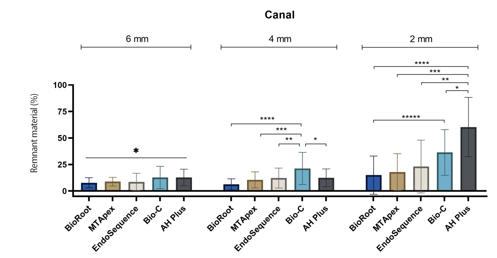
One hundred extracted mandibular molar mesial roots with isthmuses were prepared with an R25 file, obturated with one of five calcium silicate-based sealers (BioRoot RCS [Septodont], MTApex [Ultradent Products Inc.], EndoSequence BC Sealer HiFlow [Brasseler USA], Bio-C Sealer [Angelus]) or an epoxy resin-based sealer (AH Plus Jet [Dentsply Maillefer]), all stained with rhodamine B, and stored at 37ºC for 30 days to allow for setting. Retreatment was subsequently performed using R40 and XP-endo Finisher R instruments (FKG Dentaire) with 2.5% sodium hypochlorite irrigation. The presence of remaining filling material was then assessed using confocal microscopy, and setting times were tested per ISO 6876:2012.
Results
AH Plus Jet showed the most remnants at 2 mm and the longest retreatment time. Calcium silicate-based sealers exhibited prolonged setting times under dry conditions, with EndoSequence BC Sealer HiFlow showing a particularly extended setting period.
Conclusions
Despite retreatment, residues remained in all canals and isthmus regions, particularly Bio-C Sealer and AH Plus Jet in apical areas, emphasizing the difficulty of complete removal and the persistence of filling material.
- 1,813 View
- 105 Download

-
Fracture resistance after root canal filling removal using ProTaper Next, ProTaper Universal Retreatment or hybrid instrumentation: an
ex vivo study - Hadeel Hassan Hanafy, Marwa Mahmoud Bedier, Suzan Abdul Wanees Amin
- Restor Dent Endod 2024;49(4):e38. Published online October 11, 2024
- DOI: https://doi.org/10.5395/rde.2024.49.e38
-
 Abstract
 PDF
PDF PubReader
 ePub
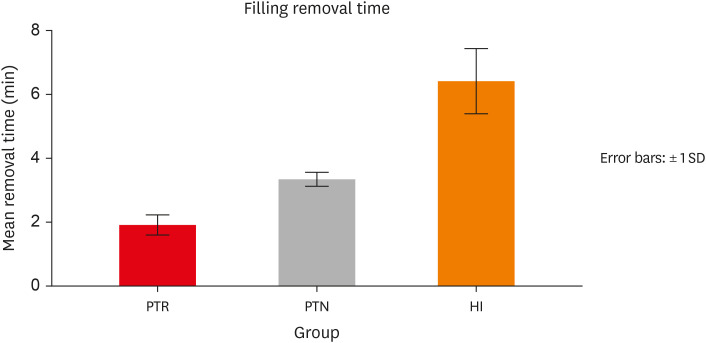
ePub Objectives This study evaluated the effect of ProTaper Next (PTN), ProTaper Universal Retreatment (PTR) and hybrid instrumentation (HI) for canal filling removal on the fracture resistance (FR), mode of failure (MoF), and filling removal time.
Materials and Methods Ninety-six, mandibular premolars were decoronated and randomly divided into 6 groups (
n = 16), as follows: sound (S), untreated canals; prepared teeth (P), canals only prepared to ProTaper Universal finishing instrument (F4); endodontically-treated (ET), prepared and obturated canals using the single-cone technique; and groups PTN, PTR, and HI where filling was removed using PTN, PTR, or HI respectively. FR under vertical loading; MoF and time were assessed. Data were analyzed (Significance level [α] = 0.05).Results There was a significant difference in FR among all groups (
p < 0.001) (HI < P < PTN < S < ET < PTR). HI showed lower FR than S, ET and PTR, and P showed lower FR than PTR (p < 0.05). For experimental groups, there was a significant difference between every group pair (p < 0.05) No significant difference was found regarding MoF distribution (p > 0.05). HI required the highest filling removal time, while PTR required the least (p < 0.05 between every group pair).Conclusions The effect of filling removal on FR may depend on the filling removal technique/system used. PTR could be faster and protect against fracture followed by PTN; HI could adversely affect FR. FR may be associated with filling removal time.
- 2,818 View
- 122 Download

- Effectiveness of endodontic retreatment using WaveOne Primary files in reciprocating and rotary motions
- Patricia Marton Costa, Renata Maíra de Souza Leal, Guilherme Hiroshi Yamanari, Bruno Cavalini Cavenago, Marco Antônio Húngaro Duarte
- Restor Dent Endod 2023;48(2):e15. Published online April 25, 2023
- DOI: https://doi.org/10.5395/rde.2023.48.e15
-
 Abstract
 PDF
PDF PubReader
 ePub
ePub Objectives This study evaluated the efficiency of WaveOne Primary files (Dentsply Sirona) for removing root canal fillings with 2 types of movement: reciprocating (RCP) and continuous counterclockwise rotation (CCR).
Materials and Methods Twenty mandibular incisors were prepared with a RCP instrument (25.08) and filled using the Tagger hybrid obturation technique. The teeth were retreated with a WaveOne Primary file and randomly allocated to 2 experimental retreatment groups (
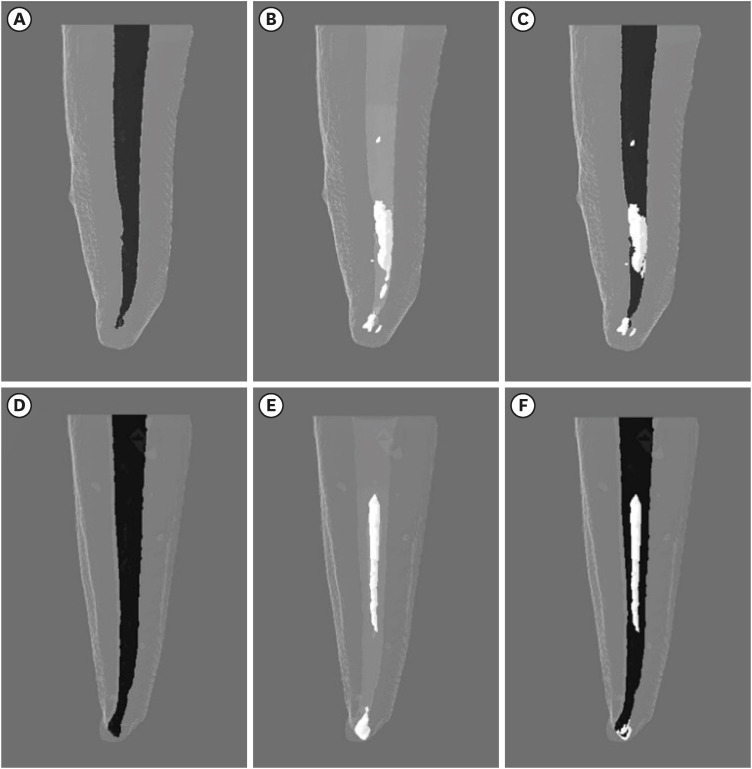
n = 10) according to movement type: RCP and CCR. The root canals were emptied of filling material in the first 3 steps of insertion, until reaching the working length. The timing of retreatment and procedure errors were recorded for all samples. The specimens were scanned before and after the retreatment procedure with micro-computed tomography to calculate the percentage and volume (mm3) of the residual filling material. The results were statistically evaluated using paired and independentt -tests, with a significance level set at 5%.Results No significant difference was found in the timing of filling removal between the groups, with a mean of 322 seconds (RCP) and 327 seconds (CCR) (
p < 0.05). There were 6 instrument fractures: 1 in a RCP motion file and 5 in continuous rotation files. The volumes of residual filling material were similar (9.94% for RCP and 15.94% for CCR;p > 0.05).Conclusions The WaveOne Primary files used in retreatment performed similarly in both RCP and CCR movements. Neither movement type completely removed the obturation material, but the RCP movement provided greater safety.
-
Citations
Citations to this article as recorded by- Micro-CT evaluation of the removal of root fillings using rotary and reciprocating systems supplemented by XP-Endo Finisher, the Self-Adjusting File, or Er,Cr:YSGG laser
Gülsen Kiraz, Bulem Üreyen Kaya, Mert Ocak, Muhammet Bora Uzuner, Hakan Hamdi Çelik
Restorative Dentistry & Endodontics.2023;[Epub] CrossRef
- Micro-CT evaluation of the removal of root fillings using rotary and reciprocating systems supplemented by XP-Endo Finisher, the Self-Adjusting File, or Er,Cr:YSGG laser
- 2,240 View
- 51 Download
- 1 Crossref

- Buckling resistance, torque, and force generation during retreatment with D-RaCe, HyFlex Remover, and Mtwo retreatment files
- Yoojin Kim, Seok Woo Chang, Soram Oh
- Restor Dent Endod 2023;48(1):e10. Published online February 6, 2023
- DOI: https://doi.org/10.5395/rde.2023.48.e10
-
 Abstract
 PDF
PDF PubReader
 ePub
ePub Objectives This study compared the buckling resistance of 3 nickel-titanium (NiTi) retreatment file systems and the torque/force generated during retreatment.
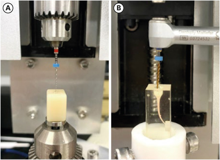
Materials and Methods The buckling resistance was compared among the D-RaCe (DR2), HyFlex Remover, and Mtwo R25/05 retreatment systems. J-shaped canals within resin blocks were prepared with ProTaper NEXT X3 and obturated by the single-cone technique with AH Plus. After 4 weeks, 4 mm of gutta-percha in the coronal aspect was removed with Gates-Glidden drills. Retreatment was then performed using DR1 (size 30, 10% taper) followed by DR2 (size 25, 4% taper), HyFlex Remover (size 30, 7% taper), or Mtrwo R25/05 (size 25, 5% taper) (15 specimens in each group). Further apical preparation was performed with WaveOne Gold Primary. The clockwise torque and upward force generated during retreatment were recorded. After retreatment, resin blocks were examined using stereomicroscopy, and the percentage of residual filling material in the canal area was calculated. Data were analyzed using 1-way analysis of variance with the Tukey test.
Results The HyFlex Remover files exhibited the greatest buckling resistance (
p < 0.05), followed by the Mtwo R25/05. The HyFlex Remover and Mtwo R25/05 files generated the highest maximum clockwise torque and upward force, respectively (p < 0.05). The DR1 and DR2 files generated the least upward force and torque (p < 0.05). The percentage of residual filling material after retreatment was not significantly different between file systems (p > 0.05).Conclusions NiTi retreatment instruments with higher buckling resistance generated greater clockwise torque and upward force.
-
Citations
Citations to this article as recorded by- Comparative evaluation of the efficacy of three different retreatment files in removing root canal filling material: An In vitro confocal microscopy study
Meghna Sarah Abraham, Aravind R. Kudva, Prathap M. S. Nair, Shravan Kini, Samreena Kalander, Faseeh Muhammed Bin Farookh
Endodontology.2025; 37(2): 136. CrossRef - Efficacy of Endodontic Files in Root Canal Retreatment: A Systematic Review of In Vitro Studies
Anna Soler-Doria, José Luis Sanz, Marcello Maddalone, Leopoldo Forner
Journal of Functional Biomaterials.2025; 16(8): 293. CrossRef - Postoperative Pain Following Single‐Visit Nonsurgical Retreatment Using Minimally Invasive Rotary vs. Reciprocating Nickel‐Titanium File Systems: A Two‐Arm Parallel Randomized Clinical Trial
Hüseyin Gürkan Güneç, Büşra Pehlivan, Celalettin Topbaş, Abdurrahman Kerim Kul, Dursun Ali Şirin, Sivakumar Nuvvula
Pain Research and Management.2025;[Epub] CrossRef - Comparative Evaluation of Canal Centering Ability of Single-file Retreatment System vs Multiple-file Retreatment System, with and without Gutta-Percha Solvent: An In Vitro Study
Sangkeetha Gnanasekaran, Arasappan Rajakumaran, Rajeswari Kalaiselvam, Mathan Rajan Rajendran, Seshan Rakkesh Ramesh, Manigandan Kuzhanchinathan
The Journal of Contemporary Dental Practice.2025; 26(9): 898. CrossRef - Effect of Different Downward Loads and Rotational Speeds on the Removal of Gutta-Percha and Root Canal Sealer Using a Nickel-Titanium Rotary Gutta-Percha Removal System: An Ex Vivo Study
Koki Toyoda, Shunsuke Kimura, Keiichiro Maki, Satoshi Omori, Keiko Hirano, Arata Ebihara, Takashi Okiji
Applied Sciences.2025; 16(1): 446. CrossRef - Cone-beam computed tomographic evaluation and fracture resistance of endodontically retreated teeth using hyflex remover, Mtwo, and ProTaper retreatment file systems: An in vitro study
Isha Singh, Dakshita Joy Sinha, Pallavi Sharma, Kunal Bedi, Priyanka Rani, Swapnil Vats
Saudi Endodontic Journal.2024; 14(1): 56. CrossRef - Comparison of torsional, bending, and buckling resistances of different nickel-titanium glide path files
Feyyaz Çeliker, İrem Çetinkaya
Matéria (Rio de Janeiro).2024;[Epub] CrossRef - Assessing the impact of obturation techniques, kinematics and irrigation protocols on apical debris extrusion and time required in endodontic retreatment
Eugenio Pedullà, Francesco Iacono, Martina Pitrolo, Giovanni Barbagallo, Giusy Rita Maria La Rosa, Chiara Pirani
Australian Endodontic Journal.2023; 49(3): 623. CrossRef
- Comparative evaluation of the efficacy of three different retreatment files in removing root canal filling material: An In vitro confocal microscopy study
- 2,373 View
- 53 Download
- 5 Web of Science
- 8 Crossref

- Endodontic micro-resurgery and guided tissue regeneration of a periapical cyst associated to recurrent root perforation: a case report
- Fernando Córdova-Malca, Hernán Coaguila-Llerena, Lucía Garré-Arnillas, Jorge Rayo-Iparraguirre, Gisele Faria
- Restor Dent Endod 2022;47(4):e35. Published online September 3, 2022
- DOI: https://doi.org/10.5395/rde.2022.47.e35
-
 Abstract
 PDF
PDF PubReader
 ePub
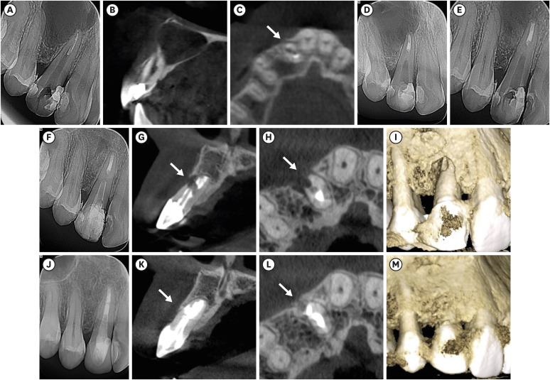
ePub Although the success rates of microsurgery and micro-resurgery are very high, the influence of a recurrent perforation combined with radicular cyst remains unclear. A 21-year-old white female patient had a history of root perforation in a previously treated right maxillary lateral incisor. Analysis using cone-beam computed tomography (CBCT) revealed an extensive and well-defined periapical radiolucency, involving the buccal and palatal bone plate. The perforation was sealed with bioceramic material (Biodentine) in the pre-surgical phase. In the surgical phase, guided tissue regeneration (GTR) was performed by combining xenograft (lyophilized bovine bone) and autologous platelet-rich fibrin applied to the bone defect. The root-end preparation was done using an ultrasonic tip. The retrograde filling was performed using a bioceramic material (Biodentine). Histopathological analysis confirmed a radicular cyst. The patient returned to her referring practitioner to continue the restorative procedures. CBCT analysis after 1-year recall revealed another perforation in the same place as the first intervention, ultimately treated by micro-resurgery using the same protocol with GTR, and a bioceramic material (MTA Angelus). The 2-year recall showed healing and bone neoformation. In conclusion, endodontic micro-resurgery with GTR showed long-term favorable results when a radicular cyst and a recurrent perforation compromised the success.
-
Citations
Citations to this article as recorded by- Outcome of endodontic micro-resurgery: A systematic review
Faisal Alnassar, Riyadh Alroomy, Qamar Hashem, Abdullah Alqedairi, Nabeel Almotairy
Saudi Endodontic Journal.2025; 15(2): 112. CrossRef - Platelet-Rich Plasma and Platelet-Rich Fibrin in Endodontics: A Scoping Review
Simão Rebimbas Guerreiro, Carlos Miguel Marto, Anabela Paula, Joana Rita de Azevedo Pereira, Eunice Carrilho, Manuel Marques-Ferreira, Siri Vicente Paulo
International Journal of Molecular Sciences.2025; 26(12): 5479. CrossRef - Non-surgical Approach to a Maxillary Cyst-Like Lesion: Orthograde Endodontic Treatment With Neodymium-Doped Yttrium Aluminum Garnet (Nd:YAG) Decontamination of the Canal System
Beatrice Spaggiari, Paolo Vescovi, Silvia Pizzi, Roberta Iaria, Ilaria Giovannacci
Cureus.2025;[Epub] CrossRef - Persistent Periradicular Lesion Associated With Concurrent Root Fracture and Odontogenic Keratocyst: A Case Report
Mehdi Vatanpour, Fatemeh Rezaei
Clinical Case Reports.2025;[Epub] CrossRef - Management of Apico-marginal Defects With Endodontic Microsurgery and Guided Tissue Regeneration: A Report of Thirteen Cases
Abayomi O. Baruwa, Jorge N.R. Martins, Mariana D. Pires, Beatriz Pereira, Pedro May Cruz, António Ginjeira
Journal of Endodontics.2023; 49(9): 1207. CrossRef
- Outcome of endodontic micro-resurgery: A systematic review
- 2,701 View
- 53 Download
- 4 Web of Science
- 5 Crossref

-
Effectiveness and safety of rotary and reciprocating kinematics for retreatment of curved root canals: a systematic review of
in vitro studies - Lucas Pinho Simões, Alexandre Henrique dos Reis-Prado, Carlos Roberto Emerenciano Bueno, Ana Cecília Diniz Viana, Marco Antônio Húngaro Duarte, Luciano Tavares Angelo Cintra, Cleidiel Aparecido Araújo Lemos, Francine Benetti
- Restor Dent Endod 2022;47(2):e22. Published online April 6, 2022
- DOI: https://doi.org/10.5395/rde.2022.47.e22
-
 Abstract
 PDF
PDF PubReader
 ePub
ePub Objectives This systematic review (register-osf.io/wg7ba) compared the efficacy and safety of rotary and reciprocating kinematics in the removal of filling material from curved root canals.
Materials and Methods Only
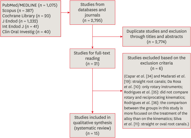
in vitro studies evaluating both kinematics during retreatment were included. A systematic search (PubMed/MEDLINE, Scopus, and other databases, until January 2021), data extraction, and risk of bias analysis (Joanna Briggs Institute checklist) were performed. Efficacy in filling removal was the primary outcome.Results The search resulted in 2,795 studies, of which 15 were included. Efficacy was measured in terms of the remaining filling material and the time required for this. Nine studies evaluated filling material removal, of which 7 found no significant differences between rotary and reciprocating kinematics. Regarding the time for filling removal, 5 studies showed no difference between both kinematics, 2 studies showed faster results with rotary systems, and other 2 showed the opposite. No significant differences were found in apical transportation, centering ability, instrument failure, dentin removed and extruded debris. A low risk of bias was observed.
Conclusions This review suggests that the choice of rotary or reciprocating kinematics does not influence the efficacy of filling removal from curved root canals. Further studies are needed to compare the kinematics safety in curved root canals.
-
Citations
Citations to this article as recorded by- EVALUATION OF MICROLEAKAGE AFTER ENDODONTIC FILLING IN TEETH WITH APICAL WIDENING: A SYSTEMATIC REVIEW
Isabella da Costa Ferreira, Gabriela da Costa Ferreira, Isabella Figueiredo de Assis Macedo, Gustavo Oliveira Campos, Isabella Faria da Cunha Peixoto, Ana Cecília Diniz Viana, Rodrigo Rodrigues Amaral, Warley Luciano Fonseca Tavares
ARACÊ .2025; 7(10): e8792. CrossRef - Fifteen years of engine‐driven nickel–titanium reciprocating instruments, what do we know so far? An umbrella review
Felipe Immich, Lucas Peixoto de Araújo, Rafaella Rodrigues da Gama, Wellington Luiz de Oliveira da Rosa, Evandro Piva, Giampiero Rossi‐Fedele
Australian Endodontic Journal.2024; 50(2): 409. CrossRef - Efficacy of Various Heat-treated Retreatment File Systems on the Apical Deformity and Canal Centering Ability in a Single-rooted Teeth using Nano CT
Swathi S, Pradeep Solete, Ganesh Jeevanandan, Delphine Priscilla Antony S, Kavalipurapu Venkata Teja, Dona Sanju
The Open Dentistry Journal.2024;[Epub] CrossRef - Micro-CT evaluation of the removal of root fillings using rotary and reciprocating systems supplemented by XP-Endo Finisher, the Self-Adjusting File, or Er,Cr:YSGG laser
Gülsen Kiraz, Bulem Üreyen Kaya, Mert Ocak, Muhammet Bora Uzuner, Hakan Hamdi Çelik
Restorative Dentistry & Endodontics.2023;[Epub] CrossRef - Influence of sodium hypochlorite on cyclic fatigue resistance of nickel–titanium instruments: A systematic review and meta-analysis of in vitro studies
Alexandre Henrique dos Reis-Prado, Lucas Guimarães Abreu, Lara Cancella de Arantes, Kiani dos Santos de Paula, Sabrina de Castro Oliveira, Juliana Goto, Ana Cecília Diniz Viana, Francine Benetti
Clinical Oral Investigations.2023; 27(11): 6291. CrossRef - Retreatment of XP-endo Shaper and R-Endo files in curved root canals
Hayam Y. Hassan, Fahd M. Hadhoud, Ayman Mandorah
BMC Oral Health.2023;[Epub] CrossRef - Advancing Endodontics through Kinematics
Shilpa Bhandi, Dario Di Nardo, Francesco Pagnoni, Rosemary Abbagnale
World Journal of Dentistry.2023; 14(6): 479. CrossRef
- EVALUATION OF MICROLEAKAGE AFTER ENDODONTIC FILLING IN TEETH WITH APICAL WIDENING: A SYSTEMATIC REVIEW
- 3,129 View
- 57 Download
- 5 Web of Science
- 7 Crossref

- Efficacy of reciprocating instruments and final irrigant activation protocols on retreatment of mesiobuccal roots of maxillary molars: a micro-CT analysis
- Lilian Tietz, Renan Diego Furlan, Ricardo Abreu da Rosa, Marco Antonio Hungaro Duarte, Murilo Priori Alcalde, Rodrigo Ricci Vivan, Theodoro Weissheimer, Marcus Vinicius Reis Só
- Restor Dent Endod 2022;47(1):e13. Published online February 15, 2022
- DOI: https://doi.org/10.5395/rde.2022.47.e13
-
 Abstract
 PDF
PDF PubReader
 ePub
ePub Objectives This study evaluated the efficacy of 3 reciprocating systems and the effects of 2 instruments for irrigant activation on filling material removal.
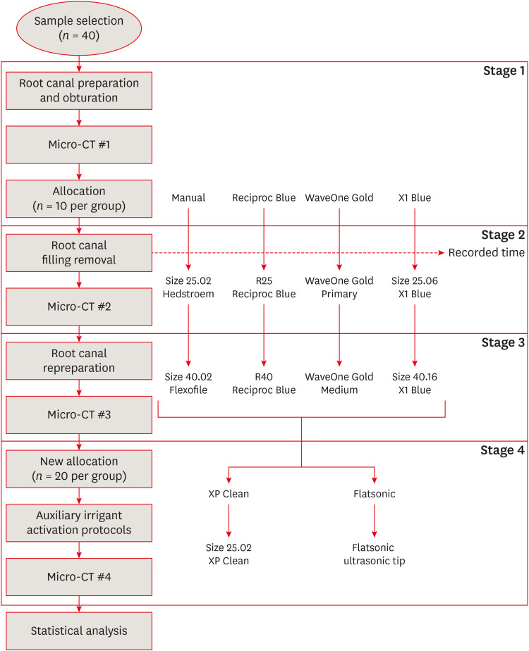
Materials and Methods Forty mesiobuccal roots of maxillary molars were prepared up to size 25.06 and obturated. Micro-computed tomography (micro-CT) examination #1 was performed. Teeth were then divided into 4 groups (
n = 10), according to the retreatment protocol: (1) manual, (2) Reciproc Blue, (3) WaveOne Gold, and (4) X1 Blue. Micro-CT examinations #2 and #3 were performed after filling removal and repreparation, respectively. Next, all teeth were divided into 2 new groups (n = 20) according to the irrigant activation protocol: XP Clean (XP Clean size 25.02) and Flatsonic (Flatsonic ultrasonic tip). Micro-CT examination #4 was performed after irrigant activation. Statistical analysis was performed with a significance level set at 5%.Results WaveOne Gold removed a significantly greater amount of filling material than the manual group (
p < 0.05). The time to reach the WL was similar for all reciprocating systems (p > 0.05). X1 Blue was faster than the manual group (p < 0.05). Only manual group improved the filling material removal after the repreparation stage (p < 0.05). Both activation protocols significantly improved the filling material removal (p < 0.05), without differences between them (p > 0.05).Conclusions None of the tested instruments completely removed the filling material. X1 Blue size 25.06 reached the working length in the shortest time. XP Clean and Flatsonic improved the filling material removal.
-
Citations
Citations to this article as recorded by- Supplementary instrumentation did not enhance the removal of residual gutta-percha: a micro-computed tomography study
Selin Nur Ayaz, Meltem Kucuk, Deniz Yanık Nalbantoğlu, Ali Keles, Amine Yigit, Fugen Dagli Comert Tasman, Bekir Karabucak
Odontology.2025;[Epub] CrossRef - Supplementary methods for filling material removal: A systematic review and meta-analysis of micro-CT imaging studies
Bruna Venzke Fischer, Taynara Santos Goulart, Filipe Colombo Vitali, Diego Leonardo de Souza, Cleonice da Silveira Teixeira, Lucas da Fonseca Roberti Garcia
Journal of Dentistry.2024; 151: 105445. CrossRef - Evaluation of the Ability of 3 Reciprocating Instruments to Remove Obturation Material: A Micro–Computed Tomography Study
Fábio Luiz Cecagno, Alexandre Sigrist De Martin, Carlos Eduardo Fontana, Bruno Cavalini Cavenago, Wayne Martins Nascimento, Ana Grasiela da Silva Limoeiro, Carlos Eduardo da Silveira Bueno
Journal of Endodontics.2024; 50(3): 376. CrossRef - Comparative evaluation of cleaning efficiency of single file NiTi rotary system during root canal treatment procedure - A scanning electron microscope study
Ruchi Vashisht, Umesh Kumar, Swaty Jhamb, Ruchi Singla
Journal of Conservative Dentistry.2023; 26(3): 316. CrossRef - Influence of rotary and reciprocating kinematics on the accuracy of an integrated apex locator
Verônica de Almeida Gardelin, Júlia Itzel Acosta Moreno Vinholes, Renata Grazziotin‐Soares, Fernanda Geraldo Pappen, Fernando Branco Barletta
Australian Endodontic Journal.2023; 49(S1): 202. CrossRef
- Supplementary instrumentation did not enhance the removal of residual gutta-percha: a micro-computed tomography study
- 2,233 View
- 34 Download
- 4 Web of Science
- 5 Crossref

- Fracture incidence of Reciproc instruments during root canal retreatment performed by postgraduate students: a cross-sectional retrospective clinical study
- Liliana Machado Ruivo, Marcos de Azevedo Rios, Alexandre Mascarenhas Villela, Alexandre Sigrist de Martin, Augusto Shoji Kato, Rina Andrea Pelegrine, Ana Flávia Almeida Barbosa, Emmanuel João Nogueira Leal Silva, Carlos Eduardo da Silveira Bueno
- Restor Dent Endod 2021;46(4):e49. Published online September 9, 2021
- DOI: https://doi.org/10.5395/rde.2021.46.e49
-
 Abstract
 PDF
PDF PubReader
 ePub
ePub Objectives To evaluate the fracture incidence of Reciproc R25 instruments (VDW) used during non-surgical root canal retreatments performed by students in a postgraduate endodontic program.
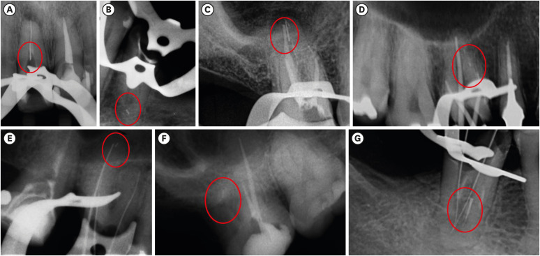
Materials and Methods From the analysis of clinical record cards and periapical radiographs of root canal retreatments performed by postgraduate students using the Reciproc R25, a total of 1,016 teeth (2,544 root canals) were selected. The instruments were discarded after a single use. The general incidence of instrument fractures and its frequency was analyzed considering the group of teeth and the root thirds where the fractures occurred. Statistical analysis was performed using the χ2 test (
p < 0.01).Results Seven instruments were separated during the procedures. The percentage of fracture in relation to the number of instrumented canals was 0.27% and 0.68% in relation to the number of instrumented teeth. Four fractures occurred in maxillary molars, 1 in a mandibular molar, 1 in a mandibular premolar and 1 in a maxillary incisor. A greater number of fractures was observed in molars when compared with the number of fractures observed in the other dental groups (
p < 0.01). Considering all of the instrument fractures, 71.43% were located in the apical third and 28.57% in the middle third (p < 0.01). One instrument fragment was removed, one bypassed, while in 5 cases, the instrument fragment remained inside the root canal.Conclusions The use of Reciproc R25 instruments in root canal retreatments carried out by postgraduate students was associated with a low incidence of fractures.
-
Citations
Citations to this article as recorded by- Reciprocating Torsional Fatigue and Mechanical Tests of Thermal-Treated Nickel Titanium Instruments
Victor Talarico Leal Vieira, Alejandro Jaime, Carlos Garcia Puente, Giuliana Soimu, Emmanuel João Nogueira Leal Silva, Carlos Nelson Elias, Gustavo de Deus
Journal of Endodontics.2025; 51(3): 359. CrossRef - Neodymium-Doped Yttrium Aluminum Perovskite (Nd:YAP) Laser in the Elimination of Endodontic Nickel-Titanium Files Fractured in Rooted Canals (Part 2: Teeth With Significant Root Curvature)
Amaury Namour, Marwan El Mobadder, Clément Cerfontaine, Patrick Matamba, Lucia Misoaga, Delphine Magnin , Praveen Arany, Samir Nammour
Cureus.2025;[Epub] CrossRef - Temperature-Dependent Effects on Cyclic Fatigue Resistance in Three Reciprocating Endodontic Systems: An In Vitro Study
Marcela Salamanca Ramos, José Aranguren, Giulia Malvicini, Cesar De Gregorio, Carmen Bonilla, Alejandro R. Perez
Materials.2025; 18(5): 952. CrossRef - The Cost of Instrument Retrieval on the Root Integrity
Marco A. Versiani, Hugo Sousa Dias, Emmanuel J. N. L. Silva, Felipe G. Belladonna, Jorge N. R. Martins, Gustavo De‐Deus
International Endodontic Journal.2025; 58(12): 1948. CrossRef - Multimethod analysis of large‐ and low‐tapered single file reciprocating instruments: Design, metallurgy, mechanical performance, and irrigation flow
Emmanuel João Nogueira Leal Silva, Fernando Peña‐Bengoa, Natasha C. Ajuz, Victor T. L. Vieira, Jorge N. R. Martins, Duarte Marques, Ricardo Pinto, Mario Rito Pereira, Francisco Manuel Braz‐Fernandes, Marco A. Versiani
International Endodontic Journal.2024; 57(5): 601. CrossRef - Nd: YAP Laser in the Elimination of Endodontic Nickel-Titanium Files Fractured in Rooted Canals (Part 1: Teeth With Minimal Root Curvature)
Amaury Namour, Marwan El Mobadder, Patrick Matamba, Lucia Misoaga, Delphine Magnin , Praveen Arany, Samir Nammour
Cureus.2024;[Epub] CrossRef - Cyclic Fatigue of Different Reciprocating Endodontic Instruments Using Matching Artificial Root Canals at Body Temperature In Vitro
Sebastian Bürklein, Paul Maßmann, Edgar Schäfer, David Donnermeyer
Materials.2024; 17(4): 827. CrossRef - Endodontic Orthograde Retreatments: Challenges and Solutions
Alessio Zanza, Rodolfo Reda, Luca Testarelli
Clinical, Cosmetic and Investigational Dentistry.2023; Volume 15: 245. CrossRef - Design, metallurgy, mechanical properties, and shaping ability of 3 heat-treated reciprocating systems: a multimethod investigation
Emmanuel J. N. L. Silva, Jorge N. R. Martins, Natasha C. Ajuz, Henrique dos Santos Antunes, Victor Talarico Leal Vieira, Francisco Manuel Braz-Fernandes, Felipe Gonçalves Belladonna, Marco Aurélio Versiani
Clinical Oral Investigations.2023; 27(5): 2427. CrossRef - Noncontact 3D evaluation of surface topography of reciprocating instruments after retreatment procedures
Miriam Fatima Zaccaro-Scelza, Renato Lenoir Cardoso Henrique Martinez, Sandro Oliveira Tavares, Fabiano Palmeira Gonçalves, Marcelo Montagnana, Emmanuel João Nogueira Leal da Silva, Pantaleo Scelza
Brazilian Dental Journal.2022; 33(3): 38. CrossRef
- Reciprocating Torsional Fatigue and Mechanical Tests of Thermal-Treated Nickel Titanium Instruments
- 2,377 View
- 21 Download
- 7 Web of Science
- 10 Crossref

- Efficacy of reciprocating and rotary retreatment nickel-titanium file systems for removing filling materials with a complementary cleaning method in oval canals
- Said Dhaimy, Hyeon-Cheol Kim, Lamyae Bedida, Imane Benkiran
- Restor Dent Endod 2021;46(1):e13. Published online February 3, 2021
- DOI: https://doi.org/10.5395/rde.2021.46.e13
-
 Abstract
 PDF
PDF PubReader
 ePub
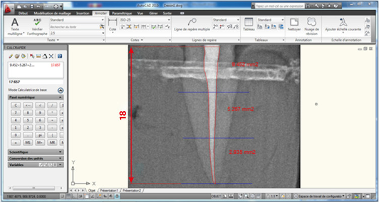
ePub Objectives This study aimed to evaluate and compare the efficacy of the S1 reciprocating system and the D-Race retreatment rotary system for filling material removal and the apical extrusion of debris.
Materials and Methods Sixty-four freshly extracted maxillary canines were shaped with size 10 and size 15 K-files, instrumented using ProTaper Gold under irrigation with 2.5% sodium hypochlorite (NaOCl), obturated according to the principle of thermo-mechanical condensation with gutta-percha and zinc oxide eugenol sealer, and allowed to set for 3 weeks at 37°C. Subsequently, the teeth were divided into a control group (
n = 4), the D-Race rotary instrument group (n = 30), and the S1 reciprocating instrument group (n = 30). After classical retreatment, the canals were subjected to a complementary approach with the XP-Endo Shaper. Desocclusol was used as a solvent, and irrigation with 2.5% NaOCl was performed. Each group was divided into subgroups according to the timing of radiographic readings. The images were imported into a software program to measure the remaining filling material, the apical extrusion, and the root canal space. The data were statistically analyzed using the Z-test and JASP graphics software.Results No significant differences were found between the D-Race and S1 groups for primary retreatment; however, using a complementary cleaning method increased the removal of remnant filling (
p < 0.05).Conclusions Classical removal of canal filling material may not be sufficient for root canal disinfection, although a complementary finishing approach improved the results. Nevertheless, all systems left some debris and caused apical extrusion.
-
Citations
Citations to this article as recorded by- Effectiveness of different supplementary protocols for remaining filling material removal in endodontic reintervention: an integrative review
Amanda Freitas da Rosa, Bruna Venzke Fischer, Luiz Carlos de Lima Dias-Junior, Anna Victoria Costa Serique, Eduardo Antunes Bortoluzzi, Cleonice da Silveira Teixeira, Lucas da Fonseca Roberti Garcia
Odontology.2024; 112(1): 51. CrossRef - Critical analysis of research methods and experimental models to study removal of root filling materials
Mahdi A. Ajina, Pratik K. Shah, Bun San Chong
International Endodontic Journal.2022; 55(S1): 119. CrossRef - Economic analysis of the different endodontic instrumentation techniques used in the Unified Health System
Laura Paredes Merchan, Livia Fernandes Probst, Ana Clara Correa Duarte Simões, Augusto Cesar Santos Raimundo, Yuri Wanderley Cavalcanti, Denise de Fátima Barros Cavalcante, João Victor Frazão Câmara, Antonio Carlos Pereira
BMC Oral Health.2022;[Epub] CrossRef - Fabrication of a Potential Electrodeposited Nanocomposite for Dental Applications
Chun-Wei Chang, Chen-Han Tsou, Bai-Hung Huang, Kuo-Sheng Hung, Yung-Chieh Cho, Takashi Saito, Chi-Hsun Tsai, Chia-Chien Hsieh, Chung-Ming Liu, Wen-Chien Lan
Inorganics.2022; 10(10): 165. CrossRef - Influence of Filling Material Remnants on the Diffusion of Hydroxyl Ions in Endodontically Retreated Teeth: An Ex Vivo Study
Vania Portela Ditzel Westphalen, Marilisa Carneiro Leao Gabardo, Natanael Henrique Ribeiro Mattos, Camila Paiva Perin, Liliane Roskamp, Cristiano Miranda de Araújo, Luiz Fernando Fariniuk, Flares Baratto–Filho
The Journal of Contemporary Dental Practice.2022; 23(8): 768. CrossRef - Efficacy of Removing Thermafil and GuttaCore from Straight Root Canal Systems Using a Novel Non-Surgical Root Canal Re-Treatment System: A Micro-Computed Tomography Analysis
Vicente Faus-Llácer, Rubén Linero Pérez, Ignacio Faus-Matoses, Celia Ruiz-Sánchez, Álvaro Zubizarreta-Macho, Salvatore Sauro, Vicente Faus-Matoses
Journal of Clinical Medicine.2021; 10(6): 1266. CrossRef
- Effectiveness of different supplementary protocols for remaining filling material removal in endodontic reintervention: an integrative review
- 1,852 View
- 37 Download
- 7 Web of Science
- 6 Crossref

- Impact of root canal curvature and instrument type on the amount of extruded debris during retreatment
- Burcu Serefoglu, Gözde Kandemir Demirci, Seniha Miçooğulları Kurt, İlknur Kaşıkçı Bilgi, Mehmet Kemal Çalışkan
- Restor Dent Endod 2021;46(1):e5. Published online December 17, 2020
- DOI: https://doi.org/10.5395/rde.2021.46.e5
-
 Abstract
 PDF
PDF PubReader
 ePub
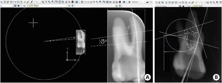
ePub Objectives The aim of the current study was to assess whether the amount of extruded debris differs for straight and severely curved root canals during retreatment using H-files, R-Endo, Reciproc and ProTaper Universal Retreatment (PTU-R) files. Additionally, the area of residual filling material was evaluated.
Materials and Methods Severely curved (
n = 104) and straight (n = 104) root canals of maxillary molar teeth were prepared with WaveOne Primary file and obturated with gutta-percha and AH Plus sealer. Root canal filling materials were removed with one of the preparation techniques: group 1: H-file; group 2: R-Endo; group 3: Reciproc; group 4: PTU-R (n = 26). The amount of extruded material and the area of the residual filling material was measured. The data were analyzed with 2-way analysis of variance (ANOVA) and 1-way ANOVA at the 0.05 significance level.Results Except for Reciproc group (
p > 0.05), PTU-R, R-Endo, and H-file systems extruded significantly more debris in severely curved canals (p < 0.05). Each file system caused more residual filling material in severely curved canals than in straight ones (p < 0.05).Conclusions All instruments used in this study caused apical debris extrusion. Root canal curvature had an effect on extruded debris, except for Reciproc system. Clinicians should be aware that the difficult morphology of the severely curved root canals is a factor increasing the amount of extruded debris during the retreatment procedure.
-
Citations
Citations to this article as recorded by- Comparative Analysis of Root Canal Curvature Measurement Methods for Permanent Mandibular Molars Distal Root: An Observational Study
Tanu Singh, Saurav Bathla, Anuraag Gurtu, Shubhi Gupta, Sana Saifi, Madhusudan Astekar
The Journal of Contemporary Dental Practice.2025; 26(10): 945. CrossRef - Do Continuous Rotating Endodontic Instruments Extrude Fewer Apical Debris Than Reciprocating Instruments in Non-Surgical Endodontic Retreatments? A Systematic Review
Francesco Puleio, Francesco Giordano, Ugo Bellezza, David Rizzo, Valentina Coppini, Roberto Lo Giudice
Applied Sciences.2024; 14(4): 1621. CrossRef - Intracanal removal and apical extrusion of filling material after retreatment using rotary or reciprocating instruments: A new approach using human cadavers
Thamyres M. Monteiro, Victor O. Cortes‐Cid, Marilia F. V. Marceliano‐Alves, Andrea F. Campello, Luan F. Bastos, Ricardo T. Lopes, José F. Siqueira, Flávio R. F. Alves
International Endodontic Journal.2024; 57(1): 100. CrossRef - Comparative analysis of methods for measuring root canal curvature based on periapical radiography: A laboratory study
Rafael Chies Hartmann, Eduardo Silva Ferraz, Theodoro Weissheimer, Jose Antônio Poli de Figueiredo, Giampiero Rossi‐Fedele, Maximiliano Schünke Gomes
International Endodontic Journal.2024; 57(12): 1848. CrossRef - Evaluation of apically extruded debris during root canal filling material removal in teeth with external apical root resorption: a comparison of different obturation techniques
Büşra Melike Çağlar, İsmail Uzun
BMC Oral Health.2024;[Epub] CrossRef - Evaluation of apically extruded debris using protaper universal, protaper next, one curve, Xp shaper, and edge file: An in vitro study
Murtada Qadir Muhaibes, Shatha Abdulkareem Alwakeel
Saudi Endodontic Journal.2024; 14(1): 31. CrossRef - A quantitative comparison of apically extruded debris during root canal preparation using NiTi full-sequence rotary and single-file rotary systems: An in vitro study
Pallavi Goel, R. Vikram, R. Anithakumari, M. S. Adarsha, M. E. Sudhanva
Endodontology.2024; 36(3): 235. CrossRef - In vitro evaluation of filling material removal and apical debris extrusion after retreatment using Reciproc blue, Hyflex EDM and ProTaper retreatment files
Passent Abdelnaby, Mohamed Ibrahim, Rania ElBackly
BMC Oral Health.2023;[Epub] CrossRef - A Comparative Study on the Shaping Ability and Cleaning Efficiency of Two Different Single-File Systems, Reciprocating Wave One Versus Continuous Rotation F360, Evaluated by Scanning Electron Microscope: An In Vitro Study
Arunkumar Samudrala, Chandrakanth Majeti, Kommineni Harika Chowdary, Lakshmi Bhavani Potru, Anusha Yaragani, Yata Prashanth Kumar, Gagandeep K Sidhu, Navneet S Kathuria
Cureus.2023;[Epub] CrossRef - COMPARATIVE EVALUATION OF THE EFFECT OF DIFFERENT ROTARY INSTRUMENT SYSTEMS ON THE AMOUNT OF APICALLY EXTRUDED DEBRIS
Recai ZAN, Bilge LENGER
Cumhuriyet Dental Journal.2022; 25(2): 172. CrossRef - A critical analysis of research methods and experimental models to study apical extrusion of debris and irrigants
Jale Tanalp
International Endodontic Journal.2022; 55(S1): 153. CrossRef - Critical analysis of research methods and experimental models to study removal of root filling materials
Mahdi A. Ajina, Pratik K. Shah, Bun San Chong
International Endodontic Journal.2022; 55(S1): 119. CrossRef
- Comparative Analysis of Root Canal Curvature Measurement Methods for Permanent Mandibular Molars Distal Root: An Observational Study
- 2,237 View
- 31 Download
- 8 Web of Science
- 12 Crossref

- Bonding of a resin-modified glass ionomer cement to dentin using universal adhesives
- Muhittin Ugurlu
- Restor Dent Endod 2020;45(3):e36. Published online June 15, 2020
- DOI: https://doi.org/10.5395/rde.2020.45.e36
-
 Abstract
 PDF
PDF PubReader
 ePub
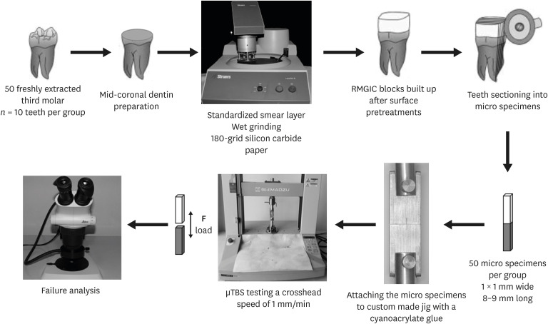
ePub Objectives This study aims to assess the effect of universal adhesives pretreatment on the bond strength of resin-modified glass ionomer cement to dentin.
Materials and Methods Fifty caries-free human third molars were employed. The teeth were randomly assigned into five groups (
n = 10) based on dentin surface pretreatments: Single Bond Universal (3M Oral Care), Gluma Bond Universal (Heraeus Kulzer), Prime&Bond Elect (Dentsply), Cavity Conditioner (GC) and control (no surface treatment). After Fuji II LC (GC) was bonded to the dentin surfaces, the specimens were stored for 7 days at 37°C. The specimens were segmented into microspecimens, and the microspecimens were subjugated to microtensile bond strength testing (1.0 mm/min). The modes of failure analyzed using a stereomicroscope and scanning electron microscopy. Data were statistically analyzed with one-way analysis of variance and Duncan tests (p = 0.05).Results The surface pretreatments with the universal adhesives and conditioner increased the bond strength of Fuji II LC to dentin (
p < 0.05). Single Bond Universal and Gluma Bond Universal provided higher bond strength to Fuji II LC than Cavity Conditioner (p < 0.05). The bond strengths obtained from Prime&Bond Elect and Cavity Conditioner were not statistically different (p > 0.05).Conclusions The universal adhesives and polyacrylic acid conditioner could increase the bond strength of resin-modified glass ionomer cement (RMGIC) to dentin. The use of universal adhesives before the application of RMGIC may be more beneficial in improving bond strength.
-
Citations
Citations to this article as recorded by- Impact of nanochitosan incorporation on the performance of resin-modified glass ionomer luting cement: a comprehensive in vitro study
Mostafa A. Abdelshafi, Nesma Elgohary, Ahmed Shams
BMC Oral Health.2026;[Epub] CrossRef - Clinical evaluation of giomer-based injectable resin composite versus resin-modified glass ionomer in class V carious lesions over 18 months: A randomized clinical trial
Reham Hendam, Rania Mosallam, Dina Kamal
Journal of Conservative Dentistry and Endodontics.2025; 28(1): 50. CrossRef - Push-Out Bond Strength of Different Luting Cements Following Post Space Irrigation with 2% Chitosan: An In Vitro Study
Shimaa Rifaat, Ahmed Rahoma, Hind Muneer Alharbi, Sawsan Jamal Kazim, Shrouq Ali Aljuaid, Basmah Omar Alakloby, Faraz A. Farooqi, Noha Taymour
Prosthesis.2025; 7(1): 18. CrossRef - Bioactive restorative materials in dentistry: a comprehensive review of mechanisms, clinical applications, and future directions
Dina Abozaid, Amr Azab, Mohammad A. Bahnsawy, Mohamed Eldebawy, Abdullah Ayad, Romesa soomro, Enas Elwakeel, Maged Ahmed Mohamed
Odontology.2025;[Epub] CrossRef - A Comparative Evaluation of Marginal Leakage and Shear Bond Strength of Cention N, Resin-Modified Glass Ionomer Cement (RMGIC), and Conventional Glass Ionomer Cement (GIC): An In Vitro Study
Khushboo Singh, Debapriya Pradhan, Saurabh Tiwari, Raksha Thakur, Priyamvada Sharma, Devika Agrawal, Mahima Singh, Devshree Jawalikar, Delphina Michael Kapoor, Jyoti Priiya Kodimela
Cureus.2025;[Epub] CrossRef - Assessment of Nanosilver Fluoride Application on the Microtensile Bond Strength of Glass Ionomer Cement and Resin-modified Glass Ionomer Cement on Primary Carious Dentin: An In Vitro Study
Ila Srinivasan, Yuthi Milit, Anushka Das, Neeraja Ramamurthy
International Journal of Clinical Pediatric Dentistry.2024; 17(5): 565. CrossRef - Effect of Surface Treatments on Shear-bond Strength of Glass Ionomer Cements to Silver Diamine Fluoride-treated Simulated Carious Dentin
WT Koh, OT Yeoh, NA Yahya, AU Yap
Operative Dentistry.2024; 49(6): 714. CrossRef - Desensitizing agents’ post-bleaching effect on orthodontic bracket bond strength
Gufa Bagus Pamungkas, Dyah Karunia, Sri Suparwitri
Dental Journal.2024; 57(1): 45. CrossRef - Successful Rehabilitation of Traumatized Immature Teeth by Different Vital Pulp Therapies in Pediatric Patients
Mohammad Kamran Khan
Journal of the Scientific Society.2023; 50(1): 111. CrossRef - Do bioactive materials show greater retention rates in restoring permanent teeth than non-bioactive materials? A systematic review and network meta-analysis of randomized controlled trials
Juliana Benace Fernandes, Sheila Mondragón Contreras, Manuela da Silva Spinola, Graziela Ribeiro Batista, Eduardo Bresciani, Taciana Marco Ferraz Caneppele
Clinical Oral Investigations.2023;[Epub] CrossRef - Effects of tooth preparation on the microleakage of fissure sealant
Gesti Kartiko Sari, Sri Kuswandari, Putri Kusuma Wardani Mahendra
Dental Journal (Majalah Kedokteran Gigi).2022; 55(2): 67. CrossRef - Rheological Properties, Surface Microhardness, and Dentin Shear Bond Strength of Resin-Modified Glass Ionomer Cements Containing Methacrylate-Functionalized Polyacids and Spherical Pre-Reacted Glass Fillers
Whithipa Thepveera, Wisitsin Potiprapanpong, Arnit Toneluck, Somruethai Channasanon, Chutikarn Khamsuk, Naruporn Monmaturapoj, Siriporn Tanodekaew, Piyaphong Panpisut
Journal of Functional Biomaterials.2021; 12(3): 42. CrossRef
- Impact of nanochitosan incorporation on the performance of resin-modified glass ionomer luting cement: a comprehensive in vitro study
- 4,043 View
- 36 Download
- 12 Crossref

- Micro-computed tomographic evaluation of canal retreatments performed by undergraduate students using different techniques
- Emmanuel João Nogueira Leal Silva, Felipe Gonçalves Belladonna, Marianna Fernandes Carapiá, Brenda Leite Muniz, Mariana Santoro Rocha, Edson Jorge Lima Moreira
- Restor Dent Endod 2018;43(1):e5. Published online January 15, 2018
- DOI: https://doi.org/10.5395/rde.2018.43.e5
-
 Abstract
 PDF
PDF PubReader
 ePub
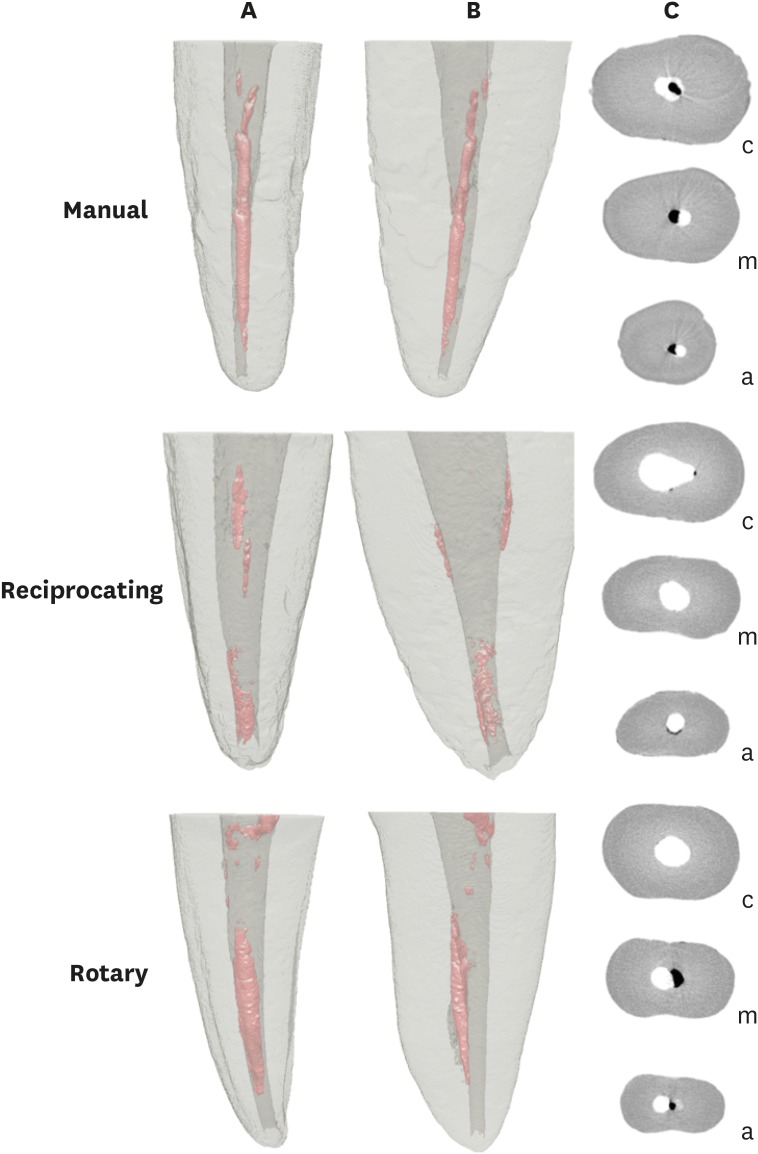
ePub Objectives This study evaluated the amount of remaining root canal filling materials after retreatment procedures performed by undergraduate students using manual, rotary, and reciprocating techniques through micro-computed tomographic analysis. The incidence of instrument fracture and the instrumentation time were also evaluated.
Materials and Methods Thirty maxillary single rooted teeth were prepared with Reciproc R25 files and filled with gutta-percha and AH Plus sealer by the continuous wave of condensation technique. Then, the specimens were assigned to 3 groups (
n = 10), according to the retreatment technique used: manual, rotary, and reciprocating groups, which used K-file, Mtwo retreatment file, and Reciproc file, respectively. Retreatments were performed by undergraduate students. The sample was scanned after root canal filling and retreatment procedures, and the images of the canals were examined to quantify the amount of remaining filling material. The incidence of instrument fracture and the instrumentation time were recorded.Results Remaining filling material was observed in all specimens regardless of the technique used. The mean volume of remaining material was significantly lower in the Reciproc group than in the manual K-file and Mtwo retreatment groups (
p < 0.05). The time required to achieve a satisfactory removal of canal filling material and refinement was significantly lower in the Mtwo retreatment and Reciproc groups (p < 0.05) when compared to the manual K-file group. No instrument fracture was observed in any of the groups.Conclusions Reciproc was the most effective instrument in the removal of canal fillings after retreatments performed by undergraduate students.
-
Citations
Citations to this article as recorded by- Assessment of isthmus filling using two obturation techniques performed by students with different levels of clinical experience
Yang Yu, Chong-Yang Yuan, Xing-Zhe Yin, Xiao-Yan Wang
Journal of Dental Sciences.2024; 19(1): 169. CrossRef - Optical microscopy evaluation of root canal filling removal by beginner operators in posterior teeth
Bogdan Dimitriu, Ioana Suciu, Oana Elena Amza, Mihai Ciocârdel, Dana Bodnar, Ana Maria Cristina Țâncu, Mihaela Tanase, Maria Sabina Branescu, Mihaela Chirilă
Journal of Medicine and Life.2024; 17(6): 555. CrossRef - Micro-CT Study on the Supplementary Effect of XP-Endo Finisher R after Endodontic Retreatment with Mtwo-R
I Tsenova-Ilieva, V Dogandzhiyska, M Raykovska, E Karova
Nigerian Journal of Clinical Practice.2023; 26(12): 1844. CrossRef - Critical analysis of research methods and experimental models to study removal of root filling materials
Mahdi A. Ajina, Pratik K. Shah, Bun San Chong
International Endodontic Journal.2022; 55(S1): 119. CrossRef - Efficiency of Supplementary Contemporary Single-file Systems in Removing Filling Remnants from Oval-shaped Canals: An In Vitro Study
Neveen A Shaheen, Dalia A Sherif, Nahla G Elhelbawy
The Journal of Contemporary Dental Practice.2021; 22(9): 1055. CrossRef - Efficacy of an arrow‐shaped ultrasonic tip for the removal of residual root canal filling materials
Emmanuel J.N.L. Silva, Carolina O. de Lima, Ana F.A. Barbosa, Cláudio M. Ferreira, Bruno M. Crozeta, Ricardo T. Lopes
Australian Endodontic Journal.2021; 47(3): 467. CrossRef - XP‐endo Finisher R instrument optimizes the removal of root filling remnants in oval‐shaped canals
G. De‐Deus, F. G. Belladonna, A. S. Zuolo, D. M. Cavalcante, J. C. A. Carvalhal, M. Simões‐Carvalho, E. M. Souza, R. T. Lopes, E. J. N. L. Silva
International Endodontic Journal.2019; 52(6): 899. CrossRef
- Assessment of isthmus filling using two obturation techniques performed by students with different levels of clinical experience
- 1,285 View
- 12 Download
- 7 Crossref

- Incidence of apical crack formation and propagation during removal of root canal filling materials with different engine driven nickel-titanium instruments
- Taha Özyürek, Vildan Tek, Koray Yılmaz, Gülşah Uslu
- Restor Dent Endod 2017;42(4):332-341. Published online November 4, 2017
- DOI: https://doi.org/10.5395/rde.2017.42.4.332
-
 Abstract
 PDF
PDF PubReader
 ePub
ePub Objectives To determine the incidence of crack formation and propagation in apical root dentin after retreatment procedures performed using ProTaper Universal Retreatment (PTR), Mtwo-R, ProTaper Next (PTN), and Twisted File Adaptive (TFA) systems.
Materials and Methods The study consisted of 120 extracted mandibular premolars. One millimeter from the apex of each tooth was ground perpendicular to the long axis of the tooth, and the apical surface was polished. Twenty teeth served as the negative control group. One hundred teeth were prepared, obturated, and then divided into 5 retreatment groups. The retreatment procedures were performed using the following files: PTR, Mtwo-R, PTN, TFA, and hand files. After filling material removal, apical enlargement was done using apical size 0.50 mm ProTaper Universal (PTU), Mtwo, PTN, TFA, and hand files. Digital images of the apical root surfaces were recorded before preparation, after preparation, after obturation, after filling removal, and after apical enlargement using a stereomicroscope. The images were then inspected for the presence of new apical cracks and crack propagation. Data were analyzed with χ2 tests using SPSS 21.0 software.
Results New cracks and crack propagation occurred in all the experimental groups during the retreatment process. Nickel-titanium rotary file systems caused significantly more apical crack formation and propagation than the hand files. The PTU system caused significantly more apical cracks than the other groups after the apical enlargement stage.
Conclusions This study showed that retreatment procedures and apical enlargement after the use of retreatment files can cause crack formation and propagation in apical dentin.
-
Citations
Citations to this article as recorded by- Microcracks induced by XP-endo retreatment system in root canals filled with bioceramic sealer: A micro-computed tomographic analysis
Sarah M. Alkahtany
The Saudi Dental Journal.2025;[Epub] CrossRef - A comparative evaluation of different retreatment methods on apical root microcracks initiation and propagation: An in vitro study
Shweta Lodha, Zinnie Nanda
Endodontology.2025; 37(2): 175. CrossRef - Efficacy of Endodontic Files in Root Canal Retreatment: A Systematic Review of In Vitro Studies
Anna Soler-Doria, José Luis Sanz, Marcello Maddalone, Leopoldo Forner
Journal of Functional Biomaterials.2025; 16(8): 293. CrossRef - Efficacy of Various Heat-treated Retreatment File Systems on Dentin Removal and Crack Analysis: An in vitro Study
Swathi Suresh, Pradeep Solete, Delphine Priscilla Antony, Kavalipurapu Venkata Teja, Adimulapu Hima Sandeep, Sruthi Sairaman, Marco Cicciù, Giuseppe Minervini
Pesquisa Brasileira em Odontopediatria e Clínica Integrada.2024;[Epub] CrossRef - A comparative evaluation of the dentinal microcracks formed and propagated during the removal of gutta-percha using hand and three rotary retreatment file systems: A micro-computed tomography study
Srivastava Sanjeev, Rita Gupta, Dubey Sandeep, Tewari Tanu, Shukla Namita, Singh Arohan
Endodontology.2023; 35(2): 155. CrossRef - Comparative evaluation of incidence of dentinal defects after root canal preparation using hand, rotary, and reciprocating files: An ex vivo study
Debanjan Das, Sudipto Barai, Rohit Kumar, Sourav Bhattacharyya, AsimB Maity, Pushpa Shankarappa
Journal of International Oral Health.2022; 14(1): 78. CrossRef - Critical analysis of research methods and experimental models to study removal of root filling materials
Mahdi A. Ajina, Pratik K. Shah, Bun San Chong
International Endodontic Journal.2022; 55(S1): 119. CrossRef - Evaluation of Dentinal Crack Propagation, Amount of Gutta Percha Remaining and Time Required During Removal of Gutta Percha Using Two Different Rotary Instruments and Hand Instruments - An In vitro Study
S Tejaswi, A Singh, S Manglekar, UK Ambikathanaya, S Shetty
Nigerian Journal of Clinical Practice.2022; 25(4): 524. CrossRef - The Influence of Root Canal Preparation with ProTaper Next, WaveOne Gold, and Twisted Files on Dentine Crack Formation
Wojciech Eliasz, Beata Czarnecka, Anna Surdacka
Machines.2021; 9(12): 332. CrossRef - The potential effect of instrumentation with different nickel titanium rotary systems on dentinal crack formation—An in vitro study
Márk Fráter, András Jakab, Gábor Braunitzer, Zsolt Tóth, Katalin Nagy, Andrej M. Kielbassa
PLOS ONE.2020; 15(9): e0238790. CrossRef - Micro–computed Tomographic Assessment of the Residual Filling Volume, Apical Transportation, and Crack Formation after Retreatment with Reciproc and Reciproc Blue Systems in Curved Root Canals
Damla Kırıcı, Sezer Demirbuga, Ertuğrul Karataş
Journal of Endodontics.2020; 46(2): 238. CrossRef - Force and vibration generated in apical direction by three endodontic files of different kinematics during simulated canal preparation: An in vitro analytical study
Ankit Nayak, PK Kankar, Prashant K Jain, Niharika Jain
Proceedings of the Institution of Mechanical Engineers, Part H: Journal of Engineering in Medicine.2019; 233(8): 839. CrossRef - Effect of Aging on Dentinal Crack Formation after Treatment and Retreatment Procedures: a Micro-CT Study
Lilian Rachel de Lima Aboud, Bernardo Camargo dos Santos, Ricardo Tadeu Lopes, Leonardo Aboud Costa Viana, Miriam Fátima Zaccaro Scelza
Brazilian Dental Journal.2018; 29(6): 530. CrossRef
- Microcracks induced by XP-endo retreatment system in root canals filled with bioceramic sealer: A micro-computed tomographic analysis
- 1,657 View
- 12 Download
- 13 Crossref

- Effect of adaptive motion on cyclic fatigue resistance of a nickel titanium instrument designed for retreatment
- Taha Özyürek, Koray Yılmaz, Gülşah Uslu
- Restor Dent Endod 2017;42(1):34-38. Published online December 19, 2016
- DOI: https://doi.org/10.5395/rde.2017.42.1.34

-
 Abstract
 PDF
PDF PubReader
 ePub
ePub Objectives The aim of this study was to evaluate the cyclic fatigue resistance of the ProTaper Universal D1 file (Dentsply Maillefer) under continuous and adaptive motion.
Materials and Methods Forty ProTaper Universal D1 files were included in this study. The cyclic fatigue tests were performed using a dynamic cyclic fatigue testing device, which had an artificial stainless steel canal with a 60° angle of curvature and a 5 mm radius of curvature. The files were randomly divided into two groups (Group 1, Rotary motion; Group 2, Adaptive motion). The time to failure of the files were recorded in seconds. The number of cycles to failure (NCF) was calculated for each group. The data were statistically analyzed using Student's
t -test. The statistical significant level was set atp < 0.05.Results The cyclic fatigue resistance of the adaptive motion group was significantly higher than the rotary motion group (
p < 0.05).Conclusion Within the limitations of the present study, the ‘Adaptive motion’ significantly increased the resistance of the ProTaper Universal D1 file to cyclic facture.
-
Citations
Citations to this article as recorded by- The Effect of Different Separated File Retrieval Strategies on the Biomechanical Behavior of a Mandibular Molar: A Finite Element Analysis Study
Anas Sira, Nawar Naguib Nawar, Shehabeldin Mohamed Saber, Hyeon-Cheol Kim
Journal of Endodontics.2025; 51(1): 64. CrossRef - Surface Alterations of Ni‐Ti Files After Retreatment of Root Canals Filled With Different Sealers: AFM and SEM Study
Duygu Aksoy, Sibel Koçak, Mustafa Murat Koçak, Baran Can Sağlam
Microscopy Research and Technique.2025; 88(10): 2704. CrossRef - Evaluatation of two nickle-titanium systems’ (Neolix and X Pro Gold) resistance to fracture after immersion in sodium hypochlorite.
Solmaz Araghi, Abbas Delvarani, Faeze dehghan, Parisa Kaghazloo
journal of research in dental sciences.2024; 21(1): 17. CrossRef - Assessment the impact of operator experience on cyclic fatigue resistance in reciprocating and rotary NiTi files: a comparative study between dental students and pediatric dentistry specialists
Hande Özyürek, Mesut Elbay, Taha Özyürek
Frontiers in Materials.2023;[Epub] CrossRef - Evaluation of gutta-percha removal from the dentinal tubules using different instrumentation techniques with or without solvent: An In vitro study
MukeshKumar Hasija, Babita Meena, Deepti Wadhwa, KulvinderKaur Wadhwani, Virender Yadav
Journal of the International Clinical Dental Research Organization.2020; 12(1): 27. CrossRef - Influences of Continuous Rotation and TF adaptive Motion on the Resistance of Different Retreatment File Systems to Deformation and Fracture: An In Vitro study
Divya Meena, Ramyadharshini LNU, V Nivedha, Anand Sherwood
Journal of Operative Dentistry & Endodontics.2018; 3(2): 71. CrossRef - Comparison of cyclic fatigue life of nickel-titanium files: an examination using high-speed camera
Taha Özyürek, Neslihan Büşra Keskin, Fatma Furuncuoğlu, Uğur İnan
Restorative Dentistry & Endodontics.2017; 42(3): 224. CrossRef
- The Effect of Different Separated File Retrieval Strategies on the Biomechanical Behavior of a Mandibular Molar: A Finite Element Analysis Study
- 1,418 View
- 6 Download
- 7 Crossref

- Nonsurgical endodontic retreatment of fused teeth with transposition: a case report
- Miguel Agostinho Beco Pinto Cardoso, Rita Brandão Noites, Miguel André Duarte Martins, Manuel Pedro da Fonseca Paulo
- Restor Dent Endod 2016;41(2):148-153. Published online February 22, 2016
- DOI: https://doi.org/10.5395/rde.2016.41.2.148
-
 Abstract
 PDF
PDF PubReader
 ePub
ePub Tooth transposition is a disorder in which a permanent tooth develops and erupts in the normal position of another permanent tooth. Fusion and gemination are developmental disturbances presenting as the union of teeth. This article reports the nonsurgical retreatment of a very rare case of fused teeth with transposition. A patient was referred for endodontic treatment of her maxillary left first molar in the position of the first premolar, which was adjacent to it on the distobuccal side. Orthopantomography and periapical radiography showed two crowns sharing the same root, with a root canal treatment and an associated periapical lesion. Tooth fusion with transposition of a maxillary molar and a premolar was diagnosed. Nonsurgical endodontic retreatment was performed. At four yr follow-up, the tooth was asymptomatic and the radiolucency around the apical region had decreased, showing the success of our intervention. The diagnosis and treatment of fused teeth require special attention. The canal system should be carefully explored to obtain a full understanding of the anatomy, allowing it to be fully cleaned and obturated. Thermoplastic techniques were useful in obtaining hermetic obturation. A correct anatomical evaluation improves the set of treatment options under consideration, leading to a higher likelihood of esthetically and functionally successful treatment.
- 1,289 View
- 12 Download

- Success and failure of endodontic microsurgery
- Minju Song, Euiseong Kim
- J Korean Acad Conserv Dent 2011;36(6):465-476. Published online November 30, 2011
- DOI: https://doi.org/10.5395/JKACD.2011.36.6.465
-
 Abstract
 PDF
PDF PubReader
 ePub
ePub In current endodontic practice, introduction of operating microscope, ultrasonic instruments, and microinstruments has induced a big change in the field of surgical retreatment. In this study, we aimed to offer key steps of endodontic microsurgery procedure compared with traditional root-end surgery, and to evaluate factors influencing success and failure based on published articles.
Endodontic microsurgery is a surgical procedure performed with the aid of a microscope, ultrasonic instruments and modern microsurgical instruments. The microscope provides magnification and illumination - essential for identifying minute details of the apical anatomy. Ultrasonic instruments facilitate the precise root-end preparation that is within the anatomical space of the canal. Modern endodontics can therefore be performed with precision and predictability, thus eliminating the disadvantages inherent in traditional periapical surgery such as large osteotomy, beveled apicoectomy, inaccurate root-end preparation and the inability to observe isthmus.
Factors influencing the outcomes of endodontic microsurgery may be diverse, but standardization of procedures can minimize its range. Among patient and tooth-related factors, periodontal status and tooth position are known to be prognostic, but there are only few articles concerning this matter. High-evidence randomized clinical trials or prospective cohort studies are needed to confirm these findings.
-
Citations
Citations to this article as recorded by- Treatment-Related Factors Affecting the Success of Endodontic Microsurgery and the Influence of GTR on Radiographic Healing—A Cone-Beam Computed Tomography Study
Daniel Bieszczad, Jarosław Wichlinski, Tomasz Kaczmarzyk
Journal of Clinical Medicine.2023; 12(19): 6382. CrossRef - Factors Affecting the Success of Endodontic Microsurgery: A Cone-Beam Computed Tomography Study
Daniel Bieszczad, Jaroslaw Wichlinski, Tomasz Kaczmarzyk
Journal of Clinical Medicine.2022; 11(14): 3991. CrossRef - Comparison of Clinical Outcomes of Endodontic Microsurgery: 1 Year versus Long-term Follow-up
Minju Song, Taekjin Nam, Su-Jung Shin, Euiseong Kim
Journal of Endodontics.2014; 40(4): 490. CrossRef - The Influence of Bone Tissue Deficiency on the Outcome of Endodontic Microsurgery: A Prospective Study
Minju Song, Sahng Gyoon Kim, Su-Jung Shin, Hyeon-Cheol Kim, Euiseong Kim
Journal of Endodontics.2013; 39(11): 1341. CrossRef - Prognostic Factors of Clinical Outcomes in Endodontic Microsurgery: A Prospective Study
Minju Song, Sahng Gyoon Kim, Seung-Jong Lee, Baekil Kim, Euiseong Kim
Journal of Endodontics.2013; 39(12): 1491. CrossRef - Is stopping of anticoagulant therapy really required in a minor dental surgery? - How about in an endodontic microsurgery?
Yong-Wook Cho, Euiseong Kim
Restorative Dentistry & Endodontics.2013; 38(3): 113. CrossRef
- Treatment-Related Factors Affecting the Success of Endodontic Microsurgery and the Influence of GTR on Radiographic Healing—A Cone-Beam Computed Tomography Study
- 2,208 View
- 27 Download
- 6 Crossref

- Evaluation of retrievability using a new soft resin based root canal filling material
- Su-Jung Shin, Yoon Lee, Jeong-Won Park
- J Korean Acad Conserv Dent 2006;31(4):323-329. Published online July 31, 2006
- DOI: https://doi.org/10.5395/JKACD.2006.31.4.323
-
 Abstract
 PDF
PDF PubReader
 ePub
ePub The aim of this study was to evaluate the retrievability of Resilon as a root canal filling material. Twenty-seven human single-rooted extracted teeth were instrumented utilizing a crown down technique with Gates-Glidden burs and ProFile system. In group1 (n = 12) canals were obturated with gutta percha and AH-26 plus sealer using a continuous wave technique and backfilled. In group 2 (n = 15) Resilon was used as a filling material. Then teeth were sealed and kept in 37℃ and 100% humidity for 7 days. For retreatment, the samples were re-accessed and filling material was removed using Gates-Glidden burs and ProFiles. Teeth were sectioned longitudinally to compare the general cleanliness and amount of debris (× 75) using SEM. Chi-square test was used (α = 0.05) to analyze the data. The total time required for removal of filling materials was expressed as mean ± SD (min) and analyzed by the Student
t -test (α = 0.05). Required time for retreatment was 3.25 ± 0.32 minutes for gutta percha/AH 26 plus sealer and 3.05 ± 0.34 minutes for Resilon. There was no statistically significant difference between the two experimental groups. There was no significant difference between the groups in the cleanliness of the root canal wall. This study showed that Resilon was effectively removed by Gates-Glidden burs and ProFiles.-
Citations
Citations to this article as recorded by- An in vitro evaluation of effectiveness of Xylene, Thyme oil and Orange oil in dissolving three different endodontic sealers
N Aiswarya, TN Girish, KC Ponnappa
Journal of Conservative Dentistry.2023; 26(3): 305. CrossRef - A Comparative Evaluation of Two Commonly Used GP Solvents on Different Epoxy Resin-based Sealers: An In Vitro Study
Sakshi Tyagi, Ekta Choudhary, Rajat Chauhan, Ashish Choudhary
International Journal of Clinical Pediatric Dentistry.2020; 13(1): 35. CrossRef - Evaluation of softening ability of Xylene & Endosolv-R on three different epoxy resin based sealers within 1 to 2 minutes - anin vitrostudy
Pratima Ramakrishna Shenoi, Gautam Pyarelal Badole, Rajiv Tarachand Khode
Restorative Dentistry & Endodontics.2014; 39(1): 17. CrossRef - Microleakage of resilon by methacrylate-based sealer and self-adhesive resin cement
Sun-Young Ham, Jin-Woo Kim, Hye-Jin Shin, Kyung-Mo Cho, Se-Hee Park
Journal of Korean Academy of Conservative Dentistry.2008; 33(3): 204. CrossRef - Microleakage of resilon: Effects of several self-etching primer
Jong-Hyeon O, Se-Hee Park, Hye-Jin Shin, Kyung-Mo Cho, Jin-Woo Kim
Journal of Korean Academy of Conservative Dentistry.2008; 33(2): 133. CrossRef
- An in vitro evaluation of effectiveness of Xylene, Thyme oil and Orange oil in dissolving three different endodontic sealers
- 1,319 View
- 2 Download
- 5 Crossref

- A NEW POST REMOVAL TECHNIQUE USING ATD TUGGING DEVICE
- Yun-Woo Park, Se-Hee Park, Hye-Jin Shin, Kyung-Mo Cho, Jin-Woo Kim
- J Korean Acad Conserv Dent 2005;30(3):215-220. Published online January 14, 2005
- DOI: https://doi.org/10.5395/JKACD.2005.30.3.215
-
 Abstract
 PDF
PDF PubReader
 ePub
ePub ABSTRACT It is common for clinicians to encounter endodontically treated teeth that contain posts within their roots. If endodontic treatment is failed, these posts must be removed to facilitate successful nonsurgical retreatment.
There have been many techniques such as ultrasonic instrument, Ruddle post removal system, Eggler post remover and Masserann kit developed to facilitate removal of posts from the root canal space. But these methods may be disadvantageous because long length of time required for post removal and fracture of post or teeth. In now days new post removal technique using ATD automatic bridge remover was introduced. Advantages of this method are simple and short time consuming compare to others.
This article served as a successful case report of post removal using ATD automatic bridge remover.
- 850 View
- 2 Download

- THE EFFECT OF GUTTA-PERCHA REMOVAL USING NICKEL-TITANIUM ROTARY INSTRUMENTS
- Jeong-Hun Jeon, Jeong-Beom Min, Ho-Keel Hwang
- J Korean Acad Conserv Dent 2004;29(3):212-218. Published online January 14, 2004
- DOI: https://doi.org/10.5395/JKACD.2004.29.3.212
-
 Abstract
 PDF
PDF PubReader
 ePub
ePub ABSTRACT The purpose of this study was to quantify the amount of remaining gutta-percha/sealer on the walls of root canals when three types of nickel-titanium rotary instruments(Profile, ProTaper and K3) and a hand instrument(Hedstrom file) used to remove these materials.
The results of this study were as follows:
In the total time for gutta-percha removal, Profile group was the fastest and followed by K3, Protaper, Hedstrom file group.
In case of the evaluation of the volume of remained gutta-percha from radiograph, K3 group got the highest score and followed by Protaper, Hedstrom file, Profile group in the apical 1/3.
In case of the evaluation of the volume of gutta-percha remained from stereomicroscope, K3 group got the highest score and followed by Protaper, Hedstrom file, Profile group in the apical 1/3.
These results showed that instrumentation using nickel-titanium rotary instrument groups was faster than that using hand instrument group. The effect of gutta-percha removal using Profile group was better than that using Protaper and K3 group in the nickel-titanium rotary instrument groups.
- 801 View
- 0 Download


 KACD
KACD

 First
First Prev
Prev


