
Search
- Page Path
- HOME > Search
Research Articles
- Magnitude of pulp space narrowing over time and contributing factors in teeth with vital pulp therapy: a retrospective cohort study
- Akarapong Boontankun, Papimon Chompu‑inwai, Chanika Manmontri, Nattakan Chaipattanawan, Areerat Nirunsittirat, Phichayut Phinyo, Trasapong Thaiupathump
- Restor Dent Endod 2026;51(2):e24. Published online May 13, 2026
- DOI: https://doi.org/10.5395/rde.2026.51.e24
-
 Abstract
 PDF
PDF Supplementary Material
 PubReader
 ePub
ePub - Objectives
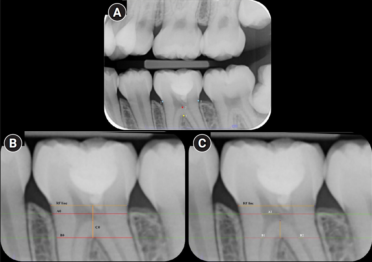
This study aimed to compare the magnitude of pulp space narrowing over time—measured as the change in pulp/tooth proportion from baseline—between mandibular molars treated with different types of vital pulp therapy (VPT) and their contralateral sound molars (controls). This study also investigated factors influencing the magnitude of pulp space narrowing in molars that have undergone VPT.
Methods
This retrospective cohort study involved the assessment of bitewing radiographs of VPT-treated molars and controls at baseline and follow-up. Using reference points and lines on the radiograph, pulp/tooth proportions were measured by examiners. The intraclass correlation coefficient (ICC) was used to report examiner reliability. The changes in pulp/tooth proportions from baselines were compared between subgroups using multilevel mixed effect linear regression and the Wald test.
Results
A total of 382 bitewing radiographs from 134 teeth were included. The follow-up period ranged from 6 to 84 months (mean, 27.12 ± 17.67 months). ICC values indicated good to excellent examiner reliability. Compared to the controls, changes in pulp/tooth proportion from baselines, indicating pulp space narrowing, were significantly greater in teeth with partial pulpotomy (at pulp chamber width) and coronal pulpotomy (at pulp canal width). Factors affecting the magnitude of pulp space narrowing included the more invasive type of VPT and the more severe preoperative diagnosis.
Conclusions
The magnitude of pulp space narrowing was greater in VPT-treated molars than in controls. The more invasive type of VPT and severe preoperative diagnosis were factors contributing to the magnitude of pulp space narrowing.
- 1,062 View
- 61 Download

- Publication rate of abstracts presented in ConsEuro meetings held between 2003 and 2019: a bibliometric analysis
- Esra Cengiz-Yanardag, Ayse Tugba Erturk-Avunduk, Izgen Karakaya
- Restor Dent Endod 2025;50(1):e10. Published online February 19, 2025
- DOI: https://doi.org/10.5395/rde.2025.50.e10
-
Abstract
PDFPubReaderePub
- Objectives
This study aimed to assess the publication rates of abstracts presented at the ConsEuro Congress using a web-scraping method and to analyze factors correlated with these publication rates.
Methods
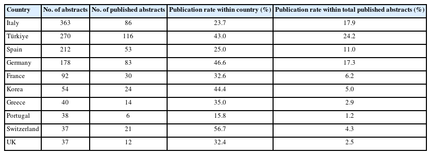
Abstracts presented at eight ConsEuro meetings held between 2003 and 2019 were evaluated for subspecialty, study design, number of authors, and the country of the principal investigator’s institution. For abstracts confirmed as subsequently published using a web-scraping method, the following data were recorded: time to publication, the journal of publication, impact factor, quartile, index status, Scientific Journal Ranking of the journal at the year of publication, and changes in the number of authors after publication.
Results
Out of 1,426 abstracts presented, 478 were published in peer-reviewed journals, yielding a publication rate of 33.5%. The median time to publication was 12 months. The leading journal in terms of publication rate was Clinical Oral Investigations. There was no statistically significant difference in publication rates across years. Abstracts related to laser therapy, caries, and dental materials had significantly higher publication rates compared to other subspecialties. Animal, basic, and clinical research studies were more likely to be published. Both study design and subspecialty influenced publication rates, which decreased over time.
Conclusions
A considerable amount of scientific data and preliminary results presented at conferences, which could contribute to scientific knowledge, are overlooked due to low publication rates. The findings of this study may encourage ConsEuro participants to submit well-planned and rigorous studies that are more likely to complete the full publication process.
- 3,155 View
- 74 Download
Review Article
- Influence of the root canal filling technique on the success rate of primary endodontic treatments: a systematic review
- Daniel Feijolo Marconi, Giovana Siocheta da Silva, Theodoro Weissheimer, Isadora Ames Silva, Gabriel Barcelos Só, Leonardo Thomasi Jahnke, Jovito Adiel Skupien, Marcus Vinicius Reis Só, Ricardo Abreu da Rosa
- Restor Dent Endod 2022;47(4):e40. Published online October 11, 2022
- DOI: https://doi.org/10.5395/rde.2022.47.e40
-
Abstract
PDFSupplementary MaterialPubReaderePub
Objectives This study aimed to investigate the influence of different obturation techniques compared to cold lateral compaction on the success rate of primary non-surgical endodontic treatments.
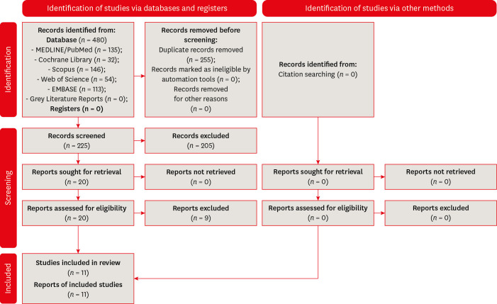
Materials and Methods Systematic searches were performed for studies published up to May 17th, 2022 in MEDLINE/PubMed, Cochrane Library, Web of Science, Scopus, EMBASE, and Grey Literature Reports. Randomized clinical trials and nonrandomized (nonrandomized clinical trials, prospective or retrospective) studies that evaluated the success rate of primary non-surgical endodontic treatments obturated with the cold lateral compaction (control) and other obturation techniques were included. The revised Cochrane risk of bias tools for randomized trials (RoB 2) and nonrandomized studies of interventions (ROBINS-I) were used to evaluate the risk of bias. The Grading of Recommendations Assessment, Development, and Evaluation (GRADE) tool was used to evaluate the certainty of evidence.
Results Eleven studies (4 randomized clinical trials (RCTs), 4 prospective, and 3 retrospectives) were included. Two RCTs were classified as having some concerns risk of bias and 2 as a low risk of bias. Two nonrandomized studies were classified as having a critical risk of bias and 5 as having a moderate risk of bias. The GRADE analysis demonstrated a very low to moderate certainty of evidence.
Conclusions This systematic review generally evidenced no differences in the success rate of primary non-surgical endodontic treatments when the cold lateral compaction technique and other obturation techniques are performed. Further well-designed studies are still necessary.
-
Citations
Citations to this article as recorded by
- Assessing Sealing Ability of C-Root SP Strontium Silicate Sealer With Different Obturation Techniques: An in vitro Study
Suixin Hu, Jianshe Li, Meng Xu, Laiqing Xu, Yangming Yin, Peng Xue, Liping Dong, Lin Wang, Huixia He, Ying Liu, Qiang Luo, Fei Chen
International Dental Journal.2026; 76(1): 109283. CrossRef - Effect of root canal filling techniques and materials on endodontic treatment outcomes: a systematic review and meta-analysis
Ahmed Mushtaq, Sura Alsanafi, Firas Elmsmari, José A. González, Marc Garcia-Font, Francesc Abella Sans, Kelvin I. Afrashtehfar, Paul V. Abbott
Scientific Reports.2026;[Epub] CrossRef - Dimensional and cross-sectional compatibility of contemporary nickel–titanium files and their corresponding gutta-percha cones: A micro-computed tomography study
Cem Sarkan, Faruk Haznedaroglu
The Saudi Dental Journal.2026;[Epub] CrossRef - Comparative Analysis Of Obturation Techniques In Endodontics: Lateral Vs. Thermoplasticized. Thermoplasticized
Juan Esteban Díaz Pacheco , Rómulo Guillermo López Torres , Verónica Alejandra Salame Ortíz
Salud, Ciencia y Tecnología.2025; 5: 1626. CrossRef - Effect of ultrasonic activation of endodontic sealers on root canal filling quality during the single-cone obturation procedure: a systematic review and meta-analysis of laboratory-based studies
Shuting Feng, Weiqing Zhou, Xiaojun Chu, Shuaimei Xu, Xiongqun Zeng
Odontology.2025; 113(4): 1380. CrossRef - In Vitro and In Vivo Evaluation of a New Experimental Polydimethylsiloxane-Based Endodontic Sealer
Fabiola Cardoso Maldonado, Cesar Gaitan Fonseca, Carlos Bermudez Jimenez, Luis Alejandro Aguilera Galaviz, Margarita L. Martinez-Fierro, Lorena Troncoso Vazquez, Martha Eugenia Reyes Ortiz
Journal of Functional Biomaterials.2025; 16(11): 402. CrossRef - Evaluation of three obturation techniques in 3D-printed models of oval canals with standardized prepared morphology: a micro-CT study
Wenjun Xia, Qisheng Gu, Yingshuang Song, Yunjia Liu, Xuetao Deng, Wenhao Qian
BMC Oral Health.2025;[Epub] CrossRef - Clinical and Radiographic Failure of Nonsurgical Endodontic Treatment and Retreatment Using Single-cone Technique With Calcium Silicate-based Sealers: A Systematic Review and Meta-analysis
Mohammad A. Sabeti, Negah Karimpourtalebi, Arash Shahravan, Omid Dianat
Journal of Endodontics.2024; 50(6): 735. CrossRef - Method of microbial decontamination of endodontic absorbent paper points: a randomised experimental study
O. A. Pavlovskaya, O. A. Kachanova, V. V. Volobuev, M. N. Mitropanova, A. R. Gazarova, V. Y. Zobenko, A. G. Uvarova
Pediatric dentistry and dental prophylaxis.2024; 24(2): 157. CrossRef - The Push-Out Bond Strength, Surface Roughness, and Antimicrobial Properties of Endodontic Bioceramic Sealers Supplemented with Silver Nanoparticles
Karla Navarrete-Olvera, Nereyda Niño-Martínez, Idania De Alba-Montero, Nuria Patiño-Marín, Facundo Ruiz, Horacio Bach, Gabriel-Alejandro Martínez-Castañón
Molecules.2024; 29(18): 4422. CrossRef - Clinical outcome of non-surgical root canal treatment using different sealers and techniques of obturation in 237 patients: A retrospective study
Mateusz Radwanski, Krystyna Pietrzycka, Tan Fırat Eyüboğlu, Mutlu Özcan, Monika Lukomska-Szymanska
Clinical Oral Investigations.2024;[Epub] CrossRef
- Assessing Sealing Ability of C-Root SP Strontium Silicate Sealer With Different Obturation Techniques: An in vitro Study
- 8,992 View
- 145 Download
- 10 Web of Science
- 11 Crossref
Research Articles
- Effect of post space preparation drills on the incidence of root dentin defects
- Thaíse Ayres Bezerra Zuli, Orlando Aguirre Guedes, Gislaine Figueiredo Zarza Arguello Gonçalves, Aurélio Rosa da Silva Júnior, Álvaro Henrique Borges, Andreza Maria Fábio Aranha
- Restor Dent Endod 2020;45(4):e53. Published online October 16, 2020
- DOI: https://doi.org/10.5395/rde.2020.45.e53
-
Abstract
PDFPubReaderePub
Objectives This study investigated the incidence of root dentin defects after the use of different post space preparation (PSP) drills.
Materials and Methods Seventy-two bovine incisors were selected and obtained 14-mm-long root sections. Twelve roots served as controls with no intervention (G1). The 60 root canals remaining were instrumented using the crown-down technique with the ProTaper Next system and obturated using the lateral condensation technique. Specimens were randomly distributed into 5 groups (
n = 12) according to the operative steps performed: G2, root canal instrumentation and filling (I+F); G3, I+F and PSP with Gates-Glidden drills; G4, I+F and PSP with Largo-Peeso reamers; G5, I+F and PSP with Exacto drill; and G6, I+F and PSP with WhitePost drill. Roots were sectioned at 3, 6, 9, and 12 mm from the apex, and digital images were captured. The presence of root dentin defects was recorded. Data were analyzed by the χ2 test, withp < 0.05 considered to indicate statistical significance.Results Root dentin defects were observed in 39.6% of the root sections. No defects were observed in G1. G5 had significantly more cracks and craze lines than G1, G2, and G3 (
p < 0.05), and more fractures than G1, G2, G3, and G4 (p < 0.05). When all root sections were analyzed together, significantly more defects were observed at the 12-mm level than at the 3-mm level (p < 0.05).Conclusions PSP drills caused defects in the root dentin. Gates-Glidden drills caused fewer root defects than Largo-Peeso reamers and Exacto drills.
-
Citations
Citations to this article as recorded by- Fracture Strength of CAD/CAM Endocrown and Post-Core Restorations with Fiber Strip Reinforcement in Mandibular Premolars
Kerem Yılmaz, Hakan Aydın, Zeynep Soylu, Özge Çiloğlu, Esma Fatıma Delican, Mehmet Mustafa Özarslan, Fehmi Gönüldaş
Journal of Functional Biomaterials.2026; 17(5): 248. CrossRef - Evaluation of dentinal crack formation during post space preparation using different fiber post systems with micro-computed tomography
Ayşe Nur Kuşuçar, Damla Kırıcı
BMC Oral Health.2025;[Epub] CrossRef - Fracture and Crack Behavior of Weakened Incisors Restored With Fiber Posts, Polyethylene Reinforcement, or 3D-Printed Endocrowns
Diana Codas-Duarte, Laís L Pelozo, Jardel F Mazzi-Chaves, Fabiane C Lopes-Olhê, Manoel D Sousa-Neto, Aline E Souza-Gabriel
Cureus.2025;[Epub] CrossRef - Selecting drill size for post space preparation based on final endodontic radiographs: An in vitro study
Farzaneh Farid, Julfikar Haider, Marjan Sadeghpour Shahab, Nika Rezaeikalantari
Technology and Health Care.2024; 32(4): 2575. CrossRef - Cone Beam Computed Tomography Analysis of Post Space in Bifurcated Premolars Using ParaPost and Peeso Reamer Drills
Abdulaziz Saleh Alqahtani, Omar Nasser Almonabhi, Abdulmajeed Moh. Almutairi, Reem R. Alnatsha
The Open Dentistry Journal.2024;[Epub] CrossRef - A Comparative Evaluation of Real-Time Guided Dynamic Navigation and Conventional Techniques for Post Space Preparation During Post Endodontic Management: An In Vitro Study
Sherifa Shervani, Sihivahanan Dhanasekaran, Vijay Venkatesh
Cureus.2024;[Epub] CrossRef - The effect of ultrasonic vibration protocols for cast post removal on the incidence of root dentin defects
Giulliano C. Serpa, Orlando A. Guedes, Neurinelma S. S. Freitas, Julio A. Silva, Carlos Estrela, Daniel A. Decurcio
Journal of Oral Science.2023; 65(3): 190. CrossRef
- Fracture Strength of CAD/CAM Endocrown and Post-Core Restorations with Fiber Strip Reinforcement in Mandibular Premolars
- 3,753 View
- 42 Download
- 7 Crossref
- Quality of root canal fillings using three gutta-percha obturation techniques
- Edith Siu Shan Ho, Jeffrey Wen Wei Chang, Gary Shun Pan Cheung
- Restor Dent Endod 2016;41(1):22-28. Published online January 4, 2016
- DOI: https://doi.org/10.5395/rde.2016.41.1.22

-
Abstract
PDFPubReaderePub
Objectives The goal of this study was to compare the density of gutta-percha root fillings obturated with the following techniques: cold lateral (CL) compaction, ultrasonic lateral (UL) compaction, and warm vertical (WV) compaction.
Materials and Methods Thirty-three extracted mandibular first molars, with two separate mesial canals in each, were selected. After instrumentation, the canals were stratified into three groups based on canal length and curvature, and underwent obturation with one of the techniques. No sealer was used in order to avoid masking any voids. The teeth were imaged pre- and post-obturation using micro-computed tomography. The reconstructed three-dimensional images were analyzed volumetrically to determine the amount of gutta-percha present in every 2 mm segment of the canal.
P values < 0.05 were considered to indicate statistical significance.Results The overall mean volume fraction of gutta-percha was 68.51 ± 6.75% for CL, 86.56 ± 5.00% for UL, and 88.91 ± 5.16% for WV. Significant differences were found between CL and UL and between CL and WV (
p < 0.05), but not between UL and WV (p = 0.526). The gutta-percha density of the roots treated with WV and UL increased towards the coronal aspect, but this trend was not noted in the CL group.Conclusions WV compaction and UL compaction produced a significantly denser gutta-percha root filling than CL compaction. The density of gutta-percha was observed to increase towards the coronal aspect when the former two techniques were used.
-
Citations
Citations to this article as recorded by- Management of internal root resorption in permanent tooth with dens invaginatus Type II: A clinical approach
Chanchal Meena, Ashwini B. Prasad, Deepak Raisingani, Harshit Srivastava, Charu Thanvi, Mohit Kumar Jyotiyana
Endodontology.2026; 38(1): 99. CrossRef - Evaluation of Postoperative Pain and Apical Outcomes Following Three Distinct Obturation Techniques: A Randomized Controlled Trial
Pragati Agarwal, Pravin Kumar, Vinay Kumar Chugh, Sharmila Shanmugam, Soundharrajan P, Karishma Pathak
Cureus.2026;[Epub] CrossRef - Effectiveness of propolis nanoparticles as endodontic therapeutic agents: a systematic review
Golnoosh Golestane, Reza Fallah Tafti, Zahra Khiali, Hadi Shakerin
Journal of Herbmed Pharmacology.2026; 15(3): 311. CrossRef - Effect of quality of radiographs taken during root canal treatment on technical quality of root canal fillings and endodontic outcome
Jia Min Ng, Yan Yee Lee, Prashanti Chippagiri, Elaheh Ahanin, Abhishek Parolia
Restorative Dentistry & Endodontics.2025; 50(1): e3. CrossRef - Restorative and endodontic clinical strategies during COVID-19 (SARS-CoV-2) pandemic: a revision of the literature
Manuele MANCINI, Flavio PALAZZI, Francesco IACONO
Minerva Dental and Oral Science.2025;[Epub] CrossRef - Evaluation of the tubular penetration of two different types of nanoparticle root canal sealers over apically separated files: a scanning electron microscopic study (in vitro study)
Alaa H. Nagdi, Nayera A. Mokhless, Mahmoud R. Aboelseoud
BMC Oral Health.2025;[Epub] CrossRef - From hopeless to regenerated: successful preservation of a tooth with massive periapical lesion in an adolescent
Adna Begović, Lajla Hasić-Branković, Samra Korać, Faruk Ljaljević, Mirna Pašić, Madžida Halilović Mehinović
Stomatological Review.2025; : 34. CrossRef - Different strategies for treating intracanal fractured instruments in a single tooth: A case report
Rong Chai, Xinpei Jiang, Ruixia Ma, Qiang Zhang, E Yang, Ansheng Zhang
Experimental and Therapeutic Medicine.2024;[Epub] CrossRef - An Experimental Anatomic CBCT Study on the Correlations Between MB1 and MB2 of the Mesio-Vestibular Root of the Upper First Molars
Luca Fiorillo, Cesare D’Amico, Giusy Rita Maria La Rosa, Francesco Calanna, Alfio Pappalardo, Eugenio Pedullà
Journal of Craniofacial Surgery.2024; 35(2): 672. CrossRef - Comparative Evaluation of Different Obturation Techniques for Root Canal Filling of Permanent Teeth: An In-Vitro Study
Adhishree S Chib, Neeta S Padmawar, Sonali Waghmare, Durgesh A Tiwari, Shahinwaz Mulani, Megna Bhatt
Cureus.2024;[Epub] CrossRef - Root canal treatment of a rhizomegaly tooth 36 mm long right permanent maxillary canine – A case report
Anita Kapri, Kiran Reddy, Varun Rana, Oliver Jacob, Pushpa Kumari
IP Annals of Prosthodontics and Restorative Dentistry.2024; 10(1): 59. CrossRef - Thermal and volumetric assessment of endodontic filling techniques using infrared thermography and micro-CT
Fernanda Clotilde M. Suassuna, Débora Ketley M. de Araújo, Ana Marly A. M. Amorim, Saulo Leonardo S. Melo, Richard J. Heck, Antonio Celso D. Antonino, Patrícia M. Bento, Diego Filipe B. Silva, Daniela P. de Melo
Journal of Oral Science.2023; 65(1): 34. CrossRef - A Comparative Evaluation of Efficacy of Various Obturating Techniques for the Presence of Voids
Rehan Ahmad Khan, Shailja Singh, Shazia Siddiqui, Mariyam Khan, Arfat Ahmad, Parul Shakarwal
Journal of Pharmacy and Bioallied Sciences.2023; 15(Suppl 2): S895. CrossRef - Continuous Wave of Condensation Improves the Filling of Curved Canals: a Micro-CT Study
Jader Camilo Pinto, Mariana Mena Barreto Pivoto-João, Juliane Maria Guerreiro-Tanomaru, Jessie Fabiola Reyes-Carmona, Mario Tanomaru-Filho
Odovtos - International Journal of Dental Sciences.2023; 25(3): 32. CrossRef - Influence of the root canal filling technique on the success rate of primary endodontic treatments: a systematic review
Daniel Feijolo Marconi, Giovana Siocheta da Silva, Theodoro Weissheimer, Isadora Ames Silva, Gabriel Barcelos Só, Leonardo Thomasi Jahnke, Jovito Adiel Skupien, Marcus Vinicius Reis Só, Ricardo Abreu da Rosa
Restorative Dentistry & Endodontics.2022;[Epub] CrossRef - Current trends in bio‐based elastomer materials
Shuai Tang, Jiao Li, Runguo Wang, Jichuan Zhang, Yonglai Lu, Guo‐Hua Hu, Zhao Wang, Liqun Zhang
SusMat.2022; 2(1): 2. CrossRef - Carrier-Based Obturation: Effect of Sonication Technique on Sealer Penetration in Dentinal Tubules: A Confocal Laser Scanning Microscope Study
Riccardo Tonini, Matteo Salvadori, Marco Bartoli, Jacopo Francinelli, Paolo Bertoletti, Maria Luisa Garo, Stefano Salgarello
Applied Sciences.2022; 12(17): 8877. CrossRef - A critical analysis of research methods and experimental models to study root canal fillings
Gustavo De‐Deus, Erick Miranda Souza, Emmanuel João Nogueira Leal Silva, Felipe Gonçalves Belladonna, Marco Simões‐Carvalho, Daniele Moreira Cavalcante, Marco Aurélio Versiani
International Endodontic Journal.2022; 55(S2): 384. CrossRef - The effect of two endodontic sealers and interval before post-preparation and cementation on the bond strength of fiber posts
He Yuanli, Wu Juan, Ji Mengzhen, Chen Xuan, Xiong Kaixin, Yang Xueqin, Qiao Xin, Hu Hantao, Gao Yuan, Zou Ling
Clinical Oral Investigations.2021; 25(11): 6211. CrossRef - Complete Obturation—Cold Lateral Condensation vs. Thermoplastic Techniques: A Systematic Review of Micro-CT Studies
Shilpa Bhandi, Mohammed Mashyakhy, Abdulaziz S. Abumelha, Mazen F. Alkahtany, Mohamed Jamal, Hitesh Chohan, A. Thirumal Raj, Luca Testarelli, Rodolfo Reda, Shankargouda Patil
Materials.2021; 14(14): 4013. CrossRef - Root canal filling quality of mandibular molars with EndoSequence BC and AH Plus sealers: A micro‐CT study
Rafael Nigri Roizenblit, Fabiola Ormiga Soares, Ricardo Tadeu Lopes, Bernardo Camargo dos Santos, Heloisa Gusman
Australian Endodontic Journal.2020; 46(1): 82. CrossRef - Effect of four different root canal obturation techniques on marginal adaptation of bioceramic sealer: An in vitro scanning electron microscopic study
NawalA Al-Sabawi, MahaM Yahya, NjwanF Shehab
Journal of International Oral Health.2020; 12(5): 455. CrossRef - Micro-computed tomographic evaluation of a new system for root canal filling using calcium silicate-based root canal sealers
Mario Tanomaru-Filho, Fernanda Ferrari Esteves Torres, Jader Camilo Pinto, Airton Oliveira Santos-Junior, Karina Ines Medina Carita Tavares, Juliane Maria Guerreiro-Tanomaru
Restorative Dentistry & Endodontics.2020;[Epub] CrossRef - Quantification of the tug-back by measuring the pulling force and micro computed tomographic evaluation
Su-Jin Jeon, Young-Mi Moon, Min-Seock Seo
Restorative Dentistry & Endodontics.2017; 42(4): 273. CrossRef
- Management of internal root resorption in permanent tooth with dens invaginatus Type II: A clinical approach
- 3,850 View
- 38 Download
- 24 Crossref
Case Report
- Pre-prosthetic minor tooth movement with elastic separating ring & provisional restoration modification: case report
- Haneol Shin, Byoung-Duck Roh, Yoo-Seok Shin, Chan-Young Lee
- Restor Dent Endod 2012;37(2):114-118. Published online May 18, 2012
- DOI: https://doi.org/10.5395/rde.2012.37.2.114
-
Abstract
PDFPubReaderePub
Proximal caries or coronal defect in posterior teeth may result in the loss of proximal space and drifting of neighboring teeth, which makes restoration difficult. Inability to restore proper contours and to align tooth axis properly are commonly encountered problems when planning tooth restoration. Moreover, tilted teeth aggravate periodontal tissue breakdown, such as pseudo-pocket, and angular osseous defect. The purpose of this case presentation is to describe a simple technique for inducing minor tooth movement with orthodontic separating ring and provisional restoration modification. This method was used to create crown placement space on mesially tilted molar. This method is easy, simple and efficient technique which could be used in interproximal space gaining in selected situation.
-
Citations
Citations to this article as recorded by- Diagnosis and treatment of teeth with primary endodontic lesions mimicking periodontal disease: three cases with long-term follow ups
Jae-Hyung Lim, Ji-Hyun Lee, Su-Jung Shin
Restorative Dentistry & Endodontics.2014; 39(1): 56. CrossRef
- Diagnosis and treatment of teeth with primary endodontic lesions mimicking periodontal disease: three cases with long-term follow ups
- 1,997 View
- 20 Download
- 1 Crossref
Basic Researchs
- Physical properties of novel composite using Portland cement for retro-filling material
- Sang-Jin Lee, Ok-In Cho, Jiwan Yum, Jeong-Kil Park, Bock Hur, Hyeon-Cheol Kim
- J Korean Acad Conserv Dent 2010;35(6):445-452. Published online November 30, 2010
- DOI: https://doi.org/10.5395/JKACD.2010.35.6.445
-
Abstract
PDFPubReaderePub
Objectives The aim of this study was to compare apical sealing ability and physical properties of MTA, MTA - AH-plus mixture (AMTA) and experimental Portland cement - Epoxy resin mixture (EPPC) for a development of a novel retro-filling material.
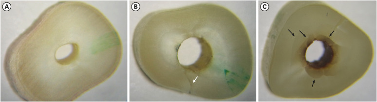
Materials and Methods Forty-nine extracted roots were instrumented and filled with gutta-percha. Apical root was resected at 3 mm and the retro-filling cavity was prepared for 3 mm depth. Roots were randomly divided into 3 groups of 15 roots each. The retro-filling was done using MTA, AMTA, and EPPC as the groups divided. Four roots were used as control groups. After setting in humid condition for 24 hours, the roots were immersed in 1% methylene blue dye solution for 72 hours to test the apical leakage. After immersion, the roots were vertically sectioned and photos were taken to evaluate microleakage. Setting times were measured with Vicat apparatus and digital radiographs were taken to evaluate aluminum equivalent thickness using aluminum step wedge. The results of microleakage and setting time were compared between groups using one-way ANOVA and Scheffe's post-hoc comparison at the significance level of 95%.
Results AMTA and EPPC showed less microleakage than MTA group (
p < 0.05). AMTA showed the highest radio-opacity than other groups and the novel EPPC showed 5 mm aluminum thickness radio-opacity. EPPC showed the shortest initial and final setting times than other groups while the MTA showed the longest (p < 0.05).Conclusions Under the condition of this study, the novel composite using Portland cement-Epoxy resin mixture may useful for retro-filling with the properties of favorable leakage resistance, radio-opacity and short setting time.
-
Citations
Citations to this article as recorded by- Comparison of Setting Time, Compressive Strength, Solubility, and pH of Four Kinds of MTA
Jing-Ling Che, Jae-Hwan Kim, Seon-Mi Kim, Nam-ki Choi, Hyun-Joo Moon, Moon-Jin Hwang, Ho-Jun Song, Yeong-Joon Park
Korean Journal of Dental Materials.2016; 43(1): 61. CrossRef - Biological Effects and Washout Resistance of a Newly Developed Fast-setting Pozzolan Cement
Yoorina Choi, Su-Jung Park, Seoung-Hoon Lee, Yun-Chan Hwang, Mi-Kyung Yu, Kyung-San Min
Journal of Endodontics.2013; 39(4): 467. CrossRef
- Comparison of Setting Time, Compressive Strength, Solubility, and pH of Four Kinds of MTA
- 1,799 View
- 2 Download
- 2 Crossref
- Evaluation of radiopacity and discriminability of various fiber reinforced composite posts
- Eun-Hye Lee, Hang-Moon Choi, Se-Hee Park, Jin-Woo Kim, Kyung-Mo Cho
- J Korean Acad Conserv Dent 2010;35(3):188-197. Published online May 31, 2010
- DOI: https://doi.org/10.5395/JKACD.2010.35.3.188
-
Abstract
PDFPubReaderePub
The purpose of this study was to compare radiopacity and radiographic discriminability of various FRC-Posts.
Six FRC-Posts were investigated ; 1) FRC Postec Plus (Ivoclar Vivadent AG, Schaan, Liechtenstein), 2) Snowlight (Carbotech, Lewis center, OH, USA), 3) Dentin Post (Komet Brasseler, Lamgo, Germany), 4) Rely-X Fiber Post (3M ESPE, St.paul, MN, USA), 5) D.T.-Light Post (BISCO, Schaumburg, IL,USA), 6) Luxapost (DMG, Hamburg, Germany)
The radiographs of each post with a reference 1 mm / 2 mm aluminum step-wedge was taken using digital sensor. The optical density were calculated by gray value of 10 × 10 pixel and compared in mm Al equivalent at five points.
Six maxillary incisors of similar radiopacity were used. Radiographs of posts in Mx. incisors of lingual side of dry mandible were taken.
We showed radiographs and asked the questionnaire to 3 radiologists, 3 endodontists, 3 general practitioners. The questionnaire was comprised of choices of the highest, lowest radiopaque individual post and the choices of best discriminable post at apical, coronal area.
The following results were obtained.
Each post system showed various radiopacity.
There was change of discriminability between each post and simulated specimens regardless of examiner.
Although each post showed various radiopacity, the difference of radiopacity did not affect on discriminability.
- 1,464 View
- 5 Download
- The effect of the amount of interdental spacing on the stress distribution in maxillary central incisors restored with porcelain laminate veneer and composite resin: A 3D-finite element analysis
- Junbae Hong, Seung-Min Tak, Seung-Ho Baek, Byeong-Hoon Cho
- J Korean Acad Conserv Dent 2010;35(1):30-39. Published online January 31, 2010
- DOI: https://doi.org/10.5395/JKACD.2010.35.1.030
-
Abstract
PDFPubReaderePub
This study evaluated the influence of the type of restoration and the amount of interdental spacing on the stress distribution in maxillary central incisors restored by means of porcelain laminate veneers and direct composite resin restorations.
Three-dimensional finite element models were fabricated to represent different types of restorations. Four clinical situations were considered. Type I, closing diastema using composite resin. Labial border of composite resin was extended just enough to cover the interdental space; Type II, closing diastema using composite resin without reduction of labial surface. Labial border of composite resin was extended distally to cover the half of the total labial surface; Type III, closing diastema using composite resin with reduction of labial surface. Labial border of the preparation and restored composite resin was extended distally two-thirds of the total labial surface; Type IV, closing diastema using porcelain laminate veneer with a feathered-edge preparation technique. Four different interdental spaces (1.0, 2.0, 3.0, 4.0 mm) were applied for each type of restorations.
For all types of restoration, adding the width of free extension of the porcelain laminate veneer and composite resin increased the stress occurred at the bonding layer. The maximum stress values observed at the bonding layer of Type IV were higher than that of Type I, II and III. However, the increasing rate of maximum stress value of Type IV was lower than that of Type I, II and III.
-
Citations
Citations to this article as recorded by- Revamping the Peg Smile: An Art of Rehabilitation of Peg Laterals with Ceramic Veneers and Composite Restorations—A Case Report
Mahendran Kavitha, Ramdhas Annapurani, Pasupathy Shakunthala, Jayavel Nandhakumar
Journal of Operative Dentistry & Endodontics.2022; 6(2): 69. CrossRef - Minimally Invasive Diastema Restoration with Prefabricated Sectional Veneers
Claudio Novelli, Andrea Scribante
Dentistry Journal.2020; 8(2): 60. CrossRef
- Revamping the Peg Smile: An Art of Rehabilitation of Peg Laterals with Ceramic Veneers and Composite Restorations—A Case Report
- 1,970 View
- 4 Download
- 2 Crossref
Original Articles
- Comparison of viability of oral epithelial cells stored by different freezing methods
- Do-Young Baek, Seung-Jong Lee, Han-Sung Jung, EuiSeong Kim
- J Korean Acad Conserv Dent 2009;34(6):491-499. Published online November 30, 2009
- DOI: https://doi.org/10.5395/JKACD.2009.34.6.491
-
Abstract
PDFPubReaderePub
This study examined the influence of the storage methods on the viability of oral epithelial cells using conventional cell freezing storage, slow freezing preservation, rapid freezing preservation, and slow freezing preservation with a pressure of 2 Mpa or 3 Mpa. The cell viability was evaluated by cell counting, WST-1 and the clonogenic capacity after 6 days of freezing storage. After 6 days, the frozen cells were thawed rapidly, and the cell counting, WST-1, and clonogenic capacity values were measured and compared.
The results from cell counting demonstrated that conventional cryopreservation, slow freezing under a 2 Mpa pressure and slow freezing under a 3 Mpa pressure showed significantly higher values than slow freezing preservation and rapid freezing preservation (p<0.05).
The results from the optical density by WST-1 demonstrated that slow freezing under a 2 Mpa pressure showed significantly higher values than slow freezing preservation and rapid freezing preservation (p<0.05).
The clonogenic capacity demonstrated that slow freezing under a 2 Mpa pressure showed significantly higher values than slow freezing preservation and rapid freezing preservation (p<0.05).
-
Citations
Citations to this article as recorded by- Evaluation of the Viability of Rat Periodontal Ligament Cells after Storing at 0℃/2 MPa Condition up to One Week: In Vivo MTT Method
Sun Mi Jang, Sin-Yeon Cho, Eui-Seong Kim, Il-Young Jung, Seung Jong Lee
Journal of Korean Dental Science.2016; 9(1): 1. CrossRef - The evaluation of periodontal ligament cells of rat teeth after low-temperature preservation under high pressure
Jin-Ho Chung, Jin Kim, Seong-Ho Choi, Eui-Seong Kim, Jiyong Park, Seung-Jong Lee
Journal of Korean Academy of Conservative Dentistry.2010; 35(4): 285. CrossRef
- Evaluation of the Viability of Rat Periodontal Ligament Cells after Storing at 0℃/2 MPa Condition up to One Week: In Vivo MTT Method
- 2,109 View
- 2 Download
- 2 Crossref
- A comparative study on radiopacity of root canal sealers
- Tae-Min Kim, Seo-Kyoung Kim, In-Nam Hwang, Yun-Chan Hwang, Byung-Cheol Kang, Suk-Ja Yoon, Jae-Seo Lee, Won-Mann Oh
- J Korean Acad Conserv Dent 2009;34(1):61-68. Published online January 31, 2009
- DOI: https://doi.org/10.5395/JKACD.2009.34.1.061
-
Abstract
PDFPubReaderePub
This study was performed to assess the radiopacity of a variety of root canal sealers according to the specification concerning root canal sealers.
Ten materials including Tubli-Seal™, Kerr Pulp Canal Sealer™, AH 26®, AH plus®, AH plus jet™, Ad sea l™, Sealapex™, NOGENOL™, ZOB seal™, Epiphany™ and dentin were evaluated in this study. In the first part, densitometric reading of an each step of aluminum step wedge on occlusal film was performed at different voltage and exposure time. In the second part, ten specimens were radiographed simultaneously with an aluminum step wedges on the occlusal films under decided condition. The mean radiographic den sity values of the materials were transformed into radiopacity expressed equivalent thickness of aluminum (mm Al).
The following results were obtained.
1. Among the various conditions, the appropriate voltage and exposure time that meet the requirement density was 60 kVp at 0.2 s
2. All of the materials had greater radiopacity than 3 mm Al requirement of ANSI/ADA specification No. 57 (2000) and ISO No. 6876 (2001) standards.
3. The radiopacity of materials increased as thickness of materials increased.
4. The mm Al value of each specimen at 1mm in thickness has a significant difference in the statistics.
It suggests that root canal sealers have a sufficient radiopacity that meet the requirement.
-
Citations
Citations to this article as recorded by- Evaluation of radiopacity and discriminability of various fiber reinforced composite posts
Eun-Hye Lee, Hang-Moon Choi, Se-Hee Park, Jin-Woo Kim, Kyung-Mo Cho
Journal of Korean Academy of Conservative Dentistry.2010; 35(3): 188. CrossRef
- Evaluation of radiopacity and discriminability of various fiber reinforced composite posts
- 2,003 View
- 4 Download
- 1 Crossref
- Shear bond strength of dentin bonding agents cured with a Plasma Arc curing light
- Youngchul Kwon, Sun-Young Kim, Sae-Joon Chung, Young-Chul Han, In-Bog Lee, Ho-Hyun Son, Chung-Moon Um, Byeong-Hoon Cho
- J Korean Acad Conserv Dent 2008;33(3):213-223. Published online May 31, 2008
- DOI: https://doi.org/10.5395/JKACD.2008.33.3.213
-
Abstract
PDFPubReaderePub
The objective of this study was to compare dentin shear bond strength (DSBS) of dentin bonding agents (DBAs) cured with a plasma arc (PAC) light curing unit (LCU) and those cured with a light emitting diode (LED) LCU. Optical properties were also analyzed for Elipar freelight 2 (3M ESPE); LED LCU, Apollo 95E (DMT Systems); PAC LCU and VIP Junior (Bisco); Halogen LCU. The DBAs used for DSBS test were Scotchbond Multipurpose (3M ESPE), Singlebond 2 (3M ESPE) and Clearfil SE Bond (Kuraray). After DSBS testing, fractured specimens were analyzed for failure modes with SEM.
The total irradiance and irradiance between 450 nm and 490 nm of the LCUs were different. LED LCU showed narrow spectral distribution around its peak at 462 nm whereas PAC and Halogen LCU showed a broad spectrum. There were no significant differences in mean shear bond strength among different LCUs (P > 0.05) but were significant differences among different DBAs (P < 0.001)
-
Citations
Citations to this article as recorded by- Temperature changes under demineralized dentin during polymerization of three resin-based restorative materials using QTH and LED units
Sayed-Mostafa Mousavinasab, Maryam Khoroushi, Mohammadreza Moharreri, Mohammad Atai
Restorative Dentistry & Endodontics.2014; 39(3): 155. CrossRef
- Temperature changes under demineralized dentin during polymerization of three resin-based restorative materials using QTH and LED units
- 2,088 View
- 2 Download
- 1 Crossref
- A comparative study on radiopacity of canal filling and retrograde root-end filling materials
- Yong-Sang Kim, Seo-Kyong Kim, Yun-Chan Hwang, In-Nam Hwang, Won-Mann Oh
- J Korean Acad Conserv Dent 2008;33(2):107-114. Published online March 31, 2008
- DOI: https://doi.org/10.5395/JKACD.2008.33.2.107
-
Abstract
PDFPubReaderePub
This study was performed to assess the radiopacity of a variety of canal filling and retrograde root-end filling materials according to the specification concerning root canal obturation materials.
Ten materials including Gutta-percha pellets, amalgam, Fuji II LC, Dyract® AP, Super EBA®, IRM®, AH 26®, Sealapex™, Tubli-Seal™ and dentin were evaluated in this study. In the first part, densitometric reading of an each step of aluminum step wedge on occlusal film were performed at 60 kVp (0.2, 0.3, 0.4 s), 70 kVp (0.2, 0.3, 0.33 s) to decide appropriate voltage and exposure time. In the second part, ten specimens which are 5 mm in diameter and 0.5, 1.0, 1.5, 2.0, 2.5, 3.0 mm in thickness, were fabricated from each material studied. The specimens were radiographed simultaneously with an aluminum step wedge under decided condition (60 kVp, 0.2 s). The mean radiographic density values of the materials were transformed into radiopacity expressed equivalent thickness of aluminum (mm Al).
The following results were obtained.
Among the various conditions including 0.2 s, 0.3 s, 0.4 s at 60 kVp and 0.2 s, 0.3 s, 0.33 s at 70 kVp, the appropriate voltage and exposure time that meet the requirement of density from 0.5 to 2.0 was 0.2 s at 60 kVp.
All of the materials in this study had greater radiopacity than the minimun level recommended by ISO No. 4049 standards.
Most of the materials had greater radiopacity than 3 mm Al requirement of ANSI/ADA specification No. 57 (2000) and ISO No. 6876 (2001) standards except for Fuji II LC and Dyract.
It suggests that all experimental canal filling and retrograde root-end filling materials have a sufficient radiopacity that meet the requirement concerning root canal obturation materials except for Fuji II LC and Dyract.
-
Citations
Citations to this article as recorded by- Evaluation of prognosis related to compliance with supportive periodontal treatment in patients with chronic periodontitis: a clinical retrospective study
Jong-Bin Lee, Hye-Jung Shin, Dae-Yeob Kim, Eun-Kyoung Pang
Journal of Periodontal & Implant Science.2019; 49(2): 76. CrossRef
- Evaluation of prognosis related to compliance with supportive periodontal treatment in patients with chronic periodontitis: a clinical retrospective study
- 1,866 View
- 0 Download
- 1 Crossref
- Evaluation of the radiopacity and cytotoxicity of resinous root canal sealers
- Chang-Kyu Kim, Hyun-Wook Ryu, Hoon-Sang Chang, Byung-Do Lee, Kyung-San Min, Chan-Ui Hong
- J Korean Acad Conserv Dent 2007;32(5):419-425. Published online September 30, 2007
- DOI: https://doi.org/10.5395/JKACD.2007.32.5.419
-
Abstract
PDFPubReaderePub
The aim of this study was to evaluate the radiopacity and cytotoxicity of three resin-based (AH 26, EZ fill and AD Seal), a zinc oxide-eugenol-based (ZOB Seal), and a calcium hydroxide-based (Sealapex) root canal sealers. Specimens, 10 mm in diameter and 1 mm in thickness, were radiographed simultaneously with an aluminum step wedge using occlusal films, according to ISO 6876/2001 standards. Radiographs were digitized, and the radiopacity of sealers was compared to the different thicknesses of the aluminum step wedge, using the Scion image software. Using the 3-(4,5-dimethylthiazol-2-yl)-2,5-diphenyltetrazolium bromide (MTT) assay, the cytotoxicity of each material was determined in immortalized human periodontal ligament (IPDL) cells.
The results demonstrated that EZ fill was the most radiopaque sealer, while Sealapex was the least radiopaque (p < 0.05). AH 26, AD Seal and ZOB Seal presented intermediate radiopacity values. All the materials evaluated, except for Sealapex, presented the minimum radiopacity required by ISO standards. The cell viabilities of resin-based root canal sealers were statistically higher than that of other type of root canal sealers through the all experimental time. Further, EZ fill showed statistically lower cell viability in 24 and 48 hours compared to AD Seal and in 72 hours compared to all other resin-based root canal sealers. However, there was no correlation between the radiopacity and cytotoxicity of three resin-based root canals sealers (p > 0.05).
These results indicate that resin-based root canal sealer is more biocompatible and has advantage in terms of radiopacity.
-
Citations
Citations to this article as recorded by- A Comparative Evaluation of Two Commonly Used GP Solvents on Different Epoxy Resin-based Sealers: An In Vitro Study
Sakshi Tyagi, Ekta Choudhary, Rajat Chauhan, Ashish Choudhary
International Journal of Clinical Pediatric Dentistry.2020; 13(1): 35. CrossRef - Evaluation of softening ability of Xylene & Endosolv-R on three different epoxy resin based sealers within 1 to 2 minutes - anin vitrostudy
Pratima Ramakrishna Shenoi, Gautam Pyarelal Badole, Rajiv Tarachand Khode
Restorative Dentistry & Endodontics.2014; 39(1): 17. CrossRef - A comparative evaluation of cytotoxicity of root canal sealers: anin vitrostudy
Gautam Pyarelal Badole, Manjusha Madhukar Warhadpande, Ganesh Kothiramji Meshram, Rakesh Namdeoraoji Bahadure, Shubha Gopal Tawani, Gopal Tawani, Shital Gautam Badole
Restorative Dentistry & Endodontics.2013; 38(4): 204. CrossRef - Evaluation of radiopacity and discriminability of various fiber reinforced composite posts
Eun-Hye Lee, Hang-Moon Choi, Se-Hee Park, Jin-Woo Kim, Kyung-Mo Cho
Journal of Korean Academy of Conservative Dentistry.2010; 35(3): 188. CrossRef - A comparative study on radiopacity of root canal sealers
Tae-Min Kim, Seo-Kyoung Kim, In-Nam Hwang, Yun-Chan Hwang, Byung-Cheol Kang, Suk-Ja Yoon, Jae-Seo Lee, Won-Mann Oh
Journal of Korean Academy of Conservative Dentistry.2009; 34(1): 61. CrossRef - A comparative study on radiopacity of canal filling and retrograde root-end filling materials
Yong-Sang Kim, Seo-Kyong Kim, Yun-Chan Hwang, In-Nam Hwang, Won-Mann Oh
Journal of Korean Academy of Conservative Dentistry.2008; 33(2): 107. CrossRef
- A Comparative Evaluation of Two Commonly Used GP Solvents on Different Epoxy Resin-based Sealers: An In Vitro Study
- 2,735 View
- 7 Download
- 6 Crossref
- Opacity and masking effect of the opaque shade composite resins
- Su Jung Park, Yun-Chan Hwang, Wonmann Oh, In-Nam Hwang
- J Korean Acad Conserv Dent 2007;32(4):356-364. Published online July 31, 2007
- DOI: https://doi.org/10.5395/JKACD.2007.32.4.356
-
Abstract
PDFPubReaderePub
The purpose of this study was to assess the background color-interceptive ability and opacity of opaque shade composites (Universal composite, Filtek Z350, Charisma, Clearfil ST, Palpaque Estelite, Esthet-X, and Metafil Flo).
Twenty four background specimens (diameter 5.5 mm, thickness 3.0 mm) with Root dentin Mustard (Bisco, Schaumburg, IL, USA) were made. The CIE L*a*b* value of background specimens was measured by a spectrophotometer (Spectrolino, GretagMacbeth, Regensdorf, Switzerland). Three specimens in every group were filled on the background specimens. The surface color of samples was measured by a spectrophotometer in 3.0 mm and every thickness to 0.5 mm while grinding. The color difference in the background color along with 3.0 mm specimen gauged the masking effect in each thickness while grinding and polishing. The opacity was calculated in 1 mm thick specimens.
The opacity was in the decreasing order of Clearfil ST, Metafil Flo, Filtek Z350, Palpaque Estelite, Universal composite, Charisma, and Esthet-X (p < 0.05). As the thickness get reduced, L* value showed decreasing, a* increasing tendency. The surface color difference between pair of the 3.0 mm thick specimen and after grinding in same opaque resin was above 3.3 except Clearfil ST and Metafil Flo. The color difference (ΔE*) between pair of background specimen and opaque resin builtup specimen showed more than 10.0 regardless kinds and thickness.
The variance in opacity characteristics and color of the opaque composites is dependent upon manufacturer. When using the opaque resin, the optical properties of each material must be considered as well as cavity.
-
Citations
Citations to this article as recorded by- Fabrication of eco-friendly nanocellulose-chitosan-calcium phosphate ternary nanocomposite for wastewater remediation
Sherin Peter, Nathalie Lyczko, Sabu Thomas, Denis Leruth, Alain Germeau, Dorina Fati, Ange Nzihou
Chemosphere.2024; 363: 142779. CrossRef - Optical characteristics of resin composite before and after polymerization
Ah-Hyang Eom, Duck-Su Kim, Soo-Hee Lee, Chang-Won Byun, Noh-Hoon Park, Kyoung-Kyu Choi
Journal of Korean Academy of Conservative Dentistry.2011; 36(3): 219. CrossRef
- Fabrication of eco-friendly nanocellulose-chitosan-calcium phosphate ternary nanocomposite for wastewater remediation
- 2,084 View
- 12 Download
- 2 Crossref
- Proposal of new dental color-space for aesthetic dental materials
- Yun-Jeong Oh, Su-Jung Park, Dong-Jun Kim, Hyun-Gu Cho, Yun-Chan Hwang, Won-Mann Oh, In-Nam Hwang
- J Korean Acad Conserv Dent 2007;32(1):19-27. Published online January 31, 2007
- DOI: https://doi.org/10.5395/JKACD.2007.32.1.019
-
Abstract
PDFPubReaderePub
The purpose of this study is to develope new dental color-space system. Twelve kinds of dental composites and one kind of dental porcelain were used in this study. Disk samples (15 mm in diameter, 4 mm in thickness) of used materials were made and sample's CIE L*a*b* value was measured by Spectrocolorimeter (MiniScan XE plus, Model 4000S, diffuse/8° viewing mode, 14.3 mm Port diameters, Hunter Lab. USA). The range of measured color distribution was analyzed. All the data were applied in the form of T### which is expression unit in CNU Cons Dental Color Chart.
The value of L* lies between 80.40 and 52.70. The value of a* are between 10.60 and 3.60 and b* are between 28.40 and 2.21. The average value of L* is 67.40, and median value is 67.30. The value of a* are 2.89 and 2.91 respectively. And for the b*, 14.30 and 13.90 were obtained. The data were converted to T### that is the unit count system in CNU-Cons Dental Color Chart. The value of L* is converted in the first digit of the numbering system. Each unit is 2.0 measured values. The second digit is the value of a* and is converted new number by 1.0 measured value. For the third digit b* is replaced and it is 2.0 measured unit apart. T555 was set to the value of L* ranging from 66.0 to 68.0, value of a* ranging from 3 to 4 and b* value ranging from 14 to 16.
-
Citations
Citations to this article as recorded by- Difference in color and translucency according to dental zirconia A3 colorant
Joo-Hee Lee, Jin-Young Park
Journal of Korean Acedemy of Dental Technology.2022; 44(4): 118. CrossRef
- Difference in color and translucency according to dental zirconia A3 colorant
- 1,713 View
- 2 Download
- 1 Crossref
- Influence of the Surface roughness on translucency and surface color of the dental composite resins
- Kyu-Jeong Cho, Su-Jung Park, Hyun-Gu Cho, Dong-Jun Kim, Yun-Chan Hwang, Won-Mann Oh, In-Nam Hwang
- J Korean Acad Conserv Dent 2006;31(4):312-322. Published online July 31, 2006
- DOI: https://doi.org/10.5395/JKACD.2006.31.4.312
-
Abstract
PDFPubReaderePub
The objectives of this study were to evaluate the effect of surface roughness on the surface color and translucency of the composite resins.
Two composite resins (Esthet-X, Dentsply, Milford, USA and Charisma, Kulzer, Domagen, Germany) were used to investigate the surface color. Charisma was used to investigate the translucency. 40 disc samples (diameter: 8 mm, thickness: 5 mm) were made by each product to measure the surface color. Polymerized each sample's one side was treated by Sof-Lex finishing and polishing system (Group C, M, F, SF). 40 disc samples (diameter: 6 mm, thickness: 1 mm) were prepared to measure the opacity. 1 mm samples were ground one side with #600, #1000, #1500 and #2000 sandpapers. CIE L*a*b* values of each 5 mm thickness samples, and XYZ values of 1 mm thickness samples on the white and black background were measured with spectrophotometer (Spectrolino, GretagMacbeth, Regensdorf, Switzerland).
Mean surface roughness (Ra) of all samples before and after surface treatment was measured using the Surface Roughness Tester SJ-301 (Mytutoyo, Tokyo, Japan).
Regardless of type and shade of the composite resin, L* values measured in group C were higher than others (p < 0.05), and L* value decreased as the Ra value decreased except B3 shade of Esthet-X. But there were no significant difference in a* values among groups. In control group and SF, highest b* values were measured (p < 0.05), except B1 shade of Esthet-X.
Contrast ratio decreased as the Ra value decreased (p < 0.05).
With the above results, difference of surface roughness has influence on surface color and translucency of dental composite resins.
-
Citations
Citations to this article as recorded by- Effects of Smokeless Tobacco on Color Stability and Surface Roughness of 3D-Printed, CAD/CAM-Milled, and Conventional Denture Base Materials: An In Vitro Study
Maryam H. Mugri, Saurabh Jain, Mohammed E. Sayed, Amjad Hussain Asiri Halawi, Safa Ahmed Ibrahim Hamzi, Raniya Abdulaziz Saad Aljohani, Zainab Mousa Ali Madkhali, Asaad Khalid, Hossam F. Jokhadar, Mai Almarzouki, Ghaida A. Alhumaidan, Ahid Amer Alshahrani
Biomedicines.2023; 11(2): 491. CrossRef - Color quality evaluation of composite resins used for splinting teeth
Ji-Hye Jung, Kyeong Jun Cheon, Yonghui Oh, Hoon-Sang Chang
Journal of Korean Society for Quality Management.2017; 45(4): 995. CrossRef - Optical characteristics of resin composite before and after polymerization
Ah-Hyang Eom, Duck-Su Kim, Soo-Hee Lee, Chang-Won Byun, Noh-Hoon Park, Kyoung-Kyu Choi
Journal of Korean Academy of Conservative Dentistry.2011; 36(3): 219. CrossRef - Surface roughness and color stability of various composite resins
Sung-Yi Lee, Hyeon-Cheol Kim, Bock Hur, Jeong-Kil Park
Journal of Korean Academy of Conservative Dentistry.2007; 32(6): 542. CrossRef
- Effects of Smokeless Tobacco on Color Stability and Surface Roughness of 3D-Printed, CAD/CAM-Milled, and Conventional Denture Base Materials: An In Vitro Study
- 2,115 View
- 2 Download
- 4 Crossref
- Obturation efficiency of non-standardized gutta-percha cone in curved root canals prepared with 0.06 taper nickel-titanium instruments
- Eun-Ah Lee, Sung-Kyo Kim
- J Korean Acad Conserv Dent 2005;30(2):79-85. Published online March 31, 2005
- DOI: https://doi.org/10.5395/JKACD.2005.30.2.079
-
Abstract
PDFPubReaderePub
The purpose of this study was to evaluate the obturation efficiency of a non-standardized gutta-percha cone in curved root canals prepared with 0.06 taper nickel-titanium instruments.
Sixty simulated curved root canals in clear resin blocks were prepared with crown-down technique using 0.06 taper rotary ProTaper™ and ProFile (Dentsply-Maillefer) until apical canal was size 30. Root canals were randomly divided into 4 groups of 15 blocks and obturated with cold-laterally compacted gutta-percha technique by using either a non-standardized size medium gutta-percha cone or an ISO-standardized size 30 one as a master cone. Gutta-percha area ratio were calculated at apical levels of 1, 3, and 5 mm using AutoCAD 2000 after cross-sectioning, and the data were analyzed with one-way and two-way ANOVAs and Duncan's multiple range test.
Non-standardized size medium cone groups showed significantly higher gutta-percha area ratio than standardized cone groups at all apical levels (
p < 0.01).Non-standardized cone groups used significantly less accessory cones than standardized cone groups (
p < 0.01).-
Citations
Citations to this article as recorded by- Efficiency of Using Different Greater Taper Gutta-Percha Cones in Continuous Warm Vertical Condensation: An Ex Vivo Study
Mamata Hebbal, Reem Barakat, Rahaf Almohareb, Ghada Alaskar, Lama Alghufaily, Nouf AlFarraj, Alia Albaz
The Journal of Contemporary Dental Practice.2021; 22(1): 56. CrossRef
- Efficiency of Using Different Greater Taper Gutta-Percha Cones in Continuous Warm Vertical Condensation: An Ex Vivo Study
- 2,657 View
- 2 Download
- 1 Crossref
- Influence of plugger penetration depth on the apical extrusion of root canal sealer in Continuous Wave of Condensation Technique
- Ho-Young So, Young-Mi Lee, Kwang-Keun Kim, Ki-Ok Kim, Young-Kyung Kim, Sung-Kyo Kim
- J Korean Acad Conserv Dent 2004;29(5):439-445. Published online January 14, 2004
- DOI: https://doi.org/10.5395/JKACD.2004.29.5.439
-
Abstract
PDFPubReaderePub
ABSTRACT The purpose of this study was to evaluate the influence of plugger penetration depth on the apical extrusion of root canal sealer during root canal obturation with Continuous Wave of Condensation Technique.
Root canals of forty extracted human teeth were divided into four groups and were prepared up to size 40 of 0.06 taper with ProFile. After drying, canals of three groups were filled with Continuous Wave of Condensation Technique with System B™ and different plugger penetration depths of 3, 5, and 7 mm from the apex. Canals of one group were filled with cold lateral compaction technique as a control. Canals were filled with non-standardized master gutta-percha cones and 0.02 mL of Sealapex. Apical extruded sealer was collected in a container and weighed. Data was analyzed with one-way ANOVA and Duncan’s Multiple Range Test. 3 and 5 mm penetration depth groups in Continuous Wave of Condensation Technique showed significantly more extrusion of root canal sealer than 7 mm penetration depth group (
p < 0.05). However, there was no significant difference between 7 mm depth group in Continuous Wave of Condensation Technique and cold lateral compaction group (p < 0.05).The result of this study demonstrates that deeper plugger penetration depth causes more extrusion of root canal sealer in root canal obturation by Continuous Wave of Condensation Technique. Therefore, special caution is needed when plugger penetration is deeper in the canal in Continuous Wave of Condensation Technique to minimize the amount of sealer extrusion beyond apex.
-
Citations
Citations to this article as recorded by- Influence of plugger penetration depth on the area of the canal space occupied by gutta-percha
Young Mi Lee, Ho-young So, Young Kyung Kim, Sung Kyo Kim
Journal of Korean Academy of Conservative Dentistry.2006; 31(1): 66. CrossRef
- Influence of plugger penetration depth on the area of the canal space occupied by gutta-percha
- 2,009 View
- 9 Download
- 1 Crossref
- Amount of polymerization shrinkage and shrinkage stress in composites and compomers for posterior restoration
- Sung-Ho Park, Soon-Young Lee, Yong-Sik Cho, Su-Sun Kim, Chang-Jae Lee, Young-Joo Kim, Bong-Hee Lee, Kouang-Sung Lee, Byung-Duk Noh
- J Korean Acad Conserv Dent 2003;28(4):348-353. Published online July 31, 2003
- DOI: https://doi.org/10.5395/JKACD.2003.28.4.348
-
Abstract
PDFPubReaderePub
The purpose of present study was to evaluate the polymerization shrinkage stress and amount of linear shrinkage of composites and compomers for posterior restoration.
For this purpose, linear polymerization shrinkage and polymerization stress were measured.
For linear polymerization shrinklage and polymerization stress measurement, custom made Linometer (R&B, Daejon, Korea) and Stress measuring machine was used (R&B, Daejon, Korea). Compositers and compomers were evaluated; Dyract AP (Dentsply Detrey, Gumbh. German) Z100 (3M Dental Products, St. Paul, USA) Surefil (Dentsply Caulk, Milford, USA) Pyramid(Bisco, Schaumburg, USA) Synergy Compact (Coltene, Altstatten, Switzerland), Heliomolar (Vivadent/Ivoclar, Liechtenstein), and Compoglass (Vivadent Ivoclar/Liechtenstein) were used. 15 measurements were made for each material. Linear polymerization shrinkage or polymerization stress for each material was compared with one way ANOVA with Tukey at 95% levels of confidence.
For linear shrinkage; Heliomolar, Surefil<Synergy Compact, Z100<Dyract AP<Pyramid, Compoglass F (p<0.05)
For Shrinkage stress; Heliomolar<Z100, Pyramid<Synergy Compact, Compoglass F<Dyract AP<Heliomolar HB, Surefil (p<0.05)
-
Citations
Citations to this article as recorded by- A comparative study on color and dimensional stability of esthetic indirect dental materials
Hye-Yun Heo, Hyo-Jin Son, Yu-Ri Heo, Mee-Kyoung Son
Oral Biology Research.2019; 43(4): 306. CrossRef - Measurement of the Internal Adaptation of Resin Composites Using Micro-CT and Its Correlation With Polymerization Shrinkage
HJ Kim, SH Park
Operative Dentistry.2014; 39(2): e57. CrossRef - Comparison of Premolar Cuspal Deflection in Bulk or in Incremental Composite Restoration Methods
ME Kim, SH Park
Operative Dentistry.2011; 36(3): 326. CrossRef - Effect of intermittent polymerization on the rate of polymerization shrinkage and cuspal deflection in composite resin
Min Kyung Kim, Sung Ho Park, Deog Gyu Seo, Yun Jung Song, Yoon Lee, Chan Young Lee
Journal of Korean Academy of Conservative Dentistry.2008; 33(4): 341. CrossRef - Correlation Between the Amount of Linear Polymerization Shrinkage and Cuspal Deflection
S-Y. Lee, S-H. Park
Operative Dentistry.2006; 31(3): 364. CrossRef - Correlation between Linear polymerization shrinkage & tooth cuspal deflection
Soon-Young Lee, Sung-Ho Park
Journal of Korean Academy of Conservative Dentistry.2005; 30(6): 442. CrossRef
- A comparative study on color and dimensional stability of esthetic indirect dental materials
- 3,608 View
- 1 Download
- 6 Crossref
- Microleakage of posterior packable composite resin at the gingival margins of class II cavities
- Su-Jin Choi, Mi-Ja Kim, Hyuk-Choon Kwon
- J Korean Acad Conserv Dent 2002;27(3):249-256. Published online May 31, 2002
- DOI: https://doi.org/10.5395/JKACD.2002.27.3.249
-
Abstract
PDFPubReaderePub
The use of flowable composite resins as liners in class II packable composite restoration has been suggested by some manufacturers. However, the contributions of this technique are unproven. The purpose of this study was to compare the gingival microleakage in class II packable composite restorations with or without the use of flowable composite resins as liners.
Slot cavities were prepared on both proximals of 80 extracted human molars and randomly assigned to 8 groups of 20 each. The gingival margins were located at 1mm above CEJ in 80 cavities (group1-4) and 1mm below CEJ in 80 cavities (group5-8). The prepared teeth were mounted in the customized tray with adjacent teeth to simulate clinical conditions and metallic matrix band (Sectional matrix) and wooden wedges were applied. After acid etching and application of Single Bond, each group was restored with the following materials using incremental placement technique: Group 1,5 (Filtek P60), group 2, 3, 4 and group 6, 7, 8 (AeliteFlo, TetricFlow, Revolution/Filtek P60). All specimens were thermocycled 500 times between 5℃ and 55℃ with 1 mimute dwell time, immersed 2% methylene blue dye for 24 hours and then rinsed with tab water. The specimens were embedded in clear resin and sectioned longitudinally through the center of restoration with a low speed diamond saw. Dye penetration at gingival margin was viewed at 20 magnification and analyzed on a scale of 0 to 4. Kruscal-Wallis One way analysis and Mann-Whitney Rank sum test were used to analyze the results.
The results of this study were as follows.
1. The leakage values seen at the enamel margin were significantly lower than those seen at the dentin margin(P<0.05).
2. On the enamel margin, packable composite resins with flowable liners showed lower leakage than those without flowable liners, but there were no significant differences among the four groups(P>0.05).
3. On the dentin margin, four groups demonstrated moderate to severe leakage, and there were no significant differences in leakage values(P>0.05).
-
Citations
Citations to this article as recorded by- Self-adhesion of low-viscosity composites to dentin surface
Tae-Hee Cho, Kyoung-Kyu Choi, Sang-Hyuk Park, Sang-Jin Park
Journal of Korean Academy of Conservative Dentistry.2003; 28(3): 209. CrossRef
- Self-adhesion of low-viscosity composites to dentin surface
- 1,849 View
- 8 Download
- 1 Crossref
 First
First Prev
Prev

