
Search
- Page Path
- HOME > Search
Review Article
- The prevalence of apical periodontitis in patients prior to hematopoietic cell transplantation: a systematic review
- Letícia Tainá de Oliveira Lemes, Carolina Horn Troian-Michel, Theodoro Weissheimer, Marcus Vinicius Reis Só
- Restor Dent Endod 2024;49(2):e22. Published online May 9, 2024
- DOI: https://doi.org/10.5395/rde.2024.49.e22
-
 Abstract
 PDF
PDF Supplementary Material
 PubReader
 ePub
ePub Objectives This systematic review addressed the question: “What is the prevalence of apical periodontitis in patients prior to hematopoietic cell transplantation?”
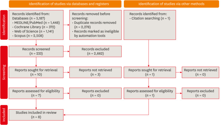
Materials and Methods A systematic search was conducted in MEDLINE/PubMed, Cochrane Library, Scopus, Web of Science, Embase, and Grey Literature Report. Eligibility criteria were based on the condition, content, and population strategy: the condition was the radiographic prevalence of apical periodontitis, the content comprised patients scheduled for hematopoietic stem cell transplantation, and the population consisted of adult and pediatric patients. The revised Risk of Bias in Nonrandomized Studies of Exposure tool was used to assess the quality of studies. The Grading Recommendations Assessments, Development, and Evaluation (GRADE) tool was used to assess the quality of evidence.
Results Eight studies were included in this review. The average number of patients with apical periodontitis was 15.65% (range, 2.1%–43.34%). One study was classified as having a very high risk of bias, 1 with a high risk of bias, and 6 with some concern for bias. GRADE analysis showed a very low certainty of evidence. Significant limitations concerning the absence of control over confounding variables were identified.
Conclusions With the caveat of the very low quality of evidence in the studies reviewed, there was a low to moderate prevalence of apical periodontitis in patients prior to undergoing hematopoietic cell transplantation.
- 3,004 View
- 63 Download

Research Articles
- Radiographic patterns of periosteal bone reactions associated with endodontic lesions
- Poorya Jalali, Jessica Riccobono, Robert A. Augsburger, Mehrnaz Tahmasbi-Arashlow
- Restor Dent Endod 2023;48(3):e23. Published online June 8, 2023
- DOI: https://doi.org/10.5395/rde.2023.48.e23
-
Abstract
PDFPubReaderePub
Objectives The formation of new bone by periosteum due to an insult is called periosteal bone reaction (PBR). This study assessed the cone beam computed tomography (CBCT) patterns of periosteal bone reactions associated with periapical inflammatory lesion (apical periodontitis/periapical rarefying osteitis).
Materials and Methods Twenty-two small field of view CBCT images of patients with PBR were selected from a database of a private practice limited to endodontics. The volume of the periapical inflammatory lesion, the presence of cortical fenestration, the distance of the root apices to the affected cortex, and the location, pattern, and longest diameter of the periosteal reaction were recorded. Statistical analysis was performed using Wilcoxon Ranksum, Fischer’s exact, Spearman Correlation Coefficient, and paired
t -test.Results In all cases, periosteal bone reaction manifested as either parallel (90.9%) or irregular (9.1%). No correlation was found between periapical inflammatory lesion volume and the periosteal reaction's longest diameter (
p > 0.05). Cortical fenestration was noted in 72.7% of the cases. In addition, the findings showed that periosteal reactions were located mostly on the buccal and were present 53.8% and 100% of the time in the mandible and maxilla, respectively.Conclusions The periosteal reactions of endodontic origin had a nonaggressive form (
i.e ., parallel or irregular), and none of the lesions resulted in a periosteal reaction with an ominous Codman’s triangle or spicule pattern.-
Citations
Citations to this article as recorded by
- ENDODONTIA E INTERCORRÊNCIAS: COMPREENDENDO OS ACIDENTES E OTIMIZANDO O PROGNÓSTICO
Ana Paula Oliveira Rocha, Flávia Cordeiro Antunes , Millena Alberto Luna , Raissa Danielle Muniz Da Silva , Gustavo Henrique Palma Durães , Juliano Magno de Valadares Bicalho , Lorena Miranda Lima , Barbara Quadros Tonelli
REMUNOM.2026; 2(03): 1. CrossRef - Endodontic Intervention in Chronic Osteomyelitis With Proliferative Periostitis: A Rare Case Report and Scoping Review
Gabriel Lima Braz, Ana Paula Neutzling Gomes, Lisandrea Rocha Schardosim, Nadia de Souza Ferreira, Jose Francisco Gomez-Sosa
Case Reports in Dentistry.2026;[Epub] CrossRef - The influence of endodontic treatment quality on periapical lesions' architecture in cone‐beam computed tomography
Ewa Mackiewicz, Tobias Bonsmann, Krzysztof Safranow, Patrycja Nowicka, Janusz Kołecki, Alicja Nowicka
Australian Endodontic Journal.2025; 51(1): 36. CrossRef - Novel radiographic pattern of maxillary periostitis induced by endodontic inflammation: A case report
Pai-Chun Huang, I-Hao Su, Meng-Ling Chiang, Jyh-Kwei Chen
Journal of Dental Sciences.2025; 20(3): 1982. CrossRef - Garre’s osteomyelitis of the mandible managed by nonsurgical re-endodontic treatment
Heegyun Kim, Jiyoung Kwon, Hyun-Jung Kim, Soram Oh, Duck-Su Kim, Ji-Hyun Jang
Restorative Dentistry & Endodontics.2024;[Epub] CrossRef
- ENDODONTIA E INTERCORRÊNCIAS: COMPREENDENDO OS ACIDENTES E OTIMIZANDO O PROGNÓSTICO
- 6,848 View
- 105 Download
- 4 Web of Science
- 5 Crossref
- Bone repair in defects filled with AH Plus sealer and different concentrations of MTA: a study in rat tibiae
- Jessica Emanuella Rocha Paz, Priscila Oliveira Costa, Albert Alexandre Costa Souza, Ingrid Macedo de Oliveira, Lucas Fernandes Falcão, Carlos Alberto Monteiro Falcão, Maria Ângela Area Leão Ferraz, Lucielma Salmito Soares Pinto
- Restor Dent Endod 2021;46(4):e48. Published online September 2, 2021
- DOI: https://doi.org/10.5395/rde.2021.46.e48
-
Abstract
PDFPubReaderePub
Objectives This study aimed to evaluate the effects on bone repair of different concentrations of mineral trioxide aggregate (MTA) added to AH Plus.
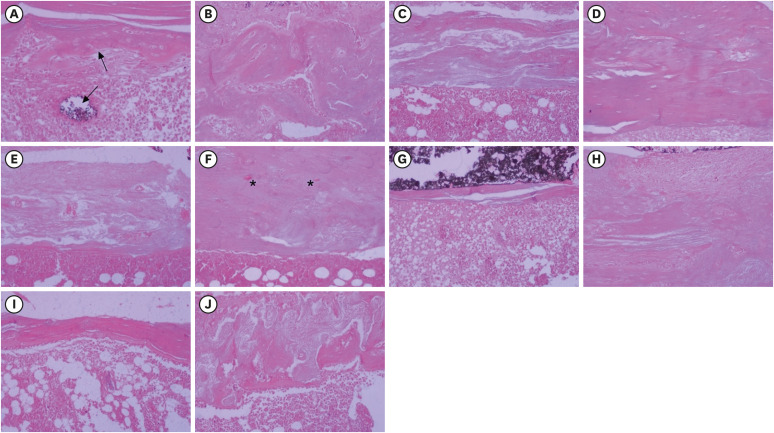
Materials and Methods Bone tissue reactions were evaluated in 30 rats (
Rattus norvegicus ) after 7 and 30 days. In the AH + MTA10, AH + MTA20, and AH + MTA30 groups, defects in the tibiae were filled with AH Plus with MTA in proportions of 10%, 20% and 30%, respectively; in the MTA-FILL group, MTA Fillapex was used; and in the control group, no sealer was used. The samples were histologically analyzed to assess bone union and maturation. The Kruskal-Wallis and Mann-Whitney tests were performed for multiple pairwise comparisons (p ≤ 0.05).Results At the 7-day time point, AH + MTA10 was superior to MTA-FILL with respect to bone union, and AH + MTA20 was superior to MTA-FILL with respect to bone maturity (
p < 0.05). At the 30-day time point, both the AH + MTA10 and AH + MTA20 experimental sealers were superior not only to MTA-FILL, but also to AH + MTA30 with respect to both parameters (p < 0.05). The results of the AH + MTA10 and AH + MTA20 groups were superior to those of the control group for both parameters and experimental time points (p < 0.05).Conclusions The results suggest the potential benefit of using a combination of these materials in situations requiring bone repair.
-
Citations
Citations to this article as recorded by- Bone Healing Response to Different Concentrations of Nano-Hydroxyapatite Incorporated into Mineral Trioxide Aggregate and Bioceramic Sealers
Arkhawan Ali Abdulhaq, Chenar Anwar Mohammad, Bassam Karem Amin
Polymers.2026; 18(14): 1743. CrossRef - Analysis of the cytotoxicity and bioactivity of CeraSeal, BioRoot™ and AH Plus® sealers in pre-osteoblast lineage cells
Luciano Aparecido de Almeida-Junior, Giuliana de Campos Chaves Lamarque, Henry Herrera, Maya Fernanda Manfrin Arnez, Francine Lorencetti-Silva, Raquel Assed Bezerra Silva, Léa Assed Bezerra Silva, Francisco Wanderley Garcia Paula-Silva
BMC Oral Health.2024;[Epub] CrossRef - A Review of the research methods and progress of biocompatibility evaluation of root canal sealers
Xiliang Yang, Tianxia Zheng, Nuoya Yang, Zihan Yin, Wuliang Wang, Yuhong Bai
Australian Endodontic Journal.2023; 49(S1): 508. CrossRef - Effect of Vitapex Combined with AH-Plus Paste on Inflammation in Middle-Aged and Elderly Patients with Periodontal-Endodontic Disease
Rong Hu, Fulan Zhang, Xiangyu Guo, Youren Jing, Xiaowan Lin, Liping Tian, Min Tang
Computational and Mathematical Methods in Medicine.2022; 2022: 1. CrossRef
- Bone Healing Response to Different Concentrations of Nano-Hydroxyapatite Incorporated into Mineral Trioxide Aggregate and Bioceramic Sealers
- 2,808 View
- 17 Download
- 4 Web of Science
- 4 Crossref
Case Report
- The application of “bone window technique” using piezoelectric saws and a CAD/CAM-guided surgical stent in endodontic microsurgery on a mandibular molar case
- Ukseong Kim, Sunil Kim, Euiseong Kim
- Restor Dent Endod 2020;45(3):e27. Published online May 21, 2020
- DOI: https://doi.org/10.5395/rde.2020.45.e27
-
Abstract
PDFPubReaderePub
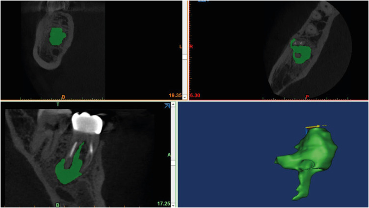
Apical surgery for a mandibular molar is still challenging for many reasons. This report describes the applications of computer-guided cortical ‘bone-window technique’ using piezoelectric saws that prevented any nerve damage in performing endodontic microsurgery of a mandibular molar. A 49-year-old woman presented with gumboil on tooth #36 (previously endodontically treated tooth) and was diagnosed with chronic apical abscess. Periapical lesions were confirmed using cone-beam computed tomography (CBCT). Endodontic microsurgery for the mesial and distal roots of tooth #36 was planned. Following the transfer of data of the CBCT images and the scanned cast to an implant surgical planning program, data from both devices were merged. A surgical stent was designed, on the superimposed three-dimensional model, to guide the preparation of a cortical window on the buccal side of tooth #36. Endodontic microsurgery was performed with a printed surgical template. Minimal osteotomy was required and preservation of the buccal cortical plate rendered this endodontic surgery less traumatic. No postoperative complications such as mental nerve damage were reported. Window technique guided by a computer-aided design/computer-aided manufacture based surgical template can be considerably useful in endodontic microsurgery in complicated cases.
-
Citations
Citations to this article as recorded by- Optimising Outcomes in Endodontic Microsurgery: Evidence, Uncertainties and Future Directions
Ukseong Kim, Euiseong Kim
International Endodontic Journal.2026; 59(5): 746. CrossRef - Accuracy of Guided Dual Technique in Esthetic Crown Lengthening: A Prospective Case‐Series Study
Meritxell Enfedaque‐Prat, Albert González‐Barnadas, Adrià Jorba‐García, Javi Vilarrasa, Jorge Toledano‐Serrabona, Rui Figueiredo, Eduard Valmaseda‐Castellón, Octavi Camps‐Font
Journal of Esthetic and Restorative Dentistry.2025; 37(6): 1284. CrossRef - Guided endodontics in the application of personalized mini-invasive treatment in clinical cases: a literature review
Shuangshuang Ren, Wanping Wang, Mingyue Cheng, Wenyue Tang, Yue Zhao, Leiying Miao
The Saudi Dental Journal.2025;[Epub] CrossRef - Accurately Defining the Location and Dimension of the Bony Lid Under the Guidance of Dynamic Navigation: Report on Three Cases
Kailiang Tang, Xiaole Zhang, Qibao Wang, Xinyu Zhao, Xijiao Yu, Yi Du
Australian Endodontic Journal.2025; 51(3): 785. CrossRef - Minimally Invasive Vertical Incision Subperiosteal Tunnelling Technique for Targeted Endodontic Surgery: Technical Overview and a Case Report
Francesc Abella Sans, Jaime Barragán Montes, Tomasz Zbozen, Nandini Suresh, Lalli Dharmarajan, Paul M. H. Dummer, Venkateshbabu Nagendrababu
International Endodontic Journal.2025; 58(11): 1799. CrossRef - Endodontic Microsurgery of Mandibular Molars with an Autonomous Robotic System
Haiying Zhang, Zi Yang, Mangnan Liu, Yaoxin Wang, Mei Fu, Benxiang Hou, Chen Zhang
Journal of Endodontics.2025; 51(12): 1830. CrossRef - Endodontic Microsurgery of a Mandibular Molar Using a Dynamic Navigation System (DNS) and Cortical Window Technique: A Case Report
Gustavo Castillo, Silvia Restrepo-Méndez, Oscar Zuluaga, Paola Escobar-Villegas
Journal of Endodontic Microsurgery.2024; 3: 1. CrossRef - The bone lid technique in endodontic microsurgery
Min Zhang, He Liu, Ya Shen
Asian Journal of Surgery.2024; 47(7): 3126. CrossRef - Guided Periradicular Surgery with Er,Cr:YSGG Laser Osteotomy: A Case Report
Julian Torres Celeita, Johanna Hernández la Rotta, Amdie Chirinos Salazar, Jorge Fandiño Rodríguez, Laura López Rincón, Mauren Orduz Solorzano, Diana Parra Galvis, Oscar Jiménez Peña
Journal of Endodontic Microsurgery.2024;[Epub] CrossRef - Piezoelectric Endodontic Microsurgery with Modified Cortical Window Technique: A Case Report
Rafael Fernández-Grisales, Wilder Rojas, Carolina Berruecos-Orozco
Journal of Endodontic Microsurgery.2023; 2: 34. CrossRef - The Impact of the Preferred Reporting Items for Case Reports in Endodontics (PRICE) 2020 Guidelines on the Reporting of Endodontic Case Reports
Sofian Youssef, Phillip Tomson, Amir Reza Akbari, Natalie Archer, Fayjel Shah, Jasmeet Heran, Sunmeet Kandhari, Sandeep Pai, Shivakar Mehrotra, Joanna M Batt
Cureus.2023;[Epub] CrossRef - Clinical and radiological outcomes of dynamic navigation in endodontic microsurgery: a prospective study
Chen Chen, Rui Zhang, Wei Zhang, Fangzhe Li, Zan Wang, Li Qin, Yun Chen, Zhuan Bian, Liuyan Meng
Clinical Oral Investigations.2023; 27(9): 5317. CrossRef - New-designed 3D printed surgical guide promotes the accuracy of endodontic microsurgery: a study of 14 upper anterior teeth
Dan Zhao, Weige Xie, Tianguo Li, Anqi Wang, Li Wu, Wen Kang, Lu Wang, Shiliang Guo, Xuna Tang, Sijing Xie
Scientific Reports.2023;[Epub] CrossRef - Failure case analysis during each stage of endodontic microsurgery: A retrospective study based on clinical databases
Changwoo Ryu, Sooil Shin, Yong-Bum Cho, Euiseong Kim, Minju Song
Saudi Endodontic Journal.2023; 13(2): 160. CrossRef - Piezoelectric Device and Dynamic Navigation System Integration for Bone Window-Guided Surgery
Frederico C. Martinho, Ina L. Griffin, Patricia A. Tordik
Journal of Endodontics.2023; 49(12): 1698. CrossRef - Bone Window Technique in Endodontic Microsurgery – Report of Two Cases
Spyros Floratos, Vasileios Molonis, Apostolos Tsolakis, Stylianos Kykalos, Konstantinos Kontzoglou
Journal of Endodontic Microsurgery.2022; 2: 24. CrossRef - An Update on Endodontic Microsurgery of Mandibular Molars: A Focused Review
Sun Mi Jang, Euiseong Kim, Kyung-San Min
Medicina.2021; 57(3): 270. CrossRef
- Optimising Outcomes in Endodontic Microsurgery: Evidence, Uncertainties and Future Directions
- 3,267 View
- 61 Download
- 17 Crossref
Research Article
- CBCT study of mandibular first molars with a distolingual root in Koreans
- Hee-Ho Kim, Hyoung-Hoon Jo, Jeong-Bum Min, Ho-Keel Hwang
- Restor Dent Endod 2018;43(3):e33. Published online July 30, 2018
- DOI: https://doi.org/10.5395/rde.2018.43.e33

-
Abstract
PDFPubReaderePub
Objectives This study aimed to investigate the prevalence of a separate distolingual root and to measure the thickness of the buccal cortical bone in mandibular first molars in Koreans using cone-beam computed tomography (CBCT) images.
Materials and Methods High-quality CBCT data from 432 patients were analyzed in this study. The prevalence of a separate distolingual root of the mandibular first molar was investigated. The distance from the distobuccal and distolingual root apices to the outer surface of the buccal cortical bone was measured. We also evaluated the thickness of the buccal cortical bone.
Results The prevalence of a separate distolingual root (2 separate distal roots with 1 canal in each root; 2R2C) was 23.26%. In mandibular first molars with 2R2C, the distance from the distobuccal root apex to the outer surface of the buccal cortical bone was 5.51 mm. Furthermore, the distance from the distolingual root apex to the outer surface of the buccal cortical bone was 12.09 mm. In mandibular first molars with 2R2C morphology, the thickness of the buccal cortical bone at the distobuccal root apex of the mandibular first molar was 3.30 mm. The buccal cortical bone at the distobuccal root apex was significantly thicker in the right side (3.38 mm) than the left side (3.09 mm) (
p < 0.05).Conclusions A separate distolingual root is not rare in mandibular first molars in the Korean population. Anatomic and morphologic knowledge of the mandibular first molar can be useful in treatment planning, including surgical endodontic treatment.
-
Citations
Citations to this article as recorded by- The association between complex root canal morphology of mandibular anteriors and distolingual roots in mandibular first molars in a Turkish population
Özge Kurt, Elif Solakoğlu
BMC Oral Health.2025;[Epub] CrossRef - Radix molaris is a hidden truth of mandibular first permanent molars: A descriptive- analytic study using cone beam computed tomography
Mohammed A. Alobaid, Saurabh Chaturvedi, Ebtihal Mobarak S. Alshahrani, Ebtsam M. Alshehri, Amal S. Shaiban, Mohamed Khaled Addas, Giuseppe Minervini
Technology and Health Care.2023; 31(5): 1957. CrossRef - Prevalence of radix entomolaris in India and its comparison with the rest of the world
Sumit MOHAN, Jyoti THAKUR
Minerva Dental and Oral Science.2022;[Epub] CrossRef - A critical analysis of laboratory and clinical research methods to study root and canal anatomy
Hany Mohamed Aly Ahmed
International Endodontic Journal.2022; 55(S2): 229. CrossRef - Three‐Rooted Permanent Mandibular First Molars: A Meta‐Analysis of Prevalence
Nyan M. Aung, Kyaw K. Myint, Luca Testarelli
International Journal of Dentistry.2022;[Epub] CrossRef - Reproducibilidad en el diagnóstico imagenológico de periodontitis apical a partir de CBCT
Sandra Milena Buitrago Rojas, Yeny Zulay Castellanos Dominguez, Jhonny Alexander Contreras Vargas, Yosdi Tomás Solano Diaz, Eder Fabián Gutierrez Argote
Acta Odontológica Colombiana.2020;[Epub] CrossRef - Assessment of Root and Root Canal Morphology of Human Primary Molars using CBCT
Yoomin Choi, Seonmi Kim, Namki Choi
THE JOURNAL OF THE KOREAN ACADEMY OF PEDTATRIC DENTISTRY.2020; 47(1): 25. CrossRef - The prevalence of radix molaris in the mandibular first molars of a Saudi subpopulation based on cone-beam computed tomography
Hassan AL-Alawi, Saad Al-Nazhan, Nassr Al-Maflehi, Mazen A. Aldosimani, Mohammed Nabil Zahid, Ghadeer N. Shihabi
Restorative Dentistry & Endodontics.2020;[Epub] CrossRef - Preferred Reporting Items for Epidemiologic Cross-sectional Studies on Root and Root Canal Anatomy Using Cone-beam Computed Tomographic Technology: A Systematized Assessment
Jorge N.R. Martins, Anil Kishen, Duarte Marques, Emmanuel João Nogueira Leal Silva, João Caramês, António Mata, Marco A. Versiani
Journal of Endodontics.2020; 46(7): 915. CrossRef - Evaluation of roots and canal systems of mandibular first molars in a vietnamese subpopulation using cone-beam computed tomography
KhoaVan Pham, AnhHoang Lan Le
Journal of International Society of Preventive and Community Dentistry.2019; 9(4): 356. CrossRef
- The association between complex root canal morphology of mandibular anteriors and distolingual roots in mandibular first molars in a Turkish population
- 2,565 View
- 10 Download
- 10 Crossref
Case Report
- Mandibular bone necrosis after use of paraformaldehyde-containing paste
- Chi-hwan Lee, Yoorina Choi, Sujung Park
- Restor Dent Endod 2016;41(4):332-337. Published online November 8, 2016
- DOI: https://doi.org/10.5395/rde.2016.41.4.332
-
Abstract
PDFPubReaderePub
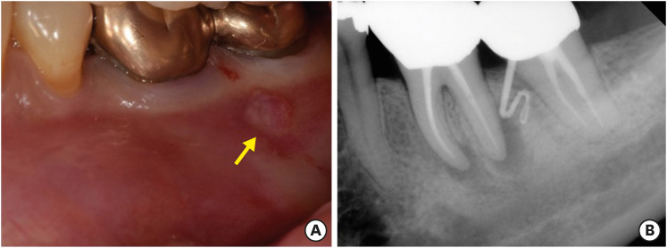
Paraformaldehyde has been used in the past as a pulpotomy agent. However, it has a severe cytotoxic effect and may cause alveolar bone necrosis. Depulpin, a devitalizing agent containing 49% paraformaldehyde, is no longer used frequently due to its severe side effects. In the two cases described in the present study, Depulpin was used as a devitalizing agent during root canal treatment. It caused a gradual loss of sensibility in adjacent teeth, gingival necrosis, and osteomyelitis. This case report demonstrates the serious side effects of using a paraformaldehyde-containing paste as a devitalizing agent for pulp, particularly mandibular bone necrosis.
-
Citations
Citations to this article as recorded by- Numb chin syndrome caused by paraformaldehyde-containing devitalizing agent – Case report
Jyh-Kwei Chen, Yeung-Yi Hsu, Chun-Pin Chiang, Meng-Ling Chiang
Journal of Dental Sciences.2023; 18(2): 955. CrossRef - Non-radiation and non-drug–induced maxillary osteomyelitis: Study of underlying risk factors, presentation, management and treatment outcomes
Kumar Nilesh, Pankaj Patil, Digvijay Patil, Monica Patil
Medical Journal Armed Forces India.2022; 78: S145. CrossRef - Acute toxicity potential and impact on periodontal and periapical tissue of Pulp Out: A paste contained jatropha, sidaguri, and melittin
Maria Tanumihardja, A.M. Windha, N. Musfirah, G.K. Punggawa, Andi Fatima, A.H.M. Nur Fadhila, Esfandiary, Nurhayaty Natsir, Husni Cangara, Lukman Muslimin
Toxicology Reports.2022; 9: 1788. CrossRef - Comparative evaluation of the effect of two pulpal medicaments on pain and bleeding status of mandibular molars with irreversible pulpitis post-failure of inferior alveolar nerve block: a double-blind, randomized, clinical trial
Naomi Ranjan Singh, Lora Mishra, Ajinkya M. Pawar, Nike Kurniawati, Dian Agustin Wahjuningrum
PeerJ.2022; 10: e13397. CrossRef - Dental implant restoration of mandibular bone necrosis defects caused by use of paraformaldehyde-containing paste: A case report
Won-Pyo Lee, Ho-Keel Hwang, Hyoung-Hoon Jo
Oral Biology Research.2019; 43(1): 110. CrossRef - Is Panoramic Radiography an Accurate Imaging Technique for the Detection of Endodontically Treated Asymptomatic Apical Periodontitis?
Cosimo Nardi, Linda Calistri, Giulia Grazzini, Isacco Desideri, Chiara Lorini, Mariaelena Occhipinti, Francesco Mungai, Stefano Colagrande
Journal of Endodontics.2018; 44(10): 1500. CrossRef - A case of high density abnormality in x-ray findings of mandible caused by leakage of root canal filling paste
Haruko Kashiwamura, Kyoko Oka, Yoko Tuchihashi, Hanako Yoshioka, Mayumi Kato, Atsuko Baba, Toyohiro Kagawa, Kazuhiko Okamura, Masao Ozaki
Pediatric Dental Journal.2017; 27(3): 162. CrossRef
- Numb chin syndrome caused by paraformaldehyde-containing devitalizing agent – Case report
- 4,673 View
- 25 Download
- 7 Crossref
Research Articles
- Proximity of the mandibular molar root apex from the buccal bone surface: a cone-beam computed tomographic study
- Dokyung Kim, Jung-Hong Ha, Myoung-Uk Jin, Young-Kyung Kim, Sung Kyo Kim
- Restor Dent Endod 2016;41(3):182-188. Published online July 14, 2016
- DOI: https://doi.org/10.5395/rde.2016.41.3.182
-
Abstract
PDFPubReaderePub
Objectives The purpose of this study was to evaluate the proximity of the mandibular molar apex to the buccal bone surface in order to provide anatomic information for apical surgery.
Materials and Methods Cone-beam computed tomography (CBCT) images of 127 mandibular first molars and 153 mandibular second molars were analyzed from 160 patients' records. The distance was measured from the buccal bone surface to the root apex and the apical 3.0 mm on the cross-sectional view of CBCT.
Results The second molar apex and apical 3 mm were located significantly deeper relative to the buccal bone surface compared with the first molar (
p < 0.01). For the mandibular second molars, the distance from the buccal bone surface to the root apex was significantly shorter in patients over 70 years of age (p < 0.05). Furthermore, this distance was significantly shorter when the first molar was missing compared to nonmissing cases (p < 0.05). For the mandibular first molars, the distance to the distal root apex of one distal-rooted tooth was significantly greater than the distance to the disto-buccal root apex (p < 0.01). In mandibular second molar, the distance to the apex of C-shaped roots was significantly greater than the distance to the mesial root apex of non-C-shaped roots (p < 0.01).Conclusions For apical surgery in mandibular molars, the distance from the buccal bone surface to the apex and apical 3 mm is significantly affected by the location, patient age, an adjacent missing anterior tooth, and root configuration.
-
Citations
Citations to this article as recorded by- Expert consensus on intentional tooth replantation
Zhengmei Lin, Dingming Huang, Shuheng Huang, Zhi Chen, Qing Yu, Benxiang Hou, Lihong Qiu, Wenxia Chen, Jiyao Li, Xiaoyan Wang, Zhengwei Huang, Jinhua Yu, Jin Zhao, Yihuai Pan, Shuang Pan, Deqin Yang, Weidong Niu, Qi Zhang, Shuli Deng, Jingzhi Ma, Xiuping
International Journal of Oral Science.2025;[Epub] CrossRef - Outcome of intentional replantation of endodontically treated teeth with periapical pathosis: A systematic review and meta‐analysis
Faizan Javed, Kamil Zafar, Farhan R. Khan
Australian Endodontic Journal.2023; 49(S1): 494. CrossRef - Proximity of maxillary molar apexes to the cortical bone surface and the maxillary sinus
Han Shin Lee, Dokyung Kim, Sung Kyo Kim
Restorative Dentistry & Endodontics.2022;[Epub] CrossRef - Alveolar bone thickness overlying healthy maxillary and mandibular teeth: A systematic review and meta-analysis
Marziyeh Shafizadeh, Azita Tehranchi, Armin Shirvani, Saeed Reza Motamedian
International Orthodontics.2021; 19(3): 389. CrossRef - Relationship between the anatomic structures and mandibular posterior teeth for endodontic surgery in a Turkish population: a cone-beam computed tomographic analysis
Zeliha Uğur Aydın, Duygu Göller Bulut
Clinical Oral Investigations.2019; 23(9): 3637. CrossRef
- Expert consensus on intentional tooth replantation
- 3,008 View
- 7 Download
- 5 Crossref
- A study on the compatibility between one-bottle dentin adhesives and composite resins using micro-shear bond strength
- Minju Song, Yooseok Shin, Jeong-Won Park, Byoung-Duck Roh
- Restor Dent Endod 2015;40(1):30-36. Published online September 26, 2014
- DOI: https://doi.org/10.5395/rde.2015.40.1.30
-
Abstract
PDFPubReaderePub
Objectives This study was performed to determine whether the combined use of one-bottle self-etch adhesives and composite resins from same manufacturers have better bond strengths than combinations of adhesive and resins from different manufacturers.
Materials and Methods 25 experimental micro-shear bond test groups were made from combinations of five dentin adhesives and five composite resins with extracted human molars stored in saline for 24 hr. Testing was performed using the wire-loop method and a universal testing machine. Bond strength data was statistically analyzed using two way analysis of variance (ANOVA) and Tukey's
post hoc test.Results Two way ANOVA revealed significant differences for the factors of dentin adhesives and composite resins, and significant interaction effect (
p < 0.001). All combinations with Xeno V (Dentsply De Trey) and Clearfil S3 Bond (Kuraray Dental) adhesives showed no significant differences in micro-shear bond strength, but other adhesives showed significant differences depending on the composite resin (p < 0.05). Contrary to the other adhesives, Xeno V and BondForce (Tokuyama Dental) had higher bond strengths with the same manufacturer's composite resin than other manufacturer's composite resin.Conclusions Not all combinations of adhesive and composite resin by same manufacturers failed to show significantly higher bond strengths than mixed manufacturer combinations.
-
Citations
Citations to this article as recorded by- Influence of etching mode and composite resin type on bond strength to dentin using universal adhesive system
Stefan Dačić, Milan Miljković, Aleksandar Mitić, Goran Radenković, Marija Anđelković‐Apostolović, Milica Jovanović
Microscopy Research and Technique.2021; 84(6): 1212. CrossRef - Is the presence of 10-MDP associated to higher bonding performance for self-etching adhesive systems? A meta-analysis of in vitro studies
Julia Fehrenbach, Cristina Pereira Isolan, Eliseu Aldrighi Münchow
Dental Materials.2021; 37(10): 1463. CrossRef - Dentin bond strengths of all-in-one adhesives combined with different manufacturers’ flowable resin composites
Koichi SHINKAI, Daiki YOSHII, Akira KOIDE, Masaya SUZUKI, Shiro SUZUKI
Dental Materials Journal.2021; 40(5): 1094. CrossRef - DİŞ HEKİMLİĞİNDE ADEZİV SİSTEMLER
Elmas TÜRKER, Buket AYNA
Atatürk Üniversitesi Diş Hekimliği Fakültesi Dergisi.2018;[Epub] CrossRef - Influence of EDC on Dentin-Resin Shear Bond Strength and Demineralized Dentin Thermal Properties
Lin Tang, Yi Zhang, Yuhua Liu, Yongsheng Zhou
Materials.2016; 9(11): 920. CrossRef
- Influence of etching mode and composite resin type on bond strength to dentin using universal adhesive system
- 2,155 View
- 9 Download
- 5 Crossref
- The effects of bone morphogenetic protein-2 and enamel matrix derivative on the bioactivity of mineral trioxide aggregate in MC3T3-E1cells
- Youngdan Jeong, Wonkyung Yang, Hyunjung Ko, Miri Kim
- Restor Dent Endod 2014;39(3):187-194. Published online June 19, 2014
- DOI: https://doi.org/10.5395/rde.2014.39.3.187
-
Abstract
PDFPubReaderePub
Objectives The effects of bone morphogenetic protein-2 (BMP-2) and enamel matrix derivative (EMD) respectively with mineral trioxide aggregate (MTA) on hard tissue regeneration have been investigated in previous studies. This study aimed to compare the osteogenic effects of MTA/BMP-2 and MTA/EMD treatment in MC3T3-E1 cells.
Materials and Methods MC3T3-E1 cells were treated with MTA (ProRoot, Dentsply), BMP-2 (R&D Systems), EMD (Emdogain, Straumann) separately and MTA/BMP-2 or MTA/EMD combination. Mineralization was evaluated by staining the calcium deposits with alkaline phosphatase (ALP, Sigma-Aldrich) and Alizarin red (Sigma-Aldrich). The effects on the osteoblast differentiation were evaluated by the expressions of osteogenic markers, including ALP, bone sialoprotein (BSP), osteocalcin (OCN), osteopontin (OPN) and osteonectin (OSN), as determined by reverse-transcription polymerase chain reaction analysis (RT-PCR, AccuPower PCR, Bioneer).
Results Mineralization increased in the BMP-2 and MTA/BMP-2 groups and increased to a lesser extent in the MTA/EMD group but appeared to decrease in the MTA-only group based on Alizarin red staining. ALP expression largely decreased in the EMD and MTA/EMD groups based on ALP staining. In the MTA/BMP-2 group, mRNA expression of OPN on day 3 and BSP and OCN on day 7 significantly increased. In the MTA/EMD group, OSN and OCN gene expression significantly increased on day 7, whereas ALP expression decreased on days 3 and 7 (
p < 0.05).Conclusions These results suggest the MTA/BMP-2 combination promoted more rapid differentiation in MC3T3-E1 cells than did MTA/EMD during the early mineralization period.
-
Citations
Citations to this article as recorded by- Non-oral tissue regeneration, angiogenesis, and healing with enamel matrix derivative
Omar Zada, Carolina Serrano-Larrea, Marcel Karperien, Egbert J. D. Veen
Academia Biology.2026;[Epub] CrossRef - Elucidating epigenetic mechanisms governing odontogenic differentiation in dental pulp stem cells: an in-depth exploration
Lei Huang, Xuan Chen, Xiaoxia Yang, Yinchun Zhang, Yiyun Liang, Xiaoling Qiu
Frontiers in Cell and Developmental Biology.2024;[Epub] CrossRef - Evaluation of the genotoxicity, cytotoxicity, and bioactivity of calcium silicate-based cements
Merve Esen, Yeliz Guven, Mehmet Fatih Seyhan, Handan Ersev, Elif Bahar Tuna-Ince
BMC Oral Health.2024;[Epub] CrossRef - GelMA‐based hydrogel biomaterial scaffold: A versatile platform for regenerative endodontics
Lei Huang, Xuan Chen, XiaoXia Yang, Yinchun Zhang, Xiaoling Qiu
Journal of Biomedical Materials Research Part B: Applied Biomaterials.2024;[Epub] CrossRef - Experimental Validation of Antiobesogenic and Osteoprotective Efficacy of Ginsenoside CK via Targeting Lipid and Atherosclerosis Pathways
Md. Niaj Morshed, Reshmi Akter, Imran Mahmud, Ah-Yeong Gwon, Jin Woo Jeang, Yeong-Geun Lee, Dae Won Park, Deok Chun Yang, Yeon Ju Kim, Se-Chan Kang
Life.2024; 15(1): 41. CrossRef - Anti-osteoporosis effects of triterpenoids from the fruit of sea buckthorn (Hippophae rhamnoides) through the promotion of osteoblast differentiation in mesenchymal stem cells, C3H10T1/2
Da Eun Lee, Kun Hee Park, Joo-Hyun Hong, Seon Hee Kim, Ki-Moon Park, Ki Hyun Kim
Archives of Pharmacal Research.2023; 46(9-10): 771. CrossRef - In Silico and In Vitro Evaluation of Antiobesogenic and Osteoprotective Effect of Pomegranate Juice Fermented by Tannin Acyl Hydrolase and Lactobacillus vespulae DCY75 via the Wnt/β-Catenin Pathway
Reshmi Akter, Vinothini Boopathi, Muhammad Awais, Juha Park, Byoung Man Kong, Se-Woung Oh, Ji-Hyung Oh, Jong Chan Ahn, Deok Chun Yang
ACS Food Science & Technology.2023; 3(11): 1975. CrossRef - Early induction of Hes1 by bone morphogenetic protein 9 plays a regulatory role in osteoblastic differentiation of a mesenchymal stem cell line
Chang‐Hwan Seong, Norika Chiba, Mardiyantoro Fredy, Joji Kusuyama, Kiyohide Ishihata, Toshiro Kibe, Muhammad Subhan Amir, Ryohei Tada, Tomokazu Ohnishi, Norifumi Nakamura, Tetsuya Matsuguchi
Journal of Cellular Biochemistry.2023; 124(9): 1366. CrossRef - Effects of Fucoidan Powder Combined with Mineral Trioxide Aggregate as a Direct Pulp-Capping Material
Mijoo Kim, Marc Hayashi, Bo Yu, Thomas K. Lee, Reuben H. Kim, Deuk-Won Jo
Polymers.2022; 14(12): 2315. CrossRef - Nerve growth factor promotes osteogenic differentiation of MC3T3-E1 cells via BMP-2/Smads pathway
Xuming Yang, Donggang Mou, Qunying Yu, Jimei Zhang, Ying Xiong, Zhimin Zhang, Shan Xing
Annals of Anatomy - Anatomischer Anzeiger.2022; 239: 151819. CrossRef - Anti-Osteoporosis Effects of the Fruit of Sea Buckthorn (Hippophae rhamnoides) through Promotion of Osteogenic Differentiation in Ovariectomized Mice
Kun Hee Park, Joo-Hyun Hong, Seon-Hee Kim, Jin-Chul Kim, Ki Hyun Kim, Ki-Moon Park
Nutrients.2022; 14(17): 3604. CrossRef - Oroactive dental biomaterials and their use in endodontic therapy
Ebrahim Patel, Priyamvada Pradeep, Pradeep Kumar, Yahya E. Choonara, Viness Pillay
Journal of Biomedical Materials Research Part B: Applied Biomaterials.2020; 108(1): 201. CrossRef - BMP-2 and type I collagen preservation in human deciduous teeth after demineralization
Nina Bono, Paolo Tarsini, Gabriele Candiani
Journal of Applied Biomaterials & Functional Materials.2019;[Epub] CrossRef - An assessment of the overexpression of BMP‐2 in transfected human osteoblast cells stimulated by mineral trioxide aggregate and Biodentine
E. M. Rodrigues, A. L. Gomes‐Cornélio, A. Soares‐Costa, L. P. Salles, M. Velayutham, C. Rossa‐Junior, J. M. Guerreiro‐Tanomaru, M. Tanomaru‐Filho
International Endodontic Journal.2017;[Epub] CrossRef - Sandblasting and fibronectin-derived peptide immobilization on titanium surface increase adhesion and differentiation of osteoblast-like cells (MC3T3-E1)
Samdharu Pramono, Kamolparn Pugdee, Jintamai Suwanprateep, Sittichai Koontongkaew
Journal of Dental Sciences.2016; 11(4): 427. CrossRef - Combined Effects of Growth Hormone and Mineral Trioxide Aggregate on Growth, Differentiation, and Angiogenesis in Human Dental Pulp Cells
Hyung-Mun Yun, Seok-Woo Chang, Kyung-Ran Park, Lan Herr, Eun-Cheol Kim
Journal of Endodontics.2016; 42(2): 269. CrossRef - Combined effects of mineral trioxide aggregate and human placental extract on rat pulp tissue and growth, differentiation and angiogenesis in human dental pulp cells
Seok-Woo Chang, Ji-Youn Kim, Mi-Joo Kim, Ga-Hyun Kim, Jin-Kyu Yi, Deok-Won Lee, Kee-Yeon Kum, Eun-Cheol Kim
Acta Odontologica Scandinavica.2016; 74(4): 298. CrossRef - Mineral trioxide aggregate induces osteoblastogenesis via Atf6
Toyonobu Maeda, Atsuko Suzuki, Satoshi Yuzawa, Yuh Baba, Yuichi Kimura, Yasumasa Kato
Bone Reports.2015; 2: 36. CrossRef - Locally controlled delivery of TNFα antibody from a novel glucose-sensitive scaffold enhances alveolar bone healing in diabetic conditions
Qi Wang, Hao Li, Yu Xiao, Shuan Li, Bo Li, Xiaowen Zhao, Lin Ye, Bin Guo, Xinmin Chen, Yi Ding, Chongyun Bao
Journal of Controlled Release.2015; 206: 232. CrossRef
- Non-oral tissue regeneration, angiogenesis, and healing with enamel matrix derivative
- 2,284 View
- 4 Download
- 19 Crossref
Case Report
- Clinical effectiveness of combining platelet rich fibrin with alloplastic bone substitute for the management of combined endodontic periodontal lesion
- Lata Goyal
- Restor Dent Endod 2014;39(1):51-55. Published online January 20, 2014
- DOI: https://doi.org/10.5395/rde.2014.39.1.51
-
Abstract
PDFPubReaderePub
The term "endo-perio" lesion has been proposed to describe the destructive lesion resulting from inflammatory products found in varying degrees in both the periodontium and the pulpal tissues. In most of the cases, clinical symptoms disappear following successful endodontic therapy. However failure after conventional root canal treatment calls for surgical intervention. A 35 year old male patient with endo-perio lesion in right maxillary lateral incisor was treated with platelet rich fibrin (PRF) and alloplastic bone substitute after conventional endodontic therapy. At the end of 6 months there was gain in clinical attachment, increased radiographic bone fill and reduction in probing depth which was maintained till 18 month follow-up. Present case report aims to evaluate the efficacy of PRF and alloplastic bone substitute in the management of intrabony defect associated with endo-perio lesion in maxillary lateral incisor because the healing potential of PRF and bone graft has not been widely studied in endodontics. The use of PRF allows the clinician to optimize tissue remodelling, wound healing and angiogenesis by the local delivery of growth factors and proteins. The novel technique described here enables the clinician to be benefited from the full regenerative capacity of this autologous biologic material.
-
Citations
Citations to this article as recorded by- Treatment of endodontic-periodontic lesions using a root resection approach associated with regenerative materials
Majid Anas Krsoum, Aliyaa Bakur Zaidan, Majed Mohammed Althubaiti, Ghada Medhat Eisa
Saudi Endodontic Journal.2026; 16(2): 280. CrossRef - To Analyze the Efficacy of Platelet-rich Plasma in Contrast to Platelet-rich Fibrin along with Synthetic Nanocrystalline Hydroxyapatite and β-tricalcium Phosphate Bone Graft in Regeneration of Bony Defects in Children
Nonie Marianne Koksi Sangma Shadap, Gunjan Yadav, Sonali Saha, Kavita Dhinsa, Anshul Sharma, Amit Rai
International Journal of Clinical Pediatric Dentistry.2024; 16(6): 842. CrossRef - Regenerative Endodontic Management of an Immature Necrotic Premolar Using Advanced Platelet‐Rich Fibrin
Sepideh Hosseini, Nazanin Chitsaz, Mohammad Hassan Hamrah, Donya Maleki, Emad Taghizadeh, Hamdi Cem Gungor
Case Reports in Dentistry.2023;[Epub] CrossRef - Effect of biodentine coated with emdogain on proliferation and differentiation of human stem cells from the apical papilla
Hamed Karkehabadi, Erfan Ahmadyani, Rezvan Najafi, Elham Khoshbin
Molecular Biology Reports.2022; 49(5): 3685. CrossRef - Healing Assessment of Osseous Defects after Surgical Removal of Periapical Lesions in the Presence of Hydroxyapatite, Nanohydroxyapatite, and a Combination of Nanohydroxyapatite and Platelet-rich Fibrin: A Clinical Study
Amira Elkholly, Maged Negm, Reham Hassan, Nada Omar
Open Access Macedonian Journal of Medical Sciences.2022; 10(D): 406. CrossRef - Case report on combining PRF with alloplastic bone substitute in Endo-Perio lesion
Mansi Bansal, Manish Khatri, Komal Puri
Restorative Dentistry & Endodontics.2021;[Epub] CrossRef Treatment of an Endo-Perio Lesion with Ozone Gas in a Patient with Aggressive Periodontitis: A Clinical Case Report and Literature Review
Maria K Makeeva, Fatima Yu Daurova, Svetlana F Byakova, Anna Yu Turkina
Clinical, Cosmetic and Investigational Dentistry.2020; Volume 12: 447. CrossRef- Revisit to endo-perio lesion a review
Roopali Sharma, Akshita Gupta, K K. Gupta, Sarah Jameel, Rashmika Kapoor
IP International Journal of Periodontology and Implantology.2020; 5(2): 48. CrossRef - Autologous platelet-rich derivatives along with alloplastic bone substitute in the management of complex perio-endo cases
Lata Goyal, Namita Gupta, NarinderDev Gupta
Journal of Indian Society of Periodontology.2020; 24(2): 182. CrossRef - Platelet-Rich Fibrin as a Bone Graft Material in Oral and Maxillofacial Bone Regeneration: Classification and Summary for Better Application
Yiping Liu, Xiaolin Sun, Jize Yu, Jia Wang, Peisong Zhai, Siyu Chen, Manxuan Liu, Yanmin Zhou
BioMed Research International.2019; 2019: 1. CrossRef - Acute periodontal lesions (periodontal abscesses and necrotizing periodontal diseases) and endo‐periodontal lesions
David Herrera, Belén Retamal‐Valdes, Bettina Alonso, Magda Feres
Journal of Clinical Periodontology.2018;[Epub] CrossRef - Acute periodontal lesions (periodontal abscesses and necrotizing periodontal diseases) and endo‐periodontal lesions
David Herrera, Belén Retamal‐Valdes, Bettina Alonso, Magda Feres
Journal of Periodontology.2018;[Epub] CrossRef - Regenerative in endodontics: how, when and where
AL Ahmar Rima, Bassam Sanaa, Salloum Sarah, El Husseini Hassan, AL Ahmar Rima
Journal of Dental Health, Oral Disorders & Therapy.2018; 9(6): 531. CrossRef - Effect of Choukroun Platelet-Rich Fibrin Combined With Autologous Micro-Morselized Bone on the Repair of Mandibular Defects in Rabbits
Tian Zhou, Hua-Wei Yang, Zhuo-Wei Tian, Yang Wang, Xiao-Shan Tang, Jing-Zhou Hu
Journal of Oral and Maxillofacial Surgery.2018; 76(1): 221. CrossRef - Preliminary Results of Bone Regeneration in Oromaxillomandibular Surgery Using Synthetic Granular Graft
Noemi Mazzone, E. Mici, A. Calvo, M. Runci, S. Crimi, F. Lauritano, E. Belli
BioMed Research International.2018; 2018: 1. CrossRef - Treatment of endo-periodontal lesion using leukocyte- platelet- rich fibrin. A case report
Pablo Betancourt, Ricardo Elgueta, Ramon Fuentes
Colombia Medica.2017; 48(4): 204. CrossRef - The impact of autologous platelet concentrates on endodontic healing: a systematic review
Nastaran Meschi, Ana B. Castro, Katleen Vandamme, Marc Quirynen, Paul Lambrechts
Platelets.2016; 27(7): 613. CrossRef - A review of the regenerative endodontic treatment procedure
Bin-Na Lee, Jong-Wook Moon, Hoon-Sang Chang, In-Nam Hwang, Won-Mann Oh, Yun-Chan Hwang
Restorative Dentistry & Endodontics.2015; 40(3): 179. CrossRef - Platelet preparations in dentistry: How? Why? Where? When?
Luigi Fabrizio Rodella
World Journal of Stomatology.2015; 4(2): 39. CrossRef
- Treatment of endodontic-periodontic lesions using a root resection approach associated with regenerative materials
- 2,484 View
- 8 Download
- 19 Crossref
Basic Research
- Histology of dental pulp healing after tooth replantation in rats
- Eun-Jin Go, Han-Seong Jung, Eui-Seong Kim, Il-Young Jung, Seung-Jong Lee
- J Korean Acad Conserv Dent 2010;35(4):273-284. Published online July 31, 2010
- DOI: https://doi.org/10.5395/JKACD.2010.35.4.273
-
Abstract
PDFPubReaderePub
The objective of this study was to observe the histology of dental pulp healing after tooth replantation in rats. The maxillary right first molars of 4-week-old rat were extracted, and then the teeth were repositioned in the original socket. At 3 days after replantation, there was localized inflammatory reaction. But, pulp revasculization and healing had already begun in the root area. At 5 days after replantation, odontoblast-like cells were observed. Tertiary dentin deposition was observed beneath the pulp-dentin border from 1 week after replantation. And tertiary dentin was increased at 2 weeks after replantation. The presence of odontoblast-like cells and the formation of tertiary dentin were continued to 4 weeks after replantation. At 4 weeks after replantation, the deposition of bone-like tissues and cementum-like tissues was observed. This results show that there is a possibility of pulp healing after tooth replantation in rats and the mineralization of tooth can progress. The mineralization of tooth after replantation was initially occurred by the deposition of tertiary dentin, but as time passed, the deposition of bone-like tissues and cementum-like tissues was begun and increased.
-
Citations
Citations to this article as recorded by- Doxycycline-Loaded Nitric Oxide-Releasing Nanomatrix Gel in Replanted Rat Molar on Pulp Regeneration
Kwan-Hee Yun, Mi-Ja Ko, Yong-Kown Chae, Koeun Lee, Ok-Hyung Nam, Hyo-Seol Lee, Kyounga Cheon, Sung-Chul Choi
Applied Sciences.2021; 11(13): 6041. CrossRef - Bio-Photonic Detection and Quantitative Evaluation Method for the Progression of Dental Caries Using Optical Frequency-Domain Imaging Method
Ruchire Wijesinghe, Nam Cho, Kibeom Park, Mansik Jeon, Jeehyun Kim
Sensors.2016; 16(12): 2076. CrossRef
- Doxycycline-Loaded Nitric Oxide-Releasing Nanomatrix Gel in Replanted Rat Molar on Pulp Regeneration
- 3,258 View
- 29 Download
- 2 Crossref
Case Report
- Anterior esthetic improvement through orthodontic extrusive remodeling and single-unit implantation in a fractured upper lateral incisor with alveolar bone loss: A case report
- Soo-Youn Hwang, Won-Jun Shon, Young-Chul Han, Kwang-Shik Bae, Seung-Ho Back, WooCheol Lee, Kee-Yeon Kum
- J Korean Acad Conserv Dent 2008;33(1):39-44. Published online January 31, 2008
- DOI: https://doi.org/10.5395/JKACD.2008.33.1.039
-
Abstract
PDFPubReaderePub
The treatment of esthetic areas with single-tooth implants represents a new challenge for the clinician. In 1993, a modification of the forced eruption technique, called "orthodontic extrusive remodelling," was proposed as a way to augment both soft- and hard-tissue profiles at potential implant sites. This case report describes augmentation of the coronal soft and hard tissues around a fractured maxillary lateral incisor associated with alveolar bone loss, which was achieved by forced orthodontic extrusion before implant placement. Through these procedures we could reconstruct esthetics and function in a hopeless tooth diagnosed with subgingival root fracture by trauma.
- 1,229 View
- 2 Download
 First
First Prev
Prev

