Articles
- Page Path
- HOME > Restor Dent Endod > Volume 33(1); 2008 > Article
- Case Report Anterior esthetic improvement through orthodontic extrusive remodeling and single-unit implantation in a fractured upper lateral incisor with alveolar bone loss: A case report
- Soo-Youn Hwang, Won-Jun Shon, Young-Chul Han, Kwang-Shik Bae, Seung-Ho Back, WooCheol Lee, Kee-Yeon Kum
-
2008;33(1):-44.
DOI: https://doi.org/10.5395/JKACD.2008.33.1.039
Published online: January 31, 2008
Department of Conservative Dentistry, Dental Research Institute, School of Dentistry, Seoul National University, Seoul, Korea.
- Corresponding Author: Kee-Yeon Kum. Department of Conservative Dentistry, School of Dentistry, Seoul National Dentistry, 92 Yunkun-Dong, Jongro-Gu, Seoul, 110-749, Korea. Tel: 82-2-2072-2651, Fax: 82-2-2072-2651, kum6139@snu.ac.kr
• Received: December 18, 2007 • Revised: January 6, 2008 • Accepted: January 7, 2008
Copyright © 2008 The Korean Academy of Conservative Dentistry
- 734 Views
- 1 Download
Abstract
- The treatment of esthetic areas with single-tooth implants represents a new challenge for the clinician. In 1993, a modification of the forced eruption technique, called "orthodontic extrusive remodelling," was proposed as a way to augment both soft- and hard-tissue profiles at potential implant sites. This case report describes augmentation of the coronal soft and hard tissues around a fractured maxillary lateral incisor associated with alveolar bone loss, which was achieved by forced orthodontic extrusion before implant placement. Through these procedures we could reconstruct esthetics and function in a hopeless tooth diagnosed with subgingival root fracture by trauma.
- 1. Phillips K, Kois JC. Aesthetic peri-implant site development. The restorative connection. Dent Clin North Am. 1998;42(1):57-70.PubMed
- 2. Chin M, Toth B. Distraction osteogenesis in maxillofacial surgery using internal devices: report of five cases. J Oral maxillofac Surg. 1996;54(1):45-53.PubMed
- 3. Hämmerle CH, Jung RE. Bone augmentation by means of barrier membranes. Periodontol 2000. 2003;33: 36-53.ArticlePubMedPDF
- 4. van Steenberghe D, Naert I, Bossuyt M, De Mars G, Calberson L, Ghyselen J, et al. The rehabilitation of the severely resorbed maxilla by simultaneous placement of autogenous bone grafts and implants: a 10-year evaluation. Clin Oral Investig. 1997;1(3):102-108.PubMed
- 5. Heithersay GS. Combined endodontic-orthodontic treatment of transverse root fractures in the region of the alveolar crest. Oral Surg Oral Med Oral Pathol. 1973;36(3):404-415.ArticlePubMed
- 6. Ingber JS. Forced eruption. I. A method of treating isolated one and two wall infrabony osseous defects-rationale and case report. J Periodontol. 1974;45(4):199-206.PubMed
- 7. Salama H, Salama M. The role of orthodontic extrusive remodeling in the enhancement of soft and hard tissue profiles prior to implant placement: a systematic approach to the management of extraction site defects. Int J Periodontics Restorative Dent. 1993;13(4):312-333.PubMed
- 8. Mantzikos T, Shamus I. Case report: forced eruption and implant site development. Angle Orthod. 1998;68(2):179-186.PubMed
- 9. Schincaglia GP, Nowzari H. Surgical treatment planning for the single-unit implant in aesthetic areas. Periodontol 2000. 2001;27: 162-182.ArticlePubMedPDF
- 10. Mantzikos T, Shamus I. Forced eruption and implant site development: soft tissue response. Am J Orthod Dentofacial Orthop. 1997;112(6):596-606.ArticlePubMed
- 11. Mantzikos T, Shamus I. Forced eruption and implant site development: an osteophysiologic response. Am J Orthod Dentofacial Orthop. 1999;115(5):583-591.PubMed
- 12. Chambrone L, Chambrone LA. Forced orthodontic eruption of fractured teeth before implant placement: Case report. J Can Dent Assoc. 2005;71(4):257-261.PubMed
- 13. Lin CD, Chang SS, Liou CS, Dong DR, Fu E. Management of interdental papillae loss with forced eruption, immediate implantation, and root-form pontic. J Periodontol. 2006;77: 135-141.ArticlePubMed
REFERENCES
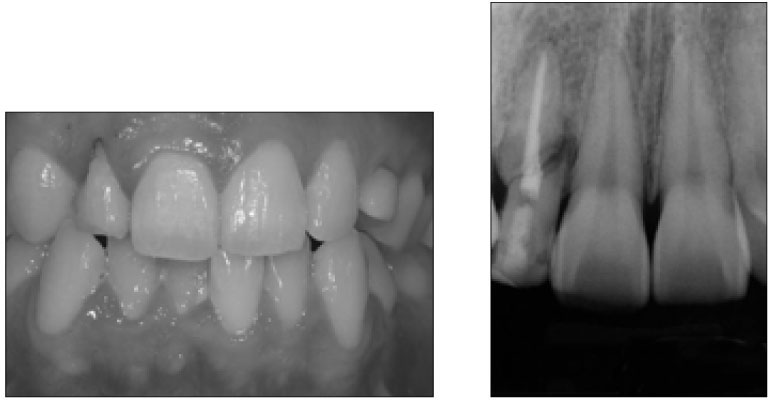
Figure 1

Clinical photo (Left) and the radiographic view of oblique crown-root fracture with alveolar bone loss in maxillary right lateral incisor (Right).

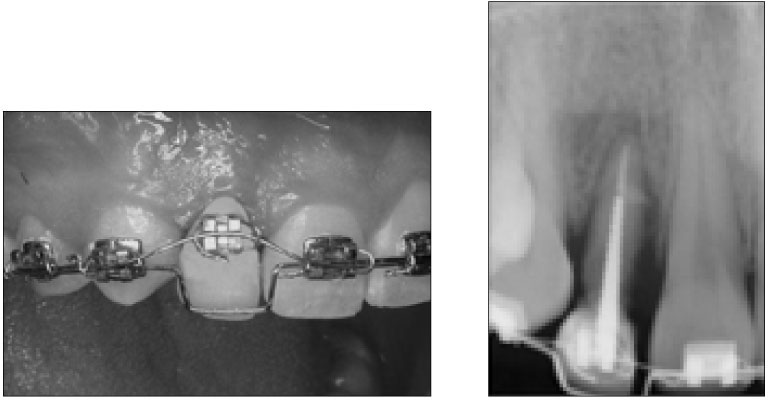
Figure 2

Clinical photo after connecting with brakets and 0.19 * 0.25 stainless steel orthodontic wire (left) and radiographic view (right).

Figure 3

Removal of upper fractured segment of crown (a), construction of post and resin core (b), stage of orthodontic eruptive force for 3 months (b, c), and retention wire for stabilization (d).

Tables & Figures
REFERENCES
Citations
Citations to this article as recorded by 

Anterior esthetic improvement through orthodontic extrusive remodeling and single-unit implantation in a fractured upper lateral incisor with alveolar bone loss: A case report


Figure 1
Clinical photo (Left) and the radiographic view of oblique crown-root fracture with alveolar bone loss in maxillary right lateral incisor (Right).
Figure 2
Clinical photo after connecting with brakets and 0.19 * 0.25 stainless steel orthodontic wire (left) and radiographic view (right).
Figure 3
Removal of upper fractured segment of crown (a), construction of post and resin core (b), stage of orthodontic eruptive force for 3 months (b, c), and retention wire for stabilization (d).
Figure 4
Single-unit implantation was inserted into the augmented bony site (a) and primary closure (b) was done using connective tissue (C) obtained from palatal mucosa.
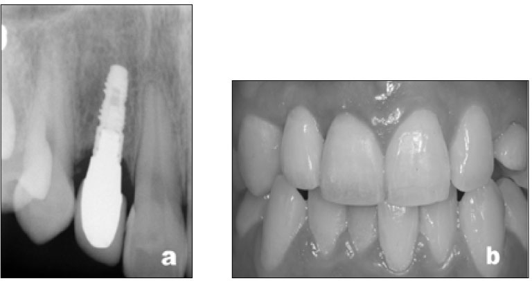
Figure 5
Radiographic view (a) and final photo (b) after ceramic restoration.
Figure 1
Figure 2
Figure 3
Figure 4
Figure 5
Anterior esthetic improvement through orthodontic extrusive remodeling and single-unit implantation in a fractured upper lateral incisor with alveolar bone loss: A case report

 KACD
KACD


 ePub Link
 Cite
Cite

